ppt on Digestive system akki
34
By – Mr ASHOK BISHNOI Lecturer
-
Upload
ashok-k-bishnoi -
Category
Health & Medicine
-
view
90 -
download
1
Transcript of ppt on Digestive system akki
By –
Mr ASHOK BISHNOI
Lecturer
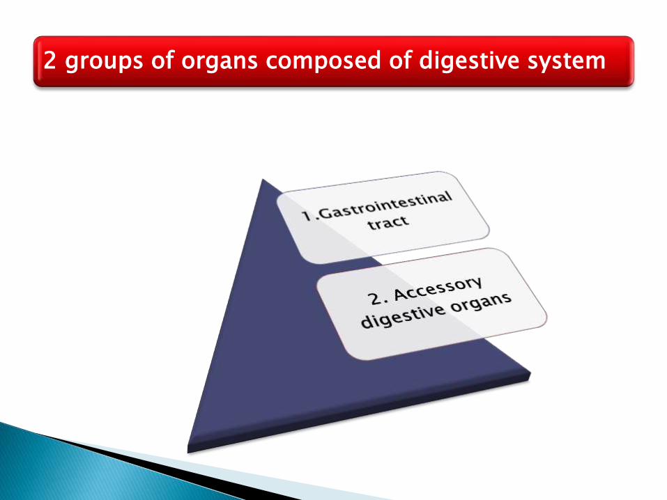
2 groups of organs composed of digestive system
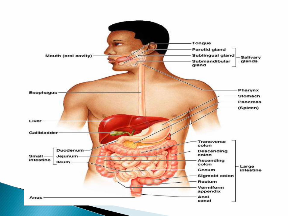
It is continuous tube that extends from the mouth to anus through the thoracic, abdominal & pelvic cavities.
1.Gastrointestinal tract/ Alimentary canal

◦ Mouth
◦ Pharynx
◦ Esophagus
◦ Stomach
◦ Small intestine
◦ Large intestine
◦ Rectum
◦ Anal canal
◦ Anus
The length of GI tract is about 5-7 meters (16.5-23 ft) in a living person.
It is long in Cadaver about 7-9 meters (23-29.5 ft)
Supply secretions contributing to the breakdown of food
2. Accessory digestive organs

◦ Teeth
◦ Lips
◦ Cheeks
◦ Tongue
◦ Salivary glands (3 pair)
◦ Gallbladder
◦ Liver
◦ Pancreas
Teeth aid in the physical breakdown of food & tongue assist in chewing & swallowing.
The other accessory digestive organs never come in to direct contact with food.
They produce secretion in the chemical breakdown of food
1. Ingestion-Taking food in to mouth
2. Secretion- Relies of H2o,acid & enzymes in to lumen of GI tract.
3. Mixing & propulsion
4. Digestion- Mechanical breakdown of food by Mastication
Chemical digestion by Enzymes
5. Absorption- Passage of digested product from the GI tract in to blood.
6. Defecation- The elimination of faeces from the GI tract
Function of DS;-

12
Structure of alimentary canal:-
The wall of GI tract from the lower oesophagus to the anal canal.
It has the “4” layers of tissue
◦ Mucosal layer◦ Submucosal layer◦ Muscularis layer◦ Serosa layer
1.Gastrointestinal tract/ Alimentary canal

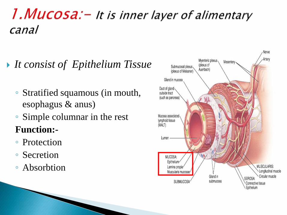
It consist of Epithelium Tissue
◦ Stratified squamous (in mouth,
esophagus & anus)
◦ Simple columnar in the rest
Function:-
◦ Protection
◦ Secretion
◦ Absorbtion

Loose connective tissue
◦ containing ,glands and lymphatic tissue
Meissner’s plexus
The secrete:-◦ Saliva from salivary gland
◦ Gastric juice from gastric gland
◦ Intestinal juice from intestinal gland
◦ Pancreatic juice from pancreas
◦ Bile from liver

Skeletal muscle = voluntary control
◦ in mouth, pharynx , upper esophagus and
anus
◦ control over swallowing and defecation
Smooth muscle = involuntary control
◦ inner circular fibers & outer longitudinal
fibers
◦ mixes, crushes & propels food along by
peristalsis

4.Serosa:-• It is outer layer
• Covers all organs and walls of cavities
• Secretes a serous fluid
• Consists of connective tissue .

Peritoneum
◦ Cavity within the abdomen
Formed by a
◦ Parietal layer- Which line the abdominal wall
◦ Visceral layer covers organs with in the abdomen & pelvic
cavity
Peritoneal cavity
◦ potential space containing a bit of serous fluid
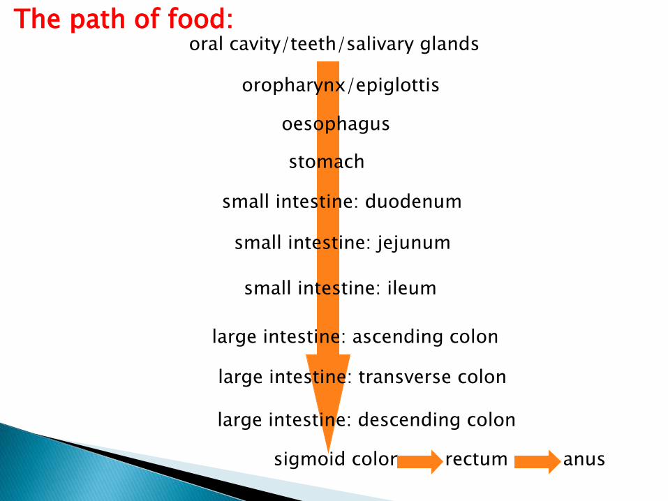
The path of food:oral cavity/teeth/salivary glands
oropharynx/epiglottis
oesophagus
stomach
small intestine: duodenum
small intestine: ileum
small intestine: jejunum
large intestine: ascending colon
large intestine: transverse colon
large intestine: descending colon
sigmoid colon rectum anus

21
It is the main window of G.I.tract
From mouth to pharynx is called oral cavity
Mouth or oral cavity is bounded by muscle & bone.
Lips- Orbicularis orismuscle
Cheeks – Buccinatormuscle
Mouth/Oral cavity:-


24
The vestibule :-is the space between gums & checks.
The oral cavity:- It is the space contained within the upper and lower dental arches.
Uvula:- Is a carved fold of muscle covered with mucus membrane hanging down from middle palate.

25
Teeth:-
Called “dentition” (like dentist)
Teeth live in sockets (alveoli) in the gum-covered margins of the mandible and maxilla
Chewing: raising and lowering the mandible and moving it from side to side while tongue positions food between teeth

26
Two sets1. Temporary/Milk/Primary
/deciduous teeth1. “Baby” teeth
2. Start at 6 months
3. 20 are out by about 2 years
4. Fall out between 2-6 years
2. Permanent: 32 total1. All but 3rd set of molars by
end of adolescence
2. 3rd set = “wisdom teeth”1. Variable
3. Some can be “impacted” (imbedded in bone)
27
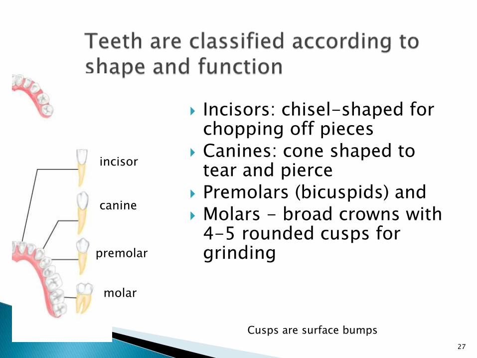
Incisors: chisel-shaped for chopping off pieces
Canines: cone shaped to tear and pierce
Premolars (bicuspids) and Molars - broad crowns with
4-5 rounded cusps for grinding
incisor
canine
premolar
molar
Cusps are surface bumps
28
29
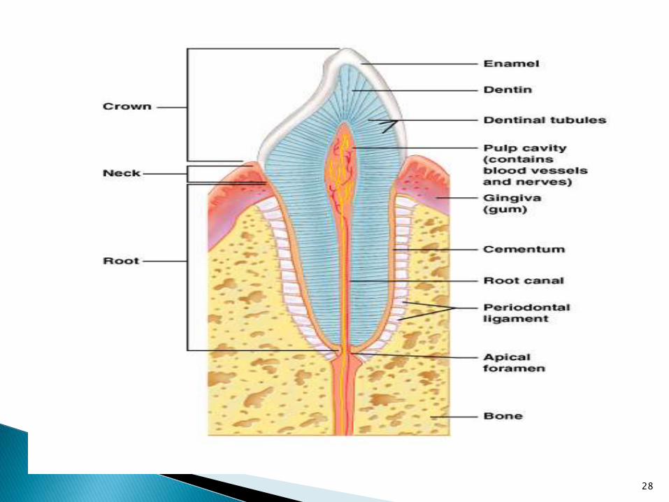
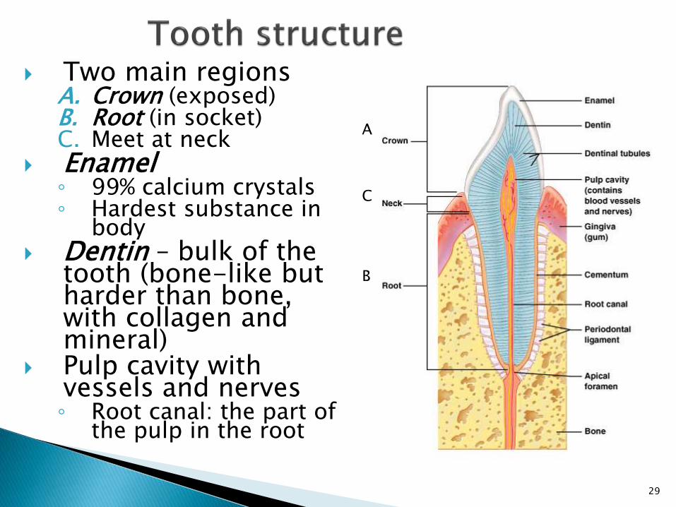
Two main regionsA. Crown (exposed)B. Root (in socket)C. Meet at neck
Enamel ◦ 99% calcium crystals◦ Hardest substance in
body Dentin – bulk of the
tooth (bone-like but harder than bone, with collagen and mineral)
Pulp cavity with vessels and nerves◦ Root canal: the part of
the pulp in the root
A
B
C
30
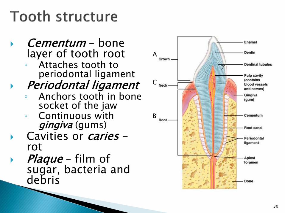
Cementum – bone layer of tooth root◦ Attaches tooth to
periodontal ligament
Periodontal ligament◦ Anchors tooth in boney
socket of the jaw◦ Continuous with
gingiva (gums)
Cavities or caries -rot
Plaque – film of sugar, bacteria and debris
A
B
C

31
Intrinsic salivary glands – within mucosa◦ Secrete saliva all the
time to keep mouth moist
Extrinsic salivary glands◦ Paired (2 each)
Parotid
Submandibular
Sublingual
◦ External to mouth◦ Ducts to mouth◦ Secrete saliva only right
before or during eating
Saliva: mixture of water, ions, mucus, enzymeskeep mouth moistdissolves food so can be tastedmoistens foodstarts enzymatic digestionbuffers acidantibacterial and antiviral
32
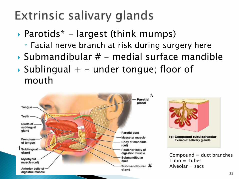
Parotids* - largest (think mumps)◦ Facial nerve branch at risk during surgery here
Submandibular # - medial surface mandible
Sublingual + - under tongue; floor of mouth
Compound = duct branchesTubo = tubesAlveolar = sacs
*
#
+

33
Oropharynx and laryngopharynx◦ Stratified squamous
epithelium
Three constrictor muscles*◦ Sequentially squeeze
bolus of food into esophagus
◦ Are skeletal muscles Voluntary action
Vagus nerve (X)
___oropharynx
___laryngopharynx
*
*
*