PowerPoint Presentation · VA 0.10 logMAR or better in each eye and equal VA between the eyes...
5
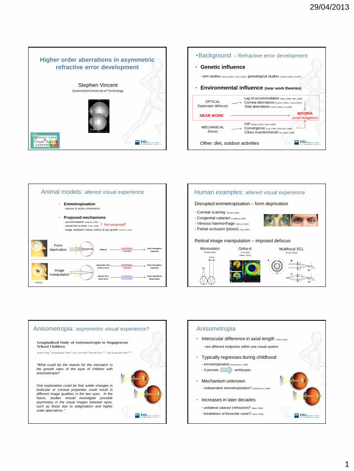
29/04/2013 1 Higher order aberrations in asymmetric refractive error development Stephen Vincent Queensland University of Technology Singh • Background – Refractive error development • Genetic influence - twin studies Sorsby (1962), Dirani (2008), genealogical studies Zadnick (1994), Ip (2007) • Environmental influence (near work theories) MYOPIA (axial elongation) OPTICAL (hyperopic defocus) MECHANICAL (force) NEAR WORK Lag of accommodation Abbot (1998), Allen (2006) Corneal aberrations Buehren (2003), Collins (2006) Total aberrations Collins (1995), He (2002) IOP Abdalla (1970), Quinn (1995) Convergence Goss (1996), Bayramlar (1999) Ciliary muscle/choroid van Alphen (1986) Other: diet, outdoor activities Animal models: altered visual experience CHOROIDAL THINNING CHOROIDAL THINNING CHOROIDAL THICKENING Axial elongation (myopia) Axial elongation (myopia) Axial retardation (hyperopia) Hyperopic blur (minus lens) Myopic blur (plus lens) Diffuser Wildsoet Form deprivation Image manipulation • Emmetropisation - passive & active components • Proposed mechanisms - accommodation Wildsoet (1991) - neural link to brain Troilo (1990) - image mediated retinal control of eye growth Wallman (1987) Not essential? Human examples: altered visual experience - Corneal scarring Tabbara (1999) - Congenital cataract Kugelberg (1996) - Vitreous haemorrhage Mohney (2002) - Partial occlusion (ptosis) Hoyt (1981) Disrupted emmetropisation – form deprivation Monovision Phillips (2005) Retinal image manipulation – imposed defocus Ortho-K Cho (2005) Kakita (2011) Multifocal SCL Anstice (2011) “What could be the reason for the mismatch in the growth rates of the eyes of children with anisometropia? One explanation could be that subtle changes in lenticular or corneal properties could result in different image qualities in the two eyes. In the future, studies should investigate possible asymmetry in the visual images between eyes, such as those due to astigmatism and higher order aberrations.” Anisometropia: asymmetric visual experience? Anisometropia • Interocular difference in axial length Sorsby (1962) - two different endpoints within one visual system • Typically regresses during childhood - emmetropisation Abrahamsson (1990) - if persists amblyopia • Mechanism unknown - independent emmetropisation? Abrahamsson (1990) • Increases in later decades - unilateral cataract (refractive)? Attebo (1999) - breakdown of binocular vision? Weale (2006)
Transcript of PowerPoint Presentation · VA 0.10 logMAR or better in each eye and equal VA between the eyes...
29/04/2013
1
Higher order aberrations in asymmetric
refractive error development
Stephen Vincent Queensland University of Technology
Singh
•Background – Refractive error development
• Genetic influence
- twin studies Sorsby (1962), Dirani (2008), genealogical studies Zadnick (1994), Ip (2007)
• Environmental influence (near work theories)
MYOPIA (axial elongation)
OPTICAL
(hyperopic defocus)
MECHANICAL
(force)
NEAR WORK
Lag of accommodation Abbot (1998), Allen (2006)
Corneal aberrations Buehren (2003), Collins (2006)
Total aberrations Collins (1995), He (2002)
IOP Abdalla (1970), Quinn (1995)
Convergence Goss (1996), Bayramlar (1999)
Ciliary muscle/choroid van Alphen (1986)
Other: diet, outdoor activities
Animal models: altered visual experience
CHOROIDAL
THINNING
CHOROIDAL
THINNING
CHOROIDAL
THICKENING
Axial elongation
(myopia)
Axial elongation
(myopia)
Axial retardation
(hyperopia)
Hyperopic blur
(minus lens)
Myopic blur
(plus lens)
Diffuser
Wildsoet
Form
deprivation
Image
manipulation
• Emmetropisation - passive & active components
• Proposed mechanisms - accommodation Wildsoet (1991)
- neural link to brain Troilo (1990)
- image mediated retinal control of eye growth Wallman (1987)
Not essential?
Human examples: altered visual experience
- Corneal scarring Tabbara (1999)
- Congenital cataract Kugelberg (1996)
- Vitreous haemorrhage Mohney (2002)
- Partial occlusion (ptosis) Hoyt (1981)
Disrupted emmetropisation – form deprivation
Monovision Phillips (2005)
Retinal image manipulation – imposed defocus
Ortho-K
Cho (2005)
Kakita (2011)
Multifocal SCL Anstice (2011)
“What could be the reason for the mismatch in
the growth rates of the eyes of children with
anisometropia?
One explanation could be that subtle changes in
lenticular or corneal properties could result in
different image qualities in the two eyes. In the
future, studies should investigate possible
asymmetry in the visual images between eyes,
such as those due to astigmatism and higher
order aberrations.”
Anisometropia: asymmetric visual experience? Anisometropia
• Interocular difference in axial length Sorsby (1962)
- two different endpoints within one visual system
• Typically regresses during childhood
- emmetropisation Abrahamsson (1990)
- if persists amblyopia
• Mechanism unknown
- independent emmetropisation? Abrahamsson (1990)
• Increases in later decades
- unilateral cataract (refractive)? Attebo (1999)
- breakdown of binocular vision? Weale (2006)
29/04/2013
2
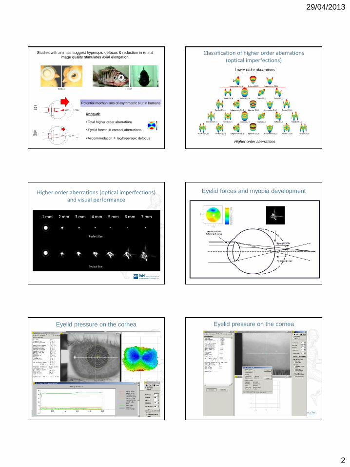
Studies with animals suggest hyperopic defocus & reduction in retinal
image quality stimulates axial elongation.
Wildsoet
OD -6.00
(6/6)
OS -3.00
(6/6)
Potential mechanisms of asymmetric blur in humans
Unequal:
• Total higher order aberrations
• Eyelid forces ± corneal aberrations
• Accommodation ± lag/hyperopic defocus
Sivak
Classification of higher order aberrations (optical imperfections)
Lower order aberrations
Higher order aberrations
Higher order aberrations (optical imperfections) and visual performance
1 mm 2 mm 3 mm 4 mm 5 mm 6 mm 7 mm
Perfect Eye
Typical Eye
Eyelid forces and myopia development
Eyelid pressure on the cornea
Previous studies:
Buehren (2003, 2005) & Shaw (2008)
Changes in corneal topography have been
observed following near work due to eyelid
pressure.
The magnitude of corneal change is proportional
to the angle of downward gaze, duration and
type of task and palpebral aperture morphology.
Myopes show larger changes compared to
emmetropes, suggesting a possibly link between
near work and myopia development.
Interocular symmetry has not been investigated.
Eyelid pressure on the cornea

29/04/2013
3
• Aim:
To examine the interocular symmetry of changes in corneal optics
following a short period of near work in a cohort of myopic
anisometropic subjects.
• Participants: 34 myopic anisometropes ≥ 1.00 D
Mean SER anisometropia 1.70 ± 0.74 D
Pre presbyopic 18-34 years of age: Mean 24 ± 4 years
31 East Asian and 3 Caucasian, 22 female and 12 male
No systemic or ocular disease, history of patching or ocular trauma
VA 0.10 logMAR or better in each eye and equal VA between the eyes
• Methods:
Digital photography of the anterior eye
(primary and down gaze)
10 minute reading task
2.5 D accommodation
25 degree downward gaze
Pre-reading
corneal topography
Post-reading
corneal topography
Right
Right
Left
Left
PRIMARY
GAZE
DOWNWARD
GAZE
• Results: Symmetry of anterior eye morphology
Right
Right
Left
Left
Symmetry of anterior eye morphology
More myopic Less myopic
More myopic Less myopic
PRIMARY
GAZE
DOWNWARD
GAZE
Symmetry of anterior eye morphology
Primary Down

29/04/2013
4
• Results: Typical change in corneal topography
Sub
ject
22
MORE MYOPIC LESS MYOPIC SCALE
-9.25/-2.50 x 2 -7.25/-2.50 x 168
PR
E TA
SK
PO
ST T
ASK
D
IFFE
REN
CE
(PO
ST –
PR
E)
25
DEG
REE
DO
WN
WA
RD
Anterior morphology
& pre/post reading
corneal topography
Change in corneal topography following reading
Less myopic More myopic Scale (D)
Change in HOA and image quality
Point Spread Function
- Measure of optical quality
- The degree of image distortion
(from a point source of light)
- In this case due to the change in
corneal optics following reading
No change
1 min arc
Less myopic More myopic
Change in aberrations and image quality
Less myopic
More myopic
For 6 mm pupil, there
was a significantly
greater reduction in
image quality for the
more myopic eyes.
Due to greater
changes in lower
order aberrations
(astigmatism) rather
than higher order
aberrations.
•Summary
• Following a short duration reading task we observed:
- Changes in corneal optics corresponding with downward gaze palpebral aperture
morphology due to eyelid pressure
- Similar changes in higher order aberrations, but significantly greater changes in corneal
astigmatism in the more myopic eye, despite symmetrical anterior eyelid morphology
• These findings lend some support to the hypothesis that image
degradation following near work may contribute to myopia/anisomyopia
development.
• However, cross-sectional study of short duration. Requires a longitudinal
study examining symmetry of optical changes during myopia
development.

29/04/2013
5
Acknowledgements
• QUT
– Professor Michael Collins
– Dr Scott Read
– Professor Leo Carney
• HKPU
– Professor Maurice Yap
– Percy Ng
2010