Powerpoint Templates Page 1 Depth Effects of DEP Chip with Microcavities Array on Impedance...
21
Powerpoint Templates Page 1 Depth Effects of DEP Chip with Microcavities Array on Impedance Measurement for Live and Dead Cells Cheng-Hsin Chuang - STUST
-
Upload
cori-bryant -
Category
Documents
-
view
219 -
download
2
Transcript of Powerpoint Templates Page 1 Depth Effects of DEP Chip with Microcavities Array on Impedance...

Powerpoint TemplatesPage 1
Depth Effects of DEP Chip with Microcavities
Array on Impedance
Measurement for Live and Dead
CellsCheng-Hsin Chuang - STUST
Powerpoint TemplatesPage 2
Outline
• I. Introduction• II. DEP theory• III. Simulation• IV. Fabrication and Measurement• V. Conclusion

Powerpoint TemplatesPage 3
I. Introduction
- Electrical detection method and the remarkable capability of positioning and registration of cell with single-cell resolution are concerned, nowadays.
- A DEP chip consisted of multilayer electrodes and microcavities array for trapping cells and further electrical measurement under single-cell level.
- Two kinds of cell lines, NB4 and HL-60 can be clearly identified, and the effects of microcavity on impedance measurement will be discussed by numerical simulation and experimental data.

Powerpoint TemplatesPage 4
II. DEP theory (1)
The time averaged dielectrophoretic force acting on a spherical particle, immersed in a medium and exposed to a spatially non-uniform electric field can be described by. The dipole component of the DEP force is
εm is the electrical permittivity of the surrounding medium,
Rp is the radius of the particle,
is the gradient of the square of applied electric field magnitude

Powerpoint TemplatesPage 5
II. DEP theory (2)
For a dielectric uniform sphere, such as a bead, it is given by
where ε and σ are the permittivity and conductivity of medium or particle, respectively, and j is √(-1).
K (ω) is the frequency dependent Claussius-Mosotti (CM) factor, ε* is the complex permittivity of the medium (m) or particle (p) and defined by

Powerpoint TemplatesPage 6
II. DEP theory (3)
> 0 it will induce positive-DEP and the particle will move toward the high intensity electric field
< 0 it will induce negative-DEP and the particle will move toward the low intensity electric field
So the direction of the DEP force is determined by the CM factor, and the magnitude of the DEP force is determined by the applied electric field and the size of the particle.

Powerpoint TemplatesPage 7
III. Simulation (1)DEP Force Simulation
Fig. 1. (a) The contour of electric field for 10μm depth SU-8 microcavity, the highest density of electric field is near middle electrode upon the SU-8 surface, and the lowest value occurred at the bottom. (b) the density of electric field along the surface of SU-8 for the different depths, 5, 10 and 15μm, respectively.

Powerpoint TemplatesPage 8
III. Simulation (2)DEP Force Simulation
Fig. 2. (a) The gradient of electric field intensity, 2E for 10μm depth SU-8 microcavity, the strongest DEP force happened near the top of SU-8 microcavity and move particle into microcavity by negative DEP (b) 2E along the surface of SU-8 for different microcavity depths 5, 10 and 15μm, respectively.

Powerpoint TemplatesPage 9
II. Simulation (3)DEP Force Simulation
Figure 3. The various profiles of 2E for different distances away from the middle electrode surface, such as 0, 2, 4 6, 10 and 28μm.

Powerpoint TemplatesPage 10
III. Simulation (4)Impedance Simulation

Powerpoint TemplatesPage 11
Figure 4. The total current density as impedance measurement by applied voltage is 0.2V on a pair of bottom electrodes, the frequency is 100kHz, and the depth of microcavity is 0μm;
(a)without particle in the SU-8 cavity; (b) the particle fixed in the SU-8 cavity.
III. Simulation (5)Impedance Simulation

Powerpoint TemplatesPage 12
Figure 5. The total current density as impedance measurement by applied voltage is 0.2V on a pair of bottom electrodes and the scan frequency is form 1kHz to 3MHz, the depth of microcavity is 10μm;
(a) without particle in the SU-8 cavity;
(b) the particle fixed in the SU-8 cavity.
III. Simulation (6)Impedance Simulation

Powerpoint TemplatesPage 13
IV. Fabrication and Measurement (1)DEP Chip
Figure 6. The microfabrication processes of multilayer electrodes DEP chip for single-cell level impedance measurement

Powerpoint TemplatesPage 14
IV. Fabrication and Measurement (2)DEP Chip
Figure 7. SEM pictures of SU-8 cayitys array, the diameter and spacing are both 16 m and the depth is 10m, (a) 2 by 2 cavitys array; (b) Nanofocus image for 5m depth (c) The picture of multilayer electrodes DEP chip; (d) The blast diagram of DEP chip.

Powerpoint TemplatesPage 15
IV. Fabrication and Measurement (3)DEP Chip
Figure 8. OM pictures of SU-8 microcavities array, the diameter and spacing are both 16 m and the depth is 10m; (a) the layout of four individual impedance electrodes under four specific the microcavities; (b) the gap of impedance electrode is 8m.

Powerpoint TemplatesPage 16
IV. Fabrication and Measurement (4)Experimental setup
Powerpoint TemplatesPage 17
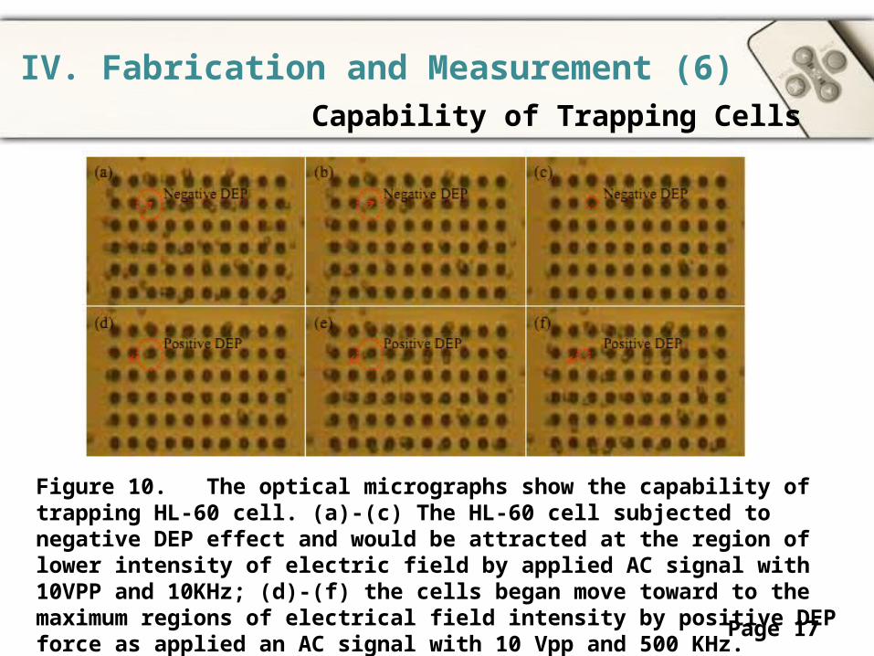
IV. Fabrication and Measurement (6)Capability of Trapping Cells
Figure 10. The optical micrographs show the capability of trapping HL-60 cell. (a)-(c) The HL-60 cell subjected to negative DEP effect and would be attracted at the region of lower intensity of electric field by applied AC signal with 10VPP and 10KHz; (d)-(f) the cells began move toward to the maximum regions of electrical field intensity by positive DEP force as applied an AC signal with 10 Vpp and 500 KHz.

Powerpoint TemplatesPage 18
IV. Fabrication and Measurement (7)Impedance Measurement
Figure 11. The results of impedance measurement for the 5m-depth microcavity under four conditions: (1) only DI water without cells, (2) only sucrose solution without cells, (3) HL60 live cell immersed in sucrose solution, (4) HL60 dead cell immersed in sucrose solution, all conditions were applied 0.2V and the frequency range is 1K to 3M Hz. (a) impedance magnitude (ohm); (b) phase (degree).

Powerpoint TemplatesPage 19
IV. Fabrication and Measurement (8)Impedance Measurement
Figure 12. The results of impedance measurement for the 10m-depth microcavity under five conditions: (1) only DI water without cells, (2) only sucrose solution without cells, (3) HL60 live cell immersed in sucrose solution, (4) NB4 live cell immersed in sucrose solution,(5) NB4 dead cell immersed in sucrose solution, all conditions were applied 0.2V and the frequency range is 1K to 3M Hz. (a) impedance magnitude (ohm); (b) phase (degree).
Powerpoint TemplatesPage 20
V. Conclusions
We have designed and fabricated a DEP chip with multilayer electrodes and microcavity array for impedance measurement of single cell.
The depth effects on impedance difference were analyzed by finite element method and verified by experimental results.
This microchip not only provides an efficient way to immobilization cells in the microcavity for a long period of time without applying DEP force but also easily identifies the live and dead cells based on impedance measurement

Powerpoint TemplatesPage 21