PowerPoint Presentationamos3.aapm.org/abstracts/pdf/137-41620-446581-142250.pdf · 2018-07-31 ·...
25
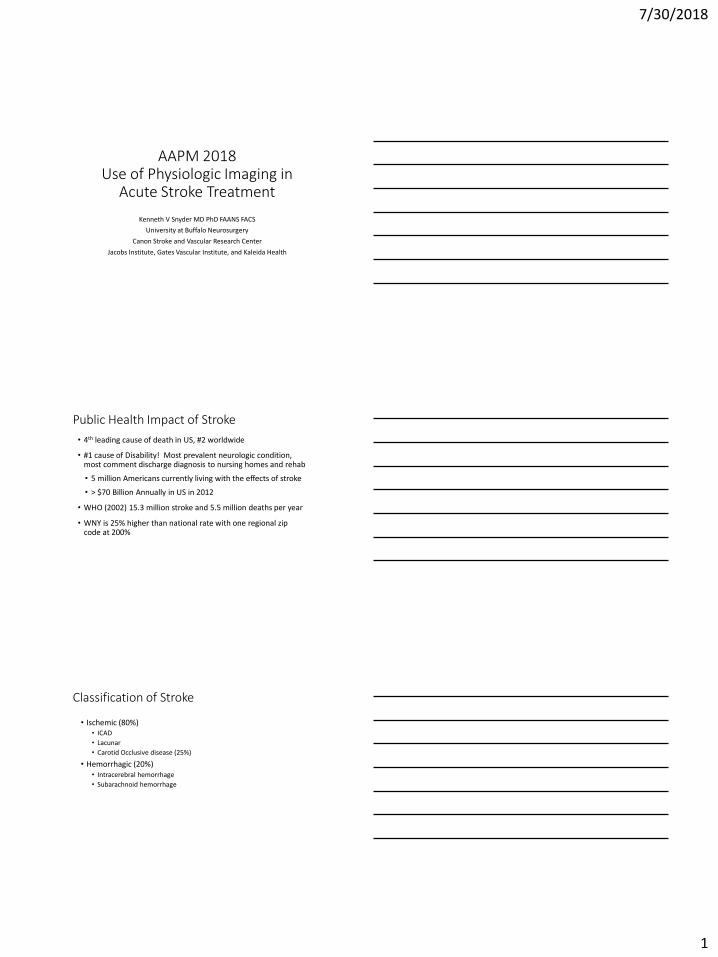
7/30/2018 1 AAPM 2018 Use of Physiologic Imaging in Acute Stroke Treatment Kenneth V Snyder MD PhD FAANS FACS University at Buffalo Neurosurgery Canon Stroke and Vascular Research Center Jacobs Institute, Gates Vascular Institute, and Kaleida Health Public Health Impact of Stroke • 4 th leading cause of death in US, #2 worldwide • #1 cause of Disability! Most prevalent neurologic condition, most comment discharge diagnosis to nursing homes and rehab • 5 million Americans currently living with the effects of stroke • > $70 Billion Annually in US in 2012 • WHO (2002) 15.3 million stroke and 5.5 million deaths per year • WNY is 25% higher than national rate with one regional zip code at 200% Classification of Stroke • Ischemic (80%) • ICAD • Lacunar • Carotid Occlusive disease (25%) • Hemorrhagic (20%) • Intracerebral hemorrhage • Subarachnoid hemorrhage
Transcript of PowerPoint Presentationamos3.aapm.org/abstracts/pdf/137-41620-446581-142250.pdf · 2018-07-31 ·...
7/30/2018
1
AAPM 2018 Use of Physiologic Imaging in
Acute Stroke Treatment
Kenneth V Snyder MD PhD FAANS FACS
University at Buffalo Neurosurgery
Canon Stroke and Vascular Research Center
Jacobs Institute, Gates Vascular Institute, and Kaleida Health
Public Health Impact of Stroke
• 4th leading cause of death in US, #2 worldwide
• #1 cause of Disability! Most prevalent neurologic condition, most comment discharge diagnosis to nursing homes and rehab
• 5 million Americans currently living with the effects of stroke
• > $70 Billion Annually in US in 2012
• WHO (2002) 15.3 million stroke and 5.5 million deaths per year
• WNY is 25% higher than national rate with one regional zip code at 200%
Classification of Stroke
• Ischemic (80%) • ICAD
• Lacunar
• Carotid Occlusive disease (25%)
• Hemorrhagic (20%) • Intracerebral hemorrhage
• Subarachnoid hemorrhage
7/30/2018
2
Stroke Treatment Options in 2013
• ASA within 24-48 hrs is recommended
• IV rtPA in appropriate patients (<3-4.5 hours)
• IA thrombolysis an option in major MCA stroke patients <6 hours if not IV rtPA candidates (dose not determined and NOT FDA approved)
• Mechanical thrombectomy devices can be offered in carefully selected patients and should continue to be studied in randomized trials
AHA/ASA Stroke Guidelines Stroke 2013
IV tPA and Large-Vessel Occlusion (35-40% of Ischemic Stroke) • Clinical response to thrombolysis is influenced by the site of occlusion
• Rate of recovery from IV tPA by occlusion • 33% for distal MCA occlusion
• 15% for proximal MCA occlusion
• 0% with ICA-T occlusions
• Mortality of LVO
Stroke. 2007;38:948-954
1. Jansen O, et al.
2. Furlan A et al. PROACT II Trial
3. Brückmann H et al.
Vessel Mortality Rate
ICA 53%1
MCA 27%2
Basilar Artery 89-90%3
7/30/2018
3
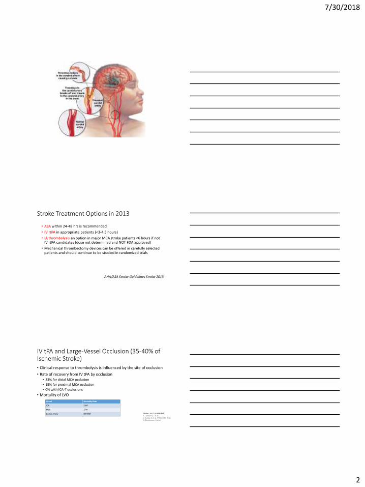
Saver JL, Stroke 2013;44:270-277
Stent retrievers
What’s A Retrievable Stent?
2015: Endovascular vs Best Medical Therapy
• 5 major studies evaluating the role of endovascular therapy in stroke treatment • MR CLEAN
• EXTEND-IA
• ESCAPE
• SWIFT PRIME
• REVASCAT
• Endovascular Therapy within 6 hours, NIHSS >7
• ALL 5 trials stopped because of significant benefit in the Endovascular arms
7/30/2018
4
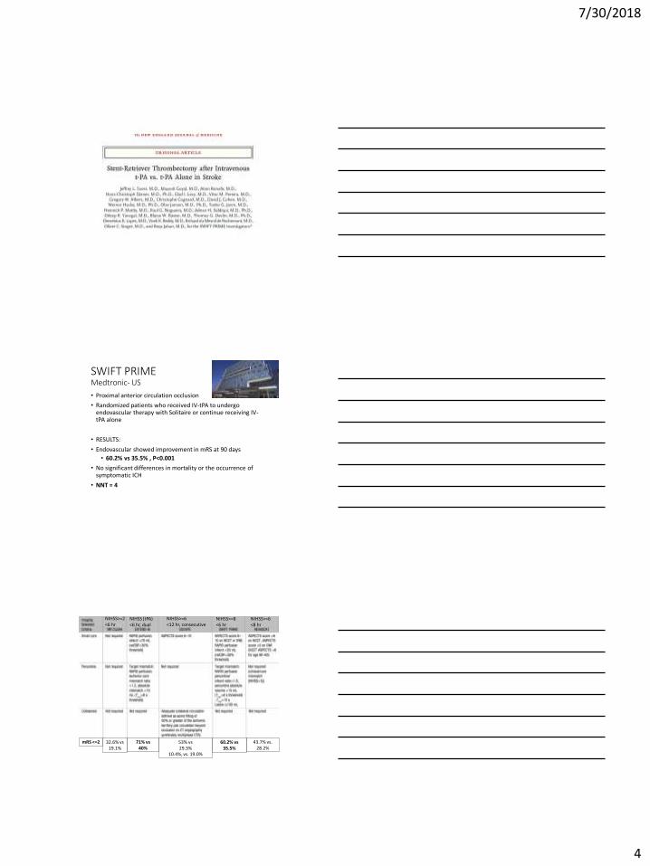
SWIFT PRIME Medtronic- US
• Proximal anterior circulation occlusion
• Randomized patients who received IV-tPA to undergo endovascular therapy with Solitaire or continue receiving IV-tPA alone
• RESULTS:
• Endovascular showed improvement in mRS at 90 days
• 60.2% vs 35.5% , P<0.001
• No significant differences in mortality or the occurrence of symptomatic ICH
• NNT = 4
32.6% vs 19.1%
71% vs 40%
60.2% vs 35.5%
43.7% vs. 28.2%
53% vs 29.3%
10.4%, vs. 19.0%
NIHSS>=2 <6 hr
NIHSS>=6 <12 hr, consecutive
NIHSS>=8 <6 hr
NIHSS (tPA) <6 hr, dual
NIHSS>=6 <8 hr
mRS <=2

7/30/2018
5
CTA Collaterals
http://www.aspectsinstroke.com
Future of Stroke Imaging
The greatest challenge is to show that advanced neuroimaging, used as a
biomarker to select patients for reperfusion therapy (in an extended time window),
improves patient outcomes
Advanced Imaging
ischemic penumbra
core ischemic zone

7/30/2018
6
Advanced Imaging
• Dynamic Studies capturing one cycle of the full transit of a contrast bolus though the tissue
• Physiologic Imaging: Transit Time, Blood Flow, Blood Volume • Parenchyma (Capillary phase NOT large vessels)
• Intravascular surrogate for Intracellular process (not biological, Xenon)
• Ability to distinguish core (infarcted tissue) from penumbra (salvageable tissue)
• Individualize stroke treatment
Buffalo Protocol
• NIHSS and CTSS (CTA head and neck and CTP) • Intervention based on perfusion parameters, clinical exam, and Time of Onset • MRI if no obvious deficit on CTP
• Post intervention CT/ LCI /MRI GRE
• CTP POD #1, NIHSS at 24hrs
• MRI at 3-5 days
• Discharge disposition, NIHSS and mRS
• CT or MRI at 1-3 months, mRS and NIHSS
• All patients collected in prospective registry
7/30/2018
7
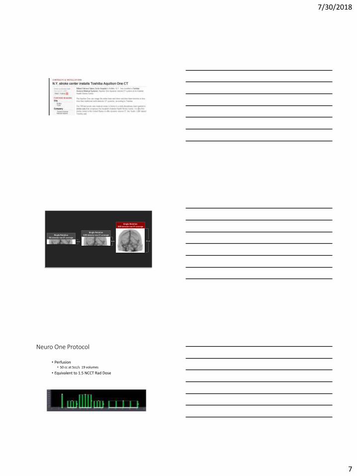
4 cm 8 cm 16 cm
Single Rotation
64 detector row CT coverage
Single Rotation
128 detector row CT coverage
Single Rotation
320 detector row CT coverage
Neuro One Protocol
• Perfusion • 50 cc at 5cc/s 19 volumes
• Equivalent to 1.5 NCCT Rad Dose
7/30/2018
8

7/30/2018
9

7/30/2018
10
7/30/2018
11
7/30/2018
12
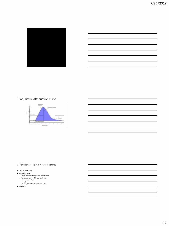
Time/Tissue Attenuation Curve
CT Perfusion Models (4 min processing time)
• Maximum Slope
• Deconvolution • Parametric R(t) has specific distribution • Non-parametric – R(t) is an unknown
• Transform – Fourier
• SVD
• Delay Insensitive Deconvolution (SVD+)
• Bayesian
7/30/2018
13
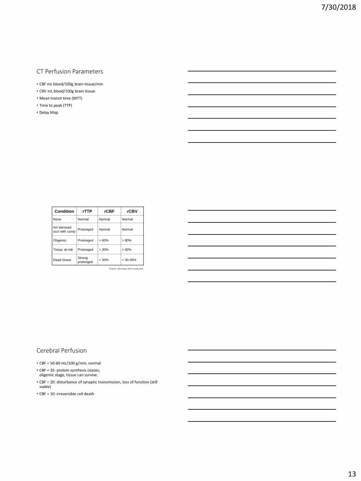
CT Perfusion Parameters
• CBF mL blood/100g brain tissue/min
• CBV mL blood/100g brain tissue
• Mean transit time (MTT)
• Time to peak (TTP)
• Delay Map
Condition rTTP rCBF rCBV
None Normal Normal Normal
Art stenosis/
occl with comp Prolonged Normal Normal
Oligemic Prolonged > 60% > 80%
Tissue at risk Prolonged > 30% > 60%
Dead tissue Strong
prolonged < 30% < 30-40%
Tomandl, 2003; Mayer 2000; Koenig 2001
Cerebral Perfusion
• CBF = 50-60 mL/100 g/min, normal
• CBF = 35: protein synthesis ceases, oligemic stage, tissue can survive.
• CBF = 20: disturbance of synaptic transmission, loss of function (still viable)
• CBF = 10: irreversible cell death

7/30/2018
14
Heiss and Rosner (1983)
0
10
20
30
40
50
60
70
80
5 8 12 15
tim
e (
min
)
CBF (ml/min/100g)
Duration of Ischemia for Infarction
Reliability?
Radiology 254(1): Jan 2010
7/30/2018
15
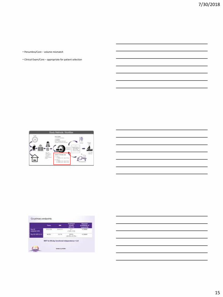
• Penumbra/Core – volume mismatch
• Clinical Exam/Core – appropriate for patient selection

7/30/2018
16
Defuse 3- Thrombectomy for Stroke at 6-16 hours with selection by Perfusion Imaging • NEJM 2018: 378:708-18
• Multicenter, randomized, MCA or ICA, Primary outcome mRS at 90 days
• Less than 70 cc core, and Ratio of >1.8
• RESULTS: • Terminated Early for efficacy (92 endo and 90 BMT)
• mRS 0-2 (45% vs 17%)
• 90 day mortality (14% vs 26%)
• No sign difference in sICH (7% vs 4%)
DAWN • Age: > 18
• NIHSS: >= 10
• Vessel: ICA/M1
• LSN: 6-24
• CTP Core: <20, <30, <50
• CTP Ratio: none
DEFUSE 3 • 18-90
• >= 6
• ICA/M1
• 6-16
• < 70
• >1.8
7/30/2018
17
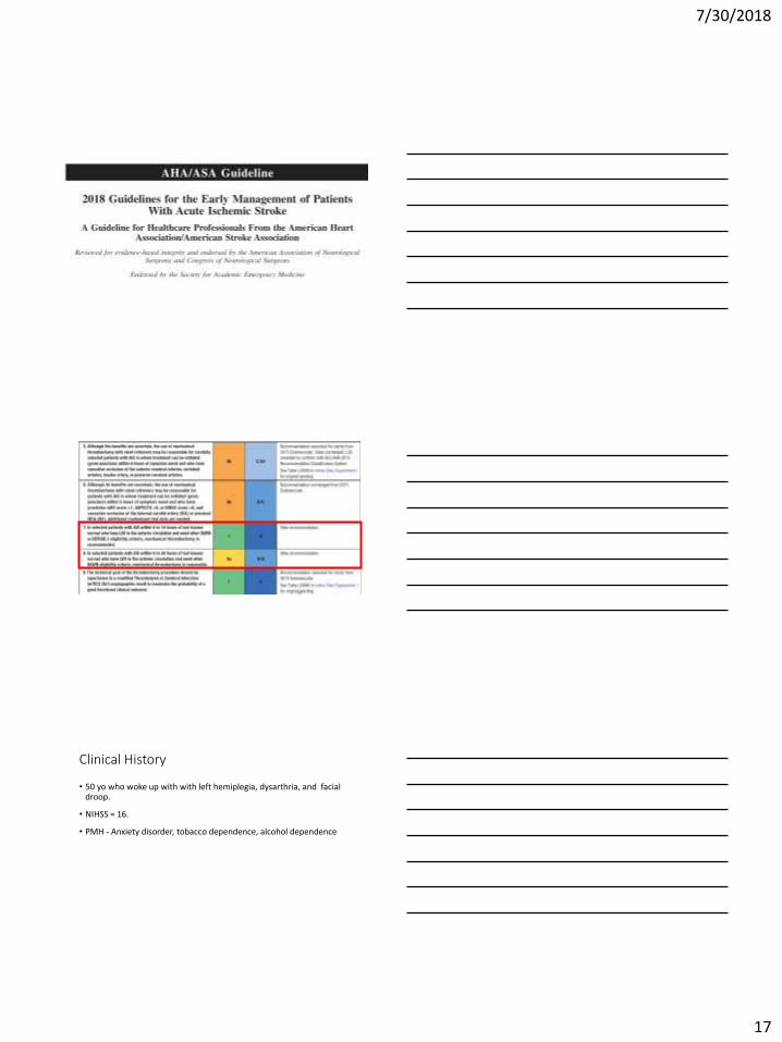
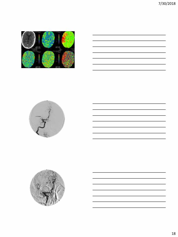
Clinical History
• 50 yo who woke up with with left hemiplegia, dysarthria, and facial droop.
• NIHSS = 16.
• PMH - Anxiety disorder, tobacco dependence, alcohol dependence
7/30/2018
18
7/30/2018
19
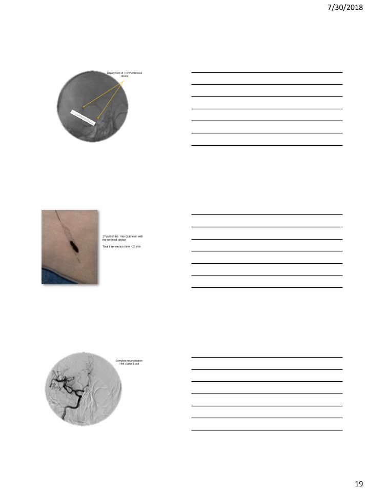
Deployment of TREVO retrieval
device
1st pull of the microcatheter with the retrieval device
Total intervention time ~20 min
Complete recanalization
TIMI-3 after 1 pull

7/30/2018
20
• In the angio suite – the patient could lift his Rt arm antigravity, improved gaze, NIHSS 16 to 5 immediately
• POD#1 NIH -3
• POD#2– NIH -0
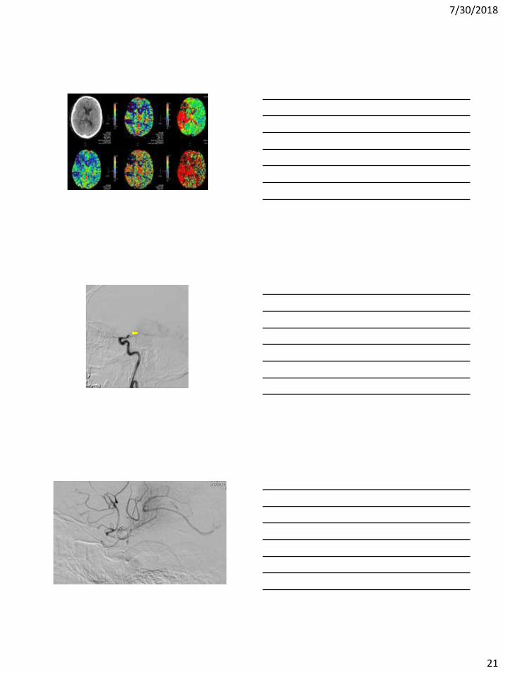
Clinical History
• 75 yo WM last seen normal at 10 pm, ? Issues at 2 am, awoke thrashing at 4 am with Right gaze preference and left HP
• NIHSS 18
7/30/2018
21

7/30/2018
22
TIME
7/30/2018
23
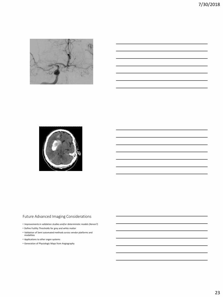
Future Advanced Imaging Considerations
• Improvements in validation studies and/or deterministic models (Xenon?)
• Define Futility Thresholds for grey and white matter
• Validation of Semi automated methods across vendor platforms and modalities
• Applications to other organ systems
• Generation of Physiologic Maps from Angiography
7/30/2018
24
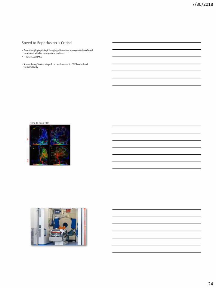
Speed to Reperfusion is Critical
• Even though physiologic imaging allows more people to be offered treatment at later time points, realize…
• IT IS STILL A RACE
• Streamlining Stroke triage from ambulance to CTP has helped tremendously
Pre
Po
st
Time To Peak(TTP)
3.8 (sec)
1.54 (sec)
0 (sec)
0 (sec)
0 (sec)
0 (sec)
2.56 (sec)
1.89 (sec)
2.12 (sec) 2.18 (sec)
0.66 (sec)
1.14 (sec)
1.2 (sec) 1.52 (sec)
1.27 (sec)
0.99 (sec)
D1
D2
E1
E2
7/30/2018
25
| See. Diagnose. Treat.
Thank you!

![Content Based Image Retrieval using Query by Approximate … · Retrieval (KBIR), Semantic Based Image Retrieval (SBIR) and Content Based Image Retrieval (CBIR) [1]. The KBIR methods](https://static.fdocuments.in/doc/165x107/604cc727f7fc662d1d5e1fe3/content-based-image-retrieval-using-query-by-approximate-retrieval-kbir-semantic.jpg)