Potential role of viruses in white plague coral disease...ORIGINAL ARTICLE Potential role of viruses...
13
ORIGINAL ARTICLE Potential role of viruses in white plague coral disease Nitzan Soffer 1,2 , Marilyn E Brandt 3 , Adrienne MS Correa 1,2,4 , Tyler B Smith 3 and Rebecca Vega Thurber 1,2 1 Department of Microbiology, Oregon State University, Corvallis, OR, USA; 2 Department of Biological Sciences, Florida International University, North Miami, FL, USA; 3 Center for Marine and Environmental Studies, University of the Virgin Islands, St Thomas, Virgin Islands, USA and 4 Ecology and Evolutionary Biology Department, Rice University, Houston, TX, USA White plague (WP)-like diseases of tropical corals are implicated in reef decline worldwide, although their etiological cause is generally unknown. Studies thus far have focused on bacterial or eukaryotic pathogens as the source of these diseases; no studies have examined the role of viruses. Using a combination of transmission electron microscopy (TEM) and 454 pyrosequencing, we compared 24 viral metagenomes generated from Montastraea annularis corals showing signs of WP-like disease and/or bleaching, control conspecific corals, and adjacent seawater. TEM was used for visual inspection of diseased coral tissue. No bacteria were visually identified within diseased coral tissues, but viral particles and sequence similarities to eukaryotic circular Rep-encoding single-stranded DNA viruses and their associated satellites (SCSDVs) were abundant in WP diseased tissues. In contrast, sequence similarities to SCSDVs were not found in any healthy coral tissues, suggesting SCSDVs might have a role in WP disease. Furthermore, Herpesviridae gene signatures dominated healthy tissues, corroborating reports that herpes-like viruses infect all corals. Nucleocytoplasmic large DNA virus (NCLDV) sequences, similar to those recently identified in cultures of Symbiodinium (the algal symbionts of corals), were most common in bleached corals. This finding further implicates that these NCLDV viruses may have a role in bleaching, as suggested in previous studies. This study determined that a specific group of viruses is associated with diseased Caribbean corals and highlights the potential for viral disease in regional coral reef decline. The ISME Journal advance online publication, 15 August 2013; doi:10.1038/ismej.2013.137 Subject Category: Microbe-microbe and microbe-host interactions Keywords: viral metagenomics; eukaryotic ssDNA virus; satellite DNAs; nucleocytolasmic large DNA virus (NCLDV); coral bleaching Introduction Difficulties associated with isolating, culturing and manipulating potential pathogens as well as in quantitatively and temporally connecting the pre- valence of a given microbe or virus to specific signs of infection have limited our understanding of the etiology of many wildlife diseases. Given these challenges, non-culture-based methods are increas- ingly used in research to characterize disease in novel environments or organisms. For example, surveys such as amplicon-based 16S ribosomal DNA profiling, can compare whole microbial assemblages from organisms of different health states, but to comprehensively survey viruses, shotgun-based metagenomic sequencing approaches are often required (Mokili et al., 2012). This is because viruses lack a common phylogenetic marker, and therefore marker-based amplicon sequencing (for example, 16S or 18S ribosomal RNA surveys) cannot necessarily be applied to diverse viral consortia. Some examples of the successful application of viral metagenomics to elucidate the pathogens associated with a disease include the identification of: an astrovirus as the probable cause for shaking mink syndrome (Blomstro ¨m et al., 2010), an anellovirus associated with a California sea lion mortality event (Ng et al., 2009) and a coronavirus that causes human respira- tory disease (Bermingham et al., 2012). Marine wildlife diseases are increasing in pre- valence and incidence (Ward and Lafferty, 2004; Harvell et al., 2004). Reports of non-bleaching coral diseases have increased over 50-fold from 1965 to 2005 (Sokolow, 2009). Such epizootics are of concern because they have contributed to declines Correspondence: N Soffer, Department of Microbiology, Oregon State University, 514 Nash Hall, Corvallis, OR 97331, USA. E-mail: [email protected] Received 9 April 2013; revised 19 June 2013; accepted 14 July 2013 The ISME Journal (2013), 1–13 & 2013 International Society for Microbial Ecology All rights reserved 1751-7362/13 www.nature.com/ismej
Transcript of Potential role of viruses in white plague coral disease...ORIGINAL ARTICLE Potential role of viruses...
ORIGINAL ARTICLE
Potential role of viruses in white plague coraldisease
Nitzan Soffer1,2, Marilyn E Brandt3, Adrienne MS Correa1,2,4, Tyler B Smith3 andRebecca Vega Thurber1,2
1Department of Microbiology, Oregon State University, Corvallis, OR, USA; 2Department of BiologicalSciences, Florida International University, North Miami, FL, USA; 3Center for Marine and EnvironmentalStudies, University of the Virgin Islands, St Thomas, Virgin Islands, USA and 4Ecology and EvolutionaryBiology Department, Rice University, Houston, TX, USA
White plague (WP)-like diseases of tropical corals are implicated in reef decline worldwide, althoughtheir etiological cause is generally unknown. Studies thus far have focused on bacterial oreukaryotic pathogens as the source of these diseases; no studies have examined the role of viruses.Using a combination of transmission electron microscopy (TEM) and 454 pyrosequencing, wecompared 24 viral metagenomes generated from Montastraea annularis corals showing signs ofWP-like disease and/or bleaching, control conspecific corals, and adjacent seawater. TEM was usedfor visual inspection of diseased coral tissue. No bacteria were visually identified within diseasedcoral tissues, but viral particles and sequence similarities to eukaryotic circular Rep-encodingsingle-stranded DNA viruses and their associated satellites (SCSDVs) were abundant in WPdiseased tissues. In contrast, sequence similarities to SCSDVs were not found in any healthy coraltissues, suggesting SCSDVs might have a role in WP disease. Furthermore, Herpesviridae genesignatures dominated healthy tissues, corroborating reports that herpes-like viruses infect allcorals. Nucleocytoplasmic large DNA virus (NCLDV) sequences, similar to those recently identifiedin cultures of Symbiodinium (the algal symbionts of corals), were most common in bleached corals.This finding further implicates that these NCLDV viruses may have a role in bleaching, as suggestedin previous studies. This study determined that a specific group of viruses is associated withdiseased Caribbean corals and highlights the potential for viral disease in regional coral reefdecline.The ISME Journal advance online publication, 15 August 2013; doi:10.1038/ismej.2013.137Subject Category: Microbe-microbe and microbe-host interactionsKeywords: viral metagenomics; eukaryotic ssDNA virus; satellite DNAs; nucleocytolasmic large DNAvirus (NCLDV); coral bleaching
Introduction
Difficulties associated with isolating, culturing andmanipulating potential pathogens as well as inquantitatively and temporally connecting the pre-valence of a given microbe or virus to specific signsof infection have limited our understanding of theetiology of many wildlife diseases. Given thesechallenges, non-culture-based methods are increas-ingly used in research to characterize disease innovel environments or organisms. For example,surveys such as amplicon-based 16S ribosomalDNA profiling, can compare whole microbialassemblages from organisms of different healthstates, but to comprehensively survey viruses,
shotgun-based metagenomic sequencing approachesare often required (Mokili et al., 2012). This isbecause viruses lack a common phylogeneticmarker, and therefore marker-based ampliconsequencing (for example, 16S or 18S ribosomalRNA surveys) cannot necessarily be applied todiverse viral consortia. Some examples of thesuccessful application of viral metagenomics toelucidate the pathogens associated with a diseaseinclude the identification of: an astrovirus as theprobable cause for shaking mink syndrome(Blomstrom et al., 2010), an anellovirus associatedwith a California sea lion mortality event (Ng et al.,2009) and a coronavirus that causes human respira-tory disease (Bermingham et al., 2012).
Marine wildlife diseases are increasing in pre-valence and incidence (Ward and Lafferty, 2004;Harvell et al., 2004). Reports of non-bleaching coraldiseases have increased over 50-fold from 1965 to2005 (Sokolow, 2009). Such epizootics are ofconcern because they have contributed to declines
Correspondence: N Soffer, Department of Microbiology, OregonState University, 514 Nash Hall, Corvallis, OR 97331, USA.E-mail: [email protected] 9 April 2013; revised 19 June 2013; accepted 14 July2013
The ISME Journal (2013), 1–13& 2013 International Society for Microbial Ecology All rights reserved 1751-7362/13
www.nature.com/ismej

in coral abundance and cover (Gardener et al., 2003;Rogers, 2009). However, of the 420 described globalcoral diseases, only 6–8 have pathogens ascribed totheir etiology (Green and Bruckner, 2000;Sutherland et al., 2004; Rosenberg et al., 2007;Bourne et al., 2009; Pollack et al., 2011). One coraldisease of particular ecological importance is whiteplague (WP), a rapid tissue loss disease that affectsmultiple species of Caribbean corals, includingdominant reef-building Montastraea species.Caribbean WP is characterized by lesions that beginbasally or peripherally on a colony, and thenprogress rapidly (mm to cm per day) across thecolony surface resulting in partial to total colonymortality (Richardson et al., 2001; Miller et al.,2006; Weil et al., 2006). Three types of WP (I, II andIII) are differentiated based on tissue loss progres-sion rates, with type I progressing the slowest (a fewmm per day), type II progressing at a maximum rateof 2 cm per day and type III progressing the fastest(42 cm per day; Dustan, 1977; Richardson et al.,1998, 2001). Although an infectious agent, thebacterium Aurantimonas coralicida, was hypothe-sized to be the cause of WP type II in the coralDichocoenia stokesii (Richardson et al., 1998;Denner et al., 2003), A. coralicida is not alwaysassociated with WP-infected coral colonies. Forexample, this bacterium was associated withhealthy but not WP-infected Montastraea annulariscolonies (Pantos et al., 2003) or WP-infectedcolonies from different geographic regions and/orhost species (Siderastrea, Diploria; Sunagawa et al.,2009; Cardenas et al., 2012).
Given the limited success in identifying thecausative agents of WP diseases, controversy existsas to whether similar disease signs represent uniqueetiologies. It is possible that the different WP typesare either the same disease variably manifested oralternatively, WP signs may represent a variety ofdifferent diseases, even in cases where progressionrates are similar (Pollock et al., 2011). For example, aWP-like disease of corals in the Red Sea has beenshown to be caused by the bacterial pathogen,Thalassomonas loyana, and not A. coralicida. Phagetherapy reduces signs of this disease in situ and inaquaria, confirming the bacterial nature of thepathogen (Thompson et al., 2006; Efrony et al.,2009; Atad et al., 2012). Alternatively, an additionalhypothesis is that a virus and not a bacterium mightbe the cause of some of these WP signs. To test thishypothesis, we determined if various viral types aredifferentially associated with a WP type I-likeoutbreak that occurred in the US Virgin Islands in 2010.
Currently little is known about viral disease incorals. Studies have characterized the virusesassociated with bleached and healthy corals, coralsexposed to different environmental stressors, andwithin the algal symbionts (Symbiodinium spp.) ofcorals (Wilson et al., 2004; Wegley et al., 2007;Marhaver et al., 2008; Vega Thurber et al., 2008;Correa et al., 2013). Based on these works, four
major groups of viruses are predicted to infect coralsand their associated microbes: bacteriophages,enveloped herpes-like viruses, nucleocytoplasmiclarge DNA viruses (NCLDVs, including members ofthe Phycodnaviridae, Mimiviridae and Iridoviridae),and small circular single-stranded DNA (ssDNA)viruses (for a review, see Vega Thurber and Correa,2011). For example, herpes-like sequences werefound in stressed Porites compressa corals as wellas in other cnidarians such as the starlet anemone,Nematostella, and the medusozoan, Hydra (VegaThurber et al., 2008). Genomic and microscopicevidence indicate that phycodnavirus-like particlescan be present within bleached and/or heat-stressedcorals, and these virus-like particles (VLPs) havebeen suggested to target Symbiodinium (Pattenet al., 2008; Wilson et al., 2009; Correa et al.,2013). Active infection of Symbiodinium by membersof the Phycodnaviridae was indirectly demonstratedbased on the identification of phycodnavirus-likecomplementary DNAs in cultures of two differentclades of Symbiodinium (Correa et al., 2013). Finally,sequence similarities to and viral-like particlesreminiscent of nano-, circo- and geminiviruses, allof which are eukaryotic circular Rep-encodingssDNA (CRESS-DNA) viruses and their associatedsatellites (collectively referred to ‘SCSDVs’ in thispaper) also have been found in corals (Davy andPatten, 2007; Wegley et al., 2007; Marhaver et al.,2008; Patten et al., 2008; Vega Thurber et al., 2008;Littman et al., 2011; Rosario et al., 2012).
Although these previous works laid the founda-tion for our understanding of viruses associatedwith diseased corals, the potential contribution ofthese viruses to colony WP disease signs isunknown, and their role in bleaching is poorlyunderstood. Henceforth, we refer to coral coloniesexhibiting signs of WP as ‘diseased’ while coloniesshowing signs of symbiont loss, which may or maynot be due to a pathogen, are described as‘bleached.’ To determine if viruses contribute toeither bleaching and/or WP signs, we appliedtransmission electron microscopy (TEM) andreplicated metagenomics to characterize virusesfrom Caribbean M. annularis corals that appearedhealthy, bleached and/or affected by a WP-likedisease. Here, we present evidence that (1) the viralconsortia among Healthy (H), Bleached (B), WPDiseased (D) and WP Diseased-Bleached (BD) coraltissues are significantly different, and (2) SCSDVsdominate WP Diseased corals but are absent fromhealthy corals. These data suggest SCSDVs mayhave a role in some manifestations of WP-likedisease in corals.
Materials and methods
Reef site and specimen collectionSampling of the scleractinian coral, M. annularis,was conducted at Brewers Bay, in St Thomas, US
Viruses associated with diseased coralsN Soffer et al
2
The ISME Journal

Virgin Islands, during a concurrent WP outbreak andbleaching event in September 2010. During thisevent, up to 90% of corals on the examined reefwere bleached, while a maximum of 7% showedsigns of WP and bleaching (Brandt et al., 2013).Collections took place over 2 days at depths of5.5–7.6 m. Temperature at depth was B29.0 1C. AllM. annularis colonies used in this study were locatedwithin B75 m of each other. In addition to beingaffected with WP disease signs, all WP Diseasedcolonies showed signs of bleaching (Figure 1). There-fore, bleached coral colonies closest to WP Diseasedcolonies were selected as controls. All apparentlyhealthy M. annularis colonies (N¼ 2) that could belocated during specimen collection dives weresampled.
WP infections were defined using the followingsigns: (1) lesions consisted of an area of recent tissueloss where denuded skeleton with little to no algalcolonization was delineated from living tissue by asmooth, undulating margin, (2) lesions were locatedperipherally or basally on the colony and (3) lesionsexpanded at an mean rate of 0.23±0.12 cm per day(Figure 1). These disease signs and epidemiologicalproperties are consistent with those previouslyreported for WP (reviewed in Bythell et al., 2004;Sutherland et al., 2004; Brandt et al., 2013), andtissue loss rates were most similar to reports of WPtype I disease (Dustan, 1977; Sutherland et al., 2004).
Coral tissue collectionsCoral tissue samples were collected using SCUBA.Two to three plugs of tissue and skeleton wereremoved from each M. annularis colony (Figure 1)using a 2-cm diameter corer and hammer (USVIDepartment of Planning and Natural Resources
permit #STT-050–10). From the seven WP Diseasedcolonies, tissues were cored from both the bleachedtop ‘BleachedþWP Diseased (BD)’ and the marginof lesions ‘WP Diseased (D),’ which did not showsigns of bleaching. Tissue from an additional five‘Bleached (B)’ non-WP Diseased corals and two‘Healthy (H)’ corals (normal pigmentation, non-diseased) were taken as controls (Figure 1). Tissuesamples were placed in individual sterile bagsunderwater, stored on ice and processed on shorewithin 4 h. All sampled colonies were examined fordiseases signs and progression (if applicable) 2 daysafter sample collections. In all sampled WP Diseasedcolonies lesions had expanded, indicating that thedisease was active when samples were taken. In allsampled non-WP Diseased colonies, no lesions haddeveloped.
Cored specimens were rinsed with 0.02 mmfiltered seawater. Tissue was removed by airbrushingwith B40 ml of 0.02mm filtered phosphate buffersaline solution (pH 7.3). Tissue homogenateswere 0.22mm filtered, preserved in molecularbiology grade chloroform (2% final concentration)and stored at 4 1C until viral metagenome generationprocessing.
Seawater sampling, viral concentration andpurificationThree liters of seawater were collected B16 cmabove three diseased coral colonies using sterilecontainers. Replicate samples were pooled into one9 l sample from which the ‘seawater BleachedþWPDiseased (SWBD)’ virome was generated. Seawateralso was collected and pooled from above threeBleached colonies for the ‘seawater Bleached (SWB)’virome.
Bleached+WP Disease (BD) Bleached (B) Healthy (H)
N=7N=5
N=2
N=7
Diseased (D)
Figure 1 Locations of sampled tissue from M. annularis colonies of different health states. BD tissue was taken from B5 cm from the Dtissue, where disease was progressing from the base of the coral colony. Seawater samples were taken B16 cm above BD and B coralcolonies.
Viruses associated with diseased coralsN Soffer et al
3
The ISME Journal

VLPs were concentrated using a 100 kD tangentialflow filter and purified via passage through a0.22 mm sterivex (Vega Thurber et al., 2009). Theresulting viral concentrate was preserved in mole-cular biology grade chloroform (2% final concentra-tion), and stored at 4 1C until DNA extraction wasperformed as described below. The cesium chloridegradient ultracentrifugation steps were not con-ducted before extracting DNA from the seawatersamples.
Viral metagenome generationVLPs were isolated from coral tissues using cesiumchloride density gradient ultracentrifugation, withbuoyant densities ranging from 1.2 to 1.7 g ml�1
before addition of samples (Vega Thurber et al.,2009). After comparing all cesium chloride densitylayers from each tissue type for the presence of viralparticles, viruses were isolated from the 1.2 g ml�1
density layer, where the majority of VLPs werepresent. To confirm the presence of VLPs and thatbacterial/eukaryotic contamination was minimal, analiquot of every viral fraction from all samples anddensity layers were stained with SYBR Gold(Invitrogen, Carlsband, CA, USA) and imaged usingan epifluorescent microscope. Following this, allsamples were again filtered through 0.22mm Sterivex(Billerica, MA, USA) to remove residual bacteria.
DNA was isolated using an organic extractionprotocol (Vega Thurber et al., 2009) and amplifiedusing nonspecific multiple displacement amplifica-tion (MDA) according to the manufacturer’s protocol(GenomPhi, GE Healthcare, Pittsburgh, PA, USA).PCR reactions using 16S and 18S primer sets wereperformed to determine whether bacterial or eukar-yotic DNA contaminated the viromes. No contam-ination was detected.
The coral viromes (24 samples total, 1 plate) werebarcoded and pyrosequenced on a Titanium 454platform from Roche at EnGencore (San Francisco,CA, USA) (University of South Carolina). The finalnumbers of replicate libraries for each coral healthstate or seawater type were: H (n¼ 2), B (n¼ 5), BD(n¼ 7), D (n¼ 7), SWB (n¼ 1) and SWBD (n¼ 1).
Virome processing and bioinformatic analysesSequence reads underwent several preliminarybioinformatic steps. SFF files were converted toFASTA/FASTQ files and de-replicated using theprogram GALAXY (Goecks et al., 2010). Low qualityreads (that is, thoseo100 bp in length and/or withquality scoresoQ20) were removed. To eliminateany potential non-viral sequences from the data sets,the program DeconSeq was used to identify andremove reads with nucleic acid homology (basedonX60% identity and 94% similarity) to eukaryotes(mouse, fish, human and mosquito), bacteria and/orarchaea (Schmieder and Edwards, 2011). Theseconservative quality and similarity parameters may
have underestimated viral sequence abundancebecause viral genomes commonly contain genesequences homologous to bacteria, archaea andeukaryotes, but importantly, they reduced the like-lihood that sequence similarities in this study werefalse positives. Sequences designated as contamina-tion by Deconseq also were run through CAMERA(see below), in order to confirm that they were infact not similarities to possible coral-infectingviruses (that is, were truly human or bacterialcontamination).
Using the CAMERA (Community Cyberinfra-structure for Advanced Microbial Ecology Researchand Analysis) platform, the tBLASTx algorithm(e values p1� 10� 5) was used to find similaritiesto sequences in the National Center for BiotechnologyInformation (NCBI) non-redundant viral database(Altschul et al., 1990; Sun et al., 2011). Hierarchicaltaxonomic information (for example, viral order andfamily membership) was manually assigned for thestrongest similarity identified to a known viralgenome. Similarities to viruses that lacked anyphylogenetic information (for example, ‘unclassifiedvirus’ or ‘fecal metagenome’) or those describingparaphyletic groupings (for example, ‘chimp virus’)were removed from the data sets. Sequence simila-rities at the viral family or satellite level werenormalized to the total number of viral similaritiesidentified in each library by dividing significant hitsto known viral types by total known viral hits.
Viral family diversity analysesSequence similarity richness and evenness weredetermined using the Shannon Index diversitymeasure, H’¼ �
Pi (pi)(logepi), where pi is the
proportion of the total sample belonging to the ithviral family/satellites (Clarke and Warwick, 2001).The index was calculated for all 24 viromes andaveraged for each virome type where applicable.
Statistical analysesTo compare differences among the ratios of phageand eukaryotic viral similarities as well as amongShannon diversity values, one-way analysis ofvariance followed by Tukey’s HSD post hoc (95%confidence) tests (when statistical differences ofPp0.05 were found) were performed using Ana-lyse-it V2.2 Ltd (Leeds, UK). In addition, significantdifferences in viral taxa among the virome types weredetermined using a Kruskal–Wallis test followed bypairwise comparisons with Bonferroni corrections,when statistical differences (Pp0.05) were found.
Viral consortia variability was determined usingBray–Curtis similarity matrices and non-metricmultidimensional scaling plots of all normalizedeukaryotic viral family similarities with 25 iterations.To test for statistical differences among the resultingmultidimensional scaling clusters, analysis ofsimilarity tests were performed while similarity of
Viruses associated with diseased coralsN Soffer et al
4
The ISME Journal

percentage analyses were executed to determinewhich viral types drove dissimilarity among sam-ples (Clarke and Warwick, 2001). To further examinethe difference in viral sequence similarities betweenthe paired WP Diseased and WP Diseased-Bleachedtissues isolated from individual colonies, a clusteranalysis was performed only on the WP Diseasedcoral colonies (D vs DB). Shannon Diversity,multidimensional scaling, analysis of similarity,similarity of percentage and cluster analyseswere all performed in PRIMER v.6 (Clarke andGorley, 2006).
SCSDV replication gene phylogenetic analysisPhylogenetic analysis of the SCSDVs was performedusing the replication (Rep) initiator protein genes(rep) found in all WP Diseased and both Seawaterviromes (SWB and SWBD) using the Metavir toolwith the ‘compute phylogenic trees from multipleviromes’ option (Roux et al., 2011). The Rep initiatorprotein was selected as a marker because of itsevolutionary conservation in all SCSDVs and theirassociated satellite DNAs (Ilyina and Koonin, 1992;Gibbs and Weiller, 1999; Martin et al., 2011; Delwartand Li, 2012). METAVIR conducts the followingsteps to construct phylogenetic comparisons:(1) assembly of reads using CAP3, (2) sequencetranslation of un-assembled reads and (3) determina-tion of reads with similarities to Rep proteins (BLASTxto the non-redundant database, e valueo10�3) toSCSDV genomes in the PFAM database. Using theMetavir FASTA output, GENEIOUS VR6 (Biomatters,http://www.geneious.com/) was used to construct aneighbor-joining tree using the Jukes–Cantor modelwith 10 000 bootstraps. Only branches with460%support were retained.
Viral metagenomic sequence alignments to SCSDVgenomes and verification of SCSDV featuresTo confirm that our libraries contained sequencessimilar to all regions of SCSDV genomes, contigsfrom each individual library were constructed inNewbler using default settings (Margulies et al.,2005). Contigs from WP Diseased libraries wereanalyzed in Metavir’s ‘Contig Map’ feature. Repre-sentative contigs of 1000–3000 bp length that hadsimilarities to SCSDVs were compared withannotated genomes from NCBI in GENEIOUS VR6(Biomatters) by generating multiple alignments inMUSCLE (1000 iterations; Edgar, 2004). Averagenucleotide identities (percentages) were derivedfrom multiple nucleotide alignments of WPDiseased contigs and the genomes of known circo-viruses, nanoviruses and satellite DNAs. Thesepercentages were compared across each majorcategory (for example, known circoviruses to knownnanoviruses or putative circoviurses to knowncircoviruses) using univariate nonparametric statis-tics. Various SCSDV features also were annotated in
GENEIOUS R6 (confirmed by tBLASTx to the non-redundant database), such as Rep and capsid genes,as well as stem loops, to further confirm that thesesequences were similar to known SCSDVs (data notshown).
Transmission electron microscopyCoral tissue and skeleton samples (B2 mm) for TEMwere collected in tandem with all samples obtainedfor viral metagenome generation. Samples werepreserved in 0.02 mm filtered 2% gluteraldehydeand 0.05 M sodium cacodylate-buffered seawater andstored at 4 1C. Samples were post-fixed in osmiumtetroxide, dehydrated in ethanol, embedded inSpurr resin, sectioned with a diamond knife in aSorval MT-2 ultra-microtome and mounted ontocopper grids. Sections were stained with lead citrateand uranyl acetate as needed and then imaged on aJOEL 1400 (Tokyo, Japan) transmission electronmicroscope at the University of Miami Center forAdvanced Microscopy (Miller et al., 2011). Twoindividuals from each health state were examinedfor the presence of VLPs.
Results
To determine if specific viruses are associated withthe documented St Thomas, USVI WP-like Diseaseoutbreak, we generated 24 pyrosequenced shotgunviromes from H, B, BD and D coral tissues, as wellas from seawater adjacent to Bleached (SWB)and BleachedþDiseased colonies (SWBD). Theselibraries totaled over 1 million reads, of which4360 000 passed quality control and in silicocontaminant screening. Over 21% (B78 000) of thecurated sequences had similarity to known anno-tated viral genomes (Supplementary Table S1).
A majority of these sequences (averaged fromall coral and seawater samples) were similar tophages (72.0%±16.8) in the Order Caudovirales(Myo-, Sipho- and Podoviridae). Within only thecoral tissues libraries, similarities to large eukary-otic viruses in the families Herpesviridae (30.0%±18.2) and Phycodnaviridae (13.8%±11.8) were themost abundant eukaryotic viral similarities. ThesePhycodnaviridae and Herpesviridae similaritieswere found to have homology to genes (that is, capsidand topoisomerase genes) located across multipleregions of the known genomes (data not shown).Of eukaryotic viral comparisons, percentage ofsimilarities to Circoviridae (15.2±17), Nanoviridae(7.9±13) and satellite DNAs (alpha satellites; 8.6±16)ranged from 0% (in Healthy tissue) to 64%(WP Diseased tissue).
Ratios of phage to eukaryotic viral similarities varyamong coral tissue health statesTheoretically, eukaryotic viruses should increase inabundance relative to bacteriophages in coral tissues
Viruses associated with diseased coralsN Soffer et al
5
The ISME Journal
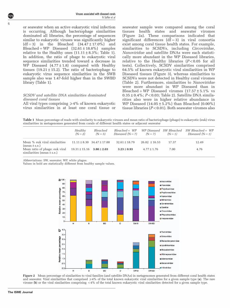
or seawater when an active eukaryotic viral infectionis occurring. Although bacteriophage similaritiesdominated all libraries, the percentage of sequencessimilar to eukaryotic viruses was significantly higher(df¼ 3) in the Bleached (34.47±17.0%) andBleachedþWP Diseased (32.61±18.8%) samplesrelative to the Healthy ones (11.11±8.3%; Table 1).In addition, the ratio of phage to eukaryotic viralsequence similarities trended toward a decrease inWP Diseased (4.77±1.8) compared with Healthytissues (19.21±15.2). The ratio of bacteriophage toeukaryotic virus sequence similarities in the SWBsample also was 1.47-fold higher than in the SWBDlibrary (Table 1).
SCSDV and satellite DNA similarities dominateddiseased coral tissuesAll viral types comprising X4% of known eukaryoticvirus similarities in at least one coral tissue or
seawater sample were compared among the coraltissues health states and seawater viromes(Figure 2a). These comparisons indicated thatsignificant differences (df¼ 3) in viral consortiaexist among coral tissue health states. For example,similarities to SCSDVs, including Circoviridae,Nanoviridae and satellite DNAs were each statisti-cally more abundant in the WP Diseased libraries,relative to the Healthy libraries (Po0.05 for alltests). Collectively, SCSDV similarities comprised64.5% of known eukaryotic viral similarities in WPDiseased tissues (Figure 3), whereas similarities toSCSDVs were not detected in Healthy coral viromes(Table 2). Furthermore, similarities to Nanoviridaewere more abundant in WP Diseased than inBleachedþWP Diseased viromes (17.57±5.1% vs0.35±0.4%; Po0.03; Table 2). Satellite DNA simila-rities also were in higher relative abundance inWP Diseased (14.05±5.2%) than Bleached (0.00%)tissue libraries (Po0.01). Both seawater viromes also
Table 1 Mean percentage of reads with similarity to eukaryotic viruses and mean ratio of bacteriophage (phage) to eukaryotic (euk) virussimilarities in metagenomes generated from corals of different health states or adjacent seawater
Healthy(N¼2)
Bleached(N¼5)
Bleachedþ WPDiseased (N¼ 7)
WP Diseased(N¼ 7)
SW Bleached(N¼1)
SW Bleachedþ WPDiseased (N¼ 1)
Mean % euk viral similarities(mean±s.e.)
11.11±8.30 34.47±17.00 32.61±18.79 26.82 ±16.53 17.37 12.49
Mean ratio of phage: euk viralsimilarities (mean±s.e.)
19.31±15.16 3.80±2.03 3.23±0.93 4.77±1.76 7.00 4.76
Abbreviations: SW, seawater; WP, white plague.Values in bold are statistically different from healthy sample values.
Figure 2 Mean percentage of similarities to viral families (and satellite DNAs) in metagenomes generated from different coral health statesand seawater. Viral similarities that comprised X4% of the total known eukaryotic viral similarities for a given sample type (a). The rarevirome (b) or the viral similarities comprising o4% of the total known eukaryotic viral similarities detected for a given sample type.
Viruses associated with diseased coralsN Soffer et al
6
The ISME Journal

contained high abundances of SCSDV similarities(SWD¼ 41.29% and SWB¼ 55.59%; Figure 2a).
Herpesviridae similarities were identified in allcoral tissue types but at a higher relative abundancein Healthy than in WP Diseased tissue (62.18±4.5%vs 16.47±3.4%; Po0.03; Table 2). Although com-mon in coral tissues, these herpesvirus similaritieswere rare in adjacent seawater and comprisedo1.5% of the similarities in either seawater virome(Figure 2a). Sequence similarities to at least somemembers of the NCLDV group also were present inall six virome types including similarities tomembers of the Phycodnaviridae, Mimiviridae andAscoviridae. A higher relative proportion (440%)of these viral sequences were identified in bothbleached (B and BD) tissue types compared withHealthy or WP Diseased tissues (33% and 18%,respectively; Figures 2a and 3). Viral sequencesimilarities that constituted a minority (that is,o4% of eukaryotic viruses similarities) of the bestannotations for each read also were examined forapparent patterns (Figure 2b). With the exception of
the Healthy viromes, Baculoviridae and Iridoviridaesimilarities were present in viromes generated fromall coral health states and seawater samples.
Diseased coral tissues contain a unique consortium ofviral sequence similaritiesTo determine whether viral consortia differedamong viromes generated from different coral healthstates as well as from adjacent seawater, multivariateanalyses were performed. The WP Diseased viromesgrouped (Supplementary Figure S1, red stars) in thisanalysis, and analysis of similarity confirmed thatthese viromes were statistically different from theother three tissue type viromes (Global R¼ 0.238and P-value¼ 0.012; Table 3). The two Healthysamples (green hexagons) were located furthestfrom the WP Diseased tissues, depicting theminimal similarity between their viral consortia(Supplementary Figure S1). Seawater viromes (darkand light blue circles) also grouped closer to eachother than to viromes generated from any coraltissue types (Supplementary Figure S1). Clusteranalysis depicted relatedness between pairedviromes generated from BleachedþWP Diseasedand WP Diseased tissue of an individual colony;viral consortia from the same individual coloniesclustered on different branches, with up to 55%dissimilarity (Supplementary Figure S2).
Similarity of percentage analysis determinedwhich viral families contributed to virome dissimi-larity (Supplementary Table S2). Although simila-rities to Herpesviridae were present in viromesgenerated from all tissue health states, thesesequences contributed to 431% of the dissimilaritybetween Healthy and WP Diseased tissue viromes,with herpesvirus similarities being the most abun-dant in Healthy tissues (Supplementary Table S2).Sequences similar to members of the SCSDVscontributed from 9% to 20% of the dissimilaritybetween WP Diseased and other coral virome types,and are strongly responsible for the uniqueness ofthe WP Diseased viral consortia (SupplementaryTable S3). Poxviridae similarities contributed todissimilarities among the BleachedþWP Diseasedand Bleached viromes compared with theHealthy viromes (9.78% and 9.46%, respectively;Supplementary Table S2). Out of all health states,similarity of percentage analysis showed that theHealthy viromes were most similar to each other(70.94% similarity). Both BleachedþWP Diseasedviromes and Bleached viromes each had approxi-mately 43% similarity, whereas WP Diseased tissuesviromes had intermediate similarity at 55.90%(Supplementary Table S2).
Diseased viromes contain higher eukaryotic viraldiversity than healthy coralsTo determine whether there were differences inviral diversity among coral tissue health states,
Figure 3 Viral ‘groups’ among different coral health states. Themean relative percentage of similarities to each viral type waslumped into ‘groups’ based on their common evolutionaryhistory. ‘NCLDVs’ are nucleocytoplasmic large double-strandedDNA (dsDNA) viruses, which include Ascoviridae, Phycodna-viridae, Poxviridae and Mimiviridae. ‘SCSDVs’ are small circularssDNA viruses and their associated satellites and include:Nanoviridae, Circoviridae and Gemniviridae, and satellite DNAs.The ‘other’ category includes similarities to all viral types notfitting into the two previous groups (that is, in this study:Herpesviridae, Papillomaviridae and Polydnaviridae).
Table 2 Univariate comparisons of the relative abundance of thesequence similarities to eukaryotic viral families and satelliteDNAs comprising X4% of coral tissue metagenomes
Viral type Significant pairwisedifferences
Kruskall–Wallisstatistic
P-value
Circoviridae H-D 8.42 p0.04Herpesviridae H-D 9.20 p0.03Nanoviridae H-D, BD-D 9.57 p0.02Satellite DNAs H-D, B-D 10.92 p0.01Papillomaviridae H-B, H-BD, H-D 9.50 p0.02Ascoviridae, — 2.33 40.05Gemniviridae — 4.22 40.05Mimiviridae — 1.45 40.05Phycodnaviridae — 1.78 40.05Polydnaviridae — 1.01 40.05Poxviridae — 4.53 40.05
Abbreviations: B, Bleached; B, BleachedþWP Diseased; D, WPDiseased; H, Healthy; WP, white plague.Bold value indicates significant difference (Po0.05).
Viruses associated with diseased coralsN Soffer et al
7
The ISME Journal

Shannon’s diversity index was calculated(Supplementary Table S3). Eukaryotic viral diversitywas statistically variable among coral tissue types(Po0.05), with WP Diseased viromes having higherdiversity than Healthy viromes (1.96±0.08 vs0.98±0.19). Diversity between different types ofbleached tissues (B and BD) was similar (1.30±0.10and 1.35±0.11; Supplementary Table S3).
Diseased coral SCSDVs are distinct from SeawaterSCSDVsPhylogenetic trees were created using the viral Repprotein of SCSDVs identified within the seven WPDiseased and two Seawater viromes. The treecontained over 50 sequences, with all of the WPDiseased and Seawater viromes contributing at leastone Rep sequence similarity (Supplementary FigureS3). Rep sequences from both Seawater viromeswere generally located on separate branches (460%support) of the tree than Rep sequences from WPDiseased coral viromes (Supplementary Figure S3).For example, two main coral SCSDV clades (stars)were distinct (72.4% and 95.0% bootstrap support)from a well-supported Seawater clade (89.9% boot-strap support; triangle; Supplementary Figure S3).
To confirm the origin of SCSDVs in WP Diseasedlibraries, representative SCSDV contigs were alignedto completely assembled and annotated genomes ofcircoviruses, nanoviruses and satellite DNAs. It wasdetermined that putative WP Diseased contigs hadbroad similarities to fully sequenced and annotatedcirco- and nanoviruses, and not just within the Repgene (Supplementary Figure S4). Further, averagenucleotide identity of putative circoviruses weremore similar to known circoviruses than other viraltypes (Supplementary Figure S4; df¼ 11, Po0.01)when all pairwise comparisons were evaluated.Surprisingly, the putative nanoviruses from WPdiseased libraries also were more similar to knowncircoviruses and our own putative circoviruses thanto known and annotated nanoviruses (df¼ 11,Po0.01).
Identification of intracellular VLPs corroboratesgenomic-based findingsElectron micrographs were used to identify poten-tial active viral infections within corals, and tocorroborate the sequence-based findings thatherpes-like viruses, SCSDVs and NCLDVs, werepresent in coral tissue. However, quantitative TEManalysis could not be performed because of theextensive variation in the quality of the tissuesamong different health states. For example, tissueswere degraded in the WP Diseased samples and celllayers were difficult to discern, whereas Bleachedtissues appeared more intact than either WPDiseased or BleachedþWP Diseased tissues.Healthy tissues exhibited intact Symbiodinium-hostcell attachment and defined tissue layers, whereassigns of degradation were evident in Bleached tissuetypes (that is, Symbiodinium detachment; data notshown). Herpesvirus-like particles with a chara-cteristic envelope that were B180 nm in diameterwere observed in BleachedþWP Diseased and WPDiseased tissues (Figure 4a). In addition, distinctivefigure 8-shaped poxvirus-like particles were visua-lized in WP Diseased tissues (Figure 4b). Finally,SCSDV-like particles were observed in WP Diseasedtissues (Figure 4c; twinned gemini-like particle),
Table 3 ANOSIM results comparing sequence similarities inviral consortia from different health states and adjacent seawater
Sample type H B BD D SWB SWBD
H (n¼ 2)B (n¼ 5) R¼ � 0.16
P¼ 0.52BD (n¼ 7) R¼ � 0.18
P¼ 0.69R¼ � 0.08P¼ 0.76
D (n¼ 7) R¼ 0.83P¼ 0.03
R¼ 0.36P¼ 0.03
R¼ 0.37P¼ 0.01
SWB (n¼ 1) R¼ 1.00P¼ 0.33
R¼ 0.24P¼ 0.33
R¼ 0.36P¼ 0.25
R¼ � 0.03P¼ 0.50
SWBD (n¼ 1) R¼ 1.00P¼ 0.33
R¼ 0.20P¼ 0.33
R¼ 0.43P¼ 0.25
R¼ 0.33P¼ 0.25
NA NA
Abbreviations: ANOSIM, analysis of similarity; B, Bleached tissue;BD, BleachedþWP Diseased tissue; D, Diseased tissue; H, Healthytissue; NA, not applicable; SWB, Seawater Bleached; SWBD, SeawaterBleachedþWP Diseased.Bold text indicates a significant difference between sample types.
Figure 4 Transmission electron micrograph of viral particles detected in diseased M. annularis tissues. A herpes-like viral particleapproximately 180 nm in width (a). A pox-like viral particle, approximately 200 nm in length (b). A gemini-like particle approximately40 nm length (c).
Viruses associated with diseased coralsN Soffer et al
8
The ISME Journal

however, TEM resolution was low, B20 nm (the sizeof a typical SCSDV particle), so the features of theseVLPs are not well defined. No bacterial cells wereobserved within the degraded WP Diseased tissue,while in Healthy tissues small cell-like structures(o0.22 um) were commonly identified (data notshown).
Discussion
SCSDVs are associated with WPCombining TEM and next-generation sequencing wehave shown here that viruses are variably associatedwith different M. annularis coral health states. Toexamine the local oceanic viral consortia and to giveinsight into transmission mechanisms, we alsogenerated viromes from seawater above Bleachedand WP diseased corals. Our robust sampling,which in some cases generated up to seven replicateviromes per health state, allowed us to determinethat a unique viral group, the coral small circularssDNA viruses (SCSDVs), was associated with a WPdisease outbreak in the Caribbean (Figures 2 and 3;Table 2).
SCSDVs are common pathogens of plants andanimals and include the families: Circoviridae,Nanoviridae, Geminiviridae, but their prevalenceand distribution in the environment was unknownuntil recently. Based on metagenomic analysesof animal hosts (for example, human, bat, rodent,pig and chimpanzee) and water samples,SCSDVs are now thought to be more common thanpreviously thought (for review, Rosario et al.,2012).
Interestingly, SCSDVs have been found to beabundant in reclaimed waters and sewage, suggestingthat SCSDVs in the diseased corals examinedhere could be linked to environment degradation(Blinkova et al., 2009; Rosario et al., 2009).Previously it has been shown that environmentalviruses such as human enteroviruses and adeno-viruses are present on coral mucus, suggesting thatthese associations were the result of anthropogenicpollution (for example, runoff; Lipp et al., 2002,2007; Futch et al., 2010). Serratia marcescens, ahuman bacterial pathogen, also found in sewage wasdemonstrated to be the cause of white pox disease inacroporids; it is likely that other pathogens includingviruses originate from sewage and negatively affectcorals (Patterson et al., 2002; Sutherland et al.,2011). However, our analysis of the Rep proteins andgenomes of coral and seawater SCSDVs sequencedin this study suggested that the coral SCSDVs aregenetically distinct from those found in thesurrounding seawater samples and are likely aunique component of the coral virome. An alternatehypothesis is that SCSDVs are present in healthycorals but are undetectable because they are eitherbelow the detection threshold of the methods usedor present in a quiescent stage.
Nevertheless, it is evident from our analysis ofboth the conserved SCSDV Rep (SupplementaryFigure S2 red lettering) sequences and the genome–genome alignments (Supplementary Figure S4) ofour putative SCSDVs and known circo-, nano- andsatellite viruses that these WP-associated SCSDVtypes are novel.
The host for the coral-associated SCSDVspresented here is yet to be determined, but mayinclude the algal endosymbiont (Symbiodiniumspp., because nanovirus/geminiviruses infectplants), the coral host itself (because circovirusesinfect animals), or another member of the coralholobiont (Yu et al., 2010; Delwart and Li, 2012;Rosario et al., 2012;). However, a previous studyexamining viruses associated with Montastraeacavernosa coral colonies searched SymbiodiniumEST libraries and found few similarities to SCSDVs(Correa et al., 2013) suggesting that the SCSDVsassociated with diseased corals in this study areinfecting the coral animal. In addition, WP diseasesigns include rapid tissue loss that suggests a coral(and not algal) pathogen is causing tissue necrosis.Furthermore if algal symbionts were affected,bleaching also would have been expected in thearea of lesions; this was not observed (Figure 1).
NCLDVs and bleachingSequence similarities to NCLDVs, includingsimilarities to members of the Phycodnaviridae,Poxviridae, Mimivirdae and Ascoviridae, were rela-tively more abundant in bleached coral tissue typesthan in non-bleached tissue types (Figure 3).Previously, poxvirus sequences were found to bemore abundant in temperature-stressed corals(Vega Thurber et al., 2008), and a recent study onthe scleractinian coral, Acropora millerpora, whichis both geographically and evolutionary distinctfrom the Caribbean M. annularis, also containedpox-like virus sequences (Littman et al., 2011). Thisstudy also corroborates findings that have impli-cated NCLDVs in the infection of the algal symbiontSymbiodinium and provides more evidence thatbleached host tissue contains viral types differentfrom those found in healthy coral tissues, suggestingviruses may be involved in some thermally inducedbleaching responses (Marhaver et al., 2008; Correaet al., 2013).
Herpesviruses dominate healthy tissuesPrevious studies have shown that herpes-likeviruses are a commonly observed in cnidarianviromes (Vega Thurber et al., 2008). This new workprovides physical evidence that herpes-likeparticles are produced in coral tissues (Figure 4a).Given their cosmopolitan presence in healthyindividuals of every coral genus tested (for example,Porites, Acropora, Montastraea and Diploria),we hypothesize that herpes-like viruses establish
Viruses associated with diseased coralsN Soffer et al
9
The ISME Journal

long-term non-fatal infections in corals, in a mannersimilar to their infections of vertebrate hosts (Knipeand Cliffe, 2008).
Viral diversity is altered in diseased coralsWP Diseased corals hosted the highest diversity ofeukaryotic viruses, bleached corals had an inter-mediate viral diversity, and healthy corals exhibitedthe lowest viral diversity. As both the WP Diseasedand Healthy tissues contained high abundances ofSymbiodinium (based on qualitative analysis ofTEM images), we suggest that the higher viraldiversity in WP Diseased viromes (relative toHealthy viromes) was not related to the relativeabundance of Symbiodinium in specimens of thetwo health states. Rather, the higher diversity ofviral types found in WP Diseased corals may be aconsequence of secondary infections resulting froma coral weakened from other viral infections.On the other hand, a higher viral diversity in WPDiseased corals may be the direct cause ofdisease signs. In either case, higher viral diversitymay be an indicator of coral stress and may be usedto diagnostically characterize corals in a diseasestate.
CaveatsMDA has been shown to bias metagenomic librariestoward ssDNA sequences and genomes because ofthe rolling circular amplification method that thePhi29 polymerase uses (Kim et al., 2008; Kim andBae, 2011). Although it is unclear whether MDA biasis stochastic (Abulencia et al., 2006) or linear(Yilmaz et al., 2010), we calculated that the prob-ability that all WP Diseased (n¼ 7) but no Healthy(n¼ 2) samples contained SCSDV-like sequenceswas o0.0079 (assuming MDA bias toward ssDNAis 50% or less). Even with a 95% bias of MDAtoward SCSDVs, we calculated that there is stillo0.7% chance of obtaining these results. Further,much of the debate about the utility of MDA for viralmetagenomics comes from studies where sampleswere processed in different ways. For example,some studies compared the results of MDA ampli-fied versus unamplified libraries, or to data setsgenerated using amplification methods that do notamplify ssDNA viruses (such as LASLs), or evenafter the elimination of a denaturing step that wouldenrich for ssDNA viruses (Kim et al.; 2008; Kim andBae, 2011). As all of our samples were processed inan identical, scientifically rigorous manner, any biastoward ssDNA sequences should be expresseduniformly in each of the sample libraries. As onlythe WP Diseased and seawater viromes, but not thebleached and healthy viromes, contained highamounts of these viral types, it is evident that thechanges detected in the relative abundance ofsimilarities to these viral types are not the result ofMDA. Finally, a previous comparative study on
stressed Pacific coral species (not infected with WP)also used MDA but detected SCSDV sequences onlyin 2 viromes out of 6 (Vega Thurber et al., 2008),while another study on another Pacific coral speciesusing MDA amplification identified o5% nano/circo viruses with no similarities to satellite DNAsor geminiviruses (Wegley et al., 2007).
These conclusions are based on correlated data,yet, ultimately only direct infection studies, such asexposing healthy corals to viruses isolated fromdiseased corals in a controlled laboratory setting,can determine whether these suspected SCSDVpathogens cause WP-like disease or are alternativelysecondary infections resulting from altered coralphysiology. Until a proper infection model isdeveloped, correlation with disease prevalence andincidence will be the best evidence as to causes ofcoral diseases. In addition, this study only examinedDNA viruses; it is possible that RNA viruses areinvolved in WP and future experiments should aimto evaluate viruses with RNA genomes.
Finally, a majority of our viral sequences (78.33%)were not similar to a known virus or group ofviruses. Therefore, although we find significantdifferences in these libraries, they are based on afraction of the total data. Yet to pointedly assess theprevalence of already known virus, described asdisease agents across these healthy and diseasestates, we narrowed our focus to taxonomicallydescribed viruses.
Conclusions
This study aimed to determine the viruses asso-ciated with WP-infected corals. Our microscopydata found no evidence of foreign microbial cellspresent in WP Diseased corals, strongly suggestingthat bacterial or small eukaryotic pathogen infectionare not the causes of the examined disease. We alsofound heterogeneity in the viral consortia amongtissue types isolated from the same coral colonies.BleachedþWP Diseased tissues were more similarto Bleached tissues than the WP-Diseased area of thesame colony. We thus hypothesize that WP infectionis only localized to the disease front, and thatbleached and WP Diseased tissues are distinct intheir viral composition. Bacteria associated withcoral mucus have been shown to be spatiallyheterogeneous (Daniels et al., 2011), and here wedemonstrate viral heterogeneity in a coral colony.Ultimately, we have shown that healthy corals havemore abundant viral similarities to Herpesviridae,bleached corals possess more viral similarities toNCLDVs, such as Phycodnaviridae and Poxviridae,and diseased tissues contain an abundance ofunique SCSDVs including members similar toCircoviridae and Nanoviridae, as well as theirassociated satellites; this novel SCSD viral grouptherefore may be responsible for WP infectionsin M. annularis.
Viruses associated with diseased coralsN Soffer et al
10
The ISME Journal

Conflict of Interest
The authors declare no conflict of interest.
Acknowledgements
We thank the USVI Department of Planning and NaturalResources for allowing us to perform coral collections(permit #STT-050-10). We thank the faculty at theUniversity of the Virgin Islands St Thomas campus fortheir hospitality. We also thank Dr Pat Blackwelder andHusain Al-Sayegh from University of Miami Center forAdvanced Microscopy for their assistance in samplepreparation and viewing. We thank four anonymousreviewers for their efforts and input. This work wasfunded by the National Science Foundation OCE Grant/Award ID 0960937 to RVT and the National GraduateResearch Fellowship to NS (1000036136). We alsothank the National Science Foundation, through the VIExperimental Program to Stimulate Competitive Research(VI-EPSCoR), which contributed valuable infrastructuresupport and research equipment.
References
Abulencia CB, Wyborski DL, Garcia JA, Podar M, Chen W,Chang SH. (2006). Environmental whole-genomeamplification to access microbial populations incontaminated sediments. Appl Environ Microbiol 72:3291–3301.
Altschul S, Gish W, Miller W, Myers E, Lipman D. (1990).Basic local alignment search tool. J Mol Biol 215:403–410.
Atad I, Zvuloni A, Loya Y, Rosenberg E. (1990). Phagetherapy of the white plague-like disease of Favia favusin the Red Sea. Coral reefs 31: 665–670.
Bermingham A, Chand M, Brown C, Aarons E, Tong C,Langrish C et al. (2012). Severe respiratory illnesscaused by a novel coronavirus, in a patient transferredto the United Kingdom from the Middle East,September 2012. Eur Comm Dis Bull 17: 1–5.
Blinkova O, Rosario K, Li L, Kapoor A, Slikas B, Bernardin Fet al. (2009). Frequent detection of highly diversevariants of cardiovirus, cosavirus, bocavirus, andcircovirus in sewage samples collected in the UnitedStates. J of clin Microbio l47: 3507–3513.
Blomstrom AL, Widen F, Hammer AS, Belak S, Berg M.(2010). Detection of a novel astrovirus in brain tissueof mink suffering from shaking mink syndromeby use of viral metagenomics. J Clin Microbiol 48:4392–4396.
Bourne DG, Garren M, Wor TM, Rosenberg E, Smith GW,Harvell CD. (2009). Microbial disease and the coralholobiont. Trends Microbiol 17: 554–562.
Brandt ME, Smith TB, AMS Correa, Vega-Thurber R.(2013). Disturbance driven colony fragmentation as adriver of a coral disease outbreak. PLoS One 8: e57164.
Bythell JC, Pantos O, Richardson L. (2004). White plague,white band and other ‘white’ diseases. In EugeneRosenberg , Loya Yossi (eds) Coral Health andDisease pp. 351–365Springer-Verlag: New York,NY, USA.
Cardenas A, Rodriguez-R LM, Pizarro V, Cadavid LF,Arevalo-Ferro C. (2012). Shifts in bacterial
communities of two Caribbean reef-building coralspecies affected by white plague disease. ISME J 6:502–512.
Correa AMS, Welsh RM, Vega Thurber RL. (2013). Uniquenucleocytoplasmic dsDNA and þ ssRNA viruses areassociated with the dinoflagellate endosymbionts ofcorals. ISME J 7: 13–27.
Clarke KR, Gorley RN. (2006). PRIMER v6: User Manual/Tutorial. PRIMER-E: Plymouth.
Clarke KR, Warwick RM. (2001). Change in MarineCommunities: An Approach to Statistical Analysisand Interpretation, 2nd edn PRIMER-EPlymouth, MA,USA.
Daniels C, Zeifman A, Heym K, Ritchie K, Watson C,Berzins I et al. (2011). Spatial heterogeneity ofbacterial communities in the mucus of Montastraeaannularis. MEPS 426: 29–40.
Davy JE, Patten NL. (2007). Morphological diversityof virus-like particles within the surface microlayerof scleractinian corals. Aquat Microb Ecol 47:37–44.
Delwart E, Li L. (2012). Rapidly expanding geneticdiversity and host range of the Circoviridae viralfamily and other Rep encoding small circular ssDNAgenomes. Virus Res 164: 1–2.
Denner EBM, Smith GW, Busse HJ, Schumann P, Narzt T,Polson SW et al. (2003). Aurantimonas coralicida gen.nov., sp. nov., the causative agent of white plague typeII on Caribbean scleractinian corals. Int J Syst EvolMicrobiol 53: 1115–1122.
Dustan P. (1977). Vitality of reef coral populations offKey Largo, Florida: recruitment and mortality. EnvironGeol 2: 51–58.
Edgar RC. (2004). MUSCLE: multiple sequence alignmentwith high accuracy and high throughput. NucleicAcids Res 32: 1792–1797.
Efrony R, Atad I, Rosenberg E. (2009). Phage therapy ofcoral white plague disease: properties of phage BA3.Curr Microbiol 58: 139–145.
Futch JC, Griffin DW, Lipp EK. (2010). Human entericviruses in groundwater indicate offshore transport ofhuman sewage to coral reefs of the Upper FloridaKeys. Environ Microbial 12: 964–974.
Gardner TA, Cote IM, Gill JA, Grant A, Watkinson AR.(2003). Long-term region-wide declines in Caribbeancorals. Science 301: 958–960.
Gibbs MJ, Weiller GF. (1999). Evidence that a plant virusswitched hosts to infect a vertebrate and thenrecombined with a vertebrate-infecting virus. ProcNatl Acad Sci USA 96: 8022–8027.
Goecks J, Nekrutenko A, Taylor JThe Galaxy Team. (2010).Galaxy: a comprehensive approach for supportingaccessible, reproducible, and transparent computa-tional research in the life sciences. Genome Biol 11:R86.
Green EP, Bruckner AW. (2000). The significance ofcoral disease epizootiology for coral reef conservation.Biol Cons 96: 347–361.
Harvell CD, Kim K, Burkholder JM, Colwell RR, EpsteinPR, Grimes DJ et al. (2004). Emerging marine diseases-climate links and anthropogenic factors. Science 285:1505–1510.
Ilyina TV, Koonin EV. (1992). Conserved sequence motifsin the initiator proteins for rolling circle DNAreplication encoded by diverse replicons from eubac-teria, eucaryotes and archaebacteria. Nucleic AcidsRes 20: 3279–3285.
Viruses associated with diseased coralsN Soffer et al
11
The ISME Journal

Kim KH, Bae JW. (2011). Amplification methods biasmetagenomics libraries of uncultured single-strandedand double-stranded DNA viruses. Appl EnvironMicrobiol 77: 7663–7668.
Kim KH, Chang HW, Nam YD, Roh SW, Kim MS, Sung Y.(2008). Amplification of uncultured single-strandedDNA viruses from rice paddy soil. Appl EnvironMicrobiol 74: 5975–5985.
Knipe DM, Cliffe A. (2008). Chromatin control of herpessimplex virus lytic and latent infection. Nat RevMicrobiol 6: 211–221.
Lipp EK, Jarrell JL, Griffin DW, Lukasik J, Jacukiewicz J,Rose JB. (2002). Preliminary evidence for human fecalcontamination in corals of the Florida Keys, USA. MarPol Bull 44: 666–670.
Lipp EK, Futch JC, Griffin DW. (2007). Analysis ofmultiple enteric viral targets as sewage markers incoral reefs. Mar Poll Bull 54: 1897–1902.
Littman R, Willis BL, Bourne DG. (2011). Metagenomicanalysis of the coral holobiont during a naturalbleaching event on the Great Barrier Reef. EnvironMicrobiol 3: 651–660.
Margulies M, Egholm M, Altman WE, Attiya S, Bader JS,Bemben LA. (2005). Genome sequencing in micro-fabricated high-density picolitre reactors. Nature 437:376–380.
Marhaver KL, Edwards RA, Rohwer F. (2008). Viralcommunities associated with healthy and bleachingcorals. Environ Microbiol 10: 2277–2286.
Martin DP, Biagini P, Lefeuvre P, Golden M, Roumagnac P,Varsani A. (2011). Recombination in eukaryotic singlestranded DNA viruses. Viruses 3: 1699–1738.
Miller AW, Blackwelder P, Al-Sayegh H, Richardson LL.(2011). Fine-structural analysis of black band disease-infected coral reveals boring cyanobacteria and novelbacteria. Dis Aqua Org 93: 179–190.
Miller J, Waara R, Muller E, Rogers C. (2006). Coralbleaching and disease combine to cause extensivemortality on reefs in US Virgin Islands. Coral Reefs 25:418.
Mokili JL, Rohwer F, Dutilh BE. (2012). Metagenomics andfuture perspectives in virus discovery. Curr Opin Virol2: 63–77.
Ng TF, Suedmeyer WK, Wheeler E, Gulland F, Breitbart M.(2009). Novel anellovirus discovered from a mortalityevent of captive California sea lions. J Gen Virol 90:1256–1261.
Pantos O, Cooney RP, MDAL Tissier, Barer MR,Donnell AGO, Bythell JC. (2003). The bacterial ecologyof a plague-like disease affecting the Caribbean coralMontastrea annularis. Environ Microbiol 5: 370–382.
Patten NL, Harrison PL, Mitchell JG. (2008). Prevalence ofvirus-like particles within a staghorn scleractiniancoral (Acropora muricata) from the Great Barrier Reef.Coral Reefs 27: 569–580.
Patterson KL, Porter JW, Ritchie KB, Polson SW,Mueller E, Peters EC et al. (2002). The etiology ofwhite pox, a lethal disease of the Caribbean elkhorncoral, Acropora palmata. Proc Natl Acad Sci USA 99:8725–8730.
Pollock FJ, Morris PJ, Willis BL, Bourne DG. (2011). Theurgent need for robust coral disease diagnostics. PLoSPath 7: 1–10.
Richardson LL, Goldberg W, Kuta K. (1998). Florida’smystery coral-killer identified. Nature 392: 557–558.
Richardson LL, Smith GW, Ritchie KB, Carlton RG. (2001).Integrating microbiological, microsensor, molecular,
and physiologic techniques in the study of coraldisease pathogenesis. Hydrobiologia 460: 71–89.
Rogers C. (2009). Coral bleaching and disease should notbe underestimated as causes of Caribbean coral reefdecline. Proc R Soc B 276: 197–198.
Rosario K, Duffy S, Breitbart M. (2012). A field guide toeukaryotic circular single-stranded DNA viruses:insights gained from metagenomics. Arch Virol 157:1851–1871.
Rosario K, Nilsson C, Lim YW, Ruan Y, Breitbart M. (2009).Metagenomic analysis of viruses in reclaimed water.Environ Microbiol 11: 2806–2820.
Rosenberg E, Koren O, Reshef L, Efrony R,Zilber-Rosenberg I. (2007). The role of microorganismsin coral health, disease and evolution. Nat RevMicrobiol 5: 355–362.
Roux S, Faubladier M, Mahul A, Paulhe N, Bernard A,Debroas D et al. (2011). Metavir: a web serverdedicated to virome analysis. Bioinformatics 27:3074–3075.
Schmieder R, Edwards R. (2011). Fast identification andremoval of sequence contamination from genomic andmetagenomic datasets. PLoS One 6: e17288.
Sokolow S. (2009). Effects of a changing climate on thedynamics of coral infectious disease: a review of theevidence. Dis Aquat Org 87: 5–18.
Sun S, Chen J, Li W, Altinatas I, Lin A, Peltier S et al.(2011). Community cyberinfrastructure for AdvancedMicrobial Ecology Research and Analysis: theCAMERA resource. Nucleic Acids Res 39: 546–551.
Sunagawa S, Desantis TZ, Piceno YM, Brodie EL,Desalvo MK, Voolstra CR et al. (2009). Bacterialdiversity and white plague disease-associated com-munity changes in the Caribbean coral Montastraeafaveolata. ISME J 131: 512–521.
Sutherland K, Porter JW, Cecilia T. (2004). Disease andimmunity in Caribbean and Indo-Pacific zooxanthel-late corals. Marine Ecol Prog Ser 266: 273–302.
Sutherland KP, Shaban S, Joyner JL, Porter JW, Lipp EK.(2011). Human pathogen shown to cause disease inthe threatened Eklhorn coral Acropora palmata. PLoSOne 6: e23468.
Thompson FL, Barash Y, Sawabe T, Sharon G, Swings J,Rosenberg E. (2006). Thalassomonas loyana sp. nov., acausative agent of the white plague-like disease ofcorals on the Eilat coral reef. Int J Syst Evol Microbiol56: 365–368.
Vega Thurber RL, Barott KL, Hall D, Liu H,Rodriguez-Mueller B, Desnues C et al. (2008).Metagenomic analysis indicates that stressorsinduce production of herpes-like viruses in thecoral Porites compressa. Proc Natl Acad Sci USA105: 18413–18418.
Vega Thurber RL, Correa AMS. (2011). Viruses ofreef-building scleractinian corals. JEMBE 408:102–113.
Vega Thurber RL, Willner-hall D, Rodriguez-mueller B,Desnues C, Edwards RA, Angly F et al. (2009).Metagenomic analysis of stressed coral holobionts.Environ Microbiol 11: 2148–2163.
Ward JR, Lafferty KD. (2004). The elusive baseline ofmarine disease: are diseases in ocean ecosystemsincreasing? PLoS Biology 2: 0543–0547.
Wegley L, Edwards R, Rodriguez-Brito B, Liu H, Rohwer F.(2007). Metagenomic analysis of the microbial com-munity associated with the coral Porites astreoides.Environ Microbiol 9: 2707–2719.
Viruses associated with diseased coralsN Soffer et al
12
The ISME Journal

Weil E, Smith G, Gil-agudelo DL. (2006). Status and progressin coral reef disease research. Dis Aqua Organ 69: 1–7.
Wilson WH, Dale AL, Davy JE, Davy SK. (2004). An enemywithin? Observations of virus-like particles in reefcorals. Coral Reefs 24: 145–148.
Wilson WH, Van Etten JL, Allen MJ. (2009). ThePhycodnaviridae: the story of how tiny giants rulethe world. Curr Top Microbiol Immunol 328: 1–42.
Yilmaz S, Allgaier M, Hugenholtz P. (2010). Multipledisplacement amplification compromises quantitativeanalysis of metagenomes. Nat Methods 7: 943–944.
Yu X, Li B, Fu YP, Jiang DH, Ghabrial SA, Li GQet al. (2010). A Gemini virus related DNA mycovirusthat confers hypovirulence to a plant patho-genic fungus. Proc Natl Acad Sci USA 107:8387–8392.
Supplementary Information accompanies this paper on The ISME Journal website (http://www.nature.com/ismej)
Viruses associated with diseased coralsN Soffer et al
13
The ISME Journal