Potential Ancillary Tests in the Evaluation of Brain Death
26
Potential Ancillary Tests in the Evaluation of Brain Death: The Value of Cerebral Blood Flow Assessment October 10, 2006
Transcript of Potential Ancillary Tests in the Evaluation of Brain Death

Potential Ancillary Tests in the Evaluation of Brain Death:
The Value of Cerebral Blood Flow Assessment
October 10, 2006
Potential Ancillary Tests in the
Evaluation of Brain Death: The Value of Cerebral Blood Flow Assessment
Prepared for the Canadian Council for Donation and Transplantation
Manraj Kanwal Singh Heran MD, FRCPC Navraj Singh Heran MD, FRCSC
October 10, 2006

1
Abstract The neurological determination of death (NDD) is primarily considered to be clinical. However, situations may arise where confounding factors make this clinical assessment difficult or impossible. As a result, ancillary tests have been developed in order to aid in the confirmation of brain death. As assessment of neuronal electrical activity (EEG) is no longer recommended in this determination, tools assessing cerebral perfusion, as reflected by the presence or absence of brain blood flow (BBF), are the mainstay of NDD. The preferred ancillary test currently is HMPAO SPECT radionuclide angiography. When this is not available, or is equivocal, 4-vessel cerebral angiography can be used to determine the presence or absence of intracranial blood flow. However, as cerebral angiography has its own limitations, other techniques are sought by physicians in the Intensive Care and Neuro-intensive Care settings to replace cerebral angiography. In this article, we briefly review the history of diagnosis of brain death, pathophysiologic issues in making this determination, and currently available CBF imaging techniques, discussing each in turn with respect to their utility in the diagnosis of brain death. Methods A literature review based on a MEDLINE search of relevant articles between January 1966 and October 2006 was conducted. Keywords included the following: brain death, neurological determination of death, CT, MRI, perfusion, xenon, transcranial Doppler, radionuclide, brain blood flow, angiography, stroke, ischemia, and infarction. Search parameters were combined to find articles relevant to the discussion of perfusion and/or angiographic imaging in the evaluation of brain death. Introduction Brain death. The very concept has stirred much debate for decades. Referred to as “coma depasse” (beyond coma) by Mollaret and Goulon in 19591, the first formalized definition of brain death was by the Ad Hoc Committee of the Harvard Medical School in 19682. Since then, there has been a greater understanding of neuronal function and mechanisms of brain cell injury3, 4, with the emergence of a concept of “death of the person rather than the body”, as described by Bonetti et al5. Brain death is now considered as complete and irreversible loss of brain function5-10, or as the Canadian Neurocritical Care Group defined it in 1999, “the irreversible loss of the capacity for consciousness combined with the irreversible loss of all brainstem functions including the capacity to breathe”11. Articles, such as those by Baron et al.12 and Bernat13, nicely review the concept of brain death, as well as discuss continuing areas of controversy, such as whole-brain death versus death of the brain stem. As modern medicine can now allow survival of an individual in situations once considered hopeless, knowing the evolution of the concept of brain death is important in order to keep perspective on the person, rather than the body.
2
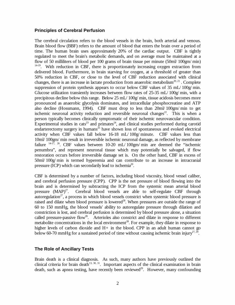
Principles of Cerebral Perfusion The cerebral circulation refers to the blood vessels in the brain, both arterial and venous. Brain blood flow (BBF) refers to the amount of blood that enters the brain over a period of time. The human brain uses approximately 20% of the cardiac output. CBF is tightly regulated to meet the brain's metabolic demands, and on average must be maintained at a flow of 50 milliliters of blood per 100 grams of brain tissue per minute (50ml/100gm/min) 14-19. With reduction in CBF, there is proportionately increasing oxygen extraction from delivered blood. Furthermore, in brain starving for oxygen, at a threshold of greater than 50% reduction in CBF, or close to the level of CBF reduction associated with clinical changes, there is an increase in lactate production from anaerobic metabolism20, 21 . Complete suppression of protein synthesis appears to occur below CBF values of 35 mL/100g/min. Glucose utilization transiently increases between flow rates of 25-35 mL/100g/min, with a precipitous decline below this range. Below 25 mL/100g/min, tissue acidosis becomes more pronounced as anaerobic glycolysis dominates, and intracellular phosphocreatine and ATP also decline (Hossmann, 1994). CBF must drop to less than 20ml/100gm/min to get ischemic neuronal activity reduction and reversible neuronal changes22. This is when a person typically becomes clinically symptomatic of their ischemic neurovascular condition. Experimental studies in cats23 and primates24, and clinical studies performed during carotid endarterectomy surgery in humans25 have shown loss of spontaneous and evoked electrical activity when CBF values fall below 16-18 mL/100g/minute. CBF values less than 10ml/100gm/min result in irreversible ischemic neuronal damage, as reflected by membrane failure 14-17, 26. CBF values between 10-20 mL/100gm/min are deemed the “ischemic penumbra”, and represent neuronal tissue which may potentially be salvaged, if flow restoration occurs before irreversible damage set is. On the other hand, CBF in excess of 50ml/100g/min is termed hyperemia and can contribute to an increase in intracranial pressure (ICP) which can secondarily lead to ischemia20. CBF is determined by a number of factors, including blood viscosity, blood vessel caliber, and cerebral perfusion pressure (CPP). CPP is the net pressure of blood flowing into the brain and is determined by subtracting the ICP from the systemic mean arterial blood pressure (MAP)27. Cerebral blood vessels are able to self-regulate CBF through autoregulation27, a process in which blood vessels constrict when systemic blood pressure is raised and dilate when blood pressure is lowered28. When pressures are outside the range of 60 to 150 mmHg, the blood vessels' ability to autoregulate pressure through dilation and constriction is lost, and cerebral perfusion is determined by blood pressure alone, a situation called pressure-passive flow28. Arterioles also constrict and dilate in response to different metabolite concentrations in the local environment28. For example, they dilate in response to higher levels of carbon dioxide and H+ in the blood. CPP in an adult human cannot go below 60-70 mmHg for a sustained period of time without causing ischemic brain injury27, 29. The Role of Ancillary Tests Brain death is a clinical diagnosis. As such, many authors have previously outlined the clinical criteria for brain death13, 30, 31. Important aspects of the clinical examination in brain death, such as apnea testing, have recently been reviewed29. However, many confounding

3
factors exist (Table 1). The clinical diagnosis of brain death may be extremely difficult in young children and neonates32. Additionally, features of the clinical exam may not be able to be safely implemented. It may not be possible for a patient to undergo an apnea test. Those patients with eye injuries may not allow for appropriate assessment of pupillary, corneal, or vestibulo-reflex testing. Those with perforated tympanic membranes will not be able to undergo ice water caloric testing for vestibulo-ocular reflexes. As a result, although confirmatory testing is not mandatory in North America, there has been much interest in ancillary tests for aiding in the diagnosis of brain death when specific components of the clinical examination cannot be reliably performed9, 30. Indeed, in certain European, Central and South American, and Asian countries, legislation requires ancillary tests as part of the diagnostic criteria for establishing brain death31, 33. The Ideal Ancillary Test The ideal ancillary test must fulfill several criteria (Table 2). There must be no false positives, and very few false negatives13. In addition, testing that could be done at the bedside would be best, as this would avoid transferring potentially hemodynamically unstable patients from the Intensive Care Unit (ICU)34. The objective of ancillary testing is to determine the point of cessation of cerebral circulation35-37. Through considered application of ancillary tests, cases where the clinical diagnosis of brain death is confounded can be assessed. Earlier determination of brain death may then allow for avoidance of protracted stays in the ICU, and potentially expedite organ donation before tissue viability becomes a concern. Types of Ancillary Testing in Brain Death There are several types of ancillary tests currently available that can assess cerebral perfusion and its effects on the brain parenchyma. These tests differ on the basis of availability, reproducibility, reliability, and sensitivity. Whereas certain tests such as radionuclide angiography are relatively easy to administer, other examinations, such as 4-vessel cerebral angiography, require transportation of the patient to specialized areas of the hospital outside the ICU and involve subspecialty expertise in order to perform and analyze the examinations. The decision to use one ancillary test over another is therefore dependent on many factors, including safety of the patient, sensitivity and specificity of the examination, and availability of the ancillary test. Existing literature regarding the use of some of these tests in the setting of brain death can be found in Appendix A, while Appendix B provides a comparative overview of existing tests assessing cerebral perfusion. In short, though several of the ancillary tests currently available meet many of the criteria outlined in Table 2, none are ideal.

4
Examinations Assessing Brain Structural Changes Computed Tomography Computed tomography (CT) is a powerful modality for evaluating neurologic disorders. It is readily available, rapidly performed, and does not necessarily require subspecialty expertise to interpret. In the setting of brain death, the classical non-contrast CT head (NCCT) findings have been well described38. These include: diffuse cerebral edema, loss of differentiation between the gray and white matter structures globally, and herniation patterns resulting from the cerebral edema. Dominguez-Roldan et al.39 concluded that two signs on NCCT were significantly associated with brain death. These were midline shift of greater than 10 mm, and compression of the ambient cistern. Geraghty and Torbey40 conducted a study assessing the prognosis of patients becoming comatose after cardiac arrest. In their article, they describe the use of a gray matter-to-white matter Hounsfield attenuation ratio (GM:WM), with a cutoff of 1.18. Using this cutoff, 100% of those patients with GM:WM ratios of <1.18 died, whereas 54% of those above this cutoff survived. However, despite the ability of NCCT to depict changes reflecting parenchymal injury, it is not sensitive or specific enough to be used as an ancillary test for determination of brain death. The concept of using an intravenous contrast-enhanced CT head examination (CECT) in the assessment for brain death is not new37, 41-43. Tan and collegues42 directly compared cerebral angiography with dynamic NCCT and CECT examinations in five patients clinically confirmed as brain dead. They found that the intracranial circulatory arrest associated with brain death in these patients resulted in no contrast opacification intracranially, whereas normal circulation was present in the scalp (supplied by external carotid artery branches). Dupas and others43 also demonstrated stagnation and arrest of contrast flow at the level of the internal carotid and vertebral arteries on their two-phase helical CT examinations. However, they also confirmed that the superior sagittal sinus could be seen in up to 50% of their study population, presumably opacifying via meningeal artery perfusion or trans-diploic emissary veins draining the scalp tissues to the superior sagittal sinus. The internal cerebral veins, vein of Galen, and straight sinus were never opacified in their study. Magnetic Resonance Imaging Magnetic resonance imaging (MRI) has revolutionized neuroimaging and its use in evaluating virtually every type of neuropathological state is beyond debate. Much like in CT, the classical findings on MRI in clinically confirmed brain death include brain swelling, various herniation patterns, obliteration of the CSF spaces, and compression of the ventricular system31, 44, 45. The ratio of signal intensity between the gray and white matter has been noted to increase after the clinical diagnosis of brain death44, 45 and serial examinations performed in a patient proceeding on to clinical brain death have demonstrated these temporal changes in progressive brain failure46. As MRI is superior to CT in the evaluation of flowing blood, this feature has been examined as well. Jones and Barnes47 confirmed absent CBF on flow-sensitive gradient-echo sequences. This lack of intracranial arterial flow voids has also been documented by others31, 44, 48. Diffusion-weighted imaging (DWI) may also be potentially useful tool in the assessment of brain death. McKinney and colleagues49 nicely reviewed the histopathologic correlation of brain tissue infarction with DWI findings and Kumada and colleagues found a significant drop in the ADC values of the involved parenchyma in those
5
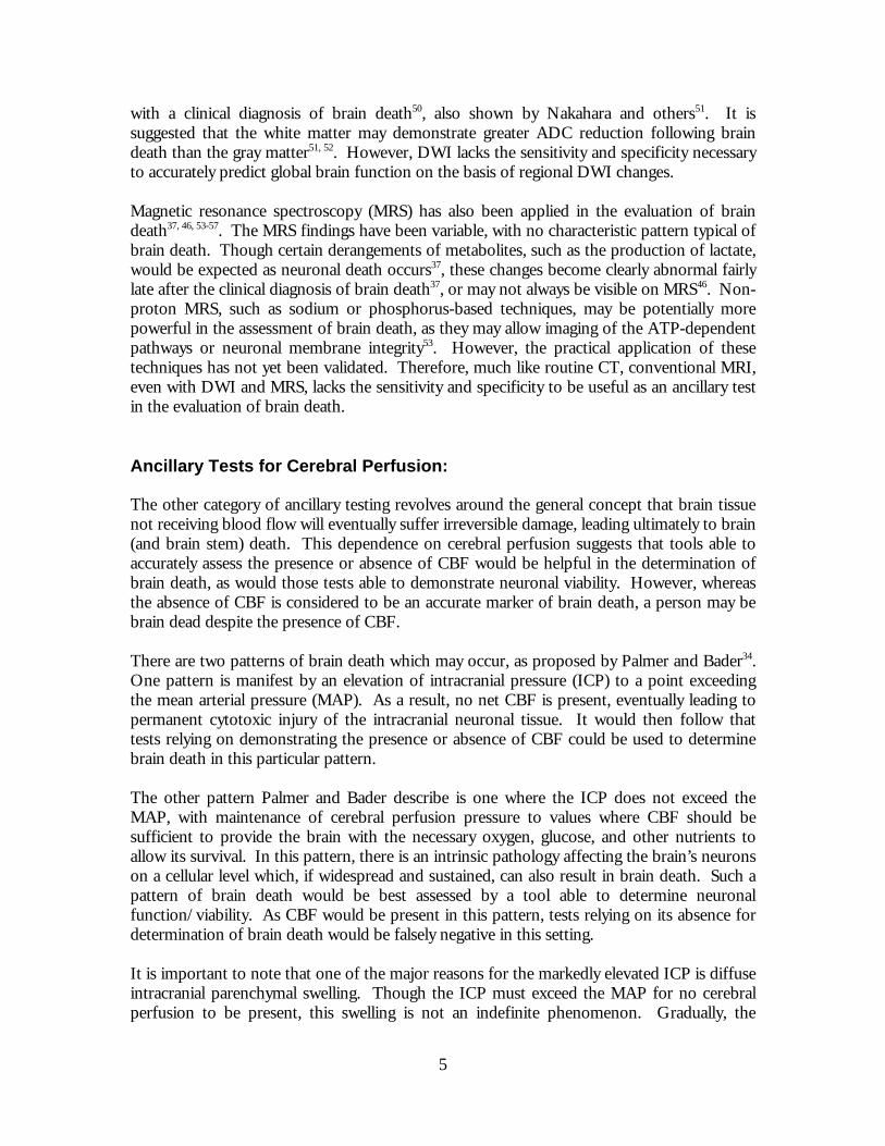
with a clinical diagnosis of brain death50, also shown by Nakahara and others51. It is suggested that the white matter may demonstrate greater ADC reduction following brain death than the gray matter51, 52. However, DWI lacks the sensitivity and specificity necessary to accurately predict global brain function on the basis of regional DWI changes. Magnetic resonance spectroscopy (MRS) has also been applied in the evaluation of brain death37, 46, 53-57. The MRS findings have been variable, with no characteristic pattern typical of brain death. Though certain derangements of metabolites, such as the production of lactate, would be expected as neuronal death occurs37, these changes become clearly abnormal fairly late after the clinical diagnosis of brain death37, or may not always be visible on MRS46. Non-proton MRS, such as sodium or phosphorus-based techniques, may be potentially more powerful in the assessment of brain death, as they may allow imaging of the ATP-dependent pathways or neuronal membrane integrity53. However, the practical application of these techniques has not yet been validated. Therefore, much like routine CT, conventional MRI, even with DWI and MRS, lacks the sensitivity and specificity to be useful as an ancillary test in the evaluation of brain death. Ancillary Tests for Cerebral Perfusion: The other category of ancillary testing revolves around the general concept that brain tissue not receiving blood flow will eventually suffer irreversible damage, leading ultimately to brain (and brain stem) death. This dependence on cerebral perfusion suggests that tools able to accurately assess the presence or absence of CBF would be helpful in the determination of brain death, as would those tests able to demonstrate neuronal viability. However, whereas the absence of CBF is considered to be an accurate marker of brain death, a person may be brain dead despite the presence of CBF. There are two patterns of brain death which may occur, as proposed by Palmer and Bader34. One pattern is manifest by an elevation of intracranial pressure (ICP) to a point exceeding the mean arterial pressure (MAP). As a result, no net CBF is present, eventually leading to permanent cytotoxic injury of the intracranial neuronal tissue. It would then follow that tests relying on demonstrating the presence or absence of CBF could be used to determine brain death in this particular pattern. The other pattern Palmer and Bader describe is one where the ICP does not exceed the MAP, with maintenance of cerebral perfusion pressure to values where CBF should be sufficient to provide the brain with the necessary oxygen, glucose, and other nutrients to allow its survival. In this pattern, there is an intrinsic pathology affecting the brain’s neurons on a cellular level which, if widespread and sustained, can also result in brain death. Such a pattern of brain death would be best assessed by a tool able to determine neuronal function/viability. As CBF would be present in this pattern, tests relying on its absence for determination of brain death would be falsely negative in this setting. It is important to note that one of the major reasons for the markedly elevated ICP is diffuse intracranial parenchymal swelling. Though the ICP must exceed the MAP for no cerebral perfusion to be present, this swelling is not an indefinite phenomenon. Gradually, the

6
swelling will subside and, if the patient is able to still maintain a reasonable MAP, CBF may eventually return. The clinical diagnosis of brain death would still be made in this setting, and tests for neuronal viability/function would also be able to accurately determine this. However, tests relying on CBF could again be falsely negative, suggesting that there may be a time-sensitive nature to the examinations using CBF as a marker of brain death. There may also be situations where an elevation in ICP resulting in cessation of CBF may not be present. This may occur as a primary phenomenon, as in the case of catastrophic brainstem or cerebellar pathology13, 58. Alternatively, this may occur when the rise in ICP has been compensated for or alleviated in some manner. This is a common phenomenon in infants who, because of their open fontanelles and soft, deformable skulls, may show only an attendant increase in their head circumference as a result of situations which would typically cause a markedly raised ICP in those patients with a “closed” cranial vault. Post-traumatic skull fractures may also allow decompression of raised ICP59. Surgical procedures, such as decompressive craniectomies60 and placement of ventricular drains61-63, are deliberately done to lower the ICP. As a result of these situations, CBF may be maintained, despite non-viability of the brain itself. In summary, though the sustained absence of CBF will eventually lead to brain death, situations where CBF can be maintained despite documented brain death exist64-66. It is important to understand the situations in which these false negative results may arise in order to assess the results in context and consider other options for confirmation of brain death. Tests for Determining CBF Non-Imaging Methods of CBF Evaluation Non-imaging methods have been used to assess the adequacy of cerebral perfusion. These methods require evaluation of arterial blood gases, as well as the venous blood leaving the brain, as sampled through a catheter placed in the internal jugular bulb. Referred to as the “cerebral oxygen extraction”67, the arteriojugular oxyhemoglobin saturation difference allows for correlation of CBF with the degree of oxygen utilization by the brain tissue, as a whole. Others have further augmented this evaluation by also assessing the venoarterial difference in carbon dioxide tension68. In addition, Artru et al.69 found that monitoring of the jugulo-arterial lactate difference, as well as the lactate oxygen index, showed that these parameters potentially became abnormal before cerebral perfusion pressure. Palmer and Bader34 describe another method which requires placement of specialized brain tissue monitors for following brain tissue oxygenation. In their study of 72 patients, they demonstrated that brain tissue oxygenation fell to 0 in all patients who were diagnosed as brain dead (clinically and on nuclear medicine imaging), and that all patients with brain tissue oxygenation of 0 were later diagnosed as brain dead. They were able to accurately determine when brain tissue oxygenation fell to 0, even in the setting of adequate cerebral perfusion pressure. Such non-imaging methods are subject to several limitations. They are invasive and intensive tests, requiring a thorough understanding of cerebral hemodynamics. No anatomic

7
correlate is provided with these techniques. Also, some techniques are inherently focal parenchymal examinations, with the results then being generalized to the whole brain34. Imaging Methods of CBF Evaluation Cerebral Angiography Among the techniques for determining CBF, cerebral angiography is classically considered to be the reference standard for imaging brain death. Experience in this setting dates back to a landmark paper published by Heiskanen in 196470, with numerous authors since contributing to the body of knowledge regarding 4-vessel cerebral angiography as an ancillary test in the determination of brain death36, 37, 43, 71-73. It is relatively simple to perform, can be done within 30-60 minutes, and is relatively straight-forward to interpret. Though early cerebral angiography examinations for brain death were actually done using iodinated contrast injections in the aortic arch, the currently accepted method of assessment is done with separate contrast injections performed in both common or internal carotid arteries, as well as in both vertebral arteries. Classical angiographic findings in brain death are summarized in Table 3, and include the lack of contrast opacification of the internal carotid arteries beyond the supraclinoid segment, and filling of the vertebral arteries to, but not beyond, their dural penetration. Despite cerebral angiography being considered the “gold standard” for determination of CBF, there are many concerns with its use30. Cerebral angiography requires specialized neuroradiologic expertise to perform and interpret the procedure. It is an invasive test, requiring the placement of a catheter into the arterial circulation, with injections performed in the cerebrovascular arterial system. Although the risk of procedural complications is low, the potential exists for vessel damage anywhere along the course of the catheter. As well, the patient must be transported to the Radiology Department for the procedure, with the attendant risks this may bring. Its availability is not always immediate, and the cost associated with this examination is not low. Although no false positive cases have been reported, false negative examinations may occur if cerebral angiography is performed on a patient in whom there is no significant elevation of the ICP36, or if angiography is performed in a manner where contrast is injected too vigorously into the downstream circulation, thereby artifactually opacifying the intracranial vasculature7, 37. Controversy also exists regarding the potential for iodinated contrast to damage transplantable tissues, such as the kidneys37. As such, though cerebral angiography is a reliable and accurate examination, other options are preferred. Radionuclide Angiography In many ways, radionuclide angiography is the ideal ancillary test in the determination of brain death7, 30, 31, 37, 47, 65, 74-76. It is safe, noninvasive, and portable. It is reliable and reproducible, and is in total agreement with cerebral angiography77. When combined with HMPAO SPECT, it has been validated for use in the setting of brain death evaluation78, 79. Several good articles review the technique for performing HMPAO SPECT radionuclide angiography as well as expected findings in the setting of brain death75, 80. Briefly, there should be no intracranial flow on the dynamic phase of imaging, with no uptake of

8
radionuclide within the brain tissue on planar and SPECT imaging. This is referred to as the “empty skull” sign. Because of the maintained external carotid artery perfusion, the nasal region can be quite prominent. This is referred to as the “hot nose” sign. Variable visualization of the superior sagittal sinus may occur37, 64, due to flow in the meningeal arteries and/or drainage of scalp venous blood via transosseous emissary veins. If focal residual brain perfusion is seen, this is interpreted as not confirmatory of brain death and a repeat HMPAO SPECT study is typically performed in 24-48 hours. This residual activity may be seen in two fairly distinct distributions, as described by Facco et al76. Perfusion may be seen in isolated hemispheric structures, such as the thalami or basal ganglia. This is usually seen in children or in adults with skull defects. Persistent perfusion may also be seen selectively in the posterior fossa structures, reflecting a rostrocaudal deterioration of intracranial tissues. CT Angiography and CT Perfusion A natural extension of CT imaging techniques is the application of CT angiography (CTA) and CT perfusion (CTP) for the evaluation of brain death9, 31, 81-83. Both techniques are readily available on modern multi-detector helical CT scanners, and are simple to implement into practice. According to Widjicks10, CTA is well accepted in all European countries, and several non-European countries, as an ancillary test for brain death. CTA is easily performed, rapidly acquired, requires only an intravenous access, and is not affected by confounding medical factors. The findings are similar to those already described for dynamic CECT in that no intracranial arterial contrast opacification should be seen in the setting of brain death, typically defined as the vertebrobasilar circulation intradurally, as well as above the level of the supraclinoid internal carotid arteries. There is expected visualization of the external carotid arterial system. Leclerc et al.83 further refine the diagnostic criteria as lack of visualization of the cortical middle cerebral artery branches, as well as lack of visualization of the internal cerebral veins. Despite its lack of portability, CTA is still highly efficacious9 and does not have the procedural risks associated with conventional digital subtraction angiography. However, as iodinated contrast is still required to perform this examination, the potential risk of contrast-mediated tissue injury is still present. CTP is a technique already in widespread use for acute stroke imaging. Interestingly, the earliest description of a perfusion-type CT scan was actually done over two decades ago41. In comparison to CTA, which is relatively simple to interpret, CTP requires post-processing of the acquired data in order to generate the mean transit time, CBF and cerebral blood volume maps needed to appropriately assess the intracranial parenchyma. As such, this interpretation is typically done by a neuroradiologist, although, in some institutions, stroke neurologists are also familiar with this technique. Though some authors tout CTP as a quantitative tool for measuring CBF84-87, this remains an area of debate22, having yet to be proven in large prospective studies. In the setting of global brain injury, the commonly performed semi-quantitative application of CTP would potentially be a major issue due to its reliance on ratio comparisons to a contralateral “normal” parenchyma. In addition, no technique has yet been developed which allows whole brain coverage through existing CTP techniques, although another level of perfusion imaging can be performed after waiting approximately 3-6 minutes. Though one may infer that the arterial input function waveform

9
should be flat, there is no actual literature reporting brain death findings on CTP, and its use in the diagnosis of brain death has never been evaluated in a formal fashion. Xenon CT Xenon CT (XeCT) is a powerful tool, and has been applied to ischemic cerebrovascular disease for many decades. It provides a quantitative measurement of CBF88-90, and is also noninvasive, fairly rapidly performed, and provides the added benefit of routine CT imaging of the brain as well. XeCT has a high spatial resolution, and flow assessment of the entire brain can be sampled accurately with the three levels of examination typically used. Also, by using inhalational Xenon, an inert gas having CT attenuation characteristics similar to iodine, XeCT avoids the use of iodinated contrast, thereby eliminating issues raised regarding contrast-mediated tissue injury (such as contrast-induced nephrotoxicity). Many authors describe the use of XeCT in brain death7, 31, 37, 91-93. Thompson et al.91 compared XeCT to dynamic brain scintigraphy in clinically brain dead children, and determined that negligible (defined as < 5 ml/100 g/minute) or no CBF globally were the CBF parameters required in order to confirm the clinical diagnosis of brain death. Pistoia et al.92 confirmed these values in 30 patients they studied over 7 years. Though the practical application of the technique is not difficult, its accessibility is a tremendous challenge, as Xenon CT is virtually unavailable apart from at a select number of academic institutions in North America. Again, the patient must be transported to the Radiology Department, and dedicated Neuroradiology expertise is required to perform and interpret the exam. As such, though XeCT remains an immensely powerful tool for quantitative examination of CBF, too many issues exist to allow its ubiquitous use as an ancillary test for the confirmation of brain death. Magnetic Resonance Angiography and Perfusion As has already been described, conventional MRI performed in the setting of clinically confirmed brain death can demonstrate the lack of normal intracranial arterial flow voids. This is thought to be a consequence of ICP being greater than MAP, thereby resulting in no net intracranial arterial blood flow. Dedicated magnetic resonance angiography (MRA) has been performed to evaluate this finding31, 35, 48, 94. All assessments thus far have utilized a “time-of-flight” (TOF) or “moving blood” imaging technique, without administration of intravenous contrast. Similar findings as those reported on CTA assessment have also been reported for TOF-MRA, namely absence of visualization of the vertebro-basilar system intracranially and the internal carotid arteries above the level of the carotid siphons44. External carotid arterial visualization can be seen, as can partial visualization of the superior sagittal sinus for the reasons described earlier. The study by Karantanas et al,35 clearly demonstrated continued visualization of the intracranial arterial circulation in 10 patients who had suffered severe closed head injuries and who did not meet clinical criteria for brain death, as opposed to total lack of visualization of intracranial blood flow in the 20 patients who were clinically thought to be brain dead prior to MR examination. All 10 patients survived, with variable neurologic outcomes. All of the 20 patients having no evidence of MRA-documented intracranial blood flow died.

10
Controversies exist around the use of MRA for brain death evaluation. Though there are several reports of its use in this setting, no formal evaluations have been done. Because contrast-enhanced MRA (CEMRA) is not prone to some of the pitfalls of TOF imaging, such as slow flow, in-plane flow, and non-laminar flow, there has been a progressive shift towards its use in the evaluation of cerebrovascular disorders. However, no reports currently exist in the literature on its use in the neurological determination of death. Although MRA is noninvasive, it still requires the transfer of the patient to the MR scanner. Subspecialty neuroradiologic expertise is again required to perform and interpret the examination. The exam takes longer than CTA, is more prone to technique-related image degradation, and requires MR compatability of all devices attached to the patient. Thus, though MRA holds promise, especially when combined with MRI, MRS, and DWI, it has not as of yet been validated for use as an ancillary test for confirmation of brain death30. MR perfusion (MRP) is a powerful technique, utilizing first-pass magnetic susceptibility effects of administered contrast agents to obtain data similar to CTP95. Its main advantage are its greater coverage of the brain during perfusion imaging (whole brain imaging versus 2-4 cm for CTP), as well as the ability to obtain diffusion weighted imaging. However, though the technique can determine CBF to a lower limit of 8 mL/100g/min, MRP is semi-quantitative, and no data is available on its use in the setting of brain death. Transcranial Duplex Ultrasonography The utility of transcranial duplex ultrasonography (TCD) as a test for evaluating the presence of intracranial blood flow has been reviewed elsewhere31, 37, 96. Classical findings in a patient who is brain dead are short systolic spikes or peaks, oscillating movement of blood within the assessed arteries, and disappearance of systolic flow on subsequent testing when previously documented as present. TCD has many potential advantages. It is safe, portable, noninvasive, and relatively rapidly performed. As well, it is relatively inexpensive, and does not require administration of potentially organotoxic contrast agents. However, TCD is not widely available, and is quite labour-intensive, requiring tremendous skill and rigor in its application to insonate all of the major intracranial arteries30, 31, 37. In addition, up to 20% of patients may fail TCD scanning as their cranial vaults are too thick to allow appropriate visualization of these arteries31. As well, many authors have reported false positives and negatives using TCD62, 97-99. Because of these drawbacks, as well as its lack of validation, TCD is not considered an accurate and reliable ancillary test in the confirmation of brain death. The Future Recognizing that false negatives may occur when an ancillary test evaluating the presence or absence of CBF is used, techniques assessing for CBF still appear to be the most promising with respect to a reliable and reproducible diagnosis of brain death in the face of confounding clinical factors. Though CTA has not been accepted for use in North America, its use as an alternative in Europe, and other non-European countries suggests that greater effort be taken to validate it as an ancillary test. CT perfusion in combination with CTA may be even better; however, no large series of patients have yet been reported and controversy remains regarding its ability to truly quantify CBF. Current advances in MR angiographic

11
techniques in the imaging of cerebrovascular pathology include the use of intravenous contrast agents100, quantitative phase contrast techniques101-103, and time-resolved MRA104-107. In addition, MR perfusion techniques such as arterial spin labelling may contribute to improved characterization of presence or absence of intracranial blood flor (Wintermark, Stroke, 2005). All of these methods may provide a greater certainty of the presence or absence of CBF, thereby ensuring no false positive exams. In the future, truly quantitative CT- or MR-based perfusion techniques may be developed, thereby allowing CBF assessment as currently only provided by XeCT or PET. In addition, issues regarding the lack of portability of these different modalities are being addressed, as portable CT scanning units are already commercially available, some with Xenon-CT perfusion capability. Other tests incorporating complementary modalities may also be developed. Conclusion The diagnosis of brain death remains primarily a clinical determination. However, it is important to recognize that confounding factors do exist that may make clinical diagnosis extremely difficult or impossible. Ancillary tests are then useful in helping make this diagnosis. Tests imaging CBF are the preferred ancillary tests, with HMPAO SPECT radionuclide angiography considered the first-line study. When this is not available or is equivocal, 4-vessel cerebral angiography is another validated examination which can be performed to determine the presence or absence of CBF. Though several case reports exist describing the use of other noninvasive angiographic or perfusion techniques, such as CTA and MRA, currently none of these techniques have been validated for use as ancillary tests in the determination of brain death. Because of the many potential advantages these noninvasive techniques may offer, further efforts should be made to evaluate them as replacement tests for conventional 4-vessel cerebral angiography.

12
References 1. Mollaret P, Goulon M. [The depassed coma (preliminary memoir).]. Rev Neurol
(Paris). Jul 1959;101:3-15. 2. A definition of irreversible coma. Report of the Ad Hoc Committee of the
Harvard Medical School to Examine the Definition of Brain Death. Jama. Aug 5 1968;205(6):337-340.
3. Popp E, Bottiger BW. Cerebral resuscitation: state of the art, experimental approaches and clinical perspectives. Neurol Clin. Feb 2006;24(1):73-87, vi.
4. Chan JY, Chang AY, Chan SH. New insights on brain stem death: from bedside to bench. Prog Neurobiol. Dec 2005;77(6):396-425.
5. Bonetti MG, Ciritella P, Valle G, Perrone E. 99mTc HM-PAO brain perfusion SPECT in brain death. Neuroradiology. Jul 1995;37(5):365-369.
6. An appraisal of the criteria of cerebral death. A summary statement. A collaborative study. Jama. Mar 7 1977;237(10):982-986.
7. Darby JM, Yonas H, Gur D, Latchaw RE. Xenon-enhanced computed tomography in brain death. Arch Neurol. May 1987;44(5):551-554.
8. Young B, Blume W, Lynch A. Brain death and the persistent vegetative state: similarities and contrasts. Can J Neurol Sci. Nov 1989;16(4):388-393.
9. Practice parameters for determining brain death in adults (summary statement). The Quality Standards Subcommittee of the American Academy of Neurology. Neurology. May 1995;45(5):1012-1014.
10. Wijdicks EF. Brain death worldwide: accepted fact but no global consensus in diagnostic criteria. Neurology. Jan 8 2002;58(1):20-25.
11. Guidelines for the diagnosis of brain death. Canadian Neurocritical Care Group. Can J Neurol Sci. Feb 1999;26(1):64-66.
12. Baron L, Shemie SD, Teitelbaum J, Doig CJ. Brief review: history, concept and controversies in the neurological determination of death. Can J Anaesth. Jun 2006;53(6):602-608.
13. Bernat JL. The concept and practice of brain death. Prog Brain Res. 2005;150:369-379.
14. Astrup J. Energy-requiring cell funcitons in the ischenic brain. Their critical suplly and possible inhibition in protective therapy. J Neursurg. 1982;56:482-497.
15. Branston NM, Ladds A, Symon L. Comparison of the effects of ischaemia on early components of thesomatosensory evoked potential in branistem, thalamus and cerebral cortex. J Cereb Blood Flow Metab. 1984;4:68-81.
16. Jones TH, Morawetz RB, Crowell RM. Thresholds of focal cerebral ischemia in awake monkeys. J Neurosurg. 1981;54:773-782.
17. Hossman KA, Schuier FJ. Experimental braininfarcts in cats. 1. Pathophysiolgoical observations. Stroke. 1980;11:583-592.
18. Siesjo BK. Pathophysiology and treatment of focal cerebral ischemia. Part I: Pathophysiology. J Neurosurg. Aug 1992;77(2):169-184.
19. Markus HS. Cerebral perfusion and stroke. J Neurol Neurosurg Psychiatry. Mar 2004;75(3):353-361.

13
20. Robertson CS, Narayan RK, Gokaslan ZL, et al. Cerebral arteriovenous oxygen difference as an estimate of cerebral blood flow in comatose patients. J Neurosurg. Feb 1989;70(2):222-230.
21. Lassen NA. The luxury-perfusion syndrome and its possible relation to acute metabolic acidosis localized within the brain. Lancet. 1996;2:1113-1115.
22. Latchaw RE, Yonas H, Hunter GJ, et al. Guidelines and recommendations for perfusion imaging in cerebral ischemia: A scientific statement for healthcare professionals by the writing group on perfusion imaging, from the Council on Cardiovascular Radiology of the American Heart Association. Stroke. Apr 2003;34(4):1084-1104.
23. Heiss WD, Hayakawa T, Waltz AG. Cortical neuronal function during ischemia. Effects of occlusion of one middle cerebral artery on single-unit activity in cats. Arch Neurol. Dec 1976;33(12):813-820.
24. Branston NM, Symon L, Crockard HA, Pasztor E. Relationship between the cortical evoked potential and local cortical blood flow following acute middle cerebral artery occlusion in the baboon. Exp Neurol. Nov 1974;45(2):195-208.
25. Sharbrough FW, Messick JM, Jr., Sundt TM, Jr. Correlation of continuous electroencephalograms with cerebral blood flow measurements during carotid endarterectomy. Stroke. Jul-Aug 1973;4(4):674-683.
26. Harris RJ, Symon L, Branston NM, Bayhan M. Changes in extracellular calcium activity in cerebral ischaemia. J Cereb Blood Flow Metab. 1981;1(2):203-209.
27. Lang EW, Chesnut RM. Intracranial pressure and cerebral perfusion pressure in severe head injury. New Horiz. Aug 1995;3(3):400-409.
28. Paulson OB, Strandgaard S, Edvinsson L. Cerebral autoregulation. Cerebrovasc Brain Metab Rev. Summer 1990;2(2):161-192.
29. Lang CJ, Heckmann JG. Apnea testing for the diagnosis of brain death. Acta Neurol Scand. Dec 2005;112(6):358-369.
30. Shemie SD, Doig C, Dickens B, et al. Severe brain injury to neurological determination of death: Canadian forum recommendations. Cmaj. Mar 14 2006;174(6):S1-13.
31. Young GB, Lee D. A critique of ancillary tests for brain death. Neurocrit Care. 2004;1(4):499-508.
32. Kohrman MH, Spivack BS. Brain death in infants: sensitivity and specificity of current criteria. Pediatr Neurol. Jan-Feb 1990;6(1):47-50.
33. Wijdicks EF. The diagnosis of brain death. N Engl J Med. Apr 19 2001;344(16):1215-1221.
34. Palmer S, Bader MK. Brain tissue oxygenation in brain death. Neurocrit Care. 2005;2(1):17-22.
35. Karantanas AH, Hadjigeorgiou GM, Paterakis K, Sfiras D, Komnos A. Contribution of MRI and MR angiography in early diagnosis of brain death. Eur Radiol. Nov 2002;12(11):2710-2716.
36. Marrache F, Megarbane B, Pirnay S, Rhaoui A, Thuong M. Difficulties in assessing brain death in a case of benzodiazepine poisoning with persistent cerebral blood flow. Hum Exp Toxicol. Oct 2004;23(10):503-505.
37. Monsein LH. The imaging of brain death. Anaesth Intensive Care. Feb 1995;23(1):44-50.

14
38. Yoshikai T, Tahara T, Kuroiwa T, et al. Plain CT findings of brain death confirmed by hollow skull sign in brain perfusion SPECT. Radiat Med. Nov-Dec 1997;15(6):419-424.
39. Dominguez-Roldan JM, Jimenez-Gonzalez PI, Garcia-Alfaro C, Hernandez-Hazanas F, Murillo-Cabezas F, Perez-Bernal J. Identification by CT scan of ischemic stroke patients with high risk of brain death. Transplant Proc. Nov 2004;36(9):2562-2563.
40. Geraghty MC, Torbey MT. Neuroimaging and serologic markers of neurologic injury after cardiac arrest. Neurol Clin. Feb 2006;24(1):107-121, vii.
41. Arnold H, Kuhne D, Rohr W, Heller M. Contrast bolus technique with rapid CT scanning. A reliable diagnostic tool for the determination of brain death. Neuroradiology. 1981;22(3):129-132.
42. Tan WS, Wilbur AC, Jafar JJ, Spigos DG, Abejo R. Brain death: use of dynamic CT and intravenous digital subtraction angiography. AJNR Am J Neuroradiol. Jan-Feb 1987;8(1):123-125.
43. Dupas B, Gayet-Delacroix M, Villers D, Antonioli D, Veccherini MF, Soulillou JP. Diagnosis of brain death using two-phase spiral CT. AJNR Am J Neuroradiol. Apr 1998;19(4):641-647.
44. Matsumura A, Meguro K, Tsurushima H, et al. Magnetic resonance imaging of brain death. Neurol Med Chir (Tokyo). Mar 1996;36(3):166-171.
45. Lee DH, Nathanson JA, Fox AJ, Pelz DM, Lownie SP. Magnetic resonance imaging of brain death. Can Assoc Radiol J. Jun 1995;46(3):174-178.
46. Falini A, Barkovich AJ, Calabrese G, Origgi D, Triulzi F, Scotti G. Progressive brain failure after diffuse hypoxic ischemic brain injury: a serial MR and proton MR spectroscopic study. AJNR Am J Neuroradiol. Apr 1998;19(4):648-652.
47. Jones KM, Barnes PD. MR diagnosis of brain death. AJNR Am J Neuroradiol. Jan-Feb 1992;13(1):65-66.
48. Lovblad KO, Bassetti C. Diffusion-weighted magnetic resonance imaging in brain death. Stroke. Feb 2000;31(2):539-542.
49. McKinney AM, Teksam M, Felice R, et al. Diffusion-weighted imaging in the setting of diffuse cortical laminar necrosis and hypoxic-ischemic encephalopathy. AJNR Am J Neuroradiol. Nov-Dec 2004;25(10):1659-1665.
50. Kumada K, Fukuda A, Yamane K, et al. [Diffusion-weighted imaging of brain death: study of apparent diffusion coefficient]. No To Shinkei. Nov 2001;53(11):1027-1031.
51. Nakahara M, Ericson K, Bellander BM. Diffusion-weighted MR and apparent diffusion coefficient in the evaluation of severe brain injury. Acta Radiol. Jul 2001;42(4):365-369.
52. Sener RN. Diffusion MRI in the postmortem brain: case report. J Neuroradiol. Dec 2004;31(5):406-408.
53. Aichner F, Felber S, Birbamer G, Luz G, Judmaier W, Schmutzhard E. Magnetic resonance: a noninvasive approach to metabolism, circulation, and morphology in human brain death. Ann Neurol. Oct 1992;32(4):507-511.
54. Garde K, Mortensen AC, Toft PB, Sorensen MB, Madsen FF, Henriksen O. Phosphorous and proton spectroscopy in relation to near incarceration and incarceration of the human brain. Acta Radiol. Mar 1994;35(2):197-200.

15
55. Terk MR, Gober JR, DeGiorgio C, Wu P, Colletti PM. Brain death in the neonate: assessment with P-31 MR spectroscopy. Radiology. Feb 1992;182(2):582-583.
56. Kato T, Tokumaru A, O'Uchi T, et al. Assessment of brain death in children by means of P-31 MR spectroscopy: preliminary note. Work in progress. Radiology. Apr 1991;179(1):95-99.
57. Wartenberg KE, Patsalides A, Yepes MS. Is magnetic resonance spectroscopy superior to conventional diagnostic tools in hypoxic-ischemic encephalopathy? J Neuroimaging. Apr 2004;14(2):180-186.
58. Kosteljanetz M, Ohrstrom JK, Skjodt S, Teglbjaerg PS. Clinical brain death with preserved cerebral arterial circulation. Acta Neurol Scand. Nov 1988;78(5):418-421.
59. Alvarez LA, Lipton RB, Hirschfeld A, Salamon O, Lantos G. Brain death determination by angiography in the setting of a skull defect. Arch Neurol. Feb 1988;45(2):225-227.
60. Braum M, Ducrocq X, Huot JC, Audibert G, Anxionnat R, Picard L. Intravenous angiography in brain death: report of 140 patients. Neuroradiology. Jun 1997;39(6):400-405.
61. Pribram HF. Angiographic appearances in acute intracranial hypertension. Neurology. Jan 1961;11:10-21.
62. Petty GW, Mohr JP, Pedley TA, et al. The role of transcranial Doppler in confirming brain death: sensitivity, specificity, and suggestions for performance and interpretation. Neurology. Feb 1990;40(2):300-303.
63. Hansen AV, Lavin PJ, Moody EB, Sandler MP. False-negative cerebral radionuclide flow study, in brain death, caused by a ventricular drain. Clin Nucl Med. Jun 1993;18(6):502-505.
64. de Campo MP. Imaging of brain death in neonates and young infants. J Paediatr Child Health. Aug 1993;29(4):255-258.
65. Kurtek RW, Lai KK, Tauxe WN, Eidelman BH, Fung JJ. Tc-99m hexamethylpropylene amine oxime scintigraphy in the diagnosis of brain death and its implications for the harvesting of organs used for transplantation. Clin Nucl Med. Jan 2000;25(1):7-10.
66. Flowers WM, Jr., Patel BR. Persistence of cerebral blood flow after brain death. South Med J. Apr 2000;93(4):364-370.
67. Cruz J. Low clinical ischemic threshold for cerebral blood flow in severe acute brain trauma. Case report. J Neurosurg. Jan 1994;80(1):143-147.
68. Stocchetti N, Zanier ER, Nicolini R, et al. Oxygen and carbon dioxide in the cerebral circulation during progression to brain death. Anesthesiology. Nov 2005;103(5):957-961.
69. Artru F, Dailler F, Burel E, et al. Assessment of jugular blood oxygen and lactate indices for detection of cerebral ischemia and prognosis. J Neurosurg Anesthesiol. Jul 2004;16(3):226-231.
70. Heiskanen O. Cerebral Circulatory Arrest Caused by Acute Increase of Intracranial Pressure. a Clinical and Roentgenological Study of 25 Cases. Acta Neurol Scand. 1964;40:SUPPL7:1-57.
71. Bergquist E, Bergstrom K. Angiography in cerebral death. Acta Radiol Diagn (Stockh). May 1972;12(3):283-288.

16
72. Bradac GB, Simon RS. Angiography in brain death. Neuroradiology. 1974;7(1):25-28.
73. Wilkening M, Louvier N, D'Athis P, Freysz M. [Validity of cerebral angiography via venous route in the diagnosis of brain death]. Bull Acad Natl Med. Jan 1995;179(1):41-48; discussion 48-50.
74. Okuyaz C, Gucuyener K, Karabacak NI, Aydin K, Serdaroglu A, Cingi E. Tc-99m-HMPAO SPECT in the diagnosis of brain death in children. Pediatr Int. Dec 2004;46(6):711-714.
75. Al-Shammri S, Al-Feeli M. Confirmation of brain death using brain radionuclide perfusion imaging technique. Med Princ Pract. Sep-Oct 2004;13(5):267-272.
76. Facco E, Zucchetta P, Munari M, et al. 99mTc-HMPAO SPECT in the diagnosis of brain death. Intensive Care Med. Sep 1998;24(9):911-917.
77. Munari M, Zucchetta P, Carollo C, et al. Confirmatory tests in the diagnosis of brain death: comparison between SPECT and contrast angiography. Crit Care Med. Sep 2005;33(9):2068-2073.
78. Wieler H, Marohl K, Kaiser KP, Klawki P, Frossler H. Tc-99m HMPAO cerebral scintigraphy. A reliable, noninvasive method for determination of brain death. Clin Nucl Med. Feb 1993;18(2):104-109.
79. Weckesser M, Schober O. Brain death revisited: utility confirmed for nuclear medicine. Eur J Nucl Med. Nov 1999;26(11):1387-1391.
80. Vander Borght T, Laloux P, Maes A, Salmon E, Goethals I, Goldman S. Guidelines for brain radionuclide imaging. Perfusion single photon computed tomography (SPECT) using Tc-99m radiopharmaceuticals and brain metabolism positron emission tomography (PET) using F-18 fluorodeoxyglucose. The Belgian Society for Nuclear Medicine. Acta Neurol Belg. Dec 2001;101(4):196-209.
81. Yu SL, Lo YK, Lin SL, Lai PH, Huang WC. Computed tomographic angiography for determination of brain death. J Comput Assist Tomogr. Jul-Aug 2005;29(4):528-531.
82. Qureshi AI, Kirmani JF, Xavier AR, Siddiqui AM. Computed tomographic angiography for diagnosis of brain death. Neurology. Feb 24 2004;62(4):652-653.
83. Leclerc X, Taschner CA, Vidal A, et al. The role of spiral CT for the assessment of the intracranial circulation in suspected brain-death. J Neuroradiol. Apr 2006;33(2):90-95.
84. Wintermark M, Thiran JP, Maeder P, Schnyder P, Meuli R. Simultaneous measurement of regional cerebral blood flow by perfusion CT and stable xenon CT: a validation study. AJNR Am J Neuroradiol. May 2001;22(5):905-914.
85. Kudo K, Terae S, Katoh C, et al. Quantitative cerebral blood flow measurement with dynamic perfusion CT using the vascular-pixel elimination method: comparison with H2(15)O positron emission tomography. AJNR Am J Neuroradiol. Mar 2003;24(3):419-426.
86. Schaefer PW, Roccatagliata L, Ledezma C, et al. First-pass quantitative CT perfusion identifies thresholds for salvageable penumbra in acute stroke patients treated with intra-arterial therapy. AJNR Am J Neuroradiol. Jan 2006;27(1):20-25.
87. Abe H, Murakami T, Kubota M, et al. Quantitative tissue blood flow evaluation of pancreatic tumor: comparison between xenon CT technique and perfusion CT

17
technique based on deconvolution analysis. Radiat Med. Aug 2005;23(5):364-370.
88. Gur D, Yonas H, Jackson DL, et al. Measurement of cerebral blood flow during xenon inhalation as measured by the microspheres method. Stroke. Sep-Oct 1985;16(5):871-874.
89. Fatouros PP, Wist AO, Kishore PR, et al. Xenon/computed tomography cerebral blood flow measurements. Methods and accuracy. Invest Radiol. Sep 1987;22(9):705-712.
90. DeWitt DS, Fatouros PP, Wist AO, et al. Stable xenon versus radiolabeled microsphere cerebral blood flow measurements in baboons. Stroke. Dec 1989;20(12):1716-1723.
91. Thompson JR, Ashwal S, Schneider S, Hasso AN, Hinshaw DB, Jr., Kirk G. Comparison of cerebral blood flow measurements by xenon computed tomography and dynamic brain scintigraphy in clinically brain dead children. Acta Radiol Suppl. 1986;369:675-679.
92. Pistoia F, Johnson DW, Darby JM, Horton JA, Applegate LJ, Yonas H. The role of xenon CT measurements of cerebral blood flow in the clinical determination of brain death. AJNR Am J Neuroradiol. Jan-Feb 1991;12(1):97-103.
93. Ashwal S, Schneider S. Brain death in the newborn. Pediatrics. Sep 1989;84(3):429-437.
94. Ishii K, Onuma T, Kinoshita T, Shiina G, Kameyama M, Shimosegawa Y. Brain death: MR and MR angiography. AJNR Am J Neuroradiol. Apr 1996;17(4):731-735.
95. Wintermark M, Sesay M, Barbier E, et al. Comparative overview of brain perfusion imaging techniques. Stroke. Sep 2005;36(9):e83-99.
96. Young GB, Shemie SD, Doig CJ, Teitelbaum J. Brief review: the role of ancillary tests in the neurological determination of death. Can J Anaesth. Jun 2006;53(6):620-627.
97. Nau R, Prange HW, Klingelhofer J, et al. Results of four technical investigations in fifty clinically brain dead patients. Intensive Care Med. 1992;18(2):82-88.
98. Rodriguez RA, Cornel G, Alghofaili F, Hutchison J, Nathan HJ. Transcranial Doppler during suspected brain death in children: Potential limitation in patients with cardiac "shunt". Pediatr Crit Care Med. Apr 2002;3(2):153-157.
99. de Freitas GR, Andre C, Bezerra M, Nunes RG, Vincent M. Persistence of isolated flow in the internal carotid artery in brain death. J Neurol Sci. Jun 15 2003;210(1-2):31-34.
100. Prince MR, Meaney JF. Expanding role of MR angiography in clinical practice. Eur Radiol. Feb 2006;16 Suppl 2:B3-8.
101. Oktar SO, Yucel C, Karaosmanoglu D, et al. Blood-flow volume quantification in internal carotid and vertebral arteries: comparison of 3 different ultrasound techniques with phase-contrast MR imaging. AJNR Am J Neuroradiol. Feb 2006;27(2):363-369.
102. Neff KW, Horn P, Schmiedek P, Duber C, Dinter DJ. 2D cine phase-contrast MRI for volume flow evaluation of the brain-supplying circulation in moyamoya disease. AJR Am J Roentgenol. Jul 2006;187(1):W107-115.

18
103. Langer DJ, Lefton DR, Ostergren L, et al. Hemispheric revascularization in the setting of carotid occlusion and subclavian steal: a diagnostic and management role for quantitative magnetic resonance angiography? Neurosurgery. Mar 2006;58(3):528-533; discussion 528-533.
104. Nael K, Michaely HJ, Villablanca P, Salamon N, Laub G, Finn JP. Time-resolved contrast enhanced magnetic resonance angiography of the head and neck at 3.0 tesla: initial results. Invest Radiol. Feb 2006;41(2):116-124.
105. Miraux S, Franconi JM, Thiaudiere E. Blood velocity assessment using 3D bright-blood time-resolved magnetic resonance angiography. Magn Reson Med. Sep 2006;56(3):469-473.
106. Meckel S, Mekle R, Taschner C, et al. Time-resolved 3D contrast-enhanced MRA with GRAPPA on a 1.5-T system for imaging of craniocervical vascular disease: initial experience. Neuroradiology. May 2006;48(5):291-299.
107. Cashen TA, Carr JC, Shin W, et al. Intracranial time-resolved contrast-enhanced MR angiography at 3T. AJNR Am J Neuroradiol. Apr 2006;27(4):822-829.

19
Abbreviations and Definitions
ADC Apparent diffusion coefficient; property of a specific tissue characterizing the degree to which its water content can move when subjected to magnetic gradients
BBF Brain blood flow; blood flow per unit brain tissue, expressed as mL/100g/minute CBV Cerebral blood volume; volume of blood per unit brain tissue, expressed as mL/100g; obeys similar autoregulatory principles as for CPP CECT Contrast-enhanced computed tomography CEMRA Contrast-enhanced magnetic resonance angiography; non-invasive MR vascular imaging technique relying on filling the lumen of the
vessels to be imaged with a MR contrast agent CPP Cerebral perfusion pressure; net pressure of blood flowing into the brain, determined by subtracting ICP from MAP; tends to stay
relatively constant over a range of blood pressures CSF Cerebrospinal fluid CT Computed tomography CTA Computed tomography angiography; non-invasive CT method of opacifying the vasculature through bolus intravenous injection of
iodinated contrast material CTP Computed tomography perfusion; technique allowing the determination of cerebral perfusion parameters through bolus intravenous
injection of iodinated contrast material DWI Diffusion weighted imaging; allows for characterization of water movement through the application of powerful magnetic gradients; may
be subject to impurities because of the specific manner in which it is acquired (ex: “T2 shine-through”) GM Grey matter HMPAO Hexamethylpropylene amine oxime ICP Intracranial pressure MAP Mean arterial pressure MRA Magnetic resonance angiography; non-invasive MR technique of imaging the vasculature; can be done by filling the lumen of the
circulation with MR-based contrast material (CEMRA), or by observing moving blood (TOF-MRA) MRI Magnetic resonance imaging MRS Magnetic resonance spectroscopy NCCT Non-contrast computed tomography NDD Neurologic determination of death PET Positron emission tomography SPECT Single photon emission computed tomography TCD Transcranial Doppler TOF Time-of-flight; technique that utilizes properties of moving blood to provide an image of the circulation in MR WM White matter XeCT Xenon computed tomography; CT done with the subject inhaling an inert gas (Xenon), which has contrast characteristics similar to

20
iodinated contrast; allows quantitative determination of CBF
21
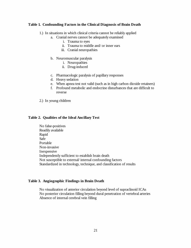
Table 1. Confounding Factors in the Clinical Diagnosis of Brain Death
1.) In situations in which clinical criteria cannot be reliably applied a. Cranial nerves cannot be adequately examined
i. Trauma to eyes ii. Trauma to middle and/or inner ears iii. Cranial neuropathies
b. Neuromuscular paralysis
i. Neuropathies ii. Drug-induced
c. Pharmacologic paralysis of papillary responses d. Heavy sedation e. When apnea test not valid (such as in high carbon dioxide retainers) f. Profound metabolic and endocrine disturbances that are difficult to
reverse
2.) In young children Table 2. Qualities of the Ideal Ancillary Test No false-positives Readily available Rapid Safe Portable Non-invasive Inexpensive Independently sufficient to establish brain death Not susceptible to external/internal confounding factors Standardized in technology, technique, and classification of results Table 3. Angiographic Findings in Brain Death No visualization of anterior circulation beyond level of supraclinoid ICAs No posterior circulation filling beyond dural penetration of vertebral arteries Absence of internal cerebral vein filling
22
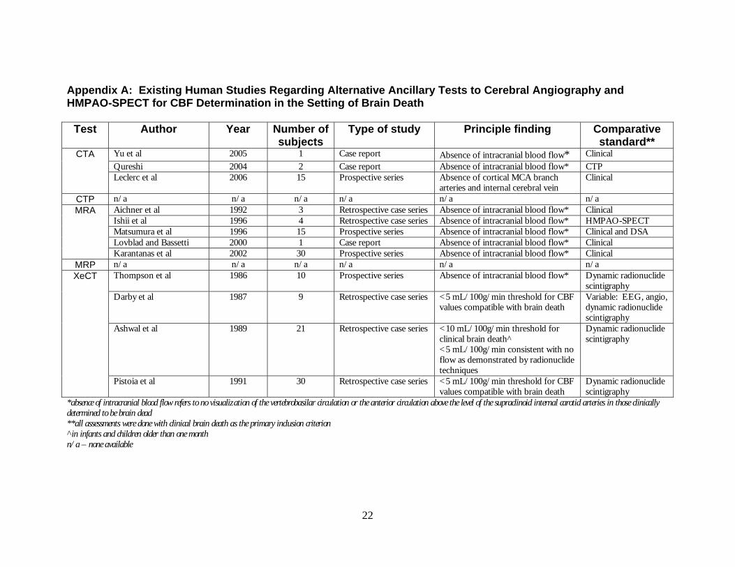
Appendix A: Existing Human Studies Regarding Alternative Ancillary Tests to Cerebral Angiography and HMPAO-SPECT for CBF Determination in the Setting of Brain Death
Test Author Year Number of subjects
Type of study Principle finding Comparative standard**
CTA Yu et al 2005 1 Case report Absence of intracranial blood flow* ClinicalQureshi 2004 2 Case report Absence of intracranial blood flow* CTPLeclerc et al 2006 15 Prospective series Absence of cortical MCA branch
arteries and internal cerebral vein Clinical
CTP n/a n/a n/a n/a n/a n/aMRA Aichner et al 1992 3 Retrospective case series Absence of intracranial blood flow* Clinical
Ishii et al 1996 4 Retrospective case series Absence of intracranial blood flow* HMPAO-SPECTMatsumura et al 1996 15 Prospective series Absence of intracranial blood flow* Clinical and DSALovblad and Bassetti 2000 1 Case report Absence of intracranial blood flow* ClinicalKarantanas et al 2002 30 Prospective series Absence of intracranial blood flow* Clinical
MRP n/a n/a n/a n/a n/a n/aXeCT
Thompson et al 1986 10 Prospective series Absence of intracranial blood flow* Dynamic radionuclide
scintigraphy Darby et al 1987 9 Retrospective case series <5 mL/100g/min threshold for CBF
values compatible with brain death Variable: EEG, angio, dynamic radionuclide scintigraphy
Ashwal et al 1989 21 Retrospective case series <10 mL/100g/min threshold for clinical brain death^ <5 mL/100g/min consistent with no flow as demonstrated by radionuclide techniques
Dynamic radionuclide scintigraphy
Pistoia et al 1991 30 Retrospective case series <5 mL/100g/min threshold for CBF values compatible with brain death
Dynamic radionuclide scintigraphy
*absence of intracranial blood flow refers to no visualization of the vertebrobasilar circulation or the anterior circulation above the level of the supraclinoid internal carotid arteries in those clinically determined to be brain dead **all assessments were done with clinical brain death as the primary inclusion criterion ^in infants and children older than one month n/a – none available

23
Appendix B: Comparison of Ancillary Tests for Determination of Brain Blood Flow in the Setting of Brain Death
Test Definition
Minimum Flow Limit (mL/100g/min)
Advantages Disadvantages Consensus
DSA Digital subtraction angiography
0 Direct visualization of CBF
Invasive Exposure to iodinated contrast Not readily available Not portable Expensive
Reference standard for NDD assessment Not preferred test due to invasiveness
SPECT
Single photon emission computed tomography
5
Widely available Portable Low cost Whole brain coverage
Not quantitative Poor spatial resolution
Reference standard for NDD assessment
PET Positron emission tomography
0
QuantitativeCan assess multiple factors, including oxygen consumption, by using different radioligands Repeated measurements possible Whole brain coverage
Impractical for everyday use Expensive Not portable
Reference standard for CBF assessment Not useful in NDD
XeCT Xenon computed tomography
0
Quantitative Allows simultaneous CT-based imaging techniques Can be repeated within 10 minutes
Only available in a few academic centers as not FDA approved Not portable Limited brain coverage (6 cm) Delivery of Xenon gas may present challenges
Reference standard for CBF assessment Not useful in NDD due to limited availability
MRA Magnetic resonance angiography
Dependent on technique of acquisition, but considered very low (<10)
Readily available No contrast necessary Can be combined with other MR-based techniques
Not readily available Expensive Not portable Scanning time lengthy
Limited data but promising

24
MRP Magnetic resonance perfusion
8
Readily availableWhole brain coverage Can be combined with other MR-based techniques Repeatable
Not quantitative for CBF Not readily available Not portable
Likely not useful as not quantitative
CTA Computed tomography angiography
Unknown but likely very low (<10)
Readily availableRapid acquisition
Exposure to iodinated contrast Not portable
Limited data but most promising as ubiquitous technology
CTP Computed tomography perfusion
0
Readily available Rapid acquisition Rapid acquisition Quantitative*
Exposure to iodinated contrast and ionizing radiation Limited anatomic coverage (2-4 cm) Requires post-processing of data Relies on intact BBB Not portable
Limited data but holds promise, especially if anatomic coverage extended
TCD Transcranial Doppler Ultrasound
Unknown RapidNon-invasive Portable
Does not quantify CBFDifficult to perform One measurement for each hemisphere
Not deemed useful in NDD
* controversy exists regarding its true quantitative abilities