POSTOPERATIVE SCAR ENDOMETROSIS: OPTIMIZATION OF …
26
Doctoral School in Medical Sciences Manuscript UDC: 616-089.84: 618.14-002 Sergiu Zaharia POSTOPERATIVE SCAR ENDOMETROSIS: OPTIMIZATION OF DIAGNOSIS AND TREATMENT 321.13 – surgery Summary of Ph.D. Thesis in Medical Sciences Chisinau, 2020
Transcript of POSTOPERATIVE SCAR ENDOMETROSIS: OPTIMIZATION OF …

Doctoral School in Medical Sciences
Manuscript
UDC: 616-089.84: 618.14-002
Sergiu Zaharia
POSTOPERATIVE SCAR ENDOMETROSIS: OPTIMIZATION
OF DIAGNOSIS AND TREATMENT
321.13 – surgery
Summary of Ph.D. Thesis in Medical Sciences
Chisinau, 2020

2
This present Ph.D. thesis was elaborated at the Department of Surgery no. 1 "Nicolae Anestiadi"
and the Laboratory of Hepato-Pancreato-Biliary Surgery of “Nicolae Testemitanu” State
University of Medicine and Pharmacy PI, based on two clinics: Mother and Child Institute IMPH
and Emergency Medicine Institute IMPH of the founding Consortium of the Doctoral School in
Medical Sciences.
Scientific advisor:
Mishin Igor, dr. habil. in med., professor ___________
Scientific co-tutor: Mishina Anna, dr. habil. in med., assoc. prof. ___________
Members of the guidance committee:
Ignatenco Sergiu, MD, PhD, assoc. prof. ___________
Berliba Sergiu, MD, PhD, assoc. prof. ___________
Tabuica Uliana, MD, PhD, assoc. prof. ___________
Ph.D. thesis defense will take place on 0202.11.81 a 00.41 t p.m., in office 205, at the meeting
of the Scientific Council 03.07.2020 (protocol no.11), "Nicolae Testemitanu" State University
of Medicine and Pharmacy, on Bd Stefan cel Mare și Sfant, 165, Republic of Moldova,
Chisinau.
The specialized scientific council for public thesis defense:
Chairman:
Casian Dumitru, dr. habil. in med., assoc. prof., Nicolae Testemiţanu SUMPh ___________
Members:
Mishin Igor, dr. habil. in med., professor, Nicolae Testemiţanu SUMPh ___________
Cernețchi Olga, dr. habil. in med., professor, Nicolae Testemiţanu SUMPh ___________
Ungureanu Sergiu, dr. habil. in med., assoc. prof., Nicolae Testemiţanu SUMPh ___________
Official Reviewers:
Berliba Sergiu, MD, PhD, assoc. prof., Nicolae Testemiţanu SUMPh ___________
Donscaia Ana, dr. habil. in med., assoc. prof., Institute of Oncology ___________
Gudima Alexandru, dr. habil. in med., assoc. prof., Institute of Oncology ___________
Author:
Zaharia Sergiu ___________
Zaharia Sergiu, 2020

3
CONTENTS
The research conceptual framework 4
PhD thesis content 8
1. Modern aspects of etiopathogenesis, diagnosis and treatment of postoperative
scar endometriosis
8
2. Material and research methods 8
3. Clinical and imaging characteristics of postoperative scar endometriosis 11
3.1 Clinical signs of postoperative scar endometriosis 11
3.2 Ultrasound and Doppler imaging patterns of surgical postoperative scar
endometriosis
13
3.3 The importance of computed tomography and magnetic resonance imaging in
the diagnosis of surgical scar endometriosis
13
4. The surgical treatment outcomes and the morphopathological characteristics of
postoperative scar endometriosis
14
4.1. Characteristics of surgical treatment, assessment of postoperative further
outcomes and life quality in patients with postoperative scar endometriosis
14
4.2 Morphological characteristics of postoperative scar endometriosis 15
4.3 The immunohistochemistry profile of postoperative scar endometriosis
17
General conclusions 18
Practical recommendations 19
Selective Bibliography 20
List of publications 21
Annotations (Romanian, Russian, English) 24
LIST OF ABBREVIATIONS
ARME - abdominal rectus muscle endometriosis
C - Cytoplasm
CM - cellular membrane
CT - computed tomography
EHP – 5 - Endometriosis Health Profile - 5
EMI - Emergency Medicine Institute
ER - estrogen receptors
IMC - Institute of Mother and Child
IMPH - Institute of Medicine and Public Health
IS - Intensity Score
MPV - mean platelet volume
MRI - magnetic resonance imaging
N - nucleus
PR - progesterone receptors
PS - Proportion Score
PSE - postoperative scar endometriosis
PVSE - perineal and vaginal scar endometriosis
QL - Quality of Life
TS - Total Score
USG - ultrasound imaging

4
THE RESEARCH CONCEPTUAL FRAMEWORK
Actuality of the research topic: Endometriosis was defined as a nosological entity,
characterized by the presence of ectopic endometrial tissue outside the uterus [1]. Postoperative
scar endometriosis (PSE) is an orphan disease (ORPHA:137820), which has been scarcely
reported in specialized literature as through unique or limited clinical cases, being related to a
number of current diagnostic and treatment challenges [2]. The etiology and pathogenesis of PSE
represents controversial issues among researchers, thus leading to non-standardized treatment
approaches [3]. However, the accuracy of clinical diagnosis, imaging methods and
histopathological investigations might directly affect the incidence of PSE.
Till now, the incidence of PSE patients with obstetric and gynecological surgery history
makes up 0.03-1.08% of cases [4]. PSE is more commonly conditioned by cesarean section (CS)
[5]. However, the risk of developing PSE regarding the emergency or scheduled CS cases has
not been studied yet.
The perineal and vaginal scar endometriosis (PVSE) is an extremely rare endometrial
ectopia localized and determined in most cases of episiotomy [6]. Thus, it should be mentioned
that no sufficient studies on the development of PSE, due to surgical interventions, have been
performed so far. Considering that few research attempts have been carried out to highlight the
risk factors responsible for developing PSE, a detailed study might contribute to prevention of
this disease. The clinical features of PSE range from common symptoms, such as catamenial
pain associated with an increased volume of the postoperative scar region, to absence of clinical
signs that might be challenging for specialists to establish a preoperative diagnosis [7]. The non-
specific clinical picture leads to diagnostic errors, whereas the diagnosis might be established
only at the histological examination later on [8].
The preoperative diagnosis of PSE is a difficult issue, due to its resemblance to a wide
range of benign or malignant tumors [9]. Therefore, the diagnosis of PSE needs to be considered,
while differentiating abdominal wall tumors in women who have undergone a cesarean section.
Currently, despite the wide range of diagnostic and treatment methods, there are no defined
criteria for the diagnosis and treatment of PSE, thus stressing the importance of clinical suspicion
in assessing an optimal treatment approach [10]. At present, a strong interest is shown to
studying of the tumor marker CA-125, the preoperative mean platelet volume (MPV) indices and
study of the peripheral systemic inflammatory response of the neutrophil/lymphocyte index in
the diagnosis of peritoneal endometriosis. Additionally, no specialized literature data have been
found on the importance of the serological markers in the diagnosis of PSE, thus justifying the
actuality of the present study [11].
Considering the difficulty of diagnosing PSE, the current specialized literature has
suggested using ultrasound imaging (USG), Doppler ultrasound, computed tomography (CT) and
magnetic resonance imaging (MRI) for preoperative diagnosis of anterior abdominal wall
endometriosis [12]. Unless specific imaging criteria and anatomical features of the PSE are
available, the establishment of these criteria will contribute to the preoperative diagnosing of
PSE, as well as to assessing the rational volume of the resected area.
Currently, surgery is considered the method of choice in the treatment and prevention of
PSE recurrence and malignancy, which includes two stages: resection (via en block removal) and
reconstruction (abdominal wall plasty) [13]. It has been established that R0 resection is the basic
first-stage, which prevents disease recurrence. Current researches have revealed that further
monitoring of patients with PSE allows determining the progression of the recurrent cases. [14].

5
However, there are a limited number of studies regarding the differentiated approach of the
reconstructive stage, thus justifying the purpose of this present research paper.
The morphological assessment of the removed samples is extremely important for
establishing the diagnosis of extragenital endometriosis [15]. According to recent studies on the
immunohistochemistry methods used for diagnosing extrapelvic endometriosis, it is worth
assessing the monoclonal antibodies (CD10, ER-α, PR, CK7, vimentin) in order to diagnose PSE
[16]. Moreover, considering the present researches on assessment of the quality of life (QL) in
patients with peritoneal endometriosis [17] and lack of specialized literature data on determining
this index in PSE patients, further targeting studies are needed.
The purpose of the research: Optimizing the diagnosis and treatment management of
patients with postoperative scar endometriosis by assessing a variety of clinical, imaging and
morphological criteria, as well as the surgical treatment outcomes.
The study objectives:
1. To study the clinical manifestations of PSE and the characteristics of its localization.
2. To determine the types of previous surgeries and define the risk factors responsible for the
development of postoperative scar endometriosis.
3. To establish the imaging method criteria (ultrasound, Doppler, computed tomography, and
magnetic resonance scanning), as well as the anatomical and topographical features of
postoperative scar endometriosis.
4. To assess the optimum surgical volume to solve postoperative scar endometriosis based on
early and further outcomes.
5. To evaluate the morphological features and the immunohistochemistry profile of surgical
scar endometriosis.
Scientific research methodology: The research paper is a prospective and retrospective
study analysis of the diagnostic procedures and treatment results, assessed in 34 patients with
PSE over a period of 26 years. A detailed information was obtained according to the approved
questionnaire. The following methods were used within the present research: (1) clinical
investigation and examination; (2) laboratory (blood biochemistry and tumor markers); (3)
imaging (ultrasound + Doppler imaging, computed tomography, magnetic resonance scanning);
(4) histopathology (light microscopy, immunohistochemistry profile). The following methods
were used for statistical processing: Kolmogorov-Smirnov test, Student criterion, U criterion -
Mann - Whitney test, Fisher's exact test, ANOVA test.Statistical data processing was carried out
via GraphPad Prism 5.0 software (GraphPad Software, Inc.). The statistical data were rendered
by graphical presentation.
The scientific novelty and originality: The primary risk factors of PSE occurrence were
identified, based on clinical data assessment, viz: (1) cesarean section cases (89.3%, p<0.0001);
(2) primiparous cases (80%, p<0.0001); (3) scheduled operations (76%, p=0.0005); (4) intact
amniotic membrane (88%, p<0.0001).
There were found non-specific clinical signs, as well as constant pain, which was reported
in 38.2% of cases (p>0.05) and inactive growth volume depending on the menstrual cycle in
29.4% cases, showing a statistically significant difference (p=0.0014), which requires a thorough
differentiated diagnosis.
The study also highlighted the following PSE specific features: prevailing anterior
abdominal wall cases (82.4%, p<0.0001), single endometriomas occurrence compared to
multiple ones (91.2% vs. 8.8%, p<0.0001), Pfannenstiel incision vs. median laparotomy (92.3%

6
vs. 7.7%, p<0.0001), left-angle endometrioma incidence of Pfannenstiel laparotomy (75%) vs.
right angle (16.6%) vs. bilateral (4.2%) vs. central (4.2%), showing a statistically significant
difference (p=0.0012).
USG and Doppler have a major role in the diagnosis of PSE, as well as the following
imaging criteria: round/oval – shaped growth masses, hypoechoic with hyperechoic contour and
presence of vascularization (91.7%), showing a statistically significant difference (p=0.0001).
The average diameter of the PSE was established according to the degree of vascularization, thus
confirming that larger endometriomas are accompanied by a gradually increasing vascularization
degree.
The MRI results in the diagnosis of PSE were analyzed by revealing the micro-
hemorrhages occurring within both the growth mass and in marked perifocal inflammation,
depending on the menstrual cycle, thus MRI scan procedure should be carried out before or
immediately after the menstrual period.
The basic principles of surgical treatment were structured in order to prevent recurrent PSE
cases, to maintain the integrity of the mass formation via en bloc surgical excision, exceeding 5–
10 mm within healthy tissues. The criteria for reconstructing the abdominal wall, following an
endometrioma excision were also developed.
For the first time, a number of particularities have been established, based on the PSE
morphopathological assessment, which included (1) presence of active and inactive evolutionary
forms; (2) presence of elastosis in the stroma and tissues; (3) unformed endometrial globoid cell
mass located remotely from primary source; (4) the morphological aspects were similar to
pseudoxanthoma, pseudomixoma or fibroelastoma. The PSE immunohistochemistry profile was
characterized by positive expression in the endometrial glands (CK7, vimentin, PR, RE –) and
in the cytogenic stroma (CD10, PR, ER–).
For the first time, the surgical intervention in patients with PSE has been successfully
proven, by assessing the quality of life index, thus showing a significant further postoperative
improvement of QL.
The scientific issue solved within this thesis is based on development and
implementation of the diagnostic and treatment methodology of PSE, which will provide better
treatment outcomes, prevent the disease relapse and improve the patient’s quality of life.
Theoretical significance of the study: The detailed analysis enabled the reveal of
potential risk factors responsible for PSE incidence. The research established the particular PSE-
related imaging signs (USG, CT, MRI), and the degree of endometrioma vascularization. The
serological assessment on CA-125 tumor marker values and the preoperative mean platelet
volume and neutrophil/lymphocyte index proved that these index values are not characteristic
for patients with PSE. There were identified the steps of the surgical treatment, R0 resection
being considered the basic principle of the first stage, in order to prevent the disease recurrence.
The reconstructive stage was performed depending on the size of the aponeurosis defect, thus in
minor defects, synthetic sutures (tension free) were used for aponeurosis closure, whereas the
major defects were reconstructed by applying a synthetic mesh. The morphological and
immunohistochemical characteristics (CD10, PR, ER-, vimentin, CK7, Ki67) of the PSE were
also described. The monitoring was carried out by assessing the local status on the absence of
relapse and postoperative hernia occurrence.
The applicative value of the PhD thesis: The specific and non-specific clinical features
of PSE have been described within this present research. The study also conducted a careful

7
assessment of the clinical and imaging criteria that may help in differentiating diagnosis for
tumors situated within postoperative scars. The PSE specific imaging signs (USG + Doppler,
CT, MRI scanning) were revealed. The study described the surgical steps, applied for PSE
treatment, as well as further surgical treatment outcomes, in terms of any potential recurrent
cases, postoperative hernia incidence and its further impact on quality of life.
Implementation of research findings: This present study enabled the implementation of
new methods of diagnosis and treatment of PSE-diseased patients, within the surgical
departments, at the Emergency Medicine Institute, the Department of Surgical Gynecology, at
the Institute of Mother and Child (IMC) (Chisinau, Republic of Moldova) and within the
teaching process of the „Nicolae Anestiadi” Department of Surgery no.1, at „Nicolae
Testemitanu” State University of Medicine and Pharmacy.
Research findings approval: The results of the study were reported and discussed at various
national and international scientific events such as: the 33rd Balkan Medical Week (Bucharest,
2014); the 16th National Congress of Obstetrics and Gynecology (Cluj-Napoca, 2014); the 36th
Annual Meeting of Surgeons from Moldova “Iacomi Răzeşu” and the 8 th Surgery Conference with
international participation (Piatra-Neamţ, 2014); Annual Scientific Conference of IMPH Institute
of Emergency Medicine on "News and controversies in the management of medical and surgical
emergencies" (Chisinau, 2014); the 37th Meeting of Surgeons from Moldova (Piatra-Neamţ, 2015);
the Young Researchers Conference at the Institute of Emergency Medicine (Chisinau, 2015); the
12th Congress of the "Nicolae Anestiadi“ Association of Surgeons from the Republic of Moldova,
with international participation (Chisinau, 2015); La XX-éme Session des Journeés Médicales
Baalkaniques la deuxième séance scientifique commune avec L´Académie Nationale de Médecine
de France (Paris, 2015); the 7th Congress of Surgeons from Russia on “Current Surgical Issues”
(Rostov-on-Don, 2015); the 23th Congress of Surgeons from Ukraine (Kiev, 2015); the 4th
Congress of the Romanian Society on Ultrasound in Obstetrics and Gynecology and the National
conference on Medical Days, "Vasile Dobrovici" 13th edition (Iași, 2016); the 6th International
Medical Congress for Students and Young Doctors MedEspera (Chisinau, 2016); the 5th National
Congress of Surgery (Sinaia, 2016); the 29th International Congress in Endoscopy on "New
technologies in the diagnosis and treatment of gynecological diseases" (Moscow, 2016); Young
Researchers Conference at the Institute of Emergency Medicine (Chisinau, 2017); the International
Scientific and Practical Conference on "Fundamental and applied research findings in the field of
natural and technical sciences" (Belgorod, 2017); Young Researchers Conference at the Institute of
Emergency Medicine (Chisinau, 2017); the Annual Scientific Conference of the IMPH Institute of
Emergency Medicine on "News and controversies in the management of medical and surgical
emergencies" (Chisinau, 2018); the 22th International Congress in Endoscopy on "New
technologies in the diagnosis and treatment of gynecological diseases" (Moscow, 2019); the 13th
Congress of „Nicolae Anestiadi" Association of Surgeons from the Republic of Moldova with
international participation (Chisinau, 2019).
The PhD thesis results were discussed and approved at the meeting of „Nicolae Anestiadi”
Department of Surgery no. 1, at „Nicolae Testemitanu” State University of Medicine and
Pharmacy (protocol no. 4, dated on 22.11.2019), at the Scientific Seminar on Surgery (321.13),
Pediatric Surgery (321.14), Urology and Andrology 321.22), and Transplantology (321.24) ”
(protocol no. 10, dated on 10.06. 2020)
Publications related to PhD thesis: The obtained scientific results were published in 23
scientific articles, including one article in the International Journal indexing in the Web Science,
8
3 articles - in international journals, 5 articles - in National Register Journals, 11 materials /
theses presented at international conferences abroad, 3 materials/theses - at international
conferences (local). There are three no co-authorship publications, including three articles
published within the journals.
Summary of the thesis: The thesis includes annotations in Romanian, Russian and
English, a list of abbreviations, introduction, 4 chapters, synthesis of results, general conclusions,
and practical recommendations. The bibliography included 274 sources, annexes, the declaration
on author’s responsibility, and the author's CV.
Keywords: postoperative scar endometriosis, abdominal rectus muscle endometriosis,
catamenial pain, cesarean section, immunohistochemistry profile, perineal and vaginal scar
endometriosis.
This present Ph.D. thesis received the positive opinion of the Research Ethics Committee
within the „Nicolae Testemitanu" State University of Medicine and Pharmacy PI (protocol no.
57/40 dated on 13 February 2017).
PhD THESIS CONTENT
1. Modern aspects of the etiopathogenesis, diagnosis and treatment of postoperative
scar endometriosis. This chapter includes a detailed analysis and synthesis of current
publications related to the topic of the thesis on the etiopathogenesis, diagnosis and treatment of
PSE. Additionally, a detailed study of topic-related data was carried out, regarding the imaging
methods for diagnosing PSE. The early and late PSE treatment outcomes were assessed,
followed by a subsequent evaluation of postoperative patient quality of life.
2. Research material and methods. The research study was conducted within the
Department of Surgery no. 1 „Nicolae Anestiadi" and Laboratory of Hepato-Pancreato-Biliary
Surgery of „Nicolae Testemitanu" State University of Medicine and Pharmacy PI, based on two
clinics: at the Department of Surgical Gynecology of Institute of Mother and Child (IMC) IMPH
and at the Surgery Departments of Emergency Medicine Institute (EMI) IMPH, during a
reference period of 1991-2017 years. This descriptive study is based on retrospective and
prospective analysis of 34 cases with PSE, following different types of obstetric and
gynecological surgeries.
The sample size calculation was based on data regarding PSE incidence, which accounted
for 0.03–1.08% [4]. Since PSE rarely occurs, the required number of subjects enrolled within the
study was estimated according to the following formula used for descriptive studies:
n = P (1 – P) (Zα/d)2, whereas n - the representative sample size
P - the best studied estimating value
(1.08% - 0.0108)
Zα - table value equal to 1.96
d - distance or tolerance (0.05)
(1) Thus, n=0.0108 x (1-0.0108) x (1.96/0.05) 2=0.0108 x 0.9892 x 1536=16.41 of
patients, which revealed 10.0% non-responsive cases, whereas the representative sample size
included 18 patients with PSE.
Prior to being included within the prospective study group, all patients agreed via an
informed consent. Patients were selected based on the inclusion and exclusion criteria, that
underwent clinical-anamnestic and paraclinical investigations based on the approved study

9
protocol. The study was authorized in accordance with ethical requirements, and received a
favorable opinion of the Research Ethics Committee of „Nicolae Testemitanu" SUMPh, no.
57/40 dated on 13 February 2017.
The study inclusion criteria were as follows: (1) preoperative PSE diagnosis; (2) PSE
diagnosis confirmed by imaging methods (USG + Doppler, CT, MRI scan); (3) morphological
and immunohistochemical confirmation of PSE. Patients were aged between 22 – 44 years old,
the average age being – 30.1±0.9 years (95% CI:28.23–32.06). The demographic structure is
presented in figure 1.
0
10
20
30
40
50
0
10
20
30
Figure 1. Demographic structure of PSE
patients
Figure 2. PSE associated with pelvic
endometriosis
Depending on its localization, PSE was more commonly reported in the anterior abdominal
wall (n=28) compared to the perineal region (n=6). According to PSE occurrence regarding
surgical interventions on the abdominal wall, the following data were revealed: cesarean sections
(n=25), myomectomies (n=1), laparoscopic interventions - laparoscopic ablation of the source of
endometriosis by monopolar coagulation (n=1), diagnostic laparoscopy (n=1).
The average cesarean delivery term was estimated at 37.7±0.4 weeks (95% CI:36.85–
38.59). Indications for emergency caesarean section (n=6) were as follows: severe preeclampsia
(n=1), labor dystocia (n=1), acute fetal hypoxia (n=1), premature membrane rupture + pelvic
presentation (n=2), placenta previa (n=1). Indications for scheduled caesarean sections (n=19)
were as follows: pelvic presentation (n=7), duplex (n=2), transverse fetal lie (n=1), placenta
previa (n=2), severe preeclampsia (n=3), uterine scarring (n=4).
Of the total number of patients with PSE (n=34) included in the study group, 6(17.6%)
cases were associated with pelvic endometriosis, whereas 1 case exhibited stage I endometriosis,
3 cases – stage II endometriosis (n=3) and 2 patients – stage III endometriosis (n=2) (figure 2).
Characteristics of laboratory and instrumental research methods
Clinical methods. The diagnosis of postoperative scar endometriosis was based on the past
obstetrical gynecological surgery history, objective clinical examination (presence of tumor
growth on the postoperative scar, the cyclical nature of pain, as well as on imaging and
laboratory data.
Laboratory methods. All patients underwent the following laboratory investigations:
ABO/Rh blood group assessment, Complete Blood Count + platelets, and neutrophil-to-
lymphocyte ratio assessment (normal range – 0.41±0.03 CU).
30.1±0.9 years (95% CI:28.23-32.06)
years
Age
n=28(82.4%)
n=6(17.6%)
vs. p<0.0001
EP(-) EP(+)

10
The tumor markers were determined via the electrochemiluminescence immunoassay
(ECLIA) by Siemens Immulite 2000 xp device (Germany), normal values being considered: CA
– 125 (0–35 IU / L).
Imaging methods. Ultrasound (USG) and Doppler scanning were performed within the
imaging units of IMPH IEM and IMPH IMC by Liliana Fuior-Bulhac, MD, PhD, by using
Esaote MyLab 15, Sono Scape 8000 (China) and Toshiba Aplio 300 (Japan) devices and via 3-5
MHz transducer for transabdominal and 5-7.5 MHz - for transvaginal (rectal) examinations. The
USG examination enabled to assess the three orthogonal diameters of each mass, to estimate the
mean value, to determine the shape (oval/round), location (postoperative scar, adjacent region
and perineum), depth (superficial/deep), to outline the characteristics (smooth/irregular). The
echogenicity was compared with the normal adjacent subcutaneous tissue and surrounding
tissue. The Doppler examination provided quantitative parameters of vascularization of the
endometrioma according to two parameters: (1) the presence or absence of vascularization and
its degree, (2) localization. The vascularization degree was assessed depending on the number of
blood vessels feeding the tumor viz. no vessels (absent vascularization), 1-3 vessels (poor
vascularization), 3-6 vessels (moderate vascularization), more than seven vessels (profuse
vascularization) [18]. There were identified three types of vascularization, based on vessel
distribution within the formation, such as: peripheral vascularization, occurring around the
formation without any ramifications into the endometrioma; central vascularization occurring
only inside; mixed - peripheral and central vessel distribution.
Spiral computed tomography (CT) was performed by SOMATOM Emotion Duo
(Siemens, Germany), Siemens Somatom Sensation 64 CT Scanner (Siemens, Germany). CT
assessed the following features: localization, depth, margin outlines, presence or absence of
cysts within the mass, density (Hounsfield Unit - HU), associated scar tissue, multiplicity, and
presence of coexisting intraperitoneal disease.
Magnetic resonance imaging (MRI) used Siemens MAGNETOM® Avanto 1.5T
(Germany), Siemens MAGNETOM® Essenza 1.5T (Germany), Siemens MAGNETOM®
Skyra 3T (Germany), AIRIS® Hitachi (Hitachi medical Systems America, Inc). The MRI
examination assessed the size, location, its correlation with the adjacent tissues, the presence or
absence of micro-hemorrhages inside the masses.
The morphological methods. The morphological assessment was performed by Vergil
Petrovici, MD, Head of the Morphopathology Department, at IMPH IMC, and Ilie Ţîple, MD,
PhD, Head of the Department at IMPH IEM. Histological examination was performed using the
scar-shaped tissue samples retrieved during surgery from patients with PSE in paraffin samples.
Prior to that the material was fixed in 10% buffered neutral Formalin solution and subsequently 6
tissue samples were retrieved from each specimen , then processed according to the histological
standards, by using the Diapath histoprocessor and Raffaello staining network (Italy). The
histological tests (5-6 tests from each sample) were performed with a thickness of ≈ 3.5-4µ via
SLEE MAINZ-CUT 6062 microtome (Germany). The staining procedure used the conventional
hematoxylin and eosin (H&E), Van Gison (VG), and Masson trichrome (tcM) stains to estimate
the connective tissue , as well as evaluation of elastic fibers pattern with orcein staining.
Microscopic examination was performed via Axiolab microscope, Carl Zeiss (Germany) at
magnification of × 2.5; × 10; × 20; × 40.
The immunohistochemical methods. The immunohistochemical examination was studied
and described by Inga Chemencedji, the head of the Morphopathology Department of IMPH

11
Institute of Oncology. The immunohistochemistry was performed on deparaffined 4 m
thicksections. For unmasked reception, the bottles with sections in Tris buffered saline were
heated in a water bath (t=97 ° C for 40 min.). Endogenous peroxidase was blocked by incubating
the sections in 3% hydrogen peroxide (for 15 min.). The immunohistochemical reactions were
visualized by using EnVision FLEX Mini Kit, High pH (Dako®, Denmark). The sections were
stained with hematoxylin. Positive control samples for each antibody were selected according to
the manufacturer's specifications.
Monoclonal antibody panel:
• CD10 expression was assessed via monoclonal mouse antibodies (clones 56C6, DAKO®,
Denmark);
• CK7 expression – via monoclonal mouse antibodies (1:50 dilution ratio, OV – TL 12/30
clones, DAKO®, Denmark);
• expression of progesterone receptors (PR) - monoclonal mouse antibodies (clones PgR636,
DAKO®, Denmark);
• expression of α estrogen receptors (ER-α) -monoclonal mouse antibodies (clones 1D5,
DAKO®, Denmark);
• Vimentin expression - monoclonal mouse antibodies (Clone V9, Dako®, Denmark);
• Ki-67 expression - monoclonal mouse antibodies (Clone MIB-1, Dako®, Denmark);
Statistical processing of quantitative values was performed by variational analysis model.
The average value (M), the average error (m) and the confidence interval (95% CI) were
calculated. The Kolmogorov – Smirnov test was used to determine the normal range of data
distribution. In normal sampling distribution, the difference of mean samples-sizes was assessed
via Student’s t-test criterion. The Mann-Whitney U test was used in a statistically significant
difference from normal distribution. The ANOVA test was used to compare the indices from the
three groups. The Fisher's exact test was applied to compare the relative values. The results were
considered statistically significant unless p<0.05. Statistical data processing was performed with
GraphPad Prism 5.0 software (GraphPad Software, Inc.).
3. CLINICAL AND IMAGING characteristics OF POSTOPERATIVE SCAR
ENDOMETROSIS
3.1. Clinical signs of postoperative scar endometriosis
The average time of PSE onset in this present study, following a surgery, was 44.1±2.6
months (95% CI:38.72–49.46). Following a cesarean section – 44.6±2.9 months (95% CI:38.55–
50.65), for gynecological surgeries - 55.1±14.4 months, for external genital surgeries - 36.5±4.8
months (95% CI:24.12–48.88), however their difference showed no statistically significant
values (p=0.4502, ANOVA test). The time of pain onset occurring at the site of tumor formation
was 7.1±0.4 months (95% CI:6.139–8.096) in the study group. This present study registered
catamenial pain in 21(61.8%) patients and permanent pain in 13(38.2%), showing no statistically
significant difference (p>0.05). Despite that catamenial pain is considered a pathognomonic sign
for PSE, only a third of cases, included within the study, reported permanent pain that is not
typical for this pathology, thus being difficult to diagnose. The increase in tumor volume,
depending on the menstrual cycle, was observed in 24(70.6%) cases, whereas in 10(29.4%) cases
it was absent.
According to PSE localization, there were determined the characteristic features for
endometrial ectopias, which mostly occurred on the anterior abdominal wall – in 28(82.4%)

12
cases compared to the perineal region – 6(17.6%) cases, the findings showing a statistically
significant value (p<0.0001). This present study reported a number of endometriomas (n=38),
the higher incidence being found in single ones (n=31, 91.2%) compared to multiple endometrial
ectopias (n=3, 8.8%), which were detected only in two cases vs. one case in the abdominal wall
region and one case in the vaginal region (p<0.0001). In the group with multiple endometriomas,
two endometrial ectopias (n=2) and three endometrial ectopias (n=1) were reported.
The study on endometrioma locations, which correlated with postoperative Pfannenstiel
incision scar (n=24) was carried out: left angle (n=18, 75%), right angle (n=4, 16.6%), bilateral
(right+left) – 1(4.2%), and central location – 1(4.2%). The predominant occurrence of the
endometriotic source in the left angle of the scarring confirmed the theory of mechanical
implantation, which is likely to be caused due to the surgeon position (obstetrician-
gynecologist), mostly on the patient's left side, the left angle being difficult to access and thus
being more vulnerable to contamination.
The PSE location within the anatomical structures of the anterior abdominal wall varies,
being detected both in the superficial layers (type 1-3) and in the deep ones (type 4). There were
reported 25 (89.3%) cases of superficial endometriosis vs. 3(10.7%) cases with deep localization,
showing a statistically significant difference (p<0.0001). Depending on PSE location (n=28)
within the anatomical layers of the abdominal wall, the following results were found: (type 1)
which were located within the subcutaneous tissue; (type 2) involving the sheath of rectus
abdominis muscle; (type 3) involving the muscles of the anterior abdominal wall; and (type 4)
which were located in the rectus abdominis muscle – EMRA (figure 3).
(type 1) n=9(32.1%)
(type 2) n=12(42.8%)
(type 3) n=4(17.9%)
(type 4) n=3(10.7%)
Figure 3. The structure of PSE localization in the anterior abdominal wall
This study confirmed the prevailing occurrence of endometrioma, depending on the
cesarean section incision: Pfannenstiel incision – 24(92.3%) cases compared to 2(7.7%) patients
who underwent median laparotomy, showing a statistically significant value (p<0.0001).
Anterior abdominal wall scar endometriomas had a higher incidence in patients, who underwent
a scheduled surgery – 19(76%) vs. emergency interventions 6(24%). Moreover, in cesarean
section deliveries the amniotic membranes were found intact compared to low amniotic fluid
cases – 22(88%) vs. 3(12%), (p<0.0001). The anamnestic data analysis of patients with PSE
revealed a higher incidence in primiparous patients - 20(80%) compared to multiparous patients -
5(20%), (p<0.0001).

13
3.2. Ultrasound and Doppler imaging patterns of postoperative scar endometriosis
The patients with PSE, included within this study, underwent an ultrasound examination,
which revealed round / oval-shaped tumor formations, hyperechoic fibrosis of irregular shape,
presence of non- homogeneous, heterogeneous and hyperechoic fibrotic changes located at
tumor peripheral margins (figures 4).
The patients complained of pain syndrome of a varying intensity, caused by the
compression of the formation in the abdominal wall when the USG transducer was applied. The
tumor size, depth and its correlation with nearby tissues were detected in all cases. Therefore, the
USG examination was performed in 18(52.9%) cases, and USG + Doppler imaging in 12(34%)
patients.
Figure 4. USG: hypoechoic nodule in the
region of surgical scar ()
Figure 5. Doppler ultrasound: mixed
vascularization of the endometrioma
The USG examination revealed single endometrial nodules in 15(83.3%) cases vs. multiple
nodules in 3(16.6%) cases. The largest endometrioma size was about 23.9±2.7 mm (95%
CI:18.25–29.45), and the minimum size - 15.9±2.1 mm (95% CI:11.65–20.18). Vascularization
of the formation was assessed by Doppler scan, which revealed vessels related to the cystic
component. The Doppler examination enabled to determine vascularization in 11(91.7%) cases
and lack of vascularization in 1(8.3%) case (p=0.0001). These study findings allowed
establishing the relevant prevalence of PSE with vascularization, compared to avascular
endometriomas. The vessel diameter ranged between 1 - 2 mm, being on average – 1.4 ± 0.2
mm. The degree of vascularization was determined in all case, according to the following
scoring: 0 points (lack of vascularization) – 1(8.3%) case, 1 point (poor vascularization) –
7(63.6%) cases and 2 points (moderate and major vascularization) – 4(36.4%) cases. Thus, the
PSE mean diameter with poor vascularization was 20.6±3.3 mm (95% CI:12.34–28.86), and the
mean diameter in cases of PSE with medium and major vascularization was – 40.1±2.9 mm
(95% CI:30.63–49.57), showing a statistically significant difference (p<0.05) (figure 5).
3.3. The importance of computed tomography and magnetic resonance imaging in the
diagnosis of postoperative scar endometriosis
CT and MRI were performed in 11(32.3%) patients enrolled within the study. CT
examination determined isodense or hyperdense formations that ranged from the smallest-sized
types, located within superficial layers, to endometriomas that were adjacent to the muscles and

14
showing a clear heterogeneous invasion (figure 6). The imaging examinations revealed solid
irregular-shaped endometriomas located within postoperative lodge and extended to the adipose
tissue, aponeurosis and rectal abdominal muscle. The size of the lesions located in the anterior
abdominal wall ranged from 10 to 46 mm, whereas the mean native density was +44 U.H., post-
contrast with moderate amplification up to +62 U.H. high sensitivity in detecting hemorrhagic
sources on T1- weighted images were reported.
Figure 6. CT: abdominal wall endometrioma () Figure 7. MRI (T1): EMRA 28x23x21
mm ()
The MRI Intravenous Contrast examination showed a reduced peripheral absorption in
large formation. MRI is a more specific method than CT for diagnosing PSE due to the ability to
better visualize the source with microhemorrhagic inclusions. The hyperintense sources on fat-
suppressed T1-weighted images are due to mild hemorrhages. The presence of micro-
hemorrhagic inclusions is a characteristic imaging sign for PSE (figure 7). The abdominal wall
endometriosis is an isointense or high-intensity signal compared to the muscles presented on T2
and T1-weighted images. T1-weighted images with fat suppression are the most sensitive for
detecting nodules smaller than 10 mm.
4. SURGICAL TREATMENT OUTCOMES AND THE MORPHOPATHOLOGICAL
FEATURES OF POSTOPERATIVE SCAR ENDOMETROSIS
4.1 Features of surgical treatment and assessment of both postoperative remote
outcomes and life quality in patients with postoperative scar endometriosis
The two stages of surgical treatment in PSE consist of resection and reconstruction. The
average time of surgical treatment was about 12.9±0.8 days (95% CI:11.20–14.74) after the
menstrual cycle. The surgical volume in most cases was determined preoperatively, based on the
imaging results. The first stage was indicated in smaller formations, located in the superficial
layers. The first stage technique is based on a full and complete excision of the endometrioma,
thus preventing the disease recurrence.
The second stage consists of plastic reconstruction of the abdominal wall integrity for
anterior abdominal wall aponeurosis, by using synthetic plastic material. Considering that the
number of primiparous patients from the study group, aged - 30.1±0.9 years, was significantly
higher (95% CI:28.23–32.06) absolute indications for applying a plastic mesh in order to
minimize further risks were required only in cases of deep invasion into the muscular layers,
followed by a large aponeurotic defect formation, which cannot be removed by tension-free

15
suturing. The following two options were suggested, depending on the size of the aponeurotic
defect: synthetic suturing materials were used in minor aponeurotic defects (tension-free
closure); reconstructive surgery was performed by using synthetic material in defects where this
principle cannot be applied. En block excision was carried out by exceeding 5-10 mm within
healthy tissue, while maintaining the formation intact, by removing the subcutaneous tissue.
Depending on the endometrioma depth, excision was performed in 7(25%) cases, located
subcutaneously, whereas 16(59%) cases required excision of the aponeurotic segment with
tension-free sutures, using separate polypropylene threads. Anterior abdominal wall
reconstruction by using a synthetic mesh was an absolute indication performed in 3(12%) cases,
due to invasion into the peritoneal surface, thus causing a large aponeurotic defect, which
couldn’t be closed by tension- free sutures. Three cases of PSE (3/28 - 10.7%), associated with
pelvic endometriosis (adenomyosis and endometriosis of the pelvic peritoneum) required
simultaneous surgeries, such as: hysterectomy with PSE excision, resection of retrocervical
endometriosis and laparoscopic excision of endometriotic source by monopolar coagulation. The
average length of hospital stay was 5.3±0.5 days (95% CI:4.206–6.382), with no early
postoperative complications. The average postoperative follow-up period of patients with PSE
was 28.7±1.8 months (95% CI:24.95–32.40). Further patient surveillance lasted from 6 to 49
months and no recurrent PSE cases were reported. The local examination of the postoperative
scars did not reveal the presence of hernias. The Endometriosis Health Profile - 5 (EHP–5)
questionnaire was used to assess quality of life. According to the data presented in figure 8, the
current study found a significant improvement in the quality of life of patients with PSE. The
overall condition of patients showed an improvement in quality of life scores with a significant
decrease in the mean scores from 13.7±0.6 (95% CI:12.37–15.11) to 2.4±0.7 (95% CI:1.205–
3.742) postoperatively, which proved the benefits of surgical interventions.
0
5
10
15
20
0
2
4
6
8
1 0
Figure 8. Quality of life assessment Figure 9. Pre- and postoperative pain
intensity
In order to quantify pain intensity that directly affects the patient quality of life, the Wong-
Baker faces pain-rating scale was used, whereas the preoperative mean score was 7.1±0.2, and
postoperative score - 1.2±0.1, showing a statistically significant difference (p<0.0001) (figure 9).
4.2. Morphological characteristics of postoperative scar endometriosis
The macro-microscopic study of colonized endometriotic scar tissue showed that, besides
being typically characterized as ectopic endometriosis with cyclic morphological and functional
features, it might change into a benign pseudonodular tumor with commonly progressive patterns
due to invasion and reproduction aspects, causing the growth of the primary endometriotic
2.4±0.7
13.7±0.6
p<0.0001
Pre-operative Post-operative
7.1±0.2
vs. p<0.0001
1.2±0.1
Pre-operative Post-operative

16
lesions. These changes characterize ectopic endometriosis and endometriotic tissues colonized as
tumors, so called abenign ectopic scar endometrioma. The morphopathology used the term
benign ectopic endometrioma within this study, characterized by the presence of typical
endometrial-like tissue (gland with inactive epithelium, circumscribed by an insignificant cuff of
endometriotic stromal vascular network) andtransformations of the glandular, glandular-stromal
and / or mixed structure through the cystic glandular component (figure 10).
Two evolutionary forms have been identified, depending on the morphological and
morphopathological changes, the inactive and active forms. The inactive form is characterized
by poorly outlined stromal cells, inactive glandular epithelium, poor vascularization and absence
or presence of cyclic reactive capacities. The active form is featured by proliferative or polypoid
patterns, presence of nuclear dysplasia without atypia, vascularized stroma and cyclic
morphological and functional reactive capacities, suggesting a risk of developing a malignant
tumor.
The present study also detected another phenomenon of aggressive endometrioma activity
manifested by the presence of loop-like, non-formed endometriotic globoid formations located
remotely from the primary source and endometriotic or stromal-glandular stromata, resembling
endometriotic satellites, sited even in normally structural tissues. (figure 11).
Figure 10. Tubular glandular complex: (1) the
epithelium reveals features of the early
proliferative stage, (2) erythrocytosis in the
stromal area (H&E × 75 staining)
Figure 11. Stromal-vascular loops with
hemosiderosis and hemosiderophages,
focal elastosis and lymphocytic infiltration
(H&E staining × 150)
This diagnosed phenomenon, in our opinion, represents the progressive activity of the
endometrioma and requires special attention for the postoperative approach. Depending on the
cyclic reactive capacities, the storage processes, inducing histiocytes, macrophage, pigmented
histiocytes and hemosiderophages eactions, as well as on the intensity of the lymphocyte and
polymorphonuclear inflammatory process, the benign endometrioma is characterized by
presence of various glandular-stromal and colonized endometriotic pseudo or xanthomatous-like
tissue and persistent inflammatory changes.
Another studied morphological feature was the presence of elastosis within the stroma and
tissues that actually cannot be determined in a healthy endometrium. The elastosis can partially
or completely replace the endometriotic source by involving the colonized tissues and their
elasto-fibrosis changes, thus defining another morphological type such as fibroelastoma.
1
2
17
The histological diagnosis of ectopic endometriosis, particularly in scar endometriosis, is quite
challenging, however its morphopathological features are highly important to consider, since it
refers to a benign ectopic endometrioma, characterized by both inactive and active evolutionary
features. The latter might be both progressive and invasive, resulting in endometriotic
reminiscences after the removal of the primary focus. Another essential feature is that PSE can
resemble to pseudoxanthoma, pseudomixoma, or fibroelastoma, which can be considered as
evolutionary forms of postoperative scar endometriosis. Any presence or suspicion regarding the
development of proliferative dysplastic processes might be predictive and suggestive of a
malignant process.
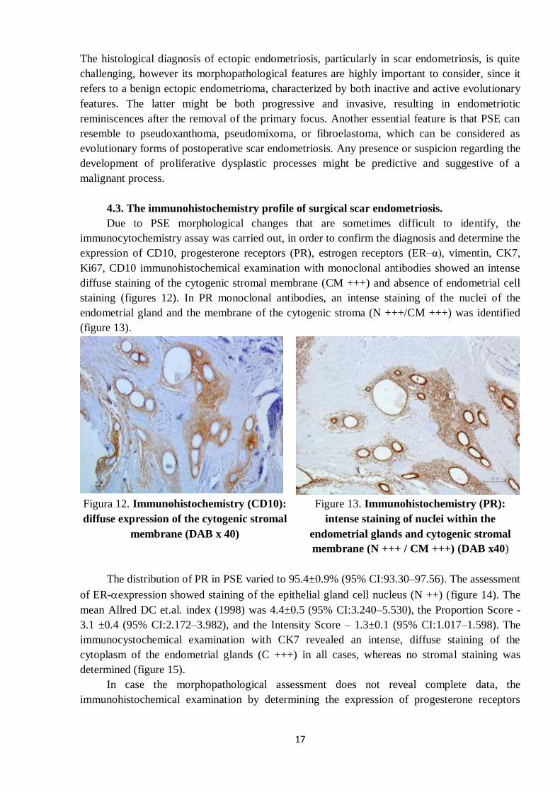
4.3. The immunohistochemistry profile of surgical scar endometriosis.
Due to PSE morphological changes that are sometimes difficult to identify, the
immunocytochemistry assay was carried out, in order to confirm the diagnosis and determine the
expression of CD10, progesterone receptors (PR), estrogen receptors (ER–α), vimentin, CK7,
Ki67, CD10 immunohistochemical examination with monoclonal antibodies showed an intense
diffuse staining of the cytogenic stromal membrane (CM +++) and absence of endometrial cell
staining (figures 12). In PR monoclonal antibodies, an intense staining of the nuclei of the
endometrial gland and the membrane of the cytogenic stroma (N +++/CM +++) was identified
(figure 13).
Figura 12. Immunohistochemistry (CD10):
diffuse expression of the cytogenic stromal
membrane (DAB x 40)
Figure 13. Immunohistochemistry (PR):
intense staining of nuclei within the
endometrial glands and cytogenic stromal
membrane (N +++ / CM +++) (DAB x40)
The distribution of PR in PSE varied to 95.4±0.9% (95% CI:93.30–97.56). The assessment
of ER-expression showed staining of the epithelial gland cell nucleus (N ++) (figure 14). The
mean Allred DC et.al. index (1998) was 4.4±0.5 (95% CI:3.240–5.530), the Proportion Score -
3.1 ±0.4 (95% CI:2.172–3.982), and the Intensity Score – 1.3±0.1 (95% CI:1.017–1.598). The
immunocystochemical examination with CK7 revealed an intense, diffuse staining of the
cytoplasm of the endometrial glands (C +++) in all cases, whereas no stromal staining was
determined (figure 15).
In case the morphopathological assessment does not reveal complete data, the
immunohistochemical examination by determining the expression of progesterone receptors

18
(PR), estrogen receptors (ER-α), vimentin, CK7, Ki67 might help establish an accurate diagnosis
as well as determine the appropriate resection amount of PSE.
Figure 14. Immunohistochemistry (ER-):
nucleus staining of epithelial gland cells (N
++) (DAB x200)
Figure 15. Immunohistochemistry (CK7):
diffuse staining of the cytoplasm of
endometrial glands (C +++) (DAB x400)
GENERAL CONCLUSIONS
1. Clinical features were manifested by typical symptoms of catamenial pain in 61.8% of cases,
and permanent pain in 38.2% of cases, that is not a pathognomonic sign for PSE (p>0.05).
An increase of the formation volume, depending on the menstrual cycle was reported in
70.6% of cases, compared to 29.4% of cases with no relevant signs, thus showing a
statistically significant difference (p=0.0014). The particular non-specific clinical signs
require a very careful differential diagnosis of abdominal wall formations.
2. The obtained findings on the particularities of the PSE localization showed the following
predominance: within the anterior abdominal wall vs. the perineal region (82.4% vs. 17.6%,
p<0.0001), Pfannenstiel incision vs. median laparotomy (92.3% vs. 7.7%, p<0.0001), having
a statistically significant difference. The Pfannenstiel's laparotomy revealed a prevailing
occurrence in the left angle (75%) vs. right angle (16.6%) vs. bilateral (4.2%) vs. central
(4.2%, p=0.0012). According to endometrioma siting as related to the postoperative scar, the
abdominal wall endometriomas were mostly found within the scar region (96.55%)
compared to distant endometriomas (3.44%, p<0.0001). Depending on the endometrioma
localization within the abdominal wall layers, superficial siting was more commonly reported
compared to deep localizations (89.3% vs.10.7%, p<0.0001).
3. As regarding the studies on the structure of surgical interventions contributing to
development of PSE, the cesarean section surgeries were more commonly reported (89.3%)
compared to traditional gynecological and laparoscopic interventions (10.7%, p<0.0001). In
PVSE cases, episiotomy was performed in 50% of cases vs. suturing postpartun ruptures -
33.3% vs. vaginal cyst removal –16.6%. The following potential risk factors were found to
be responsible for the development of PSE: cesarean section (89.3%, p<0.0001), primiparous
patients (80%, p<0.0001), scheduled operations (76%, p=0.0005), and intact amniotic
membrane (88%, p<0.0001).
4. The ultrasound and Doppler methods revealed the following PSE-related imaging criteria:
round/oval-shaped volume mass, hypoechoic with hyperechoic contour, major endometrioma

19
size was on average 23.9±2.7 mm (95% CI:18.25–29.45); whereas the minor size – 15.9±2.1
mm (95% CI:11.65-20.18); presence of vascularization was found in 11 (91.7%) cases and
1(8.3%) case revealed no vascularization, thus showing a statistically significant difference
(p=0.0001). There were reported three types of PSE vascularization viz. peripheral, mixed,
and central. CT study criteria characteristic for PSE were the homogeneous masses with
presence of linear infiltration radiating peripherally to the neighboring subcutaneous tissue
from the central node. MRI study criteria characteristic for PSE were the presence of micro-
hemorrhagic inclusions.
5. The analysis of early and further surgical treatment outcomes revealed that the optimal
surgical volume consists of the en bloc excision, exceeding 5-10 mm within the limits of
healthy tissues, thus preserving the integrity of the mass. The superficially located
endometrioma enables an extensive excision to be performed.
6. This present research determined the following morphological features: (1) presence of
active and inactive evolutionary forms; (2) presence of elastosis within the stroma and
tissues; (3) unformed endometriotic globoid formations located remotely from the primary
source, resembling endometriotic or stromal-glandular stromal loops, defined as
endometriotic satellites; (4) mimicry of the morphological features characterictic for
pseudoxandoma, pseudomixoma or fibroelastoma. The immunohistochemical profile of PSE
is characterized by positive expression in the endometrial glands (CK7, vimentin, PR, ER-)
and in the cytogenic stroma (CD10, PR, ER-).
Practical recommendations:
1. The obstetric and gynecological surgeries, resulting in opening of the uterine cavity (cesarean
section, myomectomy), require preventive measures for developing PSE, which should
include: (1) externalizing the uterus from the abdominal cavity; (2) changing the meshes used
to rehabilitate the uterine cavity; (3) using separate sutures when closing the uterus and the
anterior abdominal wall layers; (4) using high-pressure irrigation for operative wound, while
suturing the abdominal wall, particularly of the angles (in Pfannenstiel incision) by applying
a considerable amount of saline solution.
2. Although PSE is a rare disease, it is worth being included in the differential diagnostic
algorithm for major masses, located in the postoperative scar sites (infiltrated post-operative
scar, ligature abscess, incisional hernia, granuloma, tumor processes) and in patients who
have underwent obstetric and gynecological surgeries.
3. USG + Doppler ultrasound, CT and MRI scanning are recommended in patients that lack the
classic symptoms of PSE, thus revealing the specific imaging signs for determining the
degree of invaded adjacent tissues that might influence the assessment of the proper surgical
volume.
4. The correlation between the MRI imaging characteristics for PSE, the micro-hemorrhages
inside the nodules and a marked inflammation, depending on the periods of the menstrual
cycle, might suggest the importance of MRI scanning before or immediately after menstrual
cycle.
5. The surgical intervention based on the main concept of the resection stage, namely the en
bloc excision, exceeding 5-10 mm within the limits of healthy tissues, while maintaining the
integrity of the masses, might prevent the disease relapses. The reconstructive stage depends
on the aponeurosis defect size by, applying the following options: polypropylene synthetic

20
sutures are used in minor defects, for tension-free closure of aponeurosis, whereas in large
defects, the reconstruction is performed using synthetic materials due to impossibility to
apply this method.
6. Morphopathological examination and immunohistochemical profile by determining the
expression of progesterone receptors (PR), estrogen receptors (ER-α), vimentin, CK7, Ki67
will allow establishing a final diagnosis and determining the appropriate degree of PSE
resection.
These practical recommendations are provided for specialists in the field of surgery,
obstetrics and gynecology, imaging, morphopathology and immunohistochemistry, as well as
for residents and senior students of State University of Medicine and Pharmacy.
SELECTIVE BIBLIOGRAPHY
1. Rindos NB, Mansuria S. Diagnosis and management of abdominal wall endometriosis: a
systematic review and clinical recommendations. Obstet Gynecol Surv. 2017; 72(2):116-
122.
2. Ozturk A, Kaya C, Bozkurtoglu H, Tan N, Yananli ZD, Ucmakli E. Scar endometrioma: an
uncommon yet easily treated condition. J Reprod Med. 2016; 61(5-6):249-53.
3. Mistrangelo M, Gilbo N, Cassoni P, Micalef S, Faletti R, Miglietta C, Brustia R, Bonnet G,
Gregori G, Morino M. Surgical scar endometriosis. Surg Today. 2014; 44(4):767-72.
4. Zhang P, Sun Y, Zhang C, Yang Y, Zhang L, Wang N, Xu H. Cesarean scar
endometriosis: presentation of 198 cases and literature review. BMC WomensHealth.
2019;19(1):14.
5. Horton JD, Dezee KJ, Ahnfeldt EP, Wagner M. Abdominal wall endometriosis: a
surgeon's perspective and review of 445 cases. Am J Surg. 2008;196(2):207-12
6. Aytac HO, Aytac PC, Parlakgumus HA. Scar endometriosis is a gynecological
complication that general surgeons have to deal with. Clin Exp Obstet
Gynecol.2015;42(3):292-4.
7. Ecker AM, Donnellan NM, Shepherd JP, Lee TT. Abdominal wall endometriosis: 12 years
of experience at a large academic institution. Am J Obstet Gynecol. 2014;211(4):363.e1-5.
8. Medeiros FC, Cavalcante DI, Medeiros MA, Eleutério J Jr. Fine-needle aspiration cytology
of scar endometriosis: study of seven cases and literature review. Diagn Cytopathol.
2011;39(1):18-21
9. Erkan N, Haciyanli M, Sayhan H. Abdominal wall endometriomas. Int J Gynaecol Obstet.
2005;89(1):59-60.
10. Blanco RG, Parithivel VS, Shah AK, Gumbs MA, Schein M, Gerst PH. Abdominal wall
endometriomas. Am J Surg. 2003;185(6):596-8.
11. Jegan A, Dimienescu O, Dull A. M. Noi markeri pentru diagnosticul non-invaziv în
endometrioza. Jurnal Medical Brașovean. 2014;nr.2,p.39-41
12. Francica G. Reliable clinical and sonographic findings in the diagnosis of abdominal wall
endometriosis near cesarean section scar. World J Radiol. 2012;4(4):135-40.
13. Zhang J, Liu X. Clinicopathological features of endometriosis in abdominal wall--clinical
analysis of 151 cases. Clin Exp Obstet Gynecol. 2016;43(3):379-83.

21
14. Aubry G, Panel P, Thiollier G, Huchon C, Fauconnier A. Measuring health-related quality
of life in women with endometriosis: comparing the clinimetric properties of the
Endometriosis Health Profile-5 (EHP-5) and the EuroQol-5D (EQ-5D). Hum Reprod.
2017;1;32(6):1258-1269.
15. Zhu Z, Al-Beiti MA, Tang L, Liu X, Lu X. Clinical characteristic analysis of 32 patients
with abdominal incision endometriosis. J Obstet Gynaecol. 2008;28(7):742-5.
16. Fukuda H, Hideki M. Cutaneous endometriosis in the umbilical region: the usefulness of
CD10 in identifying the interstitium of ectopic endometriosis. J Dermatol. 2010;37(6):545-
9.
17. Selcuk S, Sahin S, Demirci O, Aksoy B, Eroglu M, Ay P, Cam C. Translation and
validation of the Endometriosis Health Profile (EHP-5) in patients with laparoscopically
diagnosed endometriosis. Eur J Obstet Gynecol Reprod Biol. 2015;185:41-4.
18. Savelli L, Manuzzi L, Di Donato N, Salfi N, Trivella G, Ceccaroni M, Seracchioli R.
Endometriosis of the abdominal wall: ultrasonographic and Doppler characteristics.
Ultrasound Obstet Gynecol. 2012;39(3):336-40.
LIST OF THE AUTHOR'S PUBLICATIONS ON THE THESIS
Articles published in international scientific journals:
articles in Web of Science and SCOPUS international databases
1. Mishin I., Mishina A., Zaharia S., Zastavnitsky. G. Rectus Abdominis Endometrioma after
Caesarean Section. Case Rep Surg. 2016:4312753.
articles published in recognized international scientific journals
2. Ghidirim Gh., Mișin I., Mișina A., Rojnoveanu Gh., Zaharia S., Chemencedji I.
Endometrioza mușchilui rect abdominal. Archives of the Balkan Medical Union. 2015,
vol.50, nr.2 (suppl. 1), pp.16-22. ISSN 0041-6940.
3. Mişina A., Zaharia S., Mişin I., Petrovici V. Endometrioza cicatricială perineală şi vaginală.
Archives of the Balkan Medical Union.2015, vol.50, nr.2 (suppl. 1), pp.70-75. ISSN 0041-
6940.
4. Zaharia S. Endometrioza ombilicală. Archives of the Balkan Medical Union. 2016, vol.51,
nr.1 (suppl. 1), pp.211-214. ISSN 0041-6940.
Articles published to accredited national scientific journals
articles in B-category journals
5. Mișina A., Zaharia S., Mișin I., Fuior L., Petrovici V. Endometrioza cicatricei postoperatorii
după operație cezariană. Buletinul Academiei de Ştiinţe a Moldovei Ştiinţe Medicale. 2014,
nr. 3(44), pp. 122-126. ISSN 1857-0011.
6. Zaharia S. Transformarea malignă a endometriozei cicatricei postoperatorii. Buletinul
Academiei de Ştiinţe a Moldovei Ştiinţe Medicale. 2015, nr. 4(49), pp. 73-77. ISSN 1857-
0011.
7. Mișina A., Zaharia S., Mișin I., Pochin A., Fuior-Bulhac L. Diagnostic preoperator al
endomitriomei cicatriciei postoperatorii. Buletinul Academiei de Ştiinţe a Moldovei Ştiinţe
Medicale.2015, nr. 4(49), pp. 48-51. ISSN 1857-0011.

22
8. Mișina A., Mișin I., Zaharia S., Chemencedji I. Endomitrioza cicatriciei postoperatorii
după miomectomie. Buletin de Perinatologie. 2016, nr.1(69), pp. 137-140. ISSN 1810-
5289.
9. Zaharia S., Mișina A., Mișin. I. Endometrioza cicatricei postoperatorii complicată cu
hemoragie externă: prezentare de caz. Moldovan Journal of Health Sciences. 2018, vol.15,
nr.1, pp.93-101. ISSN: 23451-6705.
Abstracts/theses published within international scientific
conferences
10. Zaharia S., Mishina A., Mishin I., Ghidirim Gh. Incisional endometrioma of the abdominal
wall. Archives of the Balkan Medical Union.2014, vol.49, nr. (suppl. 1), p.A-130(P-105).
ISSN 0041-6940.
11. Mişina A., ZahariaS., Mişin I., Ghidirim Gh. Endometrioma cicatricei postoperatorii cauzată
de operaţia cezariană. AlXVI-lea Congres Naționalal Societății de Obstetrică și Ginecologie
din România.Volum Rezumate. Cluj-Napoca, 2014, pp. 139-140. (P 15).
12. Ghidirim Gh., Mișin I., Mișina A., Zaharia S., Gheorghiţă V. Endometrioma inciziei
Pfannenstiel. A XXXVI-a Reuniune a Chirurgilor din Moldova “IacomiRăzeşu” și A VIII-a
Conferinţă de Chirurgie cu participareinternaţională. Volum Rezumate. Piatra-Neamţ,
România, 2014, pp.92-93.
13. Ghidirim Gh., Mișin I., Mișina A., Zaharia S., Gheorghiţă V. Endometrioma cicatricei
postoperatorii a peretelui abdominal: douzeci si patru cazuri consecutive. A XXXVII–a
Reuniune a chirurgilor din Moldova “Iacomi – Razesu”. Piatra–Neamţ, 2015, Volum de
Rezumate. pp.85-86. ISBN:978-606-13-2460-6.
14. Ciobanu Gh., Ghidirim Gh., Mishin I., Mishina A., Zaharia S. Les particularites
dudiagnosticet du traitement chirurgicaldel'endometriose de la cicatricepostoperatoire.La
XX-éme Session des Journeés Médicales Baalkaniques la deuxième séance scientifique
commune avec L´Académie Nationale de Médecine de France. Paris, 2015,
Programme/Livre des Résumés. A63 (T5-05). ISSN 0041-6940.
15. Гидирим Г.П., Мишин И.В., Мишина А.Е., Захария С.Л. Эндометриоз
послеоперационного рубца передней брюшной стенки. ХХІІІ з’їзді хірургів України.
Збіник наукових робит.«Клінічна хірургія». Київ, 2015, с.73. ISSN 0023-2130.
16. Гидирим Г.П., Мишин И.В., Мишина А.Е., Захария С.Л. Эндометриоз
послеоперационного рубца. Альманах Института хирургии имени А.В.Вишневского.
2015, №2, с.371-372. ISSN 2075-6895.
17. Mişina A., Zaharia S., Ghidirim Gh., Ghimpu V., Mişin I. Postoperative scar
endometriosis: 31 consecutive cases. În: Al 13-a Ediție a Conferinței Naționale Zilele
Medicale "Vasile Dobrovici" și al 4-lea Congres al Societății Române de Ultrasonografie în
Obstetrică și Ginecologie.Ginecologia.ro Journal. 2016, nr.11 (suppl.1). p.83. ISSN 2344-
2301.
18. Ghidirim Gh., Mișin I., Mișina A, Zaharia S., Vasilev V. Endometrioza cicatricei
condiționată de operația cezariană. Congresul Național de Chirurgie. Sinaia, România,
2016, pp. 260-261.
19. Мишина А.Е., Гидирим Г.П., Мишин И.В., Захария С.Л., Васильев В.Е. Эндометриоз
передней брюшной стенки после кесарева сечения. В: ХХIX Международный
Конгресс с курсом эндоскопии «Новые технологии в диагностике и лечении
гинекологических заболеваний». Москва, 2016, с.154-155. ISBN:978-5-906484-18-5
23
20. Захария С.Л. Эндометриоз послеоперационного рубцa. Международной научно-
практической конференции. Белгород, 2017, с.91-94.
Abstracts/theses published within national scientific conferences
21. Ghidirim Gh., Mișin I., Mișina A., Zaharia S. Particularități de diagnostic și tratament
chirurgical al endometriozei cicatricei postoperatorii. Arta Medica. 2015. nr.3(56). pp.41-
42. ISSN 1810-1852.
22. Zaharia S., Mișin I., Mișina A., Fuior-Bulhac L., Cuțitari I., Crăciun V. Endometrioza
cicatricei postoperatorii: caracteristici Doppler-ultrasonografice. Al IV-lea Congres al
Medicilor Imagistici din Moldova cu participarea internațională. The Moldovan Medical
Journal. Special Edition. 2018. pp.87-88. ISSN 2537-6373.
23. Zaharia S., Mișin I., Mișina Ana, Petrovici V. Particularitățile morfologice caracteristice
endometriozei cicatricei postoperatorii. Al XIII-lea Congres al Asociației
Chirurgilor"Nicolae Anestiadi" din Republica Moldova, cu participare internațională. Arta
Medica. 2019. nr. 3 (72). p. 178. ISSN 1810-1852.

24
ADNOTARE
Zaharia Sergiu „Endometrioza cicatricei postoperatorii: optimizarea diagnosticului și tratamentului”. Teza de doctor în ştiinţe medicale, Chişinău, 2020. Teza este expusă pe 120 pagini, conține - introducere, 4 capitole, sinteza rezultatelor obţinute, concluzii, recomandări practice, 5 tabele, 110 figuri, 274 surse bibliografice. La tema tezei au fost publicate 23 lucrări. Cuvinte-cheie: endometrioza cicatricei postoperatorii (ECP), endometrioza mușchiului rect abdominal, endometrioza perineală, dureri catameniale, operație cezariană, profil imunohistochimic. Domeniu de studiu: 321.13 – chirurgie Scopul lucrării: Optimizarea managementului diagnostico-curativ al pacientelor cu
endometrioza cicatricei postoperatorii prin analiza variată a criteriilor clinice, imagistice,
morfologice și a rezultatelor tratamentului chirurgical. Obiectivele lucrării: (1) studierea manifestărilor clinice și particularităților de localizare a ECP; (2) determinarea structurii intervențiilor precedente și definitivarea factorilor de risc cauzatori de dezvoltarea ECP; (3) stabilirea criteriilor imagistice (ultrasonografice (USG), dopplerografice (DG), tomografice (TC), imagistice prin rezonanță magnetică (IRM), și a particularităților anatomo-topografice ale endometriozei cicatricei postoperatorii; (4) determinarea volumului optimal al intervenției chirurgicale în endometrioza cicatricei postoperatorii bazate pe studierea rezultatelor precoce si la distanță; (5) determinarea particularităților morfologice și a profilului imunohistochimic ale endometriozei cicatricei postoperatorii. Noutatea şi originalitatea cercetării: În baza evaluării clinice au fost stabiliți factorii de risc principali de dezvoltare al ECP. Studiul prezent a demonstrat prezența semnelor clinice nespecifice. Au fost stabilite următoarele particularitățile caracteristice ECP: predominarea în regiunea peretelui abdominal anterior, dominarea endometriomelor unice, mai frecvent în incizia Pfannenstiel cu prevalența unghiului stâng a cicatricei postoperatorii. Au fost determinate criteriile imagistice de diagnostic (USG, DG, TC și IRM), cu aprecierea informativității înalte a acestor metode în diagnosticul ECP. Au fost dovedite principiile de bază a tratamentului chirurgical al ECP: păstrarea integrității formațiunii și excizia chirurgicală en bloc (R0). Elaborate criteriile aplicării metodei de reconstrucție a peretelui abdominal după excizia endometriomului. Studiate particularitățile morfologice și imunohistochimice (CD10, RE-α, RP, CK7, vimentin) al ECP. Evaluate rezultatele tratamentului chirurgical al ECP la distanță, și apreciate calitatea vieții a pacientelor cu ECP conform scorului EHP-5. Problema ştiinţifică soluționată constă în elaborarea și implementarea metodologiei de diagnostic și tratament al ECP, care va contribui la ameliorarea rezultatelor, prevenirea recidivei, și îmbunătățirea calității vieții. Semnificaţia teoretică: S-au stabilit factorii potențiali de risc ce favorizează apariția ECP. A fost justificată importanța metodelor imagistice în depistarea și stabilirea diagnosticului de ECP preoperator. Studiată informativittea testelor serologice (markerului tumoral CA-125, valorilor preoperatorii a volumului mediu trombocitar, și indexul neutrofil/limfocitar) în diagnosticarea ECP. Specificate principiile de bază ale tratamentului chirurgical al ECP. Determinat rolul examenului morfologic și profilului imunohistochimic în stabilirea definitivă a diagnosticului de ECP. Valoarea aplicativă a lucrării: Sunt argumentate și formulate principiile diagnosticului și a tratamentului chirurgical al ECP. Implementarea rezultatelor ştiinţifice: În baza cercetării, au fost implementate noi metode de diagnostic şi tratament a pacienţilor cu ECP în secţiile de chirurgie IMSP Institutul de Medicină Urgentă (Chişinău, Republica Moldova), în secția de ginecologie chirurgicală IMSP Institutul Mamei și Copilului (Chişinău, Republica Moldova) şi în procesul didactic al catedrei de chirurgie nr. 1 „Nicolae Anestiadi” a Universității de Stat de Medicină și Farmacie „Nicolae Testemițanu”. Au fost obținute 5 acte de implementare în practică.

25
РЕЗЮМЕ
Захария Сергей «Эндометриоз послеоперационного рубца: оптимизация диагностики и лечения». Диссертация на соискание ученой степени кандидата медицинских наук, Кишинев, 2020. Диссертация изложена на 120 страницах, состоит из введения, 4 глав, синтеза полученных результатов, выводов, практических рекомендаций, 5 таблиц, 110 рисунков. Библиография включает 274 источника. По теме диссертации опубликовано 23 печатных работ. Ключевые слова: эндометриоз послеоперационного рубца (ЭПР), эндометриоз прямой мышцы живота, перинеальный эндометриоз, катамениальные боли, кесарево сечение, иммуногистохимический профиль. Область исследования: 321.13 – хирургия Цель работы: Оптимизация лечебно-диагностического менеджмента больных с ЭПР на
основание многостороннего анализа клинических, радиологических, морфологических
критериев и результатов хирургического лечения.
Задачи исследования: (1) изучить клинические манифестации и особенности локализации ЭПР; (2) определить структуры предшествующих операций и факторов риска, влияющих на развитие ЭПР; (3) установить радиологические признаки (на основании ультрасонографии (УСГ), допплерографии (ДГ), компьютерной томографии (КТ) и магнитно-резонансной томографии (МРТ) и анатомо-топографические характеристики ЭПР; (4) определить оптимальный объем хирургического вмешательства при ЭПР на основании изучения ближайших и отдаленных результатов; (5) изучить морфологические особенности и иммуногистохимический профиль ЭПР. Новизна и оригинальность исследований: На основании оценки клинического материала установлены основные факторы риска ЭПР. Данное исследование продемонстрировало наличие неспецифических клинических симптомов при ЭПР. Были установлены характерные особенности ЭПР: преимущественная локализация в области передней брюшной стенки, преобладание единичных эндометриом, чаще при лапаротомии по Пфанненштилю и преимущественно локализуются в левом углу послеоперационного рубца. Установлены радиологические (УСГ, ДГ, КТ и МРТ) признаки ЭПР и определена высокая информативность методов при данной патологии. Определены основополагающие принципы хирургического лечения ЭПР: сохранение целостности эндометриомы и ее иссечение en bloc (R0). Разработаны критерии реконструкции передней брюшной стенки после иссечения эндометриомы. Изучены морфологические и иммуногистохимические (CD10, RE-α, RP, CK7, vimentin) характеристики ЭПР. Оценены отдаленные результаты и качество жизни (бальная система EHP-5) после хирургического лечения ЭПР. Решенная научная проблема состоит в разработке и внедрении методологии диагностики и хирургического лечения ЭПР, что способствует улучшению результатов, профилактики рецидивов и улучшению качества жизни. Теоретическая значимость: Установлены факторы риска развития ЭПР. Обоснована важность радиологических методов в визуализации и диагностике ЭПР. Изучена информативность серологических тестов (онкомаркера CA-125, среднего количества тромбоцитов и нейтрофильно-лимфацитарного индекса) в диагностике ЭПР. Установлена роль морфологических и иммуногистохимических методов в окончательной диагностике ЭПР. Практическая значимость: Аргументированы и сформулированы основные принципы диагностики и хирургического лечения ЭПР. Внедрение научных результатов: На основании данного исследования внедрены новые методы диагностики и лечения пациенток с ЭПР в хирургических отделениях ПМСУ Института ургентной медицины (г. Кишинев, Республика Молдова) и в отделении оперативной гинекологии ПМСУ Института матери и ребенка (г. Кишинев, Республика Молдова), а также в педагогическом процессе кафедры хирургии №1 им. Н. Анестиади Университета медицины и фармации им. Н.Тестемицану. По результатам исследования получены 5 свидетельств по внедрению в медицинскую практику.

26
ANNOTATION
Zaharia Sergiu, “Postoperative scar endometriosis: optimization of diagnosis and treatment”
PhD Thesis, Chisinau, 2020. This research work comprises 120 pages, including introduction, 4
chapters, synthesis of the obtained results, conclusions, practical recommendations, 5 tables, 110
figures, and 274 bibliographic sources. The research findings were published with in 23
scientific works.
Key-words: postoperative scar endometriosis (PSE), endometriosis of rectus abdominis muscle,
perineal endometriosis, catamenial pain, cesarean section, immunohistochemistry profile.
Research domain: 321.13 – surgery
The purpose of the study: To provide optimal diagnostic and treatment management of the
patients with PSE by assessing a range of clinical, imaging and morphological criteria, as well as
surgical treatment outcomes.
The research objectives: (1) To study the clinical manifestations and localization features of
PSE; (2) to determine the past surgery structure and define the causative risk factors for PSE; (3)
to establish the imaging criteria (ultrasound, Doppler, CT, MRI scan) and the anatomical and
topographical features of postoperative scar endometriosis; (4) to determine the optimal surgical
volume in PSE based on the study of early and long-term results; (5) to determine the
morphological features and immunohistochemistry profile of PSE.
Novelty and scientific originality: The main risk factors for PSE development were identified
based on the clinical assessment. The present study has demonstrated the presence of nonspecific
clinical signs. The following features have been established: the predominance on the anterior
abdominal wall region, dominance of single endometriomas, incision of Pfannenstiel, left angle
of the Pfannenstiel laparatomy. The diagnostic criteria (ultrasound, Doppler, CT, MRI) were
determined, which proved to be highly informative in the diagnosis of PSE. There have been
proventhe following basic principles of surgical treatment in PSE: preserving the integrity of the
structure; en bloc surgical excision. There were developed the criteria for applying the method of
reconstruction of the abdominal wall, following the endometriomaremoval. The morphological
featuresand theimmunohistochemistry profile of the PSE were also studied. Both the long-term
treatment outcomes and quality of life, based on EHP-5 score, were assessed within this
research.
The scientific issue to be solved consists of development and implementation of the PSE
diagnostic and treatment approach, which help improve patient outcomes, prevent relapses, and
enhance quality of life.
Theoretical significance: This research paper identified the potential risk factors, leading to the
onset of PSE. The importance of the imagistic methods for the preoperative diagnosis has also
been justified. The serological tests (tumor marker CA-125, preoperative values of the mean
platelet volume and neutrophil to lymphocyte ratio) proved to be highly informative in
diagnosing PSE. The basic principles of the surgical treatment were specified. This research
emphasizes the morphological features and immunohistochemistry profile for definite diagnosis
of PSE.
The applicative value of the research: The research findings formulated and justified the
principles of PSE diagnosis and surgical treatment.
Implementation of scientific results: The study results enabled implementation of the new
methods of diagnosis and treatment of the PSE patients with in the surgical units, at Emergency
Medicine Institute (Chisinau, Republic of Moldova), Department of Gynecology Surgery,
Department of Research, at the Health Care Institute of Mother and Child (Chisinau, Republic of
Moldova) and within the teaching process of „Nicolae Anestiadi” Department of Surgery No.1,
at „Nicolae Testemitanu” State University of Medicine and Pharmacy. Five certificates have
been obtained to be implemented into practice.