Postmenopausal Woman: Extremely Rare Case with...
3
Large (7.2 kg) Subserosal Fibroid with Monkenberg’s Calcificaon in a Postmenopausal Woman: Extremely Rare Case with Review of Literature Rajshree Dayanand Katke* Obstertrics and Gynaecology Department, Cama Albless Hospital, Mumbai, India *Corresponding author: Rajshree Dayanand Katke, Associate Professor, Obstertrics and Gynaecology Department, Cama Albless Hospital, Mumbai, India, Tel: 91-022-22620390, E-mail: [email protected] Received date: 15th June, 2016; Accepted date: 29th July, 2016; Published date: 1st August, 2016 Citaon: Katke RD. Large (7.2 kg) Subserosal Fibroid with Monkenberg’s Calcificaon in a Postmenopausal Woman: Extremely Rare Case with Review of Literature, Gynecol Obstet Case Rep. 2016, 2:2. Abstract We Report an unusual case of very large uterine subserosal leiomyoma mimicking ovarian mass on diagnosc modalies. A 70 year old postmenopausal woman presented with gradual distension of abdomen and diffuse pain in abdomen since 2 months. Ultrasonography was suggesve of large extrauterine mass of 20 cm × 15 cm × 18 cm displacing uterus anteriorly not separately visualised from either ovaries neoplasc origin likely ovary. While on Computed tomography scan was suggesve of 21 cm × 16cm × 19 cm mass of benign eology possibility of subserosal fibroid with Histopathology correlaon. Exploratory laparotomy with removal of huge subserosal fibroid with total abdominal hysterectomy with bilateral Salpingoophrectomy is the surgery done in our case. The majority of uterine leiomyomas are confidently diagnosed sonographically. However, large, degenerated tumours like in our case may be a diagnosc challenge and postmenopausal uterine leiomyoma with degeneraon and Mockenberg’s calcificaon in a postmenopausal woman is rare and Computed Tomgraphy may help further characterize large pelvic masses and determine their organ of origin. As in our case its diagnosed in computed tomography. Keywords: Computed tomography; Ovarian mass; Uterine subserosal leiomyoma; Ultrasonography; Subserosal fibroid; Salpingoophrectomy; Hysterectomy Introducon Leiomyomas or fibroids arise from overgrowth of the smooth muscle and connecve ssue of the uterus, and most commonly involve the uterine corpus, although they may also occur in the cervix in a minority of instances. Typical fibroids are easily recognized on imaging. However, an atypical presentaon caused by degenerave changes can cause diagnosc confusion as in our case mimicking ovarian tumour [1,2]. The use of color Doppler ultrasonography (CDUS) to visualize interface vessels between the uterus and a juxta- uterine mass is useful in the differenal diagnosis. Also, magnec resonance imaging (MRI) yielding mulplanar views can reveal the peduncle, or confirm the presence of a normal uninvolved ovary [3]. In this case report, we present mulmodal abdominopelvic radiologic imaging findings of a paent with a huge giant subserosal uterine leiomyoma, in conjuncon with histopathological findings. Case Report A 70 Year old postmenopausal woman presented to our hospital with a complaints of gradual distension of abdomen since 2 months and diffuse pain in abdomen since 3-4 months. All roune laboratory test values were within normal limits. On general examinaon paent was vitally stable. Per abdominal examinaon revealed large abdominal mass corresponding to 28 to 30 weeks Gestaonal size of uterus arising from pelvis, smooth surface, firm to hard in consistency, mobility slightly restricted. Its lower border could not be felt. Per speculum examinaon revealed cervical os was taken up. On per vaginal examinaon a mass of 28-30 weeks gestaonal size was palpable which was firm to hard in consistency, uterus could not be felt separately from the mass, bilateral fornices were free and there was no tenderness. Abdominal ultrasound examinaon showed a large heterogenous hypoechoic mass of size 20 cm × 15 cm × 18 cm in the pelvico abdominal region displacing the uterus anteriorly and is not separately visualised from either ovary query neoplasc origin likely ovary. Contrast- enhanced computed tomography of the abdomen and pelvis demonstrated a large well defined lobulated soſt ssue aenuang minimally and heterogeneously enhancing solid mass lesion of 21 cm × 16 cm × 19 cm with mulple foci of calcificaon seen within the pelvis. Computed tomography findings suggesve of benign eology query subserosal fibroid. Laboratory tests including tumour markers CA-125 and CEA were within normal limits. Paent underwent exploratory laparotomy with Total Abdominal Hysterectomy with bilateral Salpingoophrectomy with removal of huge subserosal Fibroid. Laparotomy revealed a Huge mass of 28 cm × 18 cm × 20 cm arising from anterior serosal part of uterus and the part of peritoneum near the urinary bladder, the mass was highly vascular and adherent to bladder and large bowel (Figure 1). Case Report iMedPub Journals http://www.imedpub.com/ DOI: 10.21767/2471-8165.1000026 Gynecology & Obstetrics Case report ISSN 2471-8165 Vol.2 No.2:26 2016 © Copyright iMedPub | This article is available from: http://gynecology-obstetrics.imedpub.com/ 1
Transcript of Postmenopausal Woman: Extremely Rare Case with...

Large (7.2 kg) Subserosal Fibroid with Monkenberg’s Calcification in aPostmenopausal Woman: Extremely Rare Case with Review of LiteratureRajshree Dayanand Katke*
Obstertrics and Gynaecology Department, Cama Albless Hospital, Mumbai, India
*Corresponding author: Rajshree Dayanand Katke, Associate Professor, Obstertrics and Gynaecology Department, Cama Albless Hospital,Mumbai, India, Tel: 91-022-22620390, E-mail: [email protected]
Received date: 15th June, 2016; Accepted date: 29th July, 2016; Published date: 1st August, 2016
Citation: Katke RD. Large (7.2 kg) Subserosal Fibroid with Monkenberg’s Calcification in a Postmenopausal Woman: Extremely Rare Case withReview of Literature, Gynecol Obstet Case Rep. 2016, 2:2.
Abstract
We Report an unusual case of very large uterinesubserosal leiomyoma mimicking ovarian mass ondiagnostic modalities. A 70 year old postmenopausalwoman presented with gradual distension of abdomenand diffuse pain in abdomen since 2 months.Ultrasonography was suggestive of large extrauterinemass of 20 cm × 15 cm × 18 cm displacing uterusanteriorly not separately visualised from either ovariesneoplastic origin likely ovary. While on Computedtomography scan was suggestive of 21 cm × 16cm × 19 cmmass of benign etiology possibility of subserosal fibroidwith Histopathology correlation. Exploratory laparotomywith removal of huge subserosal fibroid with totalabdominal hysterectomy with bilateralSalpingoophrectomy is the surgery done in our case. Themajority of uterine leiomyomas are confidently diagnosedsonographically. However, large, degenerated tumourslike in our case may be a diagnostic challenge andpostmenopausal uterine leiomyoma with degenerationand Mockenberg’s calcification in a postmenopausalwoman is rare and Computed Tomgraphy may helpfurther characterize large pelvic masses and determinetheir organ of origin. As in our case its diagnosed incomputed tomography.
Keywords: Computed tomography; Ovarian mass;Uterine subserosal leiomyoma; Ultrasonography; Subserosalfibroid; Salpingoophrectomy; Hysterectomy
IntroductionLeiomyomas or fibroids arise from overgrowth of the
smooth muscle and connective tissue of the uterus, and mostcommonly involve the uterine corpus, although they may alsooccur in the cervix in a minority of instances. Typical fibroidsare easily recognized on imaging. However, an atypicalpresentation caused by degenerative changes can causediagnostic confusion as in our case mimicking ovarian tumour[1,2]. The use of color Doppler ultrasonography (CDUS) tovisualize interface vessels between the uterus and a juxta-
uterine mass is useful in the differential diagnosis. Also,magnetic resonance imaging (MRI) yielding multiplanar viewscan reveal the peduncle, or confirm the presence of a normaluninvolved ovary [3]. In this case report, we presentmultimodal abdominopelvic radiologic imaging findings of apatient with a huge giant subserosal uterine leiomyoma, inconjunction with histopathological findings.
Case ReportA 70 Year old postmenopausal woman presented to our
hospital with a complaints of gradual distension of abdomensince 2 months and diffuse pain in abdomen since 3-4 months.All routine laboratory test values were within normal limits. Ongeneral examination patient was vitally stable. Per abdominalexamination revealed large abdominal mass corresponding to28 to 30 weeks Gestational size of uterus arising from pelvis,smooth surface, firm to hard in consistency, mobility slightlyrestricted. Its lower border could not be felt. Per speculumexamination revealed cervical os was taken up. On per vaginalexamination a mass of 28-30 weeks gestational size waspalpable which was firm to hard in consistency, uterus couldnot be felt separately from the mass, bilateral fornices werefree and there was no tenderness. Abdominal ultrasoundexamination showed a large heterogenous hypoechoic mass ofsize 20 cm × 15 cm × 18 cm in the pelvico abdominal regiondisplacing the uterus anteriorly and is not separately visualisedfrom either ovary query neoplastic origin likely ovary. Contrast-enhanced computed tomography of the abdomen and pelvisdemonstrated a large well defined lobulated soft tissueattenuating minimally and heterogeneously enhancing solidmass lesion of 21 cm × 16 cm × 19 cm with multiple foci ofcalcification seen within the pelvis. Computed tomographyfindings suggestive of benign etiology query subserosal fibroid.Laboratory tests including tumour markers CA-125 and CEAwere within normal limits. Patient underwent exploratorylaparotomy with Total Abdominal Hysterectomy with bilateralSalpingoophrectomy with removal of huge subserosal Fibroid.Laparotomy revealed a Huge mass of 28 cm × 18 cm × 20 cmarising from anterior serosal part of uterus and the part ofperitoneum near the urinary bladder, the mass was highlyvascular and adherent to bladder and large bowel (Figure 1).
Case Report
iMedPub Journalshttp://www.imedpub.com/
DOI: 10.21767/2471-8165.1000026
Gynecology & Obstetrics Case report
ISSN 2471-8165Vol.2 No.2:26
2016
© Copyright iMedPub | This article is available from: http://gynecology-obstetrics.imedpub.com/ 1
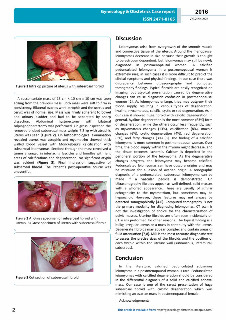
Figure 1 Intra op picture of uterus with subserosal fibroid
A succenturiate mass of 15 cm × 10 cm × 10 cm was seenarising from the previous mass. Both mass were soft to firm inconsistency. Bilateral ovaries were atrophic and the uterus andcervix was of normal size. Mass was firmly adherent to boweland urinary bladder and had to be separated by sharpdissection. Abdominal hysterectomy with bilateralsalpingoopherectomy was performed. On gross inspection theremoved bilobed subserosal mass weighs 7.2 kg with atrophicuterus was seen (Figure 2). On histopathological examinationrevealed uterus was atrophic and myometrim showed thickwalled blood vessel with Monckeberg’s calcification withsubserosal leiomyomas. Sections through the mass revealved atumor arranged in interlacing fascicles and bundles with ientareas of calcifications and degeneration. No significant atypiawas evident (Figure 3). Final impression suggestive ofsubserosal fibroid. The Patient’s post-operative course wasuneventful.
Figure 2 A) Gross specimen of subserosal fibroid withuterus, B) Gross specimen of uterus with subserosal fibroid
Figure 3 Cut section of subserosal fibroid
DiscussionLeiomyomas arise from overgrowth of the smooth muscle
and connective tissue of the uterus. Around the menopause,leiomyomas decrease in size because their growth is thoughtto be estrogen dependent, but leiomyomas may still be newlydiagnosed in postmenopausal women. A calcifiedpedunculated leiomyoma in a postmenopausal woman isextremely rare; in such cases it is more difficult to predict theclinical symptoms and physical findings. In our case there wasdiscrepancy between ultrasonography and computedtomography findings. Typical fibroids are easily recognized onimaging, but atypical presentation caused by degenerativechanges can cause diagnostic confusion in postmenopausalwomen [2]. As leiomyomas enlarge, they may outgrow theirblood supply, resulting in various types of degeneration:hyaline, myxomatous, calcific, cystic or red degeneration. As inour case it showed huge fibroid with calcific degeneration. Ingeneral, hyaline degeneration is the most common (63%) formof degeneration, while the others occur less frequently, suchas myxomatous changes (13%), calcification (8%), mucoidchanges (6%), cystic degeneration (4%), red degeneration(3%), and fatty changes (3%) [3]. The finding of a calcifiedleiomyoma is more common in postmenopausal woman. Overtime, the blood supply within the myoma might decrease, andthe tissue becomes ischemic. Calcium is deposited in theperipheral portion of the leiomyoma. As the degenerativechanges progress, the leiomyoma may become calcified.Pedunculated leiomyomas can have obscure origins and maybe mistaken for a lesion of ovarian origin. A sonographicdiagnosis of a pedunculated, subserosal leiomyoma can bemade if a vascular pedicle is demonstrated. OnUltrasonography fibroids appear as well-defined, solid masseswith a whorled appearance. These are usually of similarechogenicity to the myometrium, but sometimes may behypoechoic However, these features may not always bedetected sonographically [4-6]. Computed tomography is notthe primary modality for diagnosing leiomyomas. CT scan isnot the investigation of choice for the characterization ofpelvic masses. Uterine fibroids are often seen incidentally onCT scans performed for other reasons. The typical finding is abulky, irregular uterus or a mass in continuity with the uterus.Degenerate fibroids may appear complex and contain areas offluid attenuation [7,8]. MRI is the most accurate diagnostic testto assess the precise sizes of the fibroids and the position ofeach fibroid within the uterine wall (submucous, intramural,subserous).
ConclusionIn the literature, calcified pedunculated subserous
leiomyoma in a postmenopausal woman is rare. Pedunculatedleiomyomas with calcified degeneration should be consideredin the differential diagnosis of a solid and calcified adnexalmass. Our case is one of the rarest presentation of hugesubserosal fibroid with calcific degeneration which wasmimicking an ovarian mass in postmenopausal female.
Acknowledgement:
Gynecology & Obstetrics Case report
ISSN 2471-8165 Vol.2 No.2:26
2016
2 This article is available from: http://gynecology-obstetrics.imedpub.com/

Dr Nishtha Tripathi (Resident Doctor).
References1. Katke RD (2014) Torsion of huge cystic teratoma of ovary with
multiple fibroids uterus: a case report and review of literature.Int J Reprod Contracept Obstet Gynecol 3(3): 793-795.
2. Ciarmela P, Ciavattini A, Giannubilo SR, Lamanna P, Fiorini R, etal. (2014) Management of leiomyomas in perimenopausalwomen. Maturitas 78: 168- 173.
3. Hwang JH, Modi GV, Jeong Oh M, Lee NW, Hur JY, et al. (2010)An unusual presentation of a severely calcified parasiticleiomyoma in a postmenopausal woman. JSLS 14: 299-302.
4. Samal SK, Rathod S, Rani R, Anandraj R (2014) An unusualpresentation of a severely calcified subserous leiomyoma in a
postmenopausal woman: a case report. Int J Reprod ContraceptObstet Gynecol 3: 463-465.
5. Singh K, Prasad D, Pankaj S, Suman S, Kumar A, et al. (2014)Postmenopausal massive subserous calcified fibroid: a casereport. J of Evolution of Med and Dent Sci 3: 2255-2257.
6. Caoili EM, Hertzberg BS, Kliewer MA, DeLong D, Bowie JD (2000)Refractory shadowing from pelvic masses on sonography: auseful diagnostic sign for uterine leiomyomas. AJR Am JRoentgenol 174: 97-101.
7. Rajanna DK, Pandey V, Janardhan S, Datti SN (2013) Broadligament fibroid mimicking as ovarian tumor on ultrasonographyand computed tomography scan. J Clin Imaging Sci.
8. Owen C, Armstrong AY (2015) Clinical management ofleiomyoma. Obstet Gynecol Clin North Am 42: 67-85.
Gynecology & Obstetrics Case report
ISSN 2471-8165 Vol.2 No.2:26
2016
© Copyright iMedPub 3