Poster Department Presenting Designation Abstract Title...
41
Poster No. Department Presenting Author Designation Abstract Title Authors Phd-13 Biochemistry Ms. Gunjan Sharma PhD Student ETV6 as a regulator of the IGF2BP family of RNA binding proteins in Acute Lymphoblastic Leukemia Gunjan Sharma, Elza Boby, Ayushi Jain, Parthaprasad Chattopadhyay, Anita Chopra, Sameer Bakhshi, Jayanth Kumar Palanichamy Phd-14 Biochemistry Nitesh Mishra PhD Student Viral characteristics associated with maintenance of elite neutralizing activity in chronically HIV-1C infected monozygotic pediatric twins Mishra Nitesh, Makhdoomi Ashraf Muzamil, Sharma Shaifali, Kumar Sanjeev, Kumar Deepshikha, Chawla Himanshi, Singh Ravinder, Kanga Uma, Das Kumar Bimal, Lodha Rakesh, Kabra Kumar Sushil, Luthra Kalpana Phd-15 Biochemistry Amar Preet Kaur PhD Student Evaluating the role of Metformin on differentiation and tumor microenvironment in human colorectal carcinoma Kaur, Preet Amar, Dey, Devanjan, Bhattacharya, Aditi, Bansal, Aakriti, Das, Prasenjit, Vishnubhatla, Sreenivas, Sen, Sudip Phd-16 Biochemistry Deepti Pathak PhD Student The Involvement of Iron in Neurodegeneration associated with Friedreich’s ataxia Deepti Pathak, Achal Kumar Srivastava, Sheffali Gulati, M.V. Padma, Moganty R Rajeswari Phd-17 Biochemistry Dr. Srinivas H PhD Student Characterisation of Fat1 processing and its interaction with β- catenin in glioma Gowda H Srinivas, Gupta Yakhlesh, Malik Nargis, Goswami Sanjeev, Chattopadhyay Parthaprasad, Sarkar Chitra, Suri Ashish, Gupta K Deepak, Sinha Subrata, Chosdol Kunzang.
Transcript of Poster Department Presenting Designation Abstract Title...

Poster No.
Department Presenting Author
Designation Abstract Title Authors
Phd-13 Biochemistry Ms. Gunjan Sharma
PhD Student ETV6 as a regulator of the IGF2BP family of RNA binding proteins in Acute Lymphoblastic Leukemia
Gunjan Sharma, Elza Boby, Ayushi Jain, Parthaprasad Chattopadhyay, Anita Chopra, Sameer Bakhshi, Jayanth Kumar Palanichamy
Phd-14 Biochemistry Nitesh Mishra PhD Student Viral characteristics associated with maintenance of elite neutralizing activity in chronically HIV-1C infected monozygotic pediatric twins
Mishra Nitesh, Makhdoomi Ashraf Muzamil, Sharma Shaifali, Kumar Sanjeev, Kumar Deepshikha, Chawla Himanshi, Singh Ravinder, Kanga Uma, Das Kumar Bimal, Lodha Rakesh, Kabra Kumar Sushil, Luthra Kalpana
Phd-15 Biochemistry Amar Preet Kaur
PhD Student Evaluating the role of Metformin on differentiation and tumor microenvironment in human colorectal carcinoma
Kaur, Preet Amar, Dey, Devanjan, Bhattacharya, Aditi, Bansal, Aakriti, Das, Prasenjit, Vishnubhatla, Sreenivas, Sen, Sudip
Phd-16 Biochemistry Deepti Pathak PhD Student The Involvement of Iron in Neurodegeneration associated with Friedreich’s ataxia
Deepti Pathak, Achal Kumar Srivastava, Sheffali Gulati, M.V. Padma, Moganty R Rajeswari
Phd-17 Biochemistry Dr. Srinivas H PhD Student Characterisation of Fat1 processing and its interaction with β-catenin in glioma
Gowda H Srinivas, Gupta Yakhlesh, Malik Nargis, Goswami Sanjeev, Chattopadhyay Parthaprasad, Sarkar Chitra, Suri Ashish, Gupta K Deepak, Sinha Subrata, Chosdol Kunzang.
Phd-18 Biochemistry Vikas Kumar PhD Student Transactivation of HnRNPD/Auf-1 by NF-κB(P65/RelA) in oral cancer
Kumar Vikas, Kumar Manish, Chauhan S Shyam
Phd-21 Biochemistry Sudhasini Panda
PhD Student Role of vitamin D and its associated molecules in innate immunity modulation in pulmonary tuberculosis
Sudhasini Panda, Ambrish Tiwari, Kalpana Luthra, SK Sharma, Archana Singh
Phd-22 Biochemistry Nidhi Gupta PhD Student Targeting of verrsican by microRNAs inhibit multiple myeloma progression: A novel potential therapeutic strategy
Gupta Nidhi, Kumar Raman Seth Tulika, Garg Bhavuk, Sharma Alpana
Phd-28 Biochemistry Nargis Malik Phd. Student
FAT1 gene regulates
Programmed cell death
10 (PDCD10) via miR-
221 in glioblastoma
Nargis Malik,
Parthaprasad
Chattopadhyay,
Chitra Sarkar,
Ashish Suri,
Subrata Sinha,
Kunzang Chosdol
Phd-82 Biochemistry Khushwant Singh
PhD Student Silver nanoparticle enhance tumor associated macrophage function and decreases tumor cell proliferation
Gautam Kumar Pramod, Singh Khushwant, Anita, Garg Drishti, Bukhari Jehangir.
Phd-84 Biochemistry Himani Thakkar
PhD Student HDL abnormalities in cardiovascular disease: Quality or Quantity?
Thakkar Himani, Vincent Vinnyfred, Roy Ambuj, Singh Sandeep, Ramakrishnan Lakshmy, Singh Archna
Phd-89 Biochemistry Dayasagar Das
PhD Student Unraveling the role of Gamma Delta T cell subsets: Key players in the immunopathogenesis of Pemphigus Vulgaris
Das Dayasagar,KV Santosh, Arava Sudheer, Khandpur Sujay, Sharma Alpana
Phd-98 Biochemistry Mohit Arora PhD Student Elevated expression of Dipeptidyl Peptidase III correlates with the Tumor Infiltrating Lymphocyte(TILs) composition in multiple cancers.
Arora Mohit, Gupta Jasmeen, Prajapati Chand Subhash, Chauhan Singh Shyam

Phd-103
Biochemistry Charandeep Kaur
PhD Student Acute Febrile Illness associated malaria co-infections in tropical diseases: A spectrum and incidence in Indian population
Atreyi Pramanik, Charandeep Kaur, Rajendra Mandage, Vinod Kumar, Shounak Saha, Adarsh Singh, Manish Soneja, Pragyan Acharya
Phd-104
Biochemistry Yakhlesh Gupta
PhD Student FAT1, a novel regulator of YAP1- effector molecule of Salvador-Warts-Hippo (SWH) pathway, in human Glioblastoma
Gupta Yakhlesh, Shivajirao Sachin Shelke, Irshad Khushboo, Dikshit Bhawana, Srivastav Tapasya, Chattopadhyay Parthaprasad, Sinha Subrata & Chosdol Kunzang
Phd-109
Biochemistry Sanjeev Goswami
PhD Student Role of IGF2BP3 in migration and invasion of epithelial cancer cell lines
Sanjeev Goswami, Gunjan Sharma, Elza Boby, Parthaprasad Chattopadhyay, Jayanth Kumar Palanichamy
Phd-110
Biochemistry Mohamad Tarik
PhD Student Feasibility of measuring sodium, potassium and creatinine from urine sample on dried filter paper
Mohamad Tarik, Lakshmy Ramakrishnan, Ritvik Amarchand, Harshal R. Salve, Prashant Mathur, Pradeep Joshi, Anand Krishnan
Phd-125
Biochemistry Qazi Sahar PhD Student BESFA: Bioinformatics based Evolutionary, Structural & Functional Analysis In-silico analysis of POTE Paralogs
Qazi Sahar, Sharma Neetu, Karpf R Adam, Sharma Ashok
Phd-126
Biochemistry Nisha Manav PhD Student Epigenetic Regulation of Pericentromerically localized Cancer-Testis/Germline Antigen POTE and its use as diagnostic marker/ immunotherapeutic agent in Ovarian Cancer
Manav Nisha, Kundu Subhadip, Jha Sangit, Quazi Sahar, Sharma Neetu, Kumar Pawan, Vanamail P, Mathur Sandeep, Sharma J.B, Chauhan S.S, Kumar Lalit, Kumar Sunesh, Karpf R Adam,

Sharma Ashok
Phd-134
Biochemistry Aditi Bhattacharya
PhD Student Identifying molecules to distinguish between colorectal cancer stem cells and normal colonic stem cells to develop novel diagnostic and prognostic markers
Bhattacharya Aditi, Manhas Janvie, Dey Devanjan, Bhat Muzaffar, Das Prasenjit, Pal Sujoy, Deo SVS, Parshad Rajinder, Ghosh Debabrata, Sen Sudip
Phd-141
Biochemistry Devanjan Dey PhD Student Using human fetal neural stem cells and oligodendrocytes as a disease model to delineate the pathogenesis of cerebral palsy.
Dey D, Shrivastav V, Bhat MA, Singal C.M.S., Sharma JB, Palanichamy JK, Chattopadhyay P, Sinha S, Seth P, Sen S

Phd-13
Title: ETV6as a regulator of the IGF2BP family of RNA binding proteins in Acute
Lymphoblastic Leukemia
Authors:Gunjan Sharma1, Elza Boby
1, Ayushi Jain
1, Parthaprasad Chattopadhyay
1, Anita Chopra
3,
Sameer Bakhshi2, JayanthKumar Palanichamy
1
Affiliation and Address: 1Dept. of Biochemistry,
2Dept. of Medical Oncology,
3Dept. of Lab Oncology,
All India Institute of Medical Sciences, New Delhi, India
Email: [email protected]
Background: Acute lymphoblastic leukemia (ALL) is the most common childhood cancer,
with 85% of B-cell origin.ETV6-RUNX1 translocation is the most common genetic
aberrationin ALL. Deletion of the non-rearranged ETV6 allele is also seen commonly in
these patients. Insulin like Growth Factor 2 mRNA Binding Proteins, IGF2BP1 and
IGF2BP3are found to be specifically overexpressed inB-ALL patients with particular
translocations.We have tried to characterize the regulation IGF2BP1 and IGF2BP3 by ETV6
in ETV6-RUNX1 positive B-ALL.
Objectives:(i)To correlate the expression of IGF2BP1, IGF2BP3and ETV6 in Indian B-ALL
patients.
(ii)To study the expression of these genes in ETV6-RUNX1 positive and negative cell lines.
(iii) To study the effect of ETV6 overexpression on IGF2BP1 and IGF2BP3 levels.
Methods: RNA isolation from bone marrow samples done and 1000 ng of DNAse treated
RNA used forcDNA synthesis.ETV6-RUNX1 positive B-ALL cell line-Reh, translocation
negative cell lines-RL, and CCRF-SB, and HEK293T cell line were usedfor the study.

TheETV6CDS was amplified and cloned into an expression vector.The mRNA expression of
IGF2BP1, IGF2BP3 and ETV6 was studied by Real Time PCR.
Results:The expression of IGF2BP1 was significantly higher in ETV6-RUNX1 translocation
positive patients (N=16) as compared to translocation negative patients (N=24).ETV6
expression was nearly completely lost in all the translocation positive patients. The
expression of IGF2BP1 and IGF2BP3 negatively correlated with ETV6 expression.
In vitro, RL and CCRF-SB cells showed a lower expression of IGF2BP1 as compared to Reh
cells. IGF2BP3 showed a lower expression in Rehin comparison to RL and CCRF-SB cell
lines. Over expression of ETV6 in HEK293T cells resulted in down regulation of both
IGF2BP1 and IGF2BP3, whereas over expression in Reh cells resulted in down regulation of
IGF2BP3 alone.
Discussion and conclusion: A higher expression ofIGF2BP1 and low expression of ETV6 is
clearly seen in ETV6-RUNX1 translocation positive patients. Interestingly, IGF2BP3
expression appears to have a negative correlation with ETV6 expression whereas IGF2BP1
expression seems to be affected by the presence of ETV6-RUNX1 fusion gene too. ETV6 is a
well known transcriptional repressor. Data from the in-vitro and in-vivo studies points to
IGF2BP1 and IGF2BP3 being potential targets of ETV6. Further knockdown and
overexpression studies need to be done to confirm these findings.

Phd-14
Viral characteristics associated with maintenance of elite neutralizing activity in
chronically HIV-1C infected monozygotic pediatric twins
Mishra Nitesh*1, Makhdoomi Ashraf Muzamil*
1,2, Sharma Shaifali
1, Kumar Sanjeev
1, Kumar
Deepshikha1, Chawla Himanshi
1, Singh Ravinder
3, Kanga Uma
4, Das Kumar Bimal
3, Lodha Rakesh
5,
Kabra Kumar Sushil5, Luthra Kalpana
1#.
1Department of Biochemistry, All India Institute of Medical Sciences, New Delhi, India. 2Department of Biochemistry,
Government College for Women, Cluster University Srinagar, Srinagar, India. 3Department of Microbiology, All India
Institute of Medical Sciences, New Delhi, India. 4Department of Transplant Immunology and Immunogenetics, All India
Institute of Medical Sciences, New Delhi, India. 5Department of Pediatrics, All India Institute of Medical Sciences, New
Delhi, India.
Presenting Author: Nitesh Mishra, [email protected]
Corresponding Author: Kalpana Luthra, [email protected]
Abstract
Introduction
Broad and potent neutralizing antibodies (bnAbs) with multiple epitope specificities evolve in HIV-1
infected children. Chronically HIV-1 infected children that develop elite neutralizing activity are
suitable candidates to understand the mechanisms that lead to the co-evolution of virus and antibody
response targeting multiple epitopes.
Aims and objectives
Here, we evaluated the alterations in virus and antibody responses over time in chronically HIV-1C
infected monozygotic pediatric twins with potent plasma bnAbs, AIIMS_329 and AIIMS_330, who
had acquired the infection by vertical transmission. We used serum samples taken from a pair of HIV-
1C infected, genetically identical twins classified as long term non-progressors (LTNPs) - along with
the generation of HIV pseudoviruses - to study the evolutionary course of HIV in the context of an
elite neutralizing serological response.
Materials and methods
Utilizing standard HIV virus panel reflective of the global Env diversity, single base mutants, and
generating envelope pseudoviruses reflective of the circulating viral variants in the monozygotic
twins, we assessed the plasma antibody neutralizing activity, characterized the epitopes against which
the plasma antibody response was directed, identified mechanisms of viral escape from serum
neutralization as well as assessed the features associated with maintenance of elite plasma neutralizing
activity.
Results
In the current study, we studied the longitudinal development of antibody responses and its
association with the circulating viral immunotypes in a monozygotic twin pair infected with HIV
clade C viruses over a period 60 months with first sampling at 78 months post infection. Both HIV-1
chronically infected antiretroviral twins maintained CD4+ T cell levels and viral loads throughout and
their plasma antibodies showed cross neutralizing activity and targeted multiple epitopes of HIV-1
envelope glycoprotein. While the plasma neutralizing antibody(nAb) titers persisted and increased in
potency and breadth with time in AIIMS_330, the other twin AIIMS_329, showed an initial high nAb
titre that decreased in later time points Interestingly we observed higher viral diversity in the

pseudoviruses generated from AIIMS_330 with majority of the viral variants sensitive to autologous
plasma antibodies compared to AIIMS_329 in whom the circulating viral variants were resistant to
autologous plasma antibodies.
Conclusion
A longitudinal analysis of the alterations in the HIV-1 neutralizing activity of the plasma antibodies
and in the properties of the contemporaneous and evolving viruses in a pair of HIV-1 infected
genetically identical twin children showed that higher diversity of the viruses in AIIMS_330 as
compared to the AIIMS_329 viruses may have contributed to the development of potent bnAbs in
AIIMS_330 that persisted throughout. Such a pair of genetically identical twins that have presumably
been infected by the same founder virus serve as valuable biological samples to understand the
mechanisms that lead to the co-evolution of virus and host immune response.

Phd-15
EVALUATING THE ROLE OF METFORMIN ON DIFFERENTIATION AND TUMOR MICROENVIRONMENT IN HUMAN COLORECTAL CARCINOMA
Kaur, Preet Amar1; Dey, Devanjan1; Bhattacharya, Aditi1; Bansal, Aakriti2; Das, Prasenjit3; Vishnubhatla, Sreenivas4; Sen, Sudip1. 1Department of Biochemistry, AIIMS, New Delhi. 2Department of Biotechnology, Jamia Hamdard Deemed University, New Delhi. 3Department of Pathology, AIIMS, New Delhi. 4Department of Biostatistics, AIIMS, New Delhi. Presenting author: Name: Amar Preet Kaur Email: [email protected]
Corresponding author: Name: Sudip Sen Email: [email protected]
Introduction: Metformin, a well known antidiabetic drug has been repurposed as an anticancer drug. In this study, the effect of metformin on differentiation and tumor microenvironment of colorectal cancer was evaluated. Aim and objectives: Aim: To study the association between differentiation and tumor microenvironment in human colorectal carcinoma cancer cell lines and tumors. Objectives: 1. To study the effect of metformin as a differentiating agent in human colorectal carcinoma cell lines. 2. To analyze changes in tumor microenvironment interacting proteins in metformin treated cancer cell lines and human colorectal carcinoma samples of different histopathological grades.
Material and methods: Maximum tolerable non-toxic dose of metformin on HCT116 (poorly differentiated) and HT29 (moderately differentiated) colon cancer cells was determined by MTT assay. Cells were treated with the selected doses (1, 2.5 and 5 mM) of metformin for 2 weeks. Metformin’s ability to induce differentiation in cancer cells was assessed by analyzing Cytokeratin 20 (CK20) by flow cytometry and CDX1 (transcription factor for CK20), by RT-QPCR. Analysis of apoptosis was done by flow cytometry. Expression of Ki67 (proliferation marker) and Extracellular matrix (ECM) proteins (COL3A1, COL4A2, COL5A2, ITGA6, VTN, LAMC3 and TNXB) were studied by RT-QPCR. Immunohistochemistry (IHC) was performed in human colorectal cancer tissue of different histopathological grades to evaluate the expression of the ECM proteins. Results: After two weeks of metformin treatment (1, 2.5 and 5 mM), expression of CK20 (HCT116:Control(C)-13%, 2.5mM- 33% and HT29: C-17%, 1mM- 96%, 2.5mM-99%, 5mM-98%) and CDX1(HCT116: 4.8 fold with 2.5mM, 4.1 fold with 5mM

andHT29: 4.6 fold with 1mM, 4.6 fold with 2.5mM. 4.9 fold with 5mM) were found to be increased. In HCT116 cells, the percentage of early apoptotic cells with 2.5mM (15.8%)
metformin treatment was slightly increased as compared to control (11.6%) and the percentage of late apoptotic cells was increased with 5mM (18.1%) metformin as compared to control (7.1%). In HT29 cells, the percentage of early apoptotic cells with 2.5mM (8.9%) and 5mM metformin (12.4%) treatment was increased as compared to control (2.0%).
Expression of Ki67 was decreased with higher doses of metformin (HCT116: 5mM-2.5 fold and HT29: 5mM-3.3 fold). The expression of ECM proteins were significantly decreased with metformin in HCT116(COL3A1-5 fold, COL4A2-5 fold, COL5A2-10 fold, ITGA6-11 fold, VTN-10 fold, LAMC3-10 fold and TNXB-3 fold) and HT29 (COL3A1-5 fold, COL4A2-5 fold, COL5A2-5 fold, ITGA6-10 fold, VTN-10 fold, LAMC3-3 fold and TNXB- 2.5 fold) cells. IHC corroborated this finding in the ECM of human colorectal cancer tissue sections. Conclusion: Our study highlights that Metformin may be acting on colorectal cancer cells through multiple mechanisms like inducing differentiation, thereby reducing proliferation, as evidenced by alteration in differentiation markers and cell proliferation. Alteration in the expression of ECM proteins highlights the putative therapeutic role of metformin in regulating the niche supporting these cancer cells, thereby playing an important role in modulating cancer progression and metastasis.

Phd-16
The Involvement of Iron in Neurodegeneration associated with
Friedreich’s ataxia
Deepti Pathak1, Achal Kumar Srivastava
2, Sheffali Gulati
3, M.V. Padma
2, Moganty R
Rajeswari1
1 Department of Biochemistry, All India Institute of Medical Sciences, New Delhi, India
2 Department of Neurology, All India Institute of Medical Sciences, New Delhi, India
3 Department of Paediatrics, All India Institute of Medical Sciences, New Delhi, India
Abstract
Introduction: Friedreich's ataxia (FRDA), a progressive neurodegenerative disorder caused
by trinucleotide (GAA) repeat expansion in frataxin (fxn) gene which results in decreased
levels of frataxin protein. Insufficient frataxin levels leads to iron and copper deposits in the
brain and cardiac cells, thereby, and increased oxidative stress.
Aims and Objective: To assess their cell-free levels in FRDA patients, in order to check
whether the disturbed homeostasis of iron and copper is reflected in blood plasma.
Methods: A total of hundred and twenty patients, suspected of FRDA were screened for the
genetic analysis for (GAA) repeats in the fxn gene by PCR and those (25) patients confirmed
for FRDA were recruited in the study. The total Iron and total copper concentrations were
measured in blood plasma using Nitro PAPS and Dibrom PAESA method, respectively both
in patients and age, sex matched healthy controls.
Results: The iron levels mean ± SD (6.2 ± 3.8) in plasma of FRDA patients were found to be
significantly decreased as compared to healthy controls mean ± SD (15.2 ± 4.2). A similar
trend was also observed in case of plasma copper levels in FRDA patient (8.15± 4.6) as
compared to controls (17.5± 3.40)
Conclusion: Present results clearly prove abnormal distribution of extra-cellular iron in
FRDA patients, which is in accordance with the well established fact of intracellular iron
overload, which is the key feature of the pathogenesis of this disease. This can be of
importance in understanding the pathophysiology of the disease in association with frataxin /
iron. It appears that intracellular sequestration of trace metals in FRDA patients (due to low
frataxin) results in their sub-optimal levels in blood plasma (extra-cellular) an observation
that can find prognostic application in clinical trials.
Keywords - Friedreich's ataxia (FRDA); Neurodegenerative disorder; mitochondrial iron
accumulation; Plasma iron and copper; ROS

Phd-17
Title “CHARACTERISIATION OF FAT1 PROCESSING AND ITS INTERACTION
WITH β-CATENIN IN GLIOMA”
Gowda H Srinivas1, Gupta Yakhlesh
1, Malik Nargis
1, Goswami Sanjeev
1, Chattopadhyay
Parthaprasad1, Sarkar Chitra
2, Suri Ashish
3, Gupta K Deepak
3, Sinha Subrata
1, Chosdol
Kunzang1.
1Department of Biochemistry, AIIMS; New Delhi, India;
2Department of Pathology, AIIMS,
New Delhi; 3
Department of Neurosurgery, AIIMS, New Delhi;
Presenting Author:
Name : Dr Srinivas H
Email : [email protected]
Corresponding Author:
Name : Dr Kunzang Chosdol
Email : [email protected]
Introduction:
FAT1 is a 506kDa transmembrane protein. It functions as an adhesion molecule and has
known interaction via its intracytoplasmic region with various protein along with beta
catenin. FAT1 having differential processivity and has different subcellular localisation
suggesting its intracellular signalling functions apart from being adhesion molecule.
Glioblastoma multiforme (GBM) patients have a very poor prognosis and average survival
rate is less than 18 months, so there is a need to identify new molecular therapeutic. Studying
the role of FAT1 and its interaction with beta catenin in glioma will increase our knowledge
and enable to develop a treatment option with this molecule.
Aims: To study the oncogenic role of FAT1 in hypoxia in glioma
Objectives: 1) Characterisation of FAT1 processing in glioma and 2) Interaction of FAT1
and β-Catenin in glioma.
Materials and methods:
Experiment were carried out in U87MG glioma cells maintained under normoxia (20%O2 and
severe hypoxia (0.2%O2). Immunocytochemistry, migration/invasion, western blotting and
real-time gene expression study was carried out after FAT1 modulation (FAT1 over
expressed using trunc FAT1 plasmid/ knockdown using siFAT1). Furin inhibitor treatment
(20μmol for 48hrs) was done to analysis FAT1 processing in glioma. CO-Ip experiment was
done to study the interaction of FAT1 and β-Catenin in different subcellular fraction.

Results:
We observed along with full-length FAT1 protein band (508 kDa), multiple smaller bands in
the range of 171 to 117 kDa in different cell lines. On Furin inhibitor treatment, there was
decrease in the intensity of cleaved protein bands (171 to 117 kDa 40% reduction) and
increase in the full length FAT1 protein (1.53 fold) under hypoxia. In addition, Furin
inhibitor treatment, decreased p53, HIF1α and p300 expression as well as 60% decrease in
the migration of glioma cells, reflecting the importance of FAT1 processing for its various
cellular functioning.
On exposure to hypoxia there was 1.98 folds increase in FAT1 protein and also increased
nuclear localisation on exposure to hypoxia on ICC experiment in U87MG cells. Nuclear
localisation of FAT1 on exposure to Hypoxia was reduced on treatment with Furin Inhibitors.
β-catenin has no nuclear localisation domain and FAT1 has predicted Nuclear localisation
signals in its intracytoplasmic domain. FAT1 mediated pull down showed interaction with β-
catenin in our CO-IP experiment in nuclear lysate. FAT1 knockdown decreases β-catenin
ICC signals in nucleus both under hypoxia and normoxia. We observed the interaction of β-
catenin and FAT1 in nuclear fraction of U87MG cells (CO-IP experiments) and FAT1
modulating β-catenin downstream genes. On FAT1 over expression under hypoxia, we
observed, increase in the levels of target proteins of β-catenin (c-JUN 1.63 fold, c-MYC 1.48
fold, VEGF 1.38 fold and Cyclin D 1.17 fold) compared to hypoxia pcDNA control cells and
decreased on FAT1 knockdown samples.
Conclusion: FAT1 protein expression is increased on exposure to severe hypoxia and
enhanced nuclear translocation compared to Normoxia in glioma cells. FAT1 undergo Furin
mediated proconvertase processing in glioma under hypoxia and FAT1 may act as oncogene
by aiding transportation of known oncogene β-catenin to nucleus under hypoxia condition.
Phd-18
Title: Transactivation of HnRNPD/Auf-1 by NF-κB(P65/RelA) in oral cancer
Authors: Kumar Vikas, Kumar Manish, Chauhan S Shyam
Affiliation: Department of Biochemistry, All India Institute of Medical Sciences (AIIMS), New Delhi
Name of Presenting Authors: Vikas Kumar
Mobile No: 9899480067
Email ID: [email protected]
Corresponding Author: Prof. Shyam S Chauhan
Email: [email protected]
Abstract
Introduction: Head and neck oral squamous cell carcinoma (HNOSCC) is a heterogeneous group of
malignancies that arise in upper aero-digestive tract. Approximately 90% of HNOSCC are squamous cell in
origin. Regulation of mRNA stability is an important post transcriptional regulatory mechanism of gene
expression which has been implicated in tumorigenesis. Approximately 16% of mRNA contains AU rich regions
(ARE), in their 3’UTRs. Heterogeneous ribonucleoprotein D/Auf-1 is an RNA binding protein (AUBP), that binds
to 3’UTR ARE of many mRNA and modulate their decay rates by deploying mRNA degrading proteins.
Previously our laboratory has demonstrated over expression of hnRNPD in HNOSCC and established its
correlation with poor prognosis of the disease. However, till date no systematic study has been carried out to
elucidate the molecular mechanism of its over-expression in oral cancer. Therefore present study was
undertaken to elucidate the mechanism of transcriptional regulation of hnRNPD expression in oral cancer.
Objectives: PCR amplification and cloning of hnRNPD promoter region in pGL3-Basic vector followed by
demonstration of promoter activity in the cloned fragment and identify the functional transcription factor(s)
binding motif(s) on hnRNPD promoter by promoter deletion analysis.
Material and Methods: Upstream region of hnRNPD gene was PCR amplified and subjected to double
stranded DNA sequencing and the nucleotide sequence thus obtained was aligned with nucleotide sequence of
upstream region of hnRNPD gene. Fragment exhibiting 100% homology to the upstream region of hnRNPD
gene was cloned upstream to the promoterless luciferase reporter gene into pGL3-Basic vector. The resulting
vector was transfected in two different oral cancer cell lines SCC-4 and SCC-25 to assess promoter activity of
the cloned fragment. Promoter deletion analysis and ChIp assays were performed to identify the promoter
region and transcription factor(s) respectively involved in regulation of hnRNPD expression.
Results: Promoter reporter assay demonstrated significantly higher hnRNPD promoter activity in SCC4 cells as
compared to SCC25 cells. In silico analysis of cloned hnRNPD promoter region revealed many putative
transcription factors binding sites including four NF-κB1 and one NF-κB (RelA/P65) binding motifs. Deletion of

NF-κB (RelA/P65) binding motif located between -1150 and -1161 resulted in a significant decrease in hnRNPD
promoter activity. ChIP assay revealed binding of transcription factor NF-κB to this motif. Furthermore,
treatment of SCC4 cells with PDTC, a specific inhibitor for NF-κB specific abolished NF-kB binding to this motif
with a concomitant decrease in the level of hnRNPD in oral cancer cells.
Conclusions: Results of the present study for the first time establish the involvement of NF-κB in
transcriptional up regulation of hnRNPD

Phd-21
ROLE OF VITAMIN D AND ITS ASSOCIATED MOLECULES IN INNATE IMMUNITY
MODULATION IN PULMONARY TUBERCULOSIS
Sudhasini Panda1#, Ambrish Tiwari1, Kalpana Luthra1,SK Sharma2, Archana Singh1*
1Department of Biochemistry, All India Institute of Medical Sciences, New Delhi -110029, India.
2Department of Medicine, All India Institute of Medical Sciences, New Delhi -110029, India.
#Presenter:[email protected]
*Corresponding author:[email protected]
Introduction: Innate immunity plays an important role in pathophysiology of tuberculosis which is
influenced by various host factors. One such factor is vitamin D that along with its transport protein
vitamin D binding protein (VDBP) and its nuclear receptor vitamin D receptor (VDR), may play a
role in altering the host defense against Mtb via production of cathelicidin (antimicrobial peptide) and
regulating the expression of inducible nitric oxide synthase (iNOS) required for production of Nitric
oxide (NO) which also has bactericidal effect. With this background we attempted to investigate the
levels of Vitamin D and NO along with their associated molecules in tuberculosis patients and
household contacts as compared to healthy controls and assess the implication of these findings in
susceptibility to tuberculosis (TB).
Materials and methods: 80 active TB patients, 75 household contacts and 70 healthy controls were
taken. VDR, VDBP and iNOS mRNA levels were studied using real time PCR. Serum VDR,
cathelicidin, iNOS levels were measured using ELISA. Serum Vitamin D levels were measured in
serum samples using chemiluminiscence based immunoassay. NO was measured using colorimetry
based kit.
Results: VDR and iNOS mRNA levels were found to be lower in active TB group compared to
household contacts and healthy controls (P=0.0001 and 0.005 respectively). Though insignificant,
expression of VDBP mRNA was lower in active TB group as compared to household contact and
control group. The serum levels of Vitamin D were also found to be lower in active TB group as
compared to healthy controls (P =0.001). Levels of cathelicidin and NO was higher in patient group as
compared to other groups (p=0.01 and 0.5 respectively). However, the expression of VDR and iNOS
and levels of vitamin D was significantly (P < 0.05) higher in household contacts compared to both
active TB and healthy control groups.
Conclusion: Higher levels of Vitamin D along with VDR and iNOS expression in household contacts as
compared to patients suggest that vitamin D might have protective role against TB which prevents
activation of the disease. From our data, we can conclude that decreased vitamin D levels could be
implicated in disease progression. Supplementation of vitamin D and arginine alone in household
contacts of TB and as an adjuvant therapy along with anti tubercular treatment in active TB should
be evaluated to assess their therapeutic potential.

Phd-22
Targeting of versican by microRNAs inhibit multiple myeloma
progression: A novel potential therapeutic strategy
1Gupta Nidhi,
1Kumar Raman,
2Seth Tulika,
3Garg Bhavuk,
1*Sharma Alpana
1Department of Biochemistry, All India Institute of Medical Sciences, New Delhi, India
2Department of Hematology, All India Institute of Medical Sciences, New Delhi, India
3Department of Orthopedics, All India Institute of Medical Sciences, New Delhi, India
Presenting Author: Nidhi Gupta
*Corresponding Author: Prof. Alpana Sharma
Email id: [email protected]
ABSTRACT
Introduction: Multiple Myeloma (MM) is second most common hematological malignancy
characterized by uncontrolled proliferation of abnormal plasma cells in bone marrow (BM).
These myeloma cells require BM niche consisting of proteoglycans, cytokines and growth
factors, etc. for their growth. One of the chondroitin sulfate proteoglycan, Versican (VCAN)
has gained consideration in milieu of solid tumors where it has shown to promote tumor
progression but there is dearth of literature in hematological malignancies including MM.
Aim & Objectives: Hence, we aim to study the involvement of VCAN and its regulation by
microRNAs in MM. To achieve this hypothesis, expression of VCAN and microRNAs (miR-
144, miR-199 and miR-203) have been studied in MM patients and cell lines. Further,
involvement of VCAN in myeloma pathogenesis has been assessed by certain in vitro
experiments followed by elucidating the regulation of VCAN by microRNAs.
Materials and Methods: 30 MM patients & 20 controls were recruited and BM Stromal
Cells (BMSCs) were harvested by primary culture. Molecular expression of VCAN and
microRNAs (miR-144, miR-199 & miR-203) were examined in study subjects and MM cell
lines (RPMI8226 & U266). VCAN levels were correlated with microRNAs by Spearman
correlation analysis. Conditioned medium (CM) of BMSCs was examined for presence of
VCAN by ELISA and its effect on myeloma pathogenesis was studied in presence or absence
of VCAN antibody. Further, signaling pathways altered by VCAN were identified. VCAN
regulation by microRNAs was then assessed by transfecting miR mimics (miR-144 & miR-
199) in primary BMSCs and CM thus obtained was supplemented to MM cells to investigate
alteration in effects caused by knockdown of VCAN by miR mimics. Significance was
determined using Mann-Whitney U test and student’s t-test.
Results: Relative mRNA expression of VCAN was found significantly higher in MM
patients especially in bone marrow stroma while relative microRNA expression was
significantly lower in patients. VCAN levels showed negative correlation with microRNAs.
VCAN being produced in stroma found at lower levels in MM cell lines. Moreover, BMSCs-
CM showed presence of VCAN which upon supplementing to MM cells alter parameters in
favour of myeloma progression, however, this effect was neutralized by VCAN antibody.
The downstream signaling of VCAN was found to activate FAK and STAT3 which subsides

by VCAN antibody. The transfection of miR-144 and miR-199 mimics in BMSCs leads to
significant reduction in levels of VCAN. The effect caused by VCAN on myeloma cells has
also been neutralized by miR mimics.
Conclusion: Augmented levels of VCAN in BM of patients imply its involvement in BM
niche of MM. The neutralization of oncogenic effect of BMSCs-CM by VCAN antibody
affirms plausible role of VCAN in progression of MM. Moreover, myeloma promoting effect
of VCAN has been reversed by microRNA (miR-144 and miR-199) mimics. These findings
open up new avenues for exploring VCAN as a novel therapeutic target and microRNAs as a
mean to target VCAN for treatment of MM in clinical settings in future.
Phd-28
FAT1 gene regulates Programmed cell death 10 (PDCD10) via miR-221 in glioblastoma
Nargis Malik, Parthaprasad Chattopadhyay, Chitra Sarkar, Ashish Suri, Subrata Sinha, Kunzang Chosdol Background:-FAT1 gene is localized on chromosome 4q35.2 coding a 506KDa transmembrane
protein and functions as an oncogene or tumor suppressor depending on tissue types in human
cancers. Our lab has identified that FAT1 has oncogenic role in glioblastoma (GBM). PDCD10 gene
encodes an evolutionarily conserved protein that is widely expressed in almost all human tissues. Here
in our study we are characterizing the role of FAT1 gene in primary brain tumors and in the regulation
of miRNAs and its target gene inGBM.
Methodology:-In order to define molecular crosstalk among factors driving glioma progression, we
used knockdown method in glioma cell lines followed by expression analysis by Q-PCR. FAT1
knockdown was done usingFAT1 specific siRNA and mRNA and miRNA expression
analysis by specific primers in GBM cell lines. Expression and Spearman correlation analysis
of FAT1 with miR-221-3p and PDCD10 was done in GBM tumor samples (n=30). In a
sequential bioinformatics study, we analyzed TCGA GBM dataset for FAT1 and PDCD10
expression and perform spearman correlation in GBM tumors (n=430) as compared to normal
brain (n=10).
Results:-On FAT1 knockdown, using FAT1 specific siRNA we observed significantly
decreased expression of miR-221/222-3p. In-silico analysis recognized, PDCD10as a
potential targets of miR-221/222-3p.Furthermore, FAT1 knocked-down cells showed
significantly increased expression of PDCD10 in all studied glioma cell lines. In order to
validate our in-vitro observation and its clinical relevance, we have done expression and
correlation study in GBM tumor samples. Weobserved significantpositive spearman
correlation between FAT1 and miR-221-3p (r=0.5669, p≤0.0011)and negative correlation of
FAT1 with PDCD10 (r= -0.3492, p≤0.0585),) and miR-221-3p with PDCD10 (r=0.526,
p≤0.0028). In TCGA database we have observed negative correlation between FAT1 and
PDCD10(r=-0.4209, p≤0.0001).These results propose that FAT1 regulating the expression of
miR-221-3p leading to downregulation of PDCD10 in GBM cell lines and GBM tumors.
Conclusion:-Our in-vitro and GBM tumor data for the first time suggesting FAT1 to be a
novel molecule regulating the expression of PDCD10 via miR221-3p in GBM

PDCD10 gene encodes an evolutionarily conserved protein that is extensively expressed in nearly all
human tissues
Phd-82
Title - Silver nanoparticle enhance tumor associated macrophage function
and decreases tumor cell proliferation
Name of the authors - Gautam Kumar Pramod, Singh Khushwant, Anita, Garg Drishti, Bukhari
Jehangir.
Affiliation:
Presenting Author: Name - Khushwant Singh
Email – [email protected]
Corresponding Author:
Name – Dr. Pramod Kumar Gautam
Email - [email protected]
Abstract Introduction: Tumor-associated macrophages (TAMs) or M2 macrophages (alternatively activated) that
constitute about 10–20 % of total tumor cell mass and these cells serve for the tumor as slaves.
Tumors burden are generally characterized by nutrient deprivation, hypoxia. Tumor
microenvironment is housed with both malignant and non-malignant cell types. Tumor cells
release several immunosuppressive factors, pro-angiogenic cytokines and growth factors that
stimulate endothelial cell proliferation and promote the formation of the differentiated capillary
tube. These tumor-promoting factors further down-regulate macrophages function and change
their functional phenotype M1 to altered M2 phenotype.
Aims & Objectives: Silver has been a widely used molecule in therapeutics due to its potent anti-bacterial, anti-fungal
and anti-viral activity along with its ability to have immunomodulatory effects in the form of
silver nanoparticles. It is speculated that the potential of silver could be due to the nanoscale size
and shape of AgNPs
Materials and methods: AgNPs were synthesized using Silver nitrate by added drop wise to the solution of stirred and
cool sodium citrate of double distilled water and UV spectra were taken at a different time
interval. Further FT-IR, XRD, TEM was performed to determine the functional group upon
synthesis. Different assay like MTT assay, Nuclear morphology assay, microscopy, DNA
fragmentation assay, tumor associated macrophage Isolation, purification etc. were performed.
Results: The results showed that synthesized silver nanoparticles have anticancer activity on Daltons
Lymphoma (DL) cell line with the IC50 value of <56μg/ml confirmed by MTT assay. DL cells
treated with the AgNPs showed reduced cell viability, altered nuclear morphology, typical
apoptotic DNA ladder, and reduced mitochondrial membrane potential. It was also found that

AgNPs treatment enhanced the M2 phenotype function of macrophage harvested from tumor-host
and induced the ROS expression, fusion, adhesion, and phagocytic function.
Conclusion:
From the above finding, it can be concluded that AgNPs have the potential to decrease the
proliferation of tumor cells and enhanced the macrophage potential.

Phd-84
Title: HDL abnormalities in cardiovascular disease: Quality or Quantity?
Name of the authors: Thakkar Himani1, Vincent Vinnyfred
1, Roy Ambuj
2, Singh Sandeep
2,
Ramakrishnan Lakshmy3, Singh Archna
1
Affiliation:
1Department of Biochemistry, AIIMS, New Delhi
2Department of Cardio-Thoracic Science Centre, AIIMS, New Delhi
3Department of Cardiac Biochemistry, AIIMS, New Delhi
Presenting Author:
Name: Himani Thakkar
Email: [email protected]
Corresponding Author:
Name: Dr. Archna Singh
Email: [email protected]
Introduction:
Failure of several HDL modulating drugs in reducing the cardiac risk has raiseddoubts on
thewell accepted paradigm of HDL cholesterol’s roleas a negative risk factor for
CardiovascularDisease. Since increasing HDL-C levels alone does not appear to enhance
thebiological activities of HDL, a betterunderstanding of HDL structure-function
relationships and assessment of HDL functionality is suggested fordesigning interventions
that can produce benefit. Since, Indians tend to develop CVD at a younger ageand appear
predisposed to risk factors (low HDL-C levels) associated with acute coronary syndromes,
we assessed HDL related functional parameters in this group.
AIM:
The aim of the study was to evaluate HDL functionality in patients with ACS and its
modulation after standard therapy including statins.
OBJECTIVES:
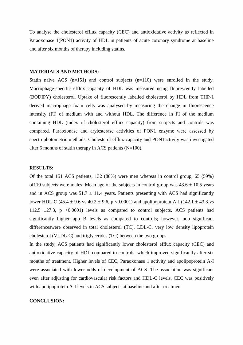
To analyse the cholesterol efflux capacity (CEC) and antioxidative activity as reflected in
Paraoxonase 1(PON1) activity of HDL in patients of acute coronary syndrome at baseline
and after six months of therapy including statins.
MATERIALS AND METHODS:
Statin naïve ACS (n=151) and control subjects (n=110) were enrolled in the study.
Macrophage-specific efflux capacity of HDL was measured using fluorescently labelled
(BODIPY) cholesterol. Uptake of fluorescently labelled cholesterol by HDL from THP-1
derived macrophage foam cells was analysed by measuring the change in fluorescence
intensity (FI) of medium with and without HDL. The difference in FI of the medium
containing HDL (index of cholesterol efflux capacity) from subjects and controls was
compared. Paraoxonase and arylesterase activities of PON1 enzyme were assessed by
spectrophotometric methods. Cholesterol efflux capacity and PON1activity was investigated
after 6 months of statin therapy in ACS patients (N=100).
RESULTS:
Of the total 151 ACS patients, 132 (88%) were men whereas in control group, 65 (59%)
of110 subjects were males. Mean age of the subjects in control group was 43.6 ± 10.5 years
and in ACS group was 51.7 ± 11.4 years. Patients presenting with ACS had significantly
lower HDL-C (45.4 ± 9.6 vs 40.2 ± 9.6, p <0.0001) and apolipoprotein A-I (142.1 ± 43.3 vs
112.5 ±27.3, p <0.0001) levels as compared to control subjects. ACS patients had
significantly higher apo B levels as compared to controls; however, noo significant
differenceswere observed in total cholesterol (TC), LDL-C, very low density lipoprotein
cholesterol (VLDL-C) and triglycerides (TG) between the two groups.
In the study, ACS patients had significantly lower cholesterol efflux capacity (CEC) and
antioxidative capacity of HDL compared to controls, which improved significantly after six
months of treatment. Higher levels of CEC, Paraoxonase 1 activity and apolipoprotein A-I
were associated with lower odds of development of ACS. The association was significant
even after adjusting for cardiovascular risk factors and HDL-C levels. CEC was positively
with apolipoprotein A-I levels in ACS subjects at baseline and after treatment
CONCLUSION:

The significant association of lower Cholesterol efflux capacity and antioxidative activity of
HDL with increased risk of development of ACS and improvement in HDL functionality
after pharmacological intervention indicates that HDL functionality would be a better
measure of cardiovascular risk than measuring static mass of HDL-cholesterol.

Phd-89
Unraveling the role of Gamma Delta T cell subsets: Key players in the
immunopathogenesis of Pemphigus Vulgaris
DasDayasagar,KV Santosh, AravaSudheer, KhandpurSujay, Sharma Alpana
Presenting author:
Dayasagar Das- PhD Student
Email ID: [email protected]
Corresponding author:
Prof. Alpana Sharma
Email ID: [email protected]
Introduction:
Autoimmune diseases are one of the challenging puzzles in immunology due to its
multiple etiologies, complex mechanisms and associated severity. Breach in tolerance
and anomalous T cell function are important phenomenon associated with autoimmune
diseases. Pemphigus Vulgaris (PV) is a severe form of autoimmune blistering disease
involving skin and mucosa. Pathophysiology of PV is due to the formation of auto-
antibodies against desmoglein3 (Dsg3) and anomalous T cell function.Gamma Delta T
cells (γδ T cells) are unique multifaceted T cells that maintain the immune surveillance at
the epithelial surface.They have unique property to recognize the antigens in MHC
independent manner and activating the adaptive arm of immune system. These cells have
found to be involved in several autoimmune diseases. However, their role and plasticity
are not yet characterized in the pathogenesis of PV.
Aims & Objectives: To explore the role of γδ T cell subsets and their plasticity in the
immunopathogenesis of PV.
Materials and methods: 30 active PV patients confirmed by clinically (H&E, DIF
staining) and 30 controls were included.Frequency of γδ-T cell subsets were assessed by
Flowcytometry. Patient and control derived γδ-T cell were isolated by MACS and
primary culture was done. Circulatory and culture levels of γδ-T cell associated markers
were estimated by ELISA. Polarization potential and the functional markersrelative m-
RNA was done by cytotoxicity assay &real time PCR respectively. Tissue localization of
γδ-T cell subsets was confirmed by Immunohistochemistry.
Results:Primary culture of γδ T cells isolated from the PV patients showed diverse
phenotypes (γδ1, γδ2, γδ17, γδreg) and functionality, such as migration, multiple cytokine
productions. We have observed the significantly higher frequency of pan γδ T cells (6.7%
vs. 4.2%) in PV patients. We also found an increased frequency of IL-17(16% vs.4.6%)
and IFN-γ (36% vs.21%) producing γδ T cells in the circulation of patients. Cytotoxic
activity of γδ T cells from PV patients (26±11%) was observed to be higher as compared
to control (12±7%) and the Th1 polarization (IFN-γ) suggesting the potential role of these

cells in the pathogenesis of PV. The molecular expression (mRNA) of γδ T cell subsets
related markers (IFN-γ, IL-1, IL-17, RORγt, CD27, and CD70) were significantly
elevated in patients. Further, tissue localization of γδ T cells and its associated receptor-
ligands were found to be overexpressed in the skin of PV patients.
Conclusion: Exploring γδ T cell subsets in PV has revealed functional facets of T cells
and their crucial involvement. Further exploitation of these potential markers associated
with γδ T cell can be utilized to develop newer diagnostic tools and novel therapeutics for
controlling autoimmune skin diseases.

Phd-98
Title: Elevated expression of Dipeptidyl Peptidase III correlates with the Tumor Infiltrating
Lymphocyte(TILs) composition in multiple cancers.
Authors: Arora Mohit1,Gupta Jasmeen
1, Prajapati Chand Subhash
2,Chauhan Singh Shyam
1*.
1Department of Biochemistry, All India Institute of Medical Sciences (AIIMS), New Delhi.
2Department of Biochemistry and Molecular Genetics, University of Virginia, USA.
Presenting Author:
Name: Mohit Arora
Email: [email protected]
Corresponding Author:
Name: Dr.Shyam Singh Chauhan
Email: [email protected]
Abstract
Introduction: Immune escape is a hallmark of cancer. Recent evidence suggests that Tumor
Infiltrating Immune Cells (TIICs) composition might play critical roles in the pathogenesis
and treatment of several cancers. Critical determinants of tumor immunogenicity include
expression levels of antigenic peptides in tumor cells, engagement of immune cells into the
tumor tissue, presentation of antigenic peptides to immune cells and expression of immune
checkpoint proteins. Concerning this, our lab previously suggested a potential role of
Dipeptidyl Peptidase III (DPP3), a ubiquitously expressed N-terminal exopeptidase, in
degrading antigenic peptides and thereby suppressing immunity against tumor antigens. Later
on, other research groups reported that DPP3along with another peptidase THOP1 potentially
cleave antigenic peptides and mediates inhibition of T cell cross-priming, thereby supporting
our hypothesis. While elevated DPP3 expression in the tumor as well in serum has been
reported in some cancers, its clinical significancehas not been evaluated. Also, the relation
between DPP3 expression and immune cell composition in cancer has not been studied.
Aim and Objectives: To study the expression of DPP3 in different cancers and its relation
with tumorinfiltrating immune cell composition.
Objective 1: To measure DPP3 mRNA level in different cancer types.
Objective 2: To assess the potential of DPP3 in degrading antigenic peptidesin vitro.
Objective 3: To correlate DPP3 expression and tumor immune cell infiltration.
Materials and Methods:

To assess the relative mRNA levels between normal tissues and tumor tissues, we performed
gene expression analysis using RNAsequencing datasets of patients of several cancer types
from The Cancer genomic Atlas (TCGA) using standard workflow of data analysis tool,Gene
expression Profiling Interactive Analysis(GEPIA). We further assessed the potential of DPP-
3 in degrading antigenic OVA and NP peptides in vitro. Toexplore the clinical significance of
DPP-3 expression in modulating antitumor immunity, we used Tumor IMmune Estimation
Resource(TIMER), which is a tool based on deconvolution algorithm using RNA-sequencing
data to extract information about the relative abundance of different immune cells within the
bulk tumor samples of TCGA. Correlation between DPP3 expression and panel of immune
cell signature genes in some cancer types were also quantified using TIMER.
Results:DPP3 was found to be differentially expressed in multiple cancers. Its high
expression in colon and stomach cancer correlated with favorable overall survival.DPP-3 was
found to degrade antigenic peptides, which was inhibited after addition of the aminopeptidase
inhibitor. Further, DPP3 expression exhibit a strong negative correlation with the number of
TIICs including B cells, CD8+ T cells, CD4+ T cells, macrophages, neutrophils, and
dendritic cells in multiple cancers.
Conclusion:
Current data supports DPP-3 as a potential biomarker for immune infiltration in cancer and as
a target for enhancing antitumor immunity.

Phd-103
Acute Febrile Illness associated malaria co-infections in tropical
diseases: A spectrum and incidence in Indian population
Atreyi Pramanik1*, Charandeep Kaur1*, Rajendra Mandage1*, Vinod Kumar2, Shounak Saha3, Adarsh
Singh3, Manish Soneja2, Pragyan Acharya1#,
*These authors have contributed equally
# Corresponding Author: [email protected] (Lab 3002, Department of Biochemistry,
Teaching Block, AIIMS, Ansari Nagar, New Delhi – 110029)
Author Affiliations: 1 Department of Biochemistry, All India Institute of Medical Sciences, New Delhi, India
2 Department of Medicine, All India Institute of Medical Sciences, New Delhi, India
3Department of Biotechnology, Indian Institute of Technology, Kharagpur, India
Background Acute Febrile Illnesses (AFI) is one of the causes of morbidity and mortality in Indian patients and
might be allied with other co-infections in tropical diseases. However, there is little information
available on co-infections incidence. Therefore, in present study, we aim to investigate spectrum and
incidence of AFI associated mono and co-infections in tropical diseases such as malaria, dengue,
scrub typhus, leptospirosis and chikungunya in Indian populations.
Method Patients having fever for more than a week were recruited as AFI cases in our study. A detailed PCR
characterization was performed on 99 AFI samples for screening of its causative agents, such as
Plasmodium falciparum (Pf), Plasmodium vivax (Pv), Plasmodium malariae (Pm), Plasmodium ovale
(Po), and Plasmodium knowlesi(Pk) along with common co-endemic and co-seasonal pathogens like
Dengue virus (DENV), Chikunguniya virus (CHIKV) Orientia tsutsugamushi (causative agent of
Scrub typhus) and Leptospira (causative agent of leptospirosis).
Results Dengue-malaria co-infection was found to be the major group in our cohort (44%, 29 out of 66) as
compared to malaria and its intra-species infections (33%, 22 out of 66). Further analysis of Dengue-
malaria co-infection revealed that Dengue subtype 4 (DENV4) has a high alliance with mild malaria
(MM) (17 out of 33, 51%), whereas P. vivax severe malaria was strongly associated with P. knowlesi,
Leptospira and Dengue virus co-infections.
Conclusions Based on the above results, it is recommended that a complete screening of multiple pathogens panel
should be conducted on AFI patients for proper and accurate diagnosis. In particular, Plasmodium
species especially P. knowlesi, Dengue virus, O. tsutsugamushi and Leptospira should be performed
along with a primary diagnosis such as malaria. Such retrospective studies are expected to aid in
understanding the range of co-infections in Indian population where malaria and dengue are co-
endemic.
Keywords: AFI, Malaria, Dengue, Co-infections, Mixed-infection

Phd-104
Title: FAT1, a novel regulator of YAP1- effector molecule of Salvador-
Warts-Hippo (SWH) pathway, in human Glioblastoma
Gupta Yakhlesh1, Shivajirao Sachin Shelke
1, Irshad Khushboo
1, Dikshit Bhawana
1,
Srivastav Tapasya2, Chattopadhyay Parthaprasad
1, Sinha Subrata
1 & Chosdol Kunzang
1
1- Department of Biochemistry, All India Institute of Medical Sciences, New Delhi-110029, India.
2- Department of Genetics, University of Delhi South Campus, New Delhi-110021, India.
Presenting Author:
Name: Yakhlesh Gupta
Email: [email protected]
Corresponding Author:
Name: Kunzang Chosdol
Email: [email protected]
Introduction: Glioblastoma(GBM) is lethal brain tumor arising from supporting cells of
brain. We has recognized the oncogenic role of FAT1 gene in GBM, regulating inflammatory
and hypoxic microenvironment of the tumor as well as migratory/invasive properties of
tumor cells. In Drosophila, fat, the ortholog of FAT1, is known to regulate the Salvador-
Warts-Hippo (SWH) pathway, but its role in human is not clear. Here, we have analyzed the
effect of FAT1 on SWH pathway in glioma.
Aims and Objectives:
AIM: Effect of FAT1 as a upstream regulator of SWH pathway in human glioblastoma.
Objectives:
1. To study the effect of FAT1 knockdown on cell morphology & survival of glioma cells
2. To study the effect of FAT1 knockdown on expression of SWH pathway molecules
3. To study the effect of FAT1 knockdown on interaction of YAP1 with TEAD1
4. To study the Effect of FAT1 knockdown on subcellular localization of YAP1 and p-YAP1.
Materials and Methods: Glioma cell lines (U87MG, U373, A172, GOS3 and SW1088)
were transfected with FAT1 specific siRNA/control siRNA and analyzed the expression of
SWH pathway molecules by qPCR/Western blot. Protein-protein interactions were analyzed
by Co-immunoprecipitation (Co-IP) after over-expression of YAP1 (wild-type and mutated)
and TEAD1 with and without FAT1 knockdown. Sub-cellular localization of proteins was
analyzed by Confocal microscopy.
Results: The mRNA expression of FAT1 and SWH pathway molecules (MST1, LATS1,
LATS2, YAP1 and TEAD1) was highest in U87MG cells followed by A172, U373MG and
GOS3. After FAT1 knockdown, the mRNA expression of MST1 and BIRC2 were

significantly decreased with no change in the levels of LATS1, LATS2, YAP1, TEAD1 and
BIRC5. At protein level, increased YAP1 and phospho-YAP1 was observed after FAT1
knockdown with increased total as well as phospho-YAP1 in the cytoplasmic extract as
compared to the nuclear extract. There was significant reduction in the interaction between
YAP1 and TEAD1 in siFAT1 treated cells as compared to siControl treated cells.
Conclusion: On FAT1 knockdown, we found (i) increased YAP1 protein level, could be by
increasing the protein stability as no change was observed at the mRNA level (ii) increased
the phospho-YAP1 level as it relieves the inhibitory effect on YAP1 phosphorylation (iii) it
affects the sub-cellular localization of YAP1 by retaining YAP1 in the cytosol and thereby
(iv) decrease in the YAP1:TEAD1 interaction with decreased expression of their target gene
Birc2.
This finding of the effect of FAT1 on YAP1 in GBM is novel with features pointing towards
the oncogenic role of FAT1 by regulating YAP1 sub-cellular localization and co-
transcriptional activity independent of SWH pathway.

Phd-109
Role of IGF2BP3 in migration and invasion of epithelial cancer
cell lines
Sanjeev Goswami, Gunjan Sharma, Elza Boby, Parthaprasad
Chattopadhyay, Jayanth Kumar Palanichamy
Department of Biochemistry, All India Institute of Medical Sciences, New Delhi, India
INTRODUCTION
RNA Binding Proteins (RBPs) play crucial role in biogenesis, stability and functioning of
RNAs. Insulin like
growth factor binding protein 3(IGF2BP3) is an oncogene correlated with poor prognosis and
metastasis in
cancers but the mechanism of oncogenesis and metastasis is not known. We try to address
these lacunae.
AIM AND OBJECTIVES
To study the role of IGF2BP3 in migration and invasion of epithelial cancer cell lines by
correlating the
expression of IGF2BP3 and its target with the migration and invasion.
MATERIALS AND METHODS
RNA isolation and cDNA synthesis followed by real time PCR for IGF2BP3 ant its target
expression in
epithelial cell lines done. Migration and invasion assays were also done. The experiments
were repeated after
IGF2BP3 knockdown by specific siRNAs.
RESULTS
IGF2BP3 and its targets (CD44, MALAT1) are highly expressed in HeLa and SiHa in
comparison to MCF7.
Knockdown of IGF2BP3 leads to a decrease in the expression level of CD44, MALAT1 and
decrease in the
migratory and invasive potential of HeLa and SiHa.
CONCLUSION
High IGF2BP3 expression correlates with high migration and invasion potential of HeLa and
SiHa compared to
MCF7. Decrease in expression level of targets after IGF2BP3 knockdown implies their
stabilisation by
IGF2BP3. Decrease in migration and invasion of HeLa cells after IGF2BP3 knockdown
implies its role in
stabilizing pro-migratory transcripts.
KEY WORDS
RNA binding protein, IGF2BP3, CD44, MALAT1.

Phd-110
Title: “Feasibility of measuring sodium, potassium and creatinine from urine sample on dried
filter paper”
Author details:
Mohamad Tarik1, Lakshmy Ramakrishnan1, Ritvik Amarchand2, Harshal R. Salve2, Prashant
Mathur3, Pradeep Joshi4, Anand Krishnan2
1. Department of Cardiac Biochemistry, All India Institute of Medical Sciences, New Delhi,
India.
2. Centre for Community Medicine, All India Institute of Medical Sciences, New Delhi,
India.
3. National Centre for Diseases Informatics & Research, Indian Council of Medical
Research, Bangalore, India.
4. World Health Organization, New Delhi India.
Presenting Author
Name: Mohamad Tarik
Email ID: [email protected]
Corresponding Author
Name: Lakshmy Ramakrishnan
Email ID: [email protected]
Abstract:
Introduction: High prevalence of hypertension has been attributed to high dietary salt intake,
which can be monitored by measurement of urinary sodium in 24hours/spot urine in a
laboratory.
Dried urine on filter strips, if found suitable, would provide a convenient alternative that
would
circumvent the need to transport urine samples in a cold chain.
Aim & Objective: The objective of this study was to standardize estimation of sodium,
potassium and creatinine in dried urine strips (DUS) and to validate the measurement by
comparing with levels of analytes in liquid urine.
Materials & Methods: Urine was collected in filter paper strips, dried at room temperature
and,
eluted prior to estimation of sodium, potassium by ISE method and creatinine by jaffe’s
method.
After standardization of measurement in DUS, the method was validated by comparing
values
obtained in 138 urine samples by DUS method with that obtained in liquid urine sample.
Correlation coefficients were computed and bland Altman was plotted to assess agreement
between the two methods. . We also assessed storage stability of these analytes after one year
of
storage of dried urine on filter paper at 4⁰C.
Results: The mean recovery of sodium, potassium and creatinine was >95% from DUS
samples.

The Limit of detection (LOD) by DUS for sodium was 0.95 mmol/L, potassium 0.47mmol/L
and
creatinine 0.59 mg/dl. Intra-assay CV was < 5% and the inter-assay CV was <7.0% for all
three
analytes. Correlation (ICC) between DUS assay and liquid urine was 0.934 (95%CI 0.909-
0.953;
p<0.0001), 0.948 (95%CI: 0.928-0.963; p<0.0001) and 0.953 (95% CI: 0.935-0.967;
p<0.0001)
respectively for sodium, potassium and creatinine. Bland-Altman analysis shows good
agreement
between the dried urine and liquid urine sample. Sodium, potassium and creatinine were
stable in
DUS during one year of storage.
Conclusion: We conclude that the sodium, potassium and creatinine are stable in dried urine
and
are readily transferable to a liquid phase for analysis offering a convenient alternative for
monitoring dietary salt intake.

Phd-125
Laboratory of Chromatin & Cancer Epigenetics
Department of Biochemistry
BESFA: Bioinformatics based Evolutionary, Structural & Functional Analysis
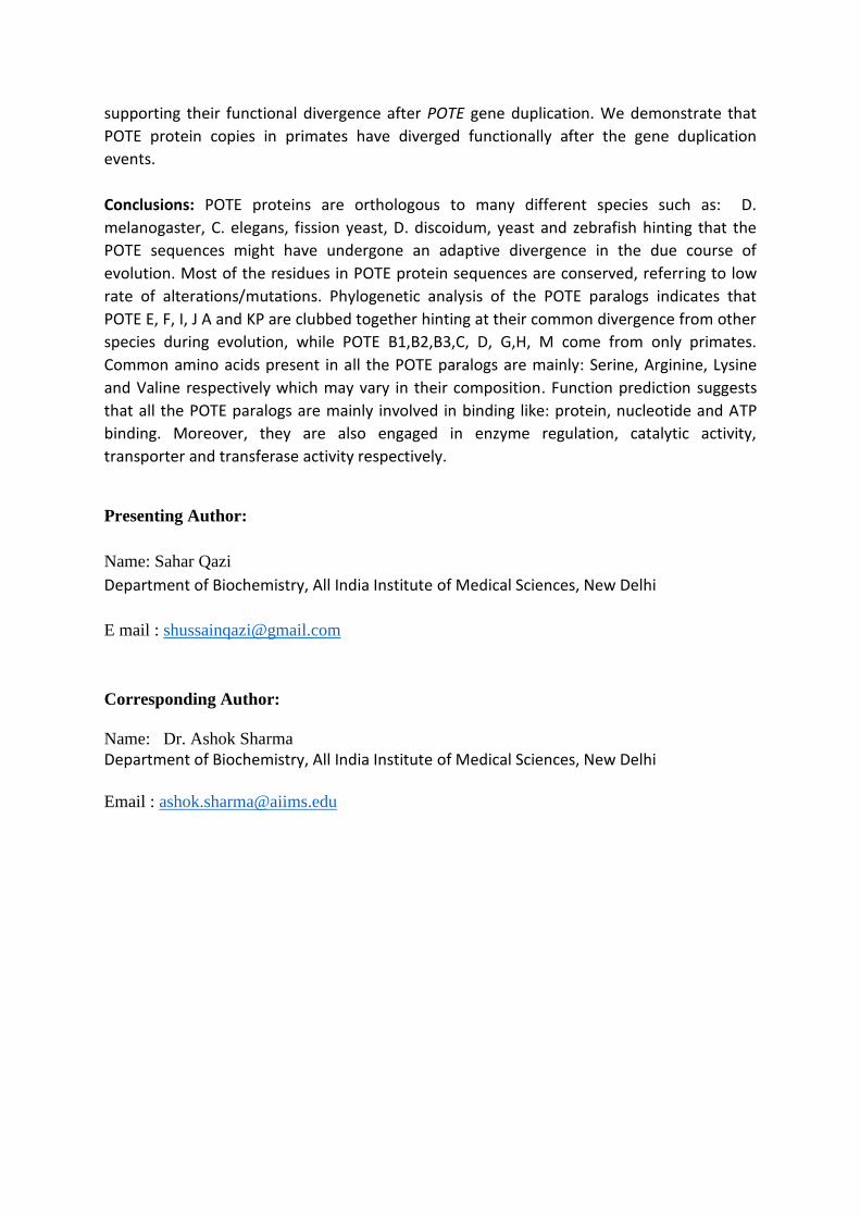
In-silico analysis of POTE Paralogs
Qazi Sahar1, Sharma Neetu, Karpf R Adam2, Sharma Ashok1
1Department of Biochemistry, All India Institute of Medical Sciences, New Delhi, 2FredUniversity of
Nebraska Medical Center, Omaha, NE, USA
Presenting Author: Sahar Qazi
Department of Biochemistry, All India Institute of Medical Sciences, New Delhi
Corresponding Author: Ashok Sharma
Department of Biochemistry, All India Institute of Medical Sciences, New Delhi
Introduction: POTE (Prostate, Placenta, Ovary, Testis, and Embryo) gene family recently
discovered paralogs belong to cancer-testis antigen family. POTE gene family is composed of
14 closely related paralogs dispersed on autosomal pericentromeric region of eight different
chromosomes in primate.
Aims & Objectives: Our basic desideratum has been epitomized below:
1. To deduce the evolutionary spectra of POTE protein paralogs.
2. Development of better and optimized tertiary structures of POTE paralogs using
homology modelling & protein threading approaches.
3. Function prediction of each POTE protein paralog.
Materials & Methodology: POTE paralogs protein sequences were first aligned by using
CLUSTAL W with default settings and then were subjected to phylogenetic analysis in MEGA
5.2 package. For structural predictions, we employed Swiss Model and MODELLER and
Phyre2 and generated models for POTE paralogs. For the best model, a critical evaluation of
the all the models was executed by -Pro Q, RAMPAGE and PROSA. ProFunc server was used
to determine the possible biochemical function of a protein from its tertiary structure.
Results: Evolutionary & structural analysis discern the functional divergence of POTE
paralogs suggesting the fact that POTE paralogs are expressed in primates, but their
orthologues are also present in several other species. The mean distance between all the
POTE proteins is 0.66 which refers to their slow evolutionary divergence. The Tajima’s χ2
test statistic was 10.29 (P = 0.00134). Further, our results point to the different protein-
protein interaction patterns between POTE paralogs and known POTE protein clients
supporting their functional divergence after POTE gene duplication. We demonstrate that
POTE protein copies in primates have diverged functionally after the gene duplication
events.
Conclusions: POTE proteins are orthologous to many different species such as: D.
melanogaster, C. elegans, fission yeast, D. discoidum, yeast and zebrafish hinting that the
POTE sequences might have undergone an adaptive divergence in the due course of
evolution. Most of the residues in POTE protein sequences are conserved, referring to low
rate of alterations/mutations. Phylogenetic analysis of the POTE paralogs indicates that
POTE E, F, I, J A and KP are clubbed together hinting at their common divergence from other
species during evolution, while POTE B1,B2,B3,C, D, G,H, M come from only primates.
Common amino acids present in all the POTE paralogs are mainly: Serine, Arginine, Lysine
and Valine respectively which may vary in their composition. Function prediction suggests
that all the POTE paralogs are mainly involved in binding like: protein, nucleotide and ATP
binding. Moreover, they are also engaged in enzyme regulation, catalytic activity,
transporter and transferase activity respectively.
Presenting Author:
Name: Sahar Qazi
Department of Biochemistry, All India Institute of Medical Sciences, New Delhi
E mail : [email protected]
Corresponding Author:
Name: Dr. Ashok Sharma
Department of Biochemistry, All India Institute of Medical Sciences, New Delhi
Email : [email protected]

Phd-126
Laboratory of Chromatin & Cancer Epigenetics Department of Biochemistry
All India Institute of Medical Sciences (AIIMS), New Delhi, India
Epigenetic Regulation of Pericentromerically localized Cancer-
Testis/Germline Antigen POTE and its use as diagnostic
marker/immunotherapeutic agent in Ovarian Cancer
Manav Nisha 1*
, Kundu Subhadip 1*
, Jha Sangit 1
, Quazi Sahar 1
, Sharma Neetu
1, Kumar Pawan
2, Vanamail P
3, Mathur Sandeep
4, Sharma J.B
3,
Chauhan S.S1, Kumar Lalit
2, Kumar Sunesh
3, Karpf R Adam
5, Sharma
Ashok1*
Department of Biochemistry1, Oncology, DR. BRA-IRCH
2, Department of Obstetrics & Gynaecology
3,
Department of Pathology4, Medical AIIMS, New Delhi, Nebraska Medical Center, Omaha, USA
5
Introduction: As per recent report of National Cancer Registry program of ICMR, ovarian
cancer is third leading cause of cancer related death amongst females in India. Ovarian
cancer (OC) is a “silent killer” and is comprised of a variety of different subtypes i.e. High-
grade serous (HGS- OvCa), endometrioid, mucinous, and clear cell tumors. We are primarily
focused on cancer epigenetics and deciphering the epigenomic mechanisms with an aim to
develop new epigenetic therapeutic drugs. The recent discovery of Cancer Testis/Germline
(CT/CG) antigen expression in cancer suggests a strong link between gametogenesis and
carcinogenesis. The research idea is to investigate HGS-OvCa in pursuit of developing a
new and highly needed biomarker and immunotherapy for OC. We are indulging to open the
new avenues for CT/CG antigens i.e. POTE antigens for cancer immunotherapy against
gynaecological cancers. POTE (Prostate, Placenta, Ovary, and Testis-expressed) is a
recently discovered gene family consisting of 14 autosomal and pericentromerically localized
cancer-testis/germline antigen genes. Epigenetic modulatory agents robustly promote
expression of CG antigens, as well as the class-1 histocompatibility complex (MHCI). Thus,
we are exploring the possible clinical use of epigenetic modulators to augment the
immunotherapeutic potential of POTE family antigens, and examining how this will ultimately
improve strategies for cancer detection and treatment.
Our overall objectives are:
1. Analyzing the POTE as a New Biomarker for Ovarian Cancer: Harnessing its Clinical
Significance for Personalized Therapy
2. Development of Novel Immunotherapy based Strategies Targeting New Cancer-
Germline Antigen POTE-Paralogs in Human Epithelial Ovarian Cancer
3. To study the Epigenetic mechanism of regulation in Pericentromeric localized
Cancer-Testis/Germline Antigen POTE Expression in Ovarian Cancer
Methodology: Affymetrix HG 1.0ST microarrays and RT-qPCR were used to determine
POTE gene expression in normal ovary (NO) and OC tumors. RNA-seq, methyl-Seq and
Pyrosequencing was used for DNA methylation and POTE gene expression analysis
fallopian tube epithelia (FTE) and OC.

Results: Recently, we defined a DNA hypomethylation phenotype in EOC that consists of
hypomethylation of global genomic DNA and a family of genes known as cancer germline (CG)
antigens. POTE gene expression levels were highly elevated in OC tumors showing global
hypomethylation of LINE1 elements, as compared to EOC tumors with hypermethylation of LINE1
elements, linking global DNA methylation status to POTE expression. POTE gene knockdown in OC
cell lines resulted in significantly reduced cell migration and cell invasion. POTE genes are widely
expressed in OC, and are significantly increased in tumors displaying global DNA hypomethylation.
We hereby explore the role of 3D nuclear architecture, lamina associate domain (LADs) interactions
to understand the molecular mechanism of activation of pericentromeric localized genes.
Conclusion: As POTEs are specifically localized on the pericentromeric regions, we will be able to
explain the regulation of the pericentromeric localized genes at a known repressed locus, and their
escape from the repression during cancer. We believe that outcomes of this project will increase our
understanding about molecular clinicopathological mechanism of ovarian cancer that will also help to
develop better cancer-testis/germline POTE based therapeutic regimen for relapsed patient in future.
Presenting Author:
Name: Ms. Nisha Manav
Department of Biochemistry, All India Institute of Medical Sciences, New Delhi
E mail : [email protected]
Corresponding Author:
Name: Dr. Ashok Sharma
Department of Biochemistry, All India Institute of Medical Sciences, New Delhi
Email : [email protected]

Phd-134
Identifying molecules to distinguish between colorectal cancer stem cells and normal
colonic stem cells to develop novel diagnostic and prognostic markers
Bhattacharya Aditi1, Manhas Janvie1, Dey Devanjan1, Bhat Muzaffar2, Das Prasenjit3, Pal Sujoy4 ,
Deo SVS5, Parshad Rajinder6, Ghosh Debabrata2, Sen Sudip1
1Dept. of Biochemistry,; 2Dept. of Physiology, 3Dept. of Pathology,; 4Dept. of GI Surgery, 5Dept. of
Surgical Oncology, 6Dept. Of General Surgery,
Presenting author: Aditi Bhattacharya, [email protected]
Corresponding author: Dr. Sudip Sen, [email protected]
Abstract Body Introduction: Cancer stem cells (CSCs) have been shown to be responsible for tumor
proliferation, metastasis and recurrence. Colorectal cancer, being asymptomatic in the earlier
stages, is already at advanced stage at the time of diagnosis.
Aim: Our study aims to identify molecular factors which are characteristic of colorectal cancer
stem cells and study if they can be developed as markers for a more accurate diagnosis and
prognosis.
Methodology: Stem cells were isolated from operative specimens of primary, untreated,
nonmetastatic, sporadic colorectal adenocarcinoma patients using documented markers antibodies
from tumor (CD44, CD166) and adjacent normal tissue (CD29, Lgr5) using fluorescence
activated cell sorting (FACS). RNA isolated from the sorted subsets were used to determine the
genome wide transcriptomic changes between CSC and normal stem cells (SC) by microarray.
Data was analysed using Flow Jo, Gene Spring GX13 and DAVID. Tumor sphere assay and
colony forming assay were done. Validation of putative gene targets identified by microarray was
done by RT-QPCR.
Results: The variability in CSC population (CD44+CD166+) was higher (range: 0.5-7%) than
normal SC (CD29+Lgr5+) (range:1-2%). The sorted CSC subset generated more tumor spheres
and had higher colony forming ability than normal SC subset. Microarray analysis comparing the
gene expression between CSC and normal SC RNA showed that both Wnt canonical and non-
canonical pathway molecules and intermediates were upregulated. Validation with real-time PCR
also showed that Wnt5a, Wnt2, Wisp1, TCF, LEF, Fzd10 and NfatC3 were upregulated in CSC
as compared to normal SCs.
Conclusion: Cancer stem cells have a greater sphere generating capacity and spheroids thus
formed are more proliferative in nature. The Wnt non-canonical pathway has been shown to play
a role in differentiation. Till date, very little research has implicated this pathway in
tumorigenesis and stemness. Our preliminary results have shown that the Wnt non-canonical pathway may contribute towards cancer stemness in colorectal cancer and studying this pathway
can help in developing better and more specific diagnostic and prognostic markers.

Phd-141
Using human fetal neural stem cells and oligodendrocytes as a disease model to delineate the
pathogenesis of cerebral palsy.
Dey D1*, Shrivastav V1, Bhat MA2, Singal C.M.S.4, Sharma JB3, Palanichamy JK1, Chattopadhyay P1,
Sinha S1, Seth P4, Sen S1
Presenting Author:
Name: Devanjan Dey
E-Mail: [email protected]
Corresponding Author:
Name: Dr. Sudip Sen
E-Mail: [email protected]
Introduction:Cerebral Palsy (CP) is a neurological disorder in children. Oligodendrocytes (OL) are
vulnerable to hypoxic injury, resulting in CP. Fetal neural stem cells (FNSCs) can differentiate into
neuronal and glial lineages and “physiological hypoxic condition” influences their growth and
differentiation.
Aim and Objective:We aim to develop an in vitro model of CP by inducing FNSCs to differentiate into
OL, followed by exposure to hypoxia.
Materials and Methods:Aborted fetuses (n=4) were collected from the Department of Obstetrics
and Gynaecology, AIIMS, New Delhi, after Institute Ethics Committee clearance and informed
consent from mothers undergoing MTP. FNSCs were isolated from the sub-ventricular zone of the
fetal brain and expanded in culture. Immunocytochemistry (ICC) and flow cytometry were used to
evaluate Nestin and Sox2 expression in FNSCs. Annexin-V was used to analyze cell death. FNSCs were
exposed to different oxygen concentrations (20%, 6%, 2% and 0.2%) for 48 hours. Hypoxia exposure
was validated by qPCR for hypoxia markers. Microarray was done using Agilent whole genome 4x44K
array slides to study transcriptomic changes between FNSCs exposed to normoxia and hypoxia. Data
was analysed using Flow Jo, Gene Spring GX13 and GeneGO MetaCore.FNSCs were differentiated
into OL and evaluated for OL specific markers using ICC and qPCR.
Results and Conclusion: Nestin and Sox2 were found to be expressed by FNSCs by ICC and
flowcytometry. CA9, VEGF and PGK1 (hypoxia markers) were observed to be up regulated in FNSCs
exposed to hypoxia. Hypoxia did not result in cell death in FNSCs. As FNSCs differentiated into OL,
they expressed OL specific Myelinating Oligodendrocyte Glycoprotein (MOG) and NG-2 associated
with a decrease in Nestin and Sox-2 expression. Gene expression analysis of FNSCs exposed to
hypoxia revealed novel genes and some interesting pathways to be modulated. Validating
transcriptomic changes associated with hypoxia will help elucidate the pathogenesis of CP.