Postcranial Pneumaticity: An Evaluation of Soft-Tissue ... · dered the identification of...
28
Postcranial Pneumaticity: An Evaluation of Soft-Tissue Influences on the Postcranial Skeleton and the Reconstruction of Pulmonary Anatomy in Archosaurs Patrick M. O’Connor* Department of Biomedical Sciences, Ohio University, College of Osteopathic Medicine, Athens, Ohio 45701 ABSTRACT Postcranial pneumaticity has been reported in numerous extinct sauropsid groups including ptero- saurs, birds, saurischian dinosaurs, and, most recently, both crurotarsan and basal archosauriform taxa. By com- parison with extant birds, pneumatic features in fossils have formed the basis for anatomical inferences concern- ing pulmonary structure and function, in addition to higher-level inferences related to growth, metabolic rate, and thermoregulation. In this study, gross dissection, vas- cular and pulmonary injection, and serial sectioning were employed to assess the manner in which different soft tissues impart their signature on the axial skeleton in a sample of birds, crocodylians, and lizards. Results from this study indicate that only cortical foramina or commu- nicating fossae connected with large internal chambers are reliable and consistent indicators of pneumatic inva- sion of bone. As both vasculature and pneumatic divertic- ula may produce foramina of similar sizes and shapes, cortical features alone do not necessarily indicate pneu- maticity. Noncommunicating (blind) vertebral fossae prove least useful, as these structures are associated with many different soft-tissue systems. This Pneumaticity Profile (PP) was used to evaluate the major clades of extinct archosauriform taxa with purported postcranial pneumaticity. Unambiguous indicators of pneumaticity are present only in certain ornithodiran archosaurs (e.g., sauropod and theropod dinosaurs, pterosaurs). In con- trast, the basal archosauriform Erythrosuchus africanus and other nonornithodiran archosaurs (e.g., parasuch- ians) fail to satisfy morphological criteria of the PP, namely, that internal cavities are absent within bone, even though blind fossae and/or cortical foramina are present on vertebral neural arches. An examination of regional pneumaticity in extant avians reveals remark- ably consistent patterns of diverticular invasion of bone, and thus provides increased resolution for inferring spe- cific components of the pulmonary air sac system in their nonavian theropod ancestors. By comparison with well- preserved exemplars from within Neotheropoda (e.g., Abe- lisauridae, Allosauroidea), the following pattern emerges: pneumaticity of cervical vertebrae and ribs suggests pneu- matization by lateral vertebral diverticula of a cervical air sac system, with sacral pneumaticity indicating the pres- ence of caudally expanding air sacs and/or diverticula. The identification of postcranial pneumaticity in extinct taxa minimally forms the basis for inferring a heterogeneous pulmonary system with distinct exchange and nonex- change (i.e., air sacs) regions. Combined with inferences supporting a rigid, dorsally fixed lung, osteological indica- tors of cervical and abdominal air sacs highlight the fun- damental layout of a flow-through pulmonary apparatus in nonavian theropods. J. Morphol. 267:1199 –1226, 2006. © 2006 Wiley-Liss, Inc. KEY WORDS: pneumaticity; air sacs; postcranial skele- ton; Archosauria; Dinosauria Osteological features preserved in the postcra- nium of fossil archosaurs, particularly saurischian dinosaurs, have been interpreted as pneumatic, with implied causal relationships between this mor- phology and a pulmonary air sac system similar to the one in extant birds (von Meyer, 1837; Owen, 1856; Seeley, 1870; Janensch, 1947; Romer, 1966; Britt, 1993, 1997; Britt et al., 1998; Gower, 2001; O’Connor, 2003; Wedel, 2003b). In contrast, recently discovered theropod dinosaurs with preserved soft tissues (e.g., Chen et al., 1998; Ji et al., 1998) have led to reconstructions of pulmonary anatomy that drastically differ from those proposed by pneumatic- ity advocates. In two different theropods, Sinosau- ropteryx prima and Scipionyx samniticus, Ruben et al. (1997, 1999, 2003) postulated the existence of a hepatic-piston ventilatory system, based on inferred similarities of body cavity organization with the con- dition observed in extant crocodylians. They further- more propose that this system would have prevented the development of an air sac-based ventilatory mode as in extant birds. Notably, these studies fail to address the significance of postcranial pneuma- Contract grant sponsors: National Science Foundation Graduate Research Fellowship; NSF funded Mahajanga Basin Project (EAR- 0116517); Society of Vertebrate Paleontology Estes Memorial Award, Society for Integrative and Comparative Biology; Paleontological So- ciety; the Jurassic Foundation; Stony Brook University Gabor Inke Graduate Research Fellowship; Ohio University College of Osteo- pathic Medicine and Department of Biomedical Sciences. *Correspondence to: Patrick M. O’Connor, Department of Biomed- ical Sciences, Ohio University, 135 Life Sciences Building, Athens, OH 45701. E-mail: [email protected] Published online 18 July 2006 in Wiley InterScience (www.interscience.wiley.com) DOI: 10.1002/jmor.10470 JOURNAL OF MORPHOLOGY 267:1199 –1226 (2006) © 2006 WILEY-LISS, INC.
Transcript of Postcranial Pneumaticity: An Evaluation of Soft-Tissue ... · dered the identification of...

Postcranial Pneumaticity: An Evaluation of Soft-TissueInfluences on the Postcranial Skeleton and theReconstruction of Pulmonary Anatomy in ArchosaursPatrick M. O’Connor*
Department of Biomedical Sciences, Ohio University, College of Osteopathic Medicine, Athens, Ohio 45701
ABSTRACT Postcranial pneumaticity has been reportedin numerous extinct sauropsid groups including ptero-saurs, birds, saurischian dinosaurs, and, most recently,both crurotarsan and basal archosauriform taxa. By com-parison with extant birds, pneumatic features in fossilshave formed the basis for anatomical inferences concern-ing pulmonary structure and function, in addition tohigher-level inferences related to growth, metabolic rate,and thermoregulation. In this study, gross dissection, vas-cular and pulmonary injection, and serial sectioning wereemployed to assess the manner in which different softtissues impart their signature on the axial skeleton in asample of birds, crocodylians, and lizards. Results fromthis study indicate that only cortical foramina or commu-nicating fossae connected with large internal chambersare reliable and consistent indicators of pneumatic inva-sion of bone. As both vasculature and pneumatic divertic-ula may produce foramina of similar sizes and shapes,cortical features alone do not necessarily indicate pneu-maticity. Noncommunicating (blind) vertebral fossaeprove least useful, as these structures are associated withmany different soft-tissue systems. This PneumaticityProfile (PP) was used to evaluate the major clades ofextinct archosauriform taxa with purported postcranialpneumaticity. Unambiguous indicators of pneumaticityare present only in certain ornithodiran archosaurs (e.g.,sauropod and theropod dinosaurs, pterosaurs). In con-trast, the basal archosauriform Erythrosuchus africanusand other nonornithodiran archosaurs (e.g., parasuch-ians) fail to satisfy morphological criteria of the PP,namely, that internal cavities are absent within bone,even though blind fossae and/or cortical foramina arepresent on vertebral neural arches. An examination ofregional pneumaticity in extant avians reveals remark-ably consistent patterns of diverticular invasion of bone,and thus provides increased resolution for inferring spe-cific components of the pulmonary air sac system in theirnonavian theropod ancestors. By comparison with well-preserved exemplars from within Neotheropoda (e.g., Abe-lisauridae, Allosauroidea), the following pattern emerges:pneumaticity of cervical vertebrae and ribs suggests pneu-matization by lateral vertebral diverticula of a cervical airsac system, with sacral pneumaticity indicating the pres-ence of caudally expanding air sacs and/or diverticula. Theidentification of postcranial pneumaticity in extinct taxaminimally forms the basis for inferring a heterogeneouspulmonary system with distinct exchange and nonex-change (i.e., air sacs) regions. Combined with inferencessupporting a rigid, dorsally fixed lung, osteological indica-tors of cervical and abdominal air sacs highlight the fun-damental layout of a flow-through pulmonary apparatus
in nonavian theropods. J. Morphol. 267:1199–1226, 2006.© 2006 Wiley-Liss, Inc.
KEY WORDS: pneumaticity; air sacs; postcranial skele-ton; Archosauria; Dinosauria
Osteological features preserved in the postcra-nium of fossil archosaurs, particularly saurischiandinosaurs, have been interpreted as pneumatic,with implied causal relationships between this mor-phology and a pulmonary air sac system similar tothe one in extant birds (von Meyer, 1837; Owen,1856; Seeley, 1870; Janensch, 1947; Romer, 1966;Britt, 1993, 1997; Britt et al., 1998; Gower, 2001;O’Connor, 2003; Wedel, 2003b). In contrast, recentlydiscovered theropod dinosaurs with preserved softtissues (e.g., Chen et al., 1998; Ji et al., 1998) haveled to reconstructions of pulmonary anatomy thatdrastically differ from those proposed by pneumatic-ity advocates. In two different theropods, Sinosau-ropteryx prima and Scipionyx samniticus, Ruben etal. (1997, 1999, 2003) postulated the existence of ahepatic-piston ventilatory system, based on inferredsimilarities of body cavity organization with the con-dition observed in extant crocodylians. They further-more propose that this system would have preventedthe development of an air sac-based ventilatorymode as in extant birds. Notably, these studies failto address the significance of postcranial pneuma-
Contract grant sponsors: National Science Foundation GraduateResearch Fellowship; NSF funded Mahajanga Basin Project (EAR-0116517); Society of Vertebrate Paleontology Estes Memorial Award,Society for Integrative and Comparative Biology; Paleontological So-ciety; the Jurassic Foundation; Stony Brook University Gabor InkeGraduate Research Fellowship; Ohio University College of Osteo-pathic Medicine and Department of Biomedical Sciences.
*Correspondence to: Patrick M. O’Connor, Department of Biomed-ical Sciences, Ohio University, 135 Life Sciences Building, Athens,OH 45701. E-mail: [email protected]
Published online 18 July 2006 inWiley InterScience (www.interscience.wiley.com)DOI: 10.1002/jmor.10470
JOURNAL OF MORPHOLOGY 267:1199–1226 (2006)
© 2006 WILEY-LISS, INC.

ticity and its bearing on reconstructions of pulmo-nary anatomy.
As an anatomical condition, pneumaticity refersto the air-filled nature of certain structures or com-partments of the body. Pneumatic features typicallyconsist of foramina in cortical bone, often leading tointernal chambers in vertebral neural arches andcentra. Other traits such as osseous fossae and dif-ferential surface texture also have been used asindicators of pneumaticity in fossil groups.
Whereas some employ pneumatic features ascharacters in phylogenetic analyses (e.g., Gauthier,1986; Rowe and Gauthier, 1990; Wilson and Sereno,1999; Carrano et al., 2002), others use them to modelrespiratory structure–function relationships and asthe basis for higher-level physiological inferencesrelated to thermoregulatory ability (Madsen, 1976;Colbert, 1989; Perry, 1992, 2001; Britt, 1993; Perryand Reuter, 1999; Wedel, 2003b; O’Connor and Clae-ssens, 2005). Yet others merely cite the necessity oflarge animals to reduce body mass, as large fossaeand internal cavities within bone (regardless ofwhether or not the author promoted an air sac originfor them) provide a means of maintaining strengthwith minimizing materials (Cope, 1877; Marsh,1877; Osborn, 1899; Romer, 1966; Carrano andO’Connor, 2005).
Generally, pneumatic spaces in bones of extantamniotes are lined by epithelium and communicatewith the external environment via pneumatic fo-ramina along the length of the respiratory tract.Many amniote groups exhibit cranial pneumaticityvia communications with: 1) the nasal cavity—as inparanasal sinuses, and 2) the pharynx—as in tym-panic pneumaticity. These two systems can pneu-matize much of the cranial skeleton in differentgroups (e.g., avian and nonavian dinosaurs; see Wit-mer, 1990, 1995b, 1997).
Postcranial pneumaticity in extant amniotes isrestricted to birds (Fig. 1) and originates from thelung–air sac system (Muller, 1908; King, 1966;Duncker, 1971, 1983; O’Connor, 2004). Whereasmain air sacs reside within the body cavity, finger-like projections (i.e., pneumatic or pulmonary diver-ticula) originate from air sacs and lungs, and extendthroughout both soft and skeletal tissues of the bodywall. A pulmonary-injection preparation of a duck(Fig. 2A) illustrates the extensive nature of the airsac system and pneumatic diverticula in birds. Avariety of morphologies are associated with air sacinvasion of bone, ranging from discreet, simple fo-ramina to complex networks of irregularly shapedopenings in the cortical surface (Fig. 2B–D). Al-though postcranial pneumaticity is generallypresent in vertebrae, ribs, girdles, and proximallimb elements, there is considerable variation in therelative extent of postcranial pneumaticity amongdifferent avian taxa (Crisp, 1857; Campana, 1875;Bignon, 1889; Muller, 1908; Groebbels, 1932; King
and Kelly, 1956; King, 1957, 1966; Hogg, 1984b;McLelland, 1989; O’Connor, 2004).
Crocodylians do not have pulmonary diverticula,but do exhibit areas of reduced parenchymal densityin the cranial, caudal, and ventral regions of thelung (Duncker, 1978, 1979; Perry, 1983, 1989, 1992,2001). These sac-like regions remain entirely withinthe body cavity and do not penetrate the postcranialskeleton. All other extant nonarchosaurian saurop-sid groups (lizards, snakes, turtles) include speciesthat also possess sac-like regions of the lung (Woodand Lenfant, 1976; Duncker, 1978, 1979, 1989;Perry, 1983, 1998), and these also do not pneumatizethe postcranial skeleton. Such specializations insauropsids are the basis for the concept of a hetero-geneous pulmonary system, whereby parenchymaldensity, and thus gas exchange potential, variesthroughout the system (e.g., see Duncker, 1978,1989; Perry, 1983).
Among extinct sauropsids, putative pneumaticfeatures have been identified in a variety of fossil
Fig. 1. Phylogenetic hypothesis of extant amniote relation-ships based on Gauthier (1994)—birds (Aves) are the only extantamniote group possessing pneumaticity of the postcranial skele-ton derived from pulmonary air sacs.
1200 P.M. O’CONNOR
Journal of Morphology DOI 10.1002/jmor

Fig. 2. The pulmonary air sac system (A) and osteological correlates (B–D) of pneumatic invasion of bone in birds. Scale bars � 1cm. A: Green-winged teal (Anas crecca, JLUG 1). Skeletal-latex preparation to demonstrate the extent of the pulmonary-air sacsystem: note intermuscular (IMDv) and lateral vertebral (LVDv) diverticula along the cervical vertebral series. Dashed arrows in insetindicate injected latex (blue) within humerus and thoracic vertebrae. B: Hawaiian goose (Branta sandvicensis, CM 14428). Thoracicvertebral series, left lateral view. C: Southern ground hornbill (Bucorvus leadbeateri, TM 76018). Thoracic vertebra—dorsal view,cranial end facing toward top of image. D: Ostrich (Struthio camelus, FMNH 222324). Thoracic vertebra, left lateral view. ABD,abdominal air sac; CAUDTH, caudal thoracic air sac; CL, clavicular (interclavicular) air sac; CPF, central pneumatic foramen; CRTH,cranial thoracic air sac; HU, humerus; IMDv, intermuscular diverticulum; LVDv, lateral vertebral diverticulum; NAPF, neural archpneumatic foramen/fossa; NS, neural spine; OT, ossified tendons; POZ, postzygapophysis; PP, parapophysis; PRZ, prezygapophysis;TR, trachea; TVP, transverse process.
1201POSTCRANIAL PNEUMATICITY IN ARCHOSAURS
Journal of Morphology DOI 10.1002/jmor

archosaurs including pterosaurs, theropod and sau-ropod dinosaurs, and basal birds (e.g., Owen, 1856;Seeley, 1870, 1901; Janensch, 1947; Romer, 1966;Bennett, 1991; Britt, 1993; Britt et al., 1998; Forsteret al., 1998; O’Connor, 2003; Wedel, 2003a). Osteo-logical features described as pneumatic in fossilspecimens include cortical foramina as well as var-ious forms of fossae associated with both vertebralcentra and neural arches (Fig. 3). Whereas postcra-nial pneumaticity is generally restricted to the axialskeleton in nonavian dinosaurs (Romer, 1966; Britt,1993; Wilson, 1999; O’Connor, 2003; Wedel, 2003a),it has been identified in both axial and appendicularelements of pterosaurs (Seeley, 1901; Bennett, 1991;O’Connor, 2002). Pneumatic features have evenbeen identified in embryonic vertebrae of troodontid(Varricchio et al., 2002) and oviraptorid (Norell etal., 2001) theropods. Finally, both axial and appen-dicular pneumaticity have been described in thepostcranial skeleton of basal avians (e.g., Archae-opteryx, Rahonavis; Britt et al., 1998; Forster et al.,1998; Christiansen and Bonde, 2000).
Taxonomic Distribution of PostcranialPneumaticity: Archosauriformes
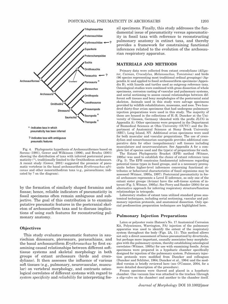
Previous interpretations of postcranial pneuma-ticity in fossil archosaurs suggest a variable distri-bution among ornithodiran taxa including exampleswithin both Pterosauria and Saurischia (Fig. 4). Al-though crurotarsan, ornithischian dinosaur, andbasal archosauriform groups are generally regardedas lacking pneumatic postcrania (Romer, 1956;Britt, 1993; Gauthier, 1986), Gower (2001) describedpneumatic vertebrae in the basal archosauriformErythrosuchus africanus and certain crurotarsantaxa (e.g., parasuchians), thereby significantly ex-panding the distribution of postcranial pneumaticityamong Archosauriformes (Fig. 4). As interpreted,postcranial pneumaticity may represent a primitivecondition for the archosauriform clade and its ab-sence in extant crocodylians, and presumably anyother taxa lacking pneumaticity (e.g., ornithischiandinosaurs, etc.), is a derived trait (see Gower, 2001).
Although postcranial pneumaticity appears tohave a much broader distribution among fossilgroups than living forms, pneumaticity research inextinct groups often lacks direct comparisons withsoft-tissue data derived from extant taxa. Most stud-ies rely on comparisons between the fossils of inter-est and avian skeletons (e.g., Britt, 1993; Chris-tiansen and Bonde, 2000). Moreover, even thecritical assessment of pneumaticity in living birdsoften lacks direct corroboration through the study ofthe variety of soft tissues that influence vertebralmorphology.
Subjective interpretations of morphology com-bined with imprecise terminology (e.g., excavation,fossa, foramen, pneumatopore, pleurocoel) have hin-dered the identification of unambiguous pneumatic
structures. Until rigorous criteria are establishedfor recognizing pneumatic features, reconstructionsof pulmonary structure and higher-level inferencesrelated to function (e.g., ventilatory and/or thermo-regulatory potential) are unreliable at best. Recentanatomical studies of extant sauropsids (e.g.,O’Connor, 2003) indicate that a number of othersoft-tissue systems influence vertebral morphology
Fig. 3. Inferred pneumatic features in fossil archosaur speci-mens. Scale bars � 1 cm in A,B; 5 cm in C,D. A: Theropoddinosaur (Majungasaurus crenatissimus, UA 8678). Caudal cer-vical vertebra, left lateral view. B: Ornithocheirid pterosaur (Or-nithocheirus sp., SM B54.302). Fused atlas-axis complex, leftlateral view. C: Sauropod dinosaur (Rapetosaurus krausei,FMNH PR 2209). Caudal cervical vertebra, left lateral view; notelarge fossae on both neural arch (1) and centrum (2). D: Sauropoddinosaur (Rapetosaurus krausei, FMNH PR 2209). Cranial tho-racic (dorsal) vertebra, left lateral view. CPF, central pneumaticforamen; NAPF, neural arch pneumatic foramen/fossa; NS, neu-ral spine; PLFS, pulmonary fossa; POZ, postzygapophysis; PP,parapophysis; PRZ, prezygapophysis; TVP, transverse process.
1202 P.M. O’CONNOR
Journal of Morphology DOI 10.1002/jmor
by the formation of similarly shaped foramina andfossae; hence, reliable indicators of pneumaticity infossil specimens often remain ambiguous and sub-jective. The goal of this contribution is to examineputative pneumatic features in the postcranial skel-eton of archosauriform taxa and to discuss implica-tions of using such features for reconstructing pul-monary anatomy.
Objectives
This study evaluates pneumatic features in sau-rischian dinosaurs, pterosaurs, parasuchians, andthe basal archosauriform Erythrosuchus by first ex-amining causal relationships between different soft-tissue systems and skeletal morphology in bothgroups of extant archosaurs (birds and croco-dylians). It then assesses the influence of varioussoft tissues (e.g., pulmonary, neurovascular, muscu-lar) on vertebral morphology, and contrasts osteo-logical correlates of different systems with regard tosystem specificity and reliability for interpreting fos-
sil specimens. Finally, this study addresses the fun-damental issue of pneumaticity versus apneumatic-ity in fossil taxa with reference to reconstructingpulmonary anatomy in extinct taxa, and therebyprovides a framework for constraining functionalinferences related to the evolution of the archosau-rian respiratory apparatus.
MATERIALS AND METHODS
Primary data were collected from extant crocodylians (Alliga-tor, Caiman, Crocodylus, Melanosuchus, Tomistoma) and birds(96 species representing most traditional ordinal groupings) (Ap-pendix A) and applied to fossil archosauriform specimens (Appen-dix B), with lizards and turtles used as outgroup reference taxa.Osteological studies were combined with gross dissection of wholespecimens, corrosion casting of vascular and pulmonary systems,and serial sectioning to assess causal relationships between dif-ferent soft tissues and bony morphologies of the postcranial axialskeleton. Animals used in this study were salvage specimensprovided by wildlife rehabilitators, museums, and zoos. Two hun-dred thirty-four avian specimens that had undergone pulmonaryinjection preparations were used in this study. The majority ofthese are housed in the collections of H.-R. Duncker at the Uni-versity of Giessen, Germany (denoted with the prefix JLUG inAppendix A). Other specimens were prepared in the Departmentof Biomedical Sciences at Ohio University (OUVC) and the De-partment of Anatomical Sciences at Stony Brook University(SBU), Long Island, NY. Additional avian specimens were usedfor both muscular and vascular preparations. The use of croco-dylian and nonarchosaurian sauropsids provides additional com-parative data for other (nonpulmonary) soft tissues includingmusculature and neurovasculature. See Appendix A for a com-plete list of species used and the type(s) of preparations for each.
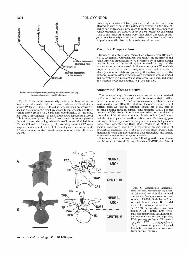
The Extant Phylogenetic Bracket (EPB) method (Witmer,1995a) was used to establish the choice of extant reference taxa(Fig. 5). The EPB constrains fundamental inferences regardingpotential tissue types in fossil groups, and is a necessary prereq-uisite before higher-level inferences concerning functional at-tributes or behavioral characteristics of fossil organisms may beassessed (Witmer, 1995a, 1997). Postcranial pneumaticity in fos-sil archosaurs represents a Level II inference, as only one of theextant sister groups (Avians) have the soft-tissue system of in-terest (Fig. 5; Witmer, 1995a). See Perry and Sander (2004) for analternative approach for inferring respiratory structure/functionrelationships in tetrapods.
Laboratory studies of extant taxa utilized standard gross ana-tomical techniques, including serial sectioning, vascular and pul-monary injection protocols, and anatomical dissection. Only spe-cial predissection preparation techniques are highlighted here.
Pulmonary Injection Preparations
Latex or polyester resin (Batson’s No. 17 Anatomical CorrosionKit, Polysciences, Warrington, PA) injection of the pulmonaryapparatus was used to identify the extent of the respiratorysystem throughout the body (Figs. 2A, 11). This method allowsnot only a direct assessment of bones pneumatized by diverticula,but perhaps more important, causally associates bony morpholo-gies with the pulmonary system, thereby establishing osteologicalcorrelates (Witmer, 1995a) for use with examining fossils. Avianspecimens were prepared in a hypobaric chamber specificallydesigned for injection of the pulmonary system. Pulmonary injec-tion protocols were modified from Duncker and colleagues(Duncker and Schluter, 1964; Duncker et al., 1964) and the mod-ified version is briefly reviewed here (see O’Connor, 2004, for amore detailed description of the procedure).
Frozen specimens were thawed and placed in a hypobaricchamber. One vacuum line was attached to the trachea (througha slip-valve on the chamber) and another to the chamber itself.
Fig. 4. Phylogenetic hypothesis of Archosauriformes based onSereno (1991), Gower and Wilkinson (1996), and Brochu (2001)showing the distribution of taxa with inferred postcranial pneu-maticity (*), traditionally limited to the Ornithodiran archosaurs.A recent study (Gower, 2001) suggested the presence of pneu-matic vertebrae in the basal archosauriform Erythrosuchus afri-canus and other nonornithodiran taxa (e.g., parasuchians; indi-cated by ? on the diagram).
1203POSTCRANIAL PNEUMATICITY IN ARCHOSAURS
Journal of Morphology DOI 10.1002/jmor
Following evacuation of both specimen and chamber, latex wasallowed to slowly enter the pulmonary system via the line at-tached to the trachea. Subsequent to infilling, the specimen wasrefrigerated in a 15% solution of acetic acid to decrease the curingtime of the latex. Specimens were then either dissected or sub-jected to whole-body maceration in order to examine the relation-ship of pneumatic diverticula to osteological features.
Vascular Preparations
Standard laboratory latex, Microfil, or polyester resin (Batson’sNo. 17 Anatomical Corrosion Kit) was used to inject arteries andveins. Arterial preparations were performed by injecting castingmedium into either the carotid system or caudal artery, and thevenous network was accessed via the jugular system. Whole-bodypreparations of birds and crocodylians were used in order toidentify vascular relationships along the entire length of thevertebral column. After injection, latex specimens were dissectedand polyester resin preparations were chemically corroded using15% sodium hydroxide solution (e.g., see Fig. 9F).
Anatomical Nomenclature
The basic anatomy of an archosaurian vertebra is summarizedin Figure 6. Soft tissues are divided into those related to eitherfossae or foramina. A “fossa” is any concavity positioned in ananatomical surface (Donath, 1969) and lacking a distinct rim ofcortical bone. An “osseous foramen” sensu lato is any hole oropening passing through cortical bone (Donath, 1969). For thepurposes of this study, foramina sensu stricto are restricted tothose identifiable at gross anatomical level (�0.5 mm) and do notinclude microscopic canals within cortical bone. Terminology per-taining to different types of internal pneumatic morphology (cam-erate, camellate, etc.; see Britt, 1993; Wedel et al., 2000), al-though potentially useful to differentiate among clades ofsaurischian dinosaurs, will not be used in this study. Table 1 listsanatomical terms and abbreviations used throughout the article,with novel items indicated by an asterisk.
Specimens were examined at the following institutions: Amer-ican Museum of Natural History, New York (AMNH); the Natural
Fig. 5. Postcranial pneumaticity in fossil archosaurs exam-ined within the context of the Extant Phylogenetic Bracket ap-proach (Witmer, 1995a). In this diagram, theropod dinosaurs areused as an example of a fossil archosaur taxon bracketed by theirextant sister groups (i.e., birds and crocodylians). In this case,postcranial pneumaticity in fossil archosaurs represents a LevelII inference, as only one (birds) of the extant sister groups possessthe soft tissue and osteological correlate of interest. Modified fromWitmer (1995a). OST, osteological correlate present; OST?, oste-ological correlate unknown; OST, osteological correlate absent;ST, soft tissue present; ST?, soft tissue unknown; ST, soft tissueabsent.
Fig. 6. Generalized archosau-rian vertebra represented by a dor-sal (thoracic) vertebra of a theropoddinosaur (Majungasaurus crenatis-simus, UA 8678). Scale bar � 5 cm.A: Left lateral view. B: Caudalview. CFS, nonspecific central fos-sa; NAFS, nonspecific neural archfossa; NAPF, neural arch pneu-matic foramen/fossa; NC, neural ca-nal; NS, neural spine; PED, pedicle;POZ, postzygapophysis; PP, parap-ophysis; PRZ, prezygapophysis;TVP, transverse process. Dashedline indicates division between cen-trum and neural arch.
1204 P.M. O’CONNOR
Journal of Morphology DOI 10.1002/jmor

History Museum, London, UK (BMNH); Carnegie Museum ofNatural History, Pittsburgh (CM); Canadian National Museum,Ottawa, Canada (CNM); Field Museum of Natural History, Chi-cago (FMNH); Museum fur Naturkunde der Humboldt-Universitat, Berlin, Germany (MB); Justus-Liebig Universitat,Giessen, Germany (JLUG); Namibian Geological Survey, Nami-bia (NGS); National Museum of Natural History, WashingtonD.C. (USNM); Ohio University Vertebrate Collections, Athens(OUVC); Transvaal Museum, Pretoria, South Africa (TM); StonyBrook University, New York (SBU); Sedgewick Museum, Univer-sity of Cambridge, UK (SM); Universite d’Antananarivo, Mada-gascar (UA); and the University of California Museum of Paleon-tology, Berkeley (UCMP).
RESULTSOsteological Correlates of Different Soft-Tissue Systems
Vertebral fossae. Fossae in archosaurian verte-brae are typically positioned on the lateral or ven-trolateral aspect of the vertebral centrum and vir-tually any surface of the neural arch (Figs. 3, 6).Extant crocodylians have vertebral centra with fos-sae of variable shape and size, similar in generalmorphology to those identified as pneumatic inmany fossil archosaurs. Such fossae are associatedwith a variety of soft tissues, depending on theirposition on the vertebra itself and location withinthe vertebral series. For example, dorsal vertebralcentra of extant crocodylians exhibit fossae of vari-able depth on the lateral central surface (Fig. 7A,CFS). Within the depth of these fossae, a foramen(or foramina) often penetrates the cortical bone, al-lowing vasculature to access the medullary cavity(see next section on vasculature). Gross dissection ofAlligator mississippiensis reveals a layer of fat (peri-vertebral adipose) contained within these fossae(Fig. 7D, FD).
Crocodylian proximal caudal vertebrae also ex-hibit concavities on the lateral surface of the cen-trum (Fig. 7B, CFS). In some cases the fossae are sodeep that only thin, bony laminae separate the rightand left compartments. Dissection reveals caud-ofemoralis musculature and fat deposits associatedwith these features (Fig. 7E,F, CFM, FD). Such ex-cavations increase surface area for attachment ofmusculature used in femoral retraction during ter-restrial locomotion. Similar lateral excavations arefound within caudal vertebral centra of many fossilarchosaurs, notably among the large theropod andsauropod dinosaurs (e.g., Wilson, 1999, fig. 4). Ver-tebrae within the cervical region have variablyshaped concavities. As in the tail, these excavationsare associated with the attachment areas of deepaxial musculature and fat.
Crocodylian vertebrae also exhibit a wide varietyof fossae on neural arches, particularly on thecraniolateral and caudolateral aspects of the neuralspine (Fig. 7B, SPFS). The cortical bone within thesefeatures is relatively smooth when compared to ad-jacent areas of the neural arch. In some fossil archo-saurs (e.g., saurischian dinosaurs) similar textural
TABLE 1. Anatomical nomenclature related to osseouspneumaticity and pulmonary soft-tissue morphology
OSSEOUS STRUCTURESGeneral descriptors traditionally used to describe osseous
pneumatic features include the following terms: chamber,excavation, foramen, fossa, groove, hole, hollow, pit,pleurocoel, pneumatopore, recess, and sulcus. Except forpleurocoel and pneumatopore, the above terms are typicallymodified with the adjective pneumatic.
*Noncommunicating (Blind) Fossae: any of the variably shapedexternal osseous concavities on bony surfaces that do notconnect with the intraosseous space.
*Communicating Fossa: any of the variably shaped externalosseous concavities that directly connect with large,cavernous spaces within pneumatic bone via foramina.
*Pulmonary Fossa: laterally facing concavities on thoracicvertebrae casually associated with dorsomedial projections oflung (Fig. 8A).
Pneumatic bone is lined by air-cells (Hunter, 1774) orpneumatic cells (Cellulae pneumaticae sensu Baumel andWitmer, 1993). These are made up of epithelial expansionsoriginating from the respiratory tract (e.g., air sacs,diverticula) that expand within bone. Also, see Britt (1993)and Witmer (1997) for a history of bony pneumatic features.
SOFT-TISSUE STRUCTURESPneumatic diverticulum (pl. diverticula): any epithelial
outpocketing that originates directly from pulmonary air sacsor other diverticula, but not the lung itself (Figs. 2A, 11). Forboth historical and current nomenclature related topulmonary morphology, see Hunter (1774), Campana (1875),Muller (1908), Locy and Larsell (1916a,b), Groebbels (1932),King (1966, 1993), McLelland (1989), and O’Connor (2004).
Note: craniocervical pneumatic diverticula originate from theupper respiratory tract (e.g., nasal, pharyngeal, laryngeal,and tympanic cavities), often pneumatizing elements of thecranial skeleton (see Witmer, 1997).
Specific named diverticula used include:● AnDv—anastomosing diverticulum: short, segmentalconnections between two or more longitudinal diverticularnetworks (e.g., between the LVDv and SMDv).● IMDv—intermuscular diverticulum: short, dorsally-directedoutpocketing originating from supramedullary orsupravertebral diverticula and expanding within epaxialmusculature (note: intermuscular diverticula are alsoassociated with other components of the axial andappendicular musculature (e.g., pectoral diverticulum).● LVDv—lateral vertebral diverticulum: longitudinal systempassing cranially through the vertebrarterial canal,occupying a position on the lateral surface of cervicalvertebral centra. The LVDv originates from the cervical airsac at the cervicothoracic junction.●SMDv—supramedullary diverticulum: longitudinal systemvariably occupying the extradural space within the vertebralcanal. The SMDv is made up of contributions from (1) thecervical air sac, (2) pulmonary diverticula of the lung, and (3)perirenal diverticula of the abdominal air sac.● SVDv—supravertebral diverticulum: short, segmentalexpansions from the SMDv that occupy a position on thedorsal surface of vertebral neural arches
*Pulmonary diverticulum: an epithelial outpocketingoriginating directly from the lung surface, extending mediallyto (1) directly pneumatize thoracic vertebral centra and (2)contribute to the formation of the supramedullarydiverticulum.
*Pulmonary protuberance: dorsomedially directed portions oflung parenchyma (Fig. 8B) that occupy fossae in thoracicvertebrae of some bird groups (e.g., procellarids, larids).
Novel terms introduced in this study indicated with an asterisk.
1205POSTCRANIAL PNEUMATICITY IN ARCHOSAURS
Journal of Morphology DOI 10.1002/jmor

differences are also observed on neural arches, par-ticularly adjacent to large foramina in the corticalsurface (Fig. 3C). Fossae are also commonly locatedadjacent to pre- and postzygapophyses and near thebase of the transverse process (Fig. 7A–C, NAFS).Similar to those on vertebral centra, neural archfossae are associated with fat deposits (Fig. 7F, FD).In lepidosaurs, similar fat deposits are also found inassociation with fossae on vertebral centra and neu-ral arches.
Among birds, expansive lateral fossae are partic-ularly well developed in thoracic vertebrae ofcharadriiform and some procellariform birds. Theseare relatively shallow and occupy only a small pro-portion of the lateral surface of the centrum in gulls.
In petrels, however, fossae (pulmonary fossae) oc-cupy the entire lateral surface of the centrum andare so deep that the centrum consists of two inter-vertebral articular facets connected by a thin, mid-line lamina (Fig. 8A, PLFS). Set within the pulmo-nary fossae (see Table 1) are medially directedparabronchial protuberances extending from thedorsomedial aspect of the lung (Fig. 8B, PLPR). Inthis case, the vertebrae are not “pneumatized,” asthe medullary space is not lined by pneumatic epi-thelium. Rather, the fossae subdivide portions of thedorsomedial lung parenchyma at the expense of thecentrum. This form of vertebral modification is dis-tinguished from that caused by the pneumatizationprocess, whereby pneumatic epithelium invades and
Fig. 7. Crocodylian vertebrae (A–C) illustratingfossae in both centrum and neural arch componentsalong with dissections (D,E) and a computed tomog-raphy (CT) scan (F) to demonstrate soft-tissue struc-tures associated with bony features. A: Americancrocodile (Crocodylus acutus, FMNH 5157). Lumbarvertebra, left lateral view. Note the central vascularforamen (CVFR) located within the fossa. Scalebar � 5 cm. B: American alligator (Alligator missis-sippiensis, AMNH 43314). Proximal caudal verte-bra, left lateral view. Scale bar � 2 cm. C: Crocodile(Crocodylus sp., CM 6450). Lumbar vertebra, caudalview. Scale bar � 2 cm. D: American alligator (A.mississippiensis, OUVC 9659). Thoracic vertebralseries, ventrolateral view illustrating fat deposits(FD) occupying fossae within the lateral surface ofvertebral centra. Similar fat deposits are associatedwith fossae within both vertebral centra (e.g., A, CFS)and neural arches (e.g., B,C, SPFS, NAFS). The fatadjacent to the middle vertebra has been reflecteddorsally to expose the nutrient foramen within thecentrum (CVFR). Also visible are corporal (nutrient)vessels supplying vertebral centra (injected orangelatex); this vasculature is causally associated withforamina like those in Figures 7A and 9A,C. Scalebar � 2 cm. E: American alligator (A. mississippien-sis, OUVC 9657). Tail, axial cross-section. F: Amer-ican alligator (A. mississippiensis, OUVC 9760).Tail, computed tomography axial cross-section tohighlight caudofemoralis musculature and fat de-posits. Note: Fat appears as radiolucent regions (i.e.,black) adjacent to the neural spine and forming aboundary layer peripheral to the caudofemoralismuscle mass. The musculature originates (at leastin part) from the fossa (CFS) on the vertebral cen-trum illustrated in B, whereas fat is associated withneural arch fossae as in B,C. CEN, centrum; CFM,caudofemoralis musculature; CFS, central fossa;COV, corporeal (nutrient) vessel; CVFR, central vas-cular (nutrient) foramen; FD, fat deposit; NAFS,neural arch fossa; NS, neural spine; SPFS, spinousfossa; TVP, transverse process.
1206 P.M. O’CONNOR
Journal of Morphology DOI 10.1002/jmor

expands throughout the medullary space (e.g., Bre-mer, 1940a,b; Schepelmann, 1990; Witmer, 1995b).In contrast to the condition in crocodylians and liz-ards, none of the birds examined in this study ex-hibits significant amounts of perivertebral fat asso-ciated with vertebral fossae.
All fossae described thus far are examples of non-communicating, or blind fossae: concavities of vari-able depth and area that do not communicate with
the medullary cavity through the cortical surface.Birds, however, have vertebral fossae that arepierced by a foramen or multiple foramina at theirdeepest points, thereby providing continuity be-tween extraosseous and medullary spaces. Thesecommunicating fossae are particularly well devel-oped on the ventrolateral and dorsal aspects of theneural arches in many avian taxa (Figs. 2B–D,8A–F, NAPF), with large-scale trabecular architec-ture often visible through the openings (e.g., Fig.2C). One extreme example of this morphology ispresent within the basal anseriform anhimids,where laminar cortical bone is virtually absent andvertebrae consist of a series of struts connectingarticular facets (Fig. 8D). Prominent vertebral fos-sae pierced by foramina result from invasion of boneby pneumatic diverticula. Diverticula, lined withsimple cuboidal to columnar epithelium, passthrough the cortical surface and spread throughoutthe medullary space (Bremer, 1940b; Hogg, 1984a).The bony morphology resulting from diverticularinvasion varies enormously in shape, size, and tex-ture (Fig. 2B–D). It is also common in birds fordistinct diverticular networks to anastomose withone another, such that those pneumatizing neuralarches are continuous with those of the centra. Theexact mechanism by which pneumatic diverticulainduce bone resorption and interact with one an-other within a given bone requires further study(e.g., Bremer, 1940b; Ikarashi et al., 1996; Honig etal., 2002).
Vascular foramina. Foramina are typically po-sitioned on lateral and ventrolateral surfaces of thecentrum. Extant crocodylians also possess pairedforamina on the dorsal surface of the centrum justmedial to the neurocentral sutures. Extant birds donot typically have the dorsal pair, but do exhibitforamina of variable size and number on the lateralsurface of the centrum. Vascular injection studies incrocodylians and avians reveal that most foraminaare causally associated with nutrient vessels supply-
Fig. 8. Osteological features related to the pulmonary systemin extant birds. Scale bars � 2 cm. A: Dark-rumped petrel (Ptero-droma phaeopygia, FMNH 313946). Thoracic vertebral series, leftlateral view. B: Latex cast of petrel lung (right) in dorsomedialview illustrating pulmonary protuberances (PLPR) that are posi-tioned within pulmonary fossae (PLFS) shown in A. Dashedwhite line on latex cast indicates separation between the lungand air sacs. C: Sarus crane (Grus antigone, SBU AV104063).Thoracic vertebra, caudal view. D: Horned screamer (Anhimacornuta, NMNH 345217). Thoracic vertebra, dorsal view—cranial end facing the top of image. E: Sarus crane (Grus anti-gone, SBU AV104063). Thoracic vertebral series, left lateral view.F: Snow goose (Chen caerulescens, CM 15047). Thoracic vertebralseries, left ventrolateral view. ABD, cast of abdominal air sac;CAUDTH, cast of caudal thoracic air sac; CEN, centrum; CERV,cast of cervical air sac; CPF, central pneumatic foramen; INTCL,cast of interclavicular (clavicular) air sac; NAPF, neural archpneumatic foramen/fossa; NS, neural spine; PLFS, pulmonaryfossa; PLPR, pulmonary protuberance; TVP, transverse process.
1207POSTCRANIAL PNEUMATICITY IN ARCHOSAURS
Journal of Morphology DOI 10.1002/jmor

ing the medullary tissues. Since nerves supplyingthe inner regions of bone tend to travel as part of aperivascular plexus (Williams, 1999), no distinctionwill be made between nutrient and nervous foram-ina in this study.
Foramina in vertebral centra vary tremendouslyin relative size, number, and position among taxastudied, in addition to exhibiting regional variationalong the vertebral column within a given species.Among crocodylians these features (Fig. 9A–C,CVFR) are related to both arteries and veins sup-plying the interior of vertebral centra (Fig. 7D,COV). Whereas arteries and veins often utilize asingle nutrient foramen within a given vertebra,occasionally there are separate foramina for each.In birds that lack vertebral pneumaticity (e.g.,loons and other diving forms), it is common toobserve similarly placed nutrient foramina in thelateral surface of vertebral centra. Vascular fo-ramina comparable to those described for croco-dylians characterize the condition in many lizardgroups (e.g., varanids, Varanus sp., CM 118507),where they also exhibit a wide range of relativesizes and positions.
Paired foramina (Figs. 9C, 10B) on the dorsal sur-face of crocodylian vertebral centra are related tothe basivertebral system of veins, which are them-selves tributaries of the internal vertebral venoussinus (Fig. 9D–F, IVVS). The IVVS is a longitudinalvenous channel occupying the dorsal half of the ver-tebral canal in extant archosaurs (although its de-velopment in extant birds is variable; e.g., Baumel,1988, 1993; Baumel et al., 1983). Numerous venoustributaries from the dorsal body wall, including thetissues of the vertebral column and spinal cord,drain into the sinus. Basivertebral foramina,through which the basivertebral veins traverse, areubiquitous both among living (e.g., Alligator missis-sippiensis, OUVC 9657) and fossil (e.g., Mahajan-gasuchus insignis, UA 8654) crocodyliform taxa. Al-though basivertebral foramina do exhibit regionalvariation, they are consistently well developed incervical, thoracic, and lumbar vertebrae. Andwhereas extant birds (i.e., avian theropods) lack ba-sivertebral foramina, the structures show a variabledistribution among nonavian theropods. For exam-ple, the abelisaurid theropod Majungasaurus crena-tissimus (UA 8678) does not have basivertebral fo-ramina in any region of the vertebral column(O’Connor, in press), whereas the allosauroid Car-charodontosaurus saharicus (CNM 41774) exhibitslarge foramina, particularly in the cervical region.The distribution of these features in other extinctarchosaurs (or even other theropod dinosaurs) isunknown, but may prove useful for systematic stud-ies of these groups.
Neural arch foramina are rare in crocodylians,occurring only as aberrant nutrient canals adjacentto transverse processes in the caudal series (e.g.,Caiman yacare, AMNH 97304). Vascular injection
Fig. 9. Vertebral foramina in crocodylians (A–C) and com-puted tomography (CT) images (D,E) and polyester resin prepa-ration (F) to highlight vascular structures related to these fea-tures. D and E were prepared as barium-latex preparations(modified protocol from Sedlmayr and Witmer, 2002) prior to CTscanning. Scale bars � 2 cm in A,B; 1 cm in C. A: Americanalligator (Alligator mississippiensis, OUVC 9412). Cervical ver-tebra, left lateral view. B: American alligator (A. mississippiensis,OUVC 9412). Cervical vertebra, caudodorsal view. C: Caiman(Caiman sp., FNNH 250822). Dorsal vertebral series, right lat-eral view. D: American alligator (A. mississippiensis, OUVC9757). Sagittal CT section, cranial to the left of image; illustratingextent of the IVVS (longitudinal radio-opaque feature within thevertebral canal); E: American alligator (A. mississippiensis,OUVC 9757). Axial cross-section CT in mid-dorsal region (dorsalto the top of image) highlighting IVVS positioned in the dorsalhalf of the vertebral canal. F: American alligator (A. mississippi-ensis, OUVC 9657). Dorsal vertebral neural arch, caudoventralview. Polyester-resin cast of venous system (blue) highlights theIVVS and its tributaries, the basivertebral veins responsible forforamina in dorsal surface of vertebral centra in crocodylians(e.g., B, BVFR). BVV, basivertebral veins; BVFR, basivertebralforamina; CVFR, central vascular foramen; IVVS, internal verte-bral venous sinus; NS, neural spine; TVP, transverse process.
1208 P.M. O’CONNOR
Journal of Morphology DOI 10.1002/jmor

studies on birds with pneumatic postcrania revealthat nutrient vessels share (i.e., co-occupy) foraminawith pneumatic diverticula to gain access to themedullary space (e.g., Gallus gallus, OUVC 9419,9420; Stuthio camelus, OUVC 9665). This is consis-tent with observations that diverticula of the pulmo-nary system first gain access to the interior of bonesby utilizing preexisting vascular foramina (e.g., Bre-mer, 1940b).
Pneumatic foramina. Whereas a range of oste-ological features can be associated with the pulmo-nary diverticular system (e.g., Figs. 2D, 8C,D), themost commonly observed are simple foramina (Figs.2B, 8E,F). These vary not only in relative size, butalso position, number, bilateral symmetry, and seri-ally within the vertebral column (e.g., Fig. 8E,F).For example, a series of four thoracic vertebrae of asnow goose (Fig. 8F) illustrate both size (e.g., be-tween the first two vertebrae) and serial variationalong the column. Furthermore, it is not uncommonto find one or two variably sized pneumatic foraminaon one side of a vertebral centrum with none presenton the other side. Birds also exhibit variable pneu-matization of vertebral, sternal, and cervical ribs.Vertebral ribs are typically pneumatized at theproximal end between capitular and tubercular pro-cesses, whereas sternal ribs receive diverticula nearthe sternal end of each element. In some “hyper-pneumatic” avian species (e.g., storks), even unci-
nate processes are pneumatized. Avian cervical ribsare pneumatized either directly by diverticulawithin the vertebrarterial canal (Fig. 11) or extra-murally (see Witmer, 1990) by intraosseous diver-ticula passing through the fused capitular and tu-bercular articulations. In the former case, ribs havepneumatic foramina on the dorsomedial aspect ofthe rib body (i.e., that portion of the rib forming theventrolateral border of the vertebrarterial canal).Finally, both ribs and vertebrae may exhibit pneu-matic foramina, with the diverticular network con-tinuous throughout the fused vertebra–rib complex.
Contrasting vascular and pneumatic foram-ina. As vascular and pulmonary tissues may resultin superficially similar cortical features (compareFigs. 8F and 9C), an examination restricted solely tothe external surface of bone (as is often the case withfossils) is necessarily limited in its ability to assign acausally related soft tissue to a given feature. Byexamining cross-sections of representative archo-saurian vertebrae (Fig. 10), it is clear that pneu-matic bone is composed of large, irregular, smooth-walled cavities deep to the cortical surface.Furthermore, this morphology is not limited to se-lect portions of a vertebra (e.g., just the centrum),but rather includes all major structural componentsof the centrum and neural arch (i.e., pedicles, lami-nae, transverse process, neural spine). These spacesare lined by pneumatic epithelium that gains access
Fig. 10. Comparative cross-sections of crocodylian and avian ver-tebrae. Scale bar � 1 cm. A: Brownpelican (Pelecanus occidentalis, SBUAV 103985). Cervical vertebra, cra-nial view illustrating internal struc-ture of pneumatic bone. B: Americanalligator (Alligator mississippiensis,OUVC 9401). Cervical vertebra,cranial view illustrating internalstructure of a nonpneumatic bone.C: Mute swan (Cygnus olor, JLUG 2).Cervical vertebra, cranial view il-lustrating internal structure of anapneumatic bone. Dashed lines in-dicate separation between adjacentvertebrae. BVFR, basivertebral fo-ramina; CEN, centrum; IODv, in-traosseous diverticula; NA, neuralarch; NC, neural canal; NS, neuralspine; PED, pedicle; TVP, trans-verse process.
1209POSTCRANIAL PNEUMATICITY IN ARCHOSAURS
Journal of Morphology DOI 10.1002/jmor
to the medullary space via foramina in the corticalsurface (Fig. 8C–F). Figure 10C illustrates the ex-tent of intraosseous diverticula (IODv) within cervi-cal vertebrae of a swan. Note that virtually theentire volume of the vertebra deep to the corticalsurface is pneumatized, as evidenced by infilled la-tex. Vertebrae are typically reduced to thin shells ofcortical bone with large (macroscopic) support tra-beculae reinforcing them internally (Fig. 10A,C).Moreover, marrow is reduced or absent in pneu-matic bone and the internal cortical surface is quitesmooth. In contrast, the cross-section of a typicalcrocodylian vertebra (Fig. 10B) reveals the absenceof large internal cavities. Trabecular bone is moredense than in birds, and cortical bone is relativelythick. Even when sizable cortical foramina arepresent (see Fig. 10B, BVFR), the macroscopic canalends abruptly when it reaches the trabecular bone.In birds that lack pneumatic bones (e.g., penguins),as in crocodylians, trabecular bone is dense and thecortices are relatively thick.
Regional Pneumaticity of the AvianPostcranial Skeleton
As the avian lung is fixed in the dorsal portion ofthe body cavity (Fig. 2), tightly packed against tho-racic vertebrae and vertebral ribs, potentially pneu-matizing soft tissues are in direct association withmany of the bones of the thoracic skeleton. Whereasa detailed anatomical description of the many diver-
ticular networks is beyond the scope of this article,this contribution will highlight those associatedpneumatization of the vertebral column and rib se-ries. See McLelland (1989), O’Connor (2004), andO’Connor and Duncker (in prep) for overviews on theother facets of air sac diverticula in birds.
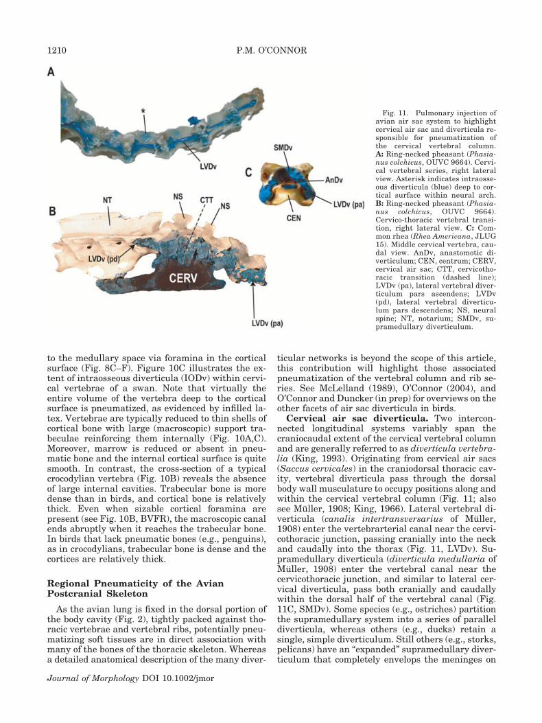
Cervical air sac diverticula. Two intercon-nected longitudinal systems variably span thecraniocaudal extent of the cervical vertebral columnand are generally referred to as diverticula vertebra-lia (King, 1993). Originating from cervical air sacs(Saccus cervicales) in the craniodorsal thoracic cav-ity, vertebral diverticula pass through the dorsalbody wall musculature to occupy positions along andwithin the cervical vertebral column (Fig. 11; alsosee Muller, 1908; King, 1966). Lateral vertebral di-verticula (canalis intertransversarius of Muller,1908) enter the vertebrarterial canal near the cervi-cothoracic junction, passing cranially into the neckand caudally into the thorax (Fig. 11, LVDv). Su-pramedullary diverticula (diverticula medullaria ofMuller, 1908) enter the vertebral canal near thecervicothoracic junction, and similar to lateral cer-vical diverticula, pass both cranially and caudallywithin the dorsal half of the vertebral canal (Fig.11C, SMDv). Some species (e.g., ostriches) partitionthe supramedullary system into a series of paralleldiverticula, whereas others (e.g., ducks) retain asingle, simple diverticulum. Still others (e.g., storks,pelicans) have an “expanded” supramedullary diver-ticulum that completely envelops the meninges on
Fig. 11. Pulmonary injection ofavian air sac system to highlightcervical air sac and diverticula re-sponsible for pneumatization ofthe cervical vertebral column.A: Ring-necked pheasant (Phasia-nus colchicus, OUVC 9664). Cervi-cal vertebral series, right lateralview. Asterisk indicates intraosse-ous diverticula (blue) deep to cor-tical surface within neural arch.B: Ring-necked pheasant (Phasia-nus colchicus, OUVC 9664).Cervico-thoracic vertebral transi-tion, right lateral view. C: Com-mon rhea (Rhea Americana, JLUG15). Middle cervical vertebra, cau-dal view. AnDv, anastomotic di-verticulum; CEN, centrum; CERV,cervical air sac; CTT, cervicotho-racic transition (dashed line);LVDv (pa), lateral vertebral diver-ticulum pars ascendens; LVDv(pd), lateral vertebral diverticu-lum pars descendens; NS, neuralspine; NT, notarium; SMDv, su-pramedullary diverticulum.
1210 P.M. O’CONNOR
Journal of Morphology DOI 10.1002/jmor

all sides, thereby forming a complete peridural di-verticulum. In addition to their circumferential dis-tribution around the spinal cord and meninges, su-pramedullary diverticula give rise to segmental,dorsally projecting extensions that occupy the spaceon the dorsal surface of neural arches (e.g., suprav-ertebral diverticula, SVDv), some of which even ex-pand between portions of the epaxial musculature(e.g., intermuscular diverticula; also see Fig. 2A,IMDv) in certain species.
Both longitudinal systems (i.e., lateral vertebraland supramedullary) may extend as far cranially asthe atlas, although there is considerable interspe-cific variation in the extent of this cranial elonga-tion. Supramedullary and lateral vertebral systemsinterconnect with one another at intervertebraljoints via small, anastomosing diverticula (Fig. 11C,AnDv). It is unclear how far caudally the two sys-tems extend, but in some taxa (e.g., hornbills, peli-cans, storks) supramedullary diverticula anasto-mose with a cranially projecting counterpart derivedfrom the abdominal air sac system (see below).Therefore, cranial (cervical air sac diverticula) andcaudal (abdominal air sac diverticula) components ofthe air sac system can communicate with one an-other via the vertebral canal. It is significant thatnone of the birds examined showed caudal extensionof cervical air sac diverticula to pneumatize post-midthoracic regions of the vertebral column. Due tothe close approximation of the medial border of theavian lung to the thoracic vertebral series, the de-scending portion of the lateral vertebral diverticu-lum (LVDv .) terminates at the cranial end of thenotarium (see Fig. 11B).
Any of the above-mentioned diverticula (i.e.,SMDv, LVDv, and SVDv), when adjacent to bone,can pneumatize vertebrae and ribs in the cervicaland thoracic regions. More specifically, cervical ver-tebrae and ribs are pneumatized by lateral vertebraldiverticula, usually around the periphery of the ver-tebrarterial canal. Extensions from lateral vertebraldiverticula also pneumatize neural arches via fo-ramina on the cranial and caudal aspects of trans-verse processes. Typically located near the base ofthe transverse process, such foramina occupy posi-tions ventral to the zygapophyses, thereby forminginfrapre- and infrapostzygapophyseal foramina. Incontrast, it is rare that supramedullary diverticuladirectly pneumatize bone forming the periphery ofthe neural canal. However, in some taxa (e.g., buce-rotids, gruids, anhimids), this system does have in-traosseous connections indirectly via its supraverte-bral extensions. Supravertebral diverticula gainaccess to vertebrae via foramina on the dorsal sur-face of the neural arch (e.g., Fig. 2C, NAPF) and byopenings on both the pre- and postspinal surfaces ofthe neural spine (e.g., Fig. 8C, NAPF). Finally, cra-nial thoracic vertebrae and ribs are also pneuma-tized by diverticula originating from cervical air sacs(Fig. 11B, LVDv .).
Lung diverticula. Whereas caudally projectingdiverticula from the cervical system may pneuma-tize the cranialmost thoracic vertebrae, it is typi-cally the case that the dorsomedially positionedavian lung directly pneumatizes adjacent vertebraeand ribs, in some cases as far caudally as the lastfree thoracic vertebra (e.g., Fig. 8E,F, CPF). Theoverall distribution of this characteristic amongextant birds (e.g., Tinamidae, Anseriformes, Galli-formes, Falconiformes, Strigiformes, Pelecaniformes,Ciconiiformes, Gruiformes, Charadriiformes, Picaf-ormes) indicates that this is a very common mode ofpneumatizing the thoracic vertebral series. Pneumaticforamina in vertebral centra typically decrease in sizeor are absent altogether in more caudal portions of thethoracic series (Fig. 8F); this general pattern is alsoobserved in nonavian theropods (see below). Althoughresulting in an air-filled bone (similar to pneumaticityderived from air sac diverticula), it seems prudent todifferentiate the anatomical soft tissues responsiblefor inducing pneumaticity in each case on whether theepithelium originates from the lung directly or via theair sacs. The term pneumatic diverticulum (divertic-ula, pl.) will be retained in the case of the latter,whereas pulmonary diverticula will be restricted toepithelial projections originating directly from thelung surface.
Abdominal air sac diverticula. Abdominal airsac diverticula responsible for osseous pneumaticityare, generally speaking, much simpler than those ofthe cervical system. Originating from the dorsal as-pect of abdominal air sacs (Saccus abdominalis; e.g.,O’Connor, 2004, fig. 2), small perirenal diverticula(diverticula pelvica of Muller, 1908) intercalate intothe space between the kidneys and the pelvic girdle,variably pneumatizing coxal elements and post-midthoracic regions of the vertebral column. Con-cerning the axial skeleton, the most commonly pneu-matized elements are synsacral and caudal thoracicvertebrae, with only very rare pneumatization offree caudal vertebrae and the pygostyle (O’Connor,2004). As with pneumatization of the cervical series,extensions from perirenal diverticula occupy a dor-sal position within the vertebral canal (i.e., forminga supramedullary diverticulum), and give rise toboth cranially and caudally directed extensions. Thecranially directed supramedullary diverticulum ofthe abdominal air sac may anastomose with its cau-dally directed counterpart derived from cervical airsacs. In these cases, abdominal air sac diverticulaunambiguously pneumatize the pelvic girdle andpost-midthoracic regions of the vertebral column(e.g., synsacrum, caudal thoracic vertebrae), onlysecondarily anastomosing with caudally projectingcomponents of the cervical air sac system. Signifi-cantly, the supramedullary diverticula of cervicalair sacs were not observed to pneumatize regions ofthe post-midthoracic vertebral column and pelvicgirdle. In other words, if pelvic girdle elements andcaudal thoracic/synsacral portions of the vertebral
1211POSTCRANIAL PNEUMATICITY IN ARCHOSAURS
Journal of Morphology DOI 10.1002/jmor

column are pneumatized in extant birds, this isachieved by the abdominal air sac system. (For adiscussion of pneumaticity in other portions of theavian skeleton, see Muller, 1908; Groebbels, 1932;King, 1966; Witmer, 1995b, 1997; O’Connor, 2004.)
Postcranial Pneumaticity in FossilArchosauriform Groups: Application of thePneumaticity Profile
The anatomical data presented above are hereused to establish a Pneumaticity Profile (PP) bywhich osteological features in fossils may be exam-ined in order to determine the relative specificity oftheir soft-tissue associations (Fig. 12). The followingcase studies use the PP to reevaluate purportedpostcranial pneumatic features in representative or-nithodiran and nonornithodiran archosauriformtaxa.
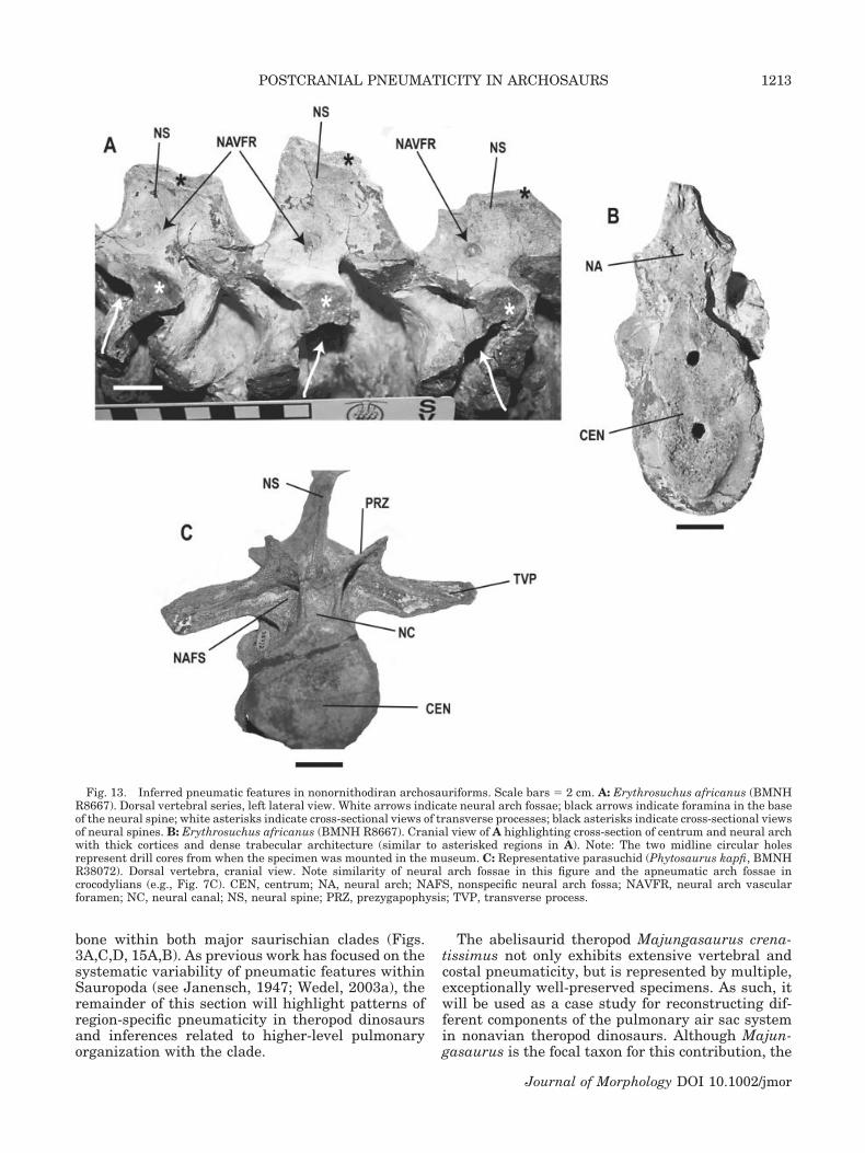
Nonornithodiran archosauriformes. Putativepneumatic features in basal archosauriforms andcertain crurotarsans (Gower, 2001) consist of fossae
on the ventral surface of the neural arch and foram-ina on the dorsal arch surface adjacent to the base ofthe neural spine (Fig. 13A,B). An examination ofErythrosuchus africanus (BMNH R8667, BMNHR3592, NGS F377) shows that although vertebralfossae are superficially similar to many featuresidentified as pneumatic in other fossil specimens,the features do not communicate with large internalchambers within the vertebrae. Due to fortuitousbreaks in BMNH R8667, it is also possible to exam-ine cross-sectional views of all components of verte-bral centra and neural arches, including both trans-verse processes and neural spines adjacent to theinferred “pneumatic” features (Fig. 13A–C). Theseviews reveal thick cortices surrounding denselypacked trabecular architecture void of the large in-ternal cavities that characterize pneumatic bone.The soft-tissue system likely responsible for the dor-sally positioned neural arch foramina is vascula-ture, as these openings lead directly into denselypacked trabecular bone, similar to vascular foram-ina in extant crocodylians (e.g., Fig. 10B). A largerand more complete specimen of Erythrosuchus (NGSF377) also exhibits similar neural arch fossae, withno apparent sign of internal cavity formation.
An examination of representative parasuchians(e.g., Phytosaurus kapfi [BMNH R38071], Rutiodonadamanensis [UCMP 26699], and R. zunii [UCMP27036]) also reveals similar noncommunicating fos-sae on dorsal neural arches (Fig. 13C). However,such features are morphologically consistent withneural arch fossae found in extant crocodylians (e.g.,Fig. 7C) and other nonavian sauropsids that houseadipose deposits, and have no relationship to anyportion of the respiratory system. Other parasuch-ians (e.g., Rutiodon [Angistorhinopsis] sp., MB1922.23.342) exhibit generally similar neural archfossae with no internal cavity formation, as indi-cated by an examination of cross-sectional views ofspecimens.
Ornithodira: Pterosauria. Examination ofpterosaur specimens reveals that vertebral pneuma-ticity is morphologically similar to that of extantbirds, particularly with regard to the position, size,and number of foramina in both central and neuralarch components (Figs. 3B, 14A). These in turn leadto spacious internal chambers within vertebrae. Ap-pendicular elements of pterosaurs, particularly longbones, are characterized by extremely thin cortices,with the placement of pneumatic foramina in posi-tions similar to those found in birds (Fig. 14B,C).See O’Connor (2003) for additional information onpostcranial pneumaticity in pterosaurs.
Ornithodira: Saurischia. Numerous workershave commented on the pneumatic postcranial skel-etons of both sauropod and nonavian theropod dino-saurs (e.g., Owen, 1856; Janensch, 1947; Romer,1956, 1966; Britt, 1993; Wilson, 1999; Wedel, 2003a;O’Connor and Claessens, 2005). This study confirmsmorphology consistent with pneumatic invasion of
Fig. 12. Pneumaticity Profile illustrating correlation and rel-ative specificity of osteological features as a function of differentsoft-tissue systems. Vertebral fossae are causally associated withmany soft-tissue systems, thus their reliability as specific indica-tors of pneumaticity remains ambiguous. Vertebral foramina arebetter indicators of pneumaticity, but only when combined withthe presence of large internal cavities within the bone. In order tojustify a positive pneumaticity assessment, these cavities mustdemonstrate clear continuity with the extraosseous space (i.e.,cortical foramina) as pneumatic diverticula originate within thebody cavity (i.e., from the air sacs/lungs) and must traverse cor-tical bone in order to gain access to the interior of bones.
1212 P.M. O’CONNOR
Journal of Morphology DOI 10.1002/jmor
bone within both major saurischian clades (Figs.3A,C,D, 15A,B). As previous work has focused on thesystematic variability of pneumatic features withinSauropoda (see Janensch, 1947; Wedel, 2003a), theremainder of this section will highlight patterns ofregion-specific pneumaticity in theropod dinosaursand inferences related to higher-level pulmonaryorganization with the clade.
The abelisaurid theropod Majungasaurus crena-tissimus not only exhibits extensive vertebral andcostal pneumaticity, but is represented by multiple,exceptionally well-preserved specimens. As such, itwill be used as a case study for reconstructing dif-ferent components of the pulmonary air sac systemin nonavian theropod dinosaurs. Although Majun-gasaurus is the focal taxon for this contribution, the
Fig. 13. Inferred pneumatic features in nonornithodiran archosauriforms. Scale bars � 2 cm. A: Erythrosuchus africanus (BMNHR8667). Dorsal vertebral series, left lateral view. White arrows indicate neural arch fossae; black arrows indicate foramina in the baseof the neural spine; white asterisks indicate cross-sectional views of transverse processes; black asterisks indicate cross-sectional viewsof neural spines. B: Erythrosuchus africanus (BMNH R8667). Cranial view of A highlighting cross-section of centrum and neural archwith thick cortices and dense trabecular architecture (similar to asterisked regions in A). Note: The two midline circular holesrepresent drill cores from when the specimen was mounted in the museum. C: Representative parasuchid (Phytosaurus kapfi, BMNHR38072). Dorsal vertebra, cranial view. Note similarity of neural arch fossae in this figure and the apneumatic arch fossae incrocodylians (e.g., Fig. 7C). CEN, centrum; NA, neural arch; NAFS, nonspecific neural arch fossa; NAVFR, neural arch vascularforamen; NC, neural canal; NS, neural spine; PRZ, prezygapophysis; TVP, transverse process.
1213POSTCRANIAL PNEUMATICITY IN ARCHOSAURS
Journal of Morphology DOI 10.1002/jmor

patterns highlighted here are also found in numer-ous other clades of neotheropods (O’Connor andClaessens, 2005; O’Connor, in press). Vertebraethroughout the entire postatlantal, precaudal series(UA 8678) exhibit pneumatic morphology consistentwith that of extant birds, namely, cortical foraminathat communicate with large, internal chamberswithin both centra and neural arches (Figs. 15, 16).Pneumaticity of the centrum and neural arch ispresent in the postatlantal cervical series and thecranial four dorsal vertebrae. The remainder of thedorsal series exhibits pneumatic features solelywithin the neural arch, with the caudal two dorsalarches (i.e., D12–13) showing a reduction in bothsize and number of pneumatic foramina and cham-bers. Pneumatic foramina are restricted to the lat-eral surface of the centrum, typically located justcaudal to the parapophysis. In contrast, pneumaticfeatures are found on virtually all components of theneural arch, including the pedicle, lamina, andtransverse processes, in addition to the neural spine(Figs. 15, 16) (O’Connor, in press). UA 8678 exhibitsthe largest pneumatic foramina (4–11 mm diame-ter) in vertebral centra between cervical vertebra 9and dorsal vertebra 4 (Figs. 3A, 15B, 16B), withforamina of more cranial vertebrae being muchsmaller in size (�1–2 mm diameter). Most neothero-pods exhibit an enlarged pneumatic foramen on the
second cervical centrum relative to the rest of thecervical series (e.g., Fig. 16A). A similar serial pat-tern (i.e., pneumatic foramina present on vertebralcentra between C2 and D4) characterizes manyother nonavian theropods including Allosaurus fra-gilis (Madsen, 1976) and Sinraptor dongi (Currieand Zhao, 1993). The largest pneumatic foramina onvertebral centra are typically located at the cervi-codorsal transition, establishing a common patternof relatively larger pneumatic features in vertebraeat the cranial end of the thorax; other theropodsshowing this trait include Spinostropheus gautieri[MNN TIG6], Allosaurus (Madsen, 1976), Sinraptor(Currie and Zhao, 1993), and Monolophosaurus(Zhao and Currie, 1993) and numerous groups ofextant birds.
Majungasaurus also exhibits pneumatic sacralneural arches (Fig. 16C), with apneumatic sacralcentra, a characteristic poorly known within Thero-poda. Whereas other theropods have been identifiedwith sacral pneumaticity, pneumatic foramina areusually restricted to sacral centra (e.g., ornithomim-ids: Gilmore, 1920; allosauroids: Harris, 1998; tyr-annosaurids: Brochu, 2003). Majungasaurus doesnot exhibit pneumaticity in the caudal vertebral se-ries, although it has been identified in other nona-
Fig. 14. Pneumatic postcranial elements in a pterosaur (A,B),with an extant bird (C) for comparison. A: Ornithocheirid ptero-saur (Ornithocheirus sp., SM B54.320). Middle cervical vertebra,left lateral view. B: Ornithocheirid pterosaur (Ornithocheirus sp.,BMNH R558). Proximal left humerus, caudal view. C: Black-necked swan (Cygnus melancoryphus, CM S-201). Proximal lefthumerus, caudal view. Scale bar � 2 cm in A; B and C are scaledto the same size in image. CPF, central pneumatic foramen; HH,humeral head; HPF, humeral pneumatic foramen.
Fig. 15. Comparison of pneumatic features in vertebrae of anonavian theropod dinosaur (A,B) and an extant bird (C,D).A: Theropod dinosaur (Majungasaurus crenatissimus, UA 8678).Third cervical vertebra, caudal view. B: Theropod dinosaur (Ma-jungasaurus crenatissimus, UA 8678). Tenth cervical vertebra,right lateral view. C: Sarus crane (Grus antigone, SBUAV104063). Thoracic vertebra, caudal view. D: Sarus crane (G.antigone, SBU AV104063). Thoracic vertebra, right lateral view.Images not to scale. CEN, centrum; CPF, central pneumatic fo-ramen; NAPF, neural arch pneumatic foramen; NC, neural canal;NS, neural spine; POZ, postzygapophysis; PP, parapophysis;TVP, transverse process.
1214 P.M. O’CONNOR
Journal of Morphology DOI 10.1002/jmor
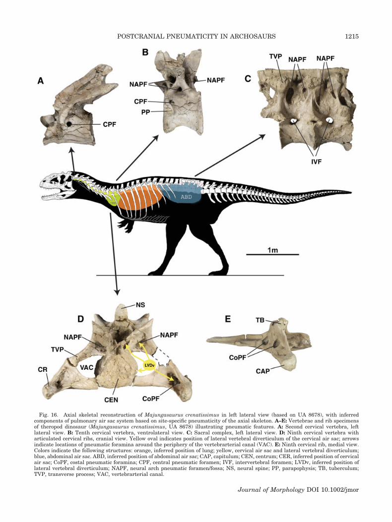
Fig. 16. Axial skeletal reconstruction of Majungasaurus crenatissimus in left lateral view (based on UA 8678), with inferredcomponents of pulmonary air sac system based on site-specific pneumaticity of the axial skeleton. A–E: Vertebrae and rib specimensof theropod dinosaur (Majungasaurus crenatissimus, UA 8678) illustrating pneumatic features. A: Second cervical vertebra, leftlateral view. B: Tenth cervical vertebra, ventrolateral view. C: Sacral complex, left lateral view. D: Ninth cervical vertebra witharticulated cervical ribs, cranial view. Yellow oval indicates position of lateral vertebral diverticulum of the cervical air sac; arrowsindicate locations of pneumatic foramina around the periphery of the vertebrarterial canal (VAC). E: Ninth cervical rib, medial view.Colors indicate the following structures: orange, inferred position of lung; yellow, cervical air sac and lateral vertebral diverticulum;blue, abdominal air sac. ABD, inferred position of abdominal air sac; CAP, capitulum; CEN, centrum; CER, inferred position of cervicalair sac; CoPF, costal pneumatic foramina; CPF, central pneumatic foramen; IVF, intervertebral foramen; LVDv, inferred position oflateral vertebral diverticulum; NAPF, neural arch pneumatic foramen/fossa; NS, neural spine; PP, parapophysis; TB, tuberculum;TVP, transverse process; VAC, vertebrarterial canal.
1215POSTCRANIAL PNEUMATICITY IN ARCHOSAURS
Journal of Morphology DOI 10.1002/jmor

vian saurischian taxa (e.g., oviraptorid theropods:Barsbold et al., 2000; diplodocid sauropods: Wedel,2003a). See Britt (1993), O’Connor and Claessens(2005), and O’Connor (in press) for a more compre-hensive analysis of the distribution of vertebralpneumaticity in nonavian theropods.
Cervical ribs of Majungasaurus demonstrate ex-tensive pneumaticity that is unparalleled in othernonavian theropod taxa. Large pneumatic foramina(5–12 mm diameter) are found bilaterally on cervicalribs 4–10, many with multiple foramina (Fig.16D,E). Costal foramina are located on both thedorsomedial surface of the rib shaft in addition tothe cranial and caudal surfaces of the capitulotuber-cular lamina (Fig. 16, CoPF). These parts of the ribform the lateral and ventral boundaries of the ver-tebrarterial canal (i.e., transverse foramen),whereas the cervical centrum and transverse pro-cess form the medial and dorsal boundaries of thecanal, respectively (Fig. 16D, VAC). Cervical trans-verse processes often exhibit accessory pneumaticforamina on their ventral surfaces. The morphologyand position of these features in cervical vertebraeand ribs of Majungasaurus are consistent withpneumatic foramina in the cervical vertebral columnof extant birds. The dorsal ribs of Majungasaurusare apneumatic.
Pulmonary fossae in Saurischian dinosaurs.Large, laterally directed fossae in dorsal vertebralcentra of sauropod (e.g., Fig. 3D; also see Diplodocuscarnegie, CM 84; Apatosaurus louisae, CM 3018;Camarasaurus sp., CM 11338) and larger-bodiedtheropod (Tyrannosaurus, FMNH PR 2081; Allosau-rus, CM 11844) dinosaurs are nearly identical inshape and position to pulmonary fossae observed insome extant birds (e.g., procellarids: Fig. 8A). Inbirds, medial projections of the dorsally positioned,rigid lung occupy the fossae. As such, these fossaeare indicative of two aspects of pulmonary morphol-ogy in birds; the first conveys topological informa-tion (i.e., the dorsomedial position of the lung withinthe thorax), with the second relating compositionalinformation (i.e., densely packed parenchyma occu-pying a fossa within a bone). Pulmonary fossae inextinct groups then represent unambiguous osteo-logical correlates of the actual lung within the tho-racic cavity, rather than a seemingly general infer-ence related to the presence of pulmonary air sacs(as has been done previously).
Pneumaticity in basal avians. As part of thisstudy, both London and Berlin specimens of Archae-opteryx lithographica were examined. The Berlinspecimen (MB 1880.81.4598) preserves a portion ofthe vertebral column in right lateral view. Cervicalvertebra 5 possesses a partially preserved foramenon the craniolateral margin of the centrum. Whereasit appears that the caudoventral rim of this foramenhas an intact ostial margin, the cranial andcraniodorsal rim of the foramen does not. Accord-ingly, it is difficult to assess the actual size and
shape of the foramen on this vertebra. Foramina onadjacent vertebral centra are ambiguously defined,particularly under microscopic examination, con-trary to diagrams provided by Britt et al. (1998, fig.1). A section of the dorsal vertebral column is alsoexposed in right lateral view. The caudalmost dorsalvertebrae exhibit shallow fossae on the lateral sur-face of the centra (“pleurocoels” of Ostrom, 1976);however, consistent with statements by Britt et al.(1998), they do not have foramina piercing the cor-tical surface. Exposed appendicular elements of MB1880.81.4598 do not exhibit any features indicativeof pneumaticity.
Putative pneumatic features recently identified(Christiansen and Bonde, 2000) on axial and appen-dicular elements of the London specimen of Archae-opteryx (BMNH 37001: main slab) were also exam-ined. The vertebral features (thoracic vertebrae 1–2)are rough-edged depressions located at the junctionof the pedicle and transverse process. Within eachfossa are 1–2 small (�1 mm) foramina, also withrough edges. Notably, the position of these featureswould allow continuity of soft-tissue structures be-tween the lateral surface of the neural arch and theneural canal, and not into the medullary cavity.Moreover, rough edges on the surrounding bone sug-gest that the structures are an artifact (of eitherpreservation or preparation) and do not representintact bone surfaces.
A large, rough-edged “fossa” on the caudodorsalaspect of the proximal left pubis has been inter-preted as pneumatic, and used to infer the existenceof abdominal air sacs in this taxon (Christiansenand Bonde, 2000). This opening leads to a shortcanal directed cranially into the body of the pubis. Itis unilateral in its development, as a similar fossa isnot visible on the right pubis (although a completeexamination of the right pubis is prevented in partby the overlying, displaced, scapula). Pneumatic pu-bes are known in a variety of basal extant avian taxa(e.g., Struthio, Rhea, Anser: O’Connor, 2004). How-ever, in contrast to the orientation of the opening inBMNH 37001, pubic pneumatic foramina are di-rected caudoventrally down the pubic shaft. Finally,similar to the condition in thoracic vertebrae, theedges of this structure are rough and appear toconsist of broken bone, and may pertain to artifactrather than invasion by air sac diverticula.
DISCUSSION
Based on the relationship between specific skele-tal morphologies and different soft-tissue systemsidentified in extant sauropsids, results from thisstudy indicate that the vast range of features previ-ously used to infer pneumaticity in extinct taxaoverestimate pneumatic invasion by pulmonary airsacs. The only reliable and consistent indicators ofpneumaticity are cortical openings (i.e., foramina orcommunicating fossae) connected directly with large
1216 P.M. O’CONNOR
Journal of Morphology DOI 10.1002/jmor

internal cavities within the bone (Figs. 12, 15, 16).In contrast, other osteological features such as ver-tebral fossae and cortical foramina alone are caus-ally related to many different types of soft tissues(e.g., vascular, muscular, etc.), indicating a lowerdegree of system specificity, and ultimately, provid-ing much less discriminating value for interpretingsoft-tissue associations when examined in extincttaxa. Application of the Pneumaticity Profile (PP)derived in this study to fossil archosauriform taxareveals the following: 1) that the ornithodiran cladesSaurischia and Pterosauria include members withunambiguous pneumatic features, thereby support-ing historical interpretations of these structures;and 2) that recently inferred pneumatic structuresin nonornithodiran archosauriforms (e.g., Erythro-suchus, parasuchians) are not consistent withknown pneumatic morphology and likely relate toother causal factors (e.g., vasculature). Finally,based on an analysis of region-specific pneumaticityin extant birds, it is possible to constrain inferencesrelated to the identification of specific components ofthe pulmonary system responsible for pneumatizingthe axial skeleton in saurischian dinosaurs.
Other osteological characteristics have been used asindicators of pneumaticity in saurischian dinosaurs.For example, noncommunicating fossae on the dorsalaspect of sauropod neural arches have been character-ized as pneumatic (Fig. 3C-1; also see Wilson, 1999).Bone within these fossae may either be rugose (“crenu-late” texture: Britt, 1993) or smooth relative to adja-cent cortical bone, and both textural categories havebeen used to identify pneumaticity in fossils (e.g.,Britt, 1993; Wedel et al., 2000; Wedel, 2003a). How-ever, an examination of extant crocodylians revealsthat neural arch fossae associated with adipose depos-its are lined by cortical bone that is relatively smoothwhen compared to adjacent bone (e.g., Fig. 7B). Mini-mally, this suggests that nonpulmonary soft tissuesmay result in cortical bone that is smooth in appear-ance and located in similar positions as putative“pneumatic” fossae in fossil taxa. From this observa-tion, causal relationships between surface morphologyand associated soft tissues may not be system specific,underscoring the necessity for a reevaluation of tex-tural characteristics used for identifying pneumaticityin fossils.
Finally, it is not necessary to invoke a “soft tissue”culprit for every bony feature on the skeleton. Somefeatures may not represent the effect of a specific soft-tissue influence at that particular spot, but the cumu-lative effect of processes in surrounding areas (seeWitmer, 1995a, for a discussion of the “hole-or-doughnut” problem). As an example, transverse pro-cesses function to support body wall musculature inaddition to serving as an articulation site for ribs. Inmany cases the transverse process is ventrally but-tressed by either a small strut or lamina connecting itto the base of the neural arch. Depending on the formof this strut, variably shaped fossae are positioned
ventral to the transverse process. When the ventralbuttress consists of a simple, craniocaudally restrictedlamina (e.g., parasuchians, Fig. 13C), distinct archfossa are located along the ventral surface of the pro-cess. Instead of assigning specific soft-tissue associa-tions to the fossae, it is reasonable that they merelyrepresent epiphenomena related to the size and shapeof the ventral buttressing unit and, ultimately, are dic-tated by mechanical demands placed on the vertebra.
Postcranial Pneumaticity in ExtinctArchosauriform Taxa
Based on compelling morphological evidence, nu-merous examples of postcranial pneumaticity existin many extinct ornithodiran clades, includingpterosaurs, and sauropod and nonavian theropoddinosaurs (Figs. 3, 4). Although both the London andthe Berlin specimens of Archaeopteryx exhibit bonyfeatures interpreted as pneumatic (Britt et al., 1998;Christiansen and Bonde, 2000), results from thisstudy indicate that in only one case (i.e., a singleforamen on the C5 vertebra of the Berlin specimen)is this morphology consistent with that observed inextant birds. Other putative pneumatic features atbest represent equivocal assessments (e.g., foraminaon cervical vertebrae other than C5 in the Berlinspecimen), or represent preservational or prepara-tion artifact (e.g., “pneumatic” vertebrae in the Lon-don specimen). In summary, variable preservation,combined with only limited access to all bony sur-faces, likely contributes to the ambiguous assess-ment of pneumaticity in Archaeopteryx (e.g., con-trast the studies of Muller, 1908; Swinton, 1960;Ostrom, 1976; Britt et al., 1998; Christiansen andBonde, 2000; current study). Similarly, evidence insupport of pneumatic postcrania in prosauropod di-nosaurs remains somewhat ambiguous (e.g., seeBritt, 1993, 1997), although recent work has identi-fied possible pneumatic features on vertebrae ofThecodontosaurus (Yates, 2003). Additional re-search is currently under way evaluating pneuma-ticity in prosauropod dinosaurs (e.g., Wedel, 2005).
Although an extensive survey of nonornithodiranarchosauriform taxa was not conducted during thisstudy, the few specific reported examples (e.g.,Gower, 2001) of vertebral pneumaticity were in-cluded, and in all cases fail to meet morphologicalcriteria consistent with a pneumatic designation.Putative pneumatic features in nonornithodiran ar-chosauriforms (e.g., parasuchians, Erythrosuchus)consist of external osteological markers that are atbest ambiguous and internal bone characteristicscompletely unlike pneumatic bone (Fig. 13).Whereas these examples fail to meet explicit criteriaof the PP, the taxa included represent a mere frac-tion of the known diversity within the group. Thestudy of Gower (2001) is significant not only in thatit suggests (even tentatively) the presence of post-cranial pneumaticity in nonornithodiran archosau-
1217POSTCRANIAL PNEUMATICITY IN ARCHOSAURS
Journal of Morphology DOI 10.1002/jmor

riforms, but in some of the earliest known (mostlyTriassic) members of the group. Notably, other non-ornithodiran archosauriform clades (e.g., rauisuch-ians, sphenosuchian crocodylomorphs; Gower, 2001)also have vertebrae with features similar to thoseidentified as pneumatic in past studies. The presentstudy, combined with those of Gower (2001) andYates (2003), indicates that a systematic examina-tion of vertebral morphology is necessary to assesspotential pneumatic features within basal archosau-riform and basal sauropodomorph archosaurs. Suchstudies will not only help to evaluate, identify, andelucidate patterns of postcranial pneumaticitywithin different clades, but will serve to constrain“first-occurrence,” both temporally and phylogeneti-cally, of postcranial pneumaticity in the fossilrecord.
General Pulmonary Structure in Taxa withPneumatic Postcrania
Ornithodiran archosaurs identified with unam-biguous postcranial pneumaticity include sauris-chian dinosaurs and pterosaurs. Such features maybe used to infer the existence of a heterogeneouspulmonary apparatus in these taxa. In this context,heterogeneous refers to a pulmonary system exhib-iting variable parenchymal densities, implying thepresence of both gas exchange (i.e., parenchyma-dense) and nonexchange (i.e., air sacs) regions(Duncker, 1978; Perry, 1983, 2001; also see Hunter,1774). The lungs in all major clades of extant sau-ropsids exhibit sac-like regions (e.g., Duncker, 1971,1978, 1989; Wood and Lenfant, 1976; Perry, 1983,1998). For example, lungs of various chameleonidand varanid species exhibit variably sized andshaped diverticula extending from the caudal sur-face of the lung (Wolf, 1933; Klaver, 1981; Becker etal., 1989; Perry, 1998, fig. 1.12). Other sauropsidgroups such as crocodiles, snakes, and turtles alsohave lungs that exhibit a large range of parenchy-mal densities, such that areas of lowest parenchy-mal density are functionally equivalent (i.e., per-forming little to no gas exchange) to avian air sacs(Hunter, 1774; Duncker, 1978, 1989; Perry, 1998;Wallach, 1998; Wyneken, 2001). Whereas regions oflow parenchymal density (i.e., air sacs) in nonaviansauropsids do not invade the postcranial skeleton,they do provide analogs for air sacs and pneumaticdiverticula observed in the avian system. As such,the extant sister taxa of all fossil sauropids, includ-ing dinosaurs and pterosaurs, have the anatomicalfoundation (i.e., the soft-tissue potential) for pneu-matizing the postcranium. Postcranial pneumaticityrequires only the presence of a heterogeneously par-titioned pulmonary system.
Perry (2001 and references therein) and Perry andReuter (1999) suggested that theropod and sauropoddinosaurs exhibiting pneumatic postcrania not onlypossessed a heterogeneous pulmonary system, but
likely possessed dorsally fixed lungs within the tho-racic cavity. Such inferences are based on two linesof reasoning. First, and perhaps the stronger of thetwo points, a dorsally fixed lung places pulmonarytissues in contact with the vertebral column andribs, thereby providing direct access for pneumatiz-ing components (i.e., diverticula) of the system tobones most commonly pneumatized. Second, a dor-sally fixed lung also provides an anchor point for theregion of relatively high parenchymal density (i.e.,the exchanger) of the heterogeneous system, andprevents collapse of this region into the less denseair sac portions. Data supporting this notion arederived from studies of extant sauropsids exhibitinghigh levels of pulmonary heterogeneity (e.g., birds,lizards), where less-dense regions are located on thecranial, caudal, and ventral portions of the lung,with parenchyma-dense regions located dorsally, of-ten “attached” to the dorsal body wall (Duncker,1978, 1989; Perry, 1983, 1998, 2001; Wyneken,2001).
Results from the current study support these con-cepts based on the identification of different “types”of osteological correlates from throughout the verte-bral column. For example, this study provides newdata to discriminate between inferences related tothe lung itself, and those related to air sacs andpneumatic diverticula. Pulmonary fossae in verte-brae of saurischian dinosaurs (e.g., Fig. 3D, PLFS)highlight two key features concerning lung struc-ture in the group. First, the lungs occupied a dorsalor dorsomedial position within the thoracic cavity,confirming the theoretical work of others (e.g., Perryand Reuter, 1999). Second, and perhaps more impor-tant, lungs were composed of densely packed, rigidparenchyma capable of influencing vertebral mor-phology, similar to the condition in extant birds.Although the rigid avian lung is always positionedin the dorsal portion of the thoracic cavity (Hunter,1774; Duncker, 1971), pulmonary fossae in vertebralcentra remain taxonomically restricted (e.g., procel-larids; Fig. 8). However, when present, they arecausally associated with medially directed para-bronchial protuberances, and hence, provide robustosteological correlates for both lung position andcomposition. Comparable vertebral modificationsare apparent in many large-bodied saurischians(Fig. 3D; e.g., Diplodocus; Hatcher, 1901, pl. 7; Al-losaurus, CM 11844; Tyrannosaurus, FMNH PR2081). The identification of similar vertebral modi-fications in fossil archosaurs helps constrain infer-ences related not only to lung position, but to aspectsof the intrapulmonary structure (i.e., rigid architec-ture � densely packed parenchyma) of the ex-changer itself. Pulmonary fossae, combined with thewealth of data on postcranial pneumaticity, supportnot just the presence of air sacs, but also of a level ofpulmonary heterogeneity (i.e., a relatively dense,rigid exchange region vs. relatively less dense, com-pliant nonexchange regions) approaching that in ex-
1218 P.M. O’CONNOR
Journal of Morphology DOI 10.1002/jmor

tant birds. Finally, whereas specific details regard-ing the intrapulmonary functional characteristics(e.g., air conduit and vascular capillary architecture)of saurischian dinosaur lungs may prove difficult todiscern, region-specific pneumaticity may be used toelucidate other aspects of higher-level pulmonaryorganization in these groups (see below).
Pulmonary Heterogeneity in OrnithischianDinosaurs
Although the presence of postcranial pneumatic-ity is indicative of pulmonary air sacs and divertic-ula, the lack of pneumaticity in no way indicates theabsence of such pulmonary specializations. The ab-sence of postcranial pneumaticity merely indicatesthat the pneumatization process is absent, but pro-vides no direct data related to lung structure. Forexample, although ornithischian dinosaurs lackpneumatic postcrania, phylogenetic inference basedon their extant relatives (i.e., the EPB) suggests thatthey possessed a heterogeneous pulmonary system.This is based not only on the inferred condition ofpulmonary structure in their extinct ornithodiranrelatives, but the condition in extant sauropsids ingeneral. Exactly why postcranial pneumaticity oc-curs in some groups (saurischians) and not others(ornithischians) remains unclear; however, similarvariability is also found among birds. This is illus-trated in a number of taxa (e.g., Apteryx, oxyurineAnseriformes, Podicepidiformes, Gaviiformes) thatcompletely lack pneumatic postcrania, althoughhaving the typical avian pulmonary air sac system,including diverticula positioned adjacent to the ver-tebral column (O’Connor, 2004). Importantly, suchvariation in birds suggests that there is an on-offswitch for the pneumatization process, and whereasit is likely that genetic factors determine this trait,specific control mechanisms are far from being un-derstood (or even hypothesized; although see Honiget al., 2002; Kurbel et al., 2004). Thus, the absence ofpneumatic postcranial bones in ornithischians re-mains intriguing, not only from the perspective ofpulmonary organization inferred from outgroupcomparison, but also from a developmental perspec-tive of how certain clades of archosaurs (e.g., divingbirds) with air sac systems restrict the pneumatiza-tion process.
Anatomical Inferences: PostcranialPneumaticity and Pulmonary Organizationin Theropod Dinosaurs
Based on modeling exercises derived from studiesof extant sauropsid lung morphology and osteologi-cal studies of select fossil specimens, Paul (2001)and Perry (2001) predicted a cranial-to-caudal pro-gression in the evolutionary acquisition of avian-likeair sacs in nonavian theropods. In these scenarios,cervical air sacs are present throughout Neothero-
poda, with abdominal air sacs “appearing” only inderived coelurosaurs (Paul, 2001). It is within suchscenarios that functional attributes (e.g., ventilatorydynamics) of the theropod pulmonary apparatushave been hypothesized.
Cervical air sacs and diverticula in theropoddinosaurs. Many theropod dinosaurs possess pneu-matic foramina that are best-developed (i.e., rela-tively larger) in the cranial dorsal and caudalmostcervical vertebrae. This is not only evident in Ma-jungasaurus (Figs. 3B, 15B, 16), but also in a num-ber of forms throughout the clade (e.g., Spinostro-pheus, Allosaurus, Monolophosaurus, Sinraptor). Asimilar pattern of pneumaticity is observed in anumber of extant birds, with relatively larger pneu-matic foramina located at the cervicodorsal transi-tion. Pulmonary injections reveal that cervical airsacs are located adjacent to the vertebral column atthis point (Fig. 11B; O’Connor, 2004, fig. 1), suggest-ing that the proximity of an air sac (and not just itsdiverticula) to bones may influence the character(e.g., size) of pneumatic features derived from it. Anumber of diving taxa (e.g., penguins) that typicallylack postcranial pneumaticity have large fossaewithin the lateral surface of vertebral centra at thecervicothoracic junction, with the medial portion ofthe cervical air sac occupying this space (pers. obs.).Based both on location and relative size, pneumaticforamina at the cervicodorsal transition in nonaviantheropods are consistent with the presence of a sig-nificant cervical air sac system similar to that inextant birds (Fig. 16). Interestingly, Hatcher (1901)also mentioned the relatively large size of vertebralcavities at the cervicodorsal transition in some sau-ropods (e.g., Diplodocus), thereby providing the ini-tial osteological basis for scenarios (e.g., Janensch,1947; Wedel, 2003a) hypothesizing cervical air sacsin this saurischian group.
The patterning of foramina on both cervical ver-tebrae and ribs of theropods is virtually identical tothe condition in birds. Variably located around theperiphery of the vertebrarterial canal, pneumaticforamina are present on both centra and neural archcomponents of vertebrae and cervical ribs (Fig. 16D;yellow arrows originating from the LVDv indicatethe typical position of foramina in these elements).Whereas the vertebrarterial canal conveys neuro-vasculature to portions of the cervical vertebral col-umn, spinal cord, and head in all sauropsids, it alsotransmits the lateral vertebral diverticulum (Fig.11, LVDv) in birds. This diverticulum originatesfrom cervical air sacs at the base of the neck, passescranially within the canal, and is responsible forpneumatization of both cervical vertebrae and ribs.Hence, the pattern and relative development ofpneumatic features in the cervical vertebral columnof Majungasaurus (and theropods generally) indi-cates the presence of a well-developed cervical airsac complex complete with ascending diverticulacomparable to that observed in extant birds (Figs.
1219POSTCRANIAL PNEUMATICITY IN ARCHOSAURS
Journal of Morphology DOI 10.1002/jmor

11, 16). A similar pattern of cervical pneumaticity,along with hypothesized cervical air sac diverticula,has also been suggested for sauropod dinosaurs firstby Janensch (1947) and most recently by Wedel(2003b).
Abdominal air sacs and diverticula in thero-pod dinosaurs. Among extant birds, pneumaticcaudal, synsacral, and caudal thoracic vertebrae arepneumatized by diverticula of abdominal air sacs(Muller, 1908; Groebbels, 1932; King and Kelly,1956; King, 1957, 1966; Hogg, 1984b; O’Connor,2004). By comparison, then, sacral pneumaticity innonavian theropods provides strong evidence for thepresence of abdominal air sacs in these taxa. Inter-estingly, Britt et al. (1998) and Wedel (2003b) havespent considerable effort to account for the possibil-ity that cervical air sac diverticula may spread cau-dally along the axial skeleton, pneumatizing thecaudal thoracic, sacral, and caudal regions of thevertebral column. Much of this effort stems fromambiguous assertions by Cover (1953), along withadditional statements perpetuating the ambiguityby King (1975), that cervical air sac diverticula inthe turkey (Meleagris gallopavo) pneumatize por-tions of the post-midthoracic vertebral series. Aspointed out in the Results section, pneumaticity ofpost-midthoracic vertebral elements by componentsof the cervical air sac system could not be demon-strated in any bird, including the suspect taxon Me-leagris.
No doubt contributing to Cover’s (1953) initialobservation is the fact that diverticula of the cervicaland abdominal air sac systems often anastomosewith one another in the midthoracic region. Sacralpneumaticity, when present in extant birds, resultsfrom invasion by diverticula of the abdominal air sacsystem. Statements by others on this point, partic-ularly with regard to interpreting region-specificpneumaticity in fossil archosaurs (e.g., Britt et al.,1998; Ruben et al., 2003; Wedel, 2003a), not onlystem from their interpretations of the ambiguousstatements of Cover (1953) and King (1975) specifi-cally, but also from the general lack of empiricaldata on avian air sac systems and diverticula asso-ciated with the vertebral column. In contrast to sce-narios based on modeling studies, then (e.g., Paul,2001; Perry, 2001), the interpretation in this studyof an abdominal air sac system in basal neothero-pods significantly expands the distribution of thischaracteristic throughout the clade. Other sauris-chian taxa with pneumatic sacra (e.g., neosauro-pods) likely also possessed abdominal air sac sys-tems (see Wedel, 2003a). The observations of actualdiverticular morphology highlighted here (ratherthan reviews of others’ work) provide novel data foruse in future modeling studies focused on pulmo-nary evolution in saurischian dinosaurs.
Finally, the strongest evidence for distinct cervi-cal and abdominal air sacs in a fossil specimenwould be the presence of both cervical and sacral
pneumaticity with an absence of pneumaticity in thedorsal region (i.e., the pneumatic hiatus of Wedel,2003a). However, most fossils are not preserved inenough detail or completeness for such studies. For-tunately, one specimen of Majungasaurus (UA 8678)preserves a near-complete vertebral column withpneumatic foramina throughout the precaudal ver-tebral series. Whereas this specimen does not have acomplete absence of pneumaticity at any point alongthe series, it does show a reduction in pneumaticityin the last two dorsal neural arches, with enhancedpneumaticity in sacral arches (Fig. 16C). While notunequivocal evidence for distinct cervical and ab-dominal air sac systems, it is suggestive of two focalcenters for the origin of pneumatic diverticula. Thisinterpretation is also consistent with results fromextant birds that indicate a relationship between therelative size of pneumatic features and the distanceof a given bone from the air sac pneumatizing it. Asdiscussed above, relatively larger pneumatic fea-tures are found closer to the main air sac giving riseto the pneumatizing diverticula, with smaller fea-tures located further away from the air sac.
In summary, the results of this project providenew perspectives regarding not only the presence ofpulmonary air sacs in theropod dinosaurs, but alsodocument specific components of the air sac systemin basal neotheropods that were generally thoughtto be present only in derived coelurosaurs. The pres-ence both of cervical and sacral pneumaticity inMajungasaurus indicates that abdominal and cervi-cal air sac systems were present in basal neothero-pod taxa, and were not restricted to coelurosaurs, ashas been predicted by modeling studies. These in-terpretations, combined with inferences positing adorsally positioned, rigid lung underscore the ana-tomical prerequisites for a flow-through ventilatorysystem relatively early in the evolution of Theropoda(Fig. 16). Results from this study do not constrainspecific physiological parameters of the pulmonarysystem and comments regarding functional at-tributes (e.g., ventilatory mechanics, uni- vs. bidi-rectional intrapulmonary air flow) or inferred met-abolic consequences of it are beyond the scope of thiswork. However, studies examining musculoskeletalanatomy and function in both extant and extinctarchosaurs (e.g., Carrier and Farmer, 2000; Farmerand Carrier, 2000; Claessens, 2004a,b, 2005) may becombined with pulmonary reconstructions to formmore robust hypotheses of pulmonary function inextinct archosaurs (e.g., see O’Connor and Claes-sens, 2005). Such complementary studies, whencombined with data derived from other sources (e.g.,growth rates based on histology or the presence ofinsulatory integumentary structures; Erickson etal., 2001; Ricqles et al., 2001; Xu et al., 2001, 2004),may provide the basis for paleontologic meta-analyses geared at elucidating metabolic potentialand other physiological inferences pertaining to ex-tinct dinosaurs.
1220 P.M. O’CONNOR
Journal of Morphology DOI 10.1002/jmor
Nonpulmonary Roles of PostcranialPneumaticity
In addition to the role in gas exchange, pulmonaryair sacs and diverticula also provide a mechanismfor pneumatizing heavy, metabolically expensiveskeletal tissues. Previous workers (e.g., Cope, 1877;Marsh, 1877; Seeley, 1901; Muller, 1908; Romer,1966; Currey and Alexander, 1985; Carrano andO’Connor, 2005) have emphasized the density-reducing effect of skeletal pneumatization in fossilarchosaurs. From studies of extant birds, pneuma-tization proceeds by the replacement of trabecularbone and marrow with air sac diverticula (Bremer,1940b; King and Kelly, 1956; King, 1957; Schepel-mann, 1990; Brackmann, 1991), resulting in a sig-nificant reduction in the density of a given bone.From an energetics perspective, then, the cost oftransport for a pneumatic bone will be less than foran apneumatic bone of the same volume. Thus,pneumatizing the entire skeleton would prove ben-eficial as long as the mechanical requirements ofbones are not jeopardized. Whereas general scalingarguments for a relationship between large organis-mal size and postcranial pneumaticity are foundthroughout the literature on both extant and fossiltaxa (e.g., Cope, 1877; Muller, 1908; King, 1966;McLelland, 1989; Carrano and O’Connor, 2005), fewstudies have actually tested this concept. As oneexample, anseriform birds ranging in body massbetween 0.30–10.65 kg indicate only a very weakrelationship between body size and relative pneu-maticity (O’Connor, 2004). Instead, clade-specificpatterns and eco/functional attributes (e.g., divingbehavior) are better predictors of overall pneumatic-ity. Similar studies examining body size and relativepneumaticity in fossil archosaurian clades are lack-ing, although such approaches may prove fruitful forestablishing general rules of construction (Witmer,1995a) in groups possessing pneumatic postcrania.
Thermoregulation and Pulmonary Air Sacs
Numerous studies have suggested that pulmo-nary air sacs in extinct taxa, inferred on the basis ofpostcranial pneumaticity, provided sites for evapo-rative heat loss, which itself has been used to sup-port “high metabolic rates” and/or sustained rapidgrowth rates in different ornithodiran taxa (e.g.,Bakker, 1972; Padian, 1983; Paul, 1988; Reid, 1997;Wedel, 2003a). However, these assertions concern-ing evaporative heat loss are not warranted basedon thermoregulatory experiments conducted on ex-tant birds. Contrary to often-cited thermoregulatorybenefits of an air sac system (e.g., “birds can dumpheat by evaporation in their air sacs…future studiesof sauropod thermal biology should at least acknowl-edge the possibility of efficient, avian-style thermo-regulation” Wedel, 2003b:253), there are in fact noexperimental studies to date that have actually
demonstrated a heat-exchanging role for air sac por-tions of the pulmonary apparatus. The studies citedsupporting this notion (e.g., Schmidt-Nielson et al.,1969; Bernstein, 1976; Dawson and Whittow, 2000)provide data that are at best equivocal, if not actu-ally documenting a virtual lack of evaporative cool-ing properties for the posttracheal respiratory sys-tem in extant birds. Instead, the richly vascularizedtrachea represents the only portion of the lowerrespiratory tract functioning in substantial amountsof heat exchange (e.g., Schmidt-Nielson et al., 1969;Dawson and Whittow, 2000, and references therein).Anatomical characteristics consistent with these ex-perimental results include the low-density vascula-ture reported for avian air sacs and diverticula(Hunter, 1774; Salt and Zeuthen, 1960; McLelland,1989). Thus, whereas osteological indicators ofpneumaticity in fossil archosaurs do have a solidmorphological basis in extant birds, putative ther-moregulatory roles for air sacs in fossil organismsshould be dismissed until experimental studies onextant species demonstrate a link between pulmo-nary air sacs and evaporative cooling.
Lung Ventilation in Taxa with PneumaticPostcrania
The identification of pneumatic postcrania inpterosaurs, theropods, and sauropods indicates thepresence of a heterogeneous pulmonary apparatus,with living birds providing the only extant referencefor anatomical and physiological properties of such asystem. However, to what extent the specific mor-phology, and perhaps more important, the function-ality of this system were similar to that of modernbirds is unclear.
Whereas this study is not a direct test of ventila-tory mechanisms in fossil groups, pneumaticity datacertainly should be factored into any reconstructionof pulmonary anatomy in fossil archosaur groupspossessing it. For example, reconstructions of thero-pod pulmonary morphology and function basedsolely on extant crocodylians (e.g., Ruben et al.,1997, 1999, 2003) seem logically incomplete, asthere are solid morphological grounds for inferringan extremely heterogeneous pulmonary system,complete with air sacs and pneumatic diverticula.Moreover, results from the present study even indi-cate the presence of individual components of an airsac system in theropod dinosaurs, including distinctcervical and abdominal air sac systems. Admittedly,other aspects of pulmonary organization (e.g., thepresence of cranial and caudal thoracic and clavicu-lar air sacs similar to living birds) remain ambigu-ous due to the absence of site-specific osteologicalmarkers for thoracic components of the air sac sys-tem. Furcular foramina (see Makovicky et al., 2005)in the new dromaeosaurid, Buitreraptor, likely rep-resent osteological evidence for clavicular air sacs innonavian theropods.
1221POSTCRANIAL PNEUMATICITY IN ARCHOSAURS
Journal of Morphology DOI 10.1002/jmor

In summary, whereas higher-level inferences(e.g., uni- vs. bidirectional air flow) of pulmonaryfunction in extinct archosaurs may remain chalk-board exercises, the basic anatomical requirementsof a flow-through pulmonary apparatus (i.e., a rigid,dorsally fixed exchanger with both cranial and cau-dal air sac ventilators) were present in basalneotheropod dinosaurs and likely characterize basicpulmonary design throughout the clade.
Whereas the potential skeletal kinematics re-quired for flow-through ventilation have yet to beelucidated in theropods, it minimally involved dif-ferential costal movements along the rib series (i.e.,relatively larger movements of the caudalmost ver-tebral ribs) to generate pressure differentials be-tween the cranial and caudal regions of the trunk(see O’Connor and Claessens, 2005). Claessens(2004b) also recently put forth a model of an “acces-sory aspiration pump” in theropod dinosaurswhereby the gastralial apparatus works to preferen-tially ventilate the caudal region of the trunk, pos-sibly facilitating air flow into caudal (i.e., abdominaland caudal thoracic) air sacs. Similar to the situa-tion in extant archosaurs, it is likely that multiplemusculo-skeletal functional units (e.g., costoverte-bral and gastralial systems) worked together to co-ordinate ventilatory air flow in theropod dinosaurs.Future work combining studies of pneumaticity withexperimental analyses of the musculoskeletal sys-tem in extant archosaurs may well refine models ofpulmonary organization and ventilatory potential intheropod dinosaurs.
CONCLUSIONS
An examination of vertebrae in extant sauropsidsindicates that different soft-tissue systems pro-foundly impact skeletal morphology, sometimes instrikingly similar ways. Vertebral fossae are associ-ated with a variety of tissues including fat and mus-cle in addition to different components of the pulmo-nary system, indicating relatively low specificity forcausally associating these features with a singlesoft-tissue system. Vertebral foramina may betterpredict pneumaticity, but only when combined withknowledge of the internal character of the bone. Themost reliable, consistent, and “system-specific” indi-cators of pneumaticity are cortical foramina thatcommunicate directly with large internal cavitieswithin bones.
Unambiguous examples of postcranial pneumatic-ity in archosaurs can be found in pterosaurs andtheropod and sauropod dinosaurs. Other archosau-riform taxa with putative pneumatic features (e.g.,Erythrosuchus africanus, parasuchians) lack crite-ria for a positive assessment based on a Pneumatic-ity Profile developed in this study. Vertebral andcostal pneumaticity in neotheropods supports theinference of cervical and abdominal air sacs in thisgroup, in addition to pneumatization directly from
the lung, and highlights the anatomical prerequisitefor a flow-through lung design. Such a condition innoncoelurosaurs indicates that a highly heteroge-neous pulmonary system likely characterized Thero-poda (at least Neotheropoda), and was not restrictedto avian members of the clade.
Pneumaticity of the postcranial skeleton repre-sents an exaptation founded on the basis of a highdegree of pulmonary heterogeneity, this latter char-acteristic being variably expressed among all extantsauropsids. Whereas its role in the adaptive radia-tion of different archosaur clades (pterosaurs, thero-pods, sauropods) remains a point of inquiry, each ofthese groups likely benefited from the density-reducing effects of skeletal pneumaticity. This mayhave facilitated not only the rapid exploitation oflarge body size in some dinosaurian clades, but theattainment of flight in pterosaurs and birds. How-ever, its presence in many small-bodied forms inconjunction with its absence in other large-bodiedtaxa (e.g., ornithischian dinosaurs) remains puz-zling, requiring additional research. Future studiesmay provide data necessary for exploring possiblefunctional or biomechanical hypotheses testing suchas the relationship between relative pneumaticity,locomotor mode, and body size in this diverse assem-blage of animals.
ACKNOWLEDGMENTS
This work was completed as a component of adoctoral dissertation in the Department of Anatom-ical Sciences at Stony Brook University. I thank M.Carrano, B. Demes, C. Forster, D. Krause, S.F.Perry, L. Witmer, and an anonymous reviewer forcomments on earlier drafts of this article. ProfessorH.-R. Duncker not only graciously provided access toan extensive collection of air sac injected birds at theInstitut fur Anatomie and Zytobiologie at Justus-Liebig-Universitat, in Giessen, Germany, but enter-tained numerous discussions on vertebrate evolu-tionary morphology during my visits there. Iespecially thank L. Witmer and his research group(A. Clifford, T. Hieronymus, C. Holliday, R. Ridgely)at Ohio University for providing a positive environ-ment in which to work during the completion of thisproject. Finally, I thank D. Krause, C. Forster, andS. Sampson for providing numerous opportunities todiscover fossils and encouraging the development ofmy doctoral dissertation topic based on those weexcavated in Madagascar. K. Curry Rogers kindlyprovided images of Rapetosaurus krausei for Figure3; L. Betti-Nash (Fig. 6) and R. Ridgely (Fig. 16)skillfully completed line drawings used in this work.Salvage specimens prepared at Stony Brook Univer-sity and Ohio University were donated by the Sweet-briar Nature Center on Long Island, New York (S.Krause, M. Smith, and W. Smith). H.-R. Duncker, R.Elsey, W. Fox, C. Holliday, K. Metzger, S. Moody, M.Newmark, T. Tully, L. Witmer, and J. Wyneken also
1222 P.M. O’CONNOR
Journal of Morphology DOI 10.1002/jmor

provided salvage specimens used in this study. H.Mayle and L. Witmer assisted with CT scanning atO’Bleness Memorial Hospital in Athens, OH. Ithank the collections managers and curators at thefollowing institutions: C. Blake, D. Dickey, P. Sweet,C. Cole, J. Cracraft, and M. Norell, AMNH; S. Chap-man and A. Milner, BMNH; A. Henrici, E. Hill, R.Panza, S. Rogers, M. Dawson, B. Livezey, and J.Wiens, CM; A. Resetar, D. Willard, G. Buckley, W.Simpson, J. Flynn, H. Voris, FMNH; D. Unwin, MB;G. Schneider, NGS; M. Brett-Surman, J. Dean, S.Olson, and R. Purdy, NMNH; M. Dorling, R. Long,and D. Norman, SM; and P. Holroyd and K. Padian,UCMP. B. Britt, L. Claessens, M. Carrano, H.-R.Duncker, R. Fajardo, A. Farke, C. Heesy, P. Makov-icky, T. Owerkowicz, K. Padian, S. Perry, N.Stevens, D. Unwin, and M. Wedel participated innumerous discussions on the topic of postcranialpneumaticity.
LITERATURE CITED
Bakker RT. 1972. Anatomical and ecological evidence of endo-thermy in dinosaurs. Nature 238:81–85.
Barsbold R, Currie PJ, Myhrvold NP, Osmolska H, TsogtbaatarK, Watabe M. 2000. A pygostyle from a non-avian theropod.Nature 403:155–156.
Baumel JJ. 1988. Functional morphology of the tail apparatus ofthe pigeon (Columba livia). Adv Anat Embryol Cell Biol 110:1–115.
Baumel JJ. 1993. System cardiovasculare. In: Baumel JJ, KingAS, Breazile JE, Evans HE, Vanden Berge JC, editors. Hand-book of avian anatomy: nomina anatomica avium. Cambridge,MA: Nuttall Ornithological Club. p 407–475.
Baumel JJ, Witmer LM. 1993. Osteologia. In: Baumel JJ, KingAS, Breazile JE, Evans HE, Vanden Berge JC, editors. Hand-book of avian anatomy: nomina anatomica avium. Cambridge,MA: Nuttall Ornithological Club. p 45–132.
Baumel JJ, Dalley AF, Quinn TH. 1983. The collar plexus ofsubcutaneous thermoregulatory veins in the pigeon, Columbalivia; its association with esophageal pulsation and gular flut-ter. Zoomorphogy 102:215–239.
Becker HO, Perry SF, Bohme W. 1989. Die Lungenmorphologieder Warane (Reptilia: Varanidae) und ihre systematisch-stammesgeschichtliche Bedeutung. Bonn Zool Beitr 40:27–56.
Bennett SC. 1991. Morphology of the Late Cretaceous pterosaurPteranodon and systematics of the Pterodactyloidea. Ph.D. Dis-sertation. Lawrence: University of Kansas.
Bernstein MH. 1976. Ventilation and respiratory evaporation inthe flying crow, Corvus ossifragus. Resp Physiol 26:371–382.
Bignon F. 1889. Contribution a l’etude de la pneumaticite chez lesoiseaux. Mem Soc Zool Fr II:260–320.
Brackmann F. 1991. Das erythropoetische Knochenmark der eu-ropaischen Wachtel: Verteilung und volumen bei Wachstumund pneumatisation des Skeletts. Ph.D. Thesis. Institut furAnatomie und Zytobiologie. Giessen: Justus-Liebig-Universitat.
Bremer JL. 1940a. The pneumatization of the head of the com-mon fowl. J Morphol 67:143–157.
Bremer JL. 1940b. The pneumatization of the humerus in thecommon fowl and the associated activity of theelin. Anat Rec77:197–211.
Britt BB. 1993. Pneumatic postcranial bones in dinosaurs andother archosaurs. Ph.D. Dissertation. University of Calgary.
Britt BB. 1997. Postcranial pneumaticity. In: Currie PJ, PadianK, editors. Encyclopedia of dinosaurs. San Diego: AcademicPress. p 590–593.
Britt BB, Makovicky PJ, Gauthier J, Bonde N. 1998. Postcranialpneumatization in Archaeopteryx. Nature 395:374–376.
Brochu CA. 2001. Progress and future directions in archosaurphylogenetics. J Paleon 75:1185–1201.
Brochu CA. 2003. Osteology of Tyrannosaurus rex: insights froma nearly complete skeleton and high-resolution computed tomo-graphic analysis of the skull. Soc Vert Paleon Mem 7, 22(Supp4):1–138.
Campana A. 1875. Physiologie de la respiration chez les oiseaux.Anatomie de l’appareil pneumatique-pulonaire, des faux dia-phragmes, des sereuses et de l’intestin chez le poulet. Paris:Masson. p 1–385.
Carrano MT, O’Connor PM. 2005. Bird’s eye view. Nat Hist 114:42–47.
Carrano MT, Sampson SD, Forster CA. 2002. The osteology ofMasiakasaurus knopfleri, a small abelisauroid (Dinosauria:Theropoda) from the Late Cretaceous of Madagascar. J VertPaleontol 22:510–534.
Carrier DR, Farmer CG. 2000. The evolution of pelvic aspirationin archosaurs. Paleobiology 26:271–293.
Chen P-J, Dong Z-M, Zhen S. 1998. An exceptionally well-preserved theropod dinosaur from the Yixian Formation ofChina. Nature 391:147–152.
Christiansen P, Bonde N. 2000. Axial and appendicular pneuma-ticity in Archaeopteryx. Proc R Soc Lond Ser B 267:2501–2505.
Claessens LPAM. 2004a. Archosaurian respiration and the pelvicgirdle of aspiration breathing in crocodyliforms. Proc R SocLond B 271:1461–1465.
Claessens LPAM. 2004b. Dinosaur gastralia; origin, morphology,and function. J Vert Paleontol 24:89–106.
Claessens LPAM. 2005. The evolution of breathing mechanismsin the Archosauria. Ph.D. Dissertation. Cambridge, MA: Har-vard University.
Colbert EH. 1989. The Triassic dinosaur Coelophysis. Mus NorthAriz Bull 57:1–174.
Cope ED. 1877. On a gigantic saurian from the Dakota Epoch ofColorado. Paleon Bull 25:5–10.
Cover MS. 1953. Gross and microscopic anatomy of the respira-tory system of the turkey. III. The air sacs. Am J Vet Res14:239–245.
Crisp E. 1857. On the presence or absence of air in the bones ofbirds. Proc Zool Soc Lond 1857:215–220.
Currey JD, Alexander RM. 1985. The thickness of the walls oftubular bones. J Zool (Lond) 206:453–468.
Currie PJ, Zhao X-J. 1993. A new carnosaur (Dinosauria, Thero-poda) from the Jurassic of Xinjiang, People’s Republic of China.Can J Earth Sci 30:2037–2081.
Dawson WR, Whittow GC. 2000. Regulation of body temperature.In: Whittow GC, editor. Sturkie’s avian physiology, 5th ed. SanDiego: Academic Press. p 343–390.
Donath T. 1969. Anatomical dictionary with nomenclature andexplanatory notes. Oxford: Pergamon Press.
Duncker H-R. 1971. The lung air sac system of birds. Adv AnatEmbryol Cell Biol 45:1–171.
Duncker H-R. 1978. Functional morphology of the respiratorysystem and coelomic subdivisions in reptiles, birds and mam-mals. Proc German Zool Soc Jena 72:91–112.
Duncker H-R. 1979. Coelomic cavities. In: King AS, McLelland J,editors. Form and function in birds, vol. I. London: AcademicPress. p 39–67.
Duncker H-R. 1983. Funktionelle Anatomie des Lungen-Luftsack-Systems. In: Mehner A, Hartfiel W, editors. Hand-buch der Geflugelphysiologie, Teil. I. Jena: G. Fischer. p 437–485.
Duncker H-R. 1989. Structural and functional integration acrossthe reptile-bird transition: locomotor and respiratory systems.In: Wake DBR, Roth G, editors. Complex organismal functions:integration and evolution in vertebrates. New York: John Wiley& Sons. p 147–169.
Duncker H-R, Schluter O. 1964. Die Darstellung der Lungen undLuftsacke der Vogel: Teil 2. Der Praparator Zeitschrift furMuseumstechnik 10:49–60.
Duncker H-R, Haufe E, Schluter O. 1964. Die Darstellung derLungen und Luftsacke der Vogel: Teil I. Der PraparatorZeitschrift fur Museumstechnik 10:9–16.
1223POSTCRANIAL PNEUMATICITY IN ARCHOSAURS
Journal of Morphology DOI 10.1002/jmor

Erickson GM, Curry-Rogers K, Yerby S. 2001. Dinosaur growthpatterns and rapid avian growth rates. Nature 412:429–433.
Farmer CG, Carrier DR. 2000. Pelvic aspiration in the Americanalligator (Alligator mississippiensis). J Exp Biol 203:1679–1687.
Forster CA, Sampson SD, Chiappe LM, Krause DW. 1998. Thetheropod ancestry of birds: new evidence from the Late Creta-ceous of Madagascar. Science 279:1915–1919.
Gauthier JA. 1986. Saursichian monophyly and the origin ofbirds. In: Padian K, editor. The origin of birds and the evolutionof flight. Mem Calif Acad Sci 8, p 1–55.
Gauthier JA. 1994. The diversification of the amniotes. In: Pro-thero DR, Schock RM, editors. Major features of vertebrateevolution, short courses in paleontology: no 7. Knoxville, TN:Paleontological Society. p 129–159.
Gilmore CW. 1920. Osteology of the carnivorous Dinosauria inthe United States National Museum, with special reference tothe genera Antrodemus (Allosaurus) and Ceratosaurus. BullU S Nat Mus (Smith Misc Cont) 110:1–159.
Gower DJ. 2001. Possible postcranial pneumaticity in the lastcommon ancestor of birds and crocodilians: evidence fromErythrosuchus and other Mesozoic archosaurs. Naturwissen-schaften 88:119–122.
Gower DJ, Wilkinson M. 1996. Is there any consensus on basalarchosaur phylogeny? Proc R Soc Lond B 263:1399–1406.
Groebbels F. 1932. Der Vogel: Bau, Funktion, Lebenserschei-nung, Einpassung. Berlin: Borntraeger.
Harris JD. 1998. A reanalysis of Acrocanthosaurus atokensis, itsphylogenetic status, and paleobiogeographic implications,based on a new specimen from Texas. New Mex Mus Nat HistSci Bull 13:1–75.
Hatcher JB. 1901. Diplodocus (Marsh): its osteology, taxonomy,and probable habits, with a restoration of the skeleton. MemCarnegie Mus 1:1–63.
Hogg DA. 1984a. The development of pneumatisation in the post-cranial skeleton of the domestic fowl. J Anat 139:105–113.
Hogg DA. 1984b. The distribution of pneumatisation in the skel-eton of the adult domestic fowl. J Anat 138:617–629.
Honig JF, Merten HA, Schutte R, Grohmann UA, Cassisis A.2002. Experimental study of frontal sinus development on Goet-tingen miniature pigs. J Craniofac Surg 13:418–426.
Hunter J. 1774. An account of certain receptacles of air, in birds,which communicate with the lungs, and are lodged both amongthe fleshy parts and in the hollow bones of those animals. PhilosTrans R Soc Lond 64:205–213.
Ikarashi F, Nakano Y, Okura T. 1996. Pneumatization of thetympanic bulla after blockage of the ventilation route throughthe eustachian tube in the pig. Ann Otol Rhin Laryn 105:784–790.
Janensch W. 1947. Pneumatizitat bei wirbeln von Sauropodenund anderen Saurischiern. Palaeontogr Supp 7:1–125.
Ji Q, Currie PJ, Norell MA, Shu-An J. 1998. Two feathereddinosaurs from northeastern China. Nature 393:753–761.
King AS. 1957. The aerated bones of Gallus domesticus. ActaAnat 31:220–230.
King AS. 1966. Structural and functional aspects of the avianlungs and air sacs. Int Rev Gen Exp Zool 2:171–267.
King AS. 1975. Aves respiratory system. In: Getty R, editor.Sisson and Grossman’s the anatomy of the domestic animals,vol. 5. Philadelphia: WB Saunders. p 1883–1918.
King AS. 1993. Apparatus respiratorius [system respiratorium].In: Baumel JJ, King AS, Breazile JE, Evans HE, Vanden BergeJC, editors. Handbook of avian anatomy: nomina anatomicaavium. Cambridge, MA: Nuttall Ornithological Club. p 257–299.
King AS, Kelly DF. 1956. The aerated bones of Gallus domesticus:the fifth thoracic vertebra and sternal ribs. Br Vet J 112:279–283.
Klaver CJJ. 1981. Lung-morphology in the Chamaeleonidae(Sauria) and its bearing upon phylogeny, systematics and zoo-geography. Zeitschr Zool Syst Evol 19:36–58.
Kurbel S, Radic R, Kristek B, Ivezic Z, Selthofer R, KotromanovicZ. 2004. Atmospheric pressure as a force that fills developingbones with marrow and air. Med Hyp 62:529–532.
Locy WA, Larsell O. 1916a. The embryology of the bird’s lung:based on observations of the domestic fowl. Part I. Am J Anat19:447–504.
Locy WA, Larsell O. 1916b. The embryology of the bird’s lungbased on observations of the domestic fowl. Part II. Am J Anat20:1–44.
Madsen JH. 1976. Allosaurus fragilis: a revised osteology. UtahGeol Surv Bull 109:1–163.
Makovicky PJ, Apesteguıa S, Agnolın FL. 2005. The earliestdromaeosaurid theropod from South America. Nature 437:1007–1011.
Marsh OC. 1877. Notice of new dinosaurian reptiles from theJurassic formation. Am J Sci 14:514–516.
McLelland J. 1989. Anatomy of the lungs and air sacs. In: KingAS, McLelland J, editors. Form and function in birds, vol. 4.London: Academic Press. p 221–279.
Muller B. 1908. The air-sacs of the pigeon. Smith Misc Coll50:365–414.
Norell MA, Clark JM, Chiappe LM. 2001. An embryonic ovirap-torid (Dinosauria: Theropoda) from the Upper Cretaceous ofMongolia. Am Mus Novit 3315:1–17.
O’Connor PM. 2002. Pulmonary pneumaticity in nondinosaurianarchosaurs with comments on Erythrosuchus and distal limbpneumaticity in pterosaurs. J Vert Paleontol 22(Supp 3):93A.
O’Connor PM. 2003. Pulmonary pneumaticity in extant birds andextinct archosaurs. Ph.D. Dissertation. Stony Brook (NY) Uni-versity.
O’Connor PM. 2004. Pulmonary pneumaticity in the postcranialskeleton of extant Aves: a case study examining Anseriformes.J Morphol 261:141–161.
O’Connor PM. The postcranial axial skeleton of Majungasauruscrenatissimus (Theropoda: Abelisauridae) from the Late Creta-ceous of Madagascar. Soc Vert Paleon Mem (in press).
O’Connor PM, Claessens LPAM. 2005. Basic avian pulmonarydesign and flow-through ventilation in non-avian theropod di-nosaurs. Nature 436:253–256.
Osborn HF. 1899. A skeleton of Diplodocus. Mem Am Mus NatHist 1:191–214.
Ostrom JH. 1976. Archaeopteryx and the origin of birds. Biol JLinn Soc 8:91–182.
Owen R. 1856. Monograph on the fossil Reptilia of the Wealdenand Purbeck Formations. Part III. Dinosauria (Megalosaurus).Palaeontogr Soc Monogr 9:1–26.
Padian K. 1983. A functional analysis of flying and walking inpterosaurs. Paleobiology 9:218–239.
Paul GS. 1988. Predatory dinosaurs of the world. New York: NewYork Academy of Sciences.
Paul GS. 2001. Were the respiratory complexes of predatorydinosaurs like crocodilians or birds? In: Gauthier J, Gall LF,editors. New perspectives on the origin and early evolution ofbirds. New Haven, CT: Peabody Museum of Natural History. p464–482.
Perry SF. 1983. Reptilian lungs: functional anatomy and evolu-tion. Adv Anat Embryol Cell Biol 79:1–81.
Perry SF. 1989. Mainstreams in the evolution of vertebrate re-spiratory structures. In: King AS, McLelland JM, editors. Formand function in birds, vol. 4. New York: Academic Press. p 1–67.
Perry SF. 1992. Gas exchange strategies in reptiles and the originof the avian lung. In: Wood SC, Weber RE, Hargens AR, MillardRW, editors. Physiological adaptations in vertebrates. Respira-tion, circulation, and metabolism. New York: Marcel Dekker. p149–167.
Perry SF. 1998. Lungs: comparative anatomy, functional mor-phology, and evolution. In: Gans C, Gaunt AS, editors. Biologyof the Reptilia: visceral organs. New York: Soc Study AmphRep. p 1–92.
Perry SF. 2001. Functional morphology of the reptilian and avianrespiratory systems and its implications for theropod dino-saurs. In: Gauthier J, Gall LF, editors. New perspectives on the
1224 P.M. O’CONNOR
Journal of Morphology DOI 10.1002/jmor
origin and early evolution of birds. New Haven, CT: PeabodyMuseum of Natural History. p 429–441.
Perry SF, Reuter C. 1999. Hypothetical lung structure of Bra-chiosaurus (Dinosauria: Sauropoda) based on functional con-straints. Mitt Mus Natkd Berl Geowiss Reihe 2:75–79.
Perry SF, Sander M. 2004. Reconstructing the evolution of therespiratory apparatus in tetrapods. Resp Phys Neurobiol 144:125–139.
Reid REH. 1987. Dinosaurian physiology: the case for “interme-diate” dinosaurs. In: Farlow JO, Brett-Surman MK, editors.The complete dinosaur. Bloomington: Indiana University Press.p 449–473.
Ricqles AJ de, Padian K, Horner JR. 2001. The bone histology ofbasal birds in phylogenetic and ontogenetic perspectives. In:Gauthier J, Gall LF, editors. New perspectives on the originand early evolution of birds. New Haven, CT: Peabody Museumof Natural History. p 411–426.
Romer AS. 1956. Osteology of the reptiles. Chicago: University ofChicago Press.
Romer AS. 1966. Vertebrate paleontology, 3rd ed. Chicago: Uni-versity of Chicago Press.
Rowe T, Gauthier JA. 1990. Ceratosauria. In: Weishampel DB,Dodson P, Osmolska H, editors. The Dinosauria. Berkeley: Uni-versity of California Press. p 151–168.
Ruben JA, Jones TD, Geist NR, Hillenius WJ. 1997. Lung struc-ture and ventilation in theropod dinosaurs and early birds.Science 278:1267–1270.
Ruben JA, Dal Sasso C, Geist NR, Hillenius WJ, Jones TD,Signore M. 1999. Pulmonary function and metabolic physiologyof theropod dinosaurs. Science 283:514–516.
Ruben JA, Jones TD, Geist NR. 2003. Respiratory and reproduc-tive paleophysiology of dinosaurs and early birds. Physiol Bio-chem Zool 76:141–164.
Salt GW, Zeuthen E. 1960. The respiratory system. In: MarshalAJ, editor. Biology and comparative physiology of birds. NewYork: Academic Press. p 363–409.
Schepelmann K. 1990. Erythropoietic bone marrow in the pigeon:development of its distribution and volume during growth andpneumatization of bones. J Morphol 203:21–34.
Schmidt-Nielsen K, Kanwisher J, Lasiewski RC, Cohn JE, BretzWL. 1969. Temperature regulation and respiration in the os-trich. Condor 71:341–352.
Sedlmayr JC, Witmer LM. 2002. Rapid technique for imaging theblood vascular system using stereoangiography. Anat Rec 267:330–336.
Seeley HG. 1870. On Ornithopsis, a gigantic animal of the ptero-dactyle kind from the Wealden. Ann Mag Nat Hist (series 4)5:279–283.
Seeley HG. 1901. Dragons of the air. New York: D. Appleton.Sereno PC. 1991. Basal archosaurs: phylogenetic relationships
and functional implications. Soc Vert Paleon Mem 1, 11(Supp4):1–53.
Swinton WE. 1960. The origin of birds. In: Marshall AJ, editor.Biology and comparative physiology of birds, vol. 1. New York:Academic Press. p 1–14.
Varricchio DJ, Horner JR, Jackson FD. 2002. Embryos and eggsfor the Cretaceous theropod dinosaur Troodon formosus. J VertPaleontol 22:564–576.
von Meyer H. 1837. Die Bayreuthen Petrefakten-Sammlungen.Neues Jahrbuch Mineralogie, Geog Geol Petrefakten-Kunde1837:314–316.
Wallach V. 1998. The lungs of snakes. In: Gans C, Gaunt AS,editors. Biology of the Reptilia: visceral organs. New York: SocStudy Amph Rep. p 93–295.
Wedel MJ. 2003a. The evolution of vertebral pneumaticity insauropod dinosaurs. J Vert Paleontol 23:344–357.
Wedel MJ. 2003b. Vertebral pneumaticity, air sacs, and the phys-iology of sauropod dinosaurs. Paleobiology 29:243–255.
Wedel MJ. 2005. What pneumaticity tells us about “prosauro-pods”, and vice versa. J Vert Paleontol 25(Supp 3):128A.
Wedel MJ, Cifelli RL, Sanders RK. 2000. Osteology, paleobiology,and relationships of the sauropod dinosaur Sauroposeidon.Acta Palaeontol Pol 45:343–388.
Williams PL. 1999. Gray’s anatomy, 38th ed. Edinburgh:Churchill Livingstone.
Wilson JA. 1999. A nomenclature for vertebral laminae in sauro-pods and other saurischian dinosaurs. J Vert Paleontol 19:639–653.
Wilson JA, Sereno PC. 1999. Early evolution and higher-levelphylogeny of sauropod dinosaurs. Soc Vert Paleontol Mem 5,18(Supp 2):1–68.
Witmer LM. 1990. The craniofacial air sac system of Mesozoicbirds (Aves). Zool J Linn Soc 100:327–378.
Witmer LM. 1995a. The extant phylogenetic bracket and theimportance of reconstructing soft tissues in fossils. In: Thoma-son J, editor. Functional morphology in vertebrate paleontol-ogy. Cambridge, UK: Cambridge University Press. p 19–33.
Witmer LM. 1995b. Homology of facial structures in extant ar-chosaurs (birds and crocodilians), with special reference toparanasal pneumaticity and nasal conchae. J Morphol 225:269–327.
Witmer LM. 1997. The evolution of the antorbital cavity of archo-saurs: a study in soft tissue reconstruction in the fossil recordwith an analysis of the function of pneumaticity. Soc VertPaleontol Mem 3, 17(Supp 1):1–73.
Wolf S. 1933. Zur Kenntnis von Bau und Funktion der Reptilien-lungen. Zool Jahrb Abt Anat Ont 57:139–190.
Wood SC, Lenfant C. 1976. Respiration mechanics: control, andgas exchange. In: Gans C, Dawson WR, editors. Biology of theReptilia, vol. 5. New York: Academic Press. p 225–274.
Wyneken J. 2001. The anatomy of sea turtles. U.S. Dept Com-merce NOAA Tech Mem NMFS SEFSC-470.
Xu X, Norell MA, Kuang X, Wang X, Zhoa Q, Jia C. 2004. Basaltyrannosauroids from China and evidence for protofeathers intyrannosauroids. Nature 431:680–684.
Xu X, Zhou HH, Prum RO. Branched integumental structures inSinornithosaurus and the origin of feathers. Nature 410:200–204.
Yates AM. 2003. A new species of the primitive dinosaur Thec-odontosaurus (Saurischia: Sauropodomorpha) and its implica-tions for the systematics of early dinosaurs. J Sys Palaeontol1:1–42.
Zhao X-J, Currie PJ. 1993. A large crested theropod from theJurassic of Xinjiang, People’s Republic of China. Can J EarthSci 30:2027–2036.
APPENDIX A
List of extant taxa examined in the study. Numbers in paren-theses indicate the type(s) of anatomical preparation(s) for eachspecimen. See Materials and Methods for list of institutionalabbreviations.
Avians: Accipiter nissus, JLUG (1,4); Aix galericulata, JLUG(1,4); A. sponsa, JULG (1,4); OUVC 9421 (1,2,3); OUVC 9663(2,4); Alca torda, JLUG (1,4); Anas crecca, JLUG 1 (1,4); A.platyrhynchos, JLUG (1,4); Anhima cornuta, NMNH 345217 (1);Anser albifrons, JLUG (1,4); A. anser, JLUG (1,4); Anthropoidesvirgo, JLUG (1,4); Aptenodytes patagonicus, JLUG (1,4); Ardeacinerea, JLUG (1,4); Asio otus, JLUG (1,4); Athene noctua, JLUG(1,4); Aythya ferina, JLUG (1,4); A. fuligula, JLUG (1,4);Balearica pavonina, JLUG (1,4); Branta sandvicensis, CM 14428(1); Bubo virginianus, JLUG (1,4); OUVC (1); Bucorvus leadbeat-eri, AMNH 3953 (1); TM 76018 (2); Buteo buteo, JLUG (1,4); B.jamaicensis, OUVC 9788 (1); OUVC 9791 (1); Cairina moschata,JLUG (1,4); Casuarius casuarius, AMNH 1106 (1); 3870 (1);JLUG (1,4); Cathartes aura, OUVC 9648 (1,2); Chauna chavaria-,JLUG (1,4); Chen caerulscens, CM 15047 (1); Chrysolophus pic-tus, JLUG (1,4); Ciconia ciconia, JLUG (1,4); Circus aeruginosus,JLUG (1,4); C. cyaneus, JLUG (1,4); OUVC 9654 (1); Colaptesauratus, OUVC 9775 (1); Columba livia, JLUG (1,4); Corvuscorone, JLUG (1,4); C. monedula, JLUG (1,4); Cygnus attratus,JLUG (1,4); C. columbianus, JLUG (1,4); C. cygnus, JLUG (1,4);C. olor, JLUG 2 (1,4); C. melancoryphus, CM S-201 (1); Dendro-copus major, JLUG (1,4); Dromaius novaehollandiae, JLUG (1,4);Eudroma elegans, OUVC 9804 (1); Eudyptes cristatus, JLUG
1225POSTCRANIAL PNEUMATICITY IN ARCHOSAURS
Journal of Morphology DOI 10.1002/jmor
(1,4); Falco tinnunculus, JLUG (1,4); Fratercula arctica, JLUG(1,4); Fulica atra, JLUG (1,4); Fulmaris glacialis, JLUG (1,4);Gallinula chloropus, JLUG (1,4); Gallus gallus, JLUG (4); OUVC9419 (1,2,3,4,5); OUCV 9420 (1,2,3,4,5); Garrulus glandarius,JLUG (1,4); Gavia arctica, JLUG (1,4); Grus antigone, SBUAV104063 (1); Larus argentatus, JLUG (1,4); L. canus, JLUG(1,4); L. marinus, JLUG (1,4); L. ridibundus, JLUG (1,4); Leptop-tilos dubius, SBU 118743 (1); Meleagris gallopavo, JLUG (1,4);Mergus albellus, JLUG (1,4); M. merganser, JLUG (1,4); Nuci-fraga caryocatactes, JLUG (1,4); Nycticorax nycticorax, JLUG(1,4); Nymphicus hallandicus, JLUG (1,4); Otus asio, OUVC 9778(1); Pelecanus erythrorhynchos, JLUG (1,4); SBU 103985 (1); P.occidentalis, AMNH 21610 (2,4); SBU AV103985 (1); Perdix per-dix, JLUG (1,4); Phalacrocorax aristotelis, JLUG (1,4); Phasianuscolchicus, OUVC 9664 (1,4,5); Phoenicoparrus ruber, JLUG 1,4);Pica pica, JLUG (1,4); Platycercus icterotis, JLUG (1,4); Podicepscristatus, JLUG (1,4); P. ruficollis, JULG (1,4); Porphyrula alleni,JLUG (1,4); Psephotus haematonotus, JLUG (1,4); Psittacus er-ithacus, JLUG (1,4); Pterodroma phaeopygia, FMNH 313946 (1);Puffinus puffinus, JLUG (1,4); Rallus aquaticus, JLUG (1,4);Ramphastos swainsonii, JLUG (1,4); Rhea American, JLUG 15(1,4); SBU 131109 (1); Scolopax rusticola, JLUG (1,4); Spheniscusdemeris, JLUG (1,4); Sterna hirundo, JLUG (1,4); Stix aluco,JLUG (1,4); Strepopelis decaocta, JLUG (1,4); Struthio camelus,AMNH 1727 (1); AMNH 1821 (1); FMNH 222324 (1); JLUG (1,4);OUVC 9422 (1,2,4); OUVC 9658 (1,2,3,5); OUVC 9665 (2,3); SBUAV20 (1,2,4); Sula bassana, CM T-21320 (2); JLUG (1,4); USNM554787 (1); S. dactylatra, JLUG (1,4); Tadorna tadorna, JLUG(1,4); Tringa hypoleucos, JLUG (1,4); Uria aalge, JLUG (1,4);Vanellus vanellus, JLUG (1,4).
Crocodylians: Alligator mississippiensis, AMNH 43314 (1);AMNH 46842 (1); OUVC 9401 (1); OUVC 9412 (1); OUVC 9657(1,2,3,4,5); OUVC 9659 (1,2,5); OUVC 9666 (1,2,5); OUVC 9667(1,2,5); OUVC 9757 (1,2,3,6); OUVC 9760 (1,2,3,6); SBU HP99013(2,3); SBU HP99014 (2,3,4); Alligator sinensis, AMNH R142507(1); Caiman sp., FMNH 250822 (1); Caiman crocodilus, AMNH20584 (1); CM 114437 (1); FMNH 98961 (1); Caiman yacare,
AMNH 97304 (1); Crocodylus sp., CM 6450 (1); Crocodylusacutus, FMNH 5157 (1); Crocodylus rhombifer, FMNH 34677 (1);Melanosuchus niger, CM 55624 (1); Tomistoma schlegeli, FMNH206755 (1);
Nonarchosaurian Sauropsids: Caretta caretta, FMNH 218507(1); *C. caretta, 1 specimen (2,4); *Chelonia mydas, 2 specimens(2,4); Ctenosaura similes, CM 144933 (1); Cyclura cornuta, CM144935 (1); Hydrosaurus sp., OUVC 9960 (1,2,3,4,5); Iguanaiguana, CM 144975 (1); CM 114410 (1); Tupinambis teguixin, CM112012 (1); Varanus sp., CM 118507 (1).
Key for anatomical preparations: 1, skeletal preparation; 2,gross dissection; 3, vascular injection preparation; 4, pulmonaryinjection preparation; 5, corrosion cast; 6, computed tomographyfollowing vascular injection with barium-latex medium. *Speci-mens provided by J. Wyneken, Florida Atlantic University (noaccession data available).
APPENDIX BList of fossil archosauriform specimens examined in the study.Ornithodiran Archosaurs: Allosaurus fragilis, CM 11844,
USNM 8367; Archaeopteryx lithographica (London), BMNH37001; Archaeopteryx lithographica (Berlin), MB 1880.81.4598;Apatosaurus louisae, CM 3018; Camarasaurus sp., CM 11338;Carcharodontosaurus saharicus, CNM 41774; Ceratosaurus na-sicornis, USNM 4735; Diplodocus carnegii, CM 84; Lonchodectessp., BMNH R3694; Majungasaurus crenatissimus, UA 8678; Or-nithocheirus sp., SM B54.302; Ornithocheirus sp., BMNH R558;Rahonavis ostromi, UA 8656; Rapetosaurus krausei, FMNH PR2209; Tyrannosaurus rex, FMNH PR 2081.
Nonornithodiran Archosauriforms: Erythrosuchus africanus,BMNH R8667; Erythrosuchus africanus, BMNH R3592; Erythro-suchus africanus, NGS F377; Leptosuchus (Rutiodon) adamanen-sis, UCMP 26699; Leptosuchus (Rutiodon) zunii, UCMP 27036;Rutiodon (Angistorhinopsis) reutimeyer, MB 1922.23.342; Maha-jangasuchus insignis, UA 8654; Phytosaurus (Nicrosaurus)kapffi, BMNH R38072.
1226 P.M. O’CONNOR
Journal of Morphology DOI 10.1002/jmor