Postcranial Myology of the California Newt, Taricha torosausers.wfu.edu/rossma/NewtMyology.pdf ·...
12
Postcranial Myology of the California Newt, Taricha torosa JULIA C. WALTHALL AND MIRIAM A. ASHLEY-ROSS* Department of Biology, Wake Forest University, Winston-Salem, North Carolina ABSTRACT Salamanders are generally agreed to represent the primitive tetrapod body plan, as well as a postural analog for early tetrapods. Dissection and description of the muscles of the forelimb, trunk, and hindlimb of the California newt, Taricha torosa, were undertaken to provide baseline data on the locomotor structures in this species. Hypaxial trunk muscles are similar to those of other vertebrates. As in other salamanders, limb muscles show a simple parallel-fibered architecture and often span multiple joints. Several differences in limb musculature were also noted. The extensor iliotibialis muscle possesses a single head in T. torosa, rather than the two heads common in larger salamander species. The ischioflexorius muscle, while divided into proximal and distal sections, is not distinct from the puboischiotibialis in its proximal portion. The femorofibularis is enlarged in this species; it is suggested that the femorofibularis and ischioflexorius muscles may be functionally analogous systems. Forelimb and hindlimb musculature show similar morphological patterns, particularly in distal limb segments, which may provide insight into the primitive arrangement of tetrapod limb muscles. © 2005 Wiley-Liss, Inc. Key words: salamander; amphibian; musculature; hindlimb; forelimb; trunk Salamanders have traditionally been of interest to com- parative anatomists, as they are thought to represent the basic tetrapod body plan; generations of comparative anatomy students have dissected Necturus as part of their studies. Indeed, salamanders are generally agreed to be the closest extant postural model for early tetrapods (Ed- wards, 1977, 1989) and salamander anatomy has been essentially unchanged for at least the last 150 million years (Gao and Shubin, 2001). A number of researchers in the field of biologically inspired robotics have turned to the salamander as their model of choice in trying to build a robot capable of coordinated autonomous locomotion (Ijspeert, 2000, 2001; Taylor and Massey, 2001; Bre- ithaupt et al., 2002). Given this seemingly central position in vertebrate anatomy and current interest in salamander locomotor systems, it is lamentable that relatively few careful de- scriptions of salamander musculature are available. From the standpoint of comparative anatomy, little appreciation has been given to divergence from the pattern seen in Necturus (Gilbert, 1973), even though aquatic versus ter- restrial habits may put different demands on the muscu- loskeletal system. From the standpoint of biomechanics and biomimetics, in order to understand how muscles and bones interact to produce locomotion, it is necessary to determine when muscles are active, usually by the tech- nique of electromyography (EMG). For EMG studies to be successful, it is necessary to have an accurate understand- ing of muscular anatomy to guide electrode implantation. In this study, we describe the postcranial muscular morphology of the California newt. Newts comprise a sub- group of salamanders with the specific characteristic of possessing a terrestrial juvenile stage, termed the eft. Adults return to the water to breed and may become secondarily aquatic, often developing a pronounced dorsal crest and tail fin (Petranka, 1998). The California newt, Taricha torosa, follows this pattern, but adults do not develop crest or fin. Indeed, California newts migrate long Grant sponsor: National Science Foundation; Grant number: IBN-0316331. *Correspondence to: Miriam A. Ashley-Ross, Department of Biology, Box 7325, Wake Forest University, Winston-Salem, NC 27109. Fax: 336-758-6008. E-mail: [email protected] Received 8 September 2005; Accepted 21 October 2005 DOI 10.1002/ar.a.20279 Published online 8 December 2005 in Wiley InterScience (www.interscience.wiley.com). THE ANATOMICAL RECORD PART A 288A:46 –57 (2006) © 2005 WILEY-LISS, INC.
Transcript of Postcranial Myology of the California Newt, Taricha torosausers.wfu.edu/rossma/NewtMyology.pdf ·...

Postcranial Myology of the CaliforniaNewt, Taricha torosa
JULIA C. WALTHALL AND MIRIAM A. ASHLEY-ROSS*Department of Biology, Wake Forest University, Winston-Salem, North Carolina
ABSTRACTSalamanders are generally agreed to represent the primitive tetrapod
body plan, as well as a postural analog for early tetrapods. Dissection anddescription of the muscles of the forelimb, trunk, and hindlimb of theCalifornia newt, Taricha torosa, were undertaken to provide baseline dataon the locomotor structures in this species. Hypaxial trunk muscles aresimilar to those of other vertebrates. As in other salamanders, limb musclesshow a simple parallel-fibered architecture and often span multiple joints.Several differences in limb musculature were also noted. The extensoriliotibialis muscle possesses a single head in T. torosa, rather than the twoheads common in larger salamander species. The ischioflexorius muscle,while divided into proximal and distal sections, is not distinct from thepuboischiotibialis in its proximal portion. The femorofibularis is enlarged inthis species; it is suggested that the femorofibularis and ischioflexoriusmuscles may be functionally analogous systems. Forelimb and hindlimbmusculature show similar morphological patterns, particularly in distallimb segments, which may provide insight into the primitive arrangementof tetrapod limb muscles. © 2005 Wiley-Liss, Inc.
Key words: salamander; amphibian; musculature; hindlimb;forelimb; trunk
Salamanders have traditionally been of interest to com-parative anatomists, as they are thought to represent thebasic tetrapod body plan; generations of comparativeanatomy students have dissected Necturus as part of theirstudies. Indeed, salamanders are generally agreed to bethe closest extant postural model for early tetrapods (Ed-wards, 1977, 1989) and salamander anatomy has beenessentially unchanged for at least the last 150 millionyears (Gao and Shubin, 2001). A number of researchers inthe field of biologically inspired robotics have turned to thesalamander as their model of choice in trying to build arobot capable of coordinated autonomous locomotion(Ijspeert, 2000, 2001; Taylor and Massey, 2001; Bre-ithaupt et al., 2002).
Given this seemingly central position in vertebrateanatomy and current interest in salamander locomotorsystems, it is lamentable that relatively few careful de-scriptions of salamander musculature are available. Fromthe standpoint of comparative anatomy, little appreciationhas been given to divergence from the pattern seen inNecturus (Gilbert, 1973), even though aquatic versus ter-restrial habits may put different demands on the muscu-loskeletal system. From the standpoint of biomechanicsand biomimetics, in order to understand how muscles andbones interact to produce locomotion, it is necessary to
determine when muscles are active, usually by the tech-nique of electromyography (EMG). For EMG studies to besuccessful, it is necessary to have an accurate understand-ing of muscular anatomy to guide electrode implantation.
In this study, we describe the postcranial muscularmorphology of the California newt. Newts comprise a sub-group of salamanders with the specific characteristic ofpossessing a terrestrial juvenile stage, termed the eft.Adults return to the water to breed and may becomesecondarily aquatic, often developing a pronounced dorsalcrest and tail fin (Petranka, 1998). The California newt,Taricha torosa, follows this pattern, but adults do notdevelop crest or fin. Indeed, California newts migrate long
Grant sponsor: National Science Foundation; Grant number:IBN-0316331.
*Correspondence to: Miriam A. Ashley-Ross, Department ofBiology, Box 7325, Wake Forest University, Winston-Salem, NC27109. Fax: 336-758-6008. E-mail: [email protected]
Received 8 September 2005; Accepted 21 October 2005DOI 10.1002/ar.a.20279Published online 8 December 2005 in Wiley InterScience(www.interscience.wiley.com).
THE ANATOMICAL RECORD PART A 288A:46–57 (2006)
© 2005 WILEY-LISS, INC.

distances annually (in some areas, roads are closed toprotect newts crossing them in mass numbers during thebreeding season) and thus maintain a primarily terres-trial existence, though they may be found walking alongthe bottom of streams. They are thus capable of locomo-tion in environments that pose differing requirements forsupport, though quadrupedal walking may be used inboth.
This anatomical study has two general goals. First, itprovides baseline morphological data for an examinationof the neuromuscular basis of newt locomotion in differentenvironments. Second, it represents a contribution to ourunderstanding of comparative morphology of urodeles andsynonymizes the forelimb muscles described with termsgiven by previous workers.
MATERIALS AND METHODSFour formalin-fixed specimens of Taricha torosa
(Rathke, 1833) were dissected in this study; all were col-lected in Orange County, California. Dissections were per-formed with a Zeiss Stemi 11V microscope outfitted with acamera lucida. An iodine stain (Bock and Shear, 1972) wasperiodically applied during dissections to better distin-guish fiber direction and connective tissue. Tracings madeusing the camera lucida were imported into a MacintoshPower PC computer using a Wacom drawing tablet(Wacom Technology, Vancouver, WA), and the anatomicalfigures were detailed and finished using Canvas 8 (ACDSystems, Miami, FL).
AbbreviationsThe following abbreviations are used for muscle names:
AbD5, abductor digiti V; AbED1, abductor et extensordigiti I; AC, anconaeus coracoideus; AHL, anconaeus hu-meralis lateralis; AHM, anconaeus humeralis medialis;ASM, anconaeus scapularis medialis; C, cucullaris; CBL,coracobrachialis longus; CCL, contrahentium caput lon-
gum; CD, contrahentes digitorum; CDF, caudofemoralis;CLMC, caput longum musculorum contrahentium; CPIT,caudalipuboischiotibialis; DS, dorsalis scapulae; EACR,extensor antebrachii et carpi radialis; EACU, extensorantebrachii et carpi ulnaris; ECT, extensor cruris tibialis;ECTF, extensor cruris et tarsi fibularis; EDB, extensoresdigitorum breves; EDC, extensor digitorum communis;ELD4, extensor lateralis digiti IV; EO, obliquus externus;ETT, extensor tarsi tibialis; FACR, flexor antebrachii etcarpi radialis; FACU, flexor antebrachii et carpi ulnaris;FAL, flexor accessorius lateralis; FAM, flexor accessoriusmedialis; FBP, flexores breves profundi; FBS, flexoresbreves superficiales; FDC, flexor digitorum communis;FMFB, femorofibularis; FPC, flexor primordialis commu-nis; HAB, humeroantebrachialis; ILC, iliocaudalis; ILFB,iliofibularis; ILT, extensor iliotibialis; IMC, intermetacar-pales; IMT, intermetatarsales; IO, obliquus internus; IOC,interosseus cruris; ISC, ischiocaudalis; ISF, ischioflexo-rius; LD, latissimus dorsi; O, opercularis; P, pectoralis;PCH, procoracohumeralis; PFM, pubifemoralis; PIFE,puboischiofemoralis externus; PIFI, puboischiofemoralisinternus; PIT, puboischiotibialis; PP, pronator profundus;PTB, pubotibialis; RA, rectus abdominis; SC, supracora-coideus; TA, transversus abdominis
RESULTSFor each of the muscles described below, the anatomical
arrangement is followed by a statement on function. Formuscles in which the activity periods have been describedby electromyography in other species, the reference isgiven. All other functions are presumed (and listed assuch), as they have not been tested experimentally.
Muscles of Abdomen
M. rectus abdominis (RA; Fig. 1). This muscle isthe most superficial of the trunk muscles. It is divided intosuperficial and deep portions; both originate on the ante-
Fig. 1. Section of trunk musculature. Anterior is toward the upper left. In this and the following figures, theattachments of the muscles are designated by the small triangular hash marks at the ends of the fibers,tendon is white and its attachments are indicated by short dashes, bone is indicated by fine stippling, andcartilage by coarse stippling. Scale bar � 1 cm.
47NEWT POSTCRANIAL MYOLOGY
rior border of the pubo-ischiac plate and ypsiloid cartilage.Right and left halves of the muscle are separated down themidline by an aponeurosis, the linea alba. Segments of themuscle are divided by a series of tendinous inscriptions.The fibers are arranged in parallel and run longitudinally.The superficial portion of the muscle inserts on the sternalcartilage, the pericardium, and the M. pectoralis (for thelateral fibers). There is also a slip that is continuous withthe M. rectus cervicis superficialis (not illustrated), whichinserts on the first ceratobranchial. The deep portion ofthe muscle is continuous with the M. rectus cervicis pro-fundus (not illustrated), which runs anteriorly to insert onthe hyoid. Presumed function: prevent sagging of thetrunk during walking, draw the pelvic and pectoral girdlesventrally toward each other.
M. ypsiloideus (not illustrated). This muscle ap-pears to be the posterior continuation of the portion of thesuperficial RA that spans the area between the ypsiloidcartilage and the pelvic girdle. Formally, it originates onthe anterior edge of the medial pubis and inserts on thearms of the ypsiloid cartilage. Presumed function: assistthe RA in preventing sagging of the trunk.
M. obliquus externus (EO; Fig. 1). The externaloblique muscle forms the superficial layer of the lateraltrunk musculature. It is parallel-fibered; the fibers rundiagonally from dorsoanterior to ventroposterior, makingan angle of approximately 50° with the body axis (where 0°would be directed toward the head and 180° would bedirected toward the tail). This muscle originates on theepaxial muscular fascia, and the two sides are insertedinto an aponeurosis that crosses the midline, deep to theRA. Function: oppose long-axis torsion generated by limbsupport during walking and bend the body during swim-ming (Carrier, 1993; Bennett et al., 2001).
M. obliquus internus (IO; Fig. 1). The thin m.obliquus internus lies deep to the EO. The fibers of thismuscle run in a nearly perpendicular direction to those ofthe EO: obliquely from ventroanterior to dorsoposterior,making an angle of approximately 145° with the body axis.It originates from connective tissue deep to the EO andinserts on an aponeurosis that crosses the midline. Func-tion: oppose long-axis torsion generated by limb supportduring walking and bend the body during swimming (Car-rier, 1993; Bennett et al., 2001).
M. transversus abdominis (TA; Fig. 1). Underboth of the oblique muscles is the m. transversus abdomi-nis. The fibers of this very thin muscle run in a dorsoven-tral direction. This muscle attaches to the transverse pro-cesses of the trunk vertebrae and crosses the midline toattach on the other side. Function: oppose long-axis tor-sion generated by limb support during walking and bendthe body during swimming (Carrier, 1993; Bennett et al.,2001); power exhalation during respiration (Brainerd,1999).
Muscles of HindlimbMuscles originating on the caudal vertebrae are as fol-
lows.
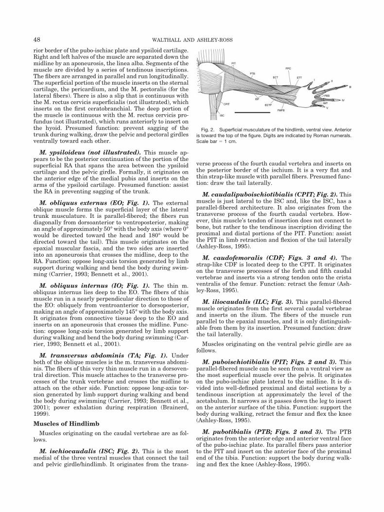
M. ischiocaudalis (ISC; Fig. 2). This is the mostmedial of the three ventral muscles that connect the tailand pelvic girdle/hindlimb. It originates from the trans-
verse process of the fourth caudal vertebra and inserts onthe posterior border of the ischium. It is a very flat andthin strap-like muscle with parallel fibers. Presumed func-tion: draw the tail laterally.
M. caudalipuboischiotibialis (CPIT; Fig. 2). Thismuscle is just lateral to the ISC and, like the ISC, has aparallel-fibered architecture. It also originates from thetransverse process of the fourth caudal vertebra. How-ever, this muscle’s tendon of insertion does not connect tobone, but rather to the tendinous inscription dividing theproximal and distal portions of the PIT. Function: assistthe PIT in limb retraction and flexion of the tail laterally(Ashley-Ross, 1995).
M. caudofemoralis (CDF; Figs. 3 and 4). Thestrap-like CDF is located deep to the CPIT. It originateson the transverse processes of the forth and fifth caudalvertebrae and inserts via a strong tendon onto the cristaventralis of the femur. Function: retract the femur (Ash-ley-Ross, 1995).
M. iliocaudalis (ILC; Fig. 3). This parallel-fiberedmuscle originates from the first several caudal vertebraeand inserts on the ilium. The fibers of the muscle runparallel to the epaxial muscles, and it is only distinguish-able from them by its insertion. Presumed function: drawthe tail laterally.
Muscles originating on the ventral pelvic girdle are asfollows.
M. puboischiotibialis (PIT; Figs. 2 and 3). Thisparallel-fibered muscle can be seen from a ventral view asthe most superficial muscle over the pelvis. It originateson the pubo-ischiac plate lateral to the midline. It is di-vided into well-defined proximal and distal sections by atendinous inscription at approximately the level of theacetabulum. It narrows as it passes down the leg to inserton the anterior surface of the tibia. Function: support thebody during walking, retract the femur and flex the knee(Ashley-Ross, 1995).
M. pubotibialis (PTB; Figs. 2 and 3). The PTBoriginates from the anterior edge and anterior ventral faceof the pubo-ischiac plate. Its parallel fibers pass anteriorto the PIT and insert on the anterior face of the proximalend of the tibia. Function: support the body during walk-ing and flex the knee (Ashley-Ross, 1995).
Fig. 2. Superficial musculature of the hindlimb, ventral view. Anterioris toward the top of the figure. Digits are indicated by Roman numerals.Scale bar � 1 cm.
48 WALTHALL AND ASHLEY-ROSS

M. ischioflexorius (ISF; Figs. 2 and 3). This mus-cle originates from the posterior and lateral edge of theischium behind the PIT. It is parallel-fibered and, like thePIT, divided into distinct proximal and distal portions bya tendinous inscription. However, the proximal section isincompletely divided from the PIT; it is possible to seewhat seems to be the division between muscles, but me-chanical separation is difficult. Only the distal portion iscompletely separate from the PIT. It inserts on the plantaraponeurosis overlying the flexor primordialis communis.Function: assist in limb retraction (Ashley-Ross, 1995).
M. puboischiofemoralis externus (PIFE; Figs. 3and 4). This fan-shaped muscle originates from much ofthe ventral surface of the pubo-ischiac plate and crossesthe hip joint to insert on the ventral surface of the femur.The insertion on the femur is extensive, covering nearlytwo-thirds of the length of the ventral surface of the bone.Function: support the body during walking and retract thefemur (Ashley-Ross, 1995).
M. pubifemoralis (PFM; Fig. 4). The PFM is theanteriormost of the ventral deep muscles of the thigh. Itoriginates from the anterior and ventral surface of thepubo-ischiac plate anterior to the PIFE. It inserts on theventral face of the femur both anterior and distal to theinsertion of the PIFE but posterior to the m. pubois-
chiofemoralis internus (PIFI). Function: support the bodyduring walking and retract the femur (Ashley-Ross, 1995).
M. ischiofemoralis (not illustrated). This shortmuscle spans the hip joint, originating on the lateral bor-der of the ischium (the posterior portion of the pubo-ischiac plate), and inserting on the posterior face of thehead of the femur. Presumed function: retract the femur.
Muscles originating on the dorsal pelvic girdle are asfollows.
M. puboischiofemoralis internus (PIFI; Figs. 3and 4). The PIFI originates from the internal (dorsal)face of the pubo-ischiac plate. It passes anterior to theilium, crossing the hip joint and inserting extensively onthe femur (covering the anterior face, as well as extendingdorsally and ventrally), anterior to the insertion of thePIFE. Function: protract and elevate the femur (Ashley-Ross, 1995).
M. extensor iliotibialis (ILT; Fig. 3). This strap-like, parallel-fibered muscle originates from the lateralsurface of the ilium above the acetabulum. It crosses thehip and knee joints to insert via a long strong tendon onthe spine of the tibia. Function: extend the knee joint(Ashley-Ross, 1995).
Fig. 3. Superficial musculature of the hindlimb, dorsal view. Anterior is toward the top of the figure. Digitsare indicated by Roman numerals. Scale bar � 1 cm.
Fig. 4. Deep musculature of the hindlimb, ventral view. Anterior is toward the top of the figure. Digits areindicated by Roman numerals. Scale bar � 1 cm.
49NEWT POSTCRANIAL MYOLOGY

M. iliofibularis (ILFB; Fig. 3). The ILFB originatesfrom the external face of the ilium just posterior to theILT. It courses parallel to, but slightly deep and posteriorto, the ILT, crossing both hip and knee joints to insert viaa tendon on the posterior side of the proximal end of thefibula. Function: elevate the femur and flex the knee joint(Ashley-Ross, 1995).
Muscles originating on the femur are as follows.
M. extensor cruris tibialis (ECT; Figs. 2–4). Thismuscle originates from the tibial epicondyle of the femur.It can be seen from both ventral and dorsal views, as itfans out from its origin to cover much of the anterior faceof the tibia, with expansions onto the dorsal and ventralfaces of that bone. It inserts extensively on anterodorsaland anteroventral faces of the tibia. Presumed function:extend the knee joint (tibia).
M. extensor tarsi tibialis (ETT; Figs. 3 and 4).This muscle originates from the tibial epicondyle of thefemur next to the ECT. The muscle fibers run parallel tothe ECT and insert on the ventral surface of the tibialebone. Presumed function: supinate the foot.
M. extensor cruris et tarsi fibularis (ECTF; Figs.2 and 3). The ECTF originates from the fibular epicon-dyle of the femur and, complementary to the ECT, insertson much of the posterodorsal and posteroventral faces ofthe fibula. A portion of the muscle inserts on the fibulare.Presumed function: extend the knee joint (fibula).
M. femorofibularis (FMFB; Figs. 2 and 4). Thisstrap-like, parallel-fibered muscle originates from the ven-tral face of the distal end of the femur. It inserts on theposterolateral face of the fibula. Unlike many othersalamander species studied, the FMFB is a fairly robustmuscle in T. torosa. Function: flex the knee joint (Ashley-Ross, 1995).
M. flexor primordialis communis (FPC; Figs. 2and 4). The bulk of the FPC originates from the posterov-entral face of the fibula, though part of it arises via atendon from the fibular epicondyle of the femur. The fibersrun ventrally and insert onto the plantar aponeurosis(plantar fascia). The large aponeurosis extends over theventral surface of the foot and divides into five tendonsthat insert on the proximal end of the terminal phalanx ofeach digit. Function: press the plantar surface of the footfirmly against the ground during walking (Ashley-Ross,1995).
M. extensor digitorum communis (EDC; Fig. 3).The thin EDC is located on the dorsal surface of the crusbetween the ETT and ECTF. It originates mostly from thelateral epicondyle of the femur anterior to the ECTF, butmay also demonstrate a small number of fibers arisingfrom the tibial spine. The fibers of the EDC fan out toinsert on the bases of the metatarsals by many smalltendons. Presumed function: extend the knee joint andabduct the foot.
Muscles of the crus and foot are as follows.
M. interosseus cruris (IOC; Fig. 4). This musclejoins the medial sides of the tibia and fibula. It arises fromthe proximal part of the fibula and inserts on the distal
portion of the tibia. Presumed function: stabilize the twolong bones of the crus.
M. flexor accessorius lateralis (FAL; Fig. 4). TheFAL originates from the lateral edge of the fibulare andinserts on the dorsal side of the plantar fascia. The fibersfollow a diagonal course toward the anterior side of thefoot. Presumed function: pronate the foot.
M. flexor accessorius medialis (FAM; Fig. 4). Thismuscle originates from the distal region of the fibula, thefibulare, and intermedium. It inserts on the plantar fascia,similar to the FAL. The muscle fibers run parallel to thoseof the FAL. Both the FAM and the FAL are parallel-fibered and generally strap-like in appearance. Presumedfunction: pronate the foot.
M. pronator profundus (PP; Fig. 4). This deepflexor muscle of the foot is roughly triangular in shape. Itoriginates from the medial side of the fibula, and its fibersconverge to insert on the lateral face of the distal end ofthe tibia, tibiale, and the base of the first metatarsal.Presumed function: pronate the foot.
M. caput longum musculorum contrahentium(CLMC; Fig. 4). The CLMC is a thin muscle that liesdeep to the plantar fascia. It originates from the distalportion of the fibula on the medial side. The fibers inserton a flat tendon that attaches to the distal tarsal bones.Presumed function: flex (depress) the tarsus.
Mm. flexores breves superficiales (FBS; Fig. 2).These small muscles originate from the dorsal side of theplantar fascia. They have a complicated insertion pattern;for all digits except digit I, the muscles are tripartite, withthe lateral portions inserting on the ventral side of thedistal ends of the metatarsals, and the medial portioninserting on the ventral surface of the proximal end of theproximal phalanx (this connection is hidden under theextensions of the plantar fascia in Fig. 4). For digit I, theinsertion is on the distal end of its associated metatarsal,without connection to the phalanx. Presumed function:flex the digits.
Mm. intermetatarsales (IMT; Figs. 2–4). Thesemuscles form the web-like structures between the digits.They attach the metatarsals of adjacent digits. They ex-tend further distally on the fibular side of each metatar-sal. Presumed function: adduct the metatarsals and theattached digits.
Mm. contrahentes digitorum (CD; Fig. 4). Thesesmall muscles originate from the flat tendon of the CLMCas well as the tarsal bones. They pass from the tarsalbones along the ventral axis of the metatarsals to insert onthe proximal phalanx of each digit. Presumed function:flex the digits.
M. abductor digiti V (AbD5; Fig. 4). The AbD5arises from the ventral surface of the distal end of thefibula. It has an extensive insertion, making a fleshy at-tachment to the posterior side of the fibulare, basale V,and the base of the fifth metatarsal. Presumed function:abduct the fifth metatarsal.
50 WALTHALL AND ASHLEY-ROSS

Mm. extensores digitorum breves (EDB; Fig. 3).These extensor muscles of the digits are a series of fiveslips, each of which originates from the distal tarsal bonesat the base of its respective digit and inserts on the dorsalsurface of the proximal end of its terminal phalanx via along tendon. The muscular portion only extends to approx-imately two-thirds of the length of the metatarsal; thetendon continues to the terminal phalanx of the digit.Presumed function: extend the digits.
M. abductor et extensor digiti I (AbED1; Fig. 3).The AbED1 originates from the intermedium and centralebones of the wrist. Its insertion is on the lateral face of themetatarsal and the phalanx via a small slip. Presumedfunction: abduct and extend the first metatarsal and itsassociated digit.
Muscles of ForelimbMuscles connecting the skull and pectoral girdle are as
follows.
M. opercularis (O; Fig. 6). The strap-like opercu-laris originates from the cartilaginous operculum associ-ated with the otic capsule. Its fibers course parasagittallyto insert on the anterior edge of the cartilaginous supras-capula. Presumed function: conduct sound, dampen move-ments of the operculum caused by head motion.
M. cucullaris (C; Fig. 6). The cucullaris muscle iscomposed of two heads: anterior and posterior. Both headsarise together from the dorsal fascia and posterior surfaceof the skull. The heads diverge as they pass posteriorly;the anterior, larger head inserts on the lateral surfaces ofthe procoracoid and scapula between the procoracohume-ralis and the dorsalis scapulae, while the fibers of theposterior head run ventrally to insert on the anterolateralborder of the scapula. Presumed function: turn or depressthe head.
Muscles originating on the ventral pectoral girdle are asfollows.
M. pectoralis (P; Fig. 5). The pectoralis is the mostsuperficial ventral chest muscle. It is fan-shaped, with thefibers converging to insert on the crista ventralis of thehumerus. The muscle has a varied origin, with some of thefibers arising from the aponeurosis that separates theright and left sides of the pectoralis, some arising from thesternum, and the posteriormost fibers taking their originfrom tendinous inscriptions of the rectus abdominis. Pre-sumed function: adduct and retract the humerus and sup-port the weight of the body on the forelimb.
M. supracoracoideus (SC; Fig. 5). The SC origi-nates from the ventral superficial surface of the coracoidcartilage. Its posterior portion lies deep to the P. Themuscle is fan-shaped and parallel-fibered; it inserts via aflat tendon on the crista ventralis of the humerus, adja-cent to the insertion of the pectoralis. Presumed function:adduct the humerus and support the weight of the body onthe forelimb.
M. procoracohumeralis (PCH; Figs. 5 and 6).This muscle is the most anteriorly located of the musclesjoining the pectoral girdle to the humerus. It originatesfrom the procoracoid cartilage and inserts on the anteriorsurface of the proximal humerus. Presumed function: pro-tract the humerus.
M. coracobrachialis longus (CBL; Fig. 5). TheCBL arises from the posterolateral surface of the coracoid.It is a parallel-fibered muscle whose fibers run along theaxis of the humerus and insert on the medial face of thedistal end of that bone. Presumed function: retract thehumerus.
M. anconaeus coracoideus (AC; Fig. 5). This mus-cle originates via a long tendon from the coracoid andextends down the posterior side of the upper forelimb,where it unites with m. anconeus scapularis medialis nearthe middle of the humerus. The joined muscles insert onthe olecranon process of the ulna. Presumed function:extend the elbow joint.
Fig. 5. Superficial musculature of the forelimb, ventral view. Anterior is toward the top of the figure. Digitsare indicated by Roman numerals. Scale bar � 1 cm.
51NEWT POSTCRANIAL MYOLOGY

Muscles originating on the dorsal pectoral girdle are asfollows.
M. latissimus dorsi (LD; Fig. 6). The LD is a fan-shaped, flat, triangular muscle that is the largest of thedorsal shoulder muscles. It originates from dorsal fasciaand the fibers converge to insert on the posterior surface ofthe crista ventralis of the humerus via a strong tendon.Presumed function: retract the humerus.
M. dorsalis scapulae (DS; Fig. 6). The dorsalisscapulae is another fan-shaped muscle joining the pecto-ral girdle and the humerus. It originates from the dorso-lateral surface of the suprascapular cartilage; the fiberspass around the anterior side of the humerus to insert onthe crista ventralis of the humerus. Presumed function:elevate the humerus.
M. anconaeus scapularis medialis (ASM; Fig.6). The ASM arises from the scapula posterior to theglenoid fossa and also from the connective tissue sur-rounding the joint capsule. Its parallel fibers run down theposterior side of the humerus and insert on the olecranonprocess. Approximately midway along the humerus, thefibers of the ASM are joined by those of the AC. Presumedfunction: extend the elbow joint.
Muscles originating on the humerus are as follows.
M. humeroantebrachialis (HAB; Figs. 5 and 6).This is a thick parallel-fibered muscle that arises from theanterior surface of the proximal end of the humerus. Itcrosses the elbow joint to insert on the proximal end of theradius. Presumed function: flex the elbow joint.
M. anconaeus humeralis lateralis (AHL; Fig. 6).The AHL is the most superficial upper forelimb musclefrom a dorsal view. It originates from the proximal two-
thirds of the lateral face of the humerus and inserts on theolecranon process of the ulna. Presumed function: extendthe elbow joint.
M. anconaeus humeralis medialis (AHM; Fig.5). This final member of the anconaeus group lies along,and arises from, the entire posterior face of the humerus.Like the other anconaeus muscles, it inserts on the olec-ranon process of the ulna. Presumed function: extend theelbow joint.
M. flexor digitorum communis (FDC; Figs. 5 and7). This thin muscle originates from the medial epicondyleof the humerus. The fibers run parallel to the direction ofthe radius and ulna and insert on a flat tendon that fillsthe entire palm region, the palmar fascia. The fascia con-tinues as four small tendons that run over the ventralsurface of the metacarpals and phalanges and finally at-taches to the proximal end of the distal phalanx of eachdigit. Presumed function: flex the carpus and digits.
M. flexor antebrachii et carpi radialis (FACR;Figs. 5, 7, and 8). The FACR is the most anterior flexormuscle of the forearm, originating from the proximal por-tion of the medial epicondyle of the humerus. It is over-lain, and thus obscured, for much of its length by the FDC.It inserts extensively on the anterior face of the radius,with a small portion of the muscle crossing the wrist jointto insert on the radiale. Presumed function: flex the car-pus and pronate the forearm.
M. flexor antebrachii et carpi ulnaris (FACU;Figs. 5 and 7). This muscle is the mirror image of theFACR. It arises from the distal portion of the medialepicondyle of the humerus, and it inserts on much of theposterior edge of the ulna. A slip of the muscle crosses thewrist joint to insert on the lateral faces of the ulnare andintermedium. Presumed function: flex the carpus and su-pinate the forearm.
Fig. 6. Superficial musculature of the forelimb, dorsal view. Anterior is toward the top of the figure. Digitsare indicated by Roman numerals. Scale bar � 1 cm.
52 WALTHALL AND ASHLEY-ROSS

M. extensor digitorum communis (EDC; Figs. 6and 8). The EDC is the most superficial of the forearmmuscles from a dorsal view. It is a thin parallel-fiberedmuscle that originates from the lateral epicondyle of thehumerus. It inserts on the dorsal surfaces of the proximalphalanges of digits II, III, and IV via paired tendons.Presumed function: extend the carpus and digits.
M. extensor antebrachii et carpi radialis(EACR; Figs. 5, 6, and 8). The EACR lies deep, andslightly anterior, to the EDC. It arises from the proximalpart of the lateral epicondyle of the humerus and insertson almost the entire anterolateral surface of the radiusand also on the lateral surface of the radiale. Presumedfunction: extend the carpus and forearm.
M. extensor antebrachii et carpi ulnaris (EACU;Figs. 6 and 8). This muscle mirrors the EACR; it is theposteriormost extensor muscle of the forearm and is par-
tially covered by the EDC. It arises from the distal portionof the lateral epicondyle of the humerus and inserts on theposterolateral face of the ulna, as well as on the ulnareand intermedium. Presumed function: extend the carpusand forearm.
Muscles of the forearm and hand are as follows.
M. contrahentium caput longum (CCL; Fig. 7).The strap-like CCL originates on the internal surface ofthe ulna and on fascia connecting the radius and ulna. Itinserts on an extensive tendon that connects the carpalbones. Presumed function: flex the carpus.
M. flexor accessorius lateralis (FAL; Fig. 7). Thisis a simple parallel-fibered, strap-like muscle that liesdeep to FDC. It originates from the distal end of the ulna,as well as the ulnare and intermedium. The fibers rundiagonally toward the radius and first digit, as well asventrally, to insert on the dorsal surface of the palmar
Fig. 7. Deep musculature of the forearm, ventral view. Anterior is toward the top of the figure. Digits areindicated by Roman numerals. Scale bar � 1 cm.
Fig. 8. Deep musculature of the forearm, dorsal view. Anterior is toward the top of the figure. Digits areindicated by Roman numerals. Scale bar � 1 cm.
53NEWT POSTCRANIAL MYOLOGY
fascia. Presumed function: assist in flexing the metacar-pals and digits.
M. flexor accessorius medialis (FAM; Fig. 7). Likethe FAL, this muscle has a simple strap-like architectureand inserts on the dorsal side of the palmar fascia. Theorigin of the FAM is somewhat more extensive than thatof the FAL, however, being approximately the distal thirdof the ulna, as well as the ulnare and intermedium. Pre-sumed function: assist in flexing the metacarpals anddigits.
M. pronator profundus (PP; Fig. 7). This very deepmuscle originates from the medial side of the ulna, theulnare, and intermedium. The PP crosses to the radial sideof the wrist and inserts on the radiale and the base of thefirst metacarpal. Presumed function: pronate the hand.
Mm. flexores breves superficiales (FBS; Fig. 5).These small muscles originate from the dorsal surface of thepalmar fascia and insert on the lateral edges of the distalends of the metacarpals. For digits II and III, slips of the FBSare present on both sides of the metacarpals. For digits I andIV, however, the FBS is only seen on the medial side of themetacarpal. Presumed function: flex the digits.
Mm. intermetacarpales (IMC; Figs. 5–8). The in-termetacarpales muscles are roughly triangular in shapeand connect adjacent metacarpals. They originate on theradial side of each digit’s metacarpal bone and insert onthe ulnar side of the adjacent digit’s metacarpal. Pre-sumed function: adduct adjacent metacarpals and theirassociated digits.
Mm. extensores digitorum breves (EDB; Figs. 6and 8). These short muscles connect the carpal boneswith the phalanges. They originate from the carpals at thebases of digits II, III, and IV and insert via long tendons onthe dorsal surface of the proximal end of the correspond-ing digit’s terminal phalanx. Presumed function: extendthe digits.
M. abductor et extensor digiti I (AbED1; Figs.5–8). The AbED1 originates from the radiale and inter-medium bones of the wrist and inserts on the distal end ofthe first metacarpal. Presumed function: abduct and ex-tend the first metacarpal and its associated digit.
M. extensor lateralis digiti IV (ELD4; Figs. 5–8).The ELD4 is a very small muscle that originates from theulnare and intermedium and inserts on the lateral edge ofthe fourth metacarpal. Presumed function: abduct andextend the fourth metacarpal and its associated digit.
Mm. contrahentes digitorum (CD; Fig. 7). Theseshort muscles lie deep to the tendons of insertion of theFBS. They originate partly from the tendon that is theinsertion of the CCL and partly from the carpals. The CDsinsert on the proximal end of the proximal phalanx of eachdigit. Presumed function: flex the digits.
Mm. flexores breves profundi (FBP; Fig. 7). Thesedeep small muscles originate from the carpals at the basesof their respective digits. They insert primarily on themetacarpal, but also on the proximal end of the proximalphalanx, of each digit. Presumed function: flex the meta-carpals and their associated digits.
DISCUSSIONOverall, the trunk and limb musculature of Taricha torosa
is similar to that of other primarily terrestrial salamanderspecies studied (e.g., Francis, 1934; Ashley-Ross, 1992). Ta-ble 1 presents a synonymy of forelimb muscles in differentsalamander species. For trunk and hindlimb muscles, equiv-alent tables are presented in Simons and Brainerd (1999)and Ashley-Ross (1992), respectively. Hypaxial trunk mus-cles follow the typical vertebrate pattern of three obliquelyoriented layers along with a superficial rectus abdominis(Fig. 1). Most muscles in both the forelimb and hindlimb aregracile, with simple parallel-fibered, strap-like architecture.Hindlimbs differ from forelimbs in that the major musclescross multiple joints and thus may be capable of complexactions during locomotion.
While basic similarity of myology among salamander spe-cies is evident, there are nonetheless several specific differ-ences, particularly in the hindlimb, that are worth noting.For instance, Francis (1934) and Mivart (1869) describe thePIT in Salamandra and Cryptobranchus (previously calledMenopoma), respectively, as a continuous sheet of muscle,without a division into proximal and distal portions. TheCPIT is inserted into the posterior border at approximatelythe level of the acetabulum by means of a tendon, but thetendinous inscription seen in Taricha, Ambystoma, and Di-camptodon (Ashley-Ross, 1992) at that location is lacking. Itis unclear what functional significance this difference mayhave; Ashley-Ross (1995) showed in Dicamptodon that thePIT and CPIT are coactive during walking, suggesting thatpassive resistance to tension that would be afforded by ten-don is not an issue.
Interestingly, the present observation of a lack of differ-entiation into anterior and posterior sections of the extensoriliotibialis muscle is at odds with Smith’s (1927) findings ona closely related species, Taricha granulosa (at that timenamed Triturus torosus). Smith (1927) described T. granu-losa as possessing two distinct parts of this muscle, whichshe named the rectus femoris and glutaeus maximus. Thepresence or absence of division of the extensor iliotibialis intotwo separate parts varies among salamanders previouslystudied; many have a distinct pars anterior and pars poste-rior of this muscle [e.g., Cryptobranchus (Mivart, 1869);Salamandra (Francis, 1934); Pseudoeurycea (Baird, 1951);Necturus (Gilbert, 1973); Ambystoma and Dicamptodon(Ashley-Ross, 1992)], though other groups have a singlesheet of muscle [e.g., Amphiuma (Davison, 1895); Typhlo-molge (Emerson, 1905); Bolitoglossa (Restrepo and Hoyos,1998)]. Whether or not a particular species has one or twoparts of this muscle may depend on limb proportions: thosespecies possessing two heads of the muscle (Cryptobranchus,Necturus, Salamandra, Pseudoeurycea, Ambystoma, Di-camptodon) have robust limbs with relatively large muscles,while those having only one head of the extensor iliotibialis(Amphiuma, Typhlomolge, Bolitoglossa, Taricha) have rela-tively slender limbs and muscles. Aquatic versus terrestrialhabits do not seem to dictate the form of the muscle(s), asCryptobranchus, Necturus, Amphiuma, and Typhlomolgeare all exclusively aquatic (Mivart, 1869; Emerson, 1905;Obst et al., 1988), yet they differ in the form of the extensoriliotibialis. Similarly, Salamandra, Pseudoeurycea, Ambys-toma, Dicamptodon, Bolitoglossa, and Taricha are all pri-marily terrestrial (Obst et al., 1988; Petranka, 1998); thelatter two species have a single head of the extensor iliotibia-lis, while the others have two.
54 WALTHALL AND ASHLEY-ROSS
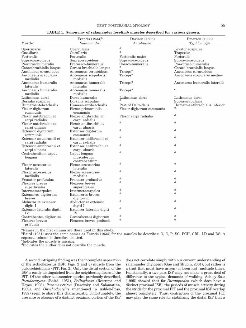
A second intriguing finding was the incomplete separationof the ischioflexorius (ISF; Figs. 2 and 3) muscle from thepuboischiotibialis (PIT; Fig. 2). Only the distal section of theISF is easily distinguished from the neighboring fibers of thePIT. Of the other salamander species previously described,Pseudoeurycea (Baird, 1951), Bolitoglossa (Restrepo andHoyos, 1998), Paramesotriton (Darevsky and Salomatina,1989), and Onychodactylus (mentioned in Ashley-Ross,1992) seem to share this characteristic. Unfortunately, thepresence or absence of a distinct proximal portion of the ISF
does not correlate simply with our current understanding ofsalamander phylogeny (Gao and Shubin, 2001), but rather isa trait that must have arisen (or been lost) multiple times.Functionally, a two-part ISF may not make a great deal ofdifference to the typical demands of walking; Ashley-Ross(1995) showed that for Dicamptodon (which does have adistinct proximal ISF), the periods of muscle activity duringthe stride for the proximal PIT and the proximal ISF overlapalmost completely. Thus, contraction of the proximal PITmay play the same role for stabilizing the distal ISF that a
TABLE 1. Synonymy of salamander forelimb muscles described for various genera.
MuscleaFrancis (1934)b Davison (1895) Emerson (1905)
Salamandra Amphiuma Typhlomolge
Opercularis Opercularis d Levator scapulaeCucullaris Cucullaris d TrapeziusPectoralis Pectoralis Pectoralis major PectoralisSupracoracoideus Supracoracoideus Supracoracoideus Supra-coracoideusProcoracohumeralis Procoraco-humeralis Coraco-humeralis Pro-coraco-humeralisCoracobrachialis longus Coraco-brachialis longus d Coraco-brachialis longusAnconaeus coracoideus Anconaeus coracoideus Triceps? Anconaeus coracoideusAnconaeus scapularis
medialisAnconaeus scapularis
medialisTriceps? Anconaeus scapularis medius
Anconaeus humeralislateralis
Anconaeus humeralislateralis
Triceps? Anconaeus humeralis lateralis
Anconaeus humeralismedialis
Anconaeus humeralismedialis
Triceps? c
Latissimus dorsi Dorso-humeralis Latissimus dorsi Latissimus dorsiDorsalis scapulae Dorsalis scapulae d Supra-scapularisHumeroantebrachialis Humero-antibrachialis Part of Deltoideus Humero-antibrachialis inferiorFlexor digitorum
communisFlexor primordialis
communisFlexor digitorum communis d
Flexor antebrachii etcarpi radialis
Flexor antibrachii etcarpi radialis
Flexor carpi radialis d
Flexor antebrachii etcarpi ulnaris
Flexor antibrachii etcarpi ulnaris
d d
Extensor digitorumcommunis
Extensor digitorumcommunis
d d
Extensor antebrachii etcarpi radialis
Extensor antibrachii etcarpi radialis
d d
Extensor antebrachii etcarpi ulnaris
Extensor antibrachii etcarpi ulnaris
d d
Contrahentium caputlongum
Caput longummusculorumcontrahentium
d d
Flexor accessoriuslateralis
Flexor accessoriuslateralis
d d
Flexor accessoriusmedialis
Flexor accessoriusmedialis
d d
Pronator profundus Pronator profundus d d
Flexores brevessuperficiales
Flexores brevessuperficiales
d d
Intermetacarpales Intermetacarpales d d
Extensores digitorumbreves
Extensores brevesdigitorum
d d
Abductor et extensordigiti I
Abductor et extensordigiti I
d d
Extensor lateralis digitiIV
Extensor lateralis digitiIV
d d
Contrahentes digitorum Contrahentes digitorum d d
Flexores brevesprofundi
Flexores breves profundi d d
aNames in the first column are those used in this study.bBaird (1951) uses the same names as Francis (1934) for the muscles he describes: O, C, P, SC, PCH, CBL, LD and DS. Aseparate column is therefore omitted.cIndicates the muscle is missing.dIndicates the author does not describe the muscle.
55NEWT POSTCRANIAL MYOLOGY

proximal ISF would during walking. A question still to beresolved, then, is why have a separate proximal ISF in sometaxa? Does the trait correlate with some other behavior, e.g.,burrowing? Recordings of muscle activity patterns duringactivities other than walking would need to be obtained toaddress this issue.
An observation that may be related to the morphology ofthe ISF is the relatively large size of the femorofibularis(FMFB; Fig. 4) in T. torosa. Ashley-Ross (1992) found thatthere appeared to be a trade-off between the two muscles
in size: Dicamptodon and newly metamorphosed Ambys-toma both possess a large ISF coupled with an extremelythin and narrow FMFB. In contrast, specimens of Amby-stoma that were well past metamorphosis had a relativelynarrow ISF and a robust FMFB. Likewise, Restrepo andHoyos (1998) describe the FMFB of Bolitoglossa as beingsubstantial, with an extensive insertion along much of thelength of the fibula. Finally, Darevsky and Salomatina(1989) note the same trade-off in size between the ISF andFMFB in Paramesotriton. Francis (1934) has noted that
TABLE 1. Synonymy of salamander forelimb muscles described for various genera (continued)
Gilbert (1973) Mivart (1869) Restrepo & Hoyos (1998) Smith (1927)Necturus Menopoma Bolitoglossa Triturusd Levator anguli scapulae Opercularis Levator scapulae
Cucullaris TrapeziusCucullaris major and
minor CucullarisPectoralis Pectoralis Pectoralis PectoralisSupracoracoideus Part of Coraco-brachialis Supracoracoideus SupracoracoideusProcoracohumeralis Subclavius Procoracohumeralis Procoracohumeralis
CoracobrachialisPart of Coraco-brachialis,
Part of Biceps Coracobrachialis longusCoraco-brachialis
longusAnconaeus Triceps Anconaeus coracoideus d
Anconaeus TricepsAnconaeus scapularis
medialisAnconaeus (Triceps)
scapularis medialis
Anconaeus TricepsAnconaeus humeralis
lateralisAnconaeus (Triceps)
humeralis lateralis
Anconaeus TricepsAnconaeus humeralis
medialisAnconeus (Triceps)
humeralis medialisLatissimus dorsi Latissimus dorsi Dorsohumeralis Latissimus dorsiDorsalis scapulae Deltoid Dorsalis scapulae Dorsalis scapulaeHumeroantebrachialis Part of Biceps Humero antibrachialis Biceps brachiiFlexor primordialis
communis Flexor longusFlexor primordialis
communisFlexor digiti
communisFlexor antebrachii et carpi
radialis Pronator teresFlexor antibrachii et carpi
radialis Flexor carpi radialisFlexor antebrachii et carpi
ulnaris dFlexor antibrachii et carpi
ulnaris Flexor carpi ulnarisExtensor digitorum
communis Extensor longusExtensor digitorum
communisExtensor digiti
communisExtensor antebrachii et
carpi radialis Supinator longusExtensor antibrachii et
carpi radialisExtensor carpi
radialisExtensor antebrachii et
carpi ulnaris UlnarisExtensor antibrachii et
carpi ulnarisExtensor carpi
ulnaris
d Flexor brevis
Caput longummusculorumcontrahentium d
d d Flexor accessories lateralis d
d dFlexor accessorius
medialis d
d d Pronator profundus d
d dFlexores breves
superficiales d
d d Intermetacarpales d
d dExtensores digitorum
breves d
d Extensor brevisAbductor et extensor digiti
I d
d d Extensor lateralis digiti IV d
d d d d
d d d
d
56 WALTHALL AND ASHLEY-ROSS

branches of the same nerve innervate the distal ISF and theFMFB; it is possible that the two muscles represent alterna-tive solutions to the same functional demands of locomotion.
The forelimb musculature does not show as much reg-ular variation as the hindlimb musculature across species.Complicating comparisons is the fact that few studieshave examined the muscles of the forearm and manus(exceptions are Francis, 1934; Restrepo and Hoyos, 1998).However, among the muscles of the shoulder and upperarm, most variation seems to be in the relative size, notposition, of various muscles. For example, the procoraco-humeralis (PCH; Figs. 5 and 6) varies in size from largeand robust in Cryptobranchus (Mivart, 1869), Pseudoeu-rycea (Baird, 1951), and Bolitoglossa (Restrepo and Hoyos,1998) through average size (Taricha) to extremely thinand narrow in Typhlomolge (Emerson, 1905). A secondexample of variation in muscle size is the latissimus dorsi(LD; Fig. 6), which is large in Typhlomolge (Emerson,1905), Pseudoeurycea (Baird, 1951), Bolitoglossa (Re-strepo and Hoyos, 1998), and Taricha; the LD has a muchless extensive origin in Cryptobranchus (Mivart, 1869).
Within an individual salamander, it is immediately ap-parent that the muscles of the forelimbs and hindlimbsshare much of the same pattern, particularly in the distallimb segments (compare Figs. 2 and 5, 3 and 6). Indeed,the forearm and crus, as well as the manus and pes, shownearly identical muscular morphology. Furthermore, dor-sal and ventral musculature (especially in the forearm)are essentially mirror images. The digits are controlledsuperficially by muscles originating from the distal end ofthe proximal limb segment; these muscles (EDCs in bothlimbs, FPC in the hindlimb, and FDC in the forelimb) allend in aponeuroses from which extend long tendons to thedistal phalanges. Below this superficial layer are musclesthat control torsional movements of the manus/pes (e.g.,FAM, FAL, and PP) and of the crus and antebrachiumitself (ECT, ETT, ECTF, EACR, EACU, FACR, andFACU). In the hindlimb, there is more differentiationbetween ventral and dorsal musculature; in the forelimb,ventral and dorsal sides appear nearly identical. Finally,the series of muscles that span from the plantar/palmaraponeurosis to the phalanges show indistinguishable ar-rangements between fore- and hindlimbs. The mirror-likearrangement of the muscles of the dorsal and ventralantebrachium would seem to argue that this limb segmenthas undergone little, if any, anatomical rearrangementdue to selective pressure on locomotor demands. The hind-limb, in contrast, is thought to provide most of the powerfor locomotion (Barclay, 1946; Daan and Belterman, 1968;Edwards, 1977; Peters and Goslow, 1983) and thereforehas altered its structures to make walking more efficientor powerful. While the salamander forelimb is known tohave undergone evolutionary reduction in digit number(loss of digit V) (Shubin, 2002), correspondence in themorphology of its flexor and extensor muscles suggeststhat it may nonetheless retain the primitive pattern ofmusculature of the tetrapod limb, which may be of use infossil reconstructions.
LITERATURE CITEDAshley-Ross MA. 1992. The comparative myology of the thigh and
crus in the salamanders Ambystoma tigrinum and Dicamptodontenebrosus. J Morphol 211:147–163.
Ashley-Ross MA. 1995. Patterns of hind limb motor output duringwalking in the salamander Dicamptodon tenebrosus, with compar-isons to other tetrapods. J Comp Physiol A 177:273–285.
Baird IL. 1951. An anatomical study of certain salamanders of thegenus Pseudoeurycea. Univ Kansas Sci Bull 34:221–265.
Barclay OR. 1946. The mechanics of amphibian locomotion. J Exp Biol23:177–203.
Bennett WO, Simons RS, Brainerd EL. 2001. Twisting and bending:the functional role of salamander lateral hypaxial musculatureduring locomotion. J Exp Biol 204:1979–1989.
Bock WJ, Shear CR. 1972. A staining method for gross dissection ofvertebrate muscles. Anat Anz Bd 130:222–227.
Brainerd EL. 1999. New perspectives on the evolution of lung venti-lation mechanisms in vertebrates. Exp Biol Online 4:11–28.
Breithaupt R, Dahnke J, Zahedi K, Hertzberg J, Pasemann F. 2002.Robo-salamander: an approach for the benefit of both robotics andbiology. In: Bedaud P, editor. Fifth international conference onclimbing and walking robots. London, UK: Professional Engineer-ing Publishing. p 55–62.
Carrier DM. 1993. Action of the hypaxial muscles during walking andswimming in the salamander Dicamptodon ensatus. J Exp Biol180:75–83.
Daan S, Belterman T. 1968. Lateral bending in locomotion of somelower tetrapods. Proc Ned Akad Wetten C 71:245–266.
Darevsky IS, Salomatina NI. 1989. Notes on hind limb structure inthe salamander, Paramesotriton deloustali, and its mode of life.J Herpetol 23:429–433.
Davison A. 1895. A contribution to the anatomy and phylogeny ofAmphiuma means (Gardner). J Morphol 11:375–410.
Edwards JL. 1977. The evolution of terrestrial locomotion. In: HechtMK, Goody PC Hecht BM, editors. Major patterns in vertebrateevolution. New York: Plenum Publishing. p 553–576.
Edwards JL. 1989. Two perspectives on the evolution of the tetrapodlimb. Am Zool 29:235–254.
Emerson ET. 1905. General anatomy of Typhlomolge rathbuni. ProcBost Soc Nat Hist 32:49–76.
Francis ETB. 1934. The anatomy of the salamander. London: OxfordUniversity Press.
Gao K-Q, Shubin NH. 2001. Late Jurassic salamanders from northernChina. Nature 410:574–577.
Gilbert SG. 1973. Pictorial anatomy of the Necturus. Seattle: Univer-sity of Washington Press.
Ijspeert AJ. 2000. A 3-D biomechanical model of the salamander. In:Heudin J-C, editor. Proceedings of the 2nd International Confer-ence of Virtual Worlds. Heidelberg: Springer-Verlag. p 225–234.
Ijspeert AJ. 2001. A connectionist central pattern generator for theaquatic and terrestrial gaits of a simulated salamander. Biol Cy-bernet 84:331–348.
Mivart SG. 1869. Notes on the myology of Menopoma alleghaniense.Proc Zool Soc Lond 1869:254–261.
Obst FJ, Richter K, Jacob U. 1988. The completely illustrated atlas ofreptiles and amphibians for the terrarium. Neptune City, NJ:T.F.H. Publications.
Peters SE, Goslow GE. 1983. From salamanders to mammals: conti-nuity in musculoskeletal function during locomotion. Brain BehavEvol 22:191–197.
Petranka JW. 1998. Salamanders of the United States and Canada.Washington, DC: Smithsonian Institution Press.
Restrepo AE, Hoyos JM. 1998. Musculatura de los miembros y de lascinturas en Bolitoglossa adspersa (Peters, 1863) (Urodela, Pleth-odontidae). Alytes 16:25–49.
Shubin NH. 2002. Origin of evolutionary novelty: examples fromlimbs. J Morphol 252:15–28.
Simons RS, Brainerd EL. 1999. Morphological variation of hypaxialmusculature in salamanders (Lissamphibia: Caudata). J Morphol241:153–164.
Smith GM. 1927. The detailed anatomy of Triturus torosus. Trans RoySoc Can 1927:451–484.
Taylor T, Massey C. 2001. Recent developments in the evolution ofmorphologies and controllers for physically simulated creatures.Artificial Life 7:77–87.
57NEWT POSTCRANIAL MYOLOGY