Post-Traumatic Osteoarthritis: Biologic Approaches to ...
30
10 Post-Traumatic Osteoarthritis: Biologic Approaches to Treatment Sukhwinderjit Lidder 1 and Susan Chubinskaya 1,2,3 1 Department of Biochemistry, Rush University Medical Center, Chicago, IL 2 Section of Rheumatology, Department of Internal Medicine 3 Department of Orthopedic Surgery, Rush University Medical Center, Chicago, IL, USA 1. Introduction Joint injuries are becoming increasingly common, with young adults between the ages of 18- 44 seeking medical attention for joint sprains, dislocation, fractures, anterior cruciate ligament (ACL) and meniscal tears, and others. The cascade of events that follow these joint injuries have been shown to increase the risk of post-traumatic osteoarthritis (PTOA) by 20- 50% (Anderson et al,2011). Therefore, understanding biological responses that predispose to PTOA should help in determining treatment strategies to delay and/or prevent the progression of the disease. Ex vivo and in vivo studies (Anderson et al,2011;Buckwalter et al,2004;Furman et al, 2007; Guilak et al,2004; Hurtig et al, 2009) have provided evidence that the force and severity of the impact applied to the joint are among the risk factors involved in the development of PTOA. Recent research on the events that follow joint trauma have shown chondrocyte death and apoptosis, inflammation (elevation of caspases, selected pro- inflammatory cytokines, matrix fragments, nitric oxide, reactive oxygen species [ROS], etc.) and matrix damage/fragmentation to be early phase responses to injury. Together they lead to the development of OA-like focal cartilage lesions characterized by the loss of matrix constituents, surface fibrillation, and fissures that if untreated have a tendency to expand and progress to fully-blown disease. Currently, the only treatments available for joint trauma are surgical interventions, such as microfracture, articular chondrocyte transplantation, autografting, allografting, debridement and lavage. There are also some experimental approaches that involve engineering of cartilage with the use of juvenile cartilage, scaffolds and various polymeric matrices, but those are still in development. To the best of our knowledge none of them could regenerate normal adult hyaline cartilage that is able to perform required functions, sustain the load and integrate with the host tissue. Furthermore, newly repaired tissue, due to its imperfect structural organization, may also be more susceptible to re-injury, an important aspect that often remains forgotten. Therefore, there is an unmet need in the development of novel therapeutic approaches based on the mechanisms that drive the onset and progression of PTOA in order to stimulate biologic repair, delay or prevent the need of surgery, or when used together, improve the outcome of surgical interventions if biologics are applied prior, during, or soon after surgery. Based on our current understanding of the molecular and www.intechopen.com
Transcript of Post-Traumatic Osteoarthritis: Biologic Approaches to ...

10
Post-Traumatic Osteoarthritis: Biologic Approaches to Treatment
Sukhwinderjit Lidder1 and Susan Chubinskaya1,2,3 1Department of Biochemistry, Rush University Medical Center, Chicago, IL
2Section of Rheumatology, Department of Internal Medicine 3Department of Orthopedic Surgery, Rush University Medical Center, Chicago, IL,
USA
1. Introduction
Joint injuries are becoming increasingly common, with young adults between the ages of 18-
44 seeking medical attention for joint sprains, dislocation, fractures, anterior cruciate
ligament (ACL) and meniscal tears, and others. The cascade of events that follow these joint
injuries have been shown to increase the risk of post-traumatic osteoarthritis (PTOA) by 20-
50% (Anderson et al,2011). Therefore, understanding biological responses that predispose to
PTOA should help in determining treatment strategies to delay and/or prevent the
progression of the disease. Ex vivo and in vivo studies (Anderson et al,2011;Buckwalter et
al,2004;Furman et al, 2007; Guilak et al,2004; Hurtig et al, 2009) have provided evidence that
the force and severity of the impact applied to the joint are among the risk factors involved
in the development of PTOA. Recent research on the events that follow joint trauma have
shown chondrocyte death and apoptosis, inflammation (elevation of caspases, selected pro-
inflammatory cytokines, matrix fragments, nitric oxide, reactive oxygen species [ROS], etc.)
and matrix damage/fragmentation to be early phase responses to injury. Together they lead
to the development of OA-like focal cartilage lesions characterized by the loss of matrix
constituents, surface fibrillation, and fissures that if untreated have a tendency to expand
and progress to fully-blown disease.
Currently, the only treatments available for joint trauma are surgical interventions, such as microfracture, articular chondrocyte transplantation, autografting, allografting, debridement and lavage. There are also some experimental approaches that involve engineering of cartilage with the use of juvenile cartilage, scaffolds and various polymeric matrices, but those are still in development. To the best of our knowledge none of them could regenerate normal adult hyaline cartilage that is able to perform required functions, sustain the load and integrate with the host tissue. Furthermore, newly repaired tissue, due to its imperfect structural organization, may also be more susceptible to re-injury, an important aspect that often remains forgotten. Therefore, there is an unmet need in the development of novel therapeutic approaches based on the mechanisms that drive the onset and progression of PTOA in order to stimulate biologic repair, delay or prevent the need of surgery, or when used together, improve the outcome of surgical interventions if biologics are applied prior, during, or soon after surgery. Based on our current understanding of the molecular and
www.intechopen.com

Principles of Osteoarthritis – Its Definition, Character, Derivation and Modality-Related Recognition
234
cellular manifestations of injury the following therapeutic options could be considered for PTOA: chondroprotection, anti-inflammatory, matrix protection, and stimulation of matrix remodeling with pro-anabolic factors. These and other approaches will be discussed in details in the current book chapter.
2. Joint injuries and the risk of post-traumatic osteoarthritis (PTOA)
Although joint injuries are not as life threatening as myocardial infarctions and strokes, they are similarly life changing. They are progressive and debilitating, with progression often leading to OA. Traumatic insult to a joint, as a precursor to OA, has been studied since it was first described by Hunter in 1743 (Key,1933). It was reported that 13 to 18% of total hip or knee patients had an identifiable acute trauma to the joint (Kern et al,1988). Further evidence of joint injury being a major factor in the development and progression of PTOA came from the work of Roos, in which it was shown that early onset of OA can occur within 10 years after injury (Roos et al,1995). With increased social and sport-related activities of today’s society there are more and more younger people (18-44 years) who present with the evidence of post-traumatic focal cartilage lesions and OA- like changes occurred as a result of joint injury. This is in comparison to idiopathic OA, where its prevalence is increased with ageing and is more evident after the age of 50 (Anderson et al,2011;Brown et al,2006;Dirschl et al,2004;Gelber et al,2000). Approximately 12% of the overall prevalence of severe OA is attributable to post-traumatic hip, knee and ankle OA which corresponds to about 5.6 million people in the US suffering from PTOA (Brown et al,2006). With such a high prevalence of a lifelong nonfatal disability, there is enormous annual socioeconomic burden on the health system estimated to be $3.06 billion (Brown et al,2006). These numbers may be underestimated because they were based on patients presenting with severe OA that required total joint replacement and did not include patients with early or less advanced OA. A more recent study in 2008 (Murphy et al,2008) revealed that a history of knee injury carried a 56.8% lifetime risk of symptomatic knee OA.
3. Pathogenesis of PTOA
The pathogenesis of PTOA is not fully understood, in part due to the lack of correlation between the disease progression and the symptoms; therefore, it is difficult to estimate the number of individuals suffering from PTOA. Diagnosis is usually based on parameters used to diagnose idiopathic OA such as joint pain, visible signs of joint deformity, radiographic changes and biochemical tests that detect inflammation. However, many of these may not be presented at early stages after joint injury (Brown et al,2006). PTOA is not a unifactorial disease and there are a number of factors that could contribute to the onset and progression of PTOA: lost structural integrity of menisci and ACL, joint incongruence, lost muscle strength, continued physical activity, excessive biomechanical overload of the joint, intra-articular inflammation, and others (Furman et al,2006). The key difference between the two types of OA is the presence of precipitating insult to the joint in patients that suffer from PTOA versus idiopathic OA associated with ageing, genetics, obesity, occupation, bone density, metabolic disease, inflammation and abnormal biomechanics. Since it takes years and decades for the development of PTOA after injury, aforementioned factors known to contribute to the idiopathic OA may also play a role in the progression of PTOA. Regardless what causes PTOA it develops as a result of poor intrinsic regenerative ability of hyaline articular cartilage
www.intechopen.com

Post-Traumatic Osteoarthritis: Biologic Approaches to Treatment
235
(Anderson et al,2011;Pelletier et al,1998;Sandell et al,2001). In clinical setting, patients with signs of PTOA usually present with an advanced form of the disease in which the meniscus, ACL and the cartilage have eroded. As a result, there is a reduction in cartilage thickness with areas of complete loss and formation of fibrocartilagenous repair tissue. Biomechanically these patients have a decreased tensile strength and compressive stiffness (Furman et al,2006). The extent of cartilage damage depends on the intensity and force of the impact (Anderson et al,2011;Butler et al,2008; Furman et al,2006). The type of damage can be categorized as: a) cartilage only disruption characterized by changes in structural components of the matrix and chondrocyte death. These may progress to focal lesions. b) Fracturing along the tidemark, in which the cartilage tissue above the calcified zone can exhibit blistering and full thickness cartilage loss can be seen. c) Fracturing through the calcified cartilage into the subchondral plate that can in most severe cases form osteochondral fragments (Anderson et al,2011).
4. Current clinical treatments for PTOA
Joint injuries have a high prevalence especially in young individuals and unfortunately, predominantly surgical interventions are currently available for their treatment: a. Arthroscopic lavaging and debridement (Avouac et al,2010;Reichenbach et al,2010),
which wash and remove pieces of degenerative cartilage, fibrous tissue and synovial fluid full of catabolic mediators that may cause joint inflammation, swelling and destruction. The removal of loose debris, cartilage flaps, torn meniscal fragments and synovial fluid may provide a temporary relief but does not prevent the generation of more fragments and more inflammation. Hence, this method has shown little to no evidence of significant improvement in pain relief or restoration of joint function (Avouac et al,2010;Reichenbach et al,2010). The arthroscopic nature of this method limits its use to patients with small cartilage defects.
b. Viscosupplementation with hyaluronic acid via intra-articular injections into the knee joint potentially improves joint lubrication and may help to restore cartilage. It also provides some relief of pain not seen with other analgesics such as ibuprofen or nonsteroidal anti-inflammatory drugs (NSAIDs) (Iannitti et al,2011;Kappler et al,2010;Migliore et al,2011). This approach has been only effective in mild to moderate OA.
c. Recently, the usage of autologous blood products became a potential breakthrough in augmenting joint tissue healing (Anitua et al,2004,2006;Sanchez et al,2009). This new method provides cellular and humeral mediators which have been greatly beneficial in regenerative processes of cartilage and other connective tissues. Autologous blood products are heavily populated with platelets, which have the capacity to release growth factors from their ┙-granules (chemokines and newly synthesised metabolites) and thus positively influence the tissues with low healing potential. Intra-articular injection of platelet concentrate may represent an innovative treatment to improve cartilage remodelling. More studies are indicated to understand the mechanism of action and to optimize, standardize, and widely implement into the clinic this new technology.
d. Osteochondral grafting makes up about 10% of surgical procedures available up to date to repair cartilage lesions (Cole et al,2009). Unlike procedures that target repair or regeneration, osteochondral grafting has garnered significant attention because of its ability to replace the lesion with true hyaline cartilage and allow for a relatively short recovery period (Convery et al,1972;Horas et al,2003;Nam et al,2004;Shasha et al,2003). Osteochondral grafting involves harvesting a cylindrical donor plug of cartilage attached
www.intechopen.com

Principles of Osteoarthritis – Its Definition, Character, Derivation and Modality-Related Recognition
236
to underlying subchondral bone and implanting the graft into the recipient site covering the cartilage lesion. This procedure has a lot of potential for the treatment of isolated cartilage defects in young, active patients; however, graft survival is still limited with survival rates under 50% after 15 years (Shasha et al,2003). Other problems with grafting are the integration with the host cartilage and reduced viability and metabolism of residing chondrocytes in case of prolong stored allograft tissue (Kirk et al,2011).
e. Microfracture and bone marrow stimulation. These surgical options are employed when cartilage damage is confined to small focal areas. The damaged cartilage is removed to expose and perforate the subchondral bone. This can stimulate new cartilage growth in the subchondrol defect through the generation of a fibrin clot and recruitment of bone marrow mesenchymal stem cells. The fibrocartilage that covers the full thickness chondral lesion does not have the biomechanical strength and resilience of the native cartilage. Although this fibrocartilage has been shown to provide relief from the symptoms for several years (Miller et al,2004), it does not alter the progression of PTOA as patients present with OA symptoms 5 years after microfracture surgery (Miller et al,2004). In a majority of the patients, the size of the cartilage defect increases after microfracture (Von Keudell et al,2011). The implantation of collagen membranes over the microfracture in a technique referred to as Autologous matrix induced chondrogenesis (AMIC) have been used to improve the chondrogenic differentiation of the mesenchymal stem cells (Behrens et al,2006) into more hyaline like cartilage. In other efforts to improve cartilage regeneration after microfracture, Saw et al (Saw et al,2009) have developed an in vivo method in goats that used intra-articular injections of autologous peripheral blood progenitor cells and hyaluronic acid. The results have been promising and a clinical pilot study has shown regeneration of articular hyaline cartilage (Saw et al,2011).
f. Autologous chondrocyte implantation (ACI) was first described by Brittberg et al (Brittberg et al,1994). Although cartilage regeneration of the defect was observed, several serious problems have been associated with this method. 1) It is a two-step procedure. 2) The need to create additional defects within the normal and un-affected joint for the extraction of autologous chondrocytes. 3) The need for a two-week in vitro cell expansion to obtain a sufficient number of chondrocyte to cover and fill the original defect. 4) Reduced viability and altered phenotype of autologous cells; and finally, the quality and properties of regenerated fibrocartilage (Horas et al,2003).
g. Articular cartilage regeneration with stem cells is another cell-based cartilage repair procedure. Similar in concept to ACI, autologous mesenchymal stem cells have been used to decrease knee pain (Kuroda et al,2007). However, for the most part all listed methods generate fibrocartilage or a mixture of hyaline-like and fibrocartilage (Kuroda et al,2007).
h. Knee replacement surgery with a metal shell is often used to treat advanced PTOA in patients that show severe destruction of the joint and exhibit increasing joint stiffness and pain. However, the knee replacement itself has been associated with chronic pain, joint stiffness, post-operative inflammation, and prolong recovery (Gonzalez et al,2004).
Current surgical approaches are mainly utilized to treat the developed disease, while the whole idea of biologic treatment is based on the premise to arrest and/or prevent the onset and progression of the disease. Ideally, biologic interventions should be applied immediately or soon after the trauma incident. But, in reality patients present with moderate to severe PTOA when meniscus and cartilage erosion has already advanced. The lack of satisfactory surgical and other therapeutic approaches to successfully restore cartilage structure and
www.intechopen.com

Post-Traumatic Osteoarthritis: Biologic Approaches to Treatment
237
function still remains a challenge in current orthopedics. Only few clinical trials have been conducted to investigate the efficacy of various classes of therapeutics in PTOA. Therefore, advances in our understanding of the mechanisms that govern the development of the disease come primarily from in vitro or in vivo animal models of the PTOA.
5. In vivo and in vitro approaches to study PTOA
Animal models that resemble human OA pathology have been difficult to develop and generally require some surgical insult. In Table 1 we summarized the majority of in vivo and in vitro studies that focused on joint trauma and the fate of cartilage after chondral or osteochondral damage (Borrelli et al,2003;Clements et al,2004;Green et al,2006a,2006b;Lewis et al,2003;Newberry et al,1998;Vener et al,1992). As outlined, this literature consistently points to three overlapping phases after acute cartilage injury that include a death/apoptosis phase, an inflammatory phase, and a limited repair phase. Studying cellular responses initiated by acute injury we identified and characterized a sequence of biologic events (both catabolic and anabolic) that cause the progressive joint degeneration leading to PTOA (Anderson et al,2011;Bajaj et al,2010;Hurtig et al,2009;Pascual Garrido et al,2009).
Target Therapeutic Agent Reference
ChondroprotectionPI88
Caspase Inhibitors ( Z-VAD-FMK; Q-VD-Oph; Caspase-3;
Caspase-9)
Anti-oxidantsiNOS inhibitors
(L-NAME; L-NIL)
Rotenone
N-acetylcysteine
(Martin et al, 2009); (Phillips&Haut, 2004); (Pascual Garrido et al, 2009 ); (Bajaj et al, 2010 )
(Martin et al, 2009); (Pascual Garrido et al, 2009 ); (D'Lima et al, 2006 ); (D'Lima et al, 2001); (Huser & Davies 2006) (Lotz, 2010)
(Kurz et al, 2004)
(Marsh et al, 2002); (Pelletier et al, 1998, 1999, 2000)
(Goodwin et al, 2010 )
(Martin et al, 2009); (Martin et al, 2009)
Anti-inflammatory IRAP/ IL-1Ra
Anti-TNFα, PEGylated soluble TNFα
(Fox & Stephens, 2010); (Frisbie et al, 2002); (Evans et al, 2004); (Meijer et al, 2003)
(Zafarullah et al, 2003); (Martel-Pelletier, 1999); (Furman et al, 2006); (Fukui et al, 2001); (Evans et al, 2004); (Elsaid et al, 2009)
Matrix protection MMP inhibitors / TIMPs
ADAMTS inhibitors (AGG-523)
(Jarvinen et al, 1995); (Murrell et al, 1995) (Chockalingam et al, 2011)
(Glasson et al, 2005); (Chockalingam et al, 2011)
Pro-anabolic, inducers of repair
BMPs (BMP-2; BMP-7)
IGF-1
FGF-18
(Hunter et al, 2010); (Hayashi et al, 2008, 2010); (Hurtig et al, 2009); (Chubinskaya et al, 2007); (Cook et al, 2003); (Badlani et al, 2008)
(Chubinskaya et al, 2011); (Fortier et al, 2002); (Im et al, 2003)
(Moore et al, 2005); (Lotz & Kraus, 2010); (Ellsworth et al, 2002)
Table 1. Potential targets and therapeutic interventions for post-traumatic osteoarthritis. iNOS, inducible Nitric oxide synthase; IRAP, interleukin receptor antagonist protein; Anti-TNF-┙, tumor necrosis factor (TNF)-┙ soluble receptor; MMP, matrix metalloproteinases; TIMPs, tissue inhibitors of matrix metalloproteinases; ADAMTS, A Disintegrin And Metalloproteinase with ThromboSpondin-like repeats; BMPs,Bone Morphogenetic Proteins; IGF-1, Insulin-like Growth Factor-1; FGF-18, fibroblast growth factor-18; L-NAME, N-Nitro-L-arginine methyl ester; L-NIL, N-iminoethyl-L-Lysine; Z-VAD-FMK, benzyloxycarbonyl-Val-Ala-Asp(OMe) fluoromethylketone; AGG-523, Aggrecanase inhibitor.
www.intechopen.com

Principles of Osteoarthritis – Its Definition, Character, Derivation and Modality-Related Recognition
238
Cartilage impact models can be divided into two types: impaction to the closed joint, which maintains normal joint biology; and open impaction applied directly to the open joint or to the surface of articular cartilage explants. Closed impactions have been studied primarily in canine, Flemish giant rabbit or mice (Ewers et al,2002,2000b;Newberry et al,1997;Oegema et al,1993;Thompson et al,1991). In the canine model, cell death and fractures in the subchondral bone and calcified cartilage were observed without full-thickness cracks in the cartilage. OA-like degenerative changes were reported at 6 months, but these had stabilized by 12 months (Thompson et al,1991). In the rabbit model, the effect of trauma depended on the level of stress and varied from cartilage softening with no thickening of subchondral bone to cartilage softening accompanied by subchondral bone thickening/remodeling at 4.5 and 12 months post-trauma. It is not clear whether the damage seen in either of these models would progress to OA if sufficient time was given for end-stage OA to become apparent. Open impact models. In open in vitro and in vivo impact models, the outcome depends on the impact forces. Forces above 500 N created more damage in the medial femoral condyle of the New Zealand white rabbits than forces below 500 N (Zhang et al,1999). The subchondral bone remained intact with only superficial fibrillation; although micro-structural injuries may have been present. When an impact was applied on an unconstrained plug of cartilage attached to subchondral bone, a stress of 25MPa at 25% strain disrupted chondrocytes and the cartilage matrix (Ewers et al,2001). Since chondrocyte death would eventually lead to matrix loss (Simon et al,1976), cell death has become the focus of cartilage trauma research and has been primarily studied in vitro in the open impact models (Ewers et al,2001;Jeffrey et al,1995;Repo et al,1977;Silyn-Roberts et al,1990;Torzilli et al,1999). Cell death was observed around cracks (Repo et al,1977) and there was a linear relationship between cell death and impact energy with stress levels up to 200 MPa (Jeffrey et al,1995). Cell death was already observed in the surface layer at 15-20 MPa, while extensive cell death in the deep layer was evident at higher levels (Torzilli et al,1999). In an open joint impact model with the impaction level of 25 MPa it has been shown (Hurtig et al,2009)that, if untreated, impact injuries progress to OA-like lesions radially from the center of impaction and present the loss of cartilage matrix components, surface fibrillation and fissures typical for OA-like pathology.
6. Immediate cellular responses to acute trauma and intervention strategies
The immediate responses that occur after joint trauma involve cell death by necrosis and apoptosis, activation of various catabolic events (inflammation, release of free radicals, nitric oxide [NO], proteinases, etc) and mechanical and enzymatic matrix disruption characterized by collagen fragmentation, loss of major matrix components (proteoglycan, hyaluronan, and other), and matrix structural disorganization. Often joint trauma is also accompanied by intra-articular bleeding. All these events identify intervention strategies that are based on specific molecular and metabolic pathways. Strategies that prevent post-traumatic cartilage degeneration and loss of cartilage and joint homeostasis would be valuable; and there is considerable experimental evidence that this goal may be attainable (Boileau et al,2002;El Hajjaji et al,2004;Ewers et al,2000a;Jovanovic et al,2001;Myers et al,1999;Pelletier et al,2000b;Phillips et al,2004;Smith et al,1999). The ideal therapy must be multi-varied and include anabolic effects on chondrocyte metabolism
www.intechopen.com

Post-Traumatic Osteoarthritis: Biologic Approaches to Treatment
239
and stimulation of intrinsic repair while protecting integrity of cell membrane and inhibiting catabolic pathways that lead to chondrocyte death and matrix loss. In other words, the following are the key mechanisms that should be considered while developing biologic intervention therapies: 1) Chondroprotection; 2) Anti-inflammatory; 3) Matrix protection; and 4) Pro-anabolic, inducers of repair (Table 1). They are discussed below in more details.
7. Chondrocyte death
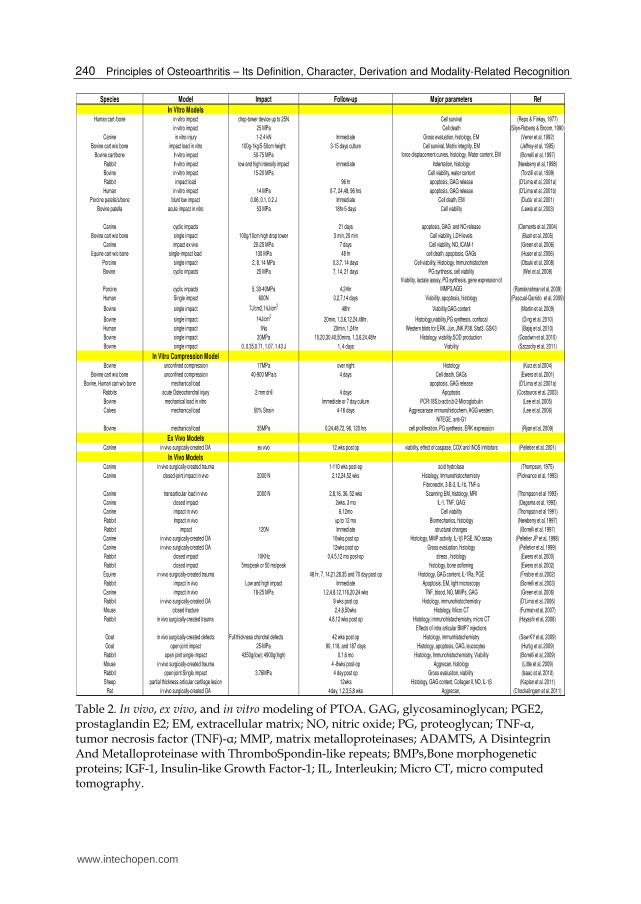
It has been well documented that cell death is the first response in all injuries that involve blunt trauma or direct insult (impaction, injurious compression, wound creation, etc) to cartilage surface or the entire joint. The role of cell death in PTOA has been widely studied using in vivo animal models, ex vivo animal and human models and in vitro culture approaches with cartilage from different species including humans (Table 2). Chondrocyte death in response to a single impact was first reported in the 1970’s (Finlay et al,1978;Repo et al,1977) and was extensively studied in the last few decades. It was shown that controlled single impacts of 15-21 MPa on bovine cartilage explants resulted in chondrocyte death within 24 hours after the injury (Oegema et al,1993; Torzilli et al,1999). This phenomena has been confirmed in multiple studies using cartilage from different species and applying various forces (15-53MPa) (Newberry et al,1998; Thompson,1975; Lewis et al,2003;Oegema et al, 1993; D'Lima et al,2001b;Ewers et al,2000b,2001,2002; Jeffrey et al,1995; Bolam et al,2006; Hurtig et al,2009; Beecher et al,2007; Pascual Garrido et al,2009; Bajaj et al,2010; Huser et al,2006a). The level of cell death and the depth of damage were proportional to the energy of the impact (Huser et al,2006a; Bolam et al,2006 ). These observations of the impact induced chondrocyte death in bovine, rabbit, canine, equine, and human cartilage point towards a general mechanism of trauma-mediated effects suggesting that cellular responses to injury could be studied in species that are readily available as models of PTOA. In the PTOA models chondrocytes could die by two mechanisms, necrosis and apoptosis. Necrosis occurs as a direct effect of impact/injury on the cell resulting in the disruption of the cellular membrane and loss of its integrity as well as the damage of the intracellular organelles. This type of death occurs immediately after the insult and is difficult to prevent in the PTOA models. Necrotic cell death also leads to the release of calcium, free radicals, nitric oxide, and the activation of intracellular catabolic mediators, including caspases, interleukins, proteinases, etc. All of them are capable of triggering the process called “apoptosis” leading to the programmed cell death. This second type of death could be prevented and arrested by using targeted therapeutics. For the most part, in the studies on PTOA types of death are not distinguished. Good examples that address both mechanisms are in vitro and in vivo studies with canine, sheep, or human cartilages (Chen et al,2001; Bajaj et al,2010;Hurtig et al,2009;Pascual Garrido et al,2009). Importantly, necrosis and apoptosis are usually spaced out in time, as it was reported for canine cartilage (Chen et al,2001), where a cyclic impaction induced necrosis during the first 4 hours after the impact, while apoptosis was seen 48 hours after the impact. As was already stated, the level of cell death depends upon the energy of the impact, but cell death is always observed at the surface and in the superficial cartilage zone; it is less pronounced in the middle and deep layers of cartilage (Beecher et al,2007;Pascual Garrido et al,2009).
www.intechopen.com
Principles of Osteoarthritis – Its Definition, Character, Derivation and Modality-Related Recognition
240
Species Model Impact Follow-up Major parameters Ref
In Vitro ModelsHuman cart /bone in vitro impact drop-tower device up to 25N Cell survival (Repo & Finkay, 1977)
in vitro impact 25 MPa Cell death (Silyn-Roberts & Broom, 1990)
Canine in vitro injury 1-2.4 kN Immediate Gross evaluation, histology, EM (Vener et al, 1992)
Bovine cart w/o bone impact load in vitro 100g-1kg/5-50cm height 3-15 days culture Cell survival, Matrix integrity, EM (Jeffrey et al, 1995)
Bovine cart/bone In vitro impact 50-75 MPa force displacement curves, histology, Water content, EM (Borrelli et al, 1997)
Rabbit In vitro impact low and high intensity impact immediate Indentation, histology (Newberry et al, 1998)
Bovine in vitro impact 15-20 MPa Cell viability, water content (Torzilli et al, 1999)
Rabbit impact load 96 hr apoptosis, GAG release (D'Lima et al, 2001a)
Human in vitro impact 14 MPa 0-7, 24,48, 96 hrs apoptosis, GAG release (D'Lima et al, 2001b)
Porcine patella's/bone blunt low impact 0.06, 0.1, 0.2 J Immediate Cell death, EM (Duda et al, 2001)
Bovine patella acute impact in vitro 53 MPa 18hr-5 days Cell viability (Lewis et al, 2003)
Canine cyclic impacts 21 days apoptosis, GAG and NO release (Clements et al, 2004)
Bovine cart w/o bone single impact 100g/10cm high drop tower 3 min, 20 min Cell viability, LDH levels (Bush et al, 2005)
Canine impact ex vivo 20-25 MPa 7 days Cell viability, NO, ICAM-1 (Green et al, 2006)
Equine cart w/o bone single-impact load 130 MPa 48 hr cell death, apoptosis, GAGs (Huser et al, 2006)
Porcine single impact 2, 8, 14 MPa 0,3,7, 14 days Cell viability, Histology, Immunohistochem (Otsuki et al, 2008)
Bovine cyclic impacts 25 MPa 7, 14, 21 days PG synthesis, cell viability (Wei et al, 2008)
Porcine cyclic impacts 5, 30-40MPa 4,24hr
Viability, lactate assay, PG synthesis, gene expression of
MMP3,AGG (Ramakrishnan et al, 2009)
Human Single impact 600N 0,2,7,14 days Viability, apoptosis, histology (Pascual-Garrido et al, 2009)
Bovine single impact 7J/cm2,14J/cm2
48hr Viability,GAG content (Martin et al, 2009)
Bovine single impact 14J/cm2
20min, 1,3,6,12,24,48hr, Histology,viability,PG synthesis, confocal (Ding et al, 2010)
Human single impact 1Ns 20min, 1,24hr Western blots for ERK, Jun, JNK,P38, Stat3, GSK3 (Bajaj et al, 2010)
Bovine single impact 20MPa 10,20,30,40,50mins, 1,3,6,24,48hr Histology, viability,SOD production (Goodwin et al, 2010)
Bovine single impact 0, 0.35,0.71, 1.07, 1.43 J 1, 4 days Viability (Szczodry et al, 2011)
In Vitro Compression ModelBovine unconfined compression 17MPa over night Histology (Kurz et al 2004)
Bovine cart w/o bone unconfined compression 40-900 MPa/s 4 days Cell death, GAGs (Ewers et al, 2001)
Bovine, Human cart w/o bone mechanical load apoptosis, GAG release (D'Lima et al, 2001a)
Rabbits acute Osteochondral injury 2 mm drill 4 days Apoptosis (Costouros et al, 2003)
Bovine mechanical load in vitro Immediate or 7 day culture PCR:18S,b-actin,b-2-Microglobulin (Lee et al, 2005)
Calves mechanical load 50% Strain 4-16 days Aggrecanase immunohistochem, AGG western, (Lee et al, 2006)
NITEGE, anti-G1
Bovine mechanical load 35MPa 0,24,48,72, 96, 120 hrs cell proliferation, PG synthesis, ERK expression (Ryan et al, 2009)
Ex Vivo ModelsCanine in vivo surgically-created OA ex vivo 12 wks post op viability, effect of caspase, COX and iNOS inhibitors (Pelletier et al, 2001)
In Vivo ModelsCanine in vivo surgically-created trauma 1-110 wks post-op acid hydrolase (Thompson, 1975)
Canine closed-joint impact in vivo 2000 N 2,12,24,52 wks Histology, Immunohistochemistry (Pickvance et al, 1993)
Fibronectin, 3-B-3, IL-1b, TNF-a
Canine transarticular load in vivo 2000 N 2,8,16, 36, 52 wks Scanning EM, histology, MRI (Thompson et al 1993)
Canine closed impact 2wks, 3 mo IL-1, TNF, GAG (Oegema et al, 1993)
Canine impact in vivo 6,12mo Cell viability (Thompson et al 1991)
Rabbit Impact in vivo up to 12 mo Biomechanics, histology (Newberry et al, 1997)
Rabbit impact 120N Immediate structural changes (Borrelli et al, 1997)
Canine in vivo surgically-created OA 10wks post op Histology, MMP activity, IL-1β PGE, NO assay (Pelletier JP et al, 1998)
Canine in vivo surgically-created OA 12wks post op Gross evaluation, histology (Pelletier et al, 1999)
Rabbit closed impact 10KHz 0,4.5,12 mo post-op stress , histology (Ewers et al, 2000)
Rabbit closed impact 5ms/peak or 50 ms/peak histology, bone softening (Ewers et al, 2002)
Equine in vivo surgically-created trauma 48 hr, 7, 14,21,28,35 and 70 day post op Histology, GAG content, IL-1Ra, PGE (Frisbie et al, 2002)
Rabbit impact in vivo Low and high impact Immediate Apoptosis, EM, light microscopy (Borrelli et al, 2003)
Canine impact in vivo 18-25 MPa 1,2,4,8,12,116,20,24 wks TNF, blood, NO, MMPs, GAG (Green et al, 2006)
Rabbit in vivo surgically-created OA 9 wks post op Histology, immunohistochemistry (D'Lima et al, 2006)
Mouse closed fracture 2,4,8,50wks Histology, Micro CT (Furman et al, 2007)
Rabbit in vivo surgically-created trauma 4,8,12 wks post op Histology, immunohistochemistry, micro CT (Hayashi et al, 2008)
Effects of intra articular BMP7 injections
Goat in vivo surgically-created defects Full thickness chondral defects 42 wks post op Histology, immunhistochemistry (Saw KY et al, 2009)
Goat open joint impact 25-MPa 90, 118, and 187 days Histology, apoptosis, GAG, leucocytes (Hurtig et al, 2009)
Rabbit open joint single impact 4350g(low); 4900g(high) 0,1,6 mo Histology, Immunohistochemistry, Viability (Borrelli et al, 2009)
Mouse in vivo surgically-created trauma 4 -8wks post-op Aggrecan, histology (Little et al, 2009)
Rabbit open joint Single impact 3.76MPa 4 day post op Gross evaluation, viability (Isaac et al, 2010)
Sheep partial thickness articular cartilage lesion 12wks Histology, GAG content, Collagen II, NO, IL-1β (Kaplan et al. 2011)
Rat in vivo surgically-created OA 4day, 1,2,3,5,8 wks Aggrecan, (Chockalingam et al, 2011)
Table 2. In vivo, ex vivo, and in vitro modeling of PTOA. GAG, glycosaminoglycan; PGE2, prostaglandin E2; EM, extracellular matrix; NO, nitric oxide; PG, proteoglycan; TNF-┙, tumor necrosis factor (TNF)-┙; MMP, matrix metalloproteinases; ADAMTS, A Disintegrin And Metalloproteinase with ThromboSpondin-like repeats; BMPs,Bone morphogenetic proteins; IGF-1, Insulin-like Growth Factor-1; IL, Interleukin; Micro CT, micro computed tomography.
www.intechopen.com

Post-Traumatic Osteoarthritis: Biologic Approaches to Treatment
241
Every model has its advantages and limitations, but each provides important information adding to our understanding of the PTOA. Studying the cartilage explants out of their joint environment eliminates the external influences that cartilage would experience in its natural environment. Factors such as weight bearing, vascularization, fluid dynamics of the joint and the biomechanics of the supporting tissue cannot be controlled in the in vitro system, but it provides an opportunity to distinguish truly cartilage responses to trauma without other contributing mechanisms. On the flip side, it makes interpretation of results and extrapolation to actual clinical situations more challenging. Investigating cellular events using humans is almost impossible due to the nature of experimental approaches and end measures. Furthermore, patients come to see the doctor only when pain and signs of degenerative changes have been already developed, which eliminates the option of studying early cellular responses to trauma. In order to overcome some of these limitations in vivo animal models have been employed. Unfortunately, many PTOA studies are conducted on rodents or rabbits (Beecher et al,2007;D'Lima et al,2001c;Guilak et al,2004;Isaac et al,2010), the animals that either have a tendency to self-repair or are far from resembling human joint structure and biomechanics. In all these models cell death by necrosis and apoptosis was the most prominent feature, though the degree of death varied between the models and the energy of the impact (Beecher et al,2007;D'Lima et al,2001c;Milentijevic et al,2005). Chondrocyte death and superficial matrix damage were always observed immediately after impaction. The changes that were reported 3 to 52 weeks post impactions were those typically seen in early and late stage osteoarthritis, such as matrix damage, chondrocytes death and proteoglycan loss (Milentijevic et al,2005;Rundell et al,2005), and at later stages, characteristic cartilage fibrillation, clone formation, matrix disorganization, and joint space narrowing (Hurtig et al,2009). All these models indicate that even smaller forces could be sufficient to initiate cell necrosis and higher forces may be needed to initiate massive apoptosis (D'Lima et al,2001b;Milentijevic et al,2005;Rundell et al,2005). The majority of studies used a drop-tower device in an open joint injury models. Borrelli et al (Borrelli et al,2003) used a pendulum type apparatus to apply a rapid impact to the articular surface of the femoral condyle. We used a pneumatically controlled impactor in our studies to generate cartilage damage (Bajaj et al,2010;Pascual Garrido et al,2009; Hurtig et al,2009). Common injuries, such as sports or vehicular, are usually attained in a closed system. Hence, studying open joint impaction may not necessarily reflect real clinical scenario. Therefore, investigating chondrocyte responses that occur in a closed fracturing system could become more relevant, especially since Gaber et al (Gaber et al,2009) also observed significant chondrocyte death after a mid- diaphyseal closed fracture of the tibia. In summary, in all PTOA models significant chondrocyte death occurs as earlier as 4 hours after the impact and persists for up to 48 hours. If left untreated, it leads to another mechanism of cell death, apoptosis and, eventually with time the cartilage develops OA-like changes. Consequently, therapeutic strategies aimed at minimizing the results of mechanical stress during the early stages post injury may help to preserve structural integrity of cartilage and slow or prevent the development of PTOA.
8. Chondroprotection as an early stage therapy
Since cell death is the first response in all injuries, the most obvious approach is to protect the cells and promote their survival and viability. Chondroprotection could be achieved via targeting different mechanisms: cell membrane protection, anti-oxidant therapy,
www.intechopen.com

Principles of Osteoarthritis – Its Definition, Character, Derivation and Modality-Related Recognition
242
mitochondria protection, inhibition of NO release, inhibition of apoptosis through the inhibition of caspase signaling, inhibition of calcium quenching, etc. These approaches are addressed below. As mentioned above, there are two major mechanisms of cell death: necrosis, in which fluid uptake increases causing the cell to swell and rupture resulting in the release of the intracellular contents that incites an inflammatory cascade; and apoptosis, in which there is a chromatin condensation, DNA fragmentation, cell shrinkage, membrane blebbing that cause the cell to self-destruct. Two cellular pathways have been identified in the apoptotic signaling, the extrinsic pathway that involves the Fas receptor pathway and the intrinsic pathway that involves the mitochondrial pathway (Borrelli,2006). Oxygen and reactive oxygen species (ROS) have a role in cartilage homeostasis and are involved in chondrocyte activation, proliferation and matrix remodeling (Henrotin et al,2003). In excess amounts, they induce chondrocyte death and matrix degradation. Mechanical injury has been associated with an increase in production of ROS (Henrotin et al,2003) and decreased antioxidant capacity (Martin et al,2004), which becomes insufficient once structural and functional cartilage damage occurred. The use of exogenous antioxidants, such as vitamin E, N-acetyl-L-cysteine (NAC) and superoxide dismutase have the potential to protect the chondrocytes from the elevated oxidants. Beecher et al (Beecher et al,2007) have shown that pre-treatment with antioxidants can significantly increase chondrocyte survival up to 40-80% in the superficial zone and up to 53-80% in the middle zone. In their study, cartilage explants from non-osteoarthritic human cadaver ankle were pre-treated with NAC, superoxide dismutase or vitamin E before cyclic impaction of either 2MPa or 5MPa was applied. Pre-treatments with NAC and superoxide dismutase were the most effective at preventing chondrocyte death, when forces of 5MPa were applied. Although these finding are promising in showing that human chondrocyte survival can be attained by the treatment with antioxidants, the timing of the antioxidant treatment is not practical, because most joint injuries occur without advance warning. This approach may be valuable when antioxidants are either injected intra-articularly or applied during surgery, when the joint area that undergoes surgical intervention is pretreated with the antioxidants. By the time of surgery, which usually takes place much after the injury, the window of opportunity for antioxidant therapy could have been missed. In a similar study by Martin et al (Martin et al,2009) bovine osteochondral explants subjected to a single blunt end impact were treated with NAC, vitamin E, poloxamer 188 (P188) or Z-VAD-FMK after impaction. The agents were administered either immediately after injury or were delayed by 4 hours. Immediate treatment with NAC improved chondrocyte viability by up to 74%, while delayed treatment also promoted cell survival, but to a lesser extent, 59%, though still being relatively high. Z-VAD-FMK, a caspase inhibitor and anti-apoptotic agent, improved chondrocyte survival to the level of delayed NAC treatment. Vitamin E and P188 did not significantly increase cell survival. In our studies with the human cartilage ex vivo injury model, NAC was effective only while it was present in culture media (first 48 hours). It promoted cell survival and inhibited apoptosis in the superficial layer, which was two times lower in the NAC treated explants than in the untreated control. However, after NAC removal, apoptosis returned to the levels of untreated impacted control. Similar observations were found for PG synthesis, which was elevated by two-fold at day 2 under the NAC treatment, but declined to the untreated controls levels as the agent was removed. Adjacent, not impacted areas remained protected by NAC treatment and cell viability was comparable to that of the non-impacted
www.intechopen.com

Post-Traumatic Osteoarthritis: Biologic Approaches to Treatment
243
controls. Though in our experiments NAC was effective in protecting the cells, it was ineffective in providing the protection for cartilage matrix integrity. Similarly to Beecher et al, Kurz et al compared the pre-treatment option versus post-injury treatment to see whether pre-treatment with antioxidants could enhance chondrocyte viability (Kurz et al,2004), though clinical relevance of this approach remains to be justified. Superoxide dismutase reduced apoptosis in a dose dependent manner with complete inhibition of apoptosis when the highest dose of 2.5µM was used, while vitamin E had no effect. Despite the efforts, a consensus cannot be achieved on whether the treatment with antioxidants prior to or after the injury has a beneficial effect in enhancing cell survival (Beecher et al,2007;Kurz et al,2004;Martin et al,2009). 1) Different time points and different methods were used to assess cell viability; 2) no distinction was made between necrosis and apoptosis; 3) different species were often used; and 4) distinct responses to the same agent were observed. Differences in responses can be attributed to species- and model-specific distinctions. It has been suggested that blunt versus cyclic impaction model may trigger distinct cellular responses and induce, for example, lipid peroxidation that has a damaging effect on plasma membrane (Beecher et al,2007). Therefore, accumulation of Vitamin E in the plasma membrane could have a protective effect (Claassen et al,2005). This is also supported by the fact that Vitamin E was the most active within the first 4 hours. Together these data indicate an anti-necrotic mechanism of Vitamin E activity. Despite the differences, above referenced studies generate one important message: a window of opportunity for treatment does exist and mechanism-based timely delivery of biologics could provide necessary protection in post-traumatic degenerative events. The beneficial effects of ROS scavenger NAC (a powerful hydroxyl and hypochlorous radical scavenger) and superoxide dismutase on chondrocyte survival implicate chondrocyte death by apoptosis being secondary to the production of ROS, although the source of ROS excess remains unclear. Chondrocyte survival experiments with superoxide dismutase suggest a role for the mitochondria in early cellular responses to injury. Rotenone, an agent that suppresses the release of superoxide from the mitochondria, has been tested to confirm the role of the mitochondrial electron transport chain in the production of ROS (Goodwin et al,2010). Although rotenone significantly reduced chondrocyte death by more than 40% when administered 2 hours post injury (Goodwin et al,2010), it is unsuitable for an in vivo use due to its high cellular toxicity. Accepting the importance of the mitochondria in ROS release, factors that control mitochondrial depolarization have to be considered. Furthermore, it has been shown that injury increases intracellular cytoplasmic calcium due to its release by the endoplasmic reticulum. This leads to depolarization of the mitochondrial membrane, which is associated with the release of cytochrome C, caspase dependent apoptosis and Bcl-2 degradation (Huser et al,2007). Altered intracellular calcium homeostasis has been implied in chondrocyte death (Browning et al,2004;Ohashi et al,2006) and studies with calcium chelating agents have shown significant reduction in chondrocyte death (Huser et al,2007). Thus, calcium quenching inhibitors may have therapeutic value, although the mechanism between the mechanical impact and the cytoplasmic calcium increase remains to be understood. NO and superoxide anion are the two main ROS produced by the chondrocyte (Hiran et al,1997;Moulton et al,1997). NO in the chondrocyte is synthesized by endothelial nitric oxide synthase (eNOS) or inducible nitric oxide synthase (iNOS) (Henrotin et al,2003) that are reportedly regulated by growth factors, cytokines and endotoxins (Henrotin et al,2003).
www.intechopen.com
Principles of Osteoarthritis – Its Definition, Character, Derivation and Modality-Related Recognition
244
Factors such as Tumor Necrosis Factor (TNF)-┙, Interleukin (IL)-┚, Interferon (INF)-┛ and Lipopolysaccharides (LPS) stimulate NO production and Transforming Growth Factor (TGF)-┚, IL-4, IL-10 and IL-13 inhibit NO production (Henrotin et al,2003). NO has been shown to be up-regulated after trauma and to possess cartilage degradative properties, which suggests a potential role for iNOS inhibitors in matrix protection. Evidence for chondroprotective effect of NO inhibition comes from studies by Beecher et al (Beecher et al,2007), where human cartilage explants, were pre-treated with the nitric oxide synthase inhibitor N-Nitro-L-arginine methyl ester (L-NAME) before cyclic impaction loads. In treated explants cell survival was considerably higher (82-90%) in the superficial and middle layers compared to the 40-53% in the same areas of the untreated explants. The mechanism, through which L-NAME exhibited its anti-apoptotic effect, is thought to be via interference with the IL-1┚ pathway (Marsh et al,2002;Pelletier et al,1999,1998). In an in vivo canine OA model, Pelletier et al (Pelletier et al,1999,2000a) tested the effect of two concentrations of another iNOS inhibitor, N-iminoethyl-L-Lysine (L-NIL). The dogs that received the higher dose of L-NIL (10mg/kg/day) showed marked decrease in Tunel-positive chondrocytes and macroscopically and histologically their cartilage lesions were less severely affected by the OA-like changes than the placebo treated dogs. In addition, a reduced level of caspase 3 and MMP activity was found in the L-NIL treated dogs (Pelletier et al,1998,2000a). These data suggest that iNOS inhibitors reduce the progression of PTOA through the caspase 3 mediated inhibition of apoptosis that results in the diminished MMP activity (Pelletier et al,1998,2000a). Apoptosis is one of the main causes of chondrocyte death after mechanical injury (Chen et al,2001;D'Lima et al,2001a; Borrelli,2006), which is mediated by a complex proteolytic system known as the cysteinyl aspartate-specific proteases (caspases). D’Lima et al have successfully demonstrated that caspase inhibitors reduce the severity of cartilage lesion in an in vivo rabbit OA model. Using intra-articular injections of the pan caspase inhibitor Z-VAD-FMK (benzyloxycarbonyl-Val-Ala-Asp(OMe) fluoromethylketone), a cell permeable fluoromethylketone, the authors demonstrated reduced cartilage degradation, reduced activity of caspase 3 and reduced p85 fragment suggesting that this broad spectrum caspase inhibitor prevents apoptosis and slows the disease progression. The effect of Z-VAD-FMK has also been studied in full thickness human cartilage explants subjected to single impacts of relatively low stress, 14MPa (D'Lima et al,2001a). These results were reliably reproduced in several other types of injury (static compression and blunt impact) applied to cartilage from different species (bovine, rabbit, equine and human) (Huser et al,2006a;Huser et al,2006b). However, in our studies with human cartilage impact induced by a higher stress, 25-30MPa, the effect of caspase inhibitors (inhibitors of caspase 3 & 9;(Pascual Garrido et al,2009) or pan-caspase inhibitors (Z -VAD-FMK or Q -VD-OPh)), was not as pronounced. In a 14-day follow-up study we found that caspase 3 inhibitor temporarily halted cartilage degenerative changes, while caspase 9 inhibitor was ineffective. Pan caspase inhibitors, contrary to studies of others (D'Lima et al,2001c;Huser et al,2006a;Huser et al,2006b), in our acute injury model on human explants, were not able to inhibit apoptosis. Yet the cells that survived impaction showed elevated PG synthesis after a treatment with caspase inhibitors. This resulted in preservation of matrix integrity (low Mankin score) especially in the areas adjacent to the impact. Both pan-caspase inhibitors demonstrated similar efficacy. Despite a wide range of effects, evidence suggests that caspase inhibitors could be and should be considered for targeted therapeutic intervention in PTOA, if they are utilized
www.intechopen.com

Post-Traumatic Osteoarthritis: Biologic Approaches to Treatment
245
immediately or soon after joint injury before the fully-blown apoptotic cascade takes place. Another way to fight cell death is physical protection of cell membrane integrity (Duke et al,1996). Therefore, a number of laboratories (including ours) focused on the use of poloxamer 188 (P188), a nontoxic nonionic surfactant that has a hydrophilic and a hydrophobic center, similar to the lipid bilayer composition (Bajaj et al,2010;Isaac et al,2010;Martin et al,2009;Pascual Garrido et al,2009;Phillips et al,2004). It has been suggested that surfactant molecules, (i.e. P188), insert into the membrane to restore the cell membrane integrity post injury, protecting the cell form subsequent catabolic activation.
P188Repaired Cell membrane
Injured Cell Membrane
Healthy Cell membrane
Released
Sealing
P188
P188
P188
Fig. 1. Diagrammatic representation of P188 surfactant restoring membrane integrity
The role of P188 in bovine chondroprotection was first discovered by the group of Haut (Phillips et al,2004) who showed that P188 statistically reduced the level of apoptosis in the ex vivo blunt impaction model. In follow-up studies, an increase in chondrocyte viability with early P188 treatment in short- and long-term has been also documented in vivo in rabbits (Isaac et al,2010). We undertook a more comprehensive approach in an attempt to understand the mechanism of P188 action. We demonstrated that P188 was superior to inhibitors of caspase 3 and 9 in promoting cell survival after acute injury (Pascual Garrido et al, 2009). We also found that a single treatment with P188 (8mg/ml; added immediately after injury) was able to inhibit cell death by necrosis and apoptosis and, more importantly, was able to prevent horizontal and longitudinal spread of cell death to the areas that were not directly affected by the impaction. Though P188 was present in the explant culture only for 48 hours, the effect was sustainable for 7 of 14 days. Furthermore, we identified the
www.intechopen.com

Principles of Osteoarthritis – Its Definition, Character, Derivation and Modality-Related Recognition
246
mechanisms through which P188 exhibited its effect (Bajaj et al,2010). Earlier, the role for mitogen-activated protein kinases (MAPKs), c-Jun-N-terminal kinase and p38, in P188 mediated effects was implied in neural tissue (Serbest et al,2006). We found that among the mechanisms, through which the surfactant directly or indirectly inhibited cell death by apoptosis and prevented its expansion, was the inhibition of the IL-6 signaling pathway. Specifically, phosphorylation of key mediators of the IL-6 pathway, Stat1, Stat3, and p38, was significantly inhibited or prevented. Furthermore, glycogen synthase kinase 3 (GSK3) signaling involved in apoptosis was also inhibited. Our data, both biochemical and histological, suggest that p38 kinase may act up-stream of Stats signaling; activation of p38 kinase as a result of injury, may be partially responsible for initiation of IL-6/Stats mediated catabolic events. A single treatment with P188 blocked phosphorylation of Stats as well as their translocation in to the nucleus, thus potentially preventing transcription of catabolic genes. Furthermore, the role of p38 in injury-induced catabolic responses was further verified by the application of specific synthetic p38 inhibitor confirming previous data (Serbest et al,2006). Treatment with p38 inhibitor not only inhibited the IL-6 pathway, but also promoted cell survival by reducing apoptotic cell death. In addition, we observed an inhibitory effect of P188 on Vascular Endothelial Growth Factor and Monocyte Chemotactic Protein-1 and a stimulatory effect on IL-7 and especially IL-12, effects that remain to be explained. Stimulation of IL-12 may indicate another anabolic response that has not been widely explored in cartilage; IL-12 is an important regulatory cytokine that functions centrally in the initiation and regulation of cellular immune responses. Because a single treatment with P188 lasted only for the first 7 days, we explored multiple applications, adding fresh agent to the culture every 48 hours to sustain its presence for the duration of the experiments. Multiple treatments with P188 were not superior to its single application, suggesting that the primary mechanism of P188 activity is sealing the cellular membrane, which prevents trauma-induced cell necrosis and thus the release of catabolic mediators by necrotic cells. Treatment with P188 prior to impaction was also ineffective, pointing to the role of this agent in repair/restoration of the membrane that was damaged. In summary, chondroprotective therapy should be seen as the earliest possible approach to treat cartilage tissue post-injury. Chondroprotection has a high potential in the development of targeted biologic interventions in PTOA, because when chondrocyte death is reduced and cartilage cellularity is preserved, there are more chances for the remaining cells to initiate anabolic responses to prevent the expansion of degenerative changes and to remodel damaged matrix.
9. Inhibition of pro-inflammatory mediators as early biologic treatment in PTOA
There are three major anti-catabolic interventions that could be considered at the present time: NAC as an antioxidant (described in details above), interleukin-1 receptor antagonist (IRAP), and TNF-┙ antagonist. NAC inhibits activation of c-Jun N-terminal kinase, p38 MAP kinase, redox-sensitive activating protein-1 and NF-B transcription factor activities regulating expression of numerous genes. NAC can also prevent apoptosis and promote cell survival by activating extracellular signal-regulated kinase pathway (Zafarullah et al,2003). Interleukin-1 (IL-1) and tumor necrosis factor (TNF)-┙ are the most studied cytokines in post-traumatic OA (Evans et al,2004;Fukui et al,2001;Furman et al,2006;Guilak et al,2004). Both are potent activators of cartilage degradation (Martel-Pelletier,1999) and their activity
www.intechopen.com

Post-Traumatic Osteoarthritis: Biologic Approaches to Treatment
247
has been significantly increased after acute injury. The levels of these mediators are shown to correlate with the disease severity (Lotz et al,2010;Marks et al,2005). Studies have also reported that IL-6, IL-8 and IL-10 (Bajaj et al,2010; Irie et al,2003;Perl et al,2003) play a role in cartilage loss and the progression of PTOA (Furman et al,2006).The increased expression of these cytokines is attributed to the stressed chondrocytes, synoviocytes and infiltrating inflammatory cells. We documented an elevation of IL-6, TNF-┙, basic fibroblast growth factor, and other cytokines as early cellular responses to injury (Anderson et al,2011;Bajaj et al,2010). Currently, the most effective approaches to inhibit activity of catabolic cytokines are the ones that could interfere with their signaling: receptor blockers, neutralizing monoclonal antibodies, soluble receptors, receptor antagonists, extracellular and intracellular binding proteins, as well as various intracellular repressors and cofactors that prevent the transcription of catabolic genes. IL-1 activity is mediated by its binding to specific IL-1 receptor (IL-1R). Therefore, the antagonist of the IL-1 receptor called interleukin receptor antagonist protein (IRAP) or IL-RA competes for binding to IL-1 and thus prevents the activation of IL-1 signaling. IRAP has been studied in the in vivo OA equine model (Frisbie et al,2002), where Ad-EqIL-1Ra was injected intra-articularly. Clinical observation based on pain (lameness) and radiographic examination showed significant improvement in treated horses compared to the untreated horses. Histological examination revealed significant reduction in subintimal edema, joint fibrillation, and chondrocyte necrosis in horses treated with IL-1Ra. Significant, improvement with IL-1Ra treatment seen in the equine model strongly supports its potential use in clinics. Methods have been developed to generate autologous conditioned serum with enriched endogenous IL-1Ra (Orthokine) (Meijer et al,2003) that could be injected into the joint. Preliminary clinical data have shown that intra-articular injections of Orthokine can improve the clinical signs and symptoms of OA such as a reduction in pain and increase in joint function (Fox et al,2010). Whether these methods are chondroprotective or alter the progression of disease is still being investigated. Recombinant IL-1Ra has been used in clinical trials in rheumatoid arthritis, sepsis and graft versus host disease (Evans et al,2004). We investigated whether recombinant IL-RA could inhibit cell death and thus affect cartilage metabolism in the ankle cartilage ex vivo acute injury model. 100ng/ml IL-RA increased the percentage of live cells by two-fold in the superficial layer of the impacted tissue compared with the untreated control; while a low dose of IL-RA (20ng/ml) was ineffective. The effect on apoptosis of both concentrations was negligible. Although ineffective in promoting cell survival, a lower concentration of IL-RA stimulated PG synthesis by three-fold. Normal histological pattern of IL-RA treated samples was observed only while the agent was present in culture. As IL-RA was removed, loss of Safranin O staining and depletion of proteoglycan became apparent. Another cytokine involved in cartilage loss of PTOA is tumor necrosis factor (TNF)-┙ (Sandell et al,2001). Antagonists of IL-1 and TNF-┙, namely, IRAP and the PEGylated soluble TNF-┙ receptor I, alone and/or in combination, down-regulated MMP-1, MMP-3,
and MMP-13 expression and promoted cartilage preservation. Early inhibition of TNF- can provide chondroprotection and cartilage preservation by decreasing release of glycosaminoglycans and increasing lubricin production in the post traumatic arthritis rat model (Elsaid et al,2009). These results suggest that the inhibition of either or both of these cytokines may offer a useful therapeutic approach to the management of PTOA by reducing gene expression of MMPs involved in cartilage matrix degradation and favoring its repair.
www.intechopen.com

Principles of Osteoarthritis – Its Definition, Character, Derivation and Modality-Related Recognition
248
10. Matrix protection
Matrix protection could be achieved by either direct inhibition of matrix proteinases or by affecting the factors responsible for their activation, such as ROS, NO, inflammatory cytokines, etc. NO has been long implicated in cartilage degradation, noting that arthritic patients showed elevated levels of nitrites (Spreng et al,2001) and lipid peroxidation products (Situnayake et al,1991a,1991b) in their biological fluids. The increased NO production has been reported to inhibit aggrecan synthesis (Evans et al,1995), increase iNOS activity, reduce proteoglycan synthesis (Jarvinen et al,1995) and increase MMP activity (Murrell et al,1995). Use of the iNOS inhibitor L-NIL has slowed the progression of PTOA in an experimental OA model in dogs (Pelletier et al,1999). It is plausible to explore iNOS inhibitors therapeutically for matrix protection in addition to chondroprotection. As a result of cartilage damage, there is a marked increase in the release of matrix components and their fragments, such as proteoglycans, aggrecan cleavage products, collagen, fibronectin, and hyaluronan fragments (Otsuki et al,2008; Ryan et al,2009; Wei et al,2009). Determining a profile of the released metabolites during disease progression may be beneficial in the development of treatment strategy and targeted therapeutic interventions. Specific effective inhibitors of the MMPs could also protect the matrix and in turn halt the disease progression. The role of MMPs has been primarily addressed in various OA models or in patients with signs of arthritis. In PTOA, there is very little, if any, available information. Selective inhibitors are not widely available and the majority of studies have utilized broad spectrum MMP inhibitors, which in addition to a direct effect on MMPs, have been also associated with adverse musculoskeletal effects such as muscle stiffness, bursitis and fibrosis (Pelletier et al,2007). Studies with MMP-13 knockout mice (Little et al,2009) showed cartilage protection when OA was surgically induced, suggesting a crucial role of MMP-13 in cartilage degradation. Oral dosing of the MMP-13 inhibitor in a rabbit OA model has shown cartilage protection without the musculoskeletal adverse effects (Johnson et al,2007). These very promising but preliminary data may have potential in PTOA therapy, although clinical trials with several MMP inhibitors in patients with established OA have failed due to side effects and lack of efficacy. ADAMTS‘s have also been implicated in the pathogenesis of OA. The use of ADAM-TS5 knockout mice have shown that these mice do not develop OA (Glasson et al,2005), but display some side effects, such as fibrosis. Inhibitors of aggrecanases with higher specificity and lower toxicity are among future therapeutic agents for the treatment of PTOA.
11. Matrix remodeling with anabolic growth factors
One of the very important directions in the development of pharmacological interventions in PTOA is the ability to stimulate production of new cartilage extracellular matrix. The best candidates are growth factors including members of the TGF-┚ superfamily, FGFs, Insulin-like Growth Factor (IGF)-1. Bone Morphogenetic Proteins (BMPs) belong to the TGF-┚ superfamily and are important stimuli of mesenchymal cell differentiation and extracellular matrix formation. BMP-2 and BMP-7 appear to be extremely potent in cartilage and bone repair. The most studied BMP for cartilage repair is BMP-7, also known as osteogenic protein-1 (OP-1). It has been studied most extensively in vitro in our laboratory on human cartilage (reviewed in (Chubinskaya et al,2007,2011) as well as in OA and PTOA animal models (Badlani et al,2008;Cook et al,2003;Hayashi et al,2008,2010;Hurtig et al,2009). The
www.intechopen.com

Post-Traumatic Osteoarthritis: Biologic Approaches to Treatment
249
results suggest that BMP-7 may be the best candidate for a disease-modifying OA drug and also for PTOA. Unlike TGF-┚ and other BMPs, BMP-7 up-regulates chondrocyte metabolism and protein synthesis without creating uncontrolled cell proliferation and formation of osteophytes. BMP-7 prevents chondrocyte catabolism induced by IL-1 or fragments of matrix components. It has synergistic anabolic effects with other growth factors such as IGF-1, in addition to its anabolic effect acts as a cell survival factor (reviewed in Chubinskaya et al,2007). BMP-7 restores the responsiveness of human chondrocytes to IGF-1 lost with ageing through the regulation of IGF-1 and its signaling pathway (Chubinskaya et al,2011; Im et al,2003). IGF-1 has chondroprotective activity in various animal models (Fortier et al,2002). In our most recent studies in the acute ex vivo cartilage trauma model, BMP-7 stimulated PG synthesis and preserved matrix integrity. Treatment with BMP-7 also significantly promoted cell survival in the impacted region (two-fold difference) and prevented expansion of cell death and matrix degeneration into the adjacent, but not impacted regions. BMP-7 has been also used in various PTOA animal models in dogs (Cook et al,2003), sheep (Hurtig et al,2009), goats (Louwerse et al,2000) and rabbits (Badlani et al,2008;Hayashi et al,2008,2010). In all these PTOA models (ACL transaction, osteochondral defect, and impaction), BMP-7 regenerated articular cartilage, increased repair tissue formation and improved integrative repair between new cartilage and the surrounding articular surface. In the impaction model (Hurtig et al,2009), a window of opportunity for the treatment with BMP-7 has been identified. It was found that therapeutic application of BMP-7 was most effective in arresting progression of cartilage degeneration if administered twice at weekly intervals either immediately after trauma or delayed by three weeks. If delayed by three months, the treatment was ineffective, suggesting that the development and progression of PTOA could be arrested and maybe even prevented if the right treatment is administered at the right time. Phase I clinical study produced very encouraging results by showing tolerability to the treatment, absence of toxic response, and a greater symptomatic improvement in patients that received a single injection of BMP-7 (Hunter et al,2010). Clinical efficacy of the BMP-7 treatment is currently being tested in a phase II clinical OA study. Other growth factors from the fibroblast growth factor family have been also tested as
potential DMOADs in PTOA. FGFs are important regulators of cartilage development and
homeostasis (Ellman et al,2008). FGF-2 can stimulate cartilage repair responses (Henson et
al,2005), but its potent mitogenic effects may lead to chondrocyte cluster formation and
poor extracellular matrix due to a relatively low level of type II collagen (Ellman et
al,2008). FGF-2 has been also shown to induce pro-catabolic and pro-inflammatory
responses (Ellman et al,2008). In a rabbit ACL transection model, sustained release
formulations of FGF-2 reduced OA severity (reviewed in Lotz et al, 2010). Another
member of the same family, FGF-18, has been shown to induce anabolic effects in
chondrocytes and chondroprogenitor cells and to stimulate cell proliferation and type II
collagen production (Ellsworth et al,2002). In a rat meniscal tear model of OA, intra-
articular FGF-18 injections induced remarkable formation of new cartilage and reduced
the severity of experimental lesions (Moore et al,2005). Thus far, only two anabolic factors,
FGF-18 and BMP-7, are currently being tested in clinical studies in patients with
established OA. At this stage of our cumulative knowledge, BMP-7 appears to be one of
the best candidate therapeutic agents for cartilage treatment after injury, since it affects
multiple catabolic and anabolic pathways.
www.intechopen.com

Principles of Osteoarthritis – Its Definition, Character, Derivation and Modality-Related Recognition
250
12. Conclusion
One of the unanswered PTOA questions is when and which therapies that have been developed as disease-modifying OA drugs are indicated for patients with post-traumatic OA and whether a different set of treatments and molecular targets has to be considered. Through basic and clinical research an impressive progress has been made towards elucidation of pathogenesis of PTOA and understanding the mechanisms that govern immediate cellular responses to injury. However, this still requires further validation in a large cohort of patients with various types of joint injuries. The ideal therapy to arrest and prevent the development and progression of PTOA must be multi-varied and include anabolic effects on chondrocyte metabolism characterized by elevated intrinsic repair, while protecting integrity of cell membrane and inhibiting catabolic pathways that lead to chondrocyte death and matrix loss (Lotz et al,2010). The following are the key mechanisms that should constitute the basis for the design of intervention therapies: 1) Chondroprotection; 2) Anti-inflammatory; 3) Matrix protection; and 4) Pro-anabolic, stimuli of cartilage remodeling and regeneration. The most beneficial agents are those that target multiple pathways and mechanisms. A number of molecular targets have been identified (Table 1) and many of the existing therapeutic agents have been already tested in vitro and in vivo. The biggest remaining challenge is the translation of this knowledge into the clinic and the development of appropriate effective therapy/therapies administered within the window of opportunity. Currently, the most suitable route for administering such therapy appears to be intra-articular injections that allow accumulation of critical doses of the drug within the damaged area and also reduce the risk of systemic side effects. To monitor the efficacy of the PTOA therapy, an appropriate set of bio- and imaging markers is needed that could predict and correlate with the progression of the disease, since it takes years and decades for the disease to develop.
13. Acknowledgements
This work was supported by the National Football League Charity Foundation, Ciba-Geigy Endowed Chair and Department of Biochemistry, Rush University Medical Center. The authors would like to acknowledge Drs. Markus A. Wimmer, Theodore R Oegema, and Jeffrey A. Borgia for their important contributions to this work. The authors would like to acknowledge Dr. Arkady Margulis for tissue procurement and Dr. Lev Rappoport and Mrs. Arnavaz Hakimiyan for their technical assistance. The authors also would like to acknowledge the Gift of Hope Organ & Tissue Donor Network and donor’s families.
14. References
Anderson, DD; Chubinskaya, S; Guilak, F; Martin, JA; Oegema, TR; Olson, SA and
Buckwalter, JA. (2011). Post-traumatic osteoarthritis: improved understanding and
opportunities for early intervention. J Orthop Res, 29(6):802-9.
Anitua, E; Andia, I; Ardanza, B; Nurden, P and Nurden, AT. (2004). Autologous platelets as
a source of proteins for healing and tissue regeneration. Thromb Haemost, 91(1):4-15.
Anitua, E; Sanchez, M; Nurden, AT; Zalduendo, M; de la Fuente, M; Orive, G; Azofra, J and
Andia, I. (2006). Autologous fibrin matrices: a potential source of biological
mediators that modulate tendon cell activities. J Biomed Mater Res A, 77(2):285-93.
www.intechopen.com

Post-Traumatic Osteoarthritis: Biologic Approaches to Treatment
251
Avouac, J; Vicaut, E; Bardin, T and Richette, P. (2010). Efficacy of joint lavage in knee
osteoarthritis: meta-analysis of randomized controlled studies. Rheumatology
(Oxford), 49(2):334-40.
Badlani, N; Inoue, A; Healey, R; Coutts, R and Amiel, D. (2008). The protective effect of OP-1
on articular cartilage in the development of osteoarthritis. Osteoarthritis Cartilage,
16(5):600-6.
Bajaj, S; Shoemaker, T; Hakimiyan, AA; Rappoport, L; Pascual-Garrido, C; Oegema, TR;
Wimmer, MA and Chubinskaya, S. (2010). Protective effect of P188 in the model of
acute trauma to human ankle cartilage: the mechanism of action. J Orthop Trauma,
24(9):571-6.
Beecher, BR; Martin, JA; Pedersen, DR; Heiner, AD and Buckwalter, JA. (2007). Antioxidants
block cyclic loading induced chondrocyte death. Iowa Orthop J, 27(1-8.
Behrens, P; Bitter, T; Kurz, B and Russlies, M. (2006). Matrix-associated autologous
chondrocyte transplantation/implantation (MACT/MACI)--5-year follow-up. Knee,
13(3):194-202.
Boileau, C; Martel-Pelletier, J; Jouzeau, JY; Netter, P; Moldovan, F; Laufer, S; Tries, S and
Pelletier, JP. (2002). Licofelone (ML-3000), a dual inhibitor of 5-lipoxygenase and
cyclooxygenase, reduces the level of cartilage chondrocyte death in vivo in
experimental dog osteoarthritis: inhibition of pro-apoptotic factors. J Rheumatol,
29(7):1446-53.
Bolam, CJ; Hurtig, MB; Cruz, A and McEwen, BJ. (2006). Characterization of experimentally
induced post-traumatic osteoarthritis in the medial femorotibial joint of horses. Am
J Vet Res, 67(3):433-47.
Borrelli, J, Jr. (2006). Chondrocyte apoptosis and posttraumatic arthrosis. J Orthop Trauma,
20(10):726-31.
Borrelli, J, Jr.; Tinsley, K; Ricci, WM; Burns, M; Karl, IE and Hotchkiss, R. (2003). Induction
of chondrocyte apoptosis following impact load. J Orthop Trauma, 17(9):635-41.
Brittberg, M; Lindahl, A; Nilsson, A; Ohlsson, C; Isaksson, O and Peterson, L. (1994).
Treatment of deep cartilage defects in the knee with autologous chondrocyte
transplantation. N Engl J Med, 331(14):889-95.
Brown, TD; Johnston, RC; Saltzman, CL; Marsh, JL and Buckwalter, JA. (2006).
Posttraumatic osteoarthritis: a first estimate of incidence, prevalence, and burden of
disease. J Orthop Trauma, 20(10):739-44.
Browning, JA; Saunders, K; Urban, JP and Wilkins, RJ. (2004). The influence and interactions
of hydrostatic and osmotic pressures on the intracellular milieu of chondrocytes.
Biorheology, 41(3-4):299-308.
Buckwalter, JA and Brown, TD. (2004). Joint injury, repair, and remodeling: roles in post-
traumatic osteoarthritis. Clin Orthop Relat Res, 423):7-16.
Butler, DL; Juncosa-Melvin, N; Boivin, GP; Galloway, MT; Shearn, JT; Gooch, C and Awad,
H. (2008). Functional tissue engineering for tendon repair: A multidisciplinary
strategy using mesenchymal stem cells, bioscaffolds, and mechanical stimulation. J
Orthop Res, 26(1):1-9.
www.intechopen.com

Principles of Osteoarthritis – Its Definition, Character, Derivation and Modality-Related Recognition
252
Chen, CT; Burton-Wurster, N; Borden, C; Hueffer, K; Bloom, SE and Lust, G. (2001).
Chondrocyte necrosis and apoptosis in impact damaged articular cartilage. J Orthop
Res, 19(4):703-11.
Chubinskaya, S; Hurtig, M and Rueger, DC. (2007). OP-1/BMP-7 in cartilage repair. Int
Orthop, 31(6):773-81.
Chubinskaya, S; Otten, L; Soeder, S; Borgia, JA; Aigner, T; Rueger, DC and Loeser, RF.
(2011). Regulation of chondrocyte gene expression by osteogenic protein-1. Arthritis
Res Ther, 13(2):R55.
Claassen, H; Schunke, M and Kurz, B. (2005). Estradiol protects cultured articular
chondrocytes from oxygen-radical-induced damage. Cell Tissue Res, 319(3):439-45.
Clements, KM; Burton-Wurster, N and Lust, G. (2004). The spread of cell death from impact
damaged cartilage: lack of evidence for the role of nitric oxide and caspases.
Osteoarthritis Cartilage, 12(7):577-85.
Cole, BJ; Pascual-Garrido, C and Grumet, RC. (2009). Surgical management of articular
cartilage defects in the knee. J Bone Joint Surg Am, 91(7):1778-90.
Convery, FR; Akeson, WH and Keown, GH. (1972). The repair of large osteochondral
defects. An experimental study in horses. Clin Orthop Relat Res, 82(253-62.
Cook, SD; Patron, LP; Salkeld, SL and Rueger, DC. (2003). Repair of articular cartilage
defects with osteogenic protein-1 (BMP-7) in dogs. J Bone Joint Surg Am, 85-A Suppl
3(116-23.
D'Lima, D; Hermida, J; Hashimoto, S; Colwell, C and Lotz, M. (2006). Caspase inhibitors
reduce severity of cartilage lesions in experimental osteoarthritis. Arthritis Rheum,
54(6):1814-21.
D'Lima, DD; Hashimoto, S; Chen, PC; Colwell, CW, Jr. and Lotz, MK. (2001a). Human
chondrocyte apoptosis in response to mechanical injury. Osteoarthritis Cartilage,
9(8):712-9.
D'Lima, DD; Hashimoto, S; Chen, PC; Colwell, CW, Jr. and Lotz, MK. (2001b). Impact of
mechanical trauma on matrix and cells. Clin Orthop Relat Res, 391 Suppl):S90-9.
D'Lima, DD; Hashimoto, S; Chen, PC; Lotz, MK and Colwell, CW, Jr. (2001c). In vitro and in
vivo models of cartilage injury. J Bone Joint Surg Am, 83-A Suppl 2(Pt 1):22-4.
Dirschl, DR; Marsh, JL; Buckwalter, JA; Gelberman, R; Olson, SA; Brown, TD and Llinias, A.
(2004). Articular fractures. J Am Acad Orthop Surg, 12(6):416-23.
Duke, RC; Ojcius, DM and Young, JD. (1996). Cell suicide in health and disease. Sci Am,
275(6):80-7.
El Hajjaji, H; Williams, JM; Devogelaer, JP; Lenz, ME; Thonar, EJ and Manicourt, DH. (2004).
Treatment with calcitonin prevents the net loss of collagen, hyaluronan and
proteoglycan aggregates from cartilage in the early stages of canine experimental
osteoarthritis. Osteoarthritis Cartilage, 12(11):904-11.
Ellman, MB; An, HS; Muddasani, P and Im, HJ. (2008). Biological impact of the fibroblast
growth factor family on articular cartilage and intervertebral disc homeostasis.
Gene, 420(1):82-9.
Ellsworth, JL; Berry, J; Bukowski, T; Claus, J; Feldhaus, A; Holderman, S; Holdren, MS; Lum,
KD; Moore, EE; Raymond, F; Ren, H; Shea, P; Sprecher, C; Storey, H; Thompson,
DL; Waggie, K; Yao, L; Fernandes, RJ; Eyre, DR and Hughes, SD. (2002). Fibroblast
www.intechopen.com

Post-Traumatic Osteoarthritis: Biologic Approaches to Treatment
253
growth factor-18 is a trophic factor for mature chondrocytes and their progenitors.
Osteoarthritis Cartilage, 10(4):308-20.
Elsaid, KA; Machan, JT; Waller, K; Fleming, BC and Jay, GD. (2009). The impact of anterior
cruciate ligament injury on lubricin metabolism and the effect of inhibiting tumor
necrosis factor alpha on chondroprotection in an animal model. Arthritis Rheum,
60(10):2997-3006.
Evans, CH; Gouze, JN; Gouze, E; Robbins, PD and Ghivizzani, SC. (2004). Osteoarthritis
gene therapy. Gene Ther, 11(4):379-89.
Evans, CH; Stefanovic-Racic, M and Lancaster, J. (1995). Nitric oxide and its role in
orthopaedic disease. Clin Orthop Relat Res, 312):275-94.
Ewers, BJ; Dvoracek-Driksna, D; Orth, MW and Haut, RC. (2001). The extent of matrix
damage and chondrocyte death in mechanically traumatized articular cartilage
explants depends on rate of loading. J Orthop Res, 19(5):779-84.
Ewers, BJ and Haut, RC. (2000a). Polysulphated glycosaminoglycan treatments can mitigate
decreases in stiffness of articular cartilage in a traumatized animal joint. J Orthop
Res, 18(5):756-61.
Ewers, BJ; Jayaraman, VM; Banglmaier, RF and Haut, RC. (2002). Rate of blunt impact
loading affects changes in retropatellar cartilage and underlying bone in the rabbit
patella. J Biomech, 35(6):747-55.
Ewers, BJ; Newberry, WN and Haut, RC. (2000b). Chronic softening of cartilage without
thickening of underlying bone in a joint trauma model. J Biomech, 33(12):1689-94.
Finlay, JB and Repo, RU. (1978). Instrumentation and procedure for the controlled impact of
articular cartilage. IEEE Trans Biomed Eng, 25(1):34-9.
Fortier, LA; Mohammed, HO; Lust, G and Nixon, AJ. (2002). Insulin-like growth factor-I
enhances cell-based repair of articular cartilage. J Bone Joint Surg Br, 84(2):276-88.
Fox, BA and Stephens, MM. (2010). Treatment of knee osteoarthritis with Orthokine-derived
autologous conditioned serum. Expert Rev Clin Immunol, 6(3):335-45.
Frisbie, DD; Ghivizzani, SC; Robbins, PD; Evans, CH and McIlwraith, CW. (2002).
Treatment of experimental equine osteoarthritis by in vivo delivery of the equine
interleukin-1 receptor antagonist gene. Gene Ther, 9(1):12-20.
Fukui, N; Purple, CR and Sandell, LJ. (2001). Cell biology of osteoarthritis: the chondrocyte's
response to injury. Curr Rheumatol Rep, 3(6):496-505.
Furman, BD; Olson, SA and Guilak, F. (2006). The development of posttraumatic arthritis
after articular fracture. J Orthop Trauma, 20(10):719-25.
Furman, BD; Strand, J; Hembree, WC; Ward, BD; Guilak, F and Olson, SA. (2007). Joint
degeneration following closed intraarticular fracture in the mouse knee: a model of
posttraumatic arthritis. J Orthop Res, 25(5):578-92.
Gaber, S; Fischerauer, EE; Frohlich, E; Janezic, G; Amerstorfer, F and Weinberg, AM. (2009).
Chondrocyte apoptosis enhanced at the growth plate: a physeal response to a
diaphyseal fracture. Cell Tissue Res, 335(3):539-49.
Gelber, AC; Hochberg, MC; Mead, LA; Wang, NY; Wigley, FM and Klag, MJ. (2000). Joint
injury in young adults and risk for subsequent knee and hip osteoarthritis. Ann
Intern Med, 133(5):321-8.
www.intechopen.com

Principles of Osteoarthritis – Its Definition, Character, Derivation and Modality-Related Recognition
254
Glasson, SS; Askew, R; Sheppard, B; Carito, B; Blanchet, T; Ma, HL; Flannery, CR; Peluso, D;
Kanki, K; Yang, Z; Majumdar, MK and Morris, EA. (2005). Deletion of active
ADAMTS5 prevents cartilage degradation in a murine model of osteoarthritis.
Nature, 434(7033):644-8.
Gonzalez, MH and Mekhail, AO. (2004). The failed total knee arthroplasty: evaluation and
etiology. J Am Acad Orthop Surg, 12(6):436-46.
Goodwin, W; McCabe, D; Sauter, E; Reese, E; Walter, M; Buckwalter, JA and Martin, JA.
(2010). Rotenone prevents impact-induced chondrocyte death. J Orthop Res,
28(8):1057-63.
Green, DM; Noble, PC; Ahuero, JS and Birdsall, HH. (2006a). Cellular events leading
to chondrocyte death after cartilage impact injury. Arthritis Rheum, 54(5):1509-
17.
Green, DM; Noble, PC; Bocell, JR, Jr.; Ahuero, JS; Poteet, BA and Birdsall, HH. (2006b).
Effect of early full weight-bearing after joint injury on inflammation and cartilage
degradation. J Bone Joint Surg Am, 88(10):2201-9.
Guilak, F; Fermor, B; Keefe, FJ; Kraus, VB; Olson, SA; Pisetsky, DS; Setton, LA and
Weinberg, JB. (2004). The role of biomechanics and inflammation in cartilage injury
and repair. Clin Orthop Relat Res, 423):17-26.
Hayashi, M; Muneta, T; Ju, YJ; Mochizuki, T and Sekiya, I. (2008). Weekly intra-articular
injections of bone morphogenetic protein-7 inhibits osteoarthritis progression.
Arthritis Res Ther, 10(5):R118.
Hayashi, M; Muneta, T; Takahashi, T; Ju, YJ; Tsuji, K and Sekiya, I. (2010). Intra-articular
injections of bone morphogenetic protein-7 retard progression of existing cartilage
degeneration. J Orthop Res, 28(11):1502-6.
Henrotin, YE; Bruckner, P and Pujol, JP. (2003). The role of reactive oxygen species in
homeostasis and degradation of cartilage. Osteoarthritis Cartilage, 11(10):747-55.
Henson, FM; Bowe, EA and Davies, ME. (2005). Promotion of the intrinsic damage-repair
response in articular cartilage by fibroblastic growth factor-2. Osteoarthritis
Cartilage, 13(6):537-44.
Hiran, TS; Moulton, PJ and Hancock, JT. (1997). Detection of superoxide and NADPH
oxidase in porcine articular chondrocytes. Free Radic Biol Med, 23(5):736-43.
Horas, U; Pelinkovic, D; Herr, G; Aigner, T and Schnettler, R. (2003). Autologous
chondrocyte implantation and osteochondral cylinder transplantation in cartilage
repair of the knee joint. A prospective, comparative trial. J Bone Joint Surg Am, 85-
A(2):185-92.
Hunter, DJ; Pike, MC; Jonas, BL; Kissin, E; Krop, J and McAlindon, T. (2010). Phase 1 safety
and tolerability study of BMP-7 in symptomatic knee osteoarthritis. BMC
Musculoskelet Disord, 11(232.
Hurtig, M; Chubinskaya, S; Dickey, J and Rueger, D. (2009). BMP-7 protects against
progression of cartilage degeneration after impact injury. J Orthop Res, 27(5):602-
11.
Huser, CA and Davies, ME. (2006a). Validation of an in vitro single-impact load model of
the initiation of osteoarthritis-like changes in articular cartilage. J Orthop Res,
24(4):725-32.
www.intechopen.com

Post-Traumatic Osteoarthritis: Biologic Approaches to Treatment
255
Huser, CA and Davies, ME. (2007). Calcium signaling leads to mitochondrial depolarization
in impact-induced chondrocyte death in equine articular cartilage explants.
Arthritis Rheum, 56(7):2322-34.
Huser, CA; Peacock, M and Davies, ME. (2006b). Inhibition of caspase-9 reduces
chondrocyte apoptosis and proteoglycan loss following mechanical trauma.
Osteoarthritis Cartilage, 14(10):1002-10.
Iannitti, T; Lodi, D and Palmieri, B. (2011). Intra-articular injections for the treatment of
osteoarthritis: focus on the clinical use of hyaluronic Acid. Drugs R D, 11(1):13-
27.
Im, HJ; Pacione, C; Chubinskaya, S; Van Wijnen, AJ; Sun, Y and Loeser, RF. (2003).
Inhibitory effects of insulin-like growth factor-1 and osteogenic protein-1 on
fibronectin fragment- and interleukin-1beta-stimulated matrix metalloproteinase-13
expression in human chondrocytes. J Biol Chem, 278(28):25386-94.
Irie, K; Uchiyama, E and Iwaso, H. (2003). Intraarticular inflammatory cytokines in acute
anterior cruciate ligament injured knee. Knee, 10(1):93-6.
Isaac, DI; Golenberg, N and Haut, RC. (2010). Acute repair of chondrocytes in the rabbit
tibiofemoral joint following blunt impact using P188 surfactant and a preliminary
investigation of its long-term efficacy. J Orthop Res, 28(4):553-8.
Jarvinen, TA; Moilanen, T; Jarvinen, TL and Moilanen, E. (1995). Nitric oxide mediates
interleukin-1 induced inhibition of glycosaminoglycan synthesis in rat articular
cartilage. Mediators Inflamm, 4(2):107-11.
Jeffrey, JE; Gregory, DW and Aspden, RM. (1995). Matrix damage and chondrocyte viability
following a single impact load on articular cartilage. Arch Biochem Biophys,
322(1):87-96.
Johnson, AR; Pavlovsky, AG; Ortwine, DF; Prior, F; Man, CF; Bornemeier, DA; Banotai, CA;
Mueller, WT; McConnell, P; Yan, C; Baragi, V; Lesch, C; Roark, WH; Wilson, M;
Datta, K; Guzman, R; Han, HK and Dyer, RD. (2007). Discovery and
characterization of a novel inhibitor of matrix metalloprotease-13 that reduces
cartilage damage in vivo without joint fibroplasia side effects. J Biol Chem,
282(38):27781-91.
Jovanovic, DV; Fernandes, JC; Martel-Pelletier, J; Jolicoeur, FC; Reboul, P; Laufer, S; Tries,
S and Pelletier, JP. (2001). In vivo dual inhibition of cyclooxygenase and
lipoxygenase by ML-3000 reduces the progression of experimental osteoarthritis:
suppression of collagenase 1 and interleukin-1beta synthesis. Arthritis Rheum,
44(10):2320-30.
Kappler, J; Kaminski, TP; Gieselmann, V; Kubitscheck, U and Jerosch, J. (2010). Single-
molecule imaging of hyaluronan in human synovial fluid. J Biomed Opt,
15(6):060504.
Kern, D; Zlatkin, MB and Dalinka, MK. (1988). Occupational and post-traumatic arthritis.
Radiol Clin North Am, 26(6):1349-58.
Key JA (1933) Experimental arthritis, Chronic and Atrophic. Med. J and Rec 138:369-372.
Kirk, S; Gitelis, S; Ruta, D; Filardo, G; Hakimiyan, AA; Rappoport, L; Cole, BJ; Chubinskaya,
S. (2011). New Insight into Allograft Cartilage: Lessons to be considered. ORS
meeting, Long Beach, CA, Jan 13-16 2011. Trans ORS, 36: Poster#1975
www.intechopen.com

Principles of Osteoarthritis – Its Definition, Character, Derivation and Modality-Related Recognition
256
Kuroda, R; Ishida, K; Matsumoto, T; Akisue, T; Fujioka, H; Mizuno, K; Ohgushi, H;
Wakitani, S and Kurosaka, M. (2007). Treatment of a full-thickness articular
cartilage defect in the femoral condyle of an athlete with autologous bone-marrow
stromal cells. Osteoarthritis Cartilage, 15(2):226-31.
Kurz, B; Lemke, A; Kehn, M; Domm, C; Patwari, P; Frank, EH; Grodzinsky, AJ and Schunke,
M. (2004). Influence of tissue maturation and antioxidants on the apoptotic
response of articular cartilage after injurious compression. Arthritis Rheum,
50(1):123-30.
Lewis, JL; Deloria, LB; Oyen-Tiesma, M; Thompson, RC, Jr.; Ericson, M and Oegema, TR, Jr.
(2003). Cell death after cartilage impact occurs around matrix cracks. J Orthop Res,
21(5):881-7.
Little, CB; Barai, A; Burkhardt, D; Smith, SM; Fosang, AJ; Werb, Z; Shah, M and Thompson,
EW. (2009). Matrix metalloproteinase 13-deficient mice are resistant to
osteoarthritic cartilage erosion but not chondrocyte hypertrophy or osteophyte
development. Arthritis Rheum, 60(12):3723-33.
Lotz, MK and Kraus, VB. (2010). New developments in osteoarthritis. Posttraumatic
osteoarthritis: pathogenesis and pharmacological treatment options. Arthritis Res
Ther, 12(3):211.
Louwerse, RT; Heyligers, IC; Klein-Nulend, J; Sugihara, S; van Kampen, GP; Semeins, CM;
Goei, SW; de Koning, MH; Wuisman, PI and Burger, EH. (2000). Use of
recombinant human osteogenic protein-1 for the repair of subchondral defects in
articular cartilage in goats. J Biomed Mater Res, 49(4):506-16.
Marks, PH and Donaldson, ML. (2005). Inflammatory cytokine profiles associated with
chondral damage in the anterior cruciate ligament-deficient knee. Arthroscopy,
21(11):1342-7.
Marsh, JL; Buckwalter, J; Gelberman, R; Dirschl, D; Olson, S; Brown, T and Llinias, A. (2002).
Articular fractures: does an anatomic reduction really change the result? J Bone Joint
Surg Am, 84-A(7):1259-71.
Martel-Pelletier, J. (1999). Proinflammatory mediators and osteoarthritis. Osteoarthritis
Cartilage, 7(3):315-6.
Martin, JA; Brown, T; Heiner, A and Buckwalter, JA. (2004). Post-traumatic osteoarthritis:
the role of accelerated chondrocyte senescence. Biorheology, 41(3-4):479-91.
Martin, JA; McCabe, D; Walter, M; Buckwalter, JA and McKinley, TO. (2009). N-
acetylcysteine inhibits post-impact chondrocyte death in osteochondral explants. J
Bone Joint Surg Am, 91(8):1890-7.
Meijer, H; Reinecke, J; Becker, C; Tholen, G and Wehling, P. (2003). The production of anti-
inflammatory cytokines in whole blood by physico-chemical induction. Inflamm
Res, 52(10):404-7.
Migliore, A; Giovannangeli, F; Bizzi, E; Massafra, U; Alimonti, A; Lagana, B; Diamanti
Picchianti, A; Germano, V; Granata, M and Piscitelli, P. (2011).
Viscosupplementation in the management of ankle osteoarthritis: a review. Arch
Orthop Trauma Surg, 131(1):139-47.
Milentijevic, D; Rubel, IF; Liew, AS; Helfet, DL and Torzilli, PA. (2005). An in vivo rabbit
model for cartilage trauma: a preliminary study of the influence of impact stress
www.intechopen.com

Post-Traumatic Osteoarthritis: Biologic Approaches to Treatment
257
magnitude on chondrocyte death and matrix damage. J Orthop Trauma, 19(7):466-
73.
Miller, BS; Steadman, JR; Briggs, KK; Rodrigo, JJ and Rodkey, WG. (2004). Patient
satisfaction and outcome after microfracture of the degenerative knee. J Knee Surg,
17(1):13-7.
Moore, EE; Bendele, AM; Thompson, DL; Littau, A; Waggie, KS; Reardon, B and Ellsworth,
JL. (2005). Fibroblast growth factor-18 stimulates chondrogenesis and cartilage
repair in a rat model of injury-induced osteoarthritis. Osteoarthritis Cartilage,
13(7):623-31.
Moulton, PJ; Hiran, TS; Goldring, MB and Hancock, JT. (1997). Detection of protein and
mRNA of various components of the NADPH oxidase complex in an immortalized
human chondrocyte line. Br J Rheumatol, 36(5):522-9.
Murphy, L; Schwartz, TA; Helmick, CG; Renner, JB; Tudor, G; Koch, G; Dragomir, A;
Kalsbeek, WD; Luta, G and Jordan, JM. (2008). Lifetime risk of symptomatic knee
osteoarthritis. Arthritis Rheum, 59(9):1207-13.
Murrell, GA; Jang, D and Williams, RJ. (1995). Nitric oxide activates metalloprotease
enzymes in articular cartilage. Biochem Biophys Res Commun, 206(1):15-21.
Myers, SL; Brandt, KD; Burr, DB; O'Connor, BL and Albrecht, M. (1999). Effects of a
bisphosphonate on bone histomorphometry and dynamics in the canine cruciate
deficiency model of osteoarthritis. J Rheumatol, 26(12):2645-53.
Nam, EK; Makhsous, M; Koh, J; Bowen, M; Nuber, G and Zhang, LQ. (2004). Biomechanical
and histological evaluation of osteochondral transplantation in a rabbit model. Am J
Sports Med, 32(2):308-16.
Newberry, WN; Garcia, JJ; Mackenzie, CD; Decamp, CE and Haut, RC. (1998). Analysis of
acute mechanical insult in an animal model of post-traumatic osteoarthrosis. J
Biomech Eng, 120(6):704-9.
Newberry, WN; Zukosky, DK and Haut, RC. (1997). Subfracture insult to a knee joint causes
alterations in the bone and in the functional stiffness of overlying cartilage. J Orthop
Res, 15(3):450-5.
Oegema, TR, Jr.; Lewis, JL and Thompson, RC, Jr. (1993). Role of acute trauma in
development of osteoarthritis. Agents Actions, 40(3-4):220-3.
Ohashi, T; Hagiwara, M; Bader, DL and Knight, MM. (2006). Intracellular mechanics and
mechanotransduction associated with chondrocyte deformation during pipette
aspiration. Biorheology, 43(3-4):201-14.
Otsuki, S; Brinson, DC; Creighton, L; Kinoshita, M; Sah, RL; D'Lima, D and Lotz, M. (2008).
The effect of glycosaminoglycan loss on chondrocyte viability: a study on porcine
cartilage explants. Arthritis Rheum, 58(4):1076-85.
Pascual Garrido, C; Hakimiyan, AA; Rappoport, L; Oegema, TR; Wimmer, MA and
Chubinskaya, S. (2009). Anti-apoptotic treatments prevent cartilage
degradation after acute trauma to human ankle cartilage. Osteoarthritis
Cartilage, 17(9):1244-51.
Pelletier, J; Jovanovic, D; Fernandes, JC; Manning, P; Connor, JR; Currie, MG and Martel-
Pelletier, J. (1999). Reduction in the structural changes of experimental
osteoarthritis by a nitric oxide inhibitor. Osteoarthritis Cartilage, 7(4):416-8.
www.intechopen.com

Principles of Osteoarthritis – Its Definition, Character, Derivation and Modality-Related Recognition
258
Pelletier, JP; Jovanovic, D; Fernandes, JC; Manning, P; Connor, JR; Currie, MG; Di Battista,
JA and Martel-Pelletier, J. (1998). Reduced progression of experimental
osteoarthritis in vivo by selective inhibition of inducible nitric oxide synthase.
Arthritis Rheum, 41(7):1275-86.
Pelletier, JP; Jovanovic, DV; Lascau-Coman, V; Fernandes, JC; Manning, PT; Connor, JR;
Currie, MG and Martel-Pelletier, J. (2000a). Selective inhibition of inducible nitric
oxide synthase reduces progression of experimental osteoarthritis in vivo: possible
link with the reduction in chondrocyte apoptosis and caspase 3 level. Arthritis
Rheum, 43(6):1290-9.
Pelletier, JP; Lajeunesse, D; Jovanovic, DV; Lascau-Coman, V; Jolicoeur, FC; Hilal, G;
Fernandes, JC and Martel-Pelletier, J. (2000b). Carprofen simultaneously reduces
progression of morphological changes in cartilage and subchondral bone in
experimental dog osteoarthritis. J Rheumatol, 27(12):2893-902.
Pelletier, JP and Martel-Pelletier, J. (2007). DMOAD developments: present and future. Bull
NYU Hosp Jt Dis, 65(3):242-8.
Perl, M; Gebhard, F; Knoferl, MW; Bachem, M; Gross, HJ; Kinzl, L and Strecker, W. (2003).
The pattern of preformed cytokines in tissues frequently affected by blunt trauma.
Shock, 19(4):299-304.
Phillips, DM and Haut, RC. (2004). The use of a non-ionic surfactant (P188) to save
chondrocytes from necrosis following impact loading of chondral explants. J Orthop
Res, 22(5):1135-42.
Reichenbach, S; Rutjes, AW; Nuesch, E; Trelle, S and Juni, P. (2010). Joint lavage for
osteoarthritis of the knee. Cochrane Database Syst Rev, 12(5):CD007320.
Repo, RU and Finlay, JB. (1977). Survival of articular cartilage after controlled impact. J Bone
Joint Surg Am, 59(8):1068-76.
Roos, H; Adalberth, T; Dahlberg, L and Lohmander, LS. (1995). Osteoarthritis of the knee
after injury to the anterior cruciate ligament or meniscus: the influence of time and
age. Osteoarthritis Cartilage, 3(4):261-7.
Rundell, SA; Baars, DC; Phillips, DM and Haut, RC. (2005). The limitation of acute necrosis
in retro-patellar cartilage after a severe blunt impact to the in vivo rabbit patello-
femoral joint. J Orthop Res, 23(6):1363-9.
Ryan, JA; Eisner, EA; DuRaine, G; You, Z and Reddi, AH. (2009). Mechanical compression of
articular cartilage induces chondrocyte proliferation and inhibits proteoglycan
synthesis by activation of the ERK pathway: implications for tissue engineering and
regenerative medicine. J Tissue Eng Regen Med, 3(2):107-16.
Sanchez, M; Anitua, E; Orive, G; Mujika, I and Andia, I. (2009). Platelet-rich therapies in the
treatment of orthopaedic sport injuries. Sports Med, 39(5):345-54.
Sandell, LJ and Aigner, T. (2001). Articular cartilage and changes in arthritis. An
introduction: cell biology of osteoarthritis. Arthritis Res, 3(2):107-13.
Saw, KY; Anz, A; Merican, S; Tay, YG; Ragavanaidu, K; Jee, CS and McGuire, DA. (2011).
Articular cartilage regeneration with autologous peripheral blood progenitor cells
and hyaluronic acid after arthroscopic subchondral drilling: a report of 5 cases with
histology. Arthroscopy, 27(4):493-506.
www.intechopen.com

Post-Traumatic Osteoarthritis: Biologic Approaches to Treatment
259
Saw, KY; Hussin, P; Loke, SC; Azam, M; Chen, HC; Tay, YG; Low, S; Wallin, KL and
Ragavanaidu, K. (2009). Articular cartilage regeneration with autologous marrow
aspirate and hyaluronic Acid: an experimental study in a goat model. Arthroscopy,
25(12):1391-400.
Serbest, G; Horwitz, J; Jost, M and Barbee, K. (2006). Mechanisms of cell death and
neuroprotection by poloxamer 188 after mechanical trauma. FASEB J, 20(2):308-
10.
Shasha, N; Krywulak, S; Backstein, D; Pressman, A and Gross, AE. (2003). Long-term follow-
up of fresh tibial osteochondral allografts for failed tibial plateau fractures. J Bone
Joint Surg Am, 85-A Suppl 2(33-9.
Silyn-Roberts, H and Broom, ND. (1990). Fracture behavior of cartilage-on-bone in response
to repeated impact loading. Connect Tissue Res, 24(2):143-56.
Simon, WH; Richardson, S; Herman, W; Parsons, JR and Lane, J. (1976). Long-term effects of
chondrocyte death on rabbit articular cartilage in vivo. J Bone Joint Surg Am,
58(4):517-26.
Situnayake, RD; Crump, BJ; Thurnham, DI and Taylor, CM. (1991a). Further evidence of
lipid peroxidation in post-enteropathic haemolytic-uraemic syndrome. Pediatr
Nephrol, 5(4):387-92.
Situnayake, RD; Thurnham, DI; Kootathep, S; Chirico, S; Lunec, J; Davis, M and McConkey,
B. (1991b). Chain breaking antioxidant status in rheumatoid arthritis: clinical and
laboratory correlates. Ann Rheum Dis, 50(2):81-6.
Smith, GN, Jr.; Myers, SL; Brandt, KD; Mickler, EA and Albrecht, ME. (1999). Diacerhein
treatment reduces the severity of osteoarthritis in the canine cruciate-deficiency
model of osteoarthritis. Arthritis Rheum, 42(3):545-54.
Spreng, D; Sigrist, N; Schweighauser, A; Busato, A and Schawalder, P. (2001). Endogenous
nitric oxide production in canine osteoarthritis: Detection in urine, serum, and
synovial fluid specimens. Vet Surg, 30(2):191-9.
Thompson, RC, Jr. (1975). An experimental study of surface injury to articular cartilage and
enzyme responses within the joint. Clin Orthop Relat Res, 107):239-48.
Thompson, RC, Jr.; Oegema, TR, Jr.; Lewis, JL and Wallace, L. (1991). Osteoarthrotic changes
after acute transarticular load. An animal model. J Bone Joint Surg Am, 73(7):990-
1001.
Torzilli, PA; Grigiene, R; Borrelli, J, Jr. and Helfet, DL. (1999). Effect of impact load on
articular cartilage: cell metabolism and viability, and matrix water content. J
Biomech Eng, 121(5):433-41.
Vener, MJ; Thompson, RC, Jr.; Lewis, JL and Oegema, TR, Jr. (1992). Subchondral damage
after acute transarticular loading: an in vitro model of joint injury. J Orthop Res,
10(6):759-65.
Von Keudell, A; Atzwanger, J; Forstner, R; Resch, H; Hoffelner, T and Mayer, M. (2011).
Radiological evaluation of cartilage after microfracture treatment: A long-term
follow-up study. Eur J Radiol,
Wei, F and Haut, RC. (2009). High levels of glucosamine-chondroitin sulfate can alter the
cyclic preload and acute overload responses of chondral explants. J Orthop Res,
27(3):353-9.
www.intechopen.com
Principles of Osteoarthritis – Its Definition, Character, Derivation and Modality-Related Recognition
260
Zafarullah, M; Li, WQ; Sylvester, J and Ahmad, M. (2003). Molecular mechanisms of N-
acetylcysteine actions. Cell Mol Life Sci, 60(1):6-20.
Zhang, H; Vrahas, MS; Baratta, RV and Rosler, DM. (1999). Damage to rabbit femoral
articular cartilage following direct impacts of uniform stresses: an in vitro study.
Clin Biomech (Bristol, Avon), 14(8):543-8.
www.intechopen.com

Principles of Osteoarthritis- Its Definition, Character, Derivationand Modality-Related RecognitionEdited by Dr. Bruce M. Rothschild
ISBN 978-953-51-0063-8Hard cover, 590 pagesPublisher InTechPublished online 22, February, 2012Published in print edition February, 2012
InTech EuropeUniversity Campus STeP Ri Slavka Krautzeka 83/A 51000 Rijeka, Croatia Phone: +385 (51) 770 447 Fax: +385 (51) 686 166www.intechopen.com
InTech ChinaUnit 405, Office Block, Hotel Equatorial Shanghai No.65, Yan An Road (West), Shanghai, 200040, China
Phone: +86-21-62489820 Fax: +86-21-62489821
This volume addresses the nature of the most common form of arthritis in humans. If osteoarthritis is inevitable(only premature death prevents all of us from being afflicted), it seems essential to facilitate its recognition,prevention, options, and indications for treatment. Progress in understanding this disease has occurred withrecognition that it is not simply a degenerative joint disease. Causative factors, such as joint malalignment,ligamentous abnormalities, overuse, and biomechanical and metabolic factors have been recognized asamenable to intervention; genetic factors, less so; with metabolic diseases, intermediate. Its diagnosis is basedon recognition of overgrowth of bone at joint margins. This contrasts with overgrowth of bone at vertebralmargins, which is not a symptomatic phenomenon and has been renamed spondylosis deformans.Osteoarthritis describes an abnormality of joints, but the severity does not necessarily produce pain. Thepatient and his/her symptoms need to be treated, not the x-ray.
How to referenceIn order to correctly reference this scholarly work, feel free to copy and paste the following:
Sukhwinderjit Lidder and Susan Chubinskaya (2012). Post-Traumatic Osteoarthritis: Biologic Approaches toTreatment, Principles of Osteoarthritis- Its Definition, Character, Derivation and Modality-Related Recognition,Dr. Bruce M. Rothschild (Ed.), ISBN: 978-953-51-0063-8, InTech, Available from:http://www.intechopen.com/books/principles-of-osteoarthritis-its-definition-character-derivation-and-modality-related-recognition/post-traumatic-osteoarthritis-biologic-approaches-to-treatment

© 2012 The Author(s). Licensee IntechOpen. This is an open access articledistributed under the terms of the Creative Commons Attribution 3.0License, which permits unrestricted use, distribution, and reproduction inany medium, provided the original work is properly cited.