Porous nanostructured poly-l-lactide scaffolds prepared by phase inversion using supercritical CO2...
7
J. of Supercritical Fluids 77 (2013) 110–116 Contents lists available at SciVerse ScienceDirect The Journal of Supercritical Fluids jou rn al h om epa ge: www.elsevier.com/locate/supflu Porous nanostructured poly-l-lactide scaffolds prepared by phase inversion using supercritical CO 2 as a nonsolvent in the presence of ammonium bicarbonate particles Aihua Deng a , Aizheng Chen a,b , Shibin Wang a,b,∗ , Yi Li c,∗∗ , Yuangang Liu a,b , Xiaoxia Cheng a , Zheng Zhao c , Dongliang Lin a a College of Chemical Engineering, Huaqiao University, Xiamen 361021, China b Institute of Pharmaceutical Engineering, Institute of Biomaterials and Tissue Engineering, Huaqiao University, Xiamen 361021, China c Institute of Textiles and Clothing, The Hong Kong Polytechnic University, Hung Hom, Kowloon 999077, Hong Kong, China a r t i c l e i n f o Article history: Received 23 November 2012 Received in revised form 18 February 2013 Accepted 22 February 2013 Keywords: Ammonium bicarbonate Phase inversion Scaffolds Supercritical fluids Tissue engineering a b s t r a c t Supercritical fluid technology has been utilized in the development of tissue engineering scaffolds. How- ever, it results in some problems, such as the poorly interconnected pores and the inability to load growth factor due to the salt leaching process for removal of the solid porogen. In this study, ammonium bicar- bonate (AB) particles were used as a porogen and mixed uniformly with poly-l-lactide (PLLA) solution. Supercritical CO 2 was used to immerse and flush through the resulting compound to allow the occurrence of phase inversion, subsequently generating a nanofibrous network. As the decomposition temperature of AB crystals is 36 ◦ C, the temperature of the CO 2 was increased to 40 ◦ C to decompose the porogen, and the decomposition products were removed by washing with CO 2 . The resulting PLLA scaffolds possessed both large pores and micro pores with a controllable pore size, a high porosity (>95%), an interconnected structure, a nanofibrous network, good mechanical properties (compressive strength up to 100 kPa), and a very low organic solvent residue (12 ppm). The results of Fourier transform infrared spectroscopy (FTIR) and X-ray diffraction (XRD) measurements indicated that the molecular structure and physical state of PLLA were not changed after supercritical processing. The results reveal that the application of AB par- ticles as a porogen in the supercritical phase inversion process is feasible to produce tissue engineering scaffolds with a high-performance. © 2013 Elsevier B.V. All rights reserved. 1. Introduction PLLA is a biodegradable aliphatic polyester derived from renew- able resources; it has been extensively studied and used in the area of industrial packaging and biomedical applications such as surgical implants, controlled drug delivery devices, and scaffolds for tissue engineering [1–3]. PLLA has been processed in different forms, such as particles, fibers and membranes, to be used in tissue engineering applications [4–6]. In these applications, PLLA scaffolds have been described as delivery systems capable of carrying active agents or bio-molecules and growth factors [7,8]. As an important part of tissue engineering, three-dimensional (3D) scaffolds play a pivotal role in cell seeding, cell prolifera- ∗ Corresponding author at: College of Chemical Engineering, Huaqiao University, Xiamen 361021, China. Tel.: +86 592 6162288; fax: +86 592 6162288. ∗∗ Corresponding author. Tel.: +86 852 2766479; fax: +86 852 2766479. E-mail addresses: [email protected], [email protected] (S. Wang), [email protected] (Y. Li). tion, and new tissue formation [9]. One of the most important stages of building scaffolds is the design and processing of a porous 3D structure with high porosity, high interconnectivity, and uni- form distribution of the pores. A variety of processing techniques have been developed and include particle leaching, gas foaming processes, thermally induced phase separation, and nonsolvent- induced phase inversion [10–13]. The main disadvantages of these methods are the use of organic solvents and the high temperatures required. Since the presence of residual organic solvents is being rigorously controlled by the international safety regulations, it is necessary to warrant the complete removal of these substances. Therefore, supercritical fluid technology appears to be an interest- ing alternative to the traditional processing method [14,15]. Nonsolvent-induced phase inversion technology has been widely used for fabricating porous polymer scaffolds [16,17]. During the process of fabricating porous polymer scaffolds, a non- solvent was added into a homogeneous polymer solution, which leads to the separation of the polymer solution into a polymer-rich phase and a polymer-lean phase. The polymer-rich phase devel- ops a continuous matrix while the polymer-lean phase mainly 0896-8446/$ – see front matter © 2013 Elsevier B.V. All rights reserved. http://dx.doi.org/10.1016/j.supflu.2013.02.020
Transcript of Porous nanostructured poly-l-lactide scaffolds prepared by phase inversion using supercritical CO2...

Pub
AZa
b
c
a
ARRA
KAPSST
1
aoieaadb
(
X
t
0h
J. of Supercritical Fluids 77 (2013) 110– 116
Contents lists available at SciVerse ScienceDirect
The Journal of Supercritical Fluids
jou rn al h om epa ge: www.elsev ier .com/ locate /supf lu
orous nanostructured poly-l-lactide scaffolds prepared by phase inversionsing supercritical CO2 as a nonsolvent in the presence of ammoniumicarbonate particles
ihua Denga, Aizheng Chena,b, Shibin Wanga,b,∗, Yi Li c,∗∗, Yuangang Liua,b, Xiaoxia Chenga,heng Zhaoc, Dongliang Lina
College of Chemical Engineering, Huaqiao University, Xiamen 361021, ChinaInstitute of Pharmaceutical Engineering, Institute of Biomaterials and Tissue Engineering, Huaqiao University, Xiamen 361021, ChinaInstitute of Textiles and Clothing, The Hong Kong Polytechnic University, Hung Hom, Kowloon 999077, Hong Kong, China
r t i c l e i n f o
rticle history:eceived 23 November 2012eceived in revised form 18 February 2013ccepted 22 February 2013
eywords:mmonium bicarbonatehase inversioncaffoldsupercritical fluids
a b s t r a c t
Supercritical fluid technology has been utilized in the development of tissue engineering scaffolds. How-ever, it results in some problems, such as the poorly interconnected pores and the inability to load growthfactor due to the salt leaching process for removal of the solid porogen. In this study, ammonium bicar-bonate (AB) particles were used as a porogen and mixed uniformly with poly-l-lactide (PLLA) solution.Supercritical CO2 was used to immerse and flush through the resulting compound to allow the occurrenceof phase inversion, subsequently generating a nanofibrous network. As the decomposition temperatureof AB crystals is 36 ◦C, the temperature of the CO2 was increased to 40 ◦C to decompose the porogen, andthe decomposition products were removed by washing with CO2. The resulting PLLA scaffolds possessedboth large pores and micro pores with a controllable pore size, a high porosity (>95%), an interconnected
issue engineering structure, a nanofibrous network, good mechanical properties (compressive strength up to 100 kPa), anda very low organic solvent residue (12 ppm). The results of Fourier transform infrared spectroscopy (FTIR)and X-ray diffraction (XRD) measurements indicated that the molecular structure and physical state ofPLLA were not changed after supercritical processing. The results reveal that the application of AB par-ticles as a porogen in the supercritical phase inversion process is feasible to produce tissue engineeringscaffolds with a high-performance.
. Introduction
PLLA is a biodegradable aliphatic polyester derived from renew-ble resources; it has been extensively studied and used in the areaf industrial packaging and biomedical applications such as surgicalmplants, controlled drug delivery devices, and scaffolds for tissuengineering [1–3]. PLLA has been processed in different forms, suchs particles, fibers and membranes, to be used in tissue engineeringpplications [4–6]. In these applications, PLLA scaffolds have beenescribed as delivery systems capable of carrying active agents or
io-molecules and growth factors [7,8].As an important part of tissue engineering, three-dimensional3D) scaffolds play a pivotal role in cell seeding, cell prolifera-
∗ Corresponding author at: College of Chemical Engineering, Huaqiao University,iamen 361021, China. Tel.: +86 592 6162288; fax: +86 592 6162288.∗∗ Corresponding author. Tel.: +86 852 2766479; fax: +86 852 2766479.
E-mail addresses: [email protected], [email protected] (S. Wang),[email protected] (Y. Li).
896-8446/$ – see front matter © 2013 Elsevier B.V. All rights reserved.ttp://dx.doi.org/10.1016/j.supflu.2013.02.020
© 2013 Elsevier B.V. All rights reserved.
tion, and new tissue formation [9]. One of the most importantstages of building scaffolds is the design and processing of a porous3D structure with high porosity, high interconnectivity, and uni-form distribution of the pores. A variety of processing techniqueshave been developed and include particle leaching, gas foamingprocesses, thermally induced phase separation, and nonsolvent-induced phase inversion [10–13]. The main disadvantages of thesemethods are the use of organic solvents and the high temperaturesrequired. Since the presence of residual organic solvents is beingrigorously controlled by the international safety regulations, it isnecessary to warrant the complete removal of these substances.Therefore, supercritical fluid technology appears to be an interest-ing alternative to the traditional processing method [14,15].
Nonsolvent-induced phase inversion technology has beenwidely used for fabricating porous polymer scaffolds [16,17].During the process of fabricating porous polymer scaffolds, a non-
solvent was added into a homogeneous polymer solution, whichleads to the separation of the polymer solution into a polymer-richphase and a polymer-lean phase. The polymer-rich phase devel-ops a continuous matrix while the polymer-lean phase mainly
itical
ctsitcbatcs
utsotboaplpow
tutacsw2pip
2
2
fLbCAr
2
2
d(TsfTtowst
A. Deng et al. / J. of Supercr
onsisting of the solvent leads to generate porous tunnels withinhe matrix, thus forming a continuous, interconnected poroustructure [18]. The supercritical CO2 can be used as a nonsolventnstead of organic solvents, and this adds several advantages tohe process. One of the most important advantages of using super-ritical CO2 is the fact that the final structure of the product cane tailored by simply tuning the operating parameters of pressurend temperature [19]. Moreover, when CO2 is used as a nonsolvent,he collapse of the structure will not occur during the drying pro-ess due to the absence of a liquid–vapor interface, and the poroustructure is free of any residual solvents [20].
Recently, a process in which supercritical CO2 replaces the liq-id nonsolvent for phase separation has been proposed [21], buthe porous scaffolds prepared by the gas foaming techniques usingupercritical CO2 have a relatively closed pore network [22,23]. Tovercome this issue, an additional porogen (sodium chloride par-icles) was incorporated into the compression molding of polymerefore gas foaming. After gas foaming, the porogen was leachedut by incubation in water for 48 h, thus creating a scaffold withn interconnected pore network [24]. However, the leaching of theorogen is a major disadvantage of this process, since it results in
oss of the majority of any incorporated growth factors [25]. If theorogen in the scaffold was replaced by AB particles, the problemf the growth factor loss would be resolved, since the AB particlesould break down at temperatures above 36 ◦C.
In this work, we attempted to produce a porous nanostruc-ured PLLA scaffold with interconnected pores by phase inversion,sing supercritical CO2 as a nonsolvent in the presence of AB par-icles. Briefly, the AB particles were added to the PLLA solutionnd mixed vigorously, and then the polymer/salt/solvent paste wasast into the steel mold. The resulting compound was immersed inupercritical CO2 to allow the occurrence of phase separation. After-ards, the system was flushed with supercritical CO2 for another
h to generate a nanofibrous network. As the decomposition tem-erature of AB crystals is 36 ◦C, the temperature of the CO2 was
ncreased to about 40 ◦C to decompose the porogen, and the decom-osition products were removed by washing with CO2.
. Materials and methods
.1. Materials
PLLA with an inherent viscosity of 3.5 dl/g (0.1% (wt/v) in chloro-orm, 25 ◦C) was purchased from the Jinan Daigang Biological Co.,td. (Jinan, China). AB and dichloromethane (99.8% purity) wereought from the Sinopharm Chemical Reagent Co., Ltd. (Shanghai,hina), and CO2 of 99.9% purity was purchased from the Rihongir Products Co., Ltd. (Xiamen, China). All materials were used aseceived.
.2. Methods
.2.1. Scaffold preparationPLLA was firstly dissolved in different solvents, namely
ichloromethane, 1,4-dioxane, and dichloromethane/1,4-dioxane1:1, v: v); the solution was stirred until it became homogeneous.hen, AB particles (size range 300–600 �m) were added into theolution and mixed vigorously. The AB/PLLA (w:w) ratios rangedrom 10 to 40 and the concentration of PLLA was set as 10% (wt/v).he polymer/salt/solvent paste was cast into the steel mold, andhen the resulting compound with a diameter of 2.0 cm and height
f 5.0 mm was placed in the high-pressure vessel. Supercritical CO2as pumped into the high-pressure vessel to the desired pres-ure (15 MPa) and temperature (35 ◦C). After treatment for 2 h,he vessel was flushed with fresh CO2 with a constant flow rate
Fluids 77 (2013) 110– 116 111
(approximately 5 g/min) for 2 h to remove the organic solvent. Dur-ing this process, the pressure and temperature were kept constant.Then, the system was rapidly depressurized to atmospheric pres-sure within 2 min. The temperature in the high-pressure vessel wasincreased to 40 ◦C, and the scaffold was flushed at a constant flowrate (approximately 5 g/min) until the AB particles were completelydecomposed.
2.2.2. Scanning electron microscopy (SEM)PLLA scaffolds were fractured in liquid nitrogen. Samples were
sputter coated with gold for 180 S (Ion Sputter E-1010, Hitch,Japan). The overall scaffold structure was analyzed using a scanningelectron microscope (SEM S-4800, Hitch, Japan).
2.2.3. Scaffold porosityThe “void space” of the scaffold was represented by the porosity
(ε); the scaffold porosity was calculated by the equation below.
ε = 1 − �s
�p
where �s is the density of the scaffold (scaffold weight/scaffold vol-ume, g/cm3) and �p is the density of as received PLLA (1.24 g/cm3).
2.2.4. Mechanical testsThe compressive mechanical properties of the scaffolds were
measured on a UTM 6102 (Suns Co., Ltd., Shenzhen, China)equipped with a 0.1 kN load cell at room temperature. Cylindri-cal samples with a diameter of 2.0 cm and height of 5.0 mm wereused. The cross-head speed was set at 1.0 mm/min. The compres-sive strength at 25% strain was determined by the ISO 604:2002.
2.2.5. Solvent residue analysisDichloromethane and 1, 4-dioxane residue was measured using
a headspace sampler (G1888, Agilent Technologies, USA) coupledto a gas chromatograph (GC) interfaced with a flame ionizationdetector (6890N, Agilent Technologies, USA).
2.2.6. FTIR analysisSamples of approximately 1 mg were pressed into a pellet with
200 mg of potassium bromide, and FTIR spectra were collected incontinuous scan mode (wavelength range: 4000–400 cm−1) and ata resolution of 10 cm−1 on a Nicolet iS10 system (Thermo, USA).
2.2.7. XRD analysisXRD analysis was carried out using a PANalytical X’Pert
PRO. The measurement was performed in the range of 10–35◦
with a step size of 0.013◦ in 2� using Cu K� radiation as thesource.
2.2.8. Differential scanning calorimetry (DSC) analysisThe melting behavior of PLLA scaffolds was investigated by
employing a Netzsch DSC 200F3 differential scanning calorime-ter. The calibration was performed with indium and all tests werecarried out in ultra-pure nitrogen as the purge gas. Samples wereheated from 30 ◦C to 200 ◦C at a rate of 10 ◦C/min.
2.2.9. Statistical analysis
All date presented are expressed as mean ± standard deviations(SD). ANOVA single factor analysis were conducted, with the level ofstatistical significance set at P < 0.05. Every experiment was carriedout in triplicate (n = 3).
1 ritical Fluids 77 (2013) 110– 116
3
3
en
trt[stipfal
pdCmbndipfos
o
wssimtiosiaaftldpTmc
fotut1tnPt
12 A. Deng et al. / J. of Superc
. Results and discussion
.1. Surface morphology
In this work, the possibility of preparing PLLA scaffolds for tissuengineering using a supercritical assisted phase inversion tech-ique in the presence of AB particles were evaluated.
Using conventional methods to produce scaffolds, in ordero improve the porosity and interconnectivity of the scaffold,esearchers have modified the process and proposed a hybridiza-ion of the supercritical fluids process with particulate leaching26,27]. They introduced an insoluble porogen into the polymericolution, such as d-fructose or sodium chloride particles. In ordero eliminate the porogen in this process, the whole scaffold must bemmersed in water or organic solvent for a long time to dissolve thearticles [28]; this may lead to the loss of growth factor in the scaf-old [25]. Sometimes, the samples were immersed into an excessmount of hot water (90 ◦C) to decompose the porogen, which mayead to the denaturation of the growth factor in the scaffold [29].
In this study, we selected AB particles of controlled size as theorogen, since it is insoluble in supercritical CO2 and will breakown at temperatures above 36 ◦C [29]. During the supercriticalO2 processing, the temperature was kept at 35 ◦C. After the for-ation of a nanofibrous network, the AB particles were removed
y increasing the temperature to 40 ◦C, to obtain an interconnectedanofibrous structure. This avoids the leaching process, which willenature the bioactive substances loaded in the scaffolds. Accord-
ng to the SEM images of PLLA scaffolds prepared under differentressures (as shown in Fig. S1 of Supplementary Material), it isound that the influence of pressures on the surface morphologyf scaffolds was not significant; hence the operating pressure waset as 15 MPa in this study.
Supplementary material related to this article found, in thenline version, at http://dx.doi.org/10.1016/j.supflu.2013.02.020.
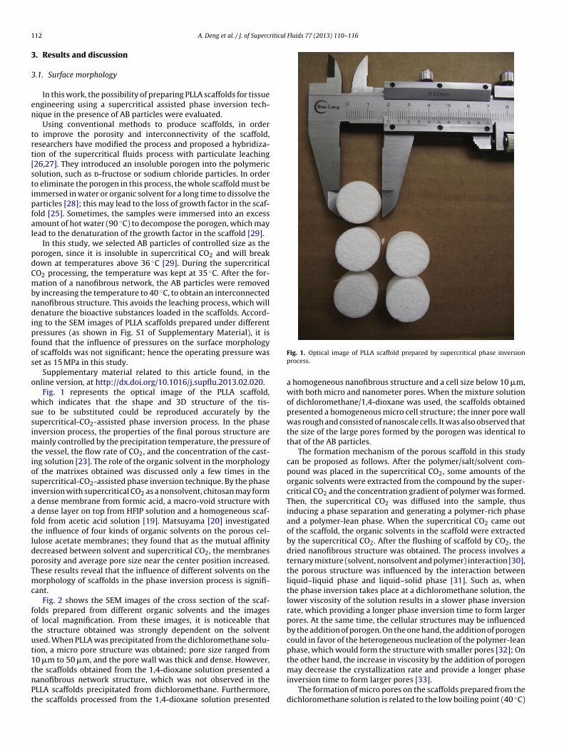
Fig. 1 represents the optical image of the PLLA scaffold,hich indicates that the shape and 3D structure of the tis-
ue to be substituted could be reproduced accurately by theupercritical-CO2-assisted phase inversion process. In the phasenversion process, the properties of the final porous structure are
ainly controlled by the precipitation temperature, the pressure ofhe vessel, the flow rate of CO2, and the concentration of the cast-ng solution [23]. The role of the organic solvent in the morphologyf the matrixes obtained was discussed only a few times in theupercritical-CO2-assisted phase inversion technique. By the phasenversion with supercritical CO2 as a nonsolvent, chitosan may form
dense membrane from formic acid, a macro-void structure with dense layer on top from HFIP solution and a homogeneous scaf-old from acetic acid solution [19]. Matsuyama [20] investigatedhe influence of four kinds of organic solvents on the porous cel-ulose acetate membranes; they found that as the mutual affinityecreased between solvent and supercritical CO2, the membranesorosity and average pore size near the center position increased.hese results reveal that the influence of different solvents on theorphology of scaffolds in the phase inversion process is signifi-
ant.Fig. 2 shows the SEM images of the cross section of the scaf-
olds prepared from different organic solvents and the imagesf local magnification. From these images, it is noticeable thathe structure obtained was strongly dependent on the solventsed. When PLLA was precipitated from the dichloromethane solu-ion, a micro pore structure was obtained; pore size ranged from0 �m to 50 �m, and the pore wall was thick and dense. However,
he scaffolds obtained from the 1,4-dioxane solution presented aanofibrous network structure, which was not observed in theLLA scaffolds precipitated from dichloromethane. Furthermore,he scaffolds processed from the 1,4-dioxane solution presentedFig. 1. Optical image of PLLA scaffold prepared by supercritical phase inversionprocess.
a homogeneous nanofibrous structure and a cell size below 10 �m,with both micro and nanometer pores. When the mixture solutionof dichloromethane/1,4-dioxane was used, the scaffolds obtainedpresented a homogeneous micro cell structure; the inner pore wallwas rough and consisted of nanoscale cells. It was also observed thatthe size of the large pores formed by the porogen was identical tothat of the AB particles.
The formation mechanism of the porous scaffold in this studycan be proposed as follows. After the polymer/salt/solvent com-pound was placed in the supercritical CO2, some amounts of theorganic solvents were extracted from the compound by the super-critical CO2 and the concentration gradient of polymer was formed.Then, the supercritical CO2 was diffused into the sample, thusinducing a phase separation and generating a polymer-rich phaseand a polymer-lean phase. When the supercritical CO2 came outof the scaffold, the organic solvents in the scaffold were extractedby the supercritical CO2. After the flushing of scaffold by CO2, thedried nanofibrous structure was obtained. The process involves aternary mixture (solvent, nonsolvent and polymer) interaction [30],the porous structure was influenced by the interaction betweenliquid–liquid phase and liquid–solid phase [31]. Such as, whenthe phase inversion takes place at a dichloromethane solution, thelower viscosity of the solution results in a slower phase inversionrate, which providing a longer phase inversion time to form largerpores. At the same time, the cellular structures may be influencedby the addition of porogen. On the one hand, the addition of porogencould in favor of the heterogeneous nucleation of the polymer-leanphase, which would form the structure with smaller pores [32]; Onthe other hand, the increase in viscosity by the addition of porogen
may decrease the crystallization rate and provide a longer phaseinversion time to form larger pores [33].The formation of micro pores on the scaffolds prepared from thedichloromethane solution is related to the low boiling point (40 ◦C)

A. Deng et al. / J. of Supercritical Fluids 77 (2013) 110– 116 113
F C and
(
otCistt1araoccmsp
bi
ig. 2. SEM images of the scaffolds prepared from different organic solutions at 35 ◦
c) 50×, (d) 1000×; 1,4-dioxane (e) 50×, (f) 1000×.
f the organic solvent, as this is close to the operating tempera-ure (35 ◦C). The solubility of dichloromethane in the supercriticalO2 is higher than that of 1,4-dioxane, which will favor the phase
nversion process because a higher affinity of the solvent to theupercritical CO2 will cause phase inversion and precipitation ofhe polymer with a porous structure. Conversely, 1,4-dioxane solu-ion is an organic solvent with a relatively high boiling point of01 ◦C, which is above the operating temperature. A lower solventffinity, that is, a higher square solubility parameter difference, willesult in a decrease in the pore size of the scaffold, and this is favor-ble for the formation of nanofibers. At the same time, the viscosityf the solution will influence the average pore size. Since the vis-osity of dichloromethane is lower than that of 1,4-dioxane, theoarsening of the micro droplets of the polymer-lean phase, whichay lead to larger pores, is favored [34,35]. These results demon-
trate that completely different morphologies can be obtained from
recipitation with different solvents.It has been proved that cell behavior can be mainly determinedy the substrate that they are cultured on, and the understand-
ng of interaction mechanism between cell and nano-topography
15.0 MPa: dichloromethane (a) 50×, (b) 1000×, dichloromethane/1,4-dioxane (1:1)
structures could provide valuable information for the design of tis-sue engineering [36]. The nanofibrous structure may influence thecell differentiation, migration and protein absorption [37,38]. Threekinds of nano-topography structures have been prepared by thesupercritical phase inversion technology in this experiment, it mayvery meaningful to induce cell growth on the scaffolds produced,to verify the effect of the mechano-topographic modulation struc-ture of the scaffold on the preferential differentiation of the cellson next step.
3.2. Porosity and mechanical strength of scaffold
In general, an increase in porosity will decrease the compres-sive strength of the scaffold, and vice versa. Hence, it is importantto deal with such relations well as porosity-porogen/PLLA ratio andcompressive strength – porogen/PLLA ratio. In this study, different
ratios of AB to PLLA were tested to find a suitable ratio to achievea high porosity and acceptable mechanical strength. Fig. 3 showsthe influence of the different ratios of AB to PLLA on the porosityand mechanical strength of the scaffold. When the mass ratio of
114 A. Deng et al. / J. of Supercritical
Fs
Ash2mr[aro
3
s
ig. 3. The influence of the different ratios of AB to PLLA on porosity and mechanicaltrength (at 25% strain).
B to PLLA increased from 10 to 40, the porosity of the scaffold wasignificantly increased (P < 0.05), from 92.0% ± 0.55 to 97.5% ± 0.45;owever, the mechanical strength was significantly decreased from25.4 kPa ± 3.16 to 36.8 ± 6.6 kPa (P < 0.05). The requirement ofechanical resistance and porosity depends on the organs to be
egenerated; for instance, 100 kPa is necessary for bone scaffolds39]. At the same time, the SEM image (Fig. 4) shows that the aver-ge pore size increased with the increase in AB/PLLA ratio. Theseesults reveal that scaffolds with desirable properties for differentrgans could be obtained by adjusting the ratios of AB to PLLA.
.3. Solvent residue and analysis
Without further treatment, the dichloromethane residue in thecaffold was only 12 ppm; this is much lower than the limit of
Fig. 4. SEM images of the scaffolds prepared from differen
Fluids 77 (2013) 110– 116
the USP 467 Pharmacopeia (600 ppm). Although organic solventswere employed, the use of supercritical CO2 allowed their completeremoval; as the supercritical CO2 has good diffusivity and masstransfer properties, its ability to diffuse and penetrate into the bulkof the 3D matrix allowed the complete extraction of the organicsolvents [21]. Reverchon and Ji [28,40] also reported that super-critical CO2 technology could produce a scaffold with an organicresidue of less than 5 ppm and 10 ppm, respectively. However, theresidual dichloromethane content of the scaffolds fabricated usingthe electrospinning method was 365 ppm after freeze drying forseven days [41], which is much higher than that of scaffolds pre-pared by supercritical CO2 technology without further downstreamprocessing. These results indicate that supercritical CO2 technol-ogy is an effective method of producing a scaffold with little or noorganic residue.
3.4. FTIR analysis
The FTIR spectra of the as-received PLLA and PLLA scaffolds areshown in Fig. 5. In these spectra, the PLLA characteristic bands arepresent; for example, the band at 1762 cm−1 was attributed to C Ostretching, the bands at 2949 and 2997 cm−1 were assigned to theCH3 and CH stretch bands, the 1196 cm−1 band was assigned to thestretching of C O C, and the 1093 cm−1 band was assigned to C Ostretching. After supercritical CO2 processing, there was no changein the chemical composition of the samples; the results were con-sistent with the report by Kang [42]. These results indicate thatthe supercritical-CO2-assisted phase inversion process is a physicalprocess capable of producing PLLA scaffolds.
3.5. XRD and DSC analysis
To investigate the physical state of PLLA before and after thesupercritical CO2 processing, XRD analysis was performed. Fig. 6shows the XRD spectra of the as-received PLLA and the PLLA scaf-fold. The main peaks at 16.3◦ and 18.5◦ proved the presence of
t AB/PLLA ratios: (a) 10:1, (b) 20:1, (c) 30:1, (d) 40:1.

A. Deng et al. / J. of Supercritical
Fig. 5. FTIR spectra of as-received PLLA and the scaffold prepared by the supercriticalfluid process.
Fig. 6. XRD spectra of as-received PLLA and the scaffold prepared by the supercriticalfluid process.
Fig. 7. DSC curves of as-received PLLA and the scaffold prepared by the supercriticalfluid process.
[
[
[
[
Fluids 77 (2013) 110– 116 115
semi-crystalline domains of PLLA specimens. Despite treating bythe supercritical CO2 process, XRD analysis showed that PLLA sam-ple was still in semi-crystalline state. This result was similar to theprevious study [43]. The DSC curves in Fig. 7 show that PLLA wasobserved as a semi-crystalline polymer with a melting temperature(Tm) of about 162.0 ◦C. The melting temperatures of the as-receivedPLLA and the scaffold are slightly shifted to a higher temperature,indicating that the supercritical-CO2-assisted phase inversion pro-cess may not affect the crystallization of PLLA significantly [43,44].
4. Conclusion
Nanofibrous PLLA scaffolds with interconnected pores were suc-cessfully prepared using ammonium bicarbonate as a pore formingagent in a supercritical CO2 process. The scaffolds possess the struc-tural characteristics of large pores and micro and nanometer poresthat have to be simultaneously present for tissue engineering appli-cations. They are characterized by a high porosity (up to 97.5%), afibrous structure that allows cell growth, a controllable cell size(depending on the particle size of AB), adequate mechanical prop-erties, and almost no solvent residues. Moreover, it is possible tosolve the problem of loss of the growth factors in the scaffold thatoccurs during the traditional preparation methods.
Acknowledgements
Financial support from the National Natural Science Foundationof China (31170939, 81171471 and 51103049) and National Sci-ence Foundation of Fujian Province (2010J05027 and 2011501223)gratefully acknowledged.
References
[1] J.P. Chen, C.H. Su, Surface modification of electrospun PLLA nanofibers byplasma treatment and cationized gelatin immobilization for cartilage tissueengineering, Acta Biomaterilia 7 (2011) 234–243.
[2] M.R. Jung, I.K. Shim, E.S. Kim, Y.J. Park, Y.I. Yang, S.K. Lee, S.J. Lee, Con-trolled release of cell-permeable gene complex from poly(l-lactide) scaffoldfor enhanced stem cell tissue engineering, Journal of Controlled Release 152(2011) 294–302.
[3] Q.W. Zhang, V.N. Mochalin, I. Neitzel, K. Hazeli, J.J. Niu, A. Kontsos, J.G.Zhou, P.I. Lelkes, Y. Gogotsi, Mechanical properties and biomineralization ofmultifunctional nanodiamond-PLLA composites for bone tissue engineering,Biomaterials 33 (2012) 5067–5075.
[4] A.Z. Chen, X.M. Pu, Y.Q. Kang, L. Liao, Y.D. Yao, G.F. Yin, Preparation of5-Fluorouracil-poly(l-lactide) microparticles using solution-enhanced disper-sion by supercritical CO2, Macromolecular Rapid Communications 27 (2006)1254–1259.
[5] J.M. Corey, C.C. Gertz, B.S. Wang, L.K. Birrell, S.L. Johnson, D.C. Martin, E.L.Feldman, The design of electrospun PLLA nanofiber scaffolds compatible withserum-free growth of primary motor and sensory neurons, Acta Biomaterialia4 (2008) 863–875.
[6] H.C. Liu, I.C. Lee, J.H. Wang, S.H. Yang, T.H. Young, Preparation of PLLA mem-branes with different morphologies of culture of MG-63 cells, Biomaterials 25(2004) 4047–4056.
[7] Z.W. Ma, C.Y. Gao, Y.H. Gong, J.C. Shen, Cartilage tissue engineering PLLA scaffoldwith surface immobilized collagen and basic fibroblast growth factor, Bioma-terials 26 (2005) 1253–1259.
[8] Z.P. Zhang, J. Hu, P.X. Ma, Nanofiber-based delivery of bioactive agents and stemcells to bone sites, Advanced Drug Delivery Reviews 64 (2012) 1129–1141.
[9] G.B. Wei, P.X. Ma, Partially nanofibrous architecture of 3D tissue engineeringscaffolds, Biomaterials 30 (2009) 6426–6434.
10] J.F. Mao, S. Duan, A. Song, Q. Cai, X.L. Deng, X.P. Yang, Macroporous andnanofibrous poly(lactide-co-glycolide)(50/50) scaffolds via phase separationcombined with particle-leaching, Materials Science and Engineering C 32(2012) 1407–1414.
11] A. Salerno, E.D. Maio, S. Iannace, P.A. Netti, Tailoring the pore structure of PCLscaffolds for tissue engineering prepared via gas foaming of multi-phase blends,Journal of Porous Materials 19 (2012) 181–188.
12] S. Rajabzadeh, C. Liang, Y. Ohmukai, T. Maruyama, H. Matsuyama, Effect ofadditives on the morphology and properties of poly(vinylidene fluoride) blend
hollow fiber membrane prepared by the thermally induced phase separationmethod, Journal of Membrane Science 423–424 (2012) 189–194.13] A.R.C. Duarte, J.F. Mano, R.L. Reis, Novel 3D scaffolds of chitosan-PLLA blendsfor tissue engineering applications: Preparation and characterization, Journalof Supercritical Fluids 54 (2010) 282–289.

1 ritical
[
[
[
[
[
[
[
[
[
[
[
[
[
[
[
[
[
[
[
[
[
[
[
[
[
[
[
[
[
[
16 A. Deng et al. / J. of Superc
14] E. Reverchon, S. Cardea, Supercritical fluids in 3-D tissue engineering, Journalof Supercritical Fluids 69 (2012) 97–107.
15] A.R.C. Duarte, J.F. Mano, R.L. Reis, Perspectives on: Supercritical fluid technol-ogy for 3D tissue engineering scaffold applications, Journal of Bioactive andCompatible Polymers 24 (2009) 385–400.
16] Y.R. Xin, T. Fujimoto, H. Uyama, Facile fabrication of polycarbonate monolith bynon-solvent induced phase separation method, Polymer 53 (2012) 2847–2853.
17] F.J. Hua, T.G. Park, D.S. Lee, A facile preparation of highly interconnected macro-porous poly(d,l-lactic acid-co-glycolic acid)(PLGA) scaffolds by liquid–liquidphase separation of a PLGA-dioxane-water ternary system, Polymer 44 (2003)1911–1920.
18] E. Danesh, S.R. Ghaffarian, P. Molla-Abbasi, Non-solvent induced phase sep-aration as a method for making high-performance chemiresistors based onconductive polymer nanocomposites, Sensors and Actuators B 155 (2011)562–567.
19] A.R.C. Duarte, J.F. Mano, R.L. Reis, The role of organic solvent on the prepara-tion of chitosan scaffolds by supercritical assisted phase inversion, Journal ofSupercritical Fluids 72 (2012) 326–332.
20] H. Matsuyama, A. Yamamoto, H. Yano, T. Maki, M. Teramoto, K. Mishima, K. Mat-suyama, Effect of organic solvents on membrane formation by phase separationwith supercritical CO2, Journal of Membrane Science 204 (2002) 81–87.
21] E. Reverchon, R. Adami, S. Cardea, G.D. Porta, Supercritical fluids processing ofpolymers for pharmaceutical and medical applications, Journal of SupercriticalFluids 47 (2009) 484–492.
22] D.J. Mooney, D.F. Baldwin, N.P. Suh, J.P. Vacanti, R. Langer, Novel approach tofabricate porous sponges of poly(d,l-lactic-co-glycolic acid) without the use oforganic solvents, Biomaterials 17 (1996) 1417–1422.
23] I. Tsivintzelis, E. Pavlidou, C. Panayiotou, Porous scaffolds prepared by phaseinversion using supercritical CO2 as antisolvent I. Poly(l-lactic acid), Journal ofSupercritical Fluids 40 (2007) 317–322.
24] L.D. Harris, B.S. Kim, D.J. Mooney, Open pore biodegradable matrices formedwith gas foaming, Journal of Biomedical Materials Research 42 (1998) 396–402.
25] O.R. Davies, A.L. Lewis, M.J. Whitaker, H.Y. Tai, K.M. Shakesheff, S.M. Howdle,Applications of supercritical CO2 in the fabrication of polymer systems for drugdelivery and tissue engineering, Advanced Drug Delivery Reviews 60 (2008)373–387.
26] G.B. Wei, P.X. Ma, Macroporous and nanofibrous polymer scaffolds andpolymer/bone-like apatite composite scaffolds generated by sugar spheres,Journal of Biomedical Materials Research 78 (2006) 306–315.
27] P.X. Ma, R.Y. Zhang, Synthetic nano-scale fibrous extracellular matrix, Journalof Biomedical Materials Research 46 (1999) 60–72.
28] E. Reverchon, S. Cardea, C. Rapuano, A new supercritical fluid-based process
to produce scaffolds for tissue replacement, Journal of Supercritical Fluids 45(2008) 365–373.29] Y.S. Nam, J.J. Yoon, T.G. Park, A novel fabrication method of macroporousbiodegradable polymer scaffolds using gas foaming salt as a porogen additive,Journal of Biomedical Materials Research 53 (2000) 1–7.
[
Fluids 77 (2013) 110– 116
30] M. Temtem, L.M.C. Silva, P.Z. Andrade, F. dos Santos, C.L. da Silva, J.M.S.Cabral, M.M. Abecasis, A. Aguiar-Ricardo, Supercritical CO2 generating chitosandevices with controlled morphology. Potential application for drug deliveryand mesenchymal stem cell culture, Journal of Supercritical Fluids 48 (2009)269–277.
31] Y.W. Kho, D.S. Kalika, B.L. Knutson, Precipitation of nylon 6 membranes usingcompressed carbon dioxide, Polymer 42 (2006) 6119–6127.
32] I. Tsivintzelis, S.I. Marras, I. Zubrtikudis, C. Panayiotou, Porous poly(l-lactic acid)nanocomposite scaffolds prepared by phase inversion using supercritical CO2
as antisolvent, Polymer 48 (2007) 6311–6318.33] J.H. Lee, T.G. Park, H.S. Park, D.S. Lee, Y.K. Lee, S.C. Yoon, J.D. Nam, Thermal and
mechanical characteristics of polymer(l-lactic acid) nanocomposite scaffold,Biomaterials 24 (2003) 2773–2778.
34] H. Matsuyama, H. Yano, T. Maki, M. Teramoto, K. Mishima, K. Matsuyama, For-mation of porous flat membrane by phase separation with supercritical CO2,Journal of Membrane Science 194 (2001) 157–163.
35] E. Reverchon, S. Cardea, Formation of cellulose acetate membranes using asupercritical fluid assisted process, Journal of Membrane Science 240 (2004)187–195.
36] V. Beachley, X.J. Wen, Polymer nafibrous structures: Fabrication, biofunc-tionalization, and cell interactions, Progress in Polymer Science 35 (2010)868–892.
37] S.H. Lim, X.Y. Liu, H.J. Song, K.J. Yarema, H.Q. Mao, The effect of nanofiber-guided cell alignment on the preferential differentiation of neural stem cells,Biomaterials 31 (2010) 9031–9039.
38] A.S. Nathan, B.M. Baker, N.L. Nerurkar, R.L. Mauck, Mechano-topographic mod-ulation of stem cell nuclear shape on nanofibrous scaffolds, Acta Biomaterialia7 (2011) 57–66.
39] J.C. Zhang, H. Zhang, L.B. Wu, J.D. Ding, Fabrication of three dimensional poly-meric scaffolds with spherical pores, Journal of Materials Science 41 (2006)1725–1731.
40] C.D. Ji, N. Annabi, M. Hosseinkhani, S. Sivaloganathan, F. Dehghani, Fabricationof poly-dl-lactide/polyethylene glycol scaffolds using the gas foaming tech-nique, Acta Biomaterialia 8 (2012) 570–578.
41] H.M. Nie, C.H. Wang, Fabrication and characterization of PLGA/Hap compositescaffolds for delivery of BMP-2 plasmid DNA, Journal of Controlled Release 120(2007) 111–121.
42] Y.Q. Kang, C. Yang, P. Ouyang, G.F. Yin, Z.B. Huang, Y.D. Yao, X.M. Liao, Thepreparation of BSA-PLLA microparticles in a batch supercritical anti-solventprocess, Carbohydrate Polymers 77 (2009) 244–249.
43] A. Vega-Gonzalez, P. Subra-Paternault, A.M. Lopez-Periago, C.A. Garcia-Gonzalez, C. Domingo, Supercritical CO2 antisolvent precipitation of polymer
networks of l-PLA, PMMA and PMMA/PCL blends for biomedical applications,European Polymer Journal 44 (2008) 1081–1094.44] S.Y. Huang, H.F. Li, S.C. Jiang, X.S. Chen, L.J. An, Crystal structure and morphologyinfluenced by shear effect of poly(l-lactide) and its melting behavior revealedby WAXD, DSC and in situ POM, Polymer 52 (2011) 3478–3487.