


Porin; OmpU; Vibrio cholerae; Programmed cell death ... · Apoptosis is a form of programmed cell...
34
OmpU induces caspase-independent programmed cell death 1 Vibrio cholerae porin OmpU induces caspase-independent programmed cell death upon translocation to the host cell mitochondria Shelly Gupta 1 , G.V.R. Krishna Prasad 1 , Arunika Mukhopadhaya 1 1 Department of Biological Sciences, Indian Institute of Science Education and Research (IISER) Mohali, Knowledge City, Sector 81, SAS Nagar, Mohali 140306, Punjab, India Running title: OmpU induces caspase-independent programmed cell death To whom correspondence should be addressed: Arunika Mukhopadhaya, Department of Biological Sciences, Indian Institute of Science Education and Research (IISER) Mohali, Knowledge City, Sector 81, SAS Nagar, Mohali 140306, Punjab, India, Tel.: 91-0172-2240266, Fax: 91-0172- 2240124, Email: [email protected] Running Title: OmpU induces caspase-independent programmed cell death. Keywords: Porin; OmpU; Vibrio cholerae; Programmed cell death; Mitochondria ______________________________________________________________________________ Abstract Porins, a major class of outer membrane proteins in gram-negative bacteria, primarily act as transport channels. OmpU is one of the major porins of human pathogen, Vibrio cholerae. In the present study, we show that V. cholerae OmpU has the ability to induce target cell death. Although OmpU-mediated cell death shows some characteristics of apoptosis such as flipping of phosphatidyl serine in the membrane, as well as cell size shrinkage and increased cell granularity, it does not show caspase-3 activation and DNA laddering pattern typical of apoptotic cells. Increased release of lactate dehydrogenase in OmpU-treated cells indicates that the OmpU- mediated cell death also has characteristics of necrosis. Further, we show that the mechanism of OmpU-mediated cell death involves major mitochondrial changes in the target cells. We observe that OmpU treatment leads to the disruption of mitochondrial membrane potential resulting in the release of cytochrome c and apoptosis inducing factor (AIF). AIF translocates to the host cell nucleus implying that it has a crucial role in OmpU-mediated cell death. Finally, we observe that OmpU translocates to the target cell mitochondria where it directly initiates mitochondrial changes leading to mitochondrial membrane permeability transition and AIF release. Partial blocking of AIF release by cyclosporine A in OmpU-treated cells further suggests that OmpU may be inducing the opening of mitochondrial permeability transition pore. All these results lead us to the conclusion that OmpU induces cell death in target cells in a programmed manner in which mitochondria play a central role. Classically, two major forms of cell death have been described: apoptosis and necrosis. Apoptosis is a form of programmed cell death that is characterized by sequential activation of events in a highly regulated manner leading to the death of cells (1-3). Necrotic cell death is supposed to be sudden or abrupt and is mainly a passive event. Apoptosis involves mainly two pathways, the extrinsic pathway and the intrinsic pathway (3,4). Sometimes extrinsic pathway initiates the intrinsic pathway or they work in isolation. Both extrinsic and intrinsic pathways involve the activation of a specialized class of enzymes called cysteine-dependent aspartate-specific proteases or caspases which are considered to be indispensable for apoptotic cell death. In case of extrinsic pathway, death receptors are involved. Upon death receptor activation, caspase-8 becomes activated initially and sequential events ultimately lead to the activation of caspase-3 (5). Mitochondria play a central role in the intrinsic pathway of apoptosis. For many years, the term apoptosis has been used synonymously with programmed cell death (PCD). However, recent years have witnessed an increase in the number reports describing different forms of PCD that occur http://www.jbc.org/cgi/doi/10.1074/jbc.M115.670182 The latest version is at JBC Papers in Press. Published on November 11, 2015 as Manuscript M115.670182 Copyright 2015 by The American Society for Biochemistry and Molecular Biology, Inc. by guest on August 25, 2019 http://www.jbc.org/ Downloaded from
Transcript of Porin; OmpU; Vibrio cholerae; Programmed cell death ... · Apoptosis is a form of programmed cell...

OmpU induces caspase-independent programmed cell death
1
Vibrio cholerae porin OmpU induces caspase-independent programmed cell death upon translocation to
the host cell mitochondria
Shelly Gupta1, G.V.R. Krishna Prasad
1, Arunika Mukhopadhaya
1
1Department of Biological Sciences, Indian Institute of Science Education and Research (IISER) Mohali,
Knowledge City, Sector 81, SAS Nagar, Mohali 140306, Punjab, India
Running title: OmpU induces caspase-independent programmed cell death
To whom correspondence should be addressed: Arunika Mukhopadhaya, Department of Biological
Sciences, Indian Institute of Science Education and Research (IISER) Mohali, Knowledge City, Sector
81, SAS Nagar, Mohali 140306, Punjab, India, Tel.: 91-0172-2240266, Fax: 91-0172- 2240124, Email:
Running Title: OmpU induces caspase-independent programmed cell death.
Keywords: Porin; OmpU; Vibrio cholerae; Programmed cell death; Mitochondria
______________________________________________________________________________
Abstract
Porins, a major class of outer membrane proteins
in gram-negative bacteria, primarily act as
transport channels. OmpU is one of the major
porins of human pathogen, Vibrio cholerae. In the
present study, we show that V. cholerae OmpU
has the ability to induce target cell death.
Although OmpU-mediated cell death shows some
characteristics of apoptosis such as flipping of
phosphatidyl serine in the membrane, as well as
cell size shrinkage and increased cell granularity,
it does not show caspase-3 activation and DNA
laddering pattern typical of apoptotic cells.
Increased release of lactate dehydrogenase in
OmpU-treated cells indicates that the OmpU-
mediated cell death also has characteristics of
necrosis. Further, we show that the mechanism of
OmpU-mediated cell death involves major
mitochondrial changes in the target cells. We
observe that OmpU treatment leads to the
disruption of mitochondrial membrane potential
resulting in the release of cytochrome c and
apoptosis inducing factor (AIF). AIF translocates
to the host cell nucleus implying that it has a
crucial role in OmpU-mediated cell death. Finally,
we observe that OmpU translocates to the target
cell mitochondria where it directly initiates
mitochondrial changes leading to mitochondrial
membrane permeability transition and AIF release.
Partial blocking of AIF release by cyclosporine A
in OmpU-treated cells further suggests that OmpU
may be inducing the opening of mitochondrial
permeability transition pore. All these results lead
us to the conclusion that OmpU induces cell death
in target cells in a programmed manner in which
mitochondria play a central role.
Classically, two major forms of cell death
have been described: apoptosis and necrosis.
Apoptosis is a form of programmed cell death that
is characterized by sequential activation of events
in a highly regulated manner leading to the death
of cells (1-3). Necrotic cell death is supposed to be
sudden or abrupt and is mainly a passive event.
Apoptosis involves mainly two pathways,
the extrinsic pathway and the intrinsic pathway
(3,4). Sometimes extrinsic pathway initiates the
intrinsic pathway or they work in isolation. Both
extrinsic and intrinsic pathways involve the
activation of a specialized class of enzymes called
cysteine-dependent aspartate-specific proteases or
caspases which are considered to be indispensable
for apoptotic cell death. In case of extrinsic
pathway, death receptors are involved. Upon death
receptor activation, caspase-8 becomes activated
initially and sequential events ultimately lead to
the activation of caspase-3 (5). Mitochondria play
a central role in the intrinsic pathway of apoptosis.
For many years, the term apoptosis has
been used synonymously with programmed cell
death (PCD). However, recent years have
witnessed an increase in the number reports
describing different forms of PCD that occur
http://www.jbc.org/cgi/doi/10.1074/jbc.M115.670182The latest version is at JBC Papers in Press. Published on November 11, 2015 as Manuscript M115.670182
Copyright 2015 by The American Society for Biochemistry and Molecular Biology, Inc.
by guest on August 25, 2019
http://ww
w.jbc.org/
Dow
nloaded from

OmpU induces caspase-independent programmed cell death
2
independent of caspases (6,7). Depending on the
type of morphological and biochemical events that
are initiated during programmed cell death,
different terms have been proposed; apoptosis like
PCD, necrosis like PCD, necroptosis and many
others (8,9). Diverse sets of molecules and
different organelles like lysosomes, ER and
mitochondria interact in a cell leading to its
demise (6).
Involvement of mitochondria in caspase
independent PCD has been established for some
cell types in response to particular stimuli (10,11).
Any disruption in the normal bio-energetic state of
mitochondria induced by stress generated in cells
by various external or internal factors can lead to a
loss of mitochondrial membrane potential.
Generally, members of Bcl-2 family such as, Bax ,
translocate to mitochondria and permeabilize the
outer mitochondrial membrane (OMM) (12-14).
Certain stimuli can affect the integrity of inner
mitochondrial membrane (IMM) and this leads to
the mitochondrial membrane permeability
transition (MMPT) further resulting in the release
of inter-membrane space molecules like
cytochrome c and apoptosis inducing factor (AIF)
into the cytosol (11). Cytochrome c may bind
Apaf-1in the presence of dATP and promote
caspase-9 and caspase-3 activation (15-17). AIF
plays a crucial role in caspase-independent
pathways. It can directly translocate to the nucleus
and cause DNA fragmentation independent of
caspases (18). Therefore, mitochondria seem to
play a key role in both caspase-dependent and -
independent pathways of PCD.
Different types of PCD play a crucial role
in host-pathogen interactions. Pathogens induce
cell death in order to invade host tissues or to
evade host immune responses (19). Gram-negative
pathogenic bacteria use such processes to damage
host tissues and cause sepsis by invading deeper
into them (20). Host uses such mechanisms to
prevent pathogenic infections by inducing the
death of infected cells.
A number of bacterial molecules have
been implicated in the induction of apoptosis or
other forms of PCD in the host cells. During
bacterial infections, some pathogens secrete toxins
which may trigger cell death (21,22). In addition,
mode of invasion of the bacteria such as
endocytosis and the structural elements of the
bacteria that help in invading the host cell can
trigger the death of target cells. In gram-negative
bacteria like Neisseria gonorrhoeae and
Pseudomonas aeruginosa, porins, a class of outer
membrane integral proteins, induce apoptosis of
the target cells (23,24).
Porins are trans-membrane channels that
allow selective uptake of nutrients, a requisite for
bacterial cell survival (25). In addition, porins may
act as pathogen-associated molecular patterns
(PAMPs) that are recognized by pattern
recognition receptors (PRRs) on host cell surfaces
and induce pro-inflammatory responses. Porins of
Shigella, Salmonella, Vibrio cholerae etc. are
known to induce the production of pro-
inflammatory cytokines (26-29). Some porins are
also reported to be anti-apoptotic such as, PorB of
Neisseria meningitidis (30).
OmpU, one of the major outer membrane
proteins of V. cholerae is porin in nature (31). In
addition to its role as a porin, OmpU has been
speculated to play a crucial role in the
pathogenesis of V. cholerae. This speculation is
mainly based on the fact that expression of OmpU
is under the control of ToxR regulon which
controls major virulence factors required for the
pathogenesis of V. cholerae (32-34). Moreover,
OmpU has been reported to facilitate intestinal
colonization of the bacterium by conferring
resistance against bile,and anti-microbial peptides.
It probably acts as an adhesin as well, although
there are contrasting reports regarding its role in
adhesion (35-37). Moreover, OmpU has been
shown to possess the ability to down-regulate
LPS-mediated pro-inflammatory effect (28).
Therefore, its regulation and reported functions
imply that OmpU may have a major role in
bacterial pathogenesis process. However, the
contribution of OmpU in the induction of cell
death has not been evaluated.
Till date a good vaccine against cholera is
not available. OmpU is considered as a good
candidate for vaccine generation mainly because
of the fact that OmpU is present in most of the
clinical isolates (38). Recently a report suggested
that OmpU can be used as bio-marker to
distinguish between epidemic and non-epidemic
strains (39). Therefore, it is very important to
characterize OmpU for its role in the induction of
multiple cellular processes in the host.
Based on all the above knowledge,
speculated role of OmpU in pathogenesis and
by guest on August 25, 2019
http://ww
w.jbc.org/
Dow
nloaded from

OmpU induces caspase-independent programmed cell death
3
weighing the importance of cell death responses in
host-pathogen interactions, in the present paper we
have studied the role of OmpU in induction of cell
death in human cells and the possible mechanism
involved in the process.
Experimental Procedures
Purification of recombinant OmpU:
Recombinant OmpU was purified as described
previously by Khan et al (40).
Detection of endotoxin contamination in
purified protein preparation: Presence of
endotoxin in different batches of purified protein
was measured by limulus amebocyte lysate test
(LAL test) using E-TOXATE™ Kit (Sigma
Aldrich, St. Louis, MO) as per the manufacturer’s
protocol.
Mammalian Cell culture: Human
monocytic cell line, THP-1 (NCCS, Pune) was
maintained in RPMI 1640 (Invitrogen Life
Technologies, Carlsbad, CA, USA) supplemented
with 10 % fetal bovine serum (FBS; Invitrogen),
100 U/ml penicillin and 100 g/ml streptomycin
(Invitrogen). Prior to each experiment, cells were
conditioned in medium containing 5 % FBS for 24
h followed by 2 % FBS for 12 h. Human
embryonic kidney cell line, HEK 293 (ATCC) and
human colon carcinoma cell line, Caco-2 (ATCC)
were maintained in DMEM (Invitrogen)
containing 10 % FBS, 100 U/ml penicillin and 100
g/ml streptomycin (Invitrogen). Similar to THP-
1, prior to each experiment, HEK 293 cells and
Caco-2 cells were conditioned in media containing
reducing concentrations of serum.
Experimental design: For majority of the
experiments, THP-1 monocytes and Caco-2 cells
were plated at a density of 1x10⁶ cells/ml and
HEK 293 cells were plated at a density of 0.5x10⁶
cells/ml respectively in media containing 2 %
serum and treated with either recombinant OmpU
or buffer (protein-buffer containing10 mM Tris-
HCl, 10 mM NaCl and 0.5 % LDAO, as per
requirement diluted in PBS+0.5 % LDAO). LDAO
was obtained from Sigma Aldrich. Following
incubations, cells were subjected to flow
cytometry or western blotting.
Most of the flow cytometry experiments
were performed using BD FACSCalibur (BD
Biosciences, San Jose, CA, USA) unless otherwise
mentioned. Acquisition for all the experiments
was done in CellQuest pro and analysis was done
using FlowJo software (Tree Star).
For western blot analysis, blots were
visualized in ImageQuant LAS 4010 (GE
Healthcare Life sciences, Uppsala, Sweden).
Detection of early morphological
changes: THP-1 monocytes were treated with
OmpU (10 g/ml) or buffer and incubated for 24
h. Cells were harvested and washed twice with
phosphate buffered saline (PBS). Finally, cells
were re-suspended in 500 l of PBS and analyzed
by flow cytometry under the parameters of FSC
and SSC.
Detection of cell viability using MTT
assay: THP-1 monocytes, Caco-2 cells and HEK
293 cells were plated in a 96-well plate format and
were treated with different concentrations of
OmpU (1.5 g/ml, 3 g/ml, 5 g/ml, 7 g/ml and
10 g/ml) or buffer and incubated for 24 h.
Following incubations, cells were subjected to
MTT assay for cell viability. MTT assay is based
on the principle that it is a tetrazolium salt which
is water soluble and gives yellow color in solution.
Active dehydrogenases of living cells can convert
the tetrazolium ring into insoluble formazan
crystals which are purple in colour. Dead cells are
incapable of carrying out the conversion.
Formazan crystals can be dissolved using acidified
isopropanol. The colour so obtained is measured
spectrophotometrically. Concentration of the dye
is proportional to the percentage of viable cells.
MTT assay was done using a kit (Sigma Aldrich)
as per the manufacturer’s instructions. Briefly,
solution provided in the kit was added to the cells
at 10 % v/v and incubated for 3 h. After
incubation, formzan crystals were dissolved by
adding equal volume of acidified propanol
(prepared by adding 0.1 N HCl in isopropanol).
After the crystals were completely dissolved,
absorbance was measured at 570 nm. Absorbance
values obtained were employed to calculate
percentage cell viability using the following
formula: (Treated cells – media only) *100/
(Untreated cells – media only). Untreated cells in
media alone were considered to be 100% viable.
Detection of cell cytotoxicity using LDH
release assay: THP-1 monocytes or Caco-2 cells
were plated and treated with different
concentrations of OmpU (5 g/ml, 7 g/ml and 10
g/ml) or buffer for 24 h. After 24 h, cells were
by guest on August 25, 2019
http://ww
w.jbc.org/
Dow
nloaded from

OmpU induces caspase-independent programmed cell death
4
subjected to LDH release assay. It is based on the
principle that upon permeabilization of plasma
membrane, lactate dehydrogenase (LDH) which is
a cytosolic enzyme, is released in the culture
supernatants. On addition of substrates like
tetrazolium salts, LDH present in the culture
supernatant converts the salt into a colored
product. Intensity of the color produced is
proportional to the number of lysed cells and can
be measured spectrophotometrically. LDH release
assay was perforned using CytoTox 96® Non-
Radioactive Cytotoxicity Assay kit (Promega
Corporation, Madison, WI) as per the
manufacturer’s instructions. Briefly, cells were
pelleted and supernatant for each treated sample
was collected separately. Substrate provided in the
kit was added to the supernatant of different
samples. Supernatant from cells treated with lysis
solution provided in the kit served as positive
control. Supernatants were incubated with
substrate for 10 min. Following incubation, stop
solution was added and absorbance was measured
at 490 nm.
Detection of DNA laddering pattern by
agrose-gel electrophoresis: THP-1 monocytes
were plated and treated with 10 g/ml of OmpU
for different time periods (24 h and 36 h) or buffer
for 36 h. Cells treated with staurosporine (1 M)
for 4 h were used as the positive control.
Following respective incubations, cells were
harvested and washed twice with 1X PBS. Cell
pellets were re-suspended in 50 l of lysis buffer
(1 % NP-40 in 20 mM EDTA, 50 mM Tris-HCl,
pH 7.5, 10 l/106
cells, minimum) and centrifuged
for 5 min at 1600 xg. Supernatant was collected
and extraction was repeated once. Further,
supernatants were combined and SDS was added
such that final concentration of SDS was 1 %
wt/vol. Then, supernatant was treated with RNase
A (final conc. 5mg/ml) at 56°C for 2 h followed
by treatment with proteinase K (final conc.
1mg/ml) at 37°C for 2 h. Following incubations,
half volume of ammonium acetate was added and
DNA was precipitated with 2.5 volumes of
absolute ethanol. After precipitation, pellets were
dissolved in DNA loading buffer and separated on
1.5 % agarose gel.
Detection of phosphatidyl serine exposure
by Annexin V-FITC staining: THP-1 monocytes
were treated with different doses of OmpU (1.5, 3,
5, 7 or 10 g/ml) or buffer, and incubated for 24 h.
Following treatment, cells were harvested and
stained using Annexin V-FITC and PI (propidium
iodide) staining kit (BD Biosciences) according to
the manufacturer’s protocol. Following staining,
cells were analyzed by flow cytometry. Quadrant
gate was applied (according to un-stained and
single-stained controls) to all the dot plots
corresponding to the tests and percentages of cells
in different quadrants were calculated using
quadrant statistics in FlowJo.
For inhibitor study, cells were incubated
with 20 M of total caspase inhibitor (zVAD-fmk,
Sigma Aldrich) for 1 h followed by treatment with
OmpU (10 g/ml) or buffer for 24 h.
Analysis of DNA fragmentation by TUNEL
Assay: THP-1 cells were treated with OmpU (10
g/ml) or buffer and incubated for different time
periods (12, 24 or 36 h). HEK 293 cells were
incubated with OmpU (10 g/ml) or buffer for 24
h. In another set, cells were pre-incubated with
zVAD-fmk (20 M) for 1 h followed by treatment
with OmpU (10 g/ml) or buffer for 24 h. Cells
were harvested and fixed using
1 % paraformaldehyde (PFA) following respective
incubations. Fixed cells were washed with ice cold
PBS, re-suspended in 70 % ethanol and incubated
at -20 ºC for 12-18 h. Finally cells were stained
using APO-BRDU kit (Sigma Aldrich) according
to the manufacturer’s protocol and subjected to
flow cytometry.
Detection of caspase-3 activation: THP-1
monocytes were treated with OmpU (10 g/ml) or
buffer and incubated for 12, 24 and 36 h. As a
positive control, cells were treated with 1 μM
staurosporine (Sigma Aldrich) and incubated for 4
h. Following respective incubations, cells were
harvested and stained with FITC conjugated rabbit
anti-human active caspase-3 antibody using a
staining kit (BD Biosciences) as per
manufacturer’s protocol and analyzed by flow
cytometry.
Analysis of change in mitochondrial
membrane potential: THP-1 monocytes were
treated with OmpU (10 g/ml) or buffer and
incubated for different time periods (4, 8, and 24
h). After respective incubations, cells were
harvested and stained with JC-1 dye (Sigma
Aldrich) according to the manufacturer’s protocol.
by guest on August 25, 2019
http://ww
w.jbc.org/
Dow
nloaded from

OmpU induces caspase-independent programmed cell death
5
Changes in mitochondrial membrane potential
were detected by flow cytometry.
Similarly, HEK 293 cells were incubated
with OmpU (10 g/ml) or buffer for 24 h.
Following incubation, cells were stained with JC-1
dye and analyzed for changes in mitochondrial
membrane potential.
For microscopic analysis, mitochondrial
membrane potential sensitive dye, mitotraker red
CMXros (Invitrogen life technologies), was used.
HEK 293 cells were seeded at a density of 0.1x10⁶
cells/ml on cover slips and incubated overnight.
Cells were treated with 10 g/ml of OmpU or
buffer and incubated for 24 h. Cells treated with
staurosporine (1 M) for 4 h were taken as
positive control. Following respective incubations,
cells were stained with 30 nM solution of
mitotraker red CMXros and incubated for 30 min
at 37 ºC. After incubation, cells were washed with
warm media (37 ºC) four times. Following
washing, cells were fixed with 4 % PFA and
washed 3-4 times with PBS. Stained cover slips
were mounted and observed under confocal
microscope. Imaging was done using Zeiss LSM
780 (Carl Zeiss, Germany) confocal laser scanning
microscope with a 63X oil immersion objective
having a 1.4 numerical aperture. Identical
parameters were used for all the samples. ImageJ
(NIH, Bethesda, MD, USA) software was used to
analyze all the images. Minimum ten images for
each sample were quantified and equal threshold
values were applied for all the samples.
Mitochondrial fluorescence for OmpU and
staurosporine treated cells was normalized with
respect to buffer-treated cells,
Detection of cytochrome c release by flow
cytometry: Release of cytochrome c was detected
using protocol given by Waterhouse et al (41) with
some modifications. Briefly, THP-1 monocytes
were treated with OmpU (10 g/ml) or buffer and
incubated for 24 h. After incubation, cells were
harvested and washed with PBS. Further, cells
were incubated in the permeabilization buffer (1 %
FBS, 0.1 % saponin and 0.1 % sodium azide in
PBS) to selectively permeabilize the cellular
membrane. Permeabilized cells were immediately
fixed with 2 % PFA by incubating for 30 min at
room temperature (RT). Fixed cells were washed
twice with ice cold PBS and incubated in blocking
buffer (3 % BSA + 0.5 % saponin in PBS) for 30
min at RT. After blocking, rabbit anti-human
cytochrome c antibody (Sigma Aldrich) was added
at a dilution of 1:50 in the blocking buffer, and
cells were incubated overnight at 4 ºC. Further,
cells were washed twice with PBS and incubated
with FITC-conjugated anti-rabbit IgG (Sigma
Aldrich) antibody at a dilution of 1:100 in
blocking buffer for 1 h at RT. Stained cells were
washed twice with PBS (ice cold), re-suspended in
500 l of blocking buffer and analyzed by flow
cytometry.
Determination of ATP: THP-1 monocytes
were plated as described previously and treated
with OmpU (10 g/ml) or buffer for 24 h.
Following incubation, cells were harvested and
subjected to ATP determination assay using ATP
Bioluminescence Assay Kit HS II (Roche
Diagnostics). It is based on the measurement of
bioluminescence produced by the enzyme
luciferase which uses ATP from lysed cells to
convert the substrate into bioluminescent product.
This luminescence is then detected using a
luminometer and is proportional to the amount of
ATP present in the cells. Assay was performed as
per the manufacturer’s instructions and data was
presented as fold change in the amount of ATP in
OmpU-treated cells with respect to buffer.
Preparation of mitochondrial fraction:
Enriched mitochondrial fraction from cultured
cells was prepared using Mitochondria Isolation
Kit (Sigma Aldrich) according to the
manufacturer’s protocol. Briefly, THP-1 cells
were treated with OmpU (10 g/ml) and incubated
for different time periods (0, 12 and 24 h). HEK
293 cells and Caco-2 cells were treated in a similar
manner as THP-1 monocytes. After respective
incubations, cells were harvested and washed
twice with ice cold PBS. Mitochondrial fraction
was prepared from cells using detergent lysis
method. Lysed cells were subjected to low speed
centrifugation (600 xg) to get rid of debris
followed by high speed centrifugation (11, 000 xg)
to pellet down the mitochondria. The pellet so
obtained was enriched in mitochondrial fraction
and the cytoplasmic fraction was present in the
supernatant.
Preparation of nuclear fraction: THP-1
monocytes, HEK 293 cells and Caco-2 cells were
treated with OmpU (10 g/ml) or buffer and
incubated for 24 h. Cells were harvested and
by guest on August 25, 2019
http://ww
w.jbc.org/
Dow
nloaded from

OmpU induces caspase-independent programmed cell death
6
washed with ice cold PBS. Washed cells were re-
suspended in 500 l of buffer A (10 mM HEPES,
1.5 mM MgCl2, 10 mM KCl, 1.5 mM DTT, 0.05
% NP-40) and incubated on ice for 5 min with
intermittent vortexing and further subjected to
centrifugation at 650 xg for 5 min. Pellet was re-
suspended in buffer B (5 mM HEPES, 1.5 mM
MgCl2, 0.2 mM EDTA, 0.5 mM DTT, 26 %
glycerol) containing 300 mM NaCl and
homogenized on ice followed by centrifugation at
24, 000 xg for 20 min at 4 ºC. Supernatant
obtained after centrifugation contained the nuclear
fraction.
Detection of AIF translocation from
mitochondria to nucleus and cytochrome c release
from mitochondria to cytoplasm: Mitochondrial,
nuclear and cytoplasmic fractions were estimated
for their protein concentrations using Bradford’s
(Sigma Aldrich) assay. Equal amounts of protein
were subjected to SDS-PAGE. Following
separation, proteins were transferred to poly-
vinylidene fluoride membrane (PVDF membrane,
Merck Millipore, Darmstadt, Germany).
Transferred membranes were blocked for 1 h at
RT with 5 % BSA in TBST (20 mM Tris, pH 7.6,
137 mM NaCl, 0.1 % Tween 20). After blocking,
membranes were incubated with rabbit anti-human
AIF antibody (Sigma Aldrich) or mouse anti-
human cytochrome c (Sigma Aldrich) diluted in
TBST (1:1000) for 4 h at RT. Membranes were
washed with TBST and incubated with anti-rabbit
IgG or anti-mouse IgG coupled with horseradish
peroxidase (1:5000, Sigma Aldrich) for 1 h.
TIM23 or TOM22 or TOM20 were used as the
loading control for mitochondrial samples and
detected with mouse anti-human TIM23 or rabbit
anti-human TOM22 or TOM20 antibodies (Sigma
Aldrich). Lamin B1 detected by rabbit anti-human
lamin B1 antibody (Santa Cruz Biotechnology,
Inc.) was used as the loading control for nuclear
lysates and GAPDH detected by rabbit anti-human
GAPDH antibody was used as the loading control
for cytoplasmic fraction.
Detection of OmpU in mitochondria:
THP-1 monocytes were treated with OmpU (10
g/ml) and incubated for different time periods
(15, 30, 60, 120 and 240 min). HEK 293 cells
were treated in a similar manner and incubated for
different time periods up to 6 h. Caco-2 cells were
also treated with same dose of OmpU and
incubated for 2 h. After respective incubations,
cells were harvested and mitochondrial lysates of
each sample were prepared using the same method
as mentioned previously. Centrifugation speeds
were modified to 1000 xg (low speed
centrifugation) and 3000 xg (high speed
centrifugation) in order to obtain more purified
mitochondrial fraction. Prepared lysates were
analyzed for translocation of OmpU by western
blotting in a similar manner as mentioned above
using primary antibody specific to V. cholerae
OmpU (rabbit anti-OmpU) at a dilution of 1:1000,
followed by secondary antibody which was anti-
rabbit IgG coupled with horse radish peroxidase
(1:5000 dilution). TIM23 or TOM22
(mitochondrial markers, Sigma Aldrich) were used
as the loading controls. Rabbit anti-human LAMP
1 (Abcam, USA) and rabbit anti-human GAPDH
(Santa Cruz Biotechnology, Inc.) antibodies were
used to check the presence of lysosomes and
cytoplasm respectively in the mitochondrial
lysates. Mouse anti-human transferrin receptor
(Invitrogen) and mouse anti-alpha-1 sodium
potassium ATPase (Abcam) antibodies were used
as plasma membrane markers in order to detect its
contamination in the mitochondrial preparation.
In order to detect the mitochondrial
translocation of OmpU by flow cytometry, THP-1
monocytes were treated with OmpU (10 g/ml)
for 60 min and 120 min or buffer for 120 min.
Following incubations, cells were harvested and
mitochondria were isolated as described
previously. Isolated mitochondria were re-
suspended in 1X storage buffer provided in the
Mitochondria Isolation Kit (Sigma Aldrich) and
incubated with rabbit anti-OmpU at a dilution of
1:50 for 30 min at RT. Mitochondria were washed
once with storage buffer by centrifugation at 17,
500 xg for 5 min and incubated with FITC tagged
anti-rabbit IgG at a dilution of 1:100 at RT.
Mitochondria were washed once with storage
buffer, re-suspended in 300 l of storage buffer
and analyzed by flow-cytometry using BD
AccuriTM
C6 Cytometer (BD Biosciences).
Detection of OmpU in different organelles
by cell fractionation: THP-1 monocytes were
plated and incubated with OmpU for 90 min.
Following incubation cells were harvested and
subjected to cell fractionation protocol for the
preparation of mitochondrial, nuclear and
cytoplasmic + light membrane fractions.
Mitochondrial fraction was prepared as described
by guest on August 25, 2019
http://ww
w.jbc.org/
Dow
nloaded from
OmpU induces caspase-independent programmed cell death
7
previously. For the preparation of nuclear fraction,
cells were ruptured by homogenization in buffer A
(1 M HEPES,1 M MgCl2, 2.5 M KCl, 1 M DTT)
following which they were centrifuged at 228 xg
for 5 min at 4 ºC to pellet down the nuclei along
with other fragments. The supernatant was stored
as the cytoplasmic + light membrane fraction from
which mitochondria were removed by centrifuging
at 3000 xg for 10 min. Nuclear pellet was further
re-suspended in 3 ml of buffer 1 (0.25 M sucrose,
10 mM MgCl2) and layered over buffer 2 (0.88 M
sucrose, 0.5 mM MgCl2) followed by
centrifugation at 2800 xg for 10 min at 4 ºC. Pellet
so obtained was re-suspended in 1X RIPA buffer
and sonicated in ice. Lysate was centrifuged at
2800 xg for 10 min at 4 ºC and the supernatant
was stored as nuclear fraction.
Detection of OmpU in plasma membrane:
HEK 293 cells were plated and incubated with
OmpU (10 g/ml) for 15 min, 30 min, 60 min and
120 min. Further, to obtain plasma membrane
enriched light membrane fraction, the supernatant
containing cytoplasm + light membranes
(described above) was ultra-centrifuged at 100,000
xg for 1 h. Pellet so obtained was washed once
with fractionation buffer, homogenized again and
finally collected as light membrane fraction. The
final pellet was resuspended in lysis buffer. Equal
amounts of all the fractions were then analysed for
the presence of OmpU along with the specific
markers as described previously.
Detection of OmpU translocation to
mitochondria from the bacterial cell: Vibrio
cholerae El Tor O1 strain (obtained from MTCC
of IMTECH Chandigarh; MTCC Code 3905) was
cultured in brain heart infusion (BHI) broth until
OD600 = 3. An aliquot of this culture was taken
separately and subjected to incubation at 60 ºC for
1 h for heat-inactivation of the bacteria.
Inactivation was confirmed by spreading the
culture on Luria agar plate and incubating at 37 ºC
for 16 h. Both live and heat-inactivated cultures
were pelleted and washed thrice with ice cold
PBS. THP-1 monocytes were treated with the
bacteria (re-suspended in sterile ice cold PBS) at
MOI = 10 and incubated at 37 ºC for 15 min, 30
min, 1 h and 2 h. Medium containing only
bacterial cells and THP-1 monocytes treated with
PBS were included as controls. All the test
samples and the controls were processed similarly.
Following respective incubations, THP-1 cells
were harvested and washed thrice with ice cold
PBS by centrifuging at 200 xg for 5 min.
Mitochondria were isolated from all samples (as
mentioned above). Streaking on agar plates from
isolated mitochondria was done to detect the
presence of any bacterial contaminants in final
lysates. Mitochondrial lysates were analyzed for
the presence of OmpU by means of western
blotting as described previously. From the
mitochondrial lysates obtained from 2 h
treatments, another blot was performed for the
detection of RNA polymerase beta as bacterial
cytoplasmic marker and lipid A, a component of
LPS, as the bacterial outer membrane marker, in
all the samples. RNA polymerase beta was
detected using mouse anti-RNA polymerase beta
antibody (Abcam) whereas, lipid A was detected
using anti-lipid A antibody (Abcam). As a positive
control for these markers, equal aliquots of live
and heat-inactivated V. cholerae cultures (OD600 =
3) were taken and pelleted. Pellets were dissolved
in sample buffer (50 l each) and heated at 100 ºC
for 15-20 min to completely lyse the bacterial
cells. Following this, equal volumes of sample
buffer containing lysed cells were analyzed in the
same blot along with mitochondrial lysates, for the
respective bacterial markers.
Analysis of direct effect of OmpU on
isolated mitochondria: Mitochondria were isolated
from THP-1 monocytes using Mitochondria
Isolation Kit (Sigma Aldrich) as per the
manufacturer’s protocol. Isolated mitochondria
were re-suspended in storage buffer provided in
the kit. Equal aliquots of isolated mitochondria
were incubated with OmpU (5 g/ml) or buffer for
30 min at 37 ºC. Following incubation,
mitochondria were stained with JC-1 dye and
analyzed for loss of aggregates in the FL-2
channel by flow cytometry.
For detection of AIF and cytochrome c
release, equal aliquots of isolated mitochondria
were treated with OmpU (5 g/ml) and incubated
for different time periods (30 min, 1 h and 2 h) or
treated with buffer and incubated for 2 h. After the
treatment, mitochondria were centrifuged at
17,500 xg for 5 min. Mitochondrial pellets were
solubilized in sample buffer and supernatant was
collected separately for each sample. Supernatants
and mitochondrial pellets were separated by SDS-
PAGE and analyzed by western blotting to
visualize the release of AIF and cytochrome c.
by guest on August 25, 2019
http://ww
w.jbc.org/
Dow
nloaded from

OmpU induces caspase-independent programmed cell death
8
TIM23 was used as the mitochondrial loading
control. For inhibition study, freshly isolated
mitochondria were pre-incubated with 1 M
cyclosporine A for 30 min followed by treatment
with OmpU for 90 min.
Statistical analysis: Data were
represented as the mean ± SEM. Statistical
analysis was done using Student’s two-sided t test.
Results
OmpU induces death of the target cells:
Different bacterial antigens can induce multitude
of host cellular responses and cell death is one of
them. To determine the cell viability, MTT is a
very well established assay. Therefore, to probe
whether any deterioration of cell health occurs in
response to OmpU, THP-1 monocytes and HEK
293 cells were treated with different
concentrations of OmpU or buffer and following
incubations cells were analyzed for their viability
using MTT assay.. The results showed that OmpU
treated cells undergo increasing loss of cell
viability with increasing doses (Fig. 1A and 1B).
In response to 10 g/ml of protein treatment,
around 18-28 % and 16-26 % loss in cell viability
was observed after 24 h in THP-1 and HEK 293
cells with respect to buffer (Fig. 1A and 1B).
These observations lead to the conclusion that
OmpU affects target cell health and induces a loss
in total cell viability.
Vibrio cholerae OmpU induces
morphological and biochemical changes that
indicate towards programmed cell death in target
cells: After we observed that OmpU induces cell
death, we probed whether OmpU induced features
that are characteristic of apoptosis. Apoptosis
involves flipping of phosphatidyl serine (PS) from
inner to outer leaflet of plasma membrane. To
investigate whether V. cholerae OmpU induces PS
flipping in target cells, THP-1 monocytes were
treated with different doses of OmpU and
incubated for 24 h. Cells were harvested and
stained with annexin V-FITC and PI. Cells with
increased PS exposure were detected as annexin
V-FITC positive population and late
apoptotic/necrotic cells were detected as annexin
V-FITC and PI double positive population. We
observed that total annexin V single positive
(lower right quadrant, LR) + annexin V /PI double
positive population (upper right quadrant, UR)
increased in a dose dependent manner from 1.5
g/ml to 10 g/ml, but a significant increase was
observed at a concentration of 10 g/ml only (38
%, stdev 10.4) compared to the buffer treatment
(16 %, stdev 4.9). The result so obtained suggested
that OmpU not only induces cell death, but also
induces certain initial changes like membrane
blebbing that might lead to the detection of PS
flipping in OmpU-treated cells. (Fig. 1C and 1D)
Endotoxin levels in different batches of
purified protein were non-detectable (EU<0.06 by
LAL test) and in a separate experiment, cells were
incubated with or without polymyxin B (pmb, 5
g/ml) for 30 min followed by incubation with
OmpU (10 g/ml) for 24 h. No change was
observed in total annexin V positive or annexin V
/PI double positive population in pmb + OmpU-
treated cells with respect to only OmpU-treated
cells suggesting that LPS plays no role in OmpU-
mediated cell death (Fig. 1E).
In flow cytometric analysis, forward
scatter (FSC) gives the measure of cell size and
side scatter (SSC) gives the measure of cell
granularity. We observed that cells treated with
OmpU showed a substantial decrease in FSC and
corresponding increase in SSC implying that
OmpU induces increased cell granularity along
with shrinking of target cells (Fig. 1F and 1G).
However, the cells undergoing morphological
changes appear higher than the cells undergoing
biochemical changes. Therefore, all the cells
undergoing morphological changes may not be
undergoing cell death at the indicated time point.
All these morphological and biochemical
events induced by OmpU suggested that it could
induce programmed cell death similar to
apoptosis.
OmpU induces DNA fragmentation but
not DNA laddering typical of apoptotic cells: In
classical apoptosis, the last step in the cascade is
removal of the inhibitor from the CAD or caspase
activated DNases. Nuclear DNA is cleaved by
CAD and this gives rise to several DNA fragments
which when resolved in an agarose gel generate a
distinct laddering pattern (42). Other types of cell
death processes also generate DNA fragments
which may be larger in size. These fragments may
not properly resolve in the agarose gel and thus do
not produce DNA ladder; like in case of AIF
mediated cell death (8,18).
by guest on August 25, 2019
http://ww
w.jbc.org/
Dow
nloaded from

OmpU induces caspase-independent programmed cell death
9
To probe whether V. cholerae OmpU
induces DNA fragmentation, THP-1 monocytes
were treated with OmpU and incubated for
different time periods. TUNEL assay was
performed and a time-dependent increase in the
percentage of cells with increased DNA
fragmentation was observed upon OmpU
treatment with respect to buffer control. Moreover,
a significant increase was observed at 36 h as
compared to the cells treated for 12 h (Fig 2A and
2C). To probe whether OmpU has the ability to
induce DNA fragmentation in HEK 293 cells, cells
were subjected to OmpU (10 g/ml) treatment for
24 h. DNA fragmentation was observed in OmpU-
treated cells with respect to buffer (Fig. 2B).
Further, to check whether OmpU induced
DNA laddering typical of apoptosis, THP-1
monocytes were treated with 10 g/ml of OmpU
for different time periods. Staurosporine-treated
cells were used as positive control and DNA from
treated cells was checked by agarose gel
electrophoresis. Resulting gel clearly showed a
DNA laddering pattern for staurosporine which is
a known inducer of apoptosis. However, OmpU-
treated cells did not show any DNA laddering
pattern (Fig. 2D).
These results lead to the conclusion that
OmpU does induce DNA fragmentation but it is
not similar to classical apoptosis. This indicates
towards a cell death mechanism which could be
programmed but is different from apoptosis.
OmpU induces LDH release in treated
cells: LDH release assay is commonly used to
determine the level of cytotoxicity but according
to some studies it determines cytotoxicity of
necrotic cells in total cell population (43). As
DNA fragmentation pattern in case of OmpU-
treated cells indicated towards a different kind of
programmed cell death mechanism, we wanted to
test whether OmpU treated cells released LDH.
Supernatants from THP-1 cells treated
with different doses of OmpU or buffer for 24 h
were collected and subjected to LDH release
assay. Results so obtained showed a significant
release of LDH indicating lysis in OmpU-treated
cells (average 33 % at a concentration of 10
g/ml) with respect to buffer (Fig. 2E). Above
results lead to the conclusion that OmpU-mediated
PCD could have features of necrosis.
OmpU induced cell death does not involve
activation of apoptosis related caspases: In case of
classical apoptosis, caspase-3 is the ultimate
caspase activated by both extrinsic and intrinsic
pathways. Therefore, we probed the activation of
caspase-3 in OmpU-mediated cell death. Cells
were treated with OmpU or buffer and incubated
for different time periods and cells treated with
staurosporine were used as the positive control.
We observed that OmpU did not induce any
activation of caspase-3 at any of the time points.
(Fig. 3A showing the plots for 24 h which is
representative of all the time points).
Further, we treated THP-1 cells with
OmpU or staurosporine with or without total
caspase inhibitor zVAD-fmk. We observed that
inhibition of caspase activation did not affect
OmpU-induced PS exposure (Fig. 3B and 3C) and
DNA fragmentation (Fig. 3E), whereas,
staurosporine-mediated apoptosis was mostly
inhibited (Fig. 3D). Similarly, treatment with
zVAD-fmk did not inhibit OmpU-mediated DNA
fragmentation in HEK 293 cells proving that
OmpU mediates caspase-independent cell death in
epithelial cells as well (Fig. 3F).
Therefore, above results indicated that
OmpU-mediated programmed cell death is not
apoptosis.
OmpU induces mitochondrial membrane
permeability transition (MMPT) in target cells:
Absence of caspase activation and DNA laddering
in OmpU-mediated PCD indicated towards a
distinct pathway and generally mitochondria are
the central units for such processes (6,44). In order
to understand whether OmpU-mediated cell death
mechanism involved MMPT, THP-1 monocytes
were treated with different doses of OmpU.
Treated cells were analyzed for changes in
mitochondrial membrane potential (MMP), using
JC-1 dye. Cells with intact mitochondrial
membrane potential accumulate JC-1 dye as
aggregates but cells with disrupted mitochondrial
membrane potential do not accumulate JC-1 in
their mitochondrial membranes and release the dye
into the cytoplasm where the dye exists as
monomers. We observed that with increase in
incubation time there was an increase in JC-1
monomers suggesting a time-dependent increase
in MMPT (Fig. 4A and 4B).
Similarly, in HEK 293 cells, OmpU-
induced MMPT was analyzed using JC-1 dye by
flow cytometry (Fig. 4C).
by guest on August 25, 2019
http://ww
w.jbc.org/
Dow
nloaded from

OmpU induces caspase-independent programmed cell death
10
Another dye, mitotraker red CMXros was
used to visualize the loss of MMP in HEK 293
cells by confocal microscopy. CMXRos is a
potential sensitive dye which accumulates in
mitochondria with intact MMP and becomes
fluorescent. Therefore, accumulation of the
dye and hence its fluorescence is proportional
to MMP. In the event of loss of MMP,
mitochondria are not able to retain the dye and
therefore relative fluorescence decreases. This
loss of fluorescence is proportional to the
extent of mitochondrial depolarization or
MMPT. HEK 293 cells were seeded on cover slips
and treated with OmpU or buffer for 24 h. Cells
treated with staurosporine for 4 h were taken as
positive control for MMPT. Resulting images
clearly show a considerable loss of mitochondrial
fluorescence corresponding to decreased MMP in
OmpU-treated cells with respect to buffer control
(Fig. 4D). Above data indicated that mitochondria
could play a crucial role in OmpU-mediated PCD.
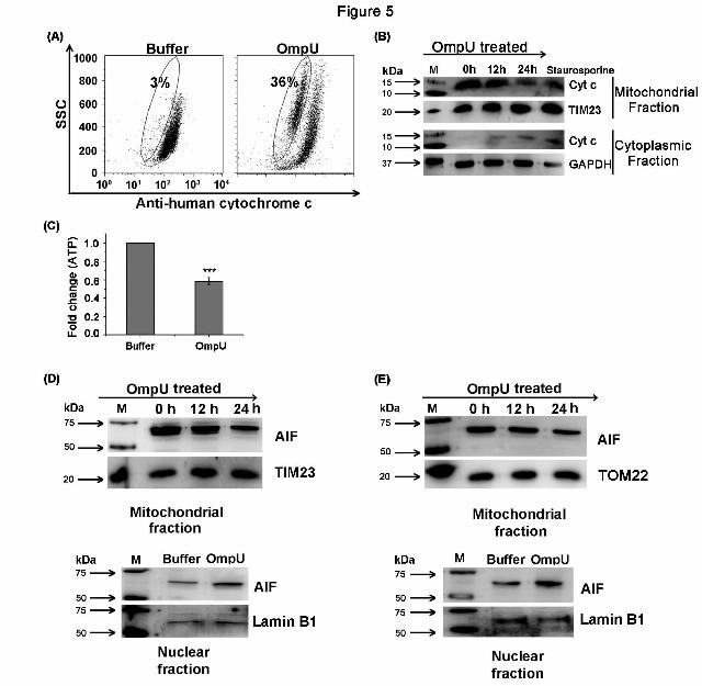
OmpU induces release of cytochrome c
from the host cell mitochondria: Cytochrome c as
a member of the electron transport chain remains
in the intact mitochondrial membrane but MMPT
usually leads to cytochrome c release from
mitochondria. Once released, cytochrome c
generally induces caspase activation, although
some reports suggest that cytochrome c is released
in case of programmed necrosis as well (45).
Therefore, cytochrome c release can be considered
as a marker of MMPT and not necessarily a
prerequisite of caspase-mediated intrinsic
apoptotic cell death. Although there is no caspase
activation in OmpU-mediated cell death, OmpU-
mediated MMPT prompted us to check whether
cytochrome c is released into the cytosol. THP-1
cells were treated with OmpU or buffer and
incubated for 24 h. It was observed that a
significant proportion of the total population of
OmpU treated cells (p<0.001 for OmpU-treated
versus buffer control) had lost cytochrome c from
their mitochondria with respect to buffer-treated
cells in a flow cytometry based assay (Fig. 5A).
Above results were confirmed by means of
western blotting in which a similar pattern of
cytochrome c release from mitochondria was
observed along with a simultaneous increase in the
cytoplasm (Fig. 5B). Stauroporine was included as
the positive control. These observations lead to the
conclusion that OmpU triggered major changes in
mitochondria by disrupting mitochondrial
membrane potential and inducing cytochrome c
release into the cytoplasm.
Since release of cytochrome c did not
induce caspase activation in THP-1 monocytes, we
probed the possibility that ATP depletion could
hamper the association of cytochrome c with
Apaf-1 thereby preventing the formation of the
complex called apoptosome. This could prevent
the activation of caspase cascade despite the
release of cytochrome c. We observed a significant
depletion (37-43 % decrease with respect to
buffer, Fig. 5C) in the amount of ATP in cells
treated with OmpU as compared to buffer.
Therefore, the loss of ATP could be the possible
reason for lack of caspase activation in OmpU-
mediated PCD.
V. cholerae OmpU induces translocation
of apoptosis inducing factor (AIF) from host cell
mitochondria to its nucleus: As a result of MMPT,
AIF could be released from the mitochondrial
inter-membrane space to the cytosol. Upon
translocation to nucleus from the cytosol, it can
cause DNA fragmentation (18). Towards probing
whether OmpU-mediated cell death involved AIF-
release from mitochondria and its translocation to
the nucleus, THP-1 monocytes were treated with
OmpU and incubated for 0, 12 or 24 h. We
observed that release of AIF from mitochondria
increased with increasing incubation periods (Fig.
5D). Since the AIF release from the mitochondria
was maximal at 24 h, nuclear lysates were
prepared from cells incubated with OmpU or
buffer for 24 h. A considerable amount of AIF
translocation was observed in the nucleus of
OmpU-treated cells as compared to the buffer
control (Fig. 5D).
Similarly, a clear translocation of AIF
from mitochondria to nucleus was observed in
HEK 293 cells as well when treated with OmpU
(Fig. 5E)
V. cholerae OmpU is translocated to the
mitochondria of the targeted host cell: THP-1 cells
and HEK 293 cells were treated with OmpU and
incubated for different time periods. Mitochondrial
fractions were prepared from treated cells and
were analyzed for the presence of OmpU. TOM22
or TIM23 were used as the mitochondrial markers
as well as the loading controls. GAPDH was used
by guest on August 25, 2019
http://ww
w.jbc.org/
Dow
nloaded from

OmpU induces caspase-independent programmed cell death
11
as the cytoplasmic marker and LAMP 1 as the
lysosomal marker. The resulting blots showed that
mitochondrial preparation was almost pure with
negligible contamination from cytoplasm or
lysosomes. A clear translocation of OmpU to the
mitochondria of treated cells was observed even at
15 min (Fig. 6A). A considerable amount of
OmpU was observed in the mitochondria of THP-
1 monocytes at 1 h and 2 h time points (Fig. 6A
and 6B) and at 2 h and 4 h in HEK 293 cells (Fig.
6C).
Furthermore, to find out whether OmpU
localizes in the mitochondria only or other
organelles as well, mitochondrial, nuclear and
light membrane + cytoplasmic fractions from
OmpU-treated cells were prepared and subjected
to western blotting for the detection of OmpU in
different fractions. The results showed that OmpU
predominantly localizes in the mitochondria of
treated cells as compared to other fractions (Fig.
6D).
We also checked the kinetics of
association of OmpU in plasma membrane and
compared it with its association in mitochondria.
HEK 293 cells were treated with 10 g/ml of
OmpU and incubated for 15 min, 30min, 1 h and 2
h. Purified mitochondrial fraction and light
membrane fraction pre-dominantly comprising of
plasma membrane were prepared from treated
cells and analyzed for the presence of OmpU by
means of western blotting. The results showed that
association of OmpU with plasma membrane at 15
min is much higher as compared to mitochondria.
Moreover, with increasing incubation periods,
amount of OmpU in mitochondria increases,
whereas in plasma membrane, it slightly declines
at 30 min and becomes almost constant with
increase in incubation time till 2 h (Fig. 6E).
Therefore, we can conclude that OmpU
associates with the plasma membrane and then
begins to translocate to the mitochondria. Initially,
its amount increases in mitochondria with
increasing incubation periods, whereas in plasma
membrane corresponding to increase of OmpU in
mitochondrial fraction, there is a decrease in the
amount of OmpU after 15 min.
Since purified OmpU translocated to
mitochondria, we checked its translocation from V.
cholerae. Live V. cholerae culture was prepared
and an aliquot of this culture was subjected to
heat-inactivation. Complete heat-inactivation of
the bacterial culture was confirmed by spreading
live and inactivated cultures on Luria agar plates
and incubating up to 16 h. Bacterial lawn was
observed in live bacterial plates within 4 h. Heat-
inactivated bacteria did not grow even after 16 h.
THP-1 monocytes were infected with live and
heat-inactivated cultures. Mitochondria were
isolated from each sample and mitochondrial
lysates were analyzed for the presence of OmpU.
In order to rule out the possibility of co-
purification of the bacteria with mitochondria,
lipid A of LPS as outer membrane marker and
RNA polymerase beta as bacterial cytoplasmic
marker were probed in the mitochondrial lysates.
OmpU translocation to the mitochondria was
observed in cells cultured with live bacteria as
well as heat-inactivated bacteria whereas none of
the bacterial cell markers appeared in the
mitochondrial lysates (Fig. 7A, 7B and 7C).
Therefore, from our results, we clearly
conclude that not only purified OmpU, but OmpU
from live or heat-inactivated V. cholerae is
translocated from the bacterial outer membrane to
the mitochondria of host cells.
Further, we observed that increasing the
amount of serum in the culture medium had no
effect on OmpU translocation to mitochondria,
and OmpU-induced PCD (Fig. 7D and 7E).
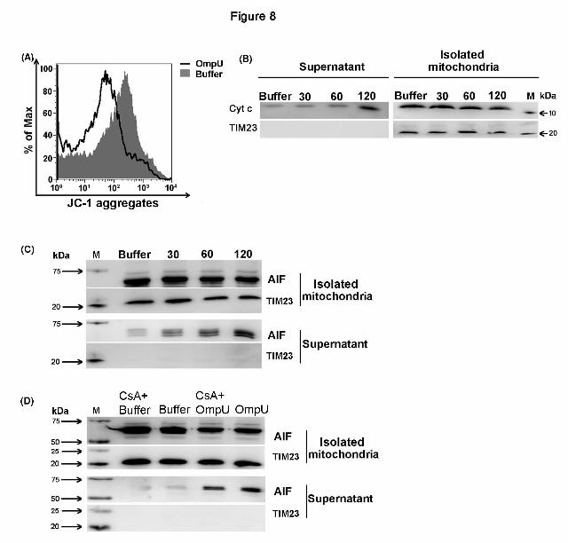
OmpU directly induces mitochondrial
changes that could lead to PCD in target cells:
The observation that V. cholerae OmpU
translocates to the mitochondria of treated cells
made it important to check if translocated OmpU
was able to directly induce the mitochondrial
changes such as MMPT and release of AIF from
the mitochondria of treated cells. Mitochondria
were isolated from untreated THP-1 monocytes
and were treated with OmpU or buffer. Treated
mitochondria showed a considerable loss of MMP
with respect to buffer which was visualized as
decreased JC-1 aggregates in isolated
mitochondria (Fig. 8A). Furthermore, to analyze
AIF and cytochrome c release, isolated
mitochondria were incubated with OmpU for
different time points. A time dependent release of
AIF and cytochrome c from OmpU-treated
mitochondria was observed which started at 30
min and increased with time with respect to buffer
(Fig. 8B and 8C ). At the same time, their amounts
in the corresponding supernatants of OmpU-
by guest on August 25, 2019
http://ww
w.jbc.org/
Dow
nloaded from

OmpU induces caspase-independent programmed cell death
12
treated mitochondria increased with increasing
incubation periods (Fig. 8B and 8C).
To check whether opening of
mitochondrial permeability transition pore
(MPTP) is involved in OmpU-mediated MMPT,
cyclosporin A (CsA) was used. CsA is a known
inhibitor that prevents that opening of MPTP and
hence MMPT. Freshly isolated mitochondria were
pre-treated with or without CsA followed by
treatment with OmpU (5 g/ml) or buffer.
Following treatment, mitochondria and
corresponding supernatants were analyzed for the
release of AIF by western blotting. Resulting blots
showed that CsA partially inhibited the release of
AIF (Fig. 8D). This result further confirmed the
role of mitochondrial permeability transition in
release of AIF from OmpU treated isolated
mitochondria.
On the basis of all the above results, we
conclude that OmpU can directly induce changes
in freshly isolated mitochondria that ultimately
lead to PCD.
OmpU induces cell death in intestinal
epithelial cells through the same mechanism as in
other cell lines: Since V. cholerae colonizes small
intestine, we probed the ability of OmpU to induce
cell death in intestinal epithelial cells using human
colon carcinoma cell line, Caco-2. Caco-2 cells
treated with different doses of OmpU or buffer
were subjected MTT assay and LDH release assay
and the results showed that a significant
percentage of OmpU treated cells were induced to
die even at 5 g/ml protein concentration (Fig. 9A
and 9B). Moreover, pre-treatment with zVAD-fmk
did not have any effect on OmpU-mediated cell
death in Caco-2 cells indicating towards a caspase-
independent mechanism as in other cell types (Fig.
9C and 9D). A substantial translocation of OmpU
to the mitochondria of Caco-2 cells was observed
at 2 h (Fig. 9E). Finally, we checked the role of
AIF and observed that OmpU induces AIF release
from mitochondria of treated cells and its
translocation to the nucleus (Fig. 9F).
Therefore, we conclude that OmpU
induces the same mechanism of caspase-
independent PCD in intestinal epithelial cells as it
induces in other cell lines that involves
translocation of OmpU to mitochondria of target
cells and translocation of AIF from mitochondria
to the nucleus of treated cells.
Discussion
Porins from gram-negative bacteria are
reported to be pro-apoptotic in two of the cases. In
Neisseria gonorrheae and Pseudomonas
aeruginosa, porin-mediated apoptosis contributes
to the mode of pathogenesis (23,24,46). In case of
N. meningitides, porin protects the target cells
from apoptosis (30). Therefore, it is evident that
porins from different bacteria need separate
evaluation regarding their effect on cell health and
viability. OmpU, one of the major porins of V.
cholerae, is considered to be crucial for bacterial
pathogenesis and is a potential vaccine candidate.
Therefore, OmpU was evaluated for its role in the
induction of host cell death
.In the current study, we established that
OmpU induces death of the target cells as
analyzed by various assays like MTT assay, LDH
release assay and annexin V-FITC /PI staining
(Fig 1A, 1B, 1C, 1D, 2E, 9A and 9B). All the
results from different assays indicated that OmpU
at 10 g/ml induced a significant amount of cell
death, although percentage of dying cells varied
slightly from assay to assay. LDH release assay
showed higher percentage of dead cells as
compared to MTT assay or PI staining but the
difference can be attributed to the differences in
assay conditions and different sensitivities of the
assays (47). Futher, LDH release assay depends
only on the permeabilization of plasma membrane
as opposed to PI staining that requires nuclear
membrane permeability as well. Therefore, it is
possible that annexin V positive cells could also
contribute to the LDH release. It has been shown
that early membrane blebbing which can start even
before PS flipping can be associated with limited
plasma membrane permeabilization. Further, it has
been shown that LDH release can be blocked by
inhibiting early membrane blebbing (48,49). The
appearance of a higher percentage in LDH release
assay could also be due to its higher sensitivity
(47). Therefore, we assume that percentage of
non-viable cells as indicated by the MTT assay is
close to actual percentage of cell death in response
to OmpU.
Results obtained on polymyxin B (pmb)
pre-treatment of OmpU-treated THP-1 monocytes
(Fig. 1E) and induction of cell death in HEK 293
cells (Fig. 1B and Fig 2B) completely rule out the
involvement of endotoxin and TLRs in OmpU-
mediated cell death.
by guest on August 25, 2019
http://ww
w.jbc.org/
Dow
nloaded from
OmpU induces caspase-independent programmed cell death
13
Moreover, we observed that OmpU also
induces certain morphological and biochemical
changes similar to that of apoptotic cell death (Fig.
1F and 1G). In normal cell types, PS is
asymmetrically distributed on the cell membrane
and is predominantly present in the inner leaflet of
the membrane lipid bilayer. In cells undergoing
apoptosis, PS flipping takes place and accordingly
its exposure increases in the outer leaflet of the
plasma membrane (50). Moreover, cells
undergoing apoptosis exhibit cell shrinking and
increased cell granularity. Our data showed that
OmpU induces above mentioned changes that are
commonly involved in apoptosis (Fig. 1).
However, DNA fragmentation leading to DNA
laddering in agarose gel is another major hallmark
of apoptosis (2,12). Though OmpU-mediated cell
death showed DNA fragmentation as evident in
Fig. 2A, 2B and 2C, but the fragments did not
resolve in the agarose gel as a typical ladder as
seen in apoptosis (Fig. 2D).
Caspases are the key enzymes involved in
apoptosis (51). Among different caspases,
caspase-3 is the main executioner enzyme
involved in all caspase-dependent apoptotic
pathways (16,52). We observed that there is no
induction of caspase-3 activation upon OmpU
treatment (Fig. 3A). Moreover, there is no
inhibition of OmpU-mediated PS flipping and/
DNA fragmentation with the use of total caspase-
inhibitor zVAD-fmk (Fig. 3B, 3D, 3E, 9C and
9D).
Results from LDH release assay (Fig. 2E)
and absence of caspase activation imply that
though the cell death mechanism induced by
OmpU has certain characteristics of apoptosis as
well as necrosis, it is different from both classical
apoptosis and necrosis.
Further, we observed that though V.
cholerae is a non-invasive bacterium, V. cholerae
OmpU translocates to the mitochondria of target
cells (Fig. 6, Fig. 7 and Fig. 9E). Mitochondria
play a central role in different kinds of cell death
processes (7,53). Many factors such as chemicals
or pathogen-derived molecules cause alterations in
cellular stress responses resulting in the dissipation
of mitochondrial membrane potential (m) i.e.,
disruption of proton gradient across inner
mitochondrial membrane. This leads to MMPT
which allows passage of small molecules
including apoptogenic factors from mitochondria
to cytosol (54,55). We observed that OmpU is
capable of inducing MMPT in both THP-1 (Fig.
4A and 4B) and HEK 293 cells (Fig. 4C and 4D ).
Cytochrome c, a heme-containing protein
complex, is a part of the electron transport chain
present in the mitochondrial inter-membrane space
(56). It is known that cytochrome c is released into
the cytoplasm following disruption of the
mitochondrial membrane potential. Cytochrome c
is instrumental in caspase-dependent apoptosis.
But some reports suggest that cytochrome c is
released in other types of cell death process such
as, programmed necrosis and may not always lead
to caspase activation and therefore, its release
leaves the cells unaffected (45,57,58). Our data
shows that OmpU induces cytochrome c release
from mitochondria (Fig. 5A and 5B). However, it
does not lead to caspase activation which could
probably be due to the loss of ATP in OmpU-
treated cells (Fig. 5C). Cytochrome c binds to
Apaf 1 in the presence of dATP which is required
for apoptosome formation and is followed by
caspase activation (59).
As a result of MMPT, AIF could also be
released from mitochondria. AIF is present in the
mitochondrial inter-membrane space and is
reported to have limited NADH or NADPH
oxidase activity (60). AIF or apoptosis inducing
factor can induce cell death by chromatin
condensation and DNA degradation upon
translocation to the nucleus from mitochondria. In
the absence of caspase activation, AIF plays a
central role in the induction of caspase-
independent PCD (8,18,61). OmpU-induced
MMPT leads to the release of AIF from
mitochondria (Fig. 5D, 5E and 9F). Further our
study showed that the released AIF translocates to
the nucleus (Fig. 5D, 5E and 9F).
Another very important observation
during the course of our study was that when
isolated mitochondria were treated directly with
OmpU, they were induced to undergo MMPT
along with AIF release (Fig. 8).
Furthermore, our results showed that CsA,
which is a known inhibitor of MMPT, leads to
partial inhibition of AIF release from freshly
isolated mitochondria (Fig. 8E). This result
confirms that OmpU directly interacts with
isolated mitochondria inducing MMPT which
further results in AIF release. Above observations
suggest that OmpU might itself act as a trigger
by guest on August 25, 2019
http://ww
w.jbc.org/
Dow
nloaded from

OmpU induces caspase-independent programmed cell death
14
inducing mitochondrial changes that lead to
caspase-independent programmed cell death in
target cells
Therefore, we conclude that V. cholerae
OmpU translocates to host cell mitochondria and
induces MMPT that leads to the release of
cytochrome c and AIF but no caspase activation.
AIF translocates to the nucleus and presumably
induces large scale DNA fragmentation and cell
death (Fig. 10).
Although PS exposure is a typical
characteristic of cells undergoing apoptosis, there
are reports that suggest that some level of PS
exposure is also observed in cells undergoing AIF-
mediated programmed cell death (8,62,63). As
reported previously, released AIF can further act
on other mitochondria leading to an amplification
of the process (18). Some reports have suggested
that AIF-mediated PCD could be referred to as
apoptosis like PCD (8,64,65) as it includes
biochemical and morphological changes similar to
that of apoptosis. In the current study, our results
evidently indicate towards this phenomenon in
case of OmpU.
PorB porin of N. gonorrhoeae is also
reported to integrate into the mitochondria but
induces caspase-mediated apoptosis (23,46).
Mitochondrial translocation of N. gonorrhoeae
porin is blocked by serum. However, we observed
that serum does not have any effect on V. cholerae
OmpU translocation to the mitochondria (Fig. 7D).
In case of P. aeruginosa, no mitochondrial
translocation of bacterial porin is reported (24).
Non-invasive gram-negative bacteria
mostly translocate their virulence factors inside the
host cell through secretory vesicles (66) or via
secretion pathways (67). In the present study we
showed that purified OmpU and OmpU from heat-
inactivated bacteria that are probably incapable of
producing vesicles can translocate to the host cell
mitochondria. This observation suggests that from
the vaccine strains also OmpU can translocate into
the host cell mitochondria and modulate cellular
responses leading to death.
To the best of our knowledge, this is the
first report regarding porin translocation from a
non-invasive bacterium to the host cell
mitochondria and induction of apoptosis-like
programmed cell death but not classical apoptosis.
Acknowledgement
This work was supported by a grant (to AM) from the Department of Biotechnology (DBT), India (DBT
Grant No. BT/PR1205/MED/29/ 318/2011) and funding from IISER Mohali.
We acknowledge Dr. Samarjit Bhattacharyya,IISER Mohali for providing us with anti-LAMP 1 antibody
and helping with microscopy experiments, Dr. Mahak Sharma from IISER Mohali for providing us with
anti-transferrin antibody and Dr. Sudip Mandal from IISER Mohali for helping us with ATP-
determination assay
Conflict of Interest
Authors declare that there is no conflict of interest.
Author Contribution
Shelly Gupta planned experiments, performed experiments, analyzed data and wrote the paper, G.V.R.
Krishna Prasad performed experiments, Arunika Mukhopadhaya planned experiments, analyzed data and
wrote the paper.
by guest on August 25, 2019
http://ww
w.jbc.org/
Dow
nloaded from

OmpU induces caspase-independent programmed cell death
15
References
1. Kerr, J. F., Wyllie, A. H., and Currie, A. R. (1972) Apoptosis: a basic biological phenomenon
with wide-ranging implications in tissue kinetics. Br J Cancer 26, 239-257
2. Saraste, A., and Pulkki, K. (2000) Morphologic and biochemical hallmarks of apoptosis.
Cardiovasc Res 45, 528-537
3. Elmore, S. (2007) Apoptosis: a review of programmed cell death. Toxicol Pathol 35, 495-516
4. Peter, M. E. (2011) Programmed cell death: Apoptosis meets necrosis. Nature 471, 310-312
5. Chandra, D., Choy, G., Deng, X., Bhatia, B., Daniel, P., and Tang, D. G. (2004) Association of
active caspase 8 with the mitochondrial membrane during apoptosis: potential roles in cleaving
BAP31 and caspase 3 and mediating mitochondrion-endoplasmic reticulum cross talk in
etoposide-induced cell death. Mol Cell Biol 24, 6592-6607
6. Broker, L. E., Kruyt, F. A., and Giaccone, G. (2005) Cell death independent of caspases: a
review. Clin Cancer Res 11, 3155-3162
7. Kroemer, G., and Martin, S. J. (2005) Caspase-independent cell death. Nat Med 11, 725-730
8. Boujrad, H., Gubkina, O., Robert, N., Krantic, S., and Susin, S. A. (2007) AIF-mediated
programmed necrosis: a highly regulated way to die. Cell Cycle 6, 2612-2619
9. Pasparakis, M., and Vandenabeele, P. (2015) Necroptosis and its role in inflammation. Nature
517, 311-320
10. Volbracht, C., Leist, M., Kolb, S. A., and Nicotera, P. (2001) Apoptosis in caspase-inhibited
neurons. Mol Med 7, 36-48
11. Weiss, J. N., Korge, P., Honda, H. M., and Ping, P. (2003) Role of the mitochondrial permeability
transition in myocardial disease. Circ Res 93, 292-301
12. Ziegler, U., and Groscurth, P. (2004) Morphological features of cell death. News Physiol Sci 19,
124-128
13. Pawlowski, J., and Kraft, A. S. (2000) Bax-induced apoptotic cell death. Proc Natl Acad Sci U S
A 97, 529-531
14. Dewson, G., and Kluck, R. M. (2009) Mechanisms by which Bak and Bax permeabilise
mitochondria during apoptosis. J Cell Sci 122, 2801-2808
15. Susin, S. A., Lorenzo, H. K., Zamzami, N., Marzo, I., Brenner, C., Larochette, N., Prevost, M. C.,
Alzari, P. M., and Kroemer, G. (1999) Mitochondrial release of caspase-2 and -9 during the
apoptotic process. J Exp Med 189, 381-394
16. Lakhani, S. A., Masud, A., Kuida, K., Porter, G. A., Jr., Booth, C. J., Mehal, W. Z., Inayat, I., and
Flavell, R. A. (2006) Caspases 3 and 7: key mediators of mitochondrial events of apoptosis.
Science 311, 847-851
17. Shi, Y. (2001) A structural view of mitochondria-mediated apoptosis. Nat Struct Biol 8, 394-401
18. Susin, S. A., Lorenzo, H. K., Zamzami, N., Marzo, I., Snow, B. E., Brothers, G. M., Mangion, J.,
Jacotot, E., Costantini, P., Loeffler, M., Larochette, N., Goodlett, D. R., Aebersold, R.,
Siderovski, D. P., Penninger, J. M., and Kroemer, G. (1999) Molecular characterization of
mitochondrial apoptosis-inducing factor. Nature 397, 441-446
19. Ashida, H., Mimuro, H., Ogawa, M., Kobayashi, T., Sanada, T., Kim, M., and Sasakawa, C.
(2012) Cell death and infection: a double-edged sword for host and pathogen survival. J Cell Biol
195, 931-942
20. Cohen, J. (2002) The immunopathogenesis of sepsis. Nature 420, 885-891
21. Lin, C. F., Chen, C. L., Huang, W. C., Cheng, Y. L., Hsieh, C. Y., Wang, C. Y., and Hong, M. Y.
(2010) Different types of cell death induced by enterotoxins. Toxins (Basel) 2, 2158-2176
22. Kennedy, C. L., Smith, D. J., Lyras, D., Chakravorty, A., and Rood, J. I. (2009) Programmed
cellular necrosis mediated by the pore-forming alpha-toxin from Clostridium septicum. PLoS
Pathog 5, e1000516
by guest on August 25, 2019
http://ww
w.jbc.org/
Dow
nloaded from

OmpU induces caspase-independent programmed cell death
16
23. Muller, A., Gunther, D., Dux, F., Naumann, M., Meyer, T. F., and Rudel, T. (1999) Neisserial
porin (PorB) causes rapid calcium influx in target cells and induces apoptosis by the activation of
cysteine proteases. EMBO J 18, 339-352
24. Buommino, E., Morelli, F., Metafora, S., Rossano, F., Perfetto, B., Baroni, A., and Tufano, M. A.
(1999) Porin from Pseudomonas aeruginosa induces apoptosis in an epithelial cell line derived
from rat seminal vesicles. Infect Immun 67, 4794-4800
25. Achouak, W., Heulin, T., and Pages, J. M. (2001) Multiple facets of bacterial porins. FEMS
Microbiol Lett 199, 1-7
26. Biswas, T. (2000) Role of porin of Shigella dysenteriae type 1 in modulation of
lipopolysaccharide mediated nitric oxide and interleukin-1 release by murine peritoneal
macrophages. FEMS Immunol Med Microbiol 29, 129-136
27. Alurkar, V., and Kamat, R. (1997) Immunomodulatory properties of porins of some members of
the family Enterobacteriaceae. Infect Immun 65, 2382-2388
28. Sakharwade, S. C., Sharma, P. K., and Mukhopadhaya, A. (2013) Vibrio cholerae porin OmpU
induces pro-inflammatory responses, but down-regulates LPS-mediated effects in RAW 264.7,
THP-1 and human PBMCs. PLoS One 8, e76583
29. Khan, J., Sharma, P. K., and Mukhopadhaya, A. (2015) Vibrio cholerae porin OmpU mediates
M1-polarization of macrophages/monocytes via TLR1/TLR2 activation. Immunobiology 220,
1199-1209
30. Massari, P., Ho, Y., and Wetzler, L. M. (2000) Neisseria meningitidis porin PorB interacts with
mitochondria and protects cells from apoptosis. Proc Natl Acad Sci U S A 97, 9070-9075
31. Chakrabarti, S. R., Chaudhuri, K., Sen, K., and Das, J. (1996) Porins of Vibrio cholerae:
purification and characterization of OmpU. J Bacteriol 178, 524-530
32. Skorupski, K., and Taylor, R. K. (1997) Control of the ToxR virulence regulon in Vibrio cholerae
by environmental stimuli. Mol Microbiol 25, 1003-1009
33. Provenzano, D., Lauriano, C. M., and Klose, K. E. (2001) Characterization of the role of the
ToxR-modulated outer membrane porins OmpU and OmpT in Vibrio cholerae virulence. J
Bacteriol 183, 3652-3662
34. Crawford, J. A., Kaper, J. B., and DiRita, V. J. (1998) Analysis of ToxR-dependent transcription
activation of ompU, the gene encoding a major envelope protein in Vibrio cholerae. Mol
Microbiol 29, 235-246
35. Mathur, J., and Waldor, M. K. (2004) The Vibrio cholerae ToxR-regulated porin OmpU confers
resistance to antimicrobial peptides. Infect Immun 72, 3577-3583
36. Nakasone, N., and Iwanaga, M. (1998) Characterization of outer membrane protein OmpU of
Vibrio cholerae O1. Infect Immun 66, 4726-4728
37. Sperandio, V., Giron, J. A., Silveira, W. D., and Kaper, J. B. (1995) The OmpU outer membrane
protein, a potential adherence factor of Vibrio cholerae. Infect Immun 63, 4433-4438
38. Goel, A. K., Jain, M., Kumar, P., and Jiang, S. C. (2010) Molecular characterization of Vibrio
cholerae outbreak strains with altered El Tor biotype from southern India. World J Microbiol
Biotechnol 26, 281-287
39. Paauw, A., Trip, H., Niemcewicz, M., Sellek, R., Heng, J. M., Mars-Groenendijk, R. H., de Jong,
A. L., Majchrzykiewicz-Koehorst, J. A., Olsen, J. S., and Tsivtsivadze, E. (2014) OmpU as a
biomarker for rapid discrimination between toxigenic and epidemic Vibrio cholerae O1/O139 and
non-epidemic Vibrio cholerae in a modified MALDI-TOF MS assay. BMC Microbiol 14, 158
40. Khan, J., Gupta, S., Chattopadhyay, K., and Mukhopadhaya, A. (2012) Refolding and functional
assembly of the Vibrio cholerae porin OmpU recombinantly expressed in the cytoplasm of
Escherichia coli. Protein Expr Purif 85, 204-210
41. Waterhouse, N. J., and Trapani, J. A. (2003) A new quantitative assay for cytochrome c release in
apoptotic cells. Cell Death Differ 10, 853-855
by guest on August 25, 2019
http://ww
w.jbc.org/
Dow
nloaded from

OmpU induces caspase-independent programmed cell death
17
42. Wolf, B. B., Schuler, M., Echeverri, F., and Green, D. R. (1999) Caspase-3 is the primary
activator of apoptotic DNA fragmentation via DNA fragmentation factor-45/inhibitor of caspase-
activated DNase inactivation. J Biol Chem 274, 30651-30656
43. Chan, F. K., Moriwaki, K., and De Rosa, M. J. (2013) Detection of necrosis by release of lactate
dehydrogenase activity. Methods Mol Biol 979, 65-70
44. Norberg, E., Orrenius, S., and Zhivotovsky, B. (2010) Mitochondrial regulation of cell death:
processing of apoptosis-inducing factor (AIF). Biochem Biophys Res Commun 396, 95-100
45. Li, Y. Z., Li, C. J., Pinto, A. V., and Pardee, A. B. (1999) Release of mitochondrial cytochrome C
in both apoptosis and necrosis induced by beta-lapachone in human carcinoma cells. Mol Med 5,
232-239
46. Muller, A., Gunther, D., Brinkmann, V., Hurwitz, R., Meyer, T. F., and Rudel, T. (2000)
Targeting of the pro-apoptotic VDAC-like porin (PorB) of Neisseria gonorrhoeae to mitochondria
of infected cells. EMBO J 19, 5332-5343
47. Bopp, S. K., and Lettieri, T. (2008) Comparison of four different colorimetric and fluorometric
cytotoxicity assays in a zebrafish liver cell line. BMC Pharmacol 8, 8
48. Andrade, R., Crisol, L., Prado, R., Boyano, M. D., Arluzea, J., and Arechaga, J. (2010) Plasma
membrane and nuclear envelope integrity during the blebbing stage of apoptosis: a time-lapse
study. Biol Cell 102, 25-35
49. Wickman, G. R., Julian, L., Mardilovich, K., Schumacher, S., Munro, J., Rath, N., Zander, S. A.,
Mleczak, A., Sumpton, D., Morrice, N., Bienvenut, W. V., and Olson, M. F. (2013) Blebs
produced by actin-myosin contraction during apoptosis release damage-associated molecular
pattern proteins before secondary necrosis occurs. Cell Death Differ 20, 1293-1305
50. Marino, G., and Kroemer, G. (2013) Mechanisms of apoptotic phosphatidylserine exposure. Cell
Res 23, 1247-1248
51. Cohen, G. M. (1997) Caspases: the executioners of apoptosis. Biochem J 326 ( Pt 1), 1-16
52. Porter, A. G., and Janicke, R. U. (1999) Emerging roles of caspase-3 in apoptosis. Cell Death
Differ 6, 99-104
53. Estaquier, J., Vallette, F., Vayssiere, J. L., and Mignotte, B. (2012) The mitochondrial pathways
of apoptosis. Adv Exp Med Biol 942, 157-183
54. Ly, J. D., Grubb, D. R., and Lawen, A. (2003) The mitochondrial membrane potential
(deltapsi(m)) in apoptosis; an update. Apoptosis 8, 115-128
55. Vander Heiden, M. G., Plas, D. R., Rathmell, J. C., Fox, C. J., Harris, M. H., and Thompson, C.
B. (2001) Growth factors can influence cell growth and survival through effects on glucose
metabolism. Mol Cell Biol 21, 5899-5912
56. Ow, Y. P., Green, D. R., Hao, Z., and Mak, T. W. (2008) Cytochrome c: functions beyond
respiration. Nat Rev Mol Cell Biol 9, 532-542
57. Jemmerson, R., LaPlante, B., and Treeful, A. (2002) Release of intact, monomeric cytochrome c
from apoptotic and necrotic cells. Cell Death Differ 9, 538-548
58. Von Ahsen, O., Waterhouse, N. J., Kuwana, T., Newmeyer, D. D., and Green, D. R. (2000) The
'harmless' release of cytochrome c. Cell Death Differ 7, 1192-1199
59. Hu, Y., Benedict, M. A., Ding, L., and Nunez, G. (1999) Role of cytochrome c and dATP/ATP
hydrolysis in Apaf-1-mediated caspase-9 activation and apoptosis. EMBO J 18, 3586-3595
60. Hangen, E., Blomgren, K., Benit, P., Kroemer, G., and Modjtahedi, N. (2010) Life with or
without AIF. Trends Biochem Sci 35, 278-287
61. Susin, S. A., Daugas, E., Ravagnan, L., Samejima, K., Zamzami, N., Loeffler, M., Costantini, P.,
Ferri, K. F., Irinopoulou, T., Prevost, M. C., Brothers, G., Mak, T. W., Penninger, J., Earnshaw,
W. C., and Kroemer, G. (2000) Two distinct pathways leading to nuclear apoptosis. J Exp Med
192, 571-580
62. Cocco, R. E., and Ucker, D. S. (2001) Distinct modes of macrophage recognition for apoptotic
and necrotic cells are not specified exclusively by phosphatidylserine exposure. Mol Biol Cell 12,
919-930
by guest on August 25, 2019
http://ww
w.jbc.org/
Dow
nloaded from

OmpU induces caspase-independent programmed cell death
18
63. Hirt, U. A., Gantner, F., and Leist, M. (2000) Phagocytosis of nonapoptotic cells dying by
caspase-independent mechanisms. J Immunol 164, 6520-6529
64. Cregan, S. P., Fortin, A., MacLaurin, J. G., Callaghan, S. M., Cecconi, F., Yu, S. W., Dawson, T.
M., Dawson, V. L., Park, D. S., Kroemer, G., and Slack, R. S. (2002) Apoptosis-inducing factor
is involved in the regulation of caspase-independent neuronal cell death. J Cell Biol 158, 507-517
65. Lorenzo, H. K., and Susin, S. A. (2004) Mitochondrial effectors in caspase-independent cell
death. FEBS Lett 557, 14-20
66. Ellis, T. N., and Kuehn, M. J. (2010) Virulence and immunomodulatory roles of bacterial outer
membrane vesicles. Microbiol Mol Biol Rev 74, 81-94
67. Coburn, B., Sekirov, I., and Finlay, B. B. (2007) Type III secretion systems and disease. Clin
Microbiol Rev 20, 535-549
by guest on August 25, 2019
http://ww
w.jbc.org/
Dow
nloaded from
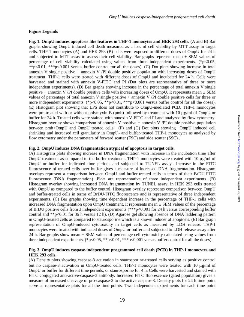
OmpU induces caspase-independent programmed cell death
19
Figure Legends
Fig. 1. OmpU induces apoptosis like features in THP-1 monocytes and HEK 293 cells. (A and B) Bar
graphs showing OmpU-induced cell death measured as a loss of cell viability by MTT assay in target
cells. THP-1 monocytes (A) and HEK 293 (B) cells were exposed to different doses of OmpU for 24 h
and subjected to MTT assay to assess their cell viability. Bar graphs represent mean ± SEM values of
percentage of cell viability calculated using values from three independent experiments. (*p<0.05,
**p<0.01, ***p<0.001 versus buffer control for all the doses). (C) Dot plots showing increase in total
annexin V single positive + annexin V /PI double positive population with increasing doses of OmpU
treatment. THP-1 cells were treated with different doses of OmpU and incubated for 24 h. Cells were
harvested and stained with annexin V-FITC and PI (Dot plots are representative of three or more
independent experiments). (D) Bar graphs showing increase in the percentage of total annexin V single
positive + annexin V /PI double positive cells with increasing doses of OmpU. It represents mean ± SEM
values of percentage of total annexin V single positive + annexin V /PI double positive cells for three or
more independent experiments. (*p<0.05, **p<0.01, ***p<0.001 versus buffer control for all the doses).
(E) Histogram plot showing that LPS does not contribute to OmpU-mediated PCD. THP-1 monocytes
were pre-treated with or without polymyxin B (pmb) followed by treatment with 10 g/ml of OmpU or
buffer for 24 h. Treated cells were stained with annexin V-FITC and PI and analyzed by flow cytometry.
Histogram overlay shows comparison of annexin V positive + annexin V /PI double positive population
between pmb+OmpU and OmpU treated cells. (F) and (G) Dot plots showing OmpU induced cell
shrinking and increased cell granularity in OmpU- and buffer-treated THP-1 monocytes as analyzed by
flow cytometry under the parameters of forward scatter (FSC) and side scatter (SSC) .
Fig. 2. OmpU induces DNA fragmentation atypical of apoptosis in target cells.
(A) Histogram plots showing increase in DNA fragmentation with increase in the incubation time after
OmpU treatment as compared to the buffer treatment. THP-1 monocytes were treated with 10 g/ml of
OmpU or buffer for indicated time periods and subjected to TUNEL assay.. Increase in the FITC
fluorescence of treated cells over buffer gives a measure of increased DNA fragmentation. Histogram
overlays represent a comparison between OmpU and buffer-treated cells in terms of their BrDU-FITC
fluorescence (DNA fragmentation). Plots are representative of three independent experiments. (B)
Histogram overlay showing increased DNA fragmentation by TUNEL assay, in HEK 293 cells treated
with OmpU as compared to the buffer control. Histogram overlay represents comparison between OmpU
and buffer-treated cells in terms of BrDU-FITC fluorescence and is representative of three independent
experiments. (C) Bar graphs showing time dependent increase in the percentage of THP-1 cells with
increased DNA fragmentation upon OmpU treatment. It represents mean ± SEM values of the percentage
of BrDU positive cells from 3 independent experiments (***p<0.001 for 24 h versus corresponding buffer
control and **p<0.01 for 36 h versus 12 h). (D) Agarose gel showing absence of DNA laddering pattern
in OmpU-treated cells as compared to staurosporine which is a known inducer of apoptosis. (E) Bar graph
representation of OmpU-induced cytotoxicity in target cells as measured by LDH release. THP-1
monocytes were treated with indicated doses of OmpU or buffer and subjected to LDH release assay after
24 h. Bar graphs show mean ± SEM values of percentage cell cytotoxicity calculated using values from
three independent experiments. (*p<0.05, **p<0.01, ***p<0.001 versus buffer control for all the doses).
Fig. 3. OmpU induces caspase-independent programmed cell death (PCD) in THP-1 monocytes and
HEK 293 cells.
(A) Density plots showing caspase-3 activation in staurosporine-treated cells serving as positive control
but no caspase-3 activation in OmpU-treated cells. THP-1 monocytes were treated with 10 g/ml of
OmpU or buffer for different time periods, or staurosporine for 4 h. Cells were harvested and stained with
FITC conjugated anti-active-caspase-3 antibody. Increased FITC fluorescence (gated population) gives a
measure of increased cleavage of pro-caspase-3 to the active caspase-3. Density plots for 24 h time point
serve as representative plots for all the time points. Two independent experiments for each time point
by guest on August 25, 2019
http://ww
w.jbc.org/
Dow
nloaded from

OmpU induces caspase-independent programmed cell death
20
were performed. (B) Histogram overlay showing no inhibition in OmpU-mediated PS flipping (annexin V
positive cells,) upon the use of total caspase inhibitor. THP-1 monocytes were pre-incubated with or
without total caspase inhibitor zVAD-fmk followed by incubation with OmpU or buffer. Cells were
harvested and stained with annexin V-FITC and PI. As mentioned in the experimental procedure section
quadrant gate was applied to all the tests. Histogram overlay represents population of cells in lower left
quadrant + lower right quadrant. This is the representative plot of three independent experiments. (C) Bar
graphs showing almost no change in percentage of total annexin V single positive (lower right
quadrant)+ annexin V /PI double positive (upper right quadrant) cells upon OmpU treatment with/without
caspase inhibitor. Quadrant gate was applied to all the tests Bar graph represents mean ± SEM values for
three independent experiments. (D) Histogram overlay showing decrease in percentage of annexin V
positive population in zVAD-fmk + staurosporine-treated cells as compared to only staurosporine-treated
cells. (E and F) Caspase inhibitor could not block DNA fragmentation induced by OmpU. THP-1
monocytes (E) or HEK 293 cells (F) were pre-treated with zVAD-fmk followed by treatment with OmpU
or buffer and incubated for 24 h. Cells were harvested and stained for TUNEL assay. Offset histogram
overlays represent a comparison between zVAD-fmk + OmpU, only OmpU, and buffer-treated cells in
terms of BrDU-FITC fluorescence (DNA fragmentation). Each plot is representative of three independent
experiments.
Fig. 4. OmpU induces mitochondrial membrane permeability transition (MMPT) in target cells.
(A) Contour plots showing increase in MMPT with increase in time of incubation after OmpU treatment.
THP-1 monocytes were treated with 10 g/ml of OmpU or buffer and incubated for 4, 8 and 24 h. Cells
were harvested and stained with JC-1 dye for detection of MMPT. Contour plots are representative of
three independent experiments. (B) Bar graphs showing increase in percentage of JC-1 monomers in
THP-1 cells with increase in time of incubation after OmpU treatment. It represents mean ± SEM values
of percentage cells with increased JC-1 monomers for three independent experiments. (*p<0.05,
**p<0.01, ***p<0.001 versus buffer control for all the time points). (C) Histogram overlay showing more
JC-1 monomers in OmpU-treated HEK 293 cells compared to buffer. HEK 293 cells were treated and
incubated with 10 g/ml of OmpU or buffer and incubated for 24 h and stained with JC-1 dye. The plot is
representative of three independent experiments. (D) Microscopic analysis of loss of MMP in target HEK
293 cells. HEK 293 cells were plated and treated with 10 g/ml of OmpU or buffer for 24 h. Cells treated
with staurosporine for 4 h were used as positive control. Cells were stained using mitotraker red CMXros
and analyzed by confocal microscopy. Images are representative of two independent experiments.
Fig. 5. OmpU induces cytochrome c and AIF release from mitochondria along with translocation of
AIF to nucleus.
(A) Dot plots showing considerable loss of cytochrome c in OmpU treated cells compared to buffer. THP-
1 monocytes were treated with 10 g/ml of OmpU or buffer and incubated for 24 h. Treated cells were
harvested and stained with anti-human cytochrome c followed by FITC-conjugated anti rabbit IgG.
Decreased FITC fluorescence (gated population) corresponds to decrease in cytochrome c in
mitochondria. Dot plots are representative of three independent experiments. (B) Western blot showing
release of cytochrome c from mitochondria to cytoplasm. THP-1 monocytes were treated with 10 g/ml
of OmpU for different incubation periods and incubated for (0, 12 and 24 h). Cells were harvested and
their mitochondrial and cytoplasmic fractions were prepared which were then analyzed for the release of
cytochrome c by means of western blot analysis. Blots are representative of three independent
experiments. (C) Bar graph showing reduction in the amount of ATP in OmpU-treated cells with respect
to buffer-treated cells. THP-1 cells were treated with OmpU or buffer for 24 h and subjected to ATP
determination assay. Bar graph represents mean ± SEM values of fold change in the amount of ATP in
OmpU-treated cells with respect to buffer and is calculated from three independent experiments. (D and
E) Western blot showing release of AIF from the mitochondrial fraction and its translocation to the
nuclear fraction in OmpU-treated cells compared to control. THP-1 monocytes (D) and HEK 293 cells
by guest on August 25, 2019
http://ww
w.jbc.org/
Dow
nloaded from
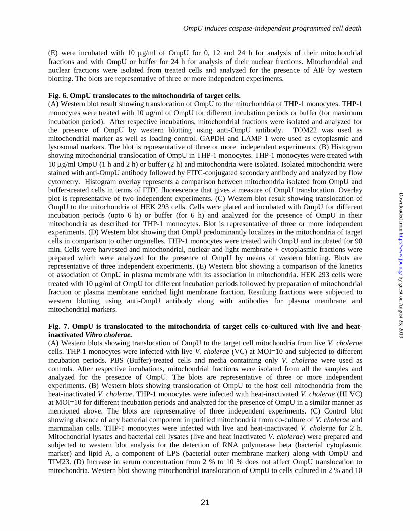
OmpU induces caspase-independent programmed cell death
21
(E) were incubated with 10 g/ml of OmpU for 0, 12 and 24 h for analysis of their mitochondrial
fractions and with OmpU or buffer for 24 h for analysis of their nuclear fractions. Mitochondrial and
nuclear fractions were isolated from treated cells and analyzed for the presence of AIF by western
blotting. The blots are representative of three or more independent experiments.
Fig. 6. OmpU translocates to the mitochondria of target cells.
(A) Western blot result showing translocation of OmpU to the mitochondria of THP-1 monocytes. THP-1
monocytes were treated with 10 g/ml of OmpU for different incubation periods or buffer (for maximum
incubation period). After respective incubations, mitochondrial fractions were isolated and analyzed for
the presence of OmpU by western blotting using anti-OmpU antibody. TOM22 was used as
mitochondrial marker as well as loading control. GAPDH and LAMP 1 were used as cytoplasmic and
lysosomal markers. The blot is representative of three or more independent experiments. (B) Histogram
showing mitochondrial translocation of OmpU in THP-1 monocytes. THP-1 monocytes were treated with
10 g/ml OmpU (1 h and 2 h) or buffer (2 h) and mitochondria were isolated. Isolated mitochondria were
stained with anti-OmpU antibody followed by FITC-conjugated secondary antibody and analyzed by flow
cytometry. Histogram overlay represents a comparison between mitochondria isolated from OmpU and
buffer-treated cells in terms of FITC fluorescence that gives a measure of OmpU translocation. Overlay
plot is representative of two independent experiments. (C) Western blot result showing translocation of
OmpU to the mitochondria of HEK 293 cells. Cells were plated and incubated with OmpU for different
incubation periods (upto 6 h) or buffer (for 6 h) and analyzed for the presence of OmpU in their
mitochondria as described for THP-1 monocytes. Blot is representative of three or more independent
experiments. (D) Western blot showing that OmpU predominantly localizes in the mitochondria of target
cells in comparison to other organelles. THP-1 monocytes were treated with OmpU and incubated for 90
min. Cells were harvested and mitochondrial, nuclear and light membrane + cytoplasmic fractions were
prepared which were analyzed for the presence of OmpU by means of western blotting. Blots are
representative of three independent experiments. (E) Western blot showing a comparison of the kinetics
of association of OmpU in plasma membrane with its association in mitochondria. HEK 293 cells were
treated with 10 g/ml of OmpU for different incubation periods followed by preparation of mitochondrial
fraction or plasma membrane enriched light membrane fraction. Resulting fractions were subjected to
western blotting using anti-OmpU antibody along with antibodies for plasma membrane and
mitochondrial markers.
Fig. 7. OmpU is translocated to the mitochondria of target cells co-cultured with live and heat-
inactivated Vibro cholerae. (A) Western blots showing translocation of OmpU to the target cell mitochondria from live V. cholerae
cells. THP-1 monocytes were infected with live V. cholerae (VC) at MOI=10 and subjected to different
incubation periods. PBS (Buffer)-treated cells and media containing only V. cholerae were used as
controls. After respective incubations, mitochondrial fractions were isolated from all the samples and
analyzed for the presence of OmpU. The blots are representative of three or more independent
experiments. (B) Western blots showing translocation of OmpU to the host cell mitochondria from the
heat-inactivated V. cholerae. THP-1 monocytes were infected with heat-inactivated V. cholerae (HI VC)
at MOI=10 for different incubation periods and analyzed for the presence of OmpU in a similar manner as
mentioned above. The blots are representative of three independent experiments. (C) Control blot
showing absence of any bacterial component in purified mitochondria from co-culture of V. cholerae and
mammalian cells. THP-1 monocytes were infected with live and heat-inactivated V. cholerae for 2 h.
Mitochondrial lysates and bacterial cell lysates (live and heat inactivated V. cholerae) were prepared and
subjected to western blot analysis for the detection of RNA polymerase beta (bacterial cytoplasmic
marker) and lipid A, a component of LPS (bacterial outer membrane marker) along with OmpU and
TIM23. (D) Increase in serum concentration from 2 % to 10 % does not affect OmpU translocation to
mitochondria. Western blot showing mitochondrial translocation of OmpU to cells cultured in 2 % and 10
by guest on August 25, 2019
http://ww
w.jbc.org/
Dow
nloaded from

OmpU induces caspase-independent programmed cell death
22
% serum containing media. THP-1 monocytes were plated in 2 % or 10 % serum-containing media,
treated with 10 g/ml of OmpU and incubated for 2 h. Mitochondrial fraction was isolated from all the
samples and analyzed for the presence of OmpU by western blotting. VDAC was used as the
mitochondrial marker and loading control. The blot is representative of two independent experiments. (E)
Increase in serum concentration from 2 % to 10 % does not affect OmpU-induced PCD. Histogram
overlay showing only annexin V positive population of OmpU treated cells cultured in 2 % and 10 %
serum-containing media. THP-1 monocytes cultured in 2 % and 10 % serum-containing media were
treated with different doses of OmpU (1.5, 5 or 10 g/ml) or buffer. Cells were harvested and stained with
annexin V-FITC and PI. Histogram overlay represents gated population comprising of only annexin V-
FITC single positive cells. The plots are representative of three independent experiments.
Fig. 8. OmpU directly induces loss of mitochondrial membrane potential and AIF release from
freshly isolated mitochondria.
(A) Histogram overlay represents increase in loss of mitochondrial membrane potential in OmpU-treated
mitochondria compared to buffer treatment. Mitochondria were isolated from 3x10⁷ THP-1 monocytes
and exactly equal aliquots of isolated mitochondria were incubated with 5 g/ml of OmpU or buffer for
30 min. Following incubation, mitochondria were stained with JC-1 dye. In corresponding histogram
overlay, decrease in red fluorescence of JC-1 aggregates gives a measure of decreased mitochondrial
membrane potential in mitochondria. The plot is representative of three independent experiments. (B and
C) Western blots showing release of cytochrome c (B) and AIF (C) from the treated mitochondria and
corresponding increase in the supernatant with increase in incubation time after OmpU treatment. Equal
aliquots of isolated mitochondria were treated with 5 g/ml of OmpU and incubated for different time
periods or with buffer for 2 h. After respective incubations, mitochondria were harvested and supernatants
were collected separately for each sample. Supernatants and mitochondria were analyzed for the presence
of cytochrome c and AIF by western blot analysis. The blots are representative of three or more
independent experiments. (D) Western blots showing inhibition of AIF release from isolated
mitochondria by CsA. Freshly isolated mitochondria were pretreated with 1 M of CsA for 30 min
followed by treatment with 5g/ml of OmpU for 90 min. Following incubation, samples were subjected
to western blotting as described above. Blots are representative of three independent experiments.
Fig. 9. OmpU induces caspase-independent cell death involving OmpU translocation to
mitochondria in Caco-2 cells.
(A) Bar graphs showing cell death induced by OmpU in Caco-2 cells measured as loss of cell viability by
MTT assay. Caco-2 cells were plated and treated with OmpU (5 g/ml and 10 g/ml) or buffer for 24 h
and subjected to MTT assay. Bar graphs represent mean ± SEM values of percentage cell viability
calculated using values from three independent experiments. (*p<0.05, **p<0.01, ***p<0.001 versus
buffer control for all the doses). (B) Bar graphs showing cell cytotoxicity measured by LDH release
induced by OmpU in Caco-2 cells. Caco-2 cells were plated and treated as described for MTT assay and
and subjected to LDH release assay. Bar graphs represent mean ± SEM values of percentage cell
cytotoxicity calculated using values from three independent experiments. (C) and (D) Bar graphs showing
that inhibition of apoptosis related caspases does not inhibit OmpU-mediated cell death. Caco-2 cells
were plated and pre-treated with 20 M of zVAD-fmk followed by treatment with OmpU (10 g/ml) or
buffer. Cells were subjected to either MTT assay or LDH release assay. Bar graphs represent mean ±
SEM values of percentage cell viability (C) or percentage cell cytotoxicity (D) and are calculated using
values from three independent experiments. (E) Western blot result showing translocation of OmpU in
Caco-2 cells. Caco-2 cells were treated with 10 g/ml of OmpU for 2 h. Mitochondrial fraction was
prepared and analyzed for the presence of different markers along with OmpU by western blotting.
(F).Western blot result showing release of AIF from mitochondria of Caco-2 cells treated with 10 g/ml
of OmpU for indicated time points and its translocation to the nucleus. Blots are representative of two
independent experiments.
by guest on August 25, 2019
http://ww
w.jbc.org/
Dow
nloaded from

OmpU induces caspase-independent programmed cell death
23
Fig. 10. Schematic diagram depicting pathway of OmpU-mediated apoptosis.
Purified OmpU or OmpU from Vibrio cholerae translocates across the cell membrane to the
mitochondria. Translocated OmpU induces loss of mitochondrial membrane potential (m) followed by
release of cytochrome c and AIF into the cytoplasm. Released cytochrome c does not induce caspase
activation. AIF translocates to the nucleus and induces DNA fragmentation and hence apoptosis like
programmed cell death.
by guest on August 25, 2019
http://ww
w.jbc.org/
Dow
nloaded from

Shelly Gupta, G. V. R. Krishna Prasad and Arunika Mukhopadhayaupon Translocation to the Host Cell Mitochondria
Porin OmpU Induces Caspase-independent Programmed Cell DeathVibrio cholerae
published online November 11, 2015J. Biol. Chem.
10.1074/jbc.M115.670182Access the most updated version of this article at doi:
Alerts:
When a correction for this article is posted•
When this article is cited•
to choose from all of JBC's e-mail alertsClick here
by guest on August 25, 2019
http://ww
w.jbc.org/
Dow
nloaded from