Polysomnography (Sleep Studies) · Polysomnography (Sleep Studies) Angelia Smith, MD Advisor:...
60
Polysomnography (Sleep Studies) Angelia Smith, MD Advisor: Harold Pine, MD, FAAP. FACS The University of Texas Medical Branch Department of Otolaryngology Grand Rounds Presentation January 31, 2011 Photographs are used with permission. Graphs, drawings, and tables are not.
Transcript of Polysomnography (Sleep Studies) · Polysomnography (Sleep Studies) Angelia Smith, MD Advisor:...
Polysomnography (Sleep Studies)
Angelia Smith, MD
Advisor: Harold Pine, MD, FAAP. FACS
The University of Texas Medical Branch
Department of Otolaryngology
Grand Rounds Presentation
January 31, 2011
Photographs are used with permission. Graphs, drawings, and tables are not.
Overview
• stages of sleep
• indications for polysomnography
• what happens to the patient
• what is measured
• how it is interpreted
• what it all means
• treatment options

“Some people talk in their sleep. Lecturers talk while other people sleep.”
Albert Camus”
Characteristics of Sleep
• Neurophysiology of arousal
– Arousal mechanisms are governed by the reticular formation in the brainstem, referred to as the reticular activating system (RAS). It communicates with the cerebral cortex through the thalamus: sensory input from the cerebral cortex can activate the RAS and cause arousal. During sleep, fewer stimuli arise from the cortex because of a feedback loop to the cortex from the RAS.

“There is only one thing people like that is good for them; a good night's sleep”
Edgar Watson Howe
Stages of Sleep
• Awake is one necessary stage of sleep
– Characterized by alpha waves on EEG
– Reactivity to external stimuli maintained
• Other stages of sleep are broken into Non-Rapid Eye Movement (NREM) and Rapid Eye Movement (REM).
– NREM has three stages
– REM is its own stage
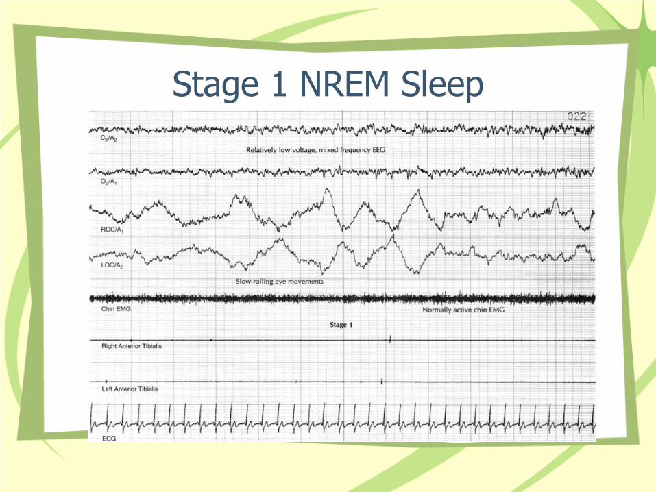
Stage 1 NREM Sleep
• Drowsiness
• EEG rhythms slow to mixed delta and theta waves
• Reduced muscle activity
• Decreased minute ventilation, increased PaCO2
• Heart rate and cardiac output decrease from increase in parasympathetic tone
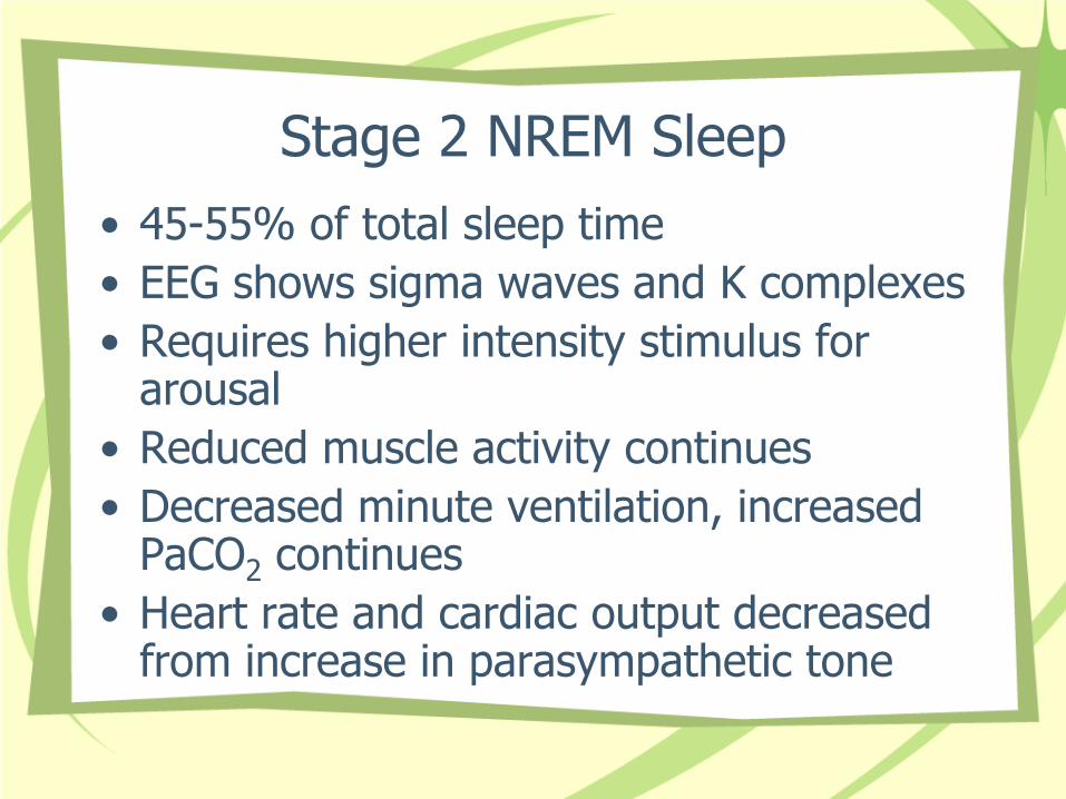
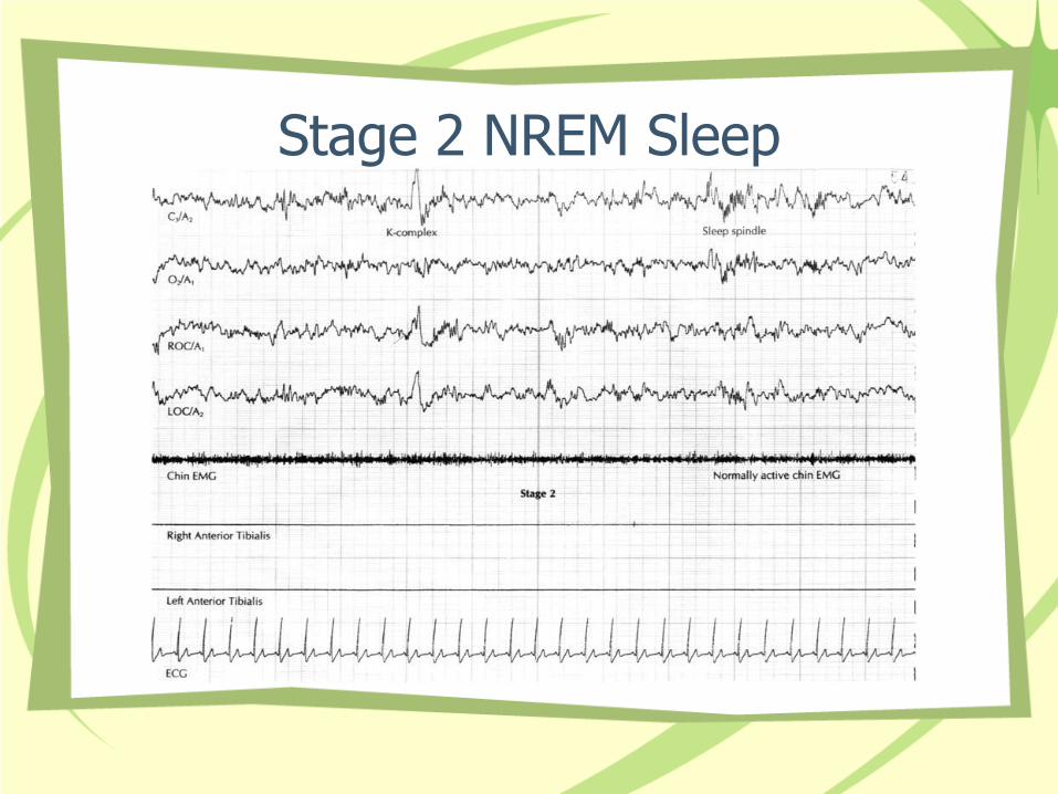
Stage 2 NREM Sleep
• 45-55% of total sleep time
• EEG shows sigma waves and K complexes
• Requires higher intensity stimulus for arousal
• Reduced muscle activity continues
• Decreased minute ventilation, increased PaCO2 continues
• Heart rate and cardiac output decreased from increase in parasympathetic tone
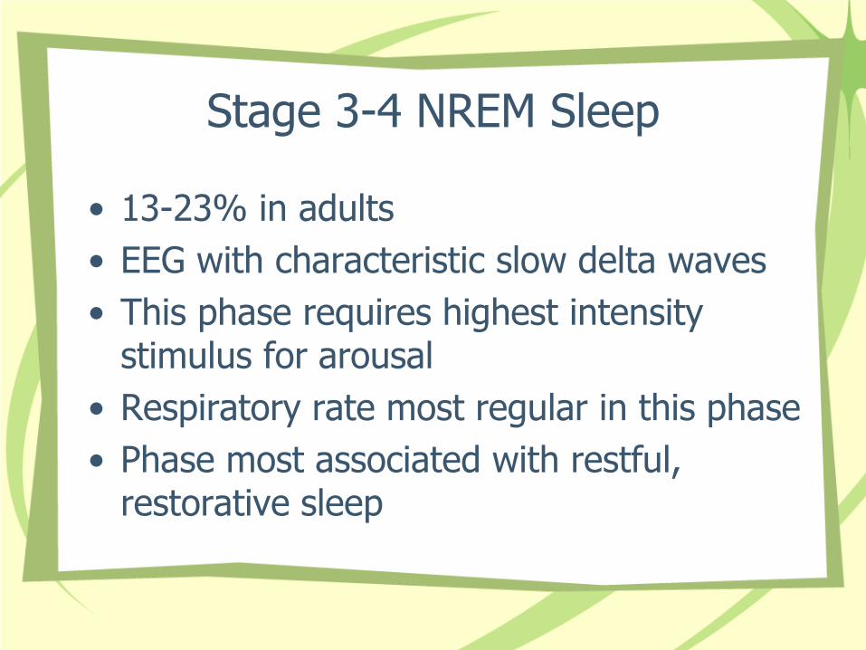
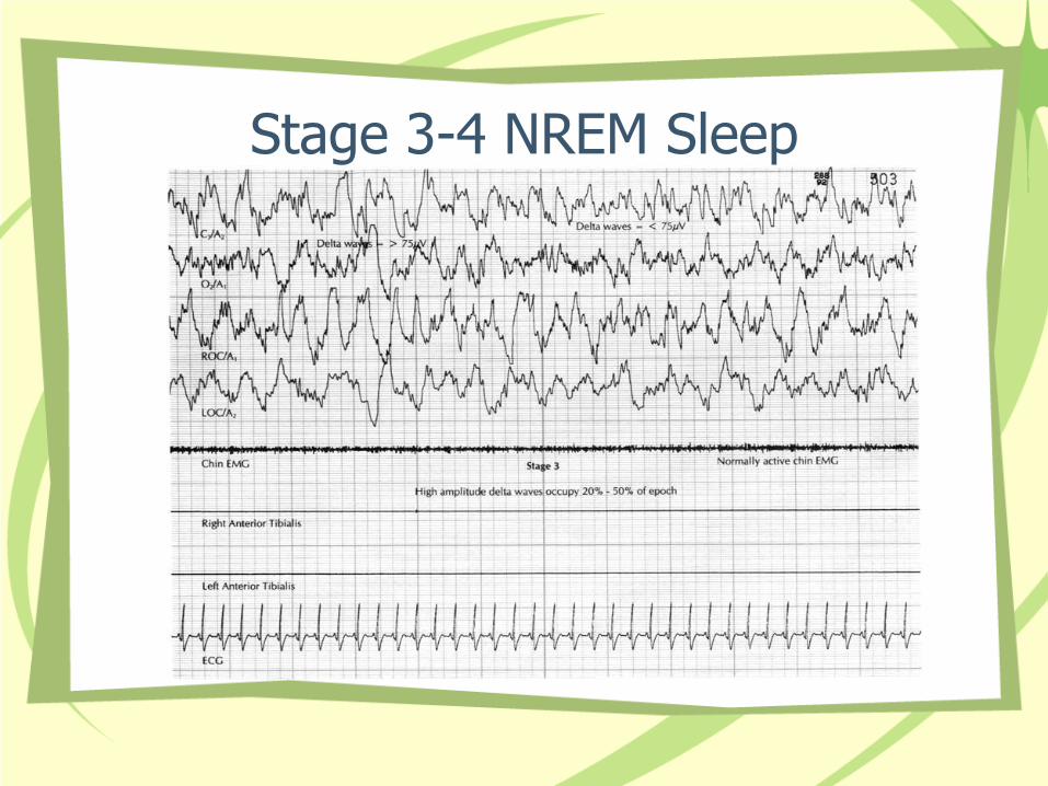
Stage 3-4 NREM Sleep
• 13-23% in adults
• EEG with characteristic slow delta waves
• This phase requires highest intensity stimulus for arousal
• Respiratory rate most regular in this phase
• Phase most associated with restful, restorative sleep
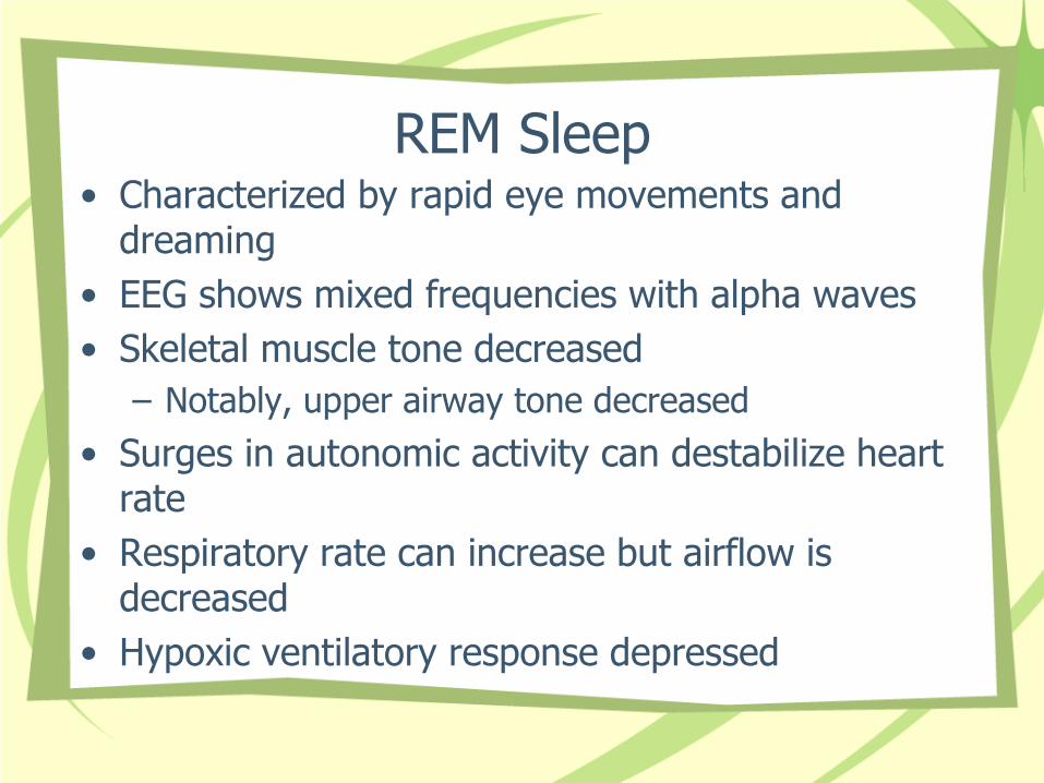
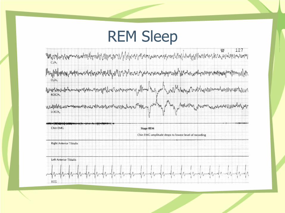
REM Sleep • Characterized by rapid eye movements and
dreaming
• EEG shows mixed frequencies with alpha waves
• Skeletal muscle tone decreased
– Notably, upper airway tone decreased
• Surges in autonomic activity can destabilize heart rate
• Respiratory rate can increase but airflow is decreased
• Hypoxic ventilatory response depressed

“Laugh and the world laughs with you, snore and you sleep alone.”
Anthony Burgess
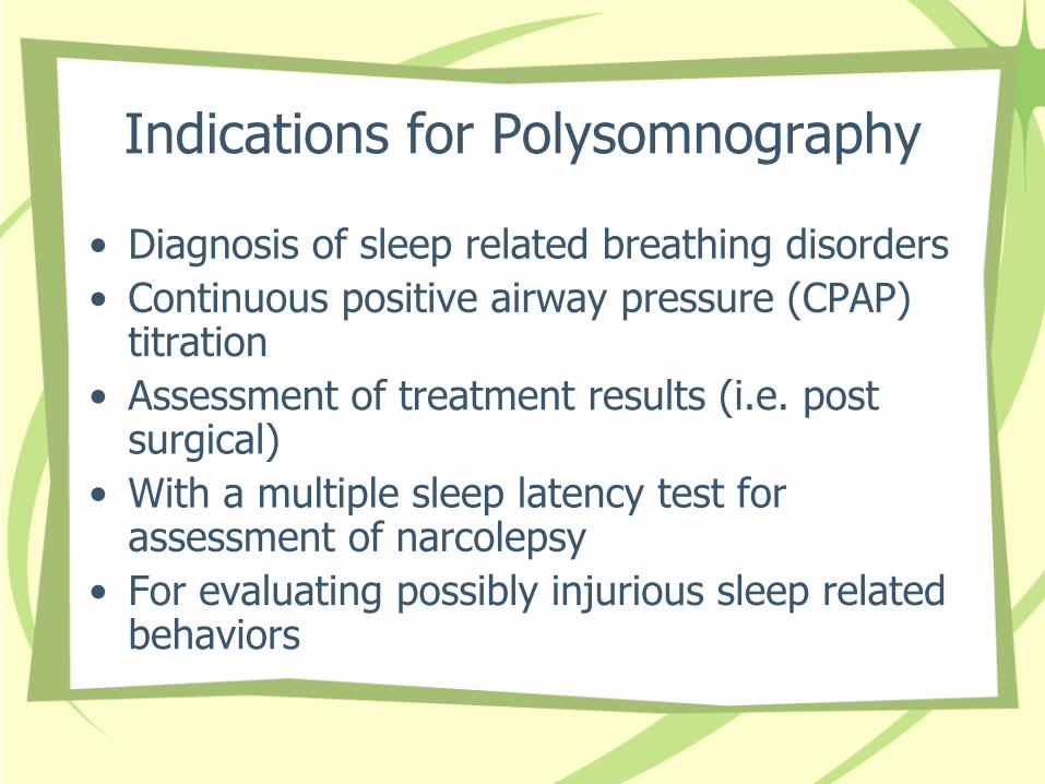
Indications for Polysomnography
• Diagnosis of sleep related breathing disorders
• Continuous positive airway pressure (CPAP) titration
• Assessment of treatment results (i.e. post surgical)
• With a multiple sleep latency test for assessment of narcolepsy
• For evaluating possibly injurious sleep related behaviors
Diagnosing Sleep Related Disorders
• Sleep history is key to diagnosis – Sleep disordered breathing symptoms include snoring,
witnessed apneas, nocturnal choking or gasping, restlessness, and excessive daytime sleepiness
– Epworth Sleepiness Scale can help distinguish severity – Evaluation of daytime sleepiness should include
evidence of sleep deprivation, use of alarm clock, shift work, snoring, recent weight gain, family history, morning headache, sore throat or dry mouth
– Also ask about alchol consumption, nasal congestion, hypothyroidism, and menopause
– A sleep log can be helpful to diagnose daytime sleepiness from lack of sleep time vs. pathologic sleep

Children vs. Adults
• In children, symptoms can be different from adults.
• Behavioral problems, learning problems, lack of attentiveness, and hyperactivity can characterize sleep disordered breathing more than classic signs of fatigue
Physical Exam
• Findings that support diagnosis of sleep disordered breathing
– Adults: obesity, HTN, cardiopulmonary disease
– Children: adenotonsillar hypertrophy, obesity
“People who snore always fall asleep first.”
Author Unknown”
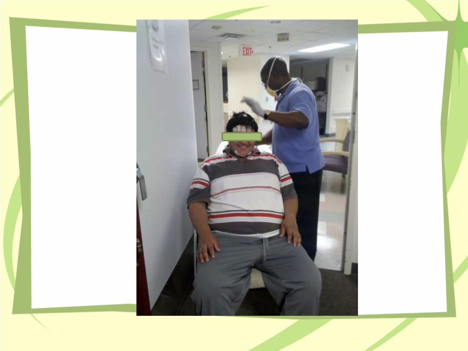
What Happens in the Sleep Lab
• In our sleep lab at UTMB, the patients arrive around 9 p.m. to be set up. In the pediatric population, families are allowed to sleep in the same room (but not in the bed with the patient)
• Introducing: “Tony”, an adolescent patient of Dr. Pine, post-T&A for OSAHS, here for follow up sleep study
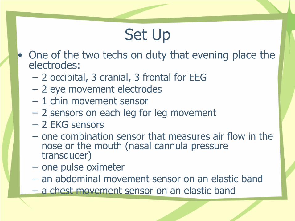
Set Up • One of the two techs on duty that evening place the
electrodes: – 2 occipital, 3 cranial, 3 frontal for EEG – 2 eye movement electrodes – 1 chin movement sensor – 2 sensors on each leg for leg movement – 2 EKG sensors – one combination sensor that measures air flow in the
nose or the mouth (nasal cannula pressure transducer)
– one pulse oximeter – an abdominal movement sensor on an elastic band – a chest movement sensor on an elastic band
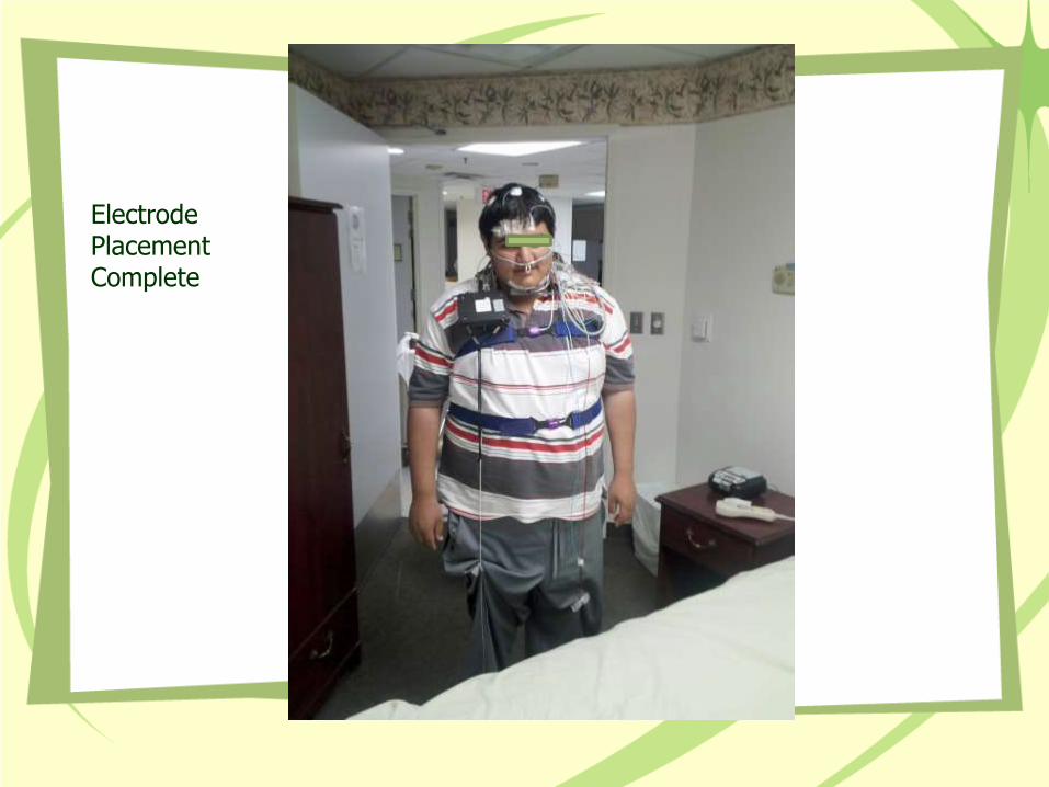
Electrode Placement Complete
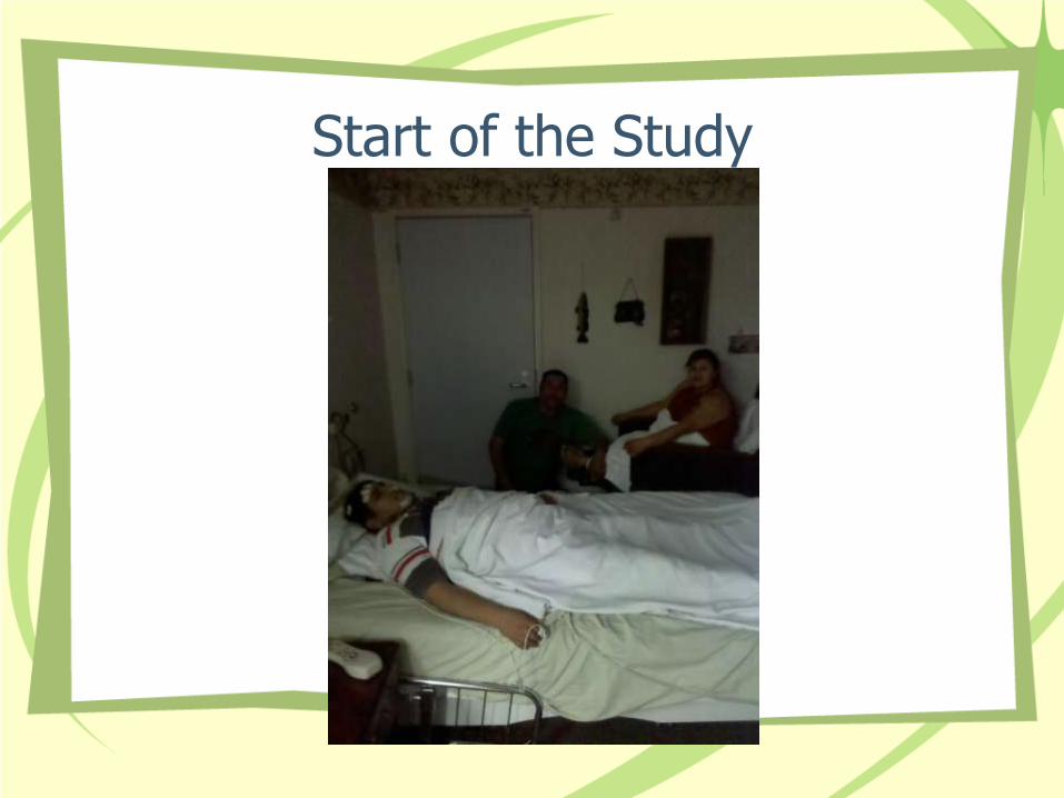
Start of the Study
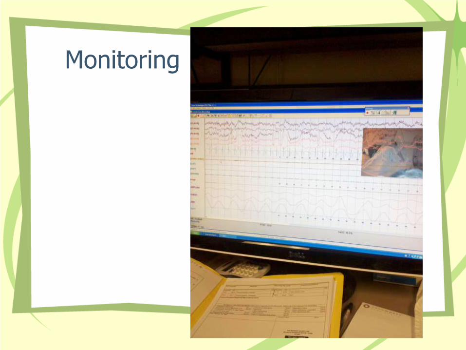
Monitoring
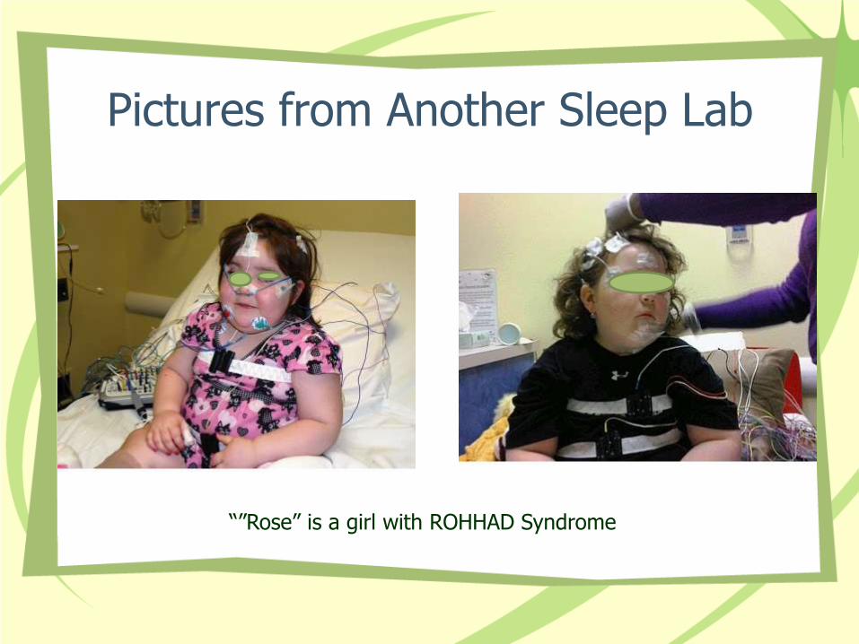
Pictures from Another Sleep Lab
“”Rose” is a girl with ROHHAD Syndrome
Brief Detour: ROHHAD Syndrome • “Rose” is a 6 year old girl with a rare syndrome that has been termed
ROHHAD: Rapid-Onset Obesity With Hypothalamic Dysfunction, Hypoventilation, and Autonomic Dysregulation.
• This syndrome is characterized most often by rapid onset obesity occuring around the age of 3. Other frequent hypothalamic dysfunction symptoms are hyperphagia, polydipsia, polyuria, and hypernatremia.
• Autonomic dysregulation often follows around the age of 4, most frequently manifested in ophthalmic problems (e.g. strabismus), GI dysmotility, altered sweating, thermal dysregulation, and tumors of neural crest origin. – "Rose" has ganglioneuroblastoma that was diagnosed in 2007 at the same
time as ROHHAD syndrome.
• Present in all patients is alveolar hypoventilation that begins to manifest at the average age of 6. Obstructive sleep apnea is common, and death can occur suddenly from cardiorespiratory arrest from central apneas. Seizures are also seen with this syndrome. – "Rose" sleeps with a pulse oximeter at night that alarms so her mom can go in
and arouse her if her sats dip too low. "Rose" undergoes sleep studies every 6 months to try and catch central apnea before it is symptomatic. She has confirmed obstructive sleep apnea and her mother and doctors are planning to have her tonsils and adenoids removed in the near future. Her CO2 used to run about 50 but she has undergone chemotherapy with cyclophosphamide, rituximab, IVIG, and prednisone that has her weight steady and her CO2 down to 45.
Polysomnography: What is Measured
• The EEG measures the brain’s neuronal activity
• Brain waves have characteristic frequencies, amplitudes, and morphologies
• Each stage of sleep has characteristic brain waves
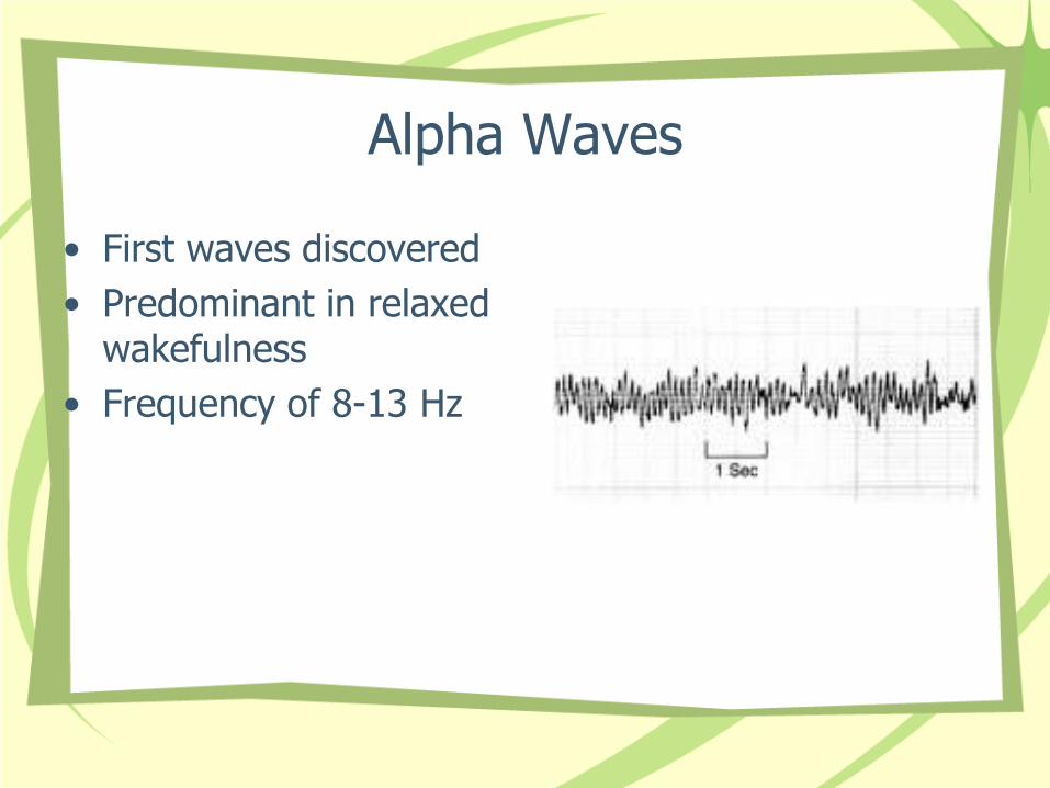
Alpha Waves
• First waves discovered
• Predominant in relaxed wakefulness
• Frequency of 8-13 Hz
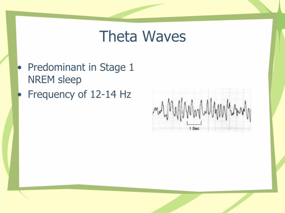
Theta Waves
• Predominant in Stage 1 NREM sleep
• Frequency of 12-14 Hz
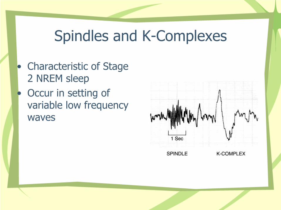
Spindles and K-Complexes
• Characteristic of Stage 2 NREM sleep
• Occur in setting of variable low frequency waves
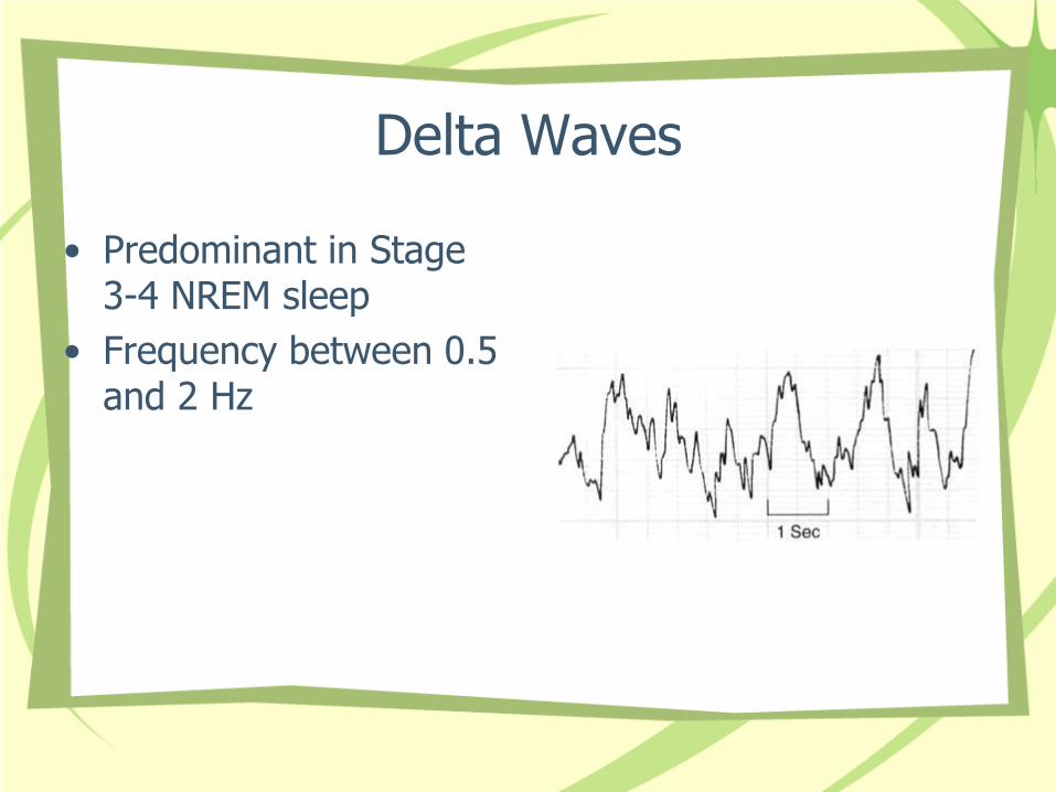
Delta Waves
• Predominant in Stage 3-4 NREM sleep
• Frequency between 0.5 and 2 Hz
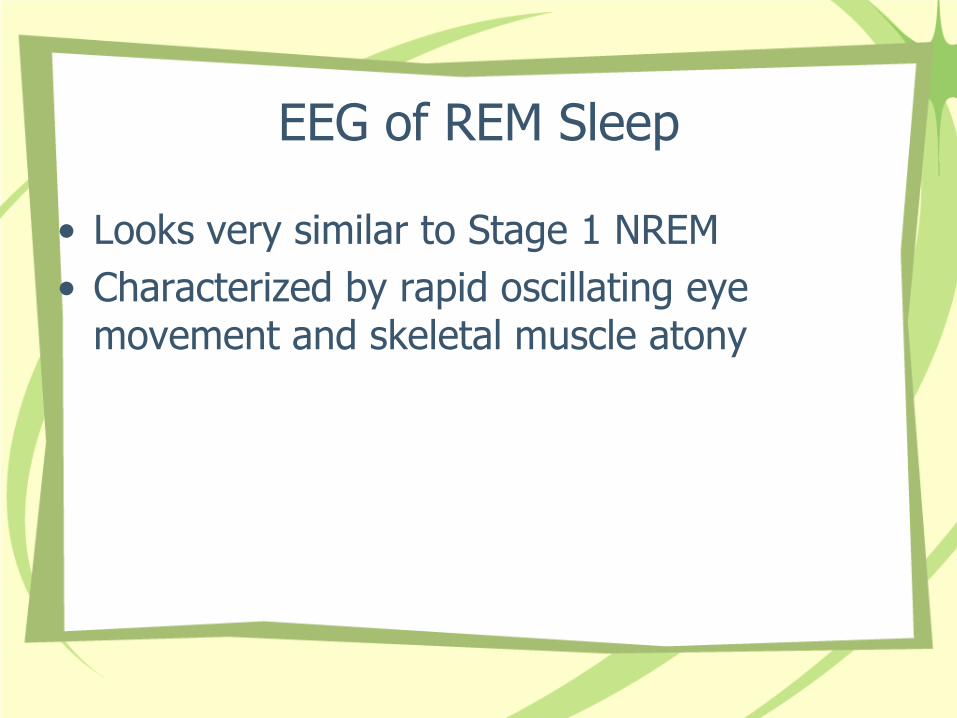
EEG of REM Sleep
• Looks very similar to Stage 1 NREM
• Characterized by rapid oscillating eye movement and skeletal muscle atony
“Sleep is a symptom of caffeine deprivation.”
Author Unknown
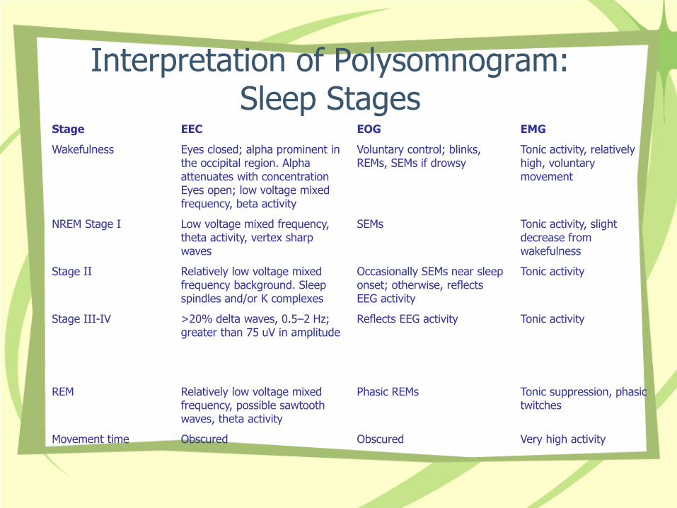
Interpretation of Polysomnogram: Sleep Stages
Stage EEC EOG EMG
Wakefulness Eyes closed; alpha prominent in the occipital region. Alpha attenuates with concentration Eyes open; low voltage mixed frequency, beta activity
Voluntary control; blinks, REMs, SEMs if drowsy
Tonic activity, relatively high, voluntary movement
NREM Stage I Low voltage mixed frequency, theta activity, vertex sharp waves
SEMs Tonic activity, slight decrease from wakefulness
Stage II Relatively low voltage mixed frequency background. Sleep spindles and/or K complexes
Occasionally SEMs near sleep onset; otherwise, reflects EEG activity
Tonic activity
Stage III-IV >20% delta waves, 0.5–2 Hz; greater than 75 uV in amplitude
Reflects EEG activity Tonic activity
REM Relatively low voltage mixed frequency, possible sawtooth waves, theta activity
Phasic REMs Tonic suppression, phasic twitches
Movement time Obscured Obscured Very high activity
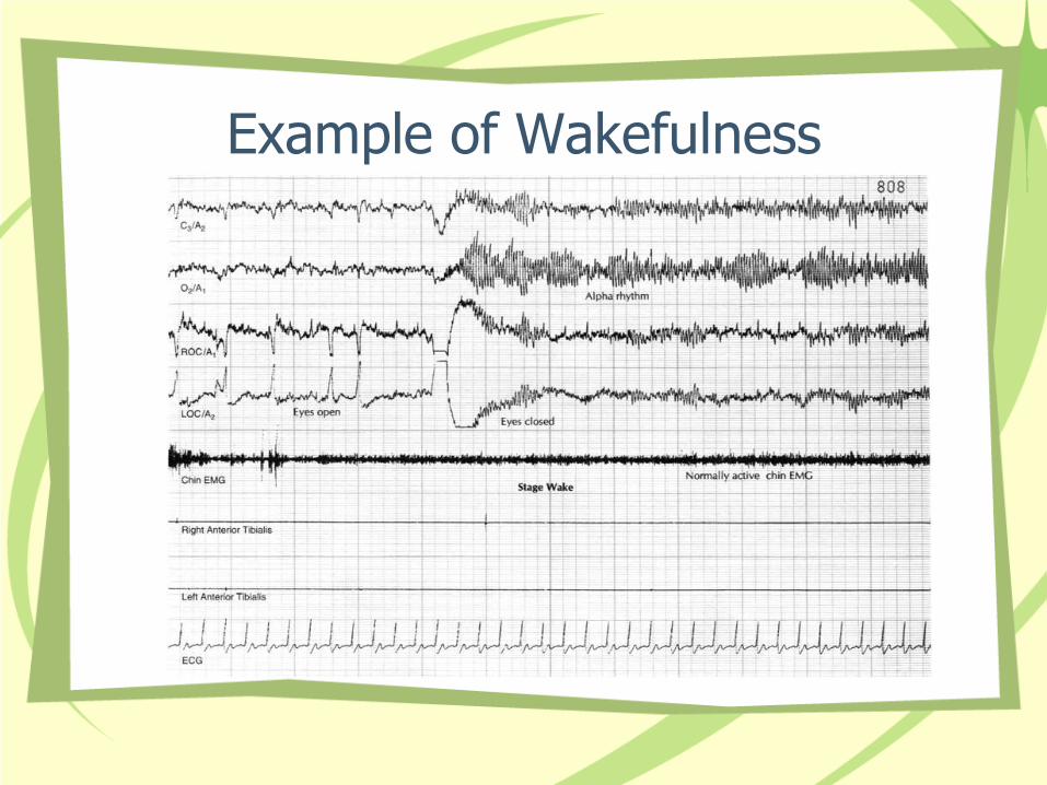
Example of Wakefulness
Stage 1 NREM Sleep
Stage 2 NREM Sleep
Stage 3-4 NREM Sleep
REM Sleep
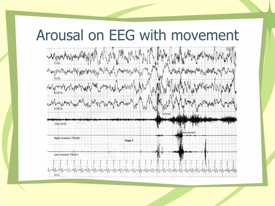
Arousal on EEG with movement
Remember all the other leads?
• Chin movement
• Leg movement
• Chest wall movement
• Abdominal movement
• Air flow, both nasal and oral
• Pulse oximetry
Characteristics of Sleep Disordered Breathing
• Apnea is the cessation or near cessation of airflow for a minimum of 10 seconds – Usually associated with desaturation and an EEG
arousal at terminus
• Hypopnea is a 50% decrease in airflow for at least 10 seconds followed by an arousal and/or 4% oxygen desaturation
• Respiratory Event Related Arousals (RERA) are periods of increased breathing effort during increased airway resistance, with subsequent arousals, but in the absence of hypopneas, apneas, or O2 desaturations
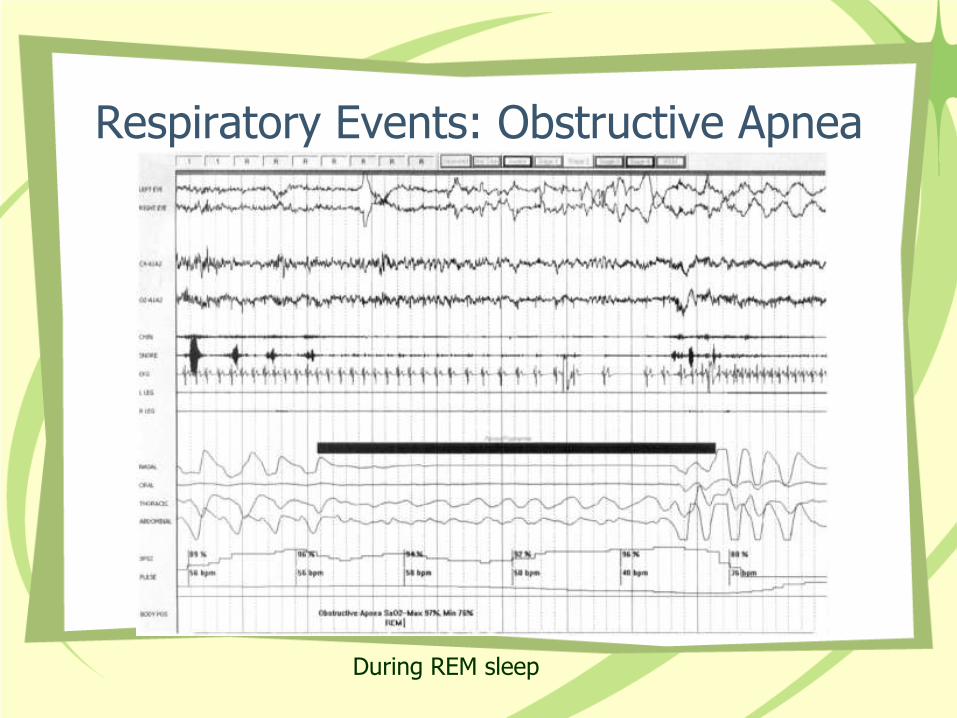
Respiratory Events: Obstructive Apnea
During REM sleep
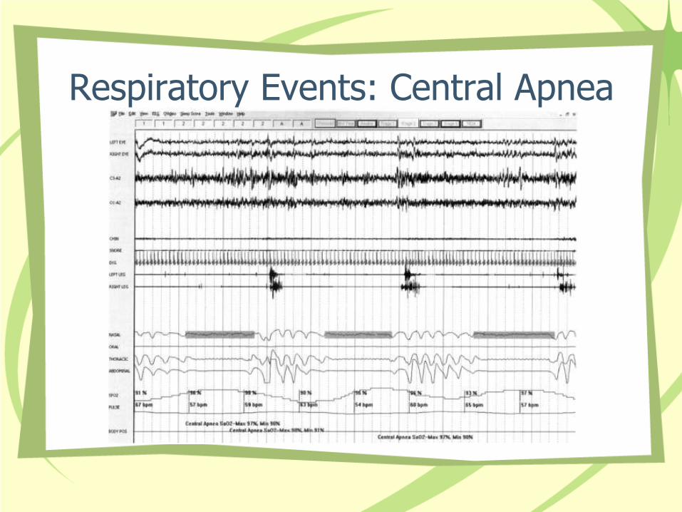
Respiratory Events: Central Apnea
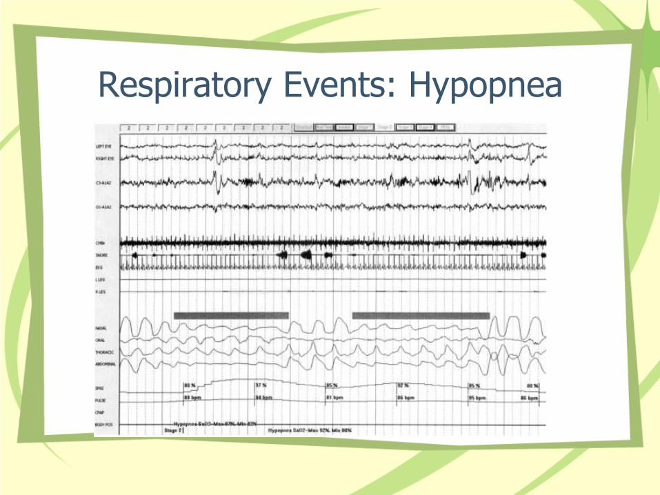
Respiratory Events: Hypopnea
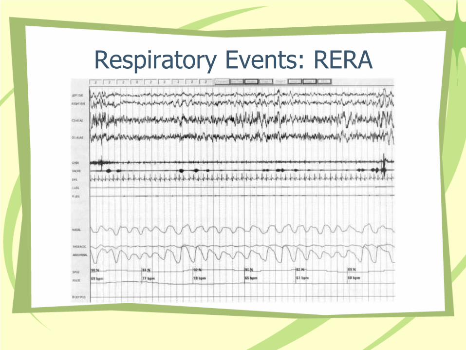
Respiratory Events: RERA
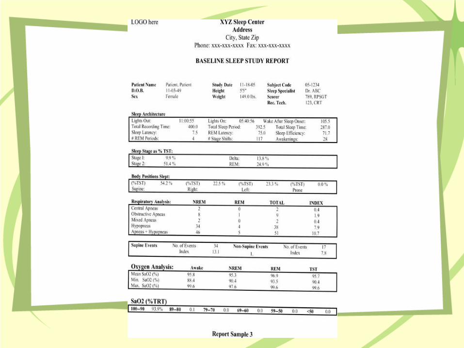
After The Polysomnogram
• The study is then scored according to AASM guidelines and a report is generated
• Measurements include total recording time, total sleep time, sleep latency (amount of time from lights out to sleep stage 1), number of REM periods, number of stage shifts, number of arousals, number of apneas and type, number of hypopneas, pulse oximetry, and leg movements
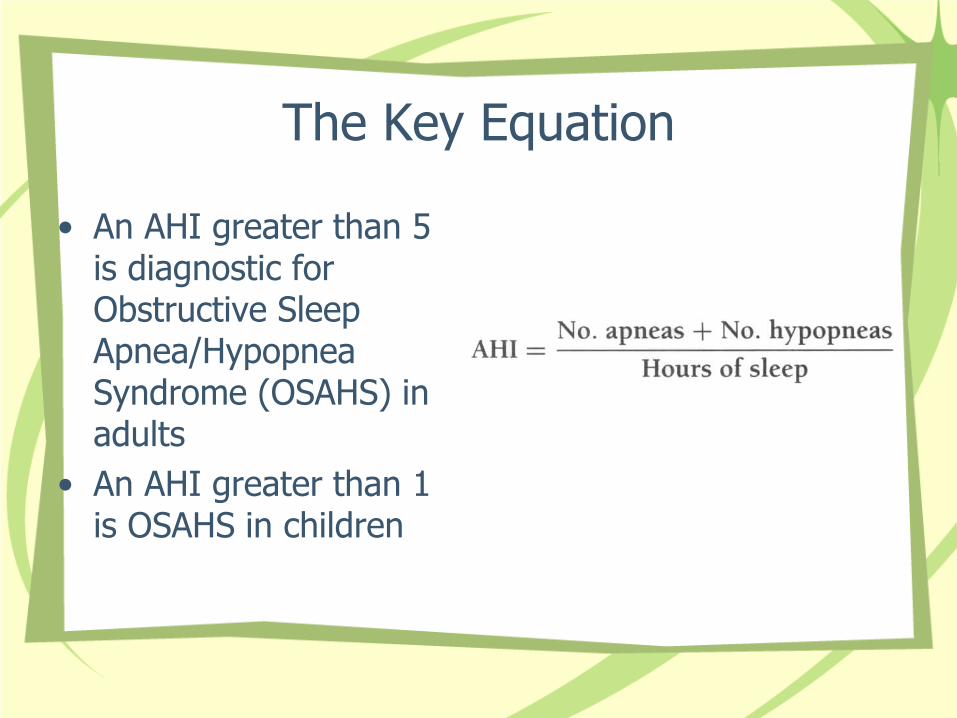
The Key Equation
• An AHI greater than 5 is diagnostic for Obstructive Sleep Apnea/Hypopnea Syndrome (OSAHS) in adults
• An AHI greater than 1 is OSAHS in children
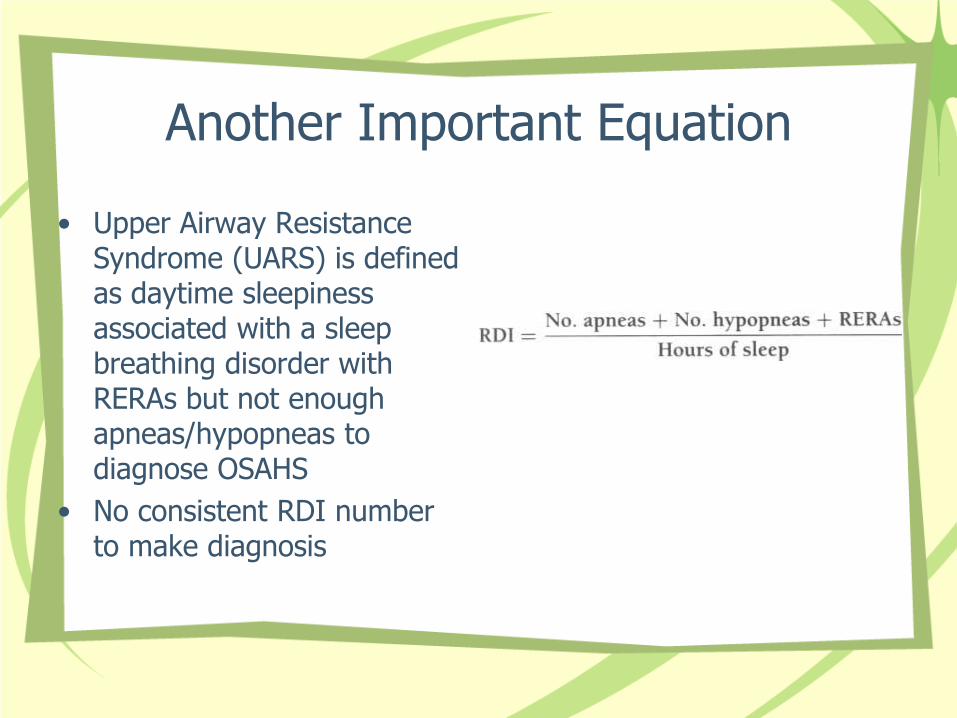
Another Important Equation
• Upper Airway Resistance Syndrome (UARS) is defined as daytime sleepiness associated with a sleep breathing disorder with RERAs but not enough apneas/hypopneas to diagnose OSAHS
• No consistent RDI number to make diagnosis

“A good laugh and a long sleep are the best cures in the doctor's book.”
Irish Proverb
Treatment Options for OSAHS
• Nothing
• Weight loss
• Oral appliances
• Positive pressure ventilation
• Surgery
• Childrens’ options
No Treatment
• It has been noted that some patients with OSA only have it in the prone position. The obstruction disappears when the patient is lying on their side, so sleep positioning can offer a cure
• These are usually mild cases with AHI <30
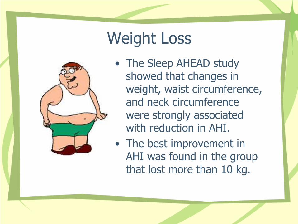
Weight Loss
• The Sleep AHEAD study showed that changes in weight, waist circumference, and neck circumference were strongly associated with reduction in AHI.
• The best improvement in AHI was found in the group that lost more than 10 kg.
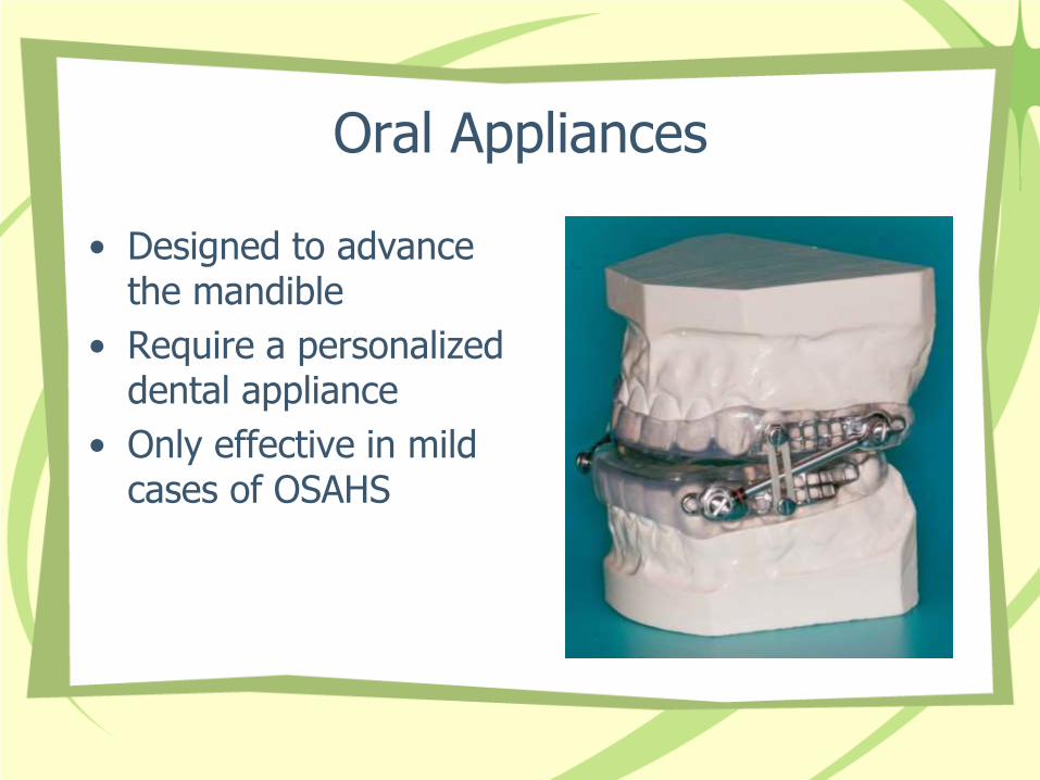
Oral Appliances
• Designed to advance the mandible
• Require a personalized dental appliance
• Only effective in mild cases of OSAHS
Positive Pressure Ventilation
• Well known treatment for OSAHS.
• Creates a “pneumatic splint” in the upper airway to prevent collapse
• Several variants: nasal CPAP, autotitrating CPAP, BiPAP
• This treatment is only as effective as the compliance to it, reported as 46-89%
• Interestingly, the least compliant patients are the ones who present for evaluation only at the urging of their spouses
Surgical Options
• Tracheostomy is the only surgical procedure consistently effective in the treatment of OSAHS, but indicated only for life threatening disease such as cor pulmonale, arrhythmias, or severe hypoxemia
• The surgery most frequently performed on adults for OSAHS is uvulopalatopharyngoplasty (UPPP). This is generally considered only 50% effective. Some research states that this is more effective in patients with lower BMI and lower AHI
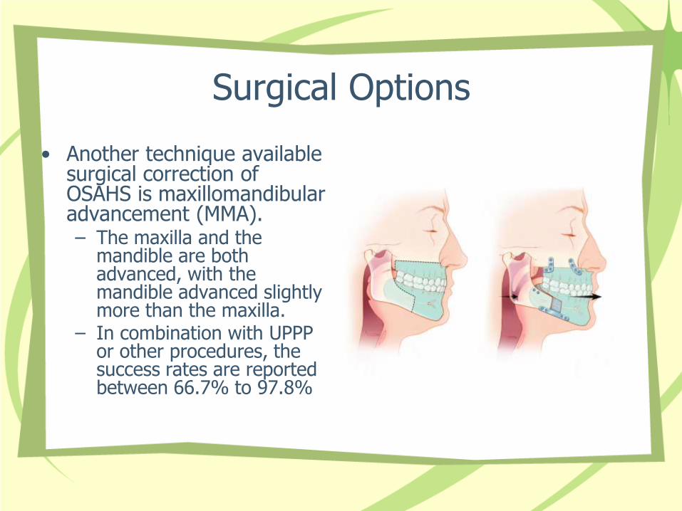
Surgical Options
• Another technique available surgical correction of OSAHS is maxillomandibular advancement (MMA). – The maxilla and the
mandible are both advanced, with the mandible advanced slightly more than the maxilla.
– In combination with UPPP or other procedures, the success rates are reported between 66.7% to 97.8%
Surgery in Children
• Treatment of choice for OSAHS in children is adenotonsillectomy.
– Complications after the procedure occurs more often in children younger than 3, those with severe OSA, and those with other medical problems
• In children with obesity, this may not completely resolve OSAHS
– In those cases, CPAP and weight loss can help

“The best bridge between despair and hope is a good night's sleep.”
E. Joseph Cossman”
References
• Fundamentals of Sleep Technology, 1st Edition Butkov, Nic; Lee-Chiong, Teofilo Copyright ©2007 Lippincott Williams & Wilkins
• Clinical Sleep Disorders by Carney, Paul R. Berry, Richard B and Geyer, James D. Copyright 2005 by Lippincott, Williams, and Wilkins
• Iber, C; Ancoli-Israel, S; Chesson, A; Quan, SF for the American Academy of Sleep Medicine (2007). The AASM Manual for the Scoring of Sleep and Associated Events: Rules, Terminology and Technical Specifications. Westchester: American Academy of Sleep Medicine
• Practice Parameters for the Indications for Polysomnography and Related Procedures: An Update for 2005 Kushida, et al, SLEEP 2005;28(4):499-521.
• Rapid-Onset Obesity With Hypothalamic Dysfunction, Hypoventilation, and Autonomic Dysregulation Presenting in Childhood, Ize-Ludlow, D, et al Pediatrics 2007;120;e179-e188
• Butkov N: Atlas of Clinical Polysomnography (vol 1). Medford, OR Synapse Media, 1996 • A randomized study on the effect of weight loss on obstructive sleep apnea among obese
patients with type 2 diabetes, Foster, GD et al., Arch Intern Med. 2009;169(17):1619-1626