Polysaccharides and Proteoglycans in Calcium Carbonate-based Biomineralization
8
Polysaccharides and Proteoglycans in Calcium Carbonate-based Biomineralization Jose ´ L. Arias* and Marı ´a S. Ferna ´ ndez Faculty of Veterinary and Animal Sciences, and Center for Advanced Interdisciplinary Research in Materials (CIMAT), Universidad de Chile, Casilla 2 Correo 15, Santiago, Chile Received January 18, 2008 Contents 1. Introduction 4475 2. Hydroxylated Polysaccharides 4475 3. Polycarboxylated Polysaccharides 4476 4. Sulfated Polysaccharides 4477 5. Polysaccharides in Biomineralization 4478 6. Calcium Carbonate Growth in the Presence of Sulfated Polymers 4479 7. Concluding Remarks 4480 8. Acknowledgments 4480 9. References 4481 1. Introduction Biomineralization is a widespread phenomenon in nature leading to the formation of a variety of solid inorganic structures by living organisms, such as intracellular crystals in prokaryotes, exoskeletons in protozoa, algae, and inver- tebrates, spicules and lenses, bone, teeth, statoliths, and otoliths, eggshells, plant mineral structures, and also patho- logical biominerals such as gall stones, kidney stones, and oyster pearls. 1–7 These biologically produced biominerals are inorganic-organic hybrid composites formed by self-as- sembled bottom up processes under mild conditions, showing interesting properties, controlled hierarchical structures, and remodeling or repair mechanisms which still remain to be developed into a practical engineering process. 8–10 Therefore, the formation of biominerals provides a unique guide for the design of materials, especially those that need to be fabricated at ambient temperatures. In biominerals, the small amount of organic component not only reinforces the mechanical properties of the resulting composite but also exerts a crucial control on the mineralization process, contributing to the determination of the size, crystal mor- phology, specific crystallographic orientation, and superb properties of the particles formed. 11–15 Therefore, biological routes of structuring biominerals are becoming valuable approaches for novel materials synthesis. Although several principles are applicable to the majority of the biominerals, herein we will focus on the role of polysaccharide polymers in calcium carbonate-based biominerals. As a general principle, the assembly of these biominerals consists of a four-stage process. It starts with the fabrication of a hydrophobic solid organic substrate or scaffolding onto which nucleation of the crystalline phase takes place closely associated with some polyanionic macromolecules. Crystal growth is then controlled by the addition of gel-structuring polyanionic macromolecules, and finally mineralization arrest is accompanied by the secretion of a new inert scaffolding of the same type or the deposition of other hydrophobic inhibitory macromolecules. 16 Currently, a large number of proteins have been described which are involved in the control of biomineralization. 17–19 These proteins are usually highly negatively charged and contain carboxylate, sulfate, or phosphate as functional groups, which may bind Ca 2+ ions and could control crystal nucleation and growth by lowering the interfacial energy between the crystal and the macromolecular substrate. 20–25 However, the precise mechanism involved in controlling crystal nucleation, growth, and morphology is far from being understood. Combinatorial biology techniques have been recently developed for testing the ability of randomly generated peptides to bind different substrates or ions, thus allowing a correlation between peptide structure and ion binding affinity. 26–29 However, the main focus is on the role of the backbone structure of the polymer due to the primary structure of the protein, because the synthetic technology does not allow the formation of post-translational modifica- tions, such as sulfation and phosphorylation, which do occur in the eukaryotic cell. Even so, the occurrence of negatively charged groups in macromolecules involved in biomineral- ization, mainly derived from acidic amino acids, has inspired many researchers to produce synthetic polymers having such groups in order to control the size, orientation, phase, and morphology of inorganic crystals. 30–43 However, since Abo- lins-Krogis’ work, 44 a slow but increasing interest has been developed to explore the role of polysaccharides in biom- ineralization, despite the fact that their involvement in biomineralization seems to appear very early in evolution. 45 There is no single type of polysaccharide associated with biominerals, but such polysaccharides are mainly hydroxy- lated, carboxylated, or sulfated or contain a mixture of these functional moieties. 2. Hydroxylated Polysaccharides After cellulose, chitin is the second most abundant biopolymer in the biosphere. As chitin or its deacetylated form chitosan, it is widely distributed in the animal, plant, fungi, and protozoa kingdoms. Although its occurrence is not necessarily directly associated with biomineralization, its presence is crucial in biominerals such as crustacean shells, carapaces and gastroliths, and mollusks shells. Chitin is a linear polysaccharide of R- or -(1-4)-2-acetamido-2- deoxy-D-glucopyranose where the monomeric residue is N-acetylglucosamine (Figure 1, upper structure). 46 It exists * E-mail: [email protected]. Chem. Rev. 2008, 108, 4475–4482 4475 10.1021/cr078269p CCC: $71.00 2008 American Chemical Society Published on Web 07/25/2008
Transcript of Polysaccharides and Proteoglycans in Calcium Carbonate-based Biomineralization
Polysaccharides and Proteoglycans in Calcium Carbonate-basedBiomineralization
Jose L. Arias* and Marıa S. Fernandez
Faculty of Veterinary and Animal Sciences, and Center for Advanced Interdisciplinary Research in Materials (CIMAT), Universidad de Chile,Casilla 2 Correo 15, Santiago, Chile
Received January 18, 2008
Contents
1. Introduction 44752. Hydroxylated Polysaccharides 44753. Polycarboxylated Polysaccharides 44764. Sulfated Polysaccharides 44775. Polysaccharides in Biomineralization 44786. Calcium Carbonate Growth in the Presence of
Sulfated Polymers4479
7. Concluding Remarks 44808. Acknowledgments 44809. References 4481
1. IntroductionBiomineralization is a widespread phenomenon in nature
leading to the formation of a variety of solid inorganicstructures by living organisms, such as intracellular crystalsin prokaryotes, exoskeletons in protozoa, algae, and inver-tebrates, spicules and lenses, bone, teeth, statoliths, andotoliths, eggshells, plant mineral structures, and also patho-logical biominerals such as gall stones, kidney stones, andoyster pearls.1–7 These biologically produced biominerals areinorganic-organic hybrid composites formed by self-as-sembled bottom up processes under mild conditions, showinginteresting properties, controlled hierarchical structures, andremodeling or repair mechanisms which still remain to bedeveloped into a practical engineering process.8–10 Therefore,the formation of biominerals provides a unique guide forthe design of materials, especially those that need to befabricated at ambient temperatures. In biominerals, the smallamount of organic component not only reinforces themechanical properties of the resulting composite but alsoexerts a crucial control on the mineralization process,contributing to the determination of the size, crystal mor-phology, specific crystallographic orientation, and superbproperties of the particles formed.11–15 Therefore, biologicalroutes of structuring biominerals are becoming valuableapproaches for novel materials synthesis. Although severalprinciples are applicable to the majority of the biominerals,herein we will focus on the role of polysaccharide polymersin calcium carbonate-based biominerals. As a generalprinciple, the assembly of these biominerals consists of afour-stage process. It starts with the fabrication of ahydrophobic solid organic substrate or scaffolding onto whichnucleation of the crystalline phase takes place closelyassociated with some polyanionic macromolecules. Crystal
growth is then controlled by the addition of gel-structuringpolyanionic macromolecules, and finally mineralization arrestis accompanied by the secretion of a new inert scaffoldingof the same type or the deposition of other hydrophobicinhibitory macromolecules.16
Currently, a large number of proteins have been describedwhich are involved in the control of biomineralization.17–19
These proteins are usually highly negatively charged andcontain carboxylate, sulfate, or phosphate as functionalgroups, which may bind Ca2+ ions and could control crystalnucleation and growth by lowering the interfacial energybetween the crystal and the macromolecular substrate.20–25
However, the precise mechanism involved in controllingcrystal nucleation, growth, and morphology is far from beingunderstood. Combinatorial biology techniques have beenrecently developed for testing the ability of randomlygenerated peptides to bind different substrates or ions, thusallowing a correlation between peptide structure and ionbinding affinity.26–29 However, the main focus is on the roleof the backbone structure of the polymer due to the primarystructure of the protein, because the synthetic technologydoes not allow the formation of post-translational modifica-tions, such as sulfation and phosphorylation, which do occurin the eukaryotic cell. Even so, the occurrence of negativelycharged groups in macromolecules involved in biomineral-ization, mainly derived from acidic amino acids, has inspiredmany researchers to produce synthetic polymers having suchgroups in order to control the size, orientation, phase, andmorphology of inorganic crystals.30–43 However, since Abo-lins-Krogis’ work,44 a slow but increasing interest has beendeveloped to explore the role of polysaccharides in biom-ineralization, despite the fact that their involvement inbiomineralization seems to appear very early in evolution.45
There is no single type of polysaccharide associated withbiominerals, but such polysaccharides are mainly hydroxy-lated, carboxylated, or sulfated or contain a mixture of thesefunctional moieties.
2. Hydroxylated PolysaccharidesAfter cellulose, chitin is the second most abundant
biopolymer in the biosphere. As chitin or its deacetylatedform chitosan, it is widely distributed in the animal, plant,fungi, and protozoa kingdoms. Although its occurrence isnot necessarily directly associated with biomineralization,its presence is crucial in biominerals such as crustaceanshells, carapaces and gastroliths, and mollusks shells. Chitinis a linear polysaccharide of R- or �-(1-4)-2-acetamido-2-deoxy-D-glucopyranose where the monomeric residue isN-acetylglucosamine (Figure 1, upper structure).46 It exists* E-mail: [email protected].
Chem. Rev. 2008, 108, 4475–4482 4475
10.1021/cr078269p CCC: $71.00 2008 American Chemical SocietyPublished on Web 07/25/2008
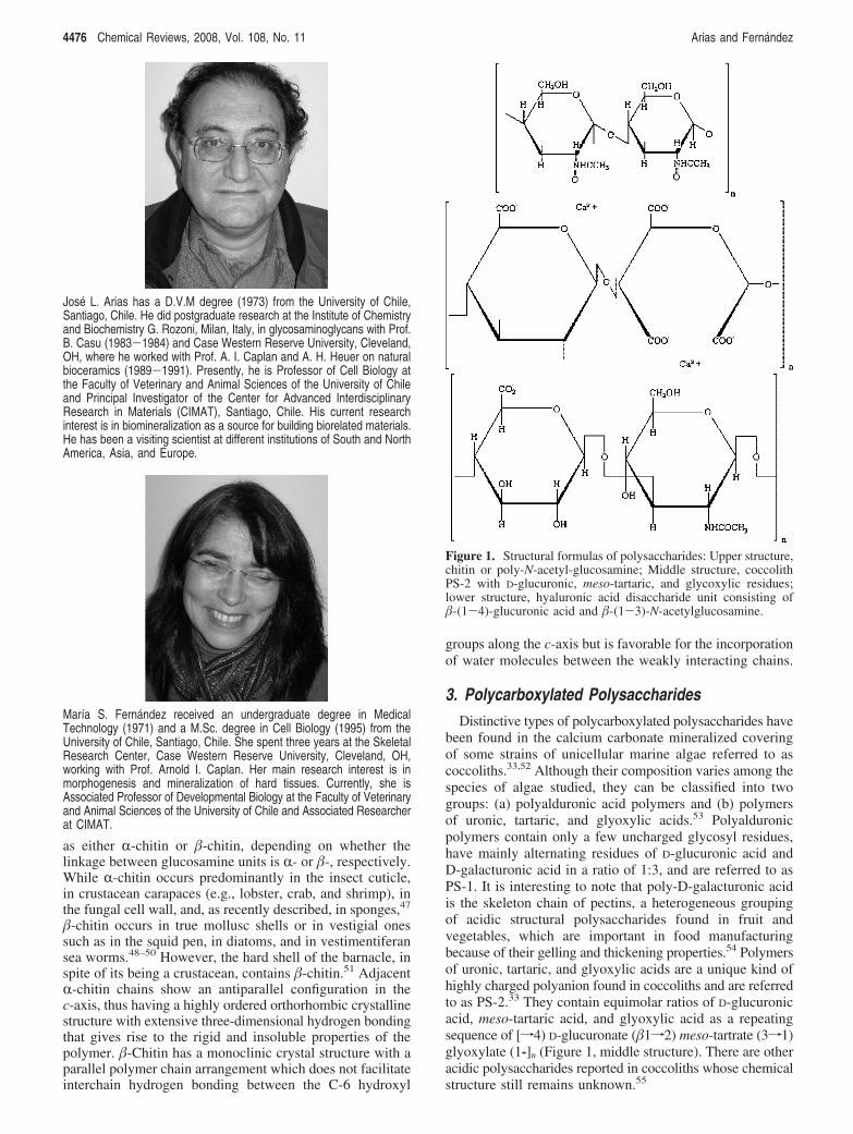
as either R-chitin or �-chitin, depending on whether thelinkage between glucosamine units is R- or �-, respectively.While R-chitin occurs predominantly in the insect cuticle,in crustacean carapaces (e.g., lobster, crab, and shrimp), inthe fungal cell wall, and, as recently described, in sponges,47
�-chitin occurs in true mollusc shells or in vestigial onessuch as in the squid pen, in diatoms, and in vestimentiferansea worms.48–50 However, the hard shell of the barnacle, inspite of its being a crustacean, contains �-chitin.51 AdjacentR-chitin chains show an antiparallel configuration in thec-axis, thus having a highly ordered orthorhombic crystallinestructure with extensive three-dimensional hydrogen bondingthat gives rise to the rigid and insoluble properties of thepolymer. �-Chitin has a monoclinic crystal structure with aparallel polymer chain arrangement which does not facilitateinterchain hydrogen bonding between the C-6 hydroxyl
groups along the c-axis but is favorable for the incorporationof water molecules between the weakly interacting chains.
3. Polycarboxylated PolysaccharidesDistinctive types of polycarboxylated polysaccharides have
been found in the calcium carbonate mineralized coveringof some strains of unicellular marine algae referred to ascoccoliths.33,52 Although their composition varies among thespecies of algae studied, they can be classified into twogroups: (a) polyalduronic acid polymers and (b) polymersof uronic, tartaric, and glyoxylic acids.53 Polyalduronicpolymers contain only a few uncharged glycosyl residues,have mainly alternating residues of D-glucuronic acid andD-galacturonic acid in a ratio of 1:3, and are referred to asPS-1. It is interesting to note that poly-D-galacturonic acidis the skeleton chain of pectins, a heterogeneous groupingof acidic structural polysaccharides found in fruit andvegetables, which are important in food manufacturingbecause of their gelling and thickening properties.54 Polymersof uronic, tartaric, and glyoxylic acids are a unique kind ofhighly charged polyanion found in coccoliths and are referredto as PS-2.33 They contain equimolar ratios of D-glucuronicacid, meso-tartaric acid, and glyoxylic acid as a repeatingsequence of [f4) D-glucuronate (�1f2) meso-tartrate (3f1)glyoxylate (1-]n (Figure 1, middle structure). There are otheracidic polysaccharides reported in coccoliths whose chemicalstructure still remains unknown.55
José L. Arias has a D.V.M degree (1973) from the University of Chile,Santiago, Chile. He did postgraduate research at the Institute of Chemistryand Biochemistry G. Rozoni, Milan, Italy, in glycosaminoglycans with Prof.B. Casu (1983-1984) and Case Western Reserve University, Cleveland,OH, where he worked with Prof. A. I. Caplan and A. H. Heuer on naturalbioceramics (1989-1991). Presently, he is Professor of Cell Biology atthe Faculty of Veterinary and Animal Sciences of the University of Chileand Principal Investigator of the Center for Advanced InterdisciplinaryResearch in Materials (CIMAT), Santiago, Chile. His current researchinterest is in biomineralization as a source for building biorelated materials.He has been a visiting scientist at different institutions of South and NorthAmerica, Asia, and Europe.
María S. Fernandez received an undergraduate degree in MedicalTechnology (1971) and a M.Sc. degree in Cell Biology (1995) from theUniversity of Chile, Santiago, Chile. She spent three years at the SkeletalResearch Center, Case Western Reserve University, Cleveland, OH,working with Prof. Arnold I. Caplan. Her main research interest is inmorphogenesis and mineralization of hard tissues. Currently, she isAssociated Professor of Developmental Biology at the Faculty of Veterinaryand Animal Sciences of the University of Chile and Associated Researcherat CIMAT.
Figure 1. Structural formulas of polysaccharides: Upper structure,chitin or poly-N-acetyl-glucosamine; Middle structure, coccolithPS-2 with D-glucuronic, meso-tartaric, and glycoxylic residues;lower structure, hyaluronic acid disaccharide unit consisting of�-(1-4)-glucuronic acid and �-(1-3)-N-acetylglucosamine.
4476 Chemical Reviews, 2008, Vol. 108, No. 11 Arias and Fernandez
4. Sulfated PolysaccharidesAlthough there are several tyrosine-sulfated proteins in
eukaryotes, whose functions are not well established,56 themain occurrence of sulfate groups are in a particular kind ofglycoconjugate called proteoglycans (formerly acid muco-polysaccharides). Proteoglycans consist of a protein core towhich highly negatively charged linear polysaccharide sidechains of variable length, called sulfated glycosaminoglycans,are covalently attached. Glycosaminoglycans (GAG) arealternating copolymers of a hexosamine and either a galac-tose or an alduronic acid. Individual GAGs differ from eachother by the type of hexosamine or alduronic acid (orhexose), the position and configuration of the glycosidiclinkages, and the degree and pattern of sulfation.57–61 Whilehyaluronic acid or hyaluronan (HA) is a nonsulfated GAG(Figure 1, lower structure), the other types of GAGs aresulfated to a variable extent and form part of proteoglycans.GAGs are huge information-dense macromolecules due tothe fact that modifications of sugar residues create anenormous molecular diversity.62 If an octasaccharide unitallows more than 106 theoretical combinations, a 50-200-disaccharide unit represents almost infinite combinatorialpossibilities.63,64 The most common sites of sulfation are atO-4, O-6, or N- of the hexosamine or at O-2 of the alduronicacid. While the alduronic acid moiety can be D-uronic orL-iduronic acid, the hexosamine can be 2-amino-2-deoxy-D-glucose or 2-amino-2-deoxy-D-galactose, and the hexoseis always galactose. The main differences among GAGs areas follows: (i) The uronic acid is D-glucuronic acid for HAand chondroitin 4- and 6-sulfate (C4S and C6S, respectively)(Figure 2, upper and middle structure), L-iduronic acid fordermatan sulfate (DS) (Figure 2, lower structure) and mainlyfor heparin (Hep) (with D-glucuronic acid as a minorconstituent) (Figure 3, upper structure), and preponderantlyD-glucuronic acid in heparan sulfate (HS) (with L-iduronicacid as a minor constituent) (Figure 3, middle structure);keratan sulfate (KS) is atypical, having D-galactose insteadof alduronic acid residues (Figure 3, lower structure). (ii)The hexosamine is 2-amino-2-deoxy-D-glucose for HA, KS,HS, and Hep, and 2-amino-2-deoxy-D-galactose for C4S,C6S, and DS. (iii) D-Glucuronic acid is �-(1-3)-linked tothe hexosamine in HA, C4S, and C6S and �-(1-4)-linkedin HS. L-Iduronic acid is linked R-(1-3) in DS and R-(1-4)in Hep. (iv) The hexosamine residue is N-acetylated in HA,KS, DS, C4S, and C6S and (partially) in HS and N-sulfatedin Hep and (partially) in HS. (v) L-Iduronic acid is 2-O-sulfated in Hep. Heparin is the most polyanionic polysac-charide known.
Biosynthesis of a proteoglycan starts with the formationof a polypeptide core composed of variable amino acids butalways having serine residues. Through the action of theenzyme D-xylosyltransferase, D-xylosyl groups are O-linkedto most of the hydroxyl groups of the L-serine of the coreprotein. Other glycosyltransferases then sequentially add tworesidues of D-galactose and one of D-glucuronic acid to eachD-xylosyl group. This tetrasaccharide segment constitutesthe protein core linkage region. Through the action ofappropriate transferases, the GAG chains grow from thesesegments by alternate addition of 2-acetamido-2-deoxy-D-hexose and D-glucuronic acid or galactose. Depending onthe GAG, these disaccharide units are then modified by aseries of enzymes by sequentially performing N-deacetylationand N-sulfation of the hexose, C-5 epimerization of D-glucuronic acid, and sulfation.57 However, KS-containing
proteoglycans are typically attached to core proteins througheither a mucin-type oligosaccharide to serine or threonine(O-linked KS) or a mannose-containing serum-type oligosac-charide to asparagine (N-linked KS).60
Depending on their GAG structure, core protein aminoacid sequence, polymer conformation, and tissue distributionand function, proteoglycans have received different names,such as aggrecan, versican, and perlecan. Therefore, themodification of the polysaccharide residues is not the onlysource of diversity but also the primary structure of the coreprotein. Due to their diversity, grouping of proteoglycanshas been a difficult task, but eventually these molecules havebeen classified into several families. One family contains thelecticans or hyalectans, of which the core protein has anN-terminal globular domain that interacts with HA and alsohas a C-terminal selectin domain. The GAG chains consistmostly of C4S, C6S, and KS. The characteristic cartilageproteoglycan, aggrecan, belongs to this group. Another familyconsists of those proteoglycans having leucine-rich repeatsequences in their core protein, thus referred to as SLRPs(small leucine-rich proteoglycans). Their GAG chains aremostly C4S, C6S, DS, and KS, and the main membersinclude decorin, biglycan, fibromodulin, and keratocan. Ingeneral, SLRPs promote protein-protein interactions, for
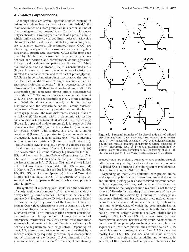
Figure 2. Structural formulas of the disaccharide unit of sulfatedglycosaminoglycans: Upper structure, chondroitin 4-sulfate consist-ing of �-(1-4)-glucuronic acid and �-(1-3)-N-acetylgalactosamine-4-O-sulfate; middle structure, chondroitin 6-sulfate consisting of�-(1-4)-glucuronic acid �-(1-3)-N-acetylgalactosamine-6-O-sulfate; lower structure, dermatan sulfate consisting of �-(1-4)-iduronic acid and �-(1-3)-N-acetylgalactosamine-4-O-sulfate.
Polysaccharides and Proteoglycans in Biomineralization Chemical Reviews, 2008, Vol. 108, No. 11 4477
example organizing collagen networks. A third group is thefamily of HS-rich proteoglycans, such as perlecan, syndecan,and agrin, which are generally associated with or form partof cellular membranes and not only function in cell adhesionto the extracellular matrix proteins but also as receptors ofsignaling for cell migration, proliferation, and differentiation.Other groups include the so-called “part-time proteoglycans”,whose core proteins do not always contain attached GAGchains, and also the spongicans. These proteoglycans functionin signal transduction processes and self-nonself recognitionand in primitive tissue formation in sponges.64–66 Becauseof their long polyanionic GAG chains, proteoglycans providehighly hydrated spaces outside of cells, forming gels ofvarying pore size and charge density, and serving as selectivesieves to regulate ions, molecules, and cell traffic. Due totheir carboxylate and sulfate groups, they show a high cation
affinity. In spite of that, not all proteoglycans are involvedin biomineralization, as described below.
Unusual sulfated polysaccharides have been found incoccoliths.33,55,67 One of these is a sulfated polysaccharidecomposed of galacturonic, glucuronic, and iduronic acids ina molar ratio of 2.4:1.1:1.0, respectively.67 A second one ispredominantly O-sulfated poly-D-galacturonic acid.67 A thirdone, referred to as APS-A92, consists of (1f3)-linkedD-mannosyl residues with sulfate groups and has abundantside chains, in which two or three D-galacturonic acidresidues occur.68 The last one is PS-3, a galacturonomannanwith high levels of rhamnose and xylose residues and asignificant amount of sulfate ester groups.33 There is noinformation concerning the linearity or branching structureof these polymers. Although these are sulfated polysaccha-rides, they are not glycosaminoglycans and have not beenreported to be attached to a protein core.
5. Polysaccharides in BiomineralizationAlthough chitin occurs in many calcium carbonate-based
biominerals, its role is almost passive. In fact, is has beendemonstrated that neither chitin nor chitosan per se modifyin Vitro calcium carbonate nucleation and growth.69,70
However, in biominerals chitin occurs not by itself butconstitutes an insoluble two-dimensional scaffolding or three-dimensional compartment wherein chitin-associated polya-nionic polysaccharides or proteins control crystallization ina confined space.16,71,72
For some polycarboxylated polysaccharides in coccoliths,the mechanism of calcium-polymer interaction could be insome sense similar to that acting in pectin. That is, carboxy-late groups cooperate together in sequestering the boundwater away from the calcium ions to form the salt links andthereby increase the gel strength.73 It has been demonstratedthat polyanionic polysaccharides accompany calcite crystalformation from nucleation to growth and also regulate crystalmorphology by enhancing precipitation of calcium carbonateions on the acute crystal face.74–76 While PS-2 facilitatescrystal nucleation, since mutants which do not produce thispolyanion produce fewer crystals,77 the sulfated polysac-charide PS-3 seems to control calcite growth and morphol-ogy.78
The first indication about the possible role of proteoglycansin calcium carbonate-based biominerals came from theoccurrence of sulfated mucopolysaccharides (presently namedGAGS) in the calcifying granules formed on glass coverslipsinserted either between the shell and the shell-forming cellsof the mantle or at the initial site of shell regeneration.79,80
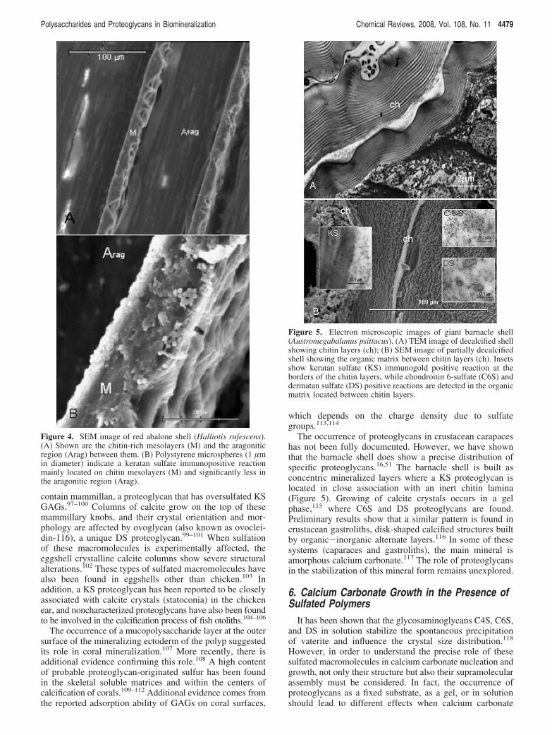
Additional evidence comes from studies in which molluskshells were decalcified in solutions which fix glycoproteins.In these studies, nucleation sites showed calcium-bindingmaterial containing sulfur, acid mucopolysaccharides, orGAGs.81–92 In fact, it has been demonstrated that sulfate isan important component of the intracrystalline organicmatrix. 93 Although it has been shown that mantle cellsproduce proteoglycans, their involvement in shell formationhas not been well established.94 An immunoultrastructurallocalization of keratan sulfate in the abalone shell is shownin Figure 4.
Additional evidence comes from eggshells. These arenatural biocomposites containing organic (5%) and inorganic(calcite) components (95%).14,95,96 Eggshell mineralizationstarts with the nucleation of calcite crystals on randomlydeposited discrete organic aggregations (mammillae), which
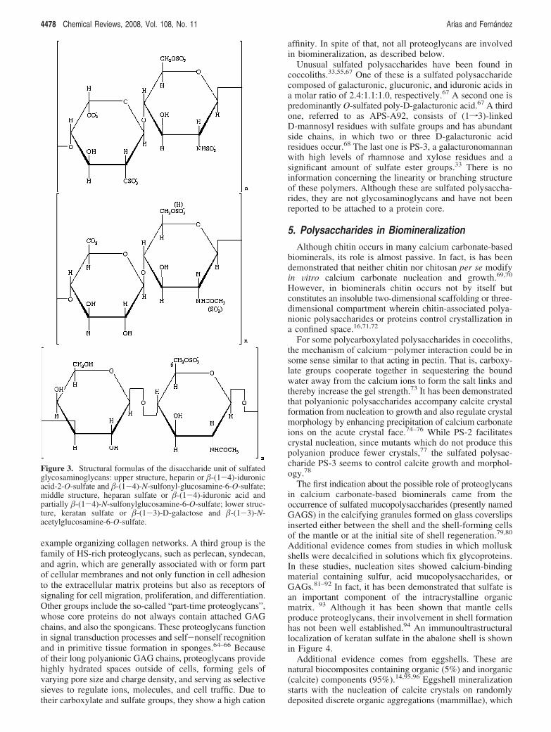
Figure 3. Structural formulas of the disaccharide unit of sulfatedglycosaminoglycans: upper structure, heparin or �-(1-4)-iduronicacid-2-O-sulfate and �-(1-4)-N-sulfonyl-glucosamine-6-O-sulfate;middle structure, heparan sulfate or �-(1-4)-iduronic acid andpartially �-(1-4)-N-sulfonylglucosamine-6-O-sulfate; lower struc-ture, keratan sulfate or �-(1-3)-D-galactose and �-(1-3)-N-acetylglucosamine-6-O-sulfate.
4478 Chemical Reviews, 2008, Vol. 108, No. 11 Arias and Fernandez
contain mammillan, a proteoglycan that has oversulfated KSGAGs.97–100 Columns of calcite grow on the top of thesemammillary knobs, and their crystal orientation and mor-phology are affected by ovoglycan (also known as ovoclei-din-116), a unique DS proteoglycan.99–101 When sulfationof these macromolecules is experimentally affected, theeggshell crystalline calcite columns show severe structuralalterations.102 These types of sulfated macromolecules havealso been found in eggshells other than chicken.103 Inaddition, a KS proteoglycan has been reported to be closelyassociated with calcite crystals (statoconia) in the chickenear, and noncharacterized proteoglycans have also been foundto be involved in the calcification process of fish otoliths.104–106
The occurrence of a mucopolysaccharide layer at the outersurface of the mineralizing ectoderm of the polyp suggestedits role in coral mineralization.107 More recently, there isadditional evidence confirming this role.108 A high contentof probable proteoglycan-originated sulfur has been foundin the skeletal soluble matrices and within the centers ofcalcification of corals.109–112 Additional evidence comes fromthe reported adsorption ability of GAGs on coral surfaces,
which depends on the charge density due to sulfategroups.113,114
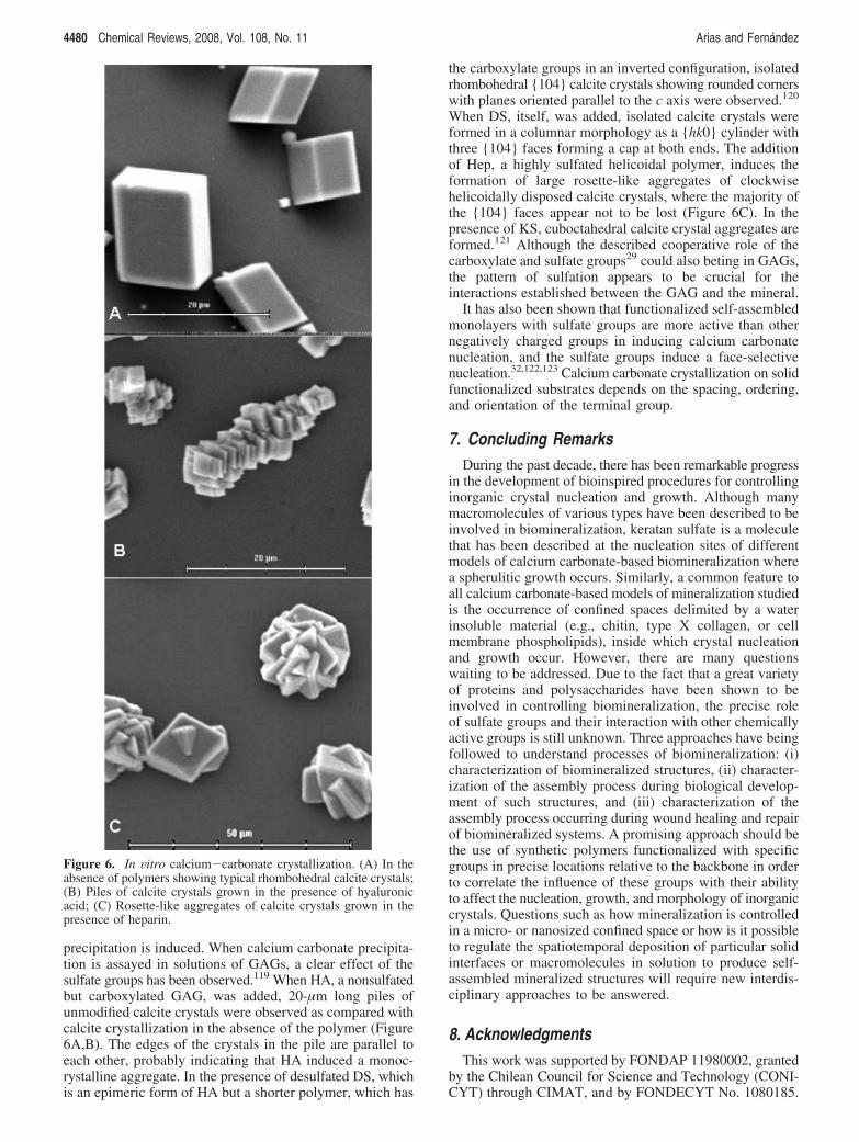
The occurrence of proteoglycans in crustacean carapaceshas not been fully documented. However, we have shownthat the barnacle shell does show a precise distribution ofspecific proteoglycans.16,51 The barnacle shell is built asconcentric mineralized layers where a KS proteoglycan islocated in close association with an inert chitin lamina(Figure 5). Growing of calcite crystals occurs in a gelphase,115 where C6S and DS proteoglycans are found.Preliminary results show that a similar pattern is found incrustacean gastroliths, disk-shaped calcified structures builtby organic-inorganic alternate layers.116 In some of thesesystems (caparaces and gastroliths), the main mineral isamorphous calcium carbonate.117 The role of proteoglycansin the stabilization of this mineral form remains unexplored.
6. Calcium Carbonate Growth in the Presence ofSulfated Polymers
It has been shown that the glycosaminoglycans C4S, C6S,and DS in solution stabilize the spontaneous precipitationof vaterite and influence the crystal size distribution.118
However, in order to understand the precise role of thesesulfated macromolecules in calcium carbonate nucleation andgrowth, not only their structure but also their supramolecularassembly must be considered. In fact, the occurrence ofproteoglycans as a fixed substrate, as a gel, or in solutionshould lead to different effects when calcium carbonate
Figure 4. SEM image of red abalone shell (Halliotis rufescens).(A) Shown are the chitin-rich mesolayers (M) and the aragoniticregion (Arag) between them. (B) Polystyrene microspheres (1 µmin diameter) indicate a keratan sulfate immunopositive reactionmainly located on chitin mesolayers (M) and significantly less inthe aragonitic region (Arag).
Figure 5. Electron microscopic images of giant barnacle shell(Austromegabalanus psittacus). (A) TEM image of decalcified shellshowing chitin layers (ch); (B) SEM image of partially decalcifiedshell showing the organic matrix between chitin layers (ch). Insetsshow keratan sulfate (KS) immunogold positive reaction at theborders of the chitin layers, while chondroitin 6-sulfate (C6S) anddermatan sulfate (DS) positive reactions are detected in the organicmatrix located between chitin layers.
Polysaccharides and Proteoglycans in Biomineralization Chemical Reviews, 2008, Vol. 108, No. 11 4479
precipitation is induced. When calcium carbonate precipita-tion is assayed in solutions of GAGs, a clear effect of thesulfate groups has been observed.119 When HA, a nonsulfatedbut carboxylated GAG, was added, 20-µm long piles ofunmodified calcite crystals were observed as compared withcalcite crystallization in the absence of the polymer (Figure6A,B). The edges of the crystals in the pile are parallel toeach other, probably indicating that HA induced a monoc-rystalline aggregate. In the presence of desulfated DS, whichis an epimeric form of HA but a shorter polymer, which has
the carboxylate groups in an inverted configuration, isolatedrhombohedral {104} calcite crystals showing rounded cornerswith planes oriented parallel to the c axis were observed.120
When DS, itself, was added, isolated calcite crystals wereformed in a columnar morphology as a {hk0} cylinder withthree {104} faces forming a cap at both ends. The additionof Hep, a highly sulfated helicoidal polymer, induces theformation of large rosette-like aggregates of clockwisehelicoidally disposed calcite crystals, where the majority ofthe {104} faces appear not to be lost (Figure 6C). In thepresence of KS, cuboctahedral calcite crystal aggregates areformed.121 Although the described cooperative role of thecarboxylate and sulfate groups29 could also beting in GAGs,the pattern of sulfation appears to be crucial for theinteractions established between the GAG and the mineral.
It has also been shown that functionalized self-assembledmonolayers with sulfate groups are more active than othernegatively charged groups in inducing calcium carbonatenucleation, and the sulfate groups induce a face-selectivenucleation.32,122,123 Calcium carbonate crystallization on solidfunctionalized substrates depends on the spacing, ordering,and orientation of the terminal group.
7. Concluding RemarksDuring the past decade, there has been remarkable progress
in the development of bioinspired procedures for controllinginorganic crystal nucleation and growth. Although manymacromolecules of various types have been described to beinvolved in biomineralization, keratan sulfate is a moleculethat has been described at the nucleation sites of differentmodels of calcium carbonate-based biomineralization wherea spherulitic growth occurs. Similarly, a common feature toall calcium carbonate-based models of mineralization studiedis the occurrence of confined spaces delimited by a waterinsoluble material (e.g., chitin, type X collagen, or cellmembrane phospholipids), inside which crystal nucleationand growth occur. However, there are many questionswaiting to be addressed. Due to the fact that a great varietyof proteins and polysaccharides have been shown to beinvolved in controlling biomineralization, the precise roleof sulfate groups and their interaction with other chemicallyactive groups is still unknown. Three approaches have beingfollowed to understand processes of biomineralization: (i)characterization of biomineralized structures, (ii) character-ization of the assembly process during biological develop-ment of such structures, and (iii) characterization of theassembly process occurring during wound healing and repairof biomineralized systems. A promising approach should bethe use of synthetic polymers functionalized with specificgroups in precise locations relative to the backbone in orderto correlate the influence of these groups with their abilityto affect the nucleation, growth, and morphology of inorganiccrystals. Questions such as how mineralization is controlledin a micro- or nanosized confined space or how is it possibleto regulate the spatiotemporal deposition of particular solidinterfaces or macromolecules in solution to produce self-assembled mineralized structures will require new interdis-ciplinary approaches to be answered.
8. AcknowledgmentsThis work was supported by FONDAP 11980002, granted
by the Chilean Council for Science and Technology (CONI-CYT) through CIMAT, and by FONDECYT No. 1080185.
Figure 6. In Vitro calcium-carbonate crystallization. (A) In theabsence of polymers showing typical rhombohedral calcite crystals;(B) Piles of calcite crystals grown in the presence of hyaluronicacid; (C) Rosette-like aggregates of calcite crystals grown in thepresence of heparin.
4480 Chemical Reviews, 2008, Vol. 108, No. 11 Arias and Fernandez

9. References(1) Lowenstam, H. A.; Weiner, S. On Biomineralization; Oxford
University Press: Oxford, 1989; p 324.(2) Mann, S.; Webb, J.; Williams, R. J. P. Biomineralization; VCH:
Weinheim, 1989; p 490.(3) Simkiss, K.; Wilbur, K. M. Biomineralization; Academic Press: San
Diego, CA,1989, p 337.(4) Baeuerlein, E. Biomineralization; Wiley-VCH: Weinheim, 2000, p
294.(5) Mann, S. Biomineralization; Oxford University Press: Oxford, 2001,
p 198.(6) Baeuerlein, E. Handbook of Biomineralization: Biological Aspects
and Structure Formation; Wiley-VCH: Weinheim, 2007; p 440.(7) Arias, J. L.; Fernandez, M. S. Biomineralization: from Paleontology
to Materials Science; Editorial Universitaria: Santiago, Chile, 2007;p 534.
(8) Heuer, A. H.; Fink, D. J.; Laraia, V. J.; Arias, J. L.; Calvert, P. D.;Kendall, K.; Messing, G. L.; Blackwell, J.; Rieke, P. C.; Thompson,D. H.; Wheeler, A. P.; Veis, A.; Caplan, A. I. Science 1992, 255,1098.
(9) Mann, S. Angew. Chem., Int. Ed. 2000, 39, 3392.(10) Dujardin, E.; Mann, S. AdV. Mater. 2002, 14, 775.(11) Belcher, A. M.; Wu, X. H.; Christensen, R. J.; Hasma, P. K.; Stucky,
G. D.; Morse, D. E. Nature 1996, 381, 56.(12) Falini, G.; Albeck, S.; Weiner, S.; Addadi, L. Science 1996, 271,
67.(13) Weiner, S.; Addadi, L. J. Mater. Chem. 1997, 7, 689.(14) Nys, Y.; Hincke, M. T.; Arias, J. L.; Garcıa-Ruiz, J. M.; Solomon,
S. E. Poult. AVian Biol. ReV. 1999, 10, 142.(15) Smith, B. L.; Schaffer, T. E.; Viani, M.; Thompson, J. B.; Frederick,
N. A.; Kindt, J.; Belcher, A. M.; Stucky, G. D.; Morse, D. E.;Hansma, P. K. Nature 1999, 399, 761.
(16) Arias, J. L.; Fernandez, M. S. Mater. Charact. 2003, 50, 189.(17) Nagasawa, H. Thalassas 2004, 20, 15.(18) Samata, T. Thalassas 2004, 20, 25.(19) Michenfelder, M.; Fu, G.; Lawrence, C.; Weaver, J. C.; Wustman,
B. A.; Taranto, L.; Evans, J. S.; Morse, D. E. Biopolymers 2003, 70,522.
(20) Weiner, S.; Hood, L. Science 1975, 190, 987.(21) Weiner, S. Calcif. Tissue Int. 1979, 29, 163.(22) Weiner, S. CRC Crit. ReV. Biochem. 1986, 20, 365.(23) De Yoreo, J. J.; Vekilov, P. G. ReV. Mineral. Geochem. 2003, 54,
57.(24) Marsh, M. E.; Sass, R. L. Biochemistry 1984, 23, 1448.(25) Marsh, M. E. J. Exp. Zool. 1986, 239, 207.(26) Belcher, A. M.; Gooch, E. E. In Biomineralization; Baeuerlein, E.
Ed.; Wiley-VCH: Weinheim, 2000; p 221.(27) Sarikaya, M.; Tamerler, C.; Jen, A. K.-Y.; Schulten, K. F.; Baneyx,
F. Nat. Mater. 2003, 2, 577.(28) Yang, W.; Jones, L. M.; Isley, L.; Ye, Y.; Lee, H. W.; Wilkins, A.;
Liu, Z.; Hollinga, H. W.; Malchow, R.; Ghazi, M.; Yang, J. J. J. Am.Chem. Soc. 2003, 125, 6165.
(29) Mao, C.; Solis, D. J.; Reiss, B. D.; Kottman, S. T.; Sweeney, R. Y.;Hayhurst, A.; Georgiou, G.; Iverson, B.; Belcher, A. M. Science 2004,303, 213.
(30) Mann, S. R.; Heywood, B. R.; Rjam, S.; Birchall, J. D. Nature 1988,334, 692.
(31) Xu, G.; Yao, N.; Aksay, I. A.; Groves, J. T. J. Am. Chem. Soc. 1998,120, 11977.
(32) Aizenberg, J.; Black, A. J.; Whitesides, G. M. J. Am. Chem. Soc.1999, 121, 4500.
(33) Marsh, M. E. In Biomineralization; Baeuerlein, E. Ed.; Wiley-VCH:Weinheim, 2000; p 251.
(34) D’Souza, S. M.; Alexander, C.; Whitcombe, M. J.; Waller, A. M.;Vulfson, E. N. Polym. Int. 2001, 50, 429.
(35) Naka, K.; Chujo, Y. Chem. Mater. 2001, 13, 3245.(36) Colfen, H.; Qi, L. Chem.sEur. J. 2001, 7, 106.(37) Kato, T.; Sugawara, A. N.; Hosoda, N. AdV. Mater. 2002, 14, 869.(38) Ajikumar, P. K.; Lakshminarayanan, R.; Ong, B. T.; Valiyaveetil,
S.; Kini, R. M. Biomacromolecules 2003, 4, 1321.(39) Colfen, H. Curr. Opin. Colloid Interface Sci. 2003, 8, 23.(40) Meldrum, F. C. Int. Mater. ReV. 2003, 48, 187.(41) Estroff, L. A.; Addadi, L.; Weiner, S.; Hamilton, A. D. Org. Biomol.
Chem. 2004, 2, 137.(42) Estroff, L. A.; Incarvito, C. D.; Hamilton, A. D. J. Am. Chem. Soc.
2004, 126, 2.(43) Grassmann, O.; Lobmann, P. Biomaterials 2004, 25, 277.(44) Abolins-Krogis, A. Acta Zool. 1958, 39, 19.(45) Benzerara, K.; Menguy, N.; Lopez-Garcia, P.; Yoon, T. H.; Ka-
zmierczak, J.; Tyliszczak, T.; Guyot, F.; Brown, G. E. Proc. Natl.Acad. Sci. U.S.A. 2006, 103, 9440.
(46) Khor, E. Chitin: Fulfilling a Biomaterials Promise; Elsevier: Oxford,U.K., 2001; p 136.
(47) Ehrlich, H.; Maldonado, M.; Spindler, K.; Eckert, C.; Hanke, T.;Born, R.; Goebel, C.; Simon, P.; Heinemann, S.; Worch, H. J. Exp.Zool. B 2007, 308, 347.
(48) Weiner, S.; Traub, W. FEBS Lett. 1980, 111, 311.(49) McLachlan, J.; McInnes, A. G.; Falk, M. Can. J. Bot. 1965, 43, 707.(50) Gaill, F.; Persson, J.; Sugiyama, J.; Vuong, R.; Chanzy, H. J. Struct.
Biol. 1992, 109, 116.(51) Fernandez, M. S.; Vergara, I.; Oyarzun, A.; Arias, J. I.; Rodriguez,
R.; Wiff, J. P.; Fuenzalida, V. M.; Arias, J. L. Mater. Res. Soc. Symp.Proc. 2002, 724, 3.
(52) van der Wal, P.; de Jong, E. W.; Westbroek, P.; de Brujin, W. C.;Mulder-Stapel, A. A. J. Ultrastruct. Biol. 1983, 85, 139.
(53) Marsh, M. E.; Chang, D.-K.; King, G. G. J. Biol. Chem. 1992, 267,20507.
(54) Clark, A. H.; Ross-Murphy, S. B. AdV. Polym. Sci. 1987, 83, 57.(55) Ozaki, N.; Okazaki, M.; Kogure, T.; Sakuda, S.; Nagasawa, H.
Thalassas 2004, 20, 59.(56) Kehoe, J. W.; Bertozzi, C. R. Chem. Biol. 2000, 7, 57.(57) Casu, B. AdV. Carbohydr. Chem. Biochem. 1985, 43, 51.(58) Iozzo, R. V. Annu. ReV. Biochem. 1998, 67, 609.(59) Sugahara, K.; Kitagawa, H. Curr. Opin. Struct. Biol. 2000, 10, 518.(60) Huckerby, T. N. Prog. Nucl. Magn. Reson. Spectrosc. 2002, 40, 35.(61) Bulow, H. E.; Hobert, O. Annu. ReV. Cell DeV. Biol. 2006, 22, 375.(62) Turnbull, J.; Powell, A.; Guimond, S. Trends Cell Biol. 2001, 11,
75.(63) Esko, J. D.; Lindahl, U. J. Clin. InVest. 2001, 108, 169.(64) Esko, J. D.; Selleck, S. B. Annu. ReV. Biochem. 2002, 71, 435.(65) Bosman, F. T.; Stamenkovic, I. J. Pathol. 2003, 200, 423.(66) Fernandez-Busquets, X.; Burger, M. M. Cell. Mol. Life Sci. 2003,
60, 88.(67) de Vrind-de Jong, E. W.; Borman, A. H.; Thierry, A. H.; Westbroek,
P.; Gruter, M.; Kamerling, J. P. In Biomineralization in Lower Plantsand Animals; Leadbeater, B. S. C., Riding, R. Eds.; Clarendon Press:Oxford, U.K., 1986; pp 205-217.
(68) Fichtinger-Schepman, A. M. J.; Kamerling, J. P.; Versluis, C.;Vliegenthart, J. F. G. Carbohydr. Res. 1981, 93, 105.
(69) Zhang, S.; Gonsalves, K. E. Langmuir 1998, 14, 6761.(70) Neira-Carrillo, A.; Yazdani-Pedram, M.; Retuert, J.; Diaz-Dosque,
M.; Gallois, S.; Arias, J. L. J. Colloid Interface Sci. 2005, 286, 134.(71) Levi-Kalisman, Y.; Falini, G.; Addadi, L.; Weiner, S. J. Struct. Biol.
2001, 135, 8.(72) Cartwright, J. H. E.; Checa, A. G. J. R. Soc. Interface 2007, 4, 491.(73) Lootens, D.; Capel, F.; Durand, D.; Nicolai, T.; Boulenguer, P.;
Langendorff, V. Food Hydrocolloid 2003, 17, 237.(74) Marsh, M. E. Protoplasma 1994, 177, 108.(75) Marsh, M. E. Protoplasma 1996, 190, 181.(76) Henriksen, K.; Stipp, S. L. S.; Young, J. R.; Marsh, M. E. Am.
Mineral. 2004, 89, 1709.(77) Marsh, M. E.; Dickinson, D. P. Protoplasma 1997, 199, 9.(78) Marsh, M. E.; Ridall, A. L.; Azadi, P.; Duke, P. J. J. Struct. Biol.
2002, 139, 39.(79) Wada, K. Bull. Jpn. Soc. Sci. Fish. 1964, 4, 993.(80) Saleuddin, A. S. M.; Chan, W. Can. J. Zool. 1969, 47, 1107.(81) Simkiss, K. Comp. Biochem. Physiol. 1965, 16, 427.(82) Crenshaw, M. A. Biomineralization 1972, 6, 6.(83) Crenshaw, M. A.; Ristedt, H. Biomineralization 1975, 8, 1.(84) Itawa, K. J. Fac. Sci. Hokkaido UniV. (Ser. 4) 1975, 17, 173.(85) Greenfield, E. M.; Wilson, D. C.; Crenshaw, M. A. Am. Zool. 1984,
24, 925.(86) Worms, D.; Weiner, S. J. Exp. Zool. 1986, 237, 11.(87) Marxen, J. C.; Becker, W. Comp. Biochem. Physiol. 1997, 118B,
23.(88) Marxen, J. C.; Hammer, M.; Gehrke, T.; Becker, W. Biol. Bull. 1998,
194, 231.(89) Marxen, J. C.; Becker, W. Comp. Biochem. Physiol. 2000, 127B,
235.(90) Pereira-Mouries, L.; Almeida, M.-J.; Ribeiro, C.; Peduzzi, J.;
Barthelemy, M.; Milet, C.; Lopez, E. Eur. J. Biochem. 2002, 269,4994.
(91) Nudelman, F.; Gotliv, B. A.; Addadi, L.; Weiner, S. J. Struct. Biol.2006, 153, 176.
(92) Guzman, N.; Ball, A. D.; Cuif, J. P.; Dauphin, Y.; Denis, A.; Ortlieb,L. Microsc. Microanal. 2007, 13, 397.
(93) Dauphin, Y.; Cuif, J.-P.; Doucet, J.; Salome, M.; Susin, J.; Williams,C. T. J. Struct. Biol. 2003, 142, 272.
(94) Poncet, J.-M.; Serpentini, A.; Thiebot, B.; Villers, C.; Bocquet, J.;Boucaud-Camou, E.; Lebel, J.-M. Mar. Biotechnol. 2000, 2, 387.
(95) Arias, J. L.; Fink, D. J.; Xiao, S.-Q.; Heuer, A. H.; Caplan, A. I. Int.ReV. Cytol. 1993, 145, 217.
(96) Arias, J. L.; Fernandez, M. S. World‘s Poult. Sci. J. 2001, 57, 349.
Polysaccharides and Proteoglycans in Biomineralization Chemical Reviews, 2008, Vol. 108, No. 11 4481
(97) Arias, J. L.; Carrino, D. A.; Fernandez, M. S.; Rodriguez, J. P.;Dennis, J. E.; Caplan, A. I. Arch. Biochem. Biophys. 1992, 298, 293.
(98) Fernandez, M. S.; Araya, M.; Arias, J. L. Matrix Biol. 1997, 16, 13.(99) Carrino, D. A.; Rodriguez, J. P.; Caplan, A. I. Connect. Tissue Res.
1997, 36, 175.(100) Hincke, M. T.; Gautron, J.; Tsang, C. P.; McKee, M. D.; Nys, Y.
J. Biol. Chem. 1999, 274, 32915.(101) Wu, T.-M.; Rodriguez, J. P.; Fink, D. J.; Carrino, D. A.; Blackwell,
J.; Caplan, A. I.; Heuer, A. H. Matrix Biol. 1994, 14, 507.(102) Fernandez, M. S. A.; Moya, A. L.; Lopez, L.; Arias, J. L. Matrix
Biol. 2001, 19, 793.(103) Panheleux, M.; Bain, M.; Fernandez, M. S.; Morales, I.; Gautron,
J.; Arias, J. L.; Solomon, S. E.; Hincke, M.; Nys, Y. Br. Poult. Sci.1999, 40, 240.
(104) Fermin, C. D.; Lovett, A. E.; Igarashi, M.; Dunner, K. Acta Anat.1990, 138, 75.
(105) Borelli, G.; Mayer-Gostan, N.; De Pontual, H.; Boeuf, G.; Payan, P.Calcif. Tissue Int. 2001, 69, 356.
(106) Borelli, G.; Mayer-Gostan, N.; Merle, P. L.; De Pontual, H.; Boeuf,G.; Allemand, D.; Payan, P. Calcif. Tissue Int. 2003, 72, 717.
(107) Goreau, T. F. Biol. Bull. 1959, 116, 59.(108) Goldberg, W. M. Tissue Cell 2001, 33, 376.(109) Constantz, B. R.; Weiner, S. J. Exp. Zool. 1988, 248, 253.(110) Allemand, D.; Cuif, J.-P.; Watabe, N.; Oishi, M.; Kawagushi, T. Bull.
Inst. Oceanogr. Monaco 1994, 14, 129.(111) Dauphin, Y. Int. J. Biol. Macromol. 2001, 28, 293.
(112) Cuif, J.-P. Y.; Dauphin, Y.; Doucet, J.; Salome, M.; Susini, J.Geochim. Cosmochim. Acta 2003, 67, 75.
(113) Volpi, N. Biomaterials 1999, 20, 1359.(114) Volpi, N. Biomaterials 2002, 23, 3015.(115) Rodrıguez-Navarro, A.; Cabral de Melo, C.; Batista, N.; Morimoto,
N.; Alvarez-Lloret, P.; Ortega-Huertas, M.; Fuenzalida, V. M.; Arias,J. I.; Wiff, J. P.; Arias, J. L. J. Struct. Biol. 2006, 156, 355.
(116) Luquet, G.; Fernandez, M. S.; Navarrete, M. J.; Arias, J. L.; Guichard,N.; Marie, B.; Marin, F. In Biomineralization, from Paleontology toMaterials Science; Arias, J. L.,. Fernandez, M. S., Eds.; EditorialUniversitaria: Santiago, Chile, 2007; pp 319-328.
(117) Addadi, L.; Raz, S.; Weiner, S. AdV. Mater. 2003, 15, 959.(118) Manoli, F.; Dalas, E. J. Cryst. Growth 2000, 217, 416.(119) Addadi, L.; Moradian, J.; Shay, J. E.; Maroudas, N. G.; Weiner, S.
Proc. Natl. Acad. Sci. U.S.A. 1987, 84, 2732.(120) Arias, J. I.; Jure, C.; Wiff, J. P.; Fernandez, M. S.; Wiff, J. P.;
Fuenzalida, V. M.; Arias, J. L. Mater. Res. Soc. Symp. Proc. 2002,711, 243.
(121) Arias, J. L.; Neira, A. C.; Arias, J. I.; Escobar, C.; Bodero, M.; David,M.; Fernandez, M. S. J. Mater. Chem. 2004, 14, 2154.
(122) Mann, S.; Heywood, B. R.; Rajam, S.; Birchall, J. D. Nature 1988,334, 692.
(123) Aizenberg, J.; Black, A. J.; Whitesides, G. M. Nature 1999, 398,495.
CR078269P
4482 Chemical Reviews, 2008, Vol. 108, No. 11 Arias and Fernandez