Polymyalgia Rheumatica and Giant Arthritis Cell Arteritis ... Rheumatica and Giant Cell Arteritis:...
10
Polymyalgia Rheumatica and Giant Cell Arteritis: From Etymology to a Clinical Understanding Terence W. Starz, MD Clinical Professor of Medicine University of Pittsburgh School of Medicine https://media.licdn.com/mpr/mpr/shrinknp_ 800_800/p/6/005/0b9/194/0dd8379.jpg Arthritis Sir William Osler, the "Father of Modern Medicine," once said, "When an arthritis patient walks in the front door, I feel like leaving by the back door." Goals • Review the clinical manifestations of polymyalgia rheumatica and giant cell arteritis and an evidence based approach to diagnosis, pathophysiology, and management • Consider if PMR and GCA are separate entities or are they related • Review factors associated with the development and expression of musculoskeletal disorders with age including: genetics, environment, regional body anatomy Polymyalgia Rheumatica • Poly- many (Greek) • Myo- one or more muscles (Greek) • Algia- painful condition (Greek) • Rheumatica- “rheuma” (Greek) Hippocrates, 4 th century BC, phlegm- one of the four primary humors • Polymyalgia rheumatica was coined by Barber in 1957. • Prominently affects the synovium of certain joints and bursae, not the muscles per se.
Transcript of Polymyalgia Rheumatica and Giant Arthritis Cell Arteritis ... Rheumatica and Giant Cell Arteritis:...

Polymyalgia Rheumatica and Giant
Cell Arteritis:
From Etymology to a Clinical
Understanding
Terence W. Starz, MD
Clinical Professor of Medicine
University of Pittsburgh
School of Medicine
https://media.licdn.com/mpr/mpr/shrinknp_
800_800/p/6/005/0b9/194/0dd8379.jpg
Arthritis
Sir William Osler, the "Father of Modern Medicine," once said,
"When an arthritis patient walks in the front door, I feel like leaving by the back door."
Goals • Review the clinical manifestations of
polymyalgia rheumatica and giant cell arteritis and an evidence based approach to diagnosis, pathophysiology, and management
• Consider if PMR and GCA are separate entities or are they related
• Review factors associated with the development and expression of musculoskeletal disorders with age including: genetics, environment, regional body anatomy
Polymyalgia Rheumatica
• Poly- many (Greek)
• Myo- one or more muscles (Greek)
• Algia- painful condition (Greek)
• Rheumatica- “rheuma” (Greek) Hippocrates, 4th century BC, phlegm- one of the four primary humors
• Polymyalgia rheumatica was coined by
Barber in 1957.
• Prominently affects the synovium of certain
joints and bursae, not the muscles per se.

PMR Epidemiology • US-the average annual incidence PMR is 52.5 cases per
100,000 persons aged 50 years and older. The prevalence is approximately 0.5-0.7%.
• Worldwide, the frequency varies by country. In Europe, the frequency decreases from north to south, with a high incidence in Scandinavia and a low incidence in Mediterranean countries. In Italy, for example, the incidence is 12.7 cases per 100,000 persons.
• Whites are affected more than other ethnic groups. PMR is only occasionally reported in African-American persons. PMR is twice as common in females.
• The incidence increases with advanced age. PMR rarely affects persons younger than 50 years. The median age at diagnosis is 72 years
-often abrupt
Polymyalgia Rheumatica
Regional musculoskeletal disorders
Lancet 2008;372;234–245
Shoulder of patient with isolated PMR- no signs or symptoms of GCA.
(A) Ultrasonography-fluid within the subacromial bursa (arrows) and surrounding the long biceps
tendon groove (arrowheads). (B) Axial T2 weighted MRI- subacromial and subdeltoid bursitis
(pentagon), joint effusion (arrow), and tenosynovitis of the long head of the biceps (arrowhead).
(C) Fluorodeoxyglucose PET- inflammatory fluorodeoxyglucose uptake in the shoulders (arrows).
Lancet 2008;372;234–245
Figure 4. Ultrasonography (A), MRI (B) of the hip and fluorodeoxyglucose PET (C) of patients with
isolated untreated PMR (A) Ultrasonography- the presence of fluid within the trochanteric bursa (surrounding white line and arrows). (B) An axial T2 weighted section- trochanteric bursitis (arrows) and
joint effusion (arrowhead). (C) Fluorodeoxyglucose PET- inflammatory fluorodeoxyglucose uptake in the
hips (arrows) and absence of vascular uptake.
Slower response
late onset RA
RF-

Rheumatoid arthritis
Overview of joints affected
Rheumatoid arthritis synovium
PMR: Etiology/Pathogenesis • The cause of PMR is unknown.
• PMR is closely linked to giant cell arteritis although it is
controversial whether GCA and PMR are two separate
diseases or part of the same spectrum of disease.
• One hypothesis is that in a genetically predisposed patient,
an environmental factor, possibly a virus, causes monocyte
activation, which helps determine the production of
cytokines that induce manifestations of PMR and GCA.
• PMR is associated with the HLA-DR4 haplotype. High level
of IL-6 is associated with increased disease activity.
• Although several infectious agents have been investigated
as possible triggers, results are inconclusive.
Polymyalgia
Rheumatica Giant cell
arteritis
Polymyalgia
Rheumatica Giant cell
arteritis
20-65%
?
PMR treatment
• Dramatically responsive to steroids
• Most response to Prednisone <20mg/day
(ACR guidelines 12.5-25 mg)
• Dose gradually tapered
• Tapering an art not science!
• Monitor for relapse, features of GCA, side-
effects of corticosteroids
• Clinical course
• Methotrexate in resistant cases
Giant Cell Arteritis
Medscape

Giant Cell Arteritis
Presenting symptoms
• Headache
• Scalp tenderness
• Thickened temporal arteries
• Jaw claudication
• Acute visual loss
• Weight loss, anorexia, fever, night sweats,
• Malaise, depression
19
1990 ACR criteria for the classification of giant cell arteritis
Autoimmunity 48-49 (2014) 73e75
Ann Intern Med. 2003;139(6):505-515
GCA- clinical features
• Many of the clinical features commonly found with
the disease are unhelpful in predicting the
likelihood of positive temporal artery biopsy results.
• Jaw claudication and diplopia substantially
increase the probability of positive biopsy results
(positive LRs = 4.2 and 3.4, respectively).
• No historical findings help rule out the diagnosis by
their absence.
Does This Patient Have Temporal Arteritis?
JAMA. 2002;287(1):92-101
GCA- physical exam findings • Among physical examination findings, synovitis makes the
diagnosis of TA less likely, while beaded, prominent, enlarged, and tender temporal arteries each increase the likelihood of positive biopsy.
• Beaded, prominent, or enlarged arteries confer the highest positive LRs of any clinical or laboratory feature and substantially increase the probability that a patient with suspected TA will have positive biopsy results.
• While these findings increase the chance of having TA, they are variably sensitive, from 16% (beaded temporal artery) to 65% (any temporal artery abnormality).
Does This Patient Have Temporal Arteritis?
JAMA. 2002;287(1):92-101
GCA- laboratory
• A normal ESR (LR = 0.2) or ESR less than
50 mm/h (LR = 0.35) each make positive
biopsy results unlikely, but setting the ESR
threshold at 100 mm/h is less efficient, as
patients with an ESR less than 100 mm/h
have an LR (0.8)
• Clinically suspected of disease, those with
an ESR greater than 100 mm/h have a
modestly increased likelihood of biopsy-
proven TA (LR = 1.9)
Does This Patient Have Temporal Arteritis?
JAMA. 2002;287(1):92-101
Conclusions:
• Review of clinical series of patients with suspected TA does not allow a determination of the predictive value of selected combinations of clinical and laboratory features.
• In addition, it is not possible to determine whether certain combinations of features would sufficiently increase the likelihood of disease that a clinician should treat presumptively for TA and not perform a biopsy at all.
• The morbidity of a prolonged course of corticosteroids, however, is such that most clinicians would favor confirmation of disease by biopsy even if
the clinical probability is high.
Does This Patient Have Temporal
Arteritis? JAMA. 2002;287(1):92-101
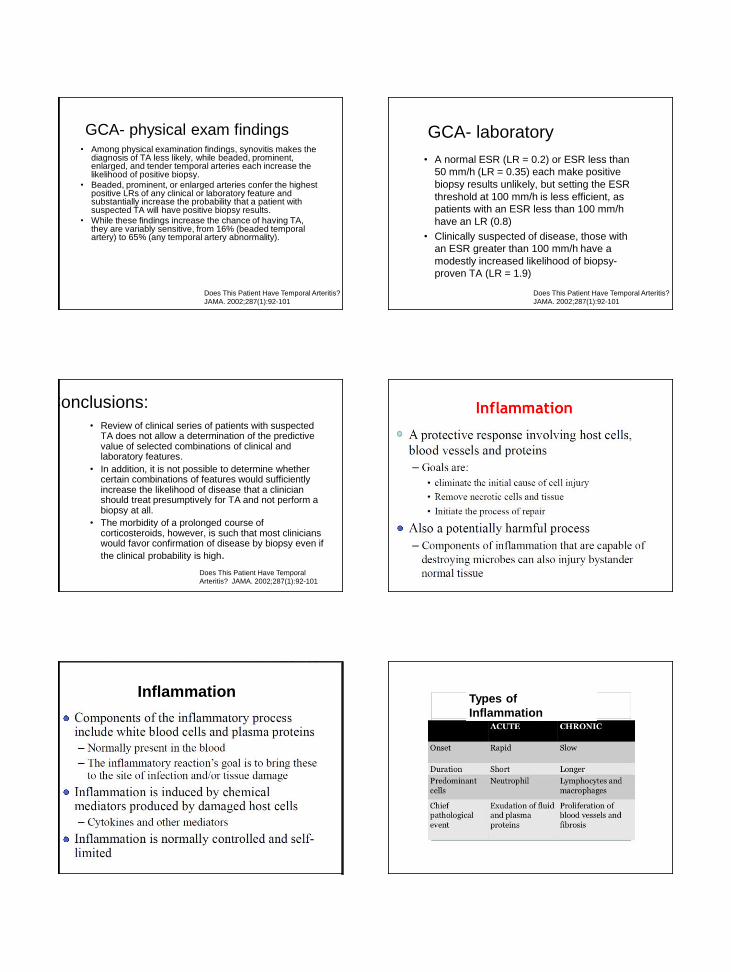
Inflammation Types of
Inflammation

Acute Inflammation
Histopathological features of giant-cell arteritis. Transverse sections of temporal artery-
untreated GCA (A) Granulomatous inflammation and multinucleated giant cells (arrows) at
junction of media and intima.. (B) A mononuclear transmural infiltrate without giant cells (C)
Vasculitis involving small vessels (arrows) close to a non-inflamed temporal artery .
Lancet 2008;372;234–245
A B C

Consequences of blood vessel wall inflammation Figure 1 Pathogenetic mechanisms operating in GCA
Salvarani, C. et al. (2012) Clinical features of polymyalgia rheumatica and giant cell arteritis
Nat. Rev. Rheumatol. doi:10.1038/nrrheum.2012.97
The pathogenic pathways implicated in granulomatous lesions in giant-cell arteritis
Front. Immunol., 12 September 2014 | http://dx.doi.org/10.3389/fimmu.2014.00432
Gross architecture of a granuloma with
macrophages, dendritic cells, and
multinucleate giant-cells forming the core
of the sphere, surrounded by a shell of
lymphocytes
Date of download: 2/29/2016
From: Giant-Cell Arteritis and Polymyalgia Rheumatica Ann Intern Med. 2003;139(6):505-515. doi:10.7326/0003-4819-139-6-200309160-00015
The systemic inflammatory response in giant-cell arteritis and polymyalgia rheumatica.ILVessel wall inflammation is preceded and accompanied by an intense acute-phase response. Circulating macrophages are activated and release interleukin ( )-1 and interleukin-6, critical inducers of a multiorgan reaction involving the liver, the central nervous system, the vascular system, the bone marrow, and the immune system. Hepatic
acute-phase reactants are useful in the laboratory diagnosis of giant-cell arteritis and polymyalgia rheumatica. The systemic inflammatory response can exist in the absence of fully developed vasculitis, as in the case of
polymyalgia rheumatica. CRP = C-reactive protein.
Figure Legend:
Copyright © American College of Physicians. All rights reserved.
The final common pathway: Mechanisms of arterial wall destruction in GCA
Figure 3 Pathologic analysis of sections adjacent to those containing VZV antigen from GCA-positive temporal arteries Temporal arteries (TAs) in which varicella-zoster virus (VZV)
antigen was detected immunohistochemically were further analyzed pathologically.
Don Gilden et al. Neurology 2015;84:1948-1955
© 2015 American Academy of Neurology

Pathogenesis of RA
Incidence of severe giant cell arteritis in the arteries of head and neck. ST
indicates superficial temporal artery; V, vertebral; 0, ophthalmic; PC, posterior
ciliary; IC, internal carotid; EC, external carotidartery and branches in the neck;
CR, central retinal Arch Neurol. 1972;27(5):378-391.
Polymyalgia
Rheumatica Giant cell
arteritis
Polymyalgia
Rheumatica Giant cell
arteritis
20-65%
?
Medicine: 2011;90:40-51
A, circumferential thickening of the subclavian artery (arrow);
B, circumferential thickening of the axillary artery (arrow
CT scan circumferential thickening of the subclavian artery
(long arrow) and primary carotid artery (short arrow).
Long stenotic segment of the humeral, axillary, and subclavian arteries (arrows).
Long stenotic segment of the superficial femoral arteries (arrows). A, circumferential thickening of the descending thoracic aorta; B,
circumferential thickening of the abdominal aorta (arrow).
Giant
Cell
Arteritis
Computed tomographic angiography
(CTA)) shows the results of aortic-root
repair and aortic-arch replacement with an
“elephant trunk graft” in a 71-year-old
woman who had biopsy-confirmed giant-
cell arteritis.
Aortic arch and its branches in a 72-year-
old woman with biopsy-positive giant-cell
arteritis. Arrows indicate stenotic lesions in
the bilateral subclavian and axillary
arteries, and arrowheads indicate long-
segment occlusions of the proximal
brachial arteries.
N Engl J Med 2014; 371:50-57
More pearls about
temporal artery biopsy

Photographs of the optic disc in patients with giant-cell arteritis and visual loss due
to anterior ischemic optic neuropathy, in the early acute phase (A) and after 3
months of prednisone therapy (B)(A) Optic disc oedema and a flame-shaped
haemorrhage. (B) Optic atrophy
Lancet 372, 9634, 2008, 234–245
Hayreh & Zimmerman
363 cases of suspected GCA
referred for TA Bx (106+, 257-)
• 21.2 % with visual loss and +TA Bx- no other
symptoms
• 55.7% with + TA Bx had new onset localized
HA- so did 45.5% with a negative TA Bx.
• 19.8% with + TA Bx had TA tenderness or
decreased pulsation- so did 12.8% with
negative TA Bx.
• Normal ESR did not rule out GCA.
Ophthalmologica 2003;217:239-259.
Ultrasonographical findings for GCA
Clinical features of polymyalgia rheumatica and giant cell
arteritis Salvarani, C. et al. (2012) Nat. Rev. Rheumatol.
Weyand CM, Goronzy JJ. N Engl J Med 2014;371:50-57.
Therapeutic Approaches to Giant-Cell Arteritis and Polymyalgia Rheumatica.

Ann Intern Med. 2003;139(6):505-515
Case 1
• A 74-year-old woman has the recent onset of daily
bitemporal headache but is otherwise well. Her general
physical examination results are normal and the
erythrocyte sedimentation rate (ESR) is moderately
elevated at 64 mm/h. You wonder whether additional
history or physical examination findings will modify
your suspicion of possible temporal arteritis (TA) or
whether the historical features alone warrant
proceeding to temporal artery biopsy.
• In our first clinical scenario, the history
of bitemporal headache and a modestly
elevated ESR would be among those
factors that may lead a clinician to
suspect TA. In this setting, one would
seek the potential additional history of
jaw claudication or diplopia, and
determine the presence of a prominent,
tender, or beaded temporal artery. If
present, these factors would
substantially increase the likelihood of
positive temporal artery biopsy results.
Case 2
• 53-year-old man has a 1-month history of fever
and fatigue and reports a single episode of
transient partial loss of vision in 1 eye. You believe
that TA is among the diagnostic considerations but
suspect that he is too young for this diagnosis. You
wonder if additional history, physical examination,
or laboratory testing will change the probability of
TA sufficiently to alter your decision about the role
of temporal artery biopsy rather than pursuing
diagnostic evaluation for carotid artery stenosis or
other considerations first.
• In the second scenario, TA is among the diagnostic
considerations for transient partial monocular visual loss in
the setting of a constitutional illness. The history in this
case is sufficiently compelling to justify a temporal artery
biopsy. Given the high prior probability and the poor
performance of historical and examination features in
excluding disease, an otherwise normal history and
physical examination would not sufficiently reduce the
likelihood of TA to avoid the need for a temporal artery
biopsy. A normal ESR would, however, reduce the
likelihood of disease by a factor of 0.2 and should prompt
consideration of alternative diagnoses.