Polycaprolactone scaffold as targeted drug delivery system ...kanglab.net/120829 pcl.pdf · system...
12
RESEARCH ARTICLE Polycaprolactone scaffold as targeted drug delivery system and cell attachment scaffold for postsurgical care of limb salvage Bin Sheng Wong & Swee-Hin Teoh & Lifeng Kang Published online: 22 August 2012 # Controlled Release Society 2012 Abstract In this paper, a dual-function drug-laden polycap- rolactone scaffold, which can serve as both targeted drug delivery system and attachment platform for tissue regener- ation for the postsurgical care of limb salvage procedure, was developed with a simple and solvent-free molding technique. Scaffolds of varying surface architecture were created using poly(ethylene glycol) diacrylate microneedle arrays. A model drug, rhodamine B, was incorporated homogenously into the scaffold. In vitro drug release studies showed that rhodamine B was released in a slow and sus- tained manner for 112 days. Its release rate was affected by drug loading and scaffold surface architecture. Release of rhodamine B from the scaffolds followed the Higuchi dif- fusion model. Other drugs, namely, doxorubicin and lido- caine hydrochloride, were also effectively loaded into and released from the scaffolds. Cell attachment study demon- strated potential for the scaffolds to provide attachment platforms for tissue regeneration. Keywords Scaffold . Polycaprolactone . Targeted drug delivery . Tissue engineering Introduction Limb salvage procedure is a type of surgery that involves local resection of bone or soft tissue cancers in order to avoid amputation [1]. This procedure has proven to be effective in the treatment of osteosarcoma due to its superior ability to maintain the external appearances and physiolog- ical functions of the patients’ limbs [2, 3]. Typically, the surgery involves the removal of tumors and some surround- ing tissues, followed by the placement of either metal pros- thesis or bone graft in the resected space to provide mechanical support and platform for bone regeneration. The use of metal prosthesis, however, is plagued with nu- merous limitations such as corrosion [4], metal hypersensi- tivity [5], stress shielding [6], growth restriction [ 7], imaging interference [8], and the need for additional remov- al operation [9]. The usage of bone grafts is also not ideal because allograft is often associated with nonunion, infec- tion, disease transmission, and limited donor availability [10, 11]. While autograft can circumvent the problem of disease transmission and tissue incompatibility, it is limited by donor site morbidity, increased operative time, and chronic pain [11, 12]. Biodegradable implants, therefore, emerged as viable alternatives for limb salvage procedure and have since demonstrated promising results in bone regeneration of cranioplasty and bone defects [13, 14]. Often, chemotherapy is given to patients following a limb salvage procedure to destroy any lingering microscopic deposits of the malignant cells and reduce cancer recurrence [15]. While chemotherapy has proven to be highly effective in increasing the disease-free survival rate of localized os- teosarcoma [16], conventional systemic chemotherapy is unfortunately highly toxic [17]. Aside from postoperative chemotherapy, radiotherapy is also used in patients follow- ing surgical resection to minimize cancer recurrence. While radiotherapy is considered generally safe and effective, it Electronic supplementary material The online version of this article (doi:10.1007/s13346-012-0096-9) contains supplementary material, which is available to authorized users. B. S. Wong : L. Kang (*) Department of Pharmacy, National University of Singapore, 18 Science Drive 4, Singapore 117543, Singapore e-mail: [email protected] S.-H. Teoh Division of Bioengineering, School of Chemical and Biomedical Engineering, Nanyang Technological University, 70, Nanyang Drive, Singapore 637457, Singapore Drug Deliv. and Transl. Res. (2012) 2:272–283 DOI 10.1007/s13346-012-0096-9
Transcript of Polycaprolactone scaffold as targeted drug delivery system ...kanglab.net/120829 pcl.pdf · system...
RESEARCH ARTICLE
Polycaprolactone scaffold as targeted drug deliverysystem and cell attachment scaffold for postsurgicalcare of limb salvage
Bin Sheng Wong & Swee-Hin Teoh & Lifeng Kang
Published online: 22 August 2012# Controlled Release Society 2012
Abstract In this paper, a dual-function drug-laden polycap-rolactone scaffold, which can serve as both targeted drugdelivery system and attachment platform for tissue regener-ation for the postsurgical care of limb salvage procedure,was developed with a simple and solvent-free moldingtechnique. Scaffolds of varying surface architecture werecreated using poly(ethylene glycol) diacrylate microneedlearrays. A model drug, rhodamine B, was incorporatedhomogenously into the scaffold. In vitro drug release studiesshowed that rhodamine B was released in a slow and sus-tained manner for 112 days. Its release rate was affected bydrug loading and scaffold surface architecture. Release ofrhodamine B from the scaffolds followed the Higuchi dif-fusion model. Other drugs, namely, doxorubicin and lido-caine hydrochloride, were also effectively loaded into andreleased from the scaffolds. Cell attachment study demon-strated potential for the scaffolds to provide attachmentplatforms for tissue regeneration.
Keywords Scaffold . Polycaprolactone . Targeted drugdelivery . Tissue engineering
Introduction
Limb salvage procedure is a type of surgery that involveslocal resection of bone or soft tissue cancers in order toavoid amputation [1]. This procedure has proven to beeffective in the treatment of osteosarcoma due to its superiorability to maintain the external appearances and physiolog-ical functions of the patients’ limbs [2, 3]. Typically, thesurgery involves the removal of tumors and some surround-ing tissues, followed by the placement of either metal pros-thesis or bone graft in the resected space to providemechanical support and platform for bone regeneration.The use of metal prosthesis, however, is plagued with nu-merous limitations such as corrosion [4], metal hypersensi-tivity [5], stress shielding [6], growth restriction [7],imaging interference [8], and the need for additional remov-al operation [9]. The usage of bone grafts is also not idealbecause allograft is often associated with nonunion, infec-tion, disease transmission, and limited donor availability[10, 11]. While autograft can circumvent the problem ofdisease transmission and tissue incompatibility, it is limitedby donor site morbidity, increased operative time, andchronic pain [11, 12]. Biodegradable implants, therefore,emerged as viable alternatives for limb salvage procedureand have since demonstrated promising results in boneregeneration of cranioplasty and bone defects [13, 14].
Often, chemotherapy is given to patients following a limbsalvage procedure to destroy any lingering microscopicdeposits of the malignant cells and reduce cancer recurrence[15]. While chemotherapy has proven to be highly effectivein increasing the disease-free survival rate of localized os-teosarcoma [16], conventional systemic chemotherapy isunfortunately highly toxic [17]. Aside from postoperativechemotherapy, radiotherapy is also used in patients follow-ing surgical resection to minimize cancer recurrence. Whileradiotherapy is considered generally safe and effective, it
Electronic supplementary material The online version of this article(doi:10.1007/s13346-012-0096-9) contains supplementary material,which is available to authorized users.
B. S. Wong : L. Kang (*)Department of Pharmacy, National University of Singapore,18 Science Drive 4,Singapore 117543, Singaporee-mail: [email protected]
S.-H. TeohDivision of Bioengineering, School of Chemical and BiomedicalEngineering, Nanyang Technological University,70, Nanyang Drive,Singapore 637457, Singapore
Drug Deliv. and Transl. Res. (2012) 2:272–283DOI 10.1007/s13346-012-0096-9
also possesses several disadvantages such as soft tissuedamage, increased pain, and the need for additional ortho-pedic procedures. Some bone cancer cells also do not re-spond well to radiotherapy [18]. One major complicationfollowing a limb salvage procedure is surgical wound andorthopedic device infections [19]. Hence, oral or intravenousbroad-spectrum antibiotics are regularly prescribed to preventor treat infections associated with limb salvage surgeries.Patients often receive prophylactic anticoagulants to preventthe formation of thrombus and embolus postsurgery. Opioids,mild narcotics, anti-inflammatory, and other analgesics arealso sometimes given to patients to counter pain experiencedfollowing the surgery [20].
To minimize systemic drug toxicities, especially for thecytotoxic agents and antibiotics, and to ensure adequatesupply of the various therapeutic agents, a targeted andcontrolled drug delivery system can be designed. By encap-sulating drugs in micropolymeric or nanopolymeric struc-tures, targeted drug delivery allows drugs to remain atspecific sites of action at sufficiently high concentration toexert their pharmacological effects, without causing severesystemic toxicities. Additionally, targeted drug delivery alsoimproves drug absorption and intracellular penetration, pro-longs retention time, enhances drug efficacy, and reducesdrug degradation [21].
In view of the existing challenges and limitations of limbsalvage surgery, a novel biodegradable and drug-laden poly-meric scaffold was envisioned to serve as viable alternativeto metal prostheses and bone grafts to promote bone andtissue regeneration and to simultaneously function as atargeted and controlled drug delivery system for the drugsthat patients would typically receive after a limb salvagesurgery to minimize undesirable systemic toxicities.
Polycaprolactone (PCL), due to its nontoxic, inert, bio-degradable, and biocompatible nature [22], has been gainingsignificant popularity in the field of targeted drug delivery.Numerous drugs, such as cisplatin, doxorubicin, and genta-micin sulfate, have been successfully incorporated into PCLin the form of nanoparticles [22–24]. The current fabricationtechniques of drug-laden PCL nanoparticles, namely, emul-sion solvent extraction/evaporation, phase separation, andspray drying, unfortunately could only achieve low drugloading efficiency and produce scaffolds with one sole func-tion of controlled drug release [25]. Failure to remove theorganic solvents, such as methylene chloride, methanol,acetone, and ethyl acetate, used in these methods post-fabrication also present significant toxicological concerns[26].
In addition to targeted drug delivery, PCL has also beenextensively used in the field of tissue engineering to providespecific microarchitecture and in vivo mechanical supportfor the regeneration of damaged cells or tissues [27]. Con-ventional methods of three-dimensional scaffold fabrication,
such as gas formation, phase separation, porogen leaching,and emulsion-free drying, that fail to offer flexibility andcontrol over the design of scaffold architecture and poreinterconnectivity have been slowly replaced by the rapidprototyping system in recent years [28, 29]. Capable ofproviding easier and greater manipulation over the scaffoldporosity and dimension, the equipment of the rapid proto-typing system is, however, exorbitant and sophisticated,making its adaptation into the laboratory costly and difficult.
While PCL has demonstrated tremendous potentials inboth targeted drug delivery and tissue engineering, there arelimited studies that combine these two distinct functions ofPCL. The drug-laden PCL nanoparticles used in targeteddrug delivery often possess no other function than to releasedrugs, while the three-dimensional PCL scaffolds developedfor tissue engineering usually contain no drug. The termtissue engineering therapeutics has been coined to describethe emerging field of three-dimensional scaffolds with ad-ditional drug delivery properties [30]. Numerous therapeuticagents, including growth factors, antibiotics, and anti-inflammatories, have been successfully loaded into three-dimensional bioactive scaffolds and demonstrated clinicalpotentials in the treatment and management of bone-relatedpathologies [31].
In this study, a biodegradable porous drug-laden PCLscaffold, which can serve as both targeted drug deliverydevice and mechanical support for cellular regeneration,was developed. A novel solvent-free fabrication procedureinvolving press-molding with poly(ethylene glycol) diacry-late (PEGDA) microneedle arrays was successfully devised.Rhodamine B was chosen as a model drug for the drugrelease kinetic study and to investigate the effects of surfacearchitecture, created using microneedle arrays of varyingdiameters and center-to-center spacings, and drug loadingconcentration on the rate of drug release. To demonstrate thedrug-delivering potentials of the scaffold, doxorubicin andlidocaine hydrochloride were incorporated into the scaffoldsand their release profiles were determined. Lastly, a cellattachment study with human dermal fibroblasts (HDF)was carried out to examine the scaffold’s ability in supportingcellular attachment.
Materials and methods
Materials
PCL (Mn010,000), PEGDA (MW0258), 2-hydroxy-2-methylpropiophenone (97 %), 3-[tris(trimethylsilyloxy)-silyl]propyl methacrylate, lidocaine hydrochloride, and InVitro Toxicology Assay Kit, XTT-based were purchasedfrom Sigma-Aldrich (St. Louis, MO, USA). Rhodamine Bwas purchased from Alfa Aesar (Lancaster, UK).
Drug Deliv. and Transl. Res. (2012) 2:272–283 273
Doxorubicin was purchased from MP Bio (California,USA). Phosphate-buffered saline (PBS; 10×, pH 7.4) wasprocured from Vivantis Technologies (Kuala Lumpur,Malaysia). HDF cells were obtained from the Institute ofMedical Biology, Singapore.
Fabrication of PEGDA microneedle array
The detailed fabrication procedures of the PEGDA micro-needle array via soft photolithography have been reported[32]. The microneedle fabrication steps employed in thispaper were similar to the reported procedures, with slightmodifications. Briefly, microscopic glass slide (Sail Brand,China), previously coated with 3-[tris(trimethylsilyloxy)-silyl]propyl methacrylate overnight, was placed on a stageformed by placing two coverslips (Cell Path, Wales, UK) ona piece of glass slide. Pre-polymer solution (PEGDA and0.5 % w/w of 2-hydroxy-2-methylpropiophenone) wasinserted into the gap between the coverslips and the coatedglass slide with a micropipette. The setup was irradiatedwith high-intensity ultraviolet (UV) light of 12.4 W/cm2
for 1 s at a distance of 11.5 cm below the light source usingEXFO OmniCure® S200-XL UV curing station (EXFO,Photonic Solutions Inc., Canada). The glass slide with thepolymerized PEGDA backing was then removed from thesetup and placed on another stage formed by placing twomicroscopic slides on a piece of glass slide. Pre-polymersolution was then inserted into the gap. An inked plastic film(photomask) containing transparent circle array (InfiniteGraphics Pte. Ltd., Singapore) was placed on top of thePEGDA-coated slide and was subsequently irradiated underUV light of 12.4 W/cm2 for 1.3 s at a distance of 2.5 cmbelow the light source to form the microneedles. The pho-tomask was removed and the setup was rinsed under waterto remove excess pre-polymer solution. Finally, the micro-needle array was detached from the setup.
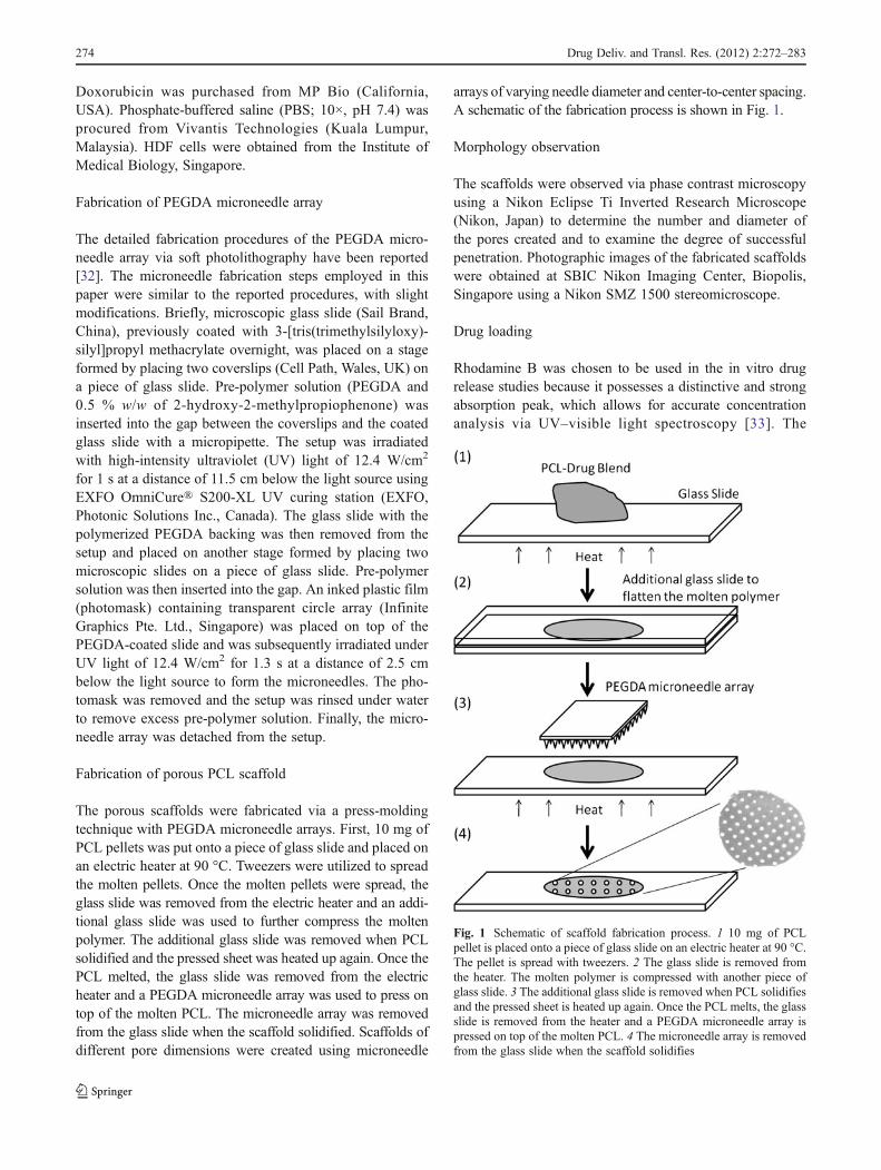
Fabrication of porous PCL scaffold
The porous scaffolds were fabricated via a press-moldingtechnique with PEGDA microneedle arrays. First, 10 mg ofPCL pellets was put onto a piece of glass slide and placed onan electric heater at 90 °C. Tweezers were utilized to spreadthe molten pellets. Once the molten pellets were spread, theglass slide was removed from the electric heater and an addi-tional glass slide was used to further compress the moltenpolymer. The additional glass slide was removed when PCLsolidified and the pressed sheet was heated up again. Once thePCL melted, the glass slide was removed from the electricheater and a PEGDA microneedle array was used to press ontop of the molten PCL. The microneedle array was removedfrom the glass slide when the scaffold solidified. Scaffolds ofdifferent pore dimensions were created using microneedle
arrays of varying needle diameter and center-to-center spacing.A schematic of the fabrication process is shown in Fig. 1.
Morphology observation
The scaffolds were observed via phase contrast microscopyusing a Nikon Eclipse Ti Inverted Research Microscope(Nikon, Japan) to determine the number and diameter ofthe pores created and to examine the degree of successfulpenetration. Photographic images of the fabricated scaffoldswere obtained at SBIC Nikon Imaging Center, Biopolis,Singapore using a Nikon SMZ 1500 stereomicroscope.
Drug loading
Rhodamine B was chosen to be used in the in vitro drugrelease studies because it possesses a distinctive and strongabsorption peak, which allows for accurate concentrationanalysis via UV–visible light spectroscopy [33]. The
Fig. 1 Schematic of scaffold fabrication process. 1 10 mg of PCLpellet is placed onto a piece of glass slide on an electric heater at 90 °C.The pellet is spread with tweezers. 2 The glass slide is removed fromthe heater. The molten polymer is compressed with another piece ofglass slide. 3 The additional glass slide is removed when PCL solidifiesand the pressed sheet is heated up again. Once the PCL melts, the glassslide is removed from the heater and a PEGDA microneedle array ispressed on top of the molten PCL. 4 The microneedle array is removedfrom the glass slide when the scaffold solidifies
274 Drug Deliv. and Transl. Res. (2012) 2:272–283
fluorescence and colored property of rhodamine B alsoenables the observation of drug distribution in the scaffoldpost-fabrication with fluorescence microscopy. A measuredamount of rhodamine B was added to a measured amount ofPCL in a beaker placed on top of an electrical heater at 90 °C. A metal spatula was used to mix the content to obtain ahomogenous blend, which was then flattened and cooled toobtain a thin sheet of stock material for scaffold fabrication.Similar procedures were employed for the encapsulation ofdoxorubicin and lidocaine hydrochloride.
In vitro drug release study
The effects of surface architecture and drug loading concen-tration on the rate of rhodamine B release were investigated.Rhodamine B–PCL scaffold was placed in a 1.5-ml micro-fuge tube filled with 1.0 ml of 1× phosphate buffer solutionat pH 7.4. Aluminum foil was used to wrap the tube tominimize photochemical degradation, and the tube’s open-ing was sealed with Parafilm to prevent solvent evaporationduring incubation. The entire setup was placed in a rack andput in an incubator at 37.0 °C. At regular time interval,0.5 ml aliquot of the sample was withdrawn for concentra-tion analysis; 0.5 ml of fresh PBS was added to replenishand to maintain the total dissolution volume at 1 ml aftereach withdrawal. The amount of rhodamine B released wasmeasured spectroscopically with a 96-well plate reader at554 nm (Tecan Infinite M200, Singapore). Triplicates wereperformed. Similar procedures were adopted to determinethe drug release profiles of doxorubicin and lidocaine hy-drochloride. Spectroscopic analysis of doxorubicin was per-formed using the Tecan plate reader at 485 nm, whilelidocaine hydrochloride was analyzed with a UV spectro-photometer (Shimadzu UV1800) at 216.5 nm.
Mathematical model fitting
The release data of rhodamine B were fitted in the zero-order, first-order, Hixson–Crowell, and Higuchi mathemat-ical models. The goodness of fit was determined by com-paring the average R2 values generated from simple linearregression. The model that best fit the data was chosen tocalculate the arbitrary release constants for the various drugrelease profiles, which were then used to compare the ratesof drug release from scaffolds of varying pore dimensionsand drug loading concentrations. In addition, the data werealso fitted to the Korsmeyer–Peppas model to determine theaverage release exponent.
Cell attachment study
HDF were cultured in Dulbecco’s modified Eagle’s medium(Invitrogen, California, USA) supplemented with 1 %
penicillin/streptomycin (PAN-Biotech, Aidenbach, Bavaria,Germany) and 10 % fetal bovine serum (Invitrogen, Cali-fornia, USA) at 37 °C and 5 % CO2. Blank porous scaffolds,fabricated with microneedle arrays of 400 μm needle diam-eter and 2× diameter center-to-center spacing, were im-mersed in 70 % ethanol overnight, followed by thoroughrinsing with sterile PBS. The scaffolds were then placed intothe wells of a nontissue culture-treated 96-well plate (Nunc,Roskilde, Denmark). Cell suspensions consisting of 104
cells were seeded onto the scaffolds and additional culturemedium were added to top up the well volume to 200 μL.Culture media were changed every 3 days and the cultureswere maintained for 5 days.
On days 2 and 5, the cultures were examined for cellattachment via an XTT assay. Briefly, culture media werefirst removed from the wells via a micropipette and the wellswere washed thoroughly with sterile PBS. Two hundredmicroliters of PBS was then added into the wells togetherwith 40 μL of XTT solution (0.5 mg/ml). One hundredmicroliters of the samples were withdrawn and placed intoa new 96-well plate after 5 h of incubation and their absor-bance at 450 nm were determined with a plate reader.
A negative control, which involves cell seeding in a 96-well plate without tissue culture treatment or PCL scaffold,and a positive control, which involves cell seeding in a 96-well plate with tissue culture treatment (Corning Incorpo-rated, Corning, NY, USA) but no PCL scaffold, wereconducted.
Statistical analysis
One-way analysis of variance was conducted using SPSS,while linear regression and graph plotting were performed inMicrosoft Excel Software.
Results
Scaffold fabrication
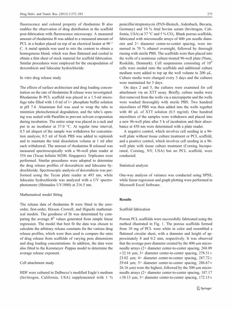
Porous PCL scaffolds were successfully fabricated using themethod illustrated in Fig. 1. The porous scaffolds formedfrom 10 mg of PCL were white in color and resembled aflattened circular sheet, with a diameter and height of ap-proximately 8 and 0.2 mm, respectively. It was observedthat the average pore diameter created by the 400-μmmicro-needle arrays (2× diameter center-to-center spacing, 268.09±32.16 μm; 3× diameter center-to-center spacing, 278.51±23.02 μm; 4× diameter center-to-center spacing, 287.72±29.64 μm; 5× diameter center-to-center spacing, 286.67±26.16 μm) were the highest, followed by the 300-μm micro-needle arrays (2× diameter center-to-center spacing, 187.17±38.13 μm; 3× diameter center-to-center spacing, 172.15±
Drug Deliv. and Transl. Res. (2012) 2:272–283 275
29.56 μm; 4× diameter center-to-center spacing, 192.97±29.87 μm; 5× diameter center-to-center spacing, 169.33±31.19 μm) and the 200-μm microneedle arrays (2× diametercenter-to-center spacing, 93.91±27.26 μm; 3× diametercenter-to-center spacing, 84.31±24.23 μm; 4× diametercenter-to-center spacing, 97.38±25.74 μm; 5× diametercenter-to-center spacing, 85.91±16.54 μm). The center-to-center spacing of the microneedle arrays showed no signif-icant effect on the average diameter of the pores created(p>0.05 for all three different microneedle base diameters).
The microscopic images of the fabricated scaffolds areshown in Fig. 2. The number of pores created decreasedwith increasing microneedle base diameter and center-to-center spacing. It was also observed that the pores createdby the 200-μm microneedle arrays were more distorted andnot fully penetrated.
Rhodamine B release profiles
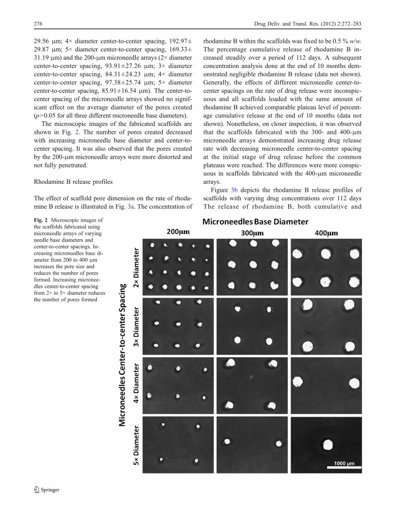
The effect of scaffold pore dimension on the rate of rhoda-mine B release is illustrated in Fig. 3a. The concentration of
rhodamine B within the scaffolds was fixed to be 0.5 % w/w.The percentage cumulative release of rhodamine B in-creased steadily over a period of 112 days. A subsequentconcentration analysis done at the end of 10 months dem-onstrated negligible rhodamine B release (data not shown).Generally, the effects of different microneedle center-to-center spacings on the rate of drug release were inconspic-uous and all scaffolds loaded with the same amount ofrhodamine B achieved comparable plateau level of percent-age cumulative release at the end of 10 months (data notshown). Nonetheless, on closer inspection, it was observedthat the scaffolds fabricated with the 300- and 400-μmmicroneedle arrays demonstrated increasing drug releaserate with decreasing microneedle center-to-center spacingat the initial stage of drug release before the commonplateaus were reached. The differences were more conspic-uous in scaffolds fabricated with the 400-μm microneedlearrays.
Figure 3b depicts the rhodamine B release profiles ofscaffolds with varying drug concentrations over 112 daysThe release of rhodamine B, both cumulative and
Fig. 2 Microscopic images ofthe scaffolds fabricated usingmicroneedle arrays of varyingneedle base diameters andcenter-to-center spacings. In-creasing microneedles base di-ameter from 200 to 400 μmincreases the pore size andreduces the number of poresformed. Increasing micronee-dles center-to-center spacingfrom 2× to 5× diameter reducesthe number of pores formed
276 Drug Deliv. and Transl. Res. (2012) 2:272–283
percentage cumulative, increased gradually over time.Again, a subsequent concentration analysis of the samplesat the end of 10 months showed negligible drug release andplateaus were obtained (data not shown). It was observedthat the rate of cumulative release increased with increasingdrug loading concentration, while the rate of percentagecumulative release increased with decreasing drug loadingconcentration.
Mathematical model fitting
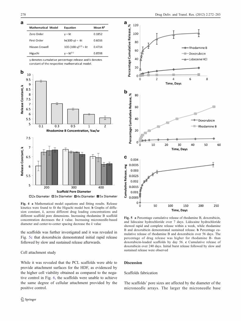
The release profiles of rhodamine B fit the Higuchimodel best, as indicated by the highest R2 obtainedand tabulated in Fig. 4a. The release constant, k, forthe various sets of experiment was generated using theHiguchi model. Figure 4b shows a plot of k acrossdifferent drug loading concentrations. It was observedthat k decreased with increasing drug loading concen-tration. The bar chart of k against different microneedlecenter-to-center spacings revealed that, with the excep-tion of scaffolds fabricated using the 200-μm micro-needle arrays, k decreased with increasing microneedlecenter-to-center spacings and the decrease was morestark in the scaffolds fabricated using the 400-μmmicroneedle arrays. These findings were consistent withthe observations made earlier with the drug releaseprofiles.
Doxorubicin and lidocaine hydrochloride release
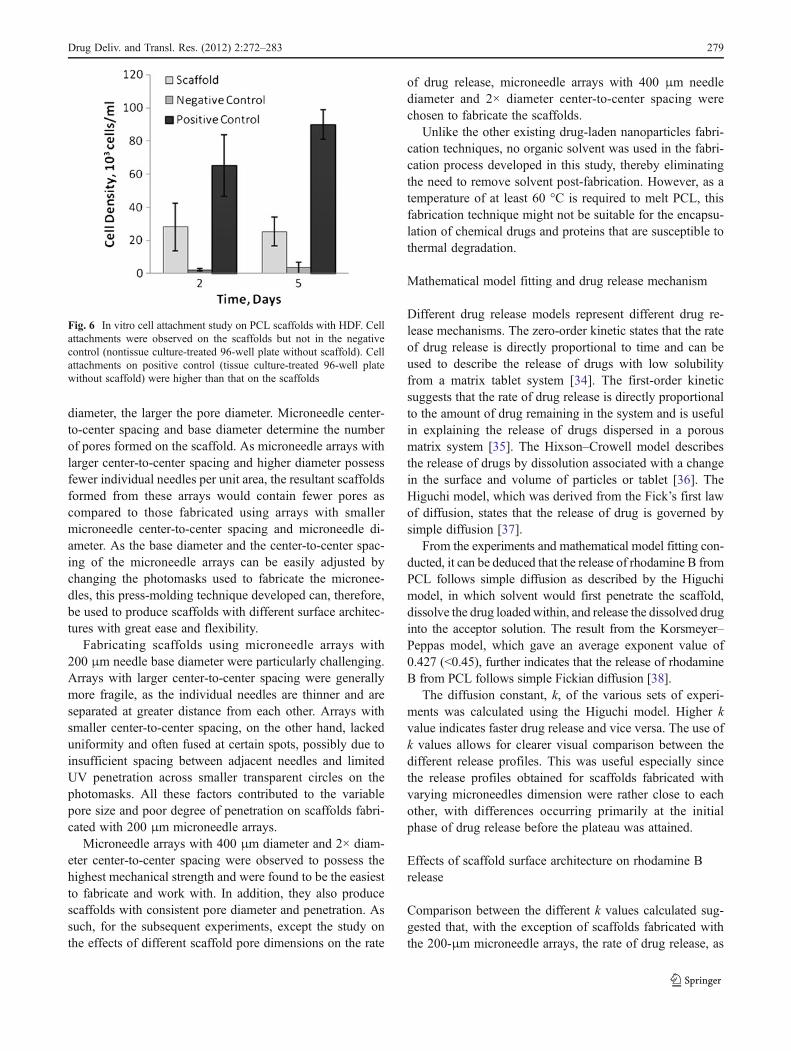
The release profiles of doxorubicin and lidocaine hydro-chloride from PCL, together with that of rhodamine B,are shown in Fig. 5a. The drug loading concentrationused for all scaffolds was 0.5 % w/w and all scaffoldswere fabricated using microneedle arrays with 400 μmneedle diameter and 2× diameter center-to-center spac-ing. Two distinct release patterns were observed, inwhich rhodamine B and doxorubicin were released ina slow and sustained manner, while lidocaine hydro-chloride demonstrated rapid release. The release ofdoxorubicin was significant lower than that of rhoda-mine B, achieving only 7 % release on day 56, asshown in Fig. 5b. The cumulative release of doxorubicin from
�Fig. 3 In vitro drug release studies. a Percentage cumulative rhoda-mine B release over 112 days for different scaffold pore dimensionsformed from microneedle arrays with microneedle-based diametersranging from 200 to 400 μm and center-to-center spacing ranging from2× to 5× diameter. Drug concentration in scaffolds was fixed at 0.5 %.b Cumulative rhodamine B release and percentage cumulative rhoda-mine B release over 112 days for different drug loading concentrationsfrom scaffolds formed from microneedle arrays with microneedle-based diameter of 400 μm and center-to-center spacing of 2× diameter
Drug Deliv. and Transl. Res. (2012) 2:272–283 277
the scaffolds was further investigated and it was revealed inFig. 5c that doxorubicin demonstrated initial rapid releasefollowed by slow and sustained release afterwards.
Cell attachment study
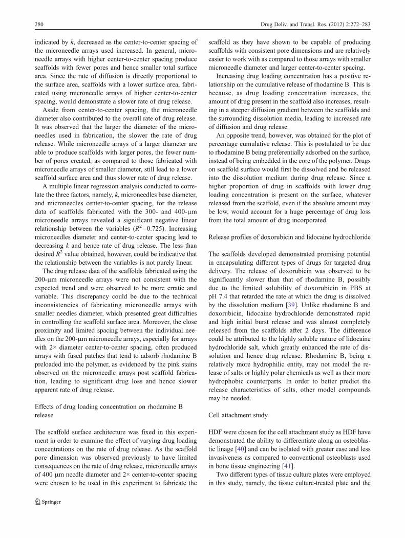
While it was revealed that the PCL scaffolds were able toprovide attachment surfaces for the HDF, as evidenced bythe higher cell viability obtained as compared to the nega-tive control in Fig. 6, the scaffolds were unable to achievethe same degree of cellular attachment provided by thepositive control.
Discussion
Scaffolds fabrication
The scaffolds’ pore sizes are affected by the diameter of themicroneedle arrays. The larger the microneedle base
Fig. 4 a Mathematical model equations and fitting results. Releasekinetics were found to fit the Higuchi model best. b Graphs of diffu-sion constant, k, across different drug loading concentrations anddifferent scaffold pore dimensions. Increasing rhodamine B scaffoldconcentration decreases the k value. Increasing microneedle-baseddiameter and center-to-center spacing decrease the k value
Fig. 5 a Percentage cumulative release of rhodamine B, doxorubicin,and lidocaine hydrochloride over 7 days. Lidocaine hydrochlorideshowed rapid and complete release within a week, while rhodamineB and doxorubicin demonstrated sustained release. b Percentage cu-mulative release of rhodamine B and doxorubicin over 56 days. Thepercentage of drug release was higher for rhodamine B- thandoxorubicin-loaded scaffolds by day 56. c Cumulative release ofdoxorubicin over 240 days. Initial burst release followed by slow andsustained release were observed
278 Drug Deliv. and Transl. Res. (2012) 2:272–283
diameter, the larger the pore diameter. Microneedle center-to-center spacing and base diameter determine the numberof pores formed on the scaffold. As microneedle arrays withlarger center-to-center spacing and higher diameter possessfewer individual needles per unit area, the resultant scaffoldsformed from these arrays would contain fewer pores ascompared to those fabricated using arrays with smallermicroneedle center-to-center spacing and microneedle di-ameter. As the base diameter and the center-to-center spac-ing of the microneedle arrays can be easily adjusted bychanging the photomasks used to fabricate the micronee-dles, this press-molding technique developed can, therefore,be used to produce scaffolds with different surface architec-tures with great ease and flexibility.
Fabricating scaffolds using microneedle arrays with200 μm needle base diameter were particularly challenging.Arrays with larger center-to-center spacing were generallymore fragile, as the individual needles are thinner and areseparated at greater distance from each other. Arrays withsmaller center-to-center spacing, on the other hand, lackeduniformity and often fused at certain spots, possibly due toinsufficient spacing between adjacent needles and limitedUV penetration across smaller transparent circles on thephotomasks. All these factors contributed to the variablepore size and poor degree of penetration on scaffolds fabri-cated with 200 μm microneedle arrays.
Microneedle arrays with 400 μm diameter and 2× diam-eter center-to-center spacing were observed to possess thehighest mechanical strength and were found to be the easiestto fabricate and work with. In addition, they also producescaffolds with consistent pore diameter and penetration. Assuch, for the subsequent experiments, except the study onthe effects of different scaffold pore dimensions on the rate
of drug release, microneedle arrays with 400 μm needlediameter and 2× diameter center-to-center spacing werechosen to fabricate the scaffolds.
Unlike the other existing drug-laden nanoparticles fabri-cation techniques, no organic solvent was used in the fabri-cation process developed in this study, thereby eliminatingthe need to remove solvent post-fabrication. However, as atemperature of at least 60 °C is required to melt PCL, thisfabrication technique might not be suitable for the encapsu-lation of chemical drugs and proteins that are susceptible tothermal degradation.
Mathematical model fitting and drug release mechanism
Different drug release models represent different drug re-lease mechanisms. The zero-order kinetic states that the rateof drug release is directly proportional to time and can beused to describe the release of drugs with low solubilityfrom a matrix tablet system [34]. The first-order kineticsuggests that the rate of drug release is directly proportionalto the amount of drug remaining in the system and is usefulin explaining the release of drugs dispersed in a porousmatrix system [35]. The Hixson–Crowell model describesthe release of drugs by dissolution associated with a changein the surface and volume of particles or tablet [36]. TheHiguchi model, which was derived from the Fick’s first lawof diffusion, states that the release of drug is governed bysimple diffusion [37].
From the experiments and mathematical model fitting con-ducted, it can be deduced that the release of rhodamine B fromPCL follows simple diffusion as described by the Higuchimodel, in which solvent would first penetrate the scaffold,dissolve the drug loadedwithin, and release the dissolved druginto the acceptor solution. The result from the Korsmeyer–Peppas model, which gave an average exponent value of0.427 (<0.45), further indicates that the release of rhodamineB from PCL follows simple Fickian diffusion [38].
The diffusion constant, k, of the various sets of experi-ments was calculated using the Higuchi model. Higher kvalue indicates faster drug release and vice versa. The use ofk values allows for clearer visual comparison between thedifferent release profiles. This was useful especially sincethe release profiles obtained for scaffolds fabricated withvarying microneedles dimension were rather close to eachother, with differences occurring primarily at the initialphase of drug release before the plateau was attained.
Effects of scaffold surface architecture on rhodamine Brelease
Comparison between the different k values calculated sug-gested that, with the exception of scaffolds fabricated withthe 200-μm microneedle arrays, the rate of drug release, as
Fig. 6 In vitro cell attachment study on PCL scaffolds with HDF. Cellattachments were observed on the scaffolds but not in the negativecontrol (nontissue culture-treated 96-well plate without scaffold). Cellattachments on positive control (tissue culture-treated 96-well platewithout scaffold) were higher than that on the scaffolds
Drug Deliv. and Transl. Res. (2012) 2:272–283 279
indicated by k, decreased as the center-to-center spacing ofthe microneedle arrays used increased. In general, micro-needle arrays with higher center-to-center spacing producescaffolds with fewer pores and hence smaller total surfacearea. Since the rate of diffusion is directly proportional tothe surface area, scaffolds with a lower surface area, fabri-cated using microneedle arrays of higher center-to-centerspacing, would demonstrate a slower rate of drug release.
Aside from center-to-center spacing, the microneedlediameter also contributed to the overall rate of drug release.It was observed that the larger the diameter of the micro-needles used in fabrication, the slower the rate of drugrelease. While microneedle arrays of a larger diameter areable to produce scaffolds with larger pores, the fewer num-ber of pores created, as compared to those fabricated withmicroneedle arrays of smaller diameter, still lead to a lowerscaffold surface area and thus slower rate of drug release.
A multiple linear regression analysis conducted to corre-late the three factors, namely, k, microneedles base diameter,and microneedles center-to-center spacing, for the releasedata of scaffolds fabricated with the 300- and 400-μmmicroneedle arrays revealed a significant negative linearrelationship between the variables (R200.725). Increasingmicroneedles diameter and center-to-center spacing lead todecreasing k and hence rate of drug release. The less thandesired R2 value obtained, however, could be indicative thatthe relationship between the variables is not purely linear.
The drug release data of the scaffolds fabricated using the200-μm microneedle arrays were not consistent with theexpected trend and were observed to be more erratic andvariable. This discrepancy could be due to the technicalinconsistencies of fabricating microneedle arrays withsmaller needles diameter, which presented great difficultiesin controlling the scaffold surface area. Moreover, the closeproximity and limited spacing between the individual nee-dles on the 200-μmmicroneedle arrays, especially for arrayswith 2× diameter center-to-center spacing, often producedarrays with fused patches that tend to adsorb rhodamine Bpreloaded into the polymer, as evidenced by the pink stainsobserved on the microneedle arrays post scaffold fabrica-tion, leading to significant drug loss and hence slowerapparent rate of drug release.
Effects of drug loading concentration on rhodamine Brelease
The scaffold surface architecture was fixed in this experi-ment in order to examine the effect of varying drug loadingconcentrations on the rate of drug release. As the scaffoldpore dimension was observed previously to have limitedconsequences on the rate of drug release, microneedle arraysof 400 μm needle diameter and 2× center-to-center spacingwere chosen to be used in this experiment to fabricate the
scaffold as they have shown to be capable of producingscaffolds with consistent pore dimensions and are relativelyeasier to work with as compared to those arrays with smallermicroneedle diameter and larger center-to-center spacing.
Increasing drug loading concentration has a positive re-lationship on the cumulative release of rhodamine B. This isbecause, as drug loading concentration increases, theamount of drug present in the scaffold also increases, result-ing in a steeper diffusion gradient between the scaffolds andthe surrounding dissolution media, leading to increased rateof diffusion and drug release.
An opposite trend, however, was obtained for the plot ofpercentage cumulative release. This is postulated to be dueto rhodamine B being preferentially adsorbed on the surface,instead of being embedded in the core of the polymer. Drugson scaffold surface would first be dissolved and be releasedinto the dissolution medium during drug release. Since ahigher proportion of drug in scaffolds with lower drugloading concentration is present on the surface, whateverreleased from the scaffold, even if the absolute amount maybe low, would account for a huge percentage of drug lossfrom the total amount of drug incorporated.
Release profiles of doxorubicin and lidocaine hydrochloride
The scaffolds developed demonstrated promising potentialin encapsulating different types of drugs for targeted drugdelivery. The release of doxorubicin was observed to besignificantly slower than that of rhodamine B, possiblydue to the limited solubility of doxorubicin in PBS atpH 7.4 that retarded the rate at which the drug is dissolvedby the dissolution medium [39]. Unlike rhodamine B anddoxorubicin, lidocaine hydrochloride demonstrated rapidand high initial burst release and was almost completelyreleased from the scaffolds after 2 days. The differencecould be attributed to the highly soluble nature of lidocainehydrochloride salt, which greatly enhanced the rate of dis-solution and hence drug release. Rhodamine B, being arelatively more hydrophilic entity, may not model the re-lease of salts or highly polar chemicals as well as their morehydrophobic counterparts. In order to better predict therelease characteristics of salts, other model compoundsmay be needed.
Cell attachment study
HDF were chosen for the cell attachment study as HDF havedemonstrated the ability to differentiate along an osteoblas-tic linage [40] and can be isolated with greater ease and lessinvasiveness as compared to conventional osteoblasts usedin bone tissue engineering [41].
Two different types of tissue culture plates were employedin this study, namely, the tissue culture-treated plate and the
280 Drug Deliv. and Transl. Res. (2012) 2:272–283
nontissue culture-treated plate. The term “tissue culture-treated” refers to treatment procedures, like corona dischargeor gas–plasma, that render the material surface more hydro-philic, via the generation of energetic oxygen ions that attachto the material surface, and therefore, more favorable forcellular attachment [42]. Nontissue culture plates, on the otherhand, offer hydrophobic surfaces and are often chosen for theculturing of cells where cellular attachments are to be mini-mized or avoided.
In this experiment, a negative control, which involved theseeding of HDF in a nontissue culture-treated plate, wasconducted to demonstrate that the cellular growth observedin the experiment set, which involved the seeding of HDF ina nontissue culture plate with PCL scaffolds, is due to theattachment surfaces provided by the scaffold per se. In thepresence of PCL scaffolds, HDF growth and attachmentwere observed to be significantly higher than that of thenegative control, suggesting that the scaffolds are capable ofproviding the cells with suitable sites for attachment. Theattachment, however, was better in the positive controlbecause anchorage-dependent cells, such as HDF, do notgrow and attach on hydrophobic surfaces well [43] andPCL, being a highly hydrophobic polymer, is unable toprovide a cell-attaching environment that is as ideal as thoseprovided by the tissue culture-treated plates.
Potential clinical applications
The scaffold developed in this paper could potentially per-form similar function to those orthopedic devices and act asa cell attachment site for the regeneration and growth ofhealthy bone cells. Long-term in vivo studies have demon-strated that the PCL implant was able to maintain its phys-ical shape after 2 years of implantation. In addition, PCLwas also shown to be capable of being degraded into lowmolecular pieces at the end of 30 months and be excretedout of the body completely, without accumulating in thebody tissues [44]. The slow degradation rate and biodegrad-able property of PCL makes it ideal to be developed intoimplants that could support long-term tissue regeneration.
As a targeted drug delivery system, the scaffolds can beused to encapsulate anticancer drugs such as doxorubicinand cisplatin to prevent cancer recurrence and minimizesystemic toxicity. Other agents, including antibiotics, anti-coagulants, opioids, mild narcotics, anti-inflammatory, andanalgesics, can also be incorporated into the scaffolds andbe released to the surrounding tissues, reducing the need fordaily dosing and possible systemic side effects.
One concern regarding the use of this dual-function scaf-fold lies on the influence of the cytotoxic agent like doxo-rubicin on bone cells attachment and regeneration. In aclinical setting, intensive chemotherapy following limb sal-vage surgery is warranted to eradicate remaining malignant
deposits. Unfortunately, bone healing occurs simultaneouslywith the administration of systemic chemotherapy. Whilepostsurgical chemotherapy has been shown to improverelapse-free survival time of patients with certain primaryosteosarcomas, postoperative chemotherapy does indeed af-fect bone growth and bone healing [45, 46]. As for thetreatment regimen, most patients would be placed on doxo-rubicin 25 mg/m2/day on days 1 to 3 and cisplatin 100 mg/m2 on day 1, every 3 weeks for six cycles [47–49]. Otheroptions include the control arm of the AOST0331 protocol(methotrexate, doxorubicin and cisplatin (MAP) chemothera-py) which involves 10 weeks of preoperative and 18 weeks ofpostoperative chemotherapywith doxorubicin, cisplatin, high-dose methotrexate, and leucovorin rescue (protocol informa-tion available online at www.cancer.gov/clinicaltrials/COG-AOST0331). Generally, 18 weeks (around 126 days) of inter-mittent postoperative chemotherapy are required. While thescaffolds developed in this paper could potentially face thesame problem of growth inhibition induced by the release ofchemotherapeutic agents like doxorubicin, they have success-fully demonstrated initial rapid release of up to around100 days for rhodamine B and around 60 days for doxorubi-cin. Specifically for rhodamine B, close to 79 % of the totaldrug loaded was released at the end of 112 days and only amere additional 2 %, making to an eventual total of 81 %, wasreleased between day 112 and day 300. Similarly for doxoru-bicin, approximately 6 % of the total drug content wasreleased at the end of 56 days and only an additional1 % was released between day 56 and day 240. Withhigher rate of initial release, sufficient quantity of thecytotoxic agent could be released at the implanted siteto destroy lingering malignancies during the initialphase of treatment, leaving negligible amount of drugto be released afterward so that the scaffold can functionprimarily as a tissue engineering platform to support cellgrowth and bone regeneration. A study has shown that58–580 ng/ml of doxorubicin has shown significant dose-dependent growth inhibition in a panel of human cell lines,representative for primary metastatic bone tumors [50]. Theamount of drug loaded in the scaffold may be adjusted toachieve this desired local concentration after release for theinitial killing of cancerous cells. Multiple chemotherapeuticagents may also be incorporated into the scaffold to bettermimic conventional multidrug chemotherapy regimens.
Conclusion
A novel porous and drug-laden biodegradable PCL scaffoldwith high loading efficiency was developed using a simple,inexpensive, and solvent-free press-molding technique withPEGDA microneedle arrays. It has been shown that thescaffolds were able to release rhodamine B slowly over a
Drug Deliv. and Transl. Res. (2012) 2:272–283 281
period of 112 days with a simple diffusion mechanism andthat the rate of drug release can be controlled by altering thedrug loading concentration and the scaffold surface architec-ture. Two other drugs, namely, doxorubicin and lidocainehydrochloride, were also successfully incorporated into thescaffolds, demonstrating the vast potential of the scaffolds tobe an effective targeted drug delivery system. The positiveresult of the cell attachment study also verified the ability forthe scaffold to support cellular attachment.
Acknowledgments The authors would like to express his gratitudeto Dr. Alexandre Chan and Dr. Richard Quek from the National CancerCenter Singapore for their comments and inputs. The authors alsothank Jaspreet S. Kochhar for his assistance in microneedle fabrication.
References
1. Lindell EB, Carroll NC. Limb salvage tumor surgery in children.Iowa Orthop J. 1993;13:124–35.
2. Messerschmitt PJ, Garcia RM, Abdul-Karim FW, Greenfield EM,Getty PJ. Osteosarcoma. J Am Acad Orthop Surg. 2009;17(8):515–27.
3. Carty CP, Dickinson IC, Watts MC, Crawford RW, Steadman P.Impairment and disability following limb salvage procedures forbone sarcoma. Knee. 2009;16(5):405–8..
4. Agins HJ, Alcock NW, Bansal M, Salvati EA, Wilson Jr PD,Pellicci PM, et al. Metallic wear in failed titanium-alloy total hipreplacements. A histological and quantitative analysis. J BoneJoint Surg Am. 1988;70(3):347–56.
5. Lalor PA, Revell PA, Gray AB, Wright S, Railton GT, FreemanMA. Sensitivity to titanium. A cause of implant failure? J BoneJoint Surg Br. 1991;73(1):25–8.
6. Uhthoff HK, Finnegan M. The effects of metal plates on post-traumatic remodelling and bone mass. J Bone Joint Surg Br.1983;65(1):66–71.
7. Yaremchuk MJ, Fiala TG, Barker F, Ragland R. The effects ofrigid fixation on craniofacial growth of rhesus monkeys. PlastReconstr Surg. 1994;93(1):1–10. discussion 1–5.
8. Sullivan PK, Smith JF, Rozzelle AA. Cranio-orbital reconstruc-tion: safety and image quality of metallic implants on CT and MRIscanning. Plast Reconstr Surg. 1994;94(5):589–96.
9. Dhillon MS, Prabhakar S, Prasanna C. Preliminary experiencewith biodegradable implants for fracture fixation. Indian J Orthop.2008;42(3):319–22.
10. Mankin HJ, Gebhardt MC, Jennings LC, Springfield DS, TomfordWW. Long-term results of allograft replacement in the manage-ment of bone tumors. Clin Orthop Relat Res. 1996;324:86–97.
11. Moore WR, Graves SE, Bain GI. Synthetic bone graft substitutes.ANZ J Surg. 2001;71(6):354–61.
12. Kurz LT, Garfin SR, Booth Jr RE. Harvesting autogenous iliacbone grafts. A review of complications and techniques. Spine(Phila Pa 1976). 1989;14(12):1324–31.
13. Schantz JT, Lim TC, Ning C, Teoh SH, Tan KC, Wang SC, et al.Cranioplasty after trephination using a novel biodegradable burrhole cover: technical case report. Neurosurgery. 2006;58(1 Suppl):ONS-E176. discussion ONS-E176.
14. Schuckert KH, Jopp S, Teoh SH. Mandibular defect reconstructionusing three-dimensional polycaprolactone scaffold in combinationwith platelet-rich plasma and recombinant human bone morpho-genetic protein-2: de novo synthesis of bone in a single case.Tissue Eng Part A. 2009;15(3):493–9.
15. Rosen G, Caparros B, Huvos AG, Kosloff C, Nirenberg A, CacavioA, et al. Preoperative chemotherapy for osteogenic-sarcoma: selec-tion of postoperative adjuvant chemotherapy based on the responseof the primary tumor to preoperative chemotherapy. Cancer. 1982;49(6):1221–30.
16. Susa M, Iyer AK, Ryu K, Hornicek FJ, Mankin H, Amiji MM, et al.Doxorubicin loaded polymeric nanoparticulate delivery system toovercome drug resistance in osteosarcoma. BMC Cancer.2009;9:399.
17. Hogendoorn PC, Athanasou N, Bielack S, De Alava E, Dei TosAP, Ferrari S, et al. Bone sarcomas: ESMO clinical practice guide-lines for diagnosis, treatment and follow-up. Ann Oncol. 2010;21Suppl 5:v204–13.
18. Matsumine A, Takegami K, Asanuma K, Matsubara T, NakamuraT, Uchida A, et al. A novel hyperthermia treatment for bonemetastases using magnetic materials. Int J Clin Oncol. 2011;16(2):101–8.
19. Gaur AH, Liu T, Knapp KM, Daw NC, Rao BN, Neel MD, et al.Infections in children and young adults with bone malignanciesundergoing limb-sparing surgery. Cancer. 2005;104(3):602–10.
20. Goff BJ, Castillo R, Raja SN. Painful sequelae following limbsalvage: etiology and management. J Am Acad Orthop Surg.2011;19 Suppl 1:S23–7.
21. Kumari A, Yadav SK, Yadav SC. Biodegradable polymeric nano-particles based drug delivery systems. Colloids Surf B Biointerfa-ces. 2010;75(1):1–18.
22. Gou M, Zheng X, Men K, Zhang J, Zheng L, Wang X, et al. Poly(epsilon-caprolactone)/poly(ethylene glycol)/poly(epsilon-capro-lactone) nanoparticles: preparation, characterization, and applica-tion in doxorubicin delivery. J Phys Chem B. 2009;113(39):12928–33.
23. Li X, Li R, Qian X, Ding Y, Tu Y, Guo R, et al. Superior antitumorefficiency of cisplatin-loaded nanoparticles by intratumoral deliv-ery with decreased tumor metabolism rate. Eur J Pharm Biopharm.2008;70(3):726–34.
24. Teo EY, Ong SY, Chong MS, Zhang Z, Lu J, Moochhala S, et al.Polycaprolactone-based fused deposition modeled mesh for deliv-ery of antibacterial agents to infected wounds. Biomaterials.2011;32(1):279–87.
25. Takada K. Microfabrication-derived DDS: from batch to individualproduction. Drug Discov Ther. 2008;2(3):140–55.
26. Lin W-J, Flanagan DR, Linhardt RJ. A novel fabrication ofpoly(ε-caprolactone) microspheres from blends of poly(ε-caprolac-tone) and poly(ethylene glycol)s. Polymer. 1999;40(7):1731–5.
27. Khademhosseini A, Vacanti JP, Langer R. Progress in tissue engi-neering. Sci Am. 2009;300(5):64–71.
28. Kim GH, Son JG. 3D polycarprolactone (PCL) scaffold withhierarchical structure fabricated by a piezoelectric transducer(PZT)-assisted bioplotter. Appl Phys A Mater. 2009;94(4):781–5.
29. Park SA, Lee SH, KimWD. Fabrication of porous polycaprolactone/hydroxyapatite (PCL/HA) blend scaffolds using a 3D plotting systemfor bone tissue engineering. Bioprocess Biosyst Eng. 2011;34(4):505–13.
30. Baroli B. From natural bone grafts to tissue engineering therapeu-tics: brainstorming on pharmaceutical formulative requirementsand challenges. J Pharm Sci. 2009;98(4):1317–75.
31. Mouriño V, Boccaccini AR. Bone tissue engineering therapeutics:controlled drug delivery in three-dimensional scaffolds. J R SocInterface. 2010;7(43):209–27.
32. Kochhar JS, Goh WJ, Chan SY, Kang L. A simple method ofmicroneedle array fabrication for transdermal drug delivery. DrugDev Ind Pharm. 2012; in press.
33. Zhang JT, Keller TF, Bhat R, Garipcan B, Jandt KD. A novel two-level microstructured poly(N-isopropylacrylamide) hydrogel forcontrolled release. Acta Biomater. 2010;6(10):3890–8.
282 Drug Deliv. and Transl. Res. (2012) 2:272–283
34. Tanaka N, Imai K, Okimoto K, Ueda S, Tokunaga Y, Ibuki R, et al.Development of novel sustained-release system, disintegration-controlled matrix tablet (DCMT) with solid dispersion granulesof nilvadipine (II): in vivo evaluation. J Control Release. 2006;112(1):51–6.
35. Mulye NV, Turco SJ. A simple-model based on first-order kineticsto explain release of highly water-soluble drugs from porousdicalcium phosphate dihydrate matrices. Drug Dev Ind Pharm.1995;21(8):943–53.
36. Hixson AW, Crowell JH. Dependence of reaction velocity uponsurface and agitation: I—theoretical consideration. Ind Eng Chem.1931;23:923–31.
37. Rodrigues MR, Lanzarini CM, Ricci E. Preparation, in vitro charac-terization and in vivo release of naproxen loaded in poly-caprolactone nanoparticles. Pharm Dev Technol. 2011;16(1):12–21.
38. Chang HI, Perrie Y, Coombes AG. Delivery of the antibioticgentamicin sulphate from precipitation cast matrices of polycapro-lactone. J Control Release. 2006;110(2):414–21.
39. Gou M, Shi H, Guo G, Men K, Zhang J, Zheng L, et al. Improvinganticancer activity and reducing systemic toxicity of doxorubicinby self-assembled polymeric micelles. Nanotechnology. 2011;22(9):095102.
40. Hee CK, Nicoll SB. Induction of osteoblast differentiation markersin human dermal fibroblasts: potential application to bone tissueengineering. Conf Proc IEEE Eng Med Biol Soc. 2006;1:521–4.
41. Notingher I, Jell G, Lohbauer U, Salih V, Hench LL. In situ non-invasive spectral discrimination between bone cell phenotypesused in tissue engineering. J Cell Biochem. 2004;92(6):1180–92.
42. Ramsey WS, Hertl W, Nowlan ED, Binkowski NJ. Surface treat-ments and cell attachment. In Vitro Cell Dev B. 1984;20(10):802–8.
43. Anselme K. Osteoblast adhesion on biomaterials. Biomaterials.2000;21(7):667–81.
44. Sun HF, Mei L, Song CX, Cui XM, Wang PY. The in vivodegradation, absorption and excretion of PCL-based implant. Bio-materials. 2006;27(9):1735–40.
45. Morcuende JA, Gomez P, Stack J, Oji G, Martin J, Fredericks DC,et al. Effect of chemotherapy on segmental bone healing enhancedby rhBMP-2. Iowa Orthop J. 2004;24:36–42.
46. Friedlaender GE, Tross RB, Doganis AC, Kirkwood JM, Baron R.Effects of chemotherapeutic agents on bone. I. Short-term metho-trexate and doxorubicin (adriamycin) treatment in a rat model. JBone Joint Surg Am. 1984;66(4):602–7.
47. Bramwell VH, Steward WP, Nooij M, Whelan J, Craft AW, GrimerRJ, et al. Neoadjuvant chemotherapy with doxorubicin andcisplatin in malignant fibrous histiocytoma of bone: a Euro-pean Osteosarcoma Intergroup study. J Clin Oncol. 1999;17(10):3260–9.
48. Souhami RL, Craft AW, Van der Eijken JW, Nooij M, Spooner D,Bramwell VH, et al. Randomised trial of two regimens of chemo-therapy in operable osteosarcoma: a study of the European Osteo-sarcoma Intergroup. Lancet. 1997;350(9082):911–7.
49. Bramwell VH, Burgers M, Sneath R, Souhami R, van OosteromAT, Voute PA, et al. A comparison of two short intensive adjuvantchemotherapy regimens in operable osteosarcoma of limbs inchildren and young adults: the first study of the European Osteo-sarcoma Intergroup. J Clin Oncol. 1992;10(10):1579–91.
50. Salerno M, Cenni E, Fotia C, Avnet S, Granchi D, Castelli F, et al.Bone-targeted doxorubicin-loaded nanoparticles as a tool for thetreatment of skeletal metastases. Curr Cancer Drug Targets.2010;10(7):649–59.
Drug Deliv. and Transl. Res. (2012) 2:272–283 283