poly( -caprolactone) and hydroxyapatite composite 3D ...
22
Page 1/22 3D printed multi-functional scaffolds based on poly( ε -caprolactone) and hydroxyapatite composite Fan Liu Wuhan Institute of Technology https://orcid.org/0000-0002-2865-2372 Honglei Kang Huazhong University of Science and Technology Zhiwei Liu Wuhan Institute of Technology Siyang Jin Wuhan University of Technology Guoping Yan ( [email protected] ) Wuhan Institute of Technology https://orcid.org/0000-0002-2649-9108 Yunlong Sun Huazhong University of Science and Technology Feng Li ( [email protected] ) Huazhong University of Science and Technology Haifei Zhan Queensland University of Technology Yuantong Gu Queensland University of Technology Research Keywords: poly( ε-caprolactone), hydroxyapatite, biodegradability, 3D printed scaffolds, bone tissue regeneration Posted Date: May 13th, 2021 DOI: https://doi.org/10.21203/rs.3.rs-486510/v1 License: This work is licensed under a Creative Commons Attribution 4.0 International License. Read Full License
Transcript of poly( -caprolactone) and hydroxyapatite composite 3D ...

Page 1/22
3D printed multi-functional scaffolds based onpoly(ε-caprolactone) and hydroxyapatite compositeFan Liu
Wuhan Institute of Technology https://orcid.org/0000-0002-2865-2372Honglei Kang
Huazhong University of Science and TechnologyZhiwei Liu
Wuhan Institute of TechnologySiyang Jin
Wuhan University of TechnologyGuoping Yan ( [email protected] )
Wuhan Institute of Technology https://orcid.org/0000-0002-2649-9108Yunlong Sun
Huazhong University of Science and TechnologyFeng Li ( [email protected] )
Huazhong University of Science and TechnologyHaifei Zhan
Queensland University of TechnologyYuantong Gu
Queensland University of Technology
Research
Keywords: poly(ε-caprolactone), hydroxyapatite, biodegradability, 3D printed scaffolds, bone tissueregeneration
Posted Date: May 13th, 2021
DOI: https://doi.org/10.21203/rs.3.rs-486510/v1
License: This work is licensed under a Creative Commons Attribution 4.0 International License. Read Full License

Page 2/22
AbstractBackground: Biodegradable polymeric scaffolds are critical to repair a large bone defect, which canprovide a porous and network microenvironment for cell attachment and bone tissue regeneration. Amultifunctional biodegradable PCL/HA composite was prepared with the blending of poly(ε-caprolactone) (PCL) and hydroxyapatite nanoparticles (HA). Subsequently, the PCL/HA scaffoldsimplants were produced by the screw extrusion/melting deposition forming method using PCL/HAcomposite as a raw material in this work.
Results: Through a serial of in vitro assessments, it is found that the PCL/HA composite possesses goodbiodegradability, good biocompatibility, and steady drug release performance, which can improve the cellproliferation of osteoblast cells MC3T3-E1. Meanwhile, in vivo experiments were carried out for the ratswith skull defect and rabbits with bone defects. It is observed that the PCL/HA scaffolds implants allowthe adhesion and penetration of bone cells, which enables the growth of bone cells and bone tissueregeneration. With a composite design to load an anticancer drug and achieve sustained drug release, thescaffolds could enhance bone repair and be expected to inhibit the tumor cells and improve patientoutcomes.
Conclusions: This work signi�es that PCL/HA composite can be used as the potential biodegradablescaffolds for bone repairing after bone malignant tumor resection.
1. IntroductionDue to the losing of osteogenic microenvironment, it is a great clinical challenge to repair a large bonedefect [1–3]. Biodegradable polymeric scaffolds possess porous and network structures, which providethe matrix and microenvironment for cell attachment and bone tissue regeneration. In particular thesepolymeric scaffolds do not need to be removed by second operation since polymers can be completelydegraded in vivo [4–6]. Biodegradable aliphatic polyesters, such as poly(lactic acid) (PLA), poly(ε-caprolactone) (PCL), and poly(trimethyl carbonate) are the attractive polymers that have been widelyused as the tissue engineering matrix and drug controlled release system [7–9]. Speci�cally, PCL is apreferred synthetic aliphatic polyester biomaterial due to their good biodegradability and biocompatibility,low toxicity, and weak in�ammatory effect of the degraded products. It has been shown to be suitable forthe fabrication of biodegradable scaffolds for tissue regeneration and tissue engineering [10–12].
The polymer composites containing nanoceramics were reported to possess better mechanical propertiesthan that of the pure polymers due to additional strength and stiffness provided by the embeddedceramic nanoparticles. As such, the polymer composite-based scaffolds can withstand a certain level ofphysiological loading and function before the new tissue replaces the scaffold matrix (during its gradualdegrading process) [13, 14]. Hydroxyapatite (HA) is a naturally occurring mineral form of calcium apatiteand an important component of human skeleton and teeth. It has been widely applied as the good bonegrafting material, replacement and supplement material because of its convenient production, low

Page 3/22
toxicity, good biocompatibility and high bioactivity [15, 16]. Thus it is of great interest to explore whetherHA can be applied as reinforcements for PCL in biomedical applications.
In the prosthodontic treatment of bone defects, the ideally implanted scaffolds should be biodegradablewhile new bone tissue is growing. They should have good bioactivity to promote the bone-binding ability,and possess other biological actions such as antitumor, antiangiogenic, anti-collagenolytic and anti-in�ammatory properties for bone regeneration. Recently, some physical and chemical methods (such assurface functionalization and blending) were used to fabricate functional scaffold materials for tissueengineering applications. For instance, Sarkar et al. [17] fabricated calcium phosphate (CaP) scaffoldswith the encapsulation of hydrophobic biomolecule curcumin (within a liposome), which eradicate theosteosarcoma cells and also promote osteoblast proliferation, offering new opportunities to treat bonedefects after tumor resection.
This work prepared a polymer composite based on poly(ε-caprolactone) and hydroxyapatite with theaddition of doxorubicin (DOX) as an anticancer drug. DOX was chosen as a model drug due to itswidespread use in bone cancer. Biodegradable scaffolds were further fabricated based on this polymercomposite using screw extrusion/melting deposition forming approach. The polymer composite was alsoevaluated to be used as the potential biodegradable scaffolds for bone repairing.
2. Materials And Methods
2.1. MaterialsAll chemicals and solvents were of analytical grade. Toluene and tetrahydrofuran (THF) were puri�ed byredistillation over sodium. Triethylamine was re�uxed under phthalic anhydride and dried over calciumhydride (CaH2) before use. Tin (II) 2-ethylhexanoate (Sn(Oct)2) was purchased from Sigma-Aldrich (Louis,MO, United States of America) and puri�ed by redistillation in vacuo before use. PCL was synthesized byring-opening bulk polymerization of ε-caprolactone using Sn(Oct)2 as a catalyst [18–20]. PCL was
characterized by the gel permeation chromatography, 1H NMR, Fourier transform infrared spectroscopy,UV, differential scanning calorimetry, and automatic contact-angle measurements. PCL: 1H NMR(300MHz, CDCl3, δ, ppm): 4.25 (m, 4H, C-CH2), 2.06 (m, 2H, C-CH2-C). FT-IR (KBr,cm− 1): 3450 (OH), 2963,
2922 (C-C), 1740 (C = O), 1461 (C-C), 1172, 1034 (C-O). The molecular weight (Mn) was 1.94×10⁵ andpolydispersity was 1.47 as determined by Gel Permeation Chromatography (GPC, Waters CorporationMilford, MA, United States of America). Hydroxyapatite (HA) nanoparticles with average diameter ofparticle sizes of 20 nm were purchased from the Beijing Deke Daojin Science and Technology Co., Ltd.(Beijing, China).
2.2.1 InstrumentationThe compounds were characterized using a UV-Vis spectrophotometer (UV-2800 series, Unico, Shanghai,China), a Nicolet is10 Fourier Transform-Infrared (FT-IR) spectrophotometer (Thermo Fisher Scienti�c Inc.,

Page 4/22
Madison, WI, United States of America), a Varian Mercury-VX300 NMR spectrometer (Varian, Inc.Corporate, Palo Alto, CA, United States of American) and an automatic contact angle meter (SL200A/B/DSeries, Solon Tech. Inc. Ltd., Shanghai, China). The molecular weight was measured by a gel permeationchromatography (GPC, Waters Corporation Milford, MA, United States of American) (Waters 2965Dseparations module, Waters 2414 Refractive Index Detector, Shodex K802.5 & K805 with Shodex K-GGuard Column, Polystyrene Standard, DMF solvent, 1.0 mL.min− 1 �ow rate, 323K Column temperatureand 323K Detector temperature). The glass transition temperature (Tg) was measured by a differentialscanning calorimeter (DSC) (NETZSCH DSC 200 F3, Erich NETZSCH GmbH & Co. Holding KG, Gebrüder-Netzsch-Strasse, Selb, Germany). The morphologies were characterized using a SEM (JEOL JSM-7001F,Japan) at 5–10 kV. Before SEM observation, the specimens were gold-sputter coated using an auto �necoater (JEOL, Ltd., Japan) under argon atmosphere for 60 s. The elemental distribution of calcium (Ca),phosphorus (P), oxygen (O), and carbon (C) in the scaffolds was investigated using an EDS (PhenomWorld BV, Netherlands). The phase composition was investigated using a XRD diffractometer (GermanBruker Co., Germany) with Cu Ka radiation (λ = 0.154056 nm, 40 kV, 40 mA). Before analysis, the scaffoldspecimens were �xed on a specimen holder by double side adhesive tape. The data were recorded in the2θ range of 10–75° with scanning speed of 8° min− 1. The absorbance (optical density: OD492) weremeasured with a DG-3022A ELISA-Reader (Hercules, CA, United States of American). The osteoblast cellsMC3T3-E1 were provided by the China Center for Type Culture Collection of Wuhan University, China. Theethical approval was obtained for the in vivo experiments in animals from the Department of Science andTechnology of Hubei Province, China and the Animal Center of Tongji Medical College, HuazhongUniversity of Science and Technology, China.
2.3. Preparation of poly(ε-caprolactone) and hydroxyapatite(PCL/HA) scaffoldsTwo different types of composite (PCL/HA and PCL/HA/DOX) samples were prepared, with onecontaining PCL and HA and the other one containing PCL, HA and doxorubicin (DOX). Thedichloromethane was used to ensure even mixture between PCL and HA (or DOX). The mixture wasslowly evaporated to dry under reduced pressure. The residue was cut into small pieces and dried undervacuum for 48 h to yield PCL/HA composite and PCL/HA/DOX composite.
Thereafter, the 3D printed PCL/HA scaffolds (diameter: 4 mm and height: 6 mm; diameter: 10 mm andheight: 2 mm) and multi-functional PCL/HA/DOX scaffolds containing DOX (diameter: 4 mm and height:6 mm) were prepared by the screw extrusion/melting deposition forming method using a 3D printer ofmelting deposition forming (Hubei Joye 3D High-tech Co., Ltd., Chibi, Hubei Province, China) with PCL/HAand PCL/HA/DOX composites as raw materials, respectively. The printing conditions were listed asfollows: the printing temperature was 130oC and preheating temperature of the operation desk was 50oC,the nozzle aperture was ≥ 0.4 mm, the extrusion speed was 220 mm/min, and the printing rate was 70mm/s. In vivo implanted PCL/HA scaffolds in a thigh-bone defect model of lower limb in rabbits werefurther loaded of 3D printed PCL/HA scaffolds and multi-functional PCL/HA/DOX scaffolds by collagen Irespectively according to the method cited in the literatures. [22–24]
Page 5/22
2.4. In vitro degradation testPCL and PCL/HA scaffolds (0.1g, diameter: 10 mm and height: 2 mm) were prepared as above and thendried in vacuum for 24 h. The scaffolds were suspended in 10 mL of PBS in a dialysis bag. The dialysisbag was sealed and then slowly shaken in 90 mL of PBS (pH 7.4) at 37oC in a 250 mL Erlenmeyer �ask.At predetermined time intervals, the samples were taken out of the degradation medium, rinsed withdistilled water and then dried in vacuo for 48 h. The molecular weight, water absorption and weight losswere calculated, respectively.
The PCL/HA samples for mechanics testing were further prepared by the thermoforming of PCL/HAcomposite materials in a standard mold on an injection molding machine. In vitro degradation test wasalso measured using PCL/HA samples according to the above same method as PCL/HA scaffolds.
2.5. In vitro drug release studyDoxorubicin (DOX, 10 mg) and PCL or PCL/HA composite materials (100 mg) were dissolved in 20 mL ofTHF. The solution was homogenized by sonication for 30s and then allowed to evaporate. The resulting�lm was cut into small pieces and dried under vacuum for 48 h to obtain 5-DOX-incorporated PCL orPCL/HA composite materials. The 5-DOX-incorporated PCL or PCL/HA scaffolds were further prepared bythe thermoforming of 5-DOX-incorporated PCL or PCL/HA composite materials in a disc mold (diameter:10 mm and height: 2 mm) on a thermocompressor with loading pressure of 10 MPa, setting temperatureof 110oC and molding time of 10 min.
The 5-DOX-incorporated PCL or PCL/HA scaffolds were suspended in 10 mL of PBS in a dialysis bag.The dialysis bag was sealed and then slowly shaken in 90 mL of PBS at 37oC in a 250-mL Erlenmeyer�ask. Aliqouts of the solution outside the dialysis membrane (25 mL) were replaced with 25 mL of PBS atvarious times intervals and tested at 256 nm by a high-performance liquid chromatographyspectrophotometer (HPLC). The changes of the concentrations of DOX were obtained from curves of theabsorption A versus concentration C of DOX in PBS on the basis of Lambert-Beer law.
2.6. Cell viability and proliferation assayThe circular 3D printed PCL and PCL/HA scaffolds (diameter: 10 mm, thickness: 2 mm, the content of HA:5%, 10%, 15%, 20% and 25%) were placed into the wells of 24 wells plate. The scaffolds were sterilizedusing Ultraviolet light for 1 hour for each side and secured with a stainless-steel ring. MC3T3-E1 cellswere seeded onto the scaffolds at a density of 4×10⁴ cells/mL and 100 µL of the RPMI-1640 growthmedium was added. Cell counting kit 8 (CCK-8) assay was performed on 1 and 3 days after culture. Thecells were incubated for 1 and 3 days in an incubator (37oC, 5% CO2) and the medium was replaced bythe fresh growth medium. And then 10µL of CCK-8 solution was added to each well and continuedincubating for 3 hours. The absorbance (optical density: OD492) was measured at 492 nm with a DG-3022A ELISA-Reader and expressed as a percentage relative to control cells (no scaffolds).

Page 6/22
2.7. In vivo implanted assay of PCL/HA scaffolds in skulldefect and histological analysisEighteen 6-week-old SD rats were divided into three groups (control, PCL and PCL/HA, for each group, n = 6) and the circular skull defect model (diameter of about 10 mm) was made in the head of each rat.Subsequently the rat was anesthetized with urethane (10%, 10mL/kg), positioned prone and �xed to apolystyrene cradle with adhesive tape to minimize motion. The scalp along the sagittal suture wasshaved and the skin was sterilized with iodine. A 15 mm long incision along the sagittal suture wasmade, and then, the calvarial bone was exposed by blunt dissection. A 10 mm diameter defect wascreated using a trephine bur (3i Implant Innovation, Palm Beach Gardens, FL, United States of America)on left parietal bone. The circular scaffolds of 10 mm diameter were was sterilized with UV light for 1hour for each side and implanted into the bone defects for each group (PCL and PCL/HA scaffolds). Forthe control group, the rats were treated the same and but without implantation. The periosteum and theoverlying skin were then stitched up using the nylon suture. After one month postsurgery, the rats wereanesthetized and perfused through heart by using 4% paraformaldehyde. The whole calvaria washarvested and further �xed with 4% paraformaldehyde at room temperature for 2 days for the followingevaluations. To assess the new bone formation in the bone defeat area, micro-computed tomography(micro-CT) (micro-CT 50, Scanco Medical AG, Bassersdorf, Switzerland) was applied under the �xedconditions (24 kV, 2 mA, 90 seconds). Area of new bone formation and percentage were measured usingthe Image Processing and Analysis in Java (ImageJ-1.51r, NIH, United States of America) software.
Nine 6-week-old SD rats were divided into three groups (control, PCL and PCL/HA scaffolds, for eachgroup, n = 3) and anesthetized with urethane (10%, 10mL/kg), positioned prone and �xed to a polystyrenecradle with adhesive tape to minimize motion. The scalp along the sagittal suture was shaved and theskin was sterilized with iodine. A 15 mm long incision along the sagittal suture was made in the muscleon the back. The circular scaffolds of 10 mm diameter were was sterilized with UV light for 1 hour foreach side and implanted into the muscle defects for each rat (PCL and PCL/HA scaffolds). For thecontrol group, the rats were treated the same and but without implantation. The periosteum and theoverlying skin were then stitched up using the nylon suture. After 3 days postsurgery, the rats wereanesthetized and the circular scaffolds were moved. The samples of muscle tissues around scaffoldswere taken out and embedded in para�n and sectioned at a thickness of 5 µm for Hematoxylin-eosin(H&E) staining. The H&E staining slides were observed under an optical microscope, and the images werecaptured under an IX-70 �uorescence inverted microscope (Olmypus Co., Ltd., Japan). Histologicalevaluation was performed by two independent examiners.
2.8. In vivo implanted assay of PCL/HA scaffolds in thigh-bone defectThe 3D PCL/HA scaffolds containing collagen I and multi-functional PCL/HA/DOX scaffolds containingcollagen I and DOX (diameter: 4 mm and height: 6 mm) were further produced by the loading of 3Dprinted PCL/HA scaffolds and multi-functional PCL/HA/DOX scaffolds with collagen I accordingly to the

Page 7/22
methods in cited literatures respectively. Eighteen 6-week-old New Zealand white rabbits were divided intothree groups (control, PCL/HA scaffolds with 25% HA content loading of collagen I and multi-functionalPCL/HA scaffolds with 25% HA content loading of collagen I and DOX, for each group, n = 6) and thecircular thigh-bone defect model (diameter of about 4 mm) was made in each rat. Subsequently therabbits was anesthetized with urethane (10%, 10mL/kg), positioned prone and �xed to a polystyrenecradle with adhesive tape to minimize motion. The scalp along the sagittal suture was shaved and theskin was sterilized with iodine. A 15 mm long incision along the sagittal suture was made, and then, thethigh-bone was exposed and a 4 mm diameter defect was created using a trephine bur in the cancellousbone. The scaffolds (diameter: 4 mm and height: 6 mm) were sterilized with UV light for 1 hour for eachside and implanted into the bone defects for each group. For the control group, the rats were treated thesame and but without implantation. The periosteum and the overlying skin were then stitched up usingthe nylon suture. After 2 months postsurgery, the rabbits were anesthetized and perfused through heart byusing 4% paraformaldehyde. The whole thigh-bone was harvested and further �xed with 4%paraformaldehyde at room temperature for 2 days for the following evaluations.
The new bone formation in the bone defeat area was assessed using micro-computed tomography(micro-CT) (Skyscan1276 X-Ray Microtomograph (Micro CT), Bruker, Belgium) under the �xed conditions(voltage: 93 kv, current: 800 µA, scanning resolution: 6.5 µm). Area of new bone formation andpercentage were measured using the Image Processing and Analysis software in the Skyscan1174 MicroCT Scanner with taking the femoral implant as the reference baseline. The cylindrical area with adiameter of 4.1 mm and a thickness of 6 mm was set as the three-dimensional reconstruction area ofinterest (ROI). The three-dimensional image was reconstructed with N-Recon software and the three-dimensional analysis was performed with CT-AN software. Moreover, the bone mineral density (BMD) ofthe new bone area was also measured.
2.9. Statistical analysisAll results were expressed as mean differences and were tested for signi�cance by a t test, P < 0.05 beingconsidered a signi�cant difference.
3. Results And Discussion
3.1. CharacterizationThe micrographs of PCL/HA scaffolds were examined by scanning electron microscopy (SEM, JEOLJSM-7001F, Japan) at 5–10 kV and the specimens were coated with gold-sputter using an auto �necoater (JEOL, Ltd., Japan) under argon atmosphere for 60 s. As shown in Fig. 1a, HA nanoparticlesscatter uniformity in the scaffolds with some aggregated occasions. Uniform pore with differentdiameters is observed from the 3D printed PCL/HA scaffolds. Figure 1b shows the mapping distributionof C, O, Ca, and P elements in the HA powders, which appears uniformly distribute in the scaffolds. Herein,the elemental distribution in the scaffolds is investigated using an EDS (Phenom World BV, Netherlands).

Page 8/22
In this work, PCL/HA scaffolds with 400–800 nm of uniform pore sizes are chosen as the implantedsamples as such pore size is expected to be bene�cial for cell proliferation/differentiation in vivo.
Chemical characterization of pure PCL, HA and PCL/HA scaffolds with different content of HA wereconducted by a Nicolet is10 Fourier Transform-Infrared (FT-IR) spectrophotometer (Thermo FisherScienti�c Inc., Madison, WI, United States of America) and their characteristic peaks are shown in Fig. 2a.For the composite samples, many of the absorption bands are overlapped but the properties of thefunctional groups remain unchanged. Whereas, the shape, location and intensity of the spectral peakschange signi�cantly. The spectra of PCL/HA scaffolds show typical ester peaks (at 1724 cm− 1, 1640cm− 1) and CH peaks (2923 cm− 1). The increase of HA content in the scaffolds (from 0 to 25%) decreasesthe peak intensities. Speci�cally, PCL/HA scaffolds show spectral features in the range of 1150 and 960cm− 1, which are related to the P-O and P = O vibrations of HA. The peak intensities of the spectral rangeof 3500 − 3200 cm− 1 that represents the absorption peak of OH groups of HA, decrease evidently.
Typical X-ray diffraction (XRD) spectra obtain for PCL/HA scaffolds are presented in Fig. 2b. The scaffoldspecimens were �xed on a specimen holder by double side adhesive tape and the XRD diffractometer(German Bruker Co., Germany) with Cu Ka radiation (λ = 0.154056 nm, 40 kV, 40 mA) was utilized. Thedata were recorded in the 2θ range of 10° − 75° with a scanning speed of 8 min− 1. As illustrated inFig. 3b, the spectra of PCL/HA scaffolds appear similar as that of pure PCL. In addition to the strongpeaks associated with the crystalline PCL phase, relatively weak peaks at ∼25.32°, 32.78°, 41.43°, 45.58°and 46.58° are observed, which correlate to the crystalline HA phase. These observations imply thepresences of both PCL and HA in the mixture powders. Combined with the above SEM image in Fig. 2a,these observations indicate that the �ne HA nanocrystals are well dispersed in the PCL matrix.
Figure 2c compares the water contact angle of the PCL/HA scaffolds containing different content of HA.The water angle was measured using an automatic contact angle meter (SL200A/B/D Series, Solon Tech.Inc. Ltd., Shanghai, China). As it is seen, the water contact angle decreases from 91.6° for pure PCL to80.70° for PCL with 15wt% HA, and then increases gradually to 81.4° when the HA content increasesfurther to 25wt%. In all examined samples, the water contact angle on the PCL/HA scaffolds is muchsmaller than that on the pure PCL. Such result demonstrates that presence of the nano-HA enhances thesurface hydrophilicity of the PCL scaffolds [21, 22].
The compressive modulus of PCL/HA scaffolds are measured and shown in Fig. 2d. Here, the scaffoldsare fabricated in the form of cylinders (with a diameter of 4 mm and a height of 6 mm) and verticallyplaced between two parallel plates in a universal testing machine (MTS Industrial Systems Co., Ltd,Shenzhen, China,). A compression rate of 1.0 mm min− 1 was utilized. Each compressive test wasrepeated �ve times. As it is seen, the compressive modulus increases from 112 MPa to 330 MPa whenthe HA content increases from 0 to 25wt%. In literature, the compressive modulus for pure PCL ismeasured between 85 MPa and 224.9 MPa [23, 24], which agrees with our measurements. Theenhancement is expected as originated from the higher intrinsic mechanical properties of HA (an averagecompressive strength of 174 MPa and a Young's modulus of 6 GPa) [25], and the uniform distribution of

Page 9/22
HA in the PCL matrix which show in the Fig. 1 play a very important role in the mechanical improvementof PCL/HA nanocomposite [24, 26].
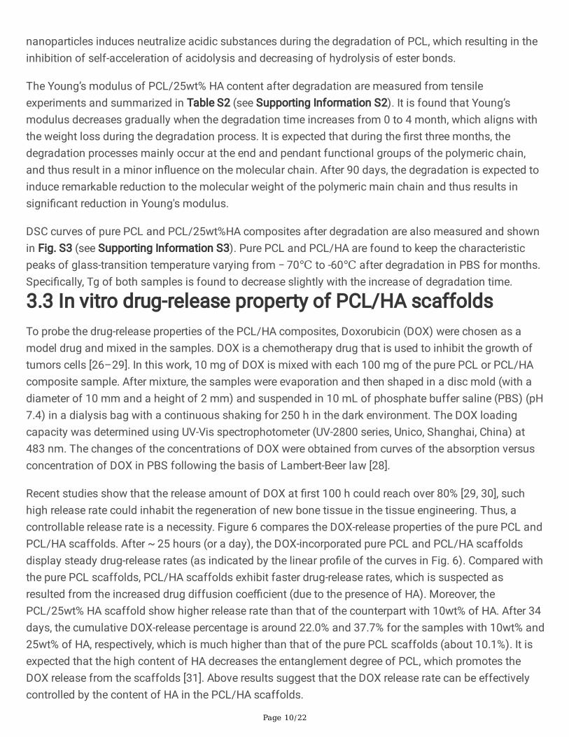
The glass transition temperature (Tg) of the PCL/HA scaffold was measured by a differential scanningcalorimeter (DSC) (NETZSCH DSC 200 F3, Erich NETZSCH GmbH & Co. Holding KG, Gebrüder-Netzsch-Strasse, Selb, Germany). For pure PCL, the glass transition temperature (Tg) is -66.4°C [27]. Figure 3ashows the DSC curves of PCL and PCL/HA composites, from which the Tg varies from − 70oC to -60oC,suggesting a good compatibility between PCL and HA. According to Fig. 3b, the degradation temperaturefor weight loss at 10% decreases from 389.7oC to 363.3oC when HA content increases from 0% to 25wt%.Similar result is also observed the degradation temperature for weight loss at 50% (from 515.0oC to414.0oC). In all, the results from thermogravimetric analysis (TGA) analysis indicate that the addition ofHA in PCL decreased the thermal stability of scaffolds as the starting decomposition temperaturedecreases.
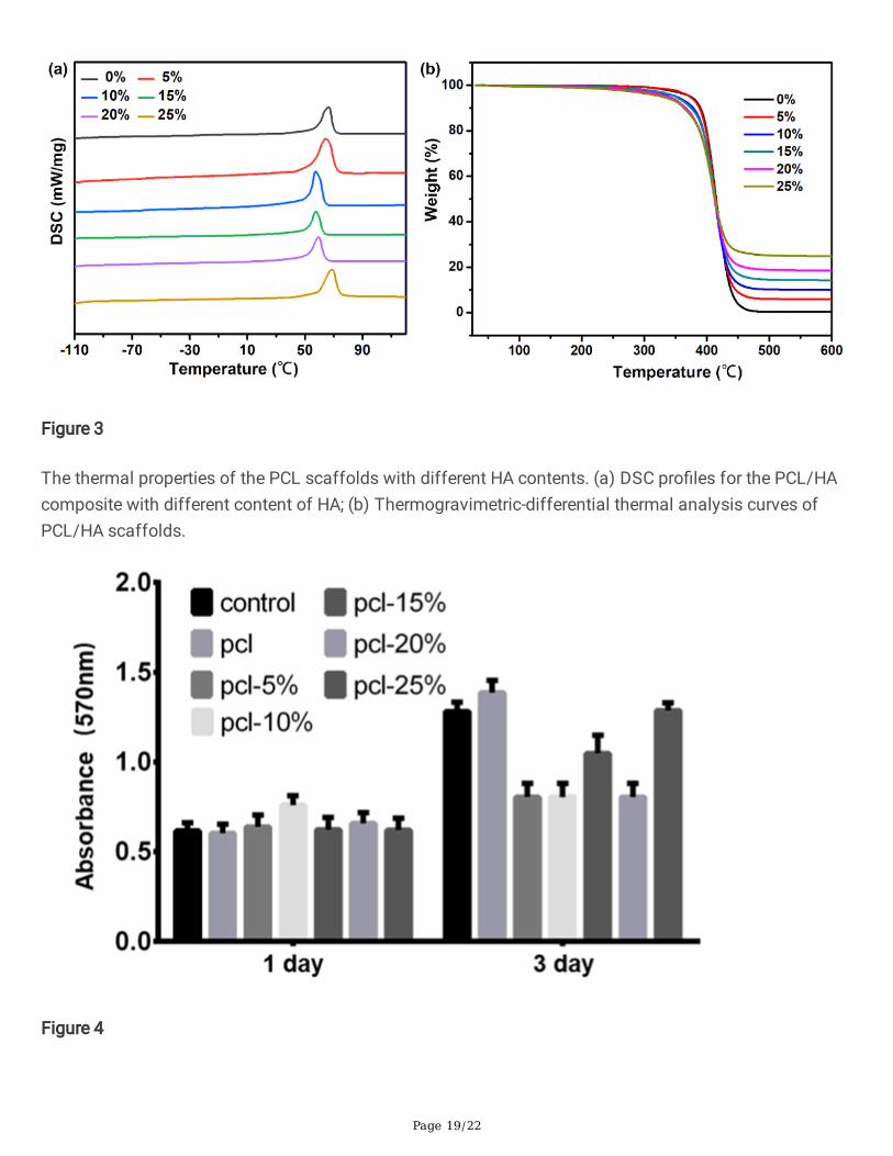
Additionally, the in vitro biocompatibility of the PCL/HA scaffolds was carried out in terms of theproliferation of MC3T3-E1 cells. The osteoblast cells MC3T3-E1 were provided by the China Center forType Culture Collection of Tongji Medical College, Huazhong University of Science and Technology, andraised according to the method described in literature [19]. In general, no signi�cant differences of cellproliferation activity among all scaffolds are observed after 1 day (Fig. 4.). Whereas, evident differencesof cell proliferation activity start to appear among all scaffolds after 3 days. Speci�cally, PCL/25wt% HAcontent exhibits the similar cell proliferation activity with the control cells. These results signify thatPCL/25wt% HA possesses good biocompatibility and can stimulate cell proliferation well. In all, based onabove analysis, PCL/25wt% HA shows the best comprehensive performances and thus being selected toassess their biomedical application potentials.
3.2 In vitro degradation property of PCL/HA scaffoldsIdeally, scaffold materials should be biodegradable during the growth of new bone tissue but maintaingood mechanical properties before the bone tissue is completely regenerated. Thus, it is important tounderstand the biodegradable behaviors of the scaffolds during the degradation for bone regeneration.For such purpose, PCL and PCL/HA scaffolds were suspended in 10 mL of PBS in a dialysis bag andthen slowly shaken in 90 mL of phosphate buffer saline (PBS) (pH 7.4) at 37°C. At predetermined timeintervals, the samples were taken out of the degradation medium, rinsed with distilled water and thendried in vacuo for 48 h.
As illustrated in Fig. 5a, the number of large pores and cracks increases in the PCL/HA scaffolds whenthe degradation time increases, and the PCL/HA scaffolds gradually lost their regularity and uniformity.The degradation of the sample can be well re�ected by the weight loss and molecular weight loss shownin Fig. 5b and 5c. Compared with pure PCL, the weight loss for PCL/25wt% HA sample is a much smallerduring degradation. For instance, after 180 days degradation, the weight losses of pure PCL andPCL/25wt% HA scaffolds are about 10% and 1%, respectively. These results indicate that mass lossoccurs at a much lower rate for samples with higher HA content. It is probably that the alkalinity of HA
Page 10/22
nanoparticles induces neutralize acidic substances during the degradation of PCL, which resulting in theinhibition of self-acceleration of acidolysis and decreasing of hydrolysis of ester bonds.
The Young’s modulus of PCL/25wt% HA content after degradation are measured from tensileexperiments and summarized in Table S2 (see Supporting Information S2). It is found that Young’smodulus decreases gradually when the degradation time increases from 0 to 4 month, which aligns withthe weight loss during the degradation process. It is expected that during the �rst three months, thedegradation processes mainly occur at the end and pendant functional groups of the polymeric chain,and thus result in a minor in�uence on the molecular chain. After 90 days, the degradation is expected toinduce remarkable reduction to the molecular weight of the polymeric main chain and thus results insigni�cant reduction in Young's modulus.
DSC curves of pure PCL and PCL/25wt%HA composites after degradation are also measured and shownin Fig. S3 (see Supporting Information S3). Pure PCL and PCL/HA are found to keep the characteristicpeaks of glass-transition temperature varying from − 70℃ to -60℃ after degradation in PBS for months.Speci�cally, Tg of both samples is found to decrease slightly with the increase of degradation time.
3.3 In vitro drug-release property of PCL/HA scaffoldsTo probe the drug-release properties of the PCL/HA composites, Doxorubicin (DOX) were chosen as amodel drug and mixed in the samples. DOX is a chemotherapy drug that is used to inhibit the growth oftumors cells [26–29]. In this work, 10 mg of DOX is mixed with each 100 mg of the pure PCL or PCL/HAcomposite sample. After mixture, the samples were evaporation and then shaped in a disc mold (with adiameter of 10 mm and a height of 2 mm) and suspended in 10 mL of phosphate buffer saline (PBS) (pH7.4) in a dialysis bag with a continuous shaking for 250 h in the dark environment. The DOX loadingcapacity was determined using UV-Vis spectrophotometer (UV-2800 series, Unico, Shanghai, China) at483 nm. The changes of the concentrations of DOX were obtained from curves of the absorption versusconcentration of DOX in PBS following the basis of Lambert-Beer law [28].
Recent studies show that the release amount of DOX at �rst 100 h could reach over 80% [29, 30], suchhigh release rate could inhabit the regeneration of new bone tissue in the tissue engineering. Thus, acontrollable release rate is a necessity. Figure 6 compares the DOX-release properties of the pure PCL andPCL/HA scaffolds. After ~ 25 hours (or a day), the DOX-incorporated pure PCL and PCL/HA scaffoldsdisplay steady drug-release rates (as indicated by the linear pro�le of the curves in Fig. 6). Compared withthe pure PCL scaffolds, PCL/HA scaffolds exhibit faster drug-release rates, which is suspected asresulted from the increased drug diffusion coe�cient (due to the presence of HA). Moreover, thePCL/25wt% HA scaffold show higher release rate than that of the counterpart with 10wt% of HA. After 34days, the cumulative DOX-release percentage is around 22.0% and 37.7% for the samples with 10wt% and25wt% of HA, respectively, which is much higher than that of the pure PCL scaffolds (about 10.1%). It isexpected that the high content of HA decreases the entanglement degree of PCL, which promotes theDOX release from the scaffolds [31]. Above results suggest that the DOX release rate can be effectivelycontrolled by the content of HA in the PCL/HA scaffolds.

Page 11/22
3.4 Cell response of BMSCs of PCL/HA scaffoldsTo further elucidate the effect of PCL/HA scaffolds on the differentiation of rat bone marrow-derivedmesenchymal stem cells (BMSCs), cell proliferation was evaluated using 3-(4, 5-dimethylthiazol-2-yl)-2, 5-diphenyltetrazolium bromide (MTT) assay after culturing for 1 and 2 days, respectively. Cells adhesionwas performed with 4, 6-diamidino-2-phenylindole (DAPI, Sigma) and FITC-Phalloidin (Sigma) aspreviously described [32]. The expression of osteogenic differentiation related genes and proteins in ratBMSCs cell was also evaluated. The mRNA transcript levels of Actin,alkaline phosphatase (ALP),collagen (COL), runt-related transcription factor-2 (RUNX2), and osteocalcin (OCN) mRNA within ratBMSCs (cultured in different supplemented osteogenic-inducing medium) were assessed by real-timepolymerase chain reaction (PCR). In both cases, cells were harvested on day 7 and day 14 then lysed inTrizol (Life Technologies, Carlsbad, CA, USA) and mRNA was extracted according to the manufacturer'sprotocol. Reverse transcription was carried out using the RNeasy Plus Micro Kit (Hilden, Germany)) andthe PCR test was performed using S1000™ Thermal Cycler (Bio-rad, Hercules).
As shown in Fig. 7a, the cell proliferation activity on PCL/25wt% HA scaffolds is obviously higher thanthe PCL/HA/DOX scaffolds at either day 1 or day 2, whereas the gap between them decreases on day 2.Fluorescence images show that the rat BMSC cells display good adhesion on the surface of thePCL/25wt% HA scaffolds (Fig. 7b), which is bene�cial for bone tissue regeneration. From Fig. 7c,PCL/25wt% HA scaffolds support the growth of BMSCs cells and evidently promote the expression ofActin, ALP, COL, RUNX2, OCN mRNA and proteins. The expressions of those genes and proteins are muchhigher at day 14 than that at day 7. These results demonstrate that the PCL/25wt% HA scaffolds possessgood biocompatibility and can stimulate cell proliferation well. Compared with the PCL/25wt% HAscaffolds, the expressions of those genes and proteins are much smaller in the PCL/HA/DOX scaffolds,which is expected as resulted from the quickly released DOX.
3.5 In vivo bone regeneration with skull defectIn vivo bone regeneration ability of PCL/HA scaffolds was investigated by measuring new boneformation using the model of rats with calvarial bone defects. Twelve 6-week-old Sprague-Dawley ratswere divided into two groups, with 6 rats in each group. A circular skull defect (with a diameter of 10 mm)was created using a trephine bur (3i Implant Innovation, Palm Beach Gardens, FL, USA) on the leftparietal bone of each rat. This size of defect was chosen because it is a defect of this size does not healby itself without intervention [11]. Thereafter, a circular scaffold (with a diameter of 10 mm and athickness of 2 mm) was sterilized and implanted into the bone defect location for each rat. To assess thenew bone formation in the bone defective area after 4 weeks, micro-computed tomography (micro-CT 50,Scanco Medical AG, Bassersdorf, Switzerland) was utilized under the �xed conditions (24 kV, 2 mA, 90seconds). The muscle tissues around the scaffolds were taken out and embedded in para�n andsectioned at a thickness of 5 µm for Hematoxylin-eosin (H&E) staining. The H&E staining slides werevisualized under an optical microscope.

Page 12/22
According to the micro-CT data, there is no observable new bone formed in the control group with purePCL scaffolds after 4 weeks post-surgery (see Fig. 8a). In comparison, the group with PCL/25wt% HAshows evident formation of new bone at the edge of the bone defect region (after 4 weeks). In themeantime, there is a certain amount of new bone deposition in the central area of the defects. Furtherhistological analysis a�rms the new bone formation after 4 weeks in the group with PCL/25wt% HAscaffolds. These observations signify that the PCL/25wt% HA scaffolds promotes the osteogenic activity,which can repair the bone defect. In the histological analysis,no obvious in�ammation was observed inthe tissue sections in the H&E staining micrographs after 4 weeks post-surgery (Fig. 8b). This observationfurther suggests that the PCL/25wt% HA scaffolds possess good biocompatibility and can provide agood microenvironment for osteoblast proliferation and differentiation.
3.6 In vivo bone regeneration with thigh-bone defectThe effects of the scaffolds on bone formation in vivo are also evaluated in rabbits with a thigh bonedefect in their lower limbs. Eighteen 6-week-old New Zealand white rabbits were divided into three groupswith 6 in each group, including the control group (with no scaffold), the group with PCL/25wt% HAscaffolds and the group with PCL/HA/DOX scaffolds. A circular thigh-bone defect (with a diameter of 4mm) was introduced to the thigh bone of each rabbit. The PCL/25wt% HA and PCL/HA/DOX scaffolds(with a diameter of 4 mm and a height of 6 mm) were �rstly coated with collagen I (see SupportingInformation S4), and then sterilized and implanted into the bone defects for each rabbit following themethod used in literatures [33–37]. The implanted position was determined by the magnetic resonanceimaging using M7 Small animal MRI system (1.0 Tesla, Aspect Imaging Ltd, Israel) (see SupportingInformation S5). Area of new bone formation and percentage were measured using the Image Processingand Analysis software taking the femoral implant as the reference baseline. The cylindrical area with adiameter of 4.1 mm and a thickness of 6 mm was set as the three-dimensional reconstruction area ofinterest. The three-dimensional image was reconstructed with N-Recon software and the three-dimensional analysis was performed with CT-AN software.
As shown in Fig. 9, there is a large amount of new bone formed in the rabbits either with PCL/25wt% HAscaffolds or PCL/HA/DOX scaffolds after 8 weeks. Bone tissues are found to gradually penetrate into thescaffold, adhere on the scaffold surface, and then grow to form a network. Along with the repair process,the PCL/HA scaffolds degrade gradually. These results indicate both PCL/25wt% HA and PCL/HA/DOXscaffolds promote the osteogenic activity and can repair the bone defect. To further assess the newformed bone, Fig. 9b compares bone tissue volume/total tissue volume (BV/TV, %), trabecular thickness(Tb.Th, mm), number of trabecula (Tb.N, mm− 1), and bone mineral density (BMD, g.cm− 3) after 8 weekssurgery. Interestingly, more new bone formation is observed from the rabbit with PCL/25wt% HAscaffolds than the counterpart with PCL/HA/DOX scaffolds after 4 weeks. Probably the large amount ofDOX released from the PCL/HA/DOX scaffolds inhibit the cell proliferation activity due to its high cellcytotoxicity to the bone cell. The PCL/25wt% HA scaffolds are found to result in higher bone volumefraction, larger trabecular thickness, larger number of trabecula and higher bone density. It is expected

Page 13/22
that, the slowly released DOX at the earlier stage of implantation is conducive to cell proliferation andinhibits in�ammation.
4. ConclusionsA multifunctional biodegradable PCL/HA composite was prepared by blending PCL with HA and then the3D printed PCL/HA scaffolds implants were produced by the screw extrusion/melting deposition formingmethod using PCL/HA composite as a raw material in this work. In vitro assessments reveal that thePCL/HA composite possesses good biodegradability and biocompatibility and promotes cellproliferation. With the addition of a chemotherapy drug (DOX), it is found that the PCL/HA scaffolds hasa steady drug release performance, and the release rate can be effectively controlled by the content ofHA. Subsequently, in vivo experiments for the rats with bone defects and rabbits with skull defect revealthat the PCL/HA scaffolds implants allow the penetration of bone cells, which enables the growth ofbone cells and bone tissue regeneration. Therefore the results suggest that PCL/HA composite can be anideal biodegradable scaffolds for bone repairing after bone malignant tumor resection.
AbbreviationsPCL: poly(ε-caprolactone); HA: hydroxyapatite nanoparticles; DOX: Doxorubicin; ALP: alkalinephosphatase; COL: collagen; RUNX2: runt-related transcription factor-2; OCN: Osteocalcin.
DeclarationsSupporting Information
The Supporting Information is available free of charge, including: The thermo properties of pure PCL andPCL/HA scaffolds, degradation property of PCL/HA scaffolds in vitro, characterization of PCL/HA/DOXscaffolds coated with collagen I, magnetic resonance imaging of the position of implanted PCL/HAscaffolds in the thigh-bone of rabbit in vivo.
Authors’ contributions
GY and FLI designed the experiments, FL, ZL, and SJ manufactured and characterized the samples. HKand YS carried out the experiments in vitro. FL, HZ and YG wrote the paper and conducted the analysisand discussion, all authors discussed the results and commented on the manuscript. HK and FLcontributed equally to this work.
Funding
This work was supported by the National Key R&D Program of China (Grant No. 2018YFB1105502,2016YFB1101302), Innovative Talents Project of Yellow Crane Talents Plan of Wuhan City (Grant [2017]No.1), Frontier Project of Application Foundation of Wuhan Former Funded Science and TechnologyProgram (Grant No. 2020020601012252), Scienti�c research projects for high level talents in the new

Page 14/22
century of Hubei Province (Grant [2017] No.344), Australian Research Council Discovery Project (GrantNo. DP180103009), Scienti�c Research Fund Project of Wuhan Institute of Technology (Grant No.K201861), and South Hubei Talents Project of Innovation and Entrepreneurship (Grant [2019] No.11). Theauthors also acknowledge the �nancial support from the China Scholarship Council (CSC, Grant No.201908420084).
Availability of data and materials
The data that support the �ndings of this study are available from the corresponding author uponreasonable request.
Declarations
Ethics approval and consent to participate
All animal experiments were performed at Tongji Hospital of Huazhong University of Science andTechnology in accordance with protocols approved by the Institutional Animal Care and Use Committee
Consent for publication
Not applicable.
Competing interests
All authors declare that they have no con�ict of interest.
References1. Matai I, Kaur G, Seyedsalehi A, McClinton A, Laurencin CT. Progress in 3D bioprinting technology for
tissue/organ regenerative engineering. Biomaterials. 2020;226:119536.
2. Lian H, Zhang L, Meng Z. Biomimetic hydroxyapatite/gelatin composites for bone tissueregeneration: fabrication, characterization, and osteogenic differentiation in vitro. Materials Design.2018;156:381–8.
3. McKeea C, Chaudhry GR. Advances and challenges in stem cell culture. Colloid Surface B.2017;159:62–77.
4. Yan Y, Chen H, Zhang H, Guo C, Yang K, Chen K, Cheng R, Qian N, Sandler N, Zhang YS. Vascularized3D printed scaffolds for promoting bone regeneration. Biomaterials. 2019;190:97–110.
5. Bunpetch V, Zhang X, Li T, Lin J, Maswikiti EP, Wu Y, Cai D, Li J, Zhang S, Wu C. Silicate-basedbioceramic scaffolds for dual-lineage regeneration of osteochondral defect. Biomaterials.2019;192:323–33.
�. Zhuang P, Sun AX, An J, Chua CK. S. Y. Chew, 3D neural tissue models: From spheroids to bioprinting.Biomaterials. 2018;154:113–33.

Page 15/22
7. Daly AC, Pitacco P, Nulty J, Cunniffe GM. D. J. Kelly, 3D printed microchannel networks to directvascularisation during endochondral bone repair Biomaterials 162 (2018) 34–46.
�. Jang J, Park JY, Gao G, Cho DW. Biomaterials-based 3D cell printing for next-generation therapeuticsand diagnostics. Biomaterials. 2018;156:88–106.
9. Hu B, Zhai MY, Yan GP, Zhuo RX, Wu Y, Fan CL. Polycarbonate magnetic microspheres containingtumor necrosis factor-α for potential targeted hepatic carcinoma therapeutics. J Drug Deliv Sci Tec.2014;24(1):57–60.
10. Hu B, Du HJ, Yan GP, Zhuo RX, Wu Y, Fan CL. Magnetic polycarbonate microspheres for tumor-targeted delivery of tumor necrosis factor. Drug Deliv. 2014;21(3):204–12.
11. Liu D, Nie W, Li D, Wang W, Zheng L, Zhang J, Zhang J, Peng C, Mo X, He C. 3D printed PCL/SrHAscaffold for enhanced bone regeneration. Chem Eng J. 2019;362:269–79.
12. Wang Q, Yang X, Wang G, Wan L, Wang S, Niu X, Wu J, Pan J. Osteogenic growth peptide-loaded 3D-printed PCL scaffolds for the promotion of osteogenesis through the ERK pathway. Materials Design.2020;193:108811.
13. Güney A, Malda J, Dhert WJ, Grijpma DW. Triblock copolymers based on ε-caprolactone andtrimethylene carbonate for the 3D printing of tissue engineering scaffolds. Int J Artif Organs.2017;40(4):176–84.
14. Huang KH, Lin YH, Shie MY, Lin CP. Effects of bone morphogenic protein-2 loaded on the 3D-printedMesoCS scaffolds. J Formosan Med Assoc. 2018;117(10):879–87.
15. Chen L, Deng C, Li J, Yao Q, Chang J, Wang L, Wu C. 3D printing of a lithium-calcium-silicate crystalbioscaffold with dual bioactivities for osteochondral interface reconstruction. Biomaterials.2019;196:138–50.
1�. Ma H, Feng C, Chang J, Wu C. 3D-printed bioceramic scaffolds: from bone tissue engineering totumor therapy. Acta Biomater. 2018;79:37–59.
17. Sarkar N, Bose S. Liposome-encapsulated curcumin-loaded 3D printed scaffold for bone tissueengineering. ACS Appl Mater Inter. 2019;11(19):17184–92.
1�. Chen H, Yan GP, Li L, Ai CW, Yu XH. Synthesis, characterization, and properties of ε-caprolactone andcarbonate copolymers. J Appl Polym Sci. 2009;114(5):3087–96.
19. Li D, Zhang K, Shi C, Liu L, Yan G, Liu C, Zhou Y, Hu Y, Sun H, Yang B. Small molecules modi�edbiomimetic gelatin/hydroxyapatite nano�bers constructing an ideal osteogenic microenvironmentwith signi�cantly enhanced cranial bone formation. Int J Nanomed. 2018;13:7167.
20. Lu K, Yan G, Chen H, Li L, Ai C, Yu X. Microwave-assisted ring-opening copolymerization of -caprolactone and 2-phenyl-5,5-bis(oxymethyl) trimethylene carbonate. Chin Sci Bull.2009;54(18):3237–43.
21. Xia Y, Zhou P, Cheng X, Xie Y, Liang C, Li C, Xu S. Selective laser sintering fabrication of nano-hydroxyapatite/poly-ε-caprolactone scaffolds for bone tissue engineering applications. Int JNanomed. 2013;8:4197.

Page 16/22
22. Medeiros GS, Muñoz PA, de Oliveira CF, da Silva LC, Malhotra R, Gonçalves MC, Rosa V, Fechine GJ.Polymer nanocomposites based on poly (ε-caprolactone), hydroxyapatite and graphene oxide. JPolym Environ. 2020;28(1):331–42.
23. Li Y, Han C, Yu Y, Xiao L. Effect of loadings of nanocellulose on the signi�cantly improvedcrystallization and mechanical properties of biodegradable poly(ε-caprolactone). Int J BiolMacromol. 2020;147:34–45.
24. Choi WY, Kim HE, Oh SY, Koh YH. Synthesis of poly (ε-caprolactone)/hydroxyapatite nanocompositesusing in-situ co-precipitation. Mat Sci Eng C Mater. 2010;30(5):777–80.
25. Martin R, Brown P. Mechanical properties of hydroxyapatite formed at physiological temperature. JMater Sci Mater Med. 1995;6(3):138–43.
2�. Corcione CE, Gervaso F, Scalera F, Padmanabhan SK, Madaghiele M, Montagna F, Sannino A, LicciulliA, Maffezzoli A. Highly loaded hydroxyapatite microsphere/PLA porous scaffolds obtained by fuseddeposition modelling. Ceram Int. 2019;45(2):2803–10.
27. Ramírez Agudelo R, Scheuermann K, Gala García A, Monteiro APF, Pinzón García AD, Cortés ME,Sinisterra RD. Hybrid nano�bers based on poly-caprolactone/gelatin/hydroxyapatite nanoparticles-loaded doxycycline: effective anti-tumoral and antibacterial activity. Mat Sci Eng C Mater.2018;83:25–34.
2�. Javanbakht S, Namazi H. Doxorubicin loaded carboxymethyl cellulose/graphene quantum dotnanocomposite hydrogel �lms as a potential anticancer drug delivery system. Mat Sci Eng C Mater.2018;87:50–9.
29. Kozlu S, Sahin A, Ultav G, Yerlikaya F, Calis S, Capan Y. Development and in vitro evaluation ofdoxorubicin and celecoxib co-loaded bone targeted nanoparticles. J Drug Deliv Sci Tec.2018;45:213–9.
30. Lai YL, Cheng YM, Yen SK. Doxorubicin-chitosan-hydroxyapatite composite coatings on titaniumalloy for localized cancer therapy. Mat Sci Eng C Mater. 2019;104:109953.
31. Ouyang L, Sun Z, Wang D, Qiao Y, Zhu H, Ma X, Liu X. Smart release of doxorubicin loaded onpolyetheretherketone (PEEK) surface with 3D porous structure. Colloid Surface B. 2018;163:175–83.
32. Sun Y, Kang H, Lin K, Guan H, Liu J, Li F. Evaluation of mechanical properties and osteogenesisability of porous titanium alloy scaffolds manufactured by selective laser melting technique. OrthopBiomech Mater Clin Study. 2019;016(002):1–5.
33. Du YW, Zhang LN, Hou ZT, Ye X, Gu HS, Yan GP, Shang P. Physical modi�cation ofpolyetheretherketone for orthopedic implants. Front Mater Sci. 2014;8(4):313–24.
34. Du YW, Zhang LN, Ye X, Nie HM, Hou ZT, Zeng TH, Yan GP, Shang P. In vitro and in vivo evaluation ofbone morphogenetic protein-2 (BMP-2) immobilized collagen-coated polyetheretherketone (PEEK).Front Mater Sci. 2015;9(1):38–50.
35. Feng P, Wu P, Gao C, Yang Y, Guo W, Yang W, Shuai C. A multimaterial scaffold with tunableproperties: toward bone tissue repair. Adv Sci. 2018;5(6):1700817.

Page 17/22
3�. Liu Y, Li T, Ma H, Zhai D, Deng C, Wang J, Zhuo S, Chang J, Wu C. 3D-printed scaffolds with bioactiveelements-induced photothermal effect for bone tumor therapy. Acta Biomater. 2018;73:531–46.
37. Shi F. Medical animal experiment method, China. Beijing: People’s Medical Publishing House; 1990.
Figures

Page 18/22
Figure 1
The characteristics of scaffolds. (a) Micromorphology of PCL/HA scaffolds in different content (0%,10wt%, 25wt%) (upper panel: ×50, middle panel: ×103, lower panel: ×105); (b) The optical graphs and EDSmapping distribution of C, O, Ca, and P elements in the surface of PCL/25wt% HA scaffolds.
Figure 2
Characterization of different PCL scaffolds with different content of HA ranging from 0% to 25wt%. (a)FT-IR result; (b) XRD spectra; (c) Water contact angle as a function of the HA content; and (d)Compressive modulus as a function of the HA content.
Page 19/22
Figure 3
The thermal properties of the PCL scaffolds with different HA contents. (a) DSC pro�les for the PCL/HAcomposite with different content of HA; (b) Thermogravimetric-differential thermal analysis curves ofPCL/HA scaffolds.
Figure 4

Page 20/22
Cell proliferation results of MC3T3-E1 cells after culturing for 1 and 3 days on the PCL/HA scaffolds with0%-25wt% content of HA
Figure 5
The degradation behaviors of the PCL/25wt% HA scaffolds after PBS immersion. (a) SEM micrographsof the scaffolds with PCL/25wt% HA scaffolds after PBS immersion for 3 months; (b) Weight loss andweight-average molecular weight (Mn) of the PCL scaffold with 0%, 25wt% of HA content for six months.

Page 21/22
Figure 6
Release pro�les of DOX-incorporated PCL scaffold with 0%, 10%, 25wt% of HA content for 250 h.
Figure 8

Page 22/22
In vivo micro-CT images and histological analysis in skull defect. (a) Micro-CT of the skull defect repair inrats with pure PCL and PCL/25wt% HA scaffolds at 4 weeks; (b) H&E staining of the muscle on the backof the rats at 4 weeks post-surgery (HE ×300).
Figure 9
Micro-CT analysis of the effect of scaffolds on thigh-bone defect in vivo. (a) Representative three-dimensional reconstructed micro-CT images showing the effect of PCL/HA scaffolds on the new bonetissue formation inside the defect site (red dashed line). (left: PCL/25wt% HA scaffolds for 4 weeks and 8weeks, right: PCL/HA/DOX scaffolds for 4 weeks and 8 weeks); (b) Summarized data showing the micro-architectural parameters of the new formed bone tissue at 4 and 8 weeks by analyzing the three-dimensional reconstructed micro-CT images using image analysis software. BMD, BV/TV, Tb.Th andTb.N were shown in the panel.
Supplementary Files
This is a list of supplementary �les associated with this preprint. Click to download.
supplementaryinformation20210507.docx