Polarized Rac-dependent protrusions drive epithelial intercalation in ...
26
RESEARCH ARTICLE Polarized Rac-dependent protrusions drive epithelial intercalation in the embryonic epidermis of C. elegans Elise Walck-Shannon 1 , David Reiner 2,3 and Jeff Hardin 1,4, * ABSTRACT Cell intercalation is a fundamental, coordinated cell rearrangement process that shapes tissues throughout animal development. Studies of intercalation within epithelia have focused almost exclusively on the localized constriction of specific apical junctions. Another widely deployed yet poorly understood alternative mechanism of epithelial intercalation relies on basolateral protrusive activity. Using the dorsal embryonic epidermis of Caenorhabditis elegans, we have investigated this alternative mechanism using high-resolution live cell microscopy and genetic analysis. We find that as dorsal epidermal cells migrate past one another they produce F-actin-rich protrusions polarized at their extending (medial) edges. These protrusions are controlled by the C. elegans Rac and RhoG orthologs CED-10 and MIG-2, which function redundantly to polarize actin polymerization upstream of the WAVE complex and WASP, respectively. We also identify UNC-73, the C. elegans ortholog of Trio, as a guanine nucleotide exchange factor (GEF) upstream of both CED-10 and MIG-2. Further, we identify a novel polarizing cue, CRML-1, which is the ortholog of human capping Arp2/3 myosin I linker (CARMIL), that localizes to the nonprotrusive lateral edges of dorsal cells. CRML-1 genetically suppresses UNC-73 function and, indirectly, actin polymerization. This network identifies a novel, molecularly conserved cassette that regulates epithelial intercalation via basolateral protrusive activity. KEY WORDS: Cell intercalation, Trio, Morphogenesis, Rac GTPase, CARMIL, LRRC16A INTRODUCTION Morphogenesis requires the concurrent rearrangement of many cell types during animal development. One such coordinated cell movement, mediolateral cell intercalation, is a ubiquitous process deployed in many developmental contexts to make a tissue longer and thinner (Walck-Shannon and Hardin, 2014). Cells derived from all three germ layers can undergo mediolateral intercalation, including epithelia. The intercalation of epithelial cells is especially complex because apicobasal polarity must be maintained while cells move along the mediolateral axis. Thus far, most research on epithelial cell intercalation has focused on RhoA-mediated shrinkage of apical junctions oriented along the axis of tissue shortening (Bertet et al., 2004; Blankenship et al., 2006; Chacon-Heszele et al., 2012; Karner et al., 2009; Lienkamp et al., 2012; Nishimura et al., 2012; Simões et al., 2014). A less well understood mechanism of epithelial intercalation involves basolateral protrusive activity. Although basolateral protrusions have been described in several systems, including the sea urchin archenteron (Hardin, 1989), the ascidian notochordal primordium (Munro and Odell, 2002a,b) and the vertebrate neural tube (Williams et al., 2014), little information is available regarding how they form. Intriguingly, recent evidence suggests that a cooperative mechanism between apical junctional constriction and basal protrusions mediates cell intercalation in the mouse neuroepithelium (Williams et al., 2014). Together, these examples suggest that basolateral protrusive activity is an important but underappreciated mechanism of epithelial cell intercalation. A promising context for exploring basolateral protrusive activity during epithelial cell rearrangement is the dorsal epidermis of the C. elegans embryo. Four hours after fertilization, 20 dorsal epidermal cells migrate past one another in the process of dorsal intercalation (Sulston et al., 1983). These intercalating cells are bona fide, apicobasally polarized epithelia, with intact apical junctions before, during and after intercalation (McMahon et al., 2001; Patel et al., 2008; Soto et al., 2002; Williams-Masson et al., 1998; E.W.-S. and J.H., unpublished). As cells become mediolaterally polarized they adopt a wedge shape; their medial tips extend contralaterally, while their lateral edges remain rounded. Eventually, the cell body and nucleus follow as an intercalating cell makes contact with contralateral seam epidermal cells, completing intercalation (Fig. 1A). Dorsal intercalation is marked by the formation of medially directed protrusions that extend basal to the apical junction (Williams-Masson et al., 1998). These basolateral protrusions, and not apical junction rearrangement, appear to be the major driver of dorsal intercalation, as null mutants for cadherin complex components can intercalate successfully (E.W.-S. and J.H., unpublished; Costa et al., 1998). Four-dimensional microscopy indicates that basolateral protrusions in dorsal epidermal cells bear similarities to lamellipodia in other migrating cell types (Heid et al., 2001). Pharmacological studies suggest that actin filaments are required for intercalation (Williams-Masson et al., 1998). Beyond a basic role for actin, however, the molecular basis for medial tip extension is not well understood. Actin networks can be formed through nucleation of existing filaments via the Arp2/3 complex, which must be activated by nucleation-promoting factors of the Wiskott-Aldrich syndrome protein (WASP) family, namely WASP and WAVE (Takenawa and Suetsugu, 2007). Rho family GTPases are the predominant regulators of such F-actin networks. In its GTP-bound state, Rac can activate the highly conserved, pentameric WAVE complex, while Cdc42 can activate WASP by relieving its autoinhibition (Kim et al., 2000; Miki et al., 1998; Patel et al., 2008). The RhoG ortholog mig-2 also genetically interacts with wsp-1 (Shakir et al., 2008), although it is unclear if the activation is direct. Rather Received 17 June 2015; Accepted 26 August 2015 1 Graduate Program in Genetics, University of Wisconsin-Madison, 1117 W. Johnson Street, Madison, WI 53706, USA. 2 Department of Pharmacology and Lineberger Comprehensive Cancer Center, University of North Carolina, 101 Manning Drive, Chapel Hill, NC 27514, USA. 3 Center for Translational Cancer Research, Institute of Biosciences and Technology and Department of Medical Physiology, Texas A&M Health Science Center, 2121 W. Holcombe Boulevard, Houston, TX 77030, USA. 4 Department of Zoology, University of Wisconsin- Madison, 1117 W. Johnson Street, Madison, WI 53706, USA. *Author for correspondence ( [email protected]) 3549 © 2015. Published by The Company of Biologists Ltd | Development (2015) 142, 3549-3560 doi:10.1242/dev.127597 DEVELOPMENT
Transcript of Polarized Rac-dependent protrusions drive epithelial intercalation in ...
RESEARCH ARTICLE
Polarized Rac-dependent protrusions drive epithelial intercalationin the embryonic epidermis of C. elegansElise Walck-Shannon1, David Reiner2,3 and Jeff Hardin1,4,*
ABSTRACTCell intercalation is a fundamental, coordinated cell rearrangementprocess that shapes tissues throughout animal development. Studiesof intercalation within epithelia have focused almost exclusively onthe localized constriction of specific apical junctions. Another widelydeployed yet poorly understood alternative mechanism of epithelialintercalation relies on basolateral protrusive activity. Using the dorsalembryonic epidermis ofCaenorhabditis elegans, we have investigatedthis alternative mechanism using high-resolution live cell microscopyand genetic analysis. We find that as dorsal epidermal cells migratepast one another they produce F-actin-rich protrusions polarized attheir extending (medial) edges. These protrusions are controlled bythe C. elegans Rac and RhoG orthologs CED-10 and MIG-2, whichfunction redundantly to polarize actin polymerization upstream of theWAVEcomplex andWASP, respectively.We also identify UNC-73, theC. elegans ortholog of Trio, as a guanine nucleotide exchange factor(GEF) upstream of both CED-10 and MIG-2. Further, we identify anovel polarizing cue, CRML-1, which is the ortholog of human cappingArp2/3 myosin I linker (CARMIL), that localizes to the nonprotrusivelateral edges of dorsal cells. CRML-1 genetically suppresses UNC-73function and, indirectly, actin polymerization. This network identifiesa novel, molecularly conserved cassette that regulates epithelialintercalation via basolateral protrusive activity.
KEY WORDS: Cell intercalation, Trio, Morphogenesis, Rac GTPase,CARMIL, LRRC16A
INTRODUCTIONMorphogenesis requires the concurrent rearrangement of many celltypes during animal development. One such coordinated cellmovement, mediolateral cell intercalation, is a ubiquitous processdeployed in many developmental contexts to make a tissue longerand thinner (Walck-Shannon and Hardin, 2014). Cells derived fromall three germ layers can undergo mediolateral intercalation,including epithelia. The intercalation of epithelial cells is especiallycomplex because apicobasal polarity must be maintained while cellsmove along the mediolateral axis. Thus far, most research onepithelial cell intercalation has focused on RhoA-mediated shrinkageof apical junctions oriented along the axis of tissue shortening (Bertetet al., 2004; Blankenship et al., 2006; Chacon-Heszele et al., 2012;
Karner et al., 2009; Lienkamp et al., 2012; Nishimura et al., 2012;Simões et al., 2014).
A less well understood mechanism of epithelial intercalationinvolves basolateral protrusive activity. Although basolateralprotrusions have been described in several systems, including thesea urchin archenteron (Hardin, 1989), the ascidian notochordalprimordium (Munro and Odell, 2002a,b) and the vertebrate neuraltube (Williams et al., 2014), little information is available regardinghow they form. Intriguingly, recent evidence suggests that acooperative mechanism between apical junctional constriction andbasal protrusions mediates cell intercalation in the mouseneuroepithelium (Williams et al., 2014). Together, these examplessuggest that basolateral protrusive activity is an important butunderappreciated mechanism of epithelial cell intercalation.
A promising context for exploring basolateral protrusive activityduring epithelial cell rearrangement is the dorsal epidermis of theC. elegans embryo. Four hours after fertilization, 20 dorsal epidermalcells migrate past one another in the process of dorsal intercalation(Sulston et al., 1983). These intercalating cells are bona fide,apicobasally polarized epithelia, with intact apical junctions before,during and after intercalation (McMahon et al., 2001; Patel et al.,2008; Soto et al., 2002; Williams-Masson et al., 1998; E.W.-S. andJ.H., unpublished). As cells become mediolaterally polarized theyadopt a wedge shape; their medial tips extend contralaterally, whiletheir lateral edges remain rounded. Eventually, the cell body andnucleus followas an intercalating cell makes contact with contralateralseam epidermal cells, completing intercalation (Fig. 1A).
Dorsal intercalation is marked by the formation of mediallydirected protrusions that extend basal to the apical junction(Williams-Masson et al., 1998). These basolateral protrusions,and not apical junction rearrangement, appear to be the major driverof dorsal intercalation, as null mutants for cadherin complexcomponents can intercalate successfully (E.W.-S. and J.H.,unpublished; Costa et al., 1998). Four-dimensional microscopyindicates that basolateral protrusions in dorsal epidermal cells bearsimilarities to lamellipodia in other migrating cell types (Heid et al.,2001). Pharmacological studies suggest that actin filaments arerequired for intercalation (Williams-Masson et al., 1998). Beyond abasic role for actin, however, the molecular basis for medial tipextension is not well understood.
Actin networks can be formed through nucleation of existingfilaments via the Arp2/3 complex, which must be activated bynucleation-promoting factors of the Wiskott-Aldrich syndromeprotein (WASP) family, namely WASP and WAVE (Takenawa andSuetsugu, 2007). Rho family GTPases are the predominantregulators of such F-actin networks. In its GTP-bound state, Raccan activate the highly conserved, pentameric WAVE complex,while Cdc42 can activate WASP by relieving its autoinhibition(Kim et al., 2000; Miki et al., 1998; Patel et al., 2008). The RhoGortholog mig-2 also genetically interacts with wsp-1 (Shakir et al.,2008), although it is unclear if the activation is direct. RatherReceived 17 June 2015; Accepted 26 August 2015
1Graduate Program in Genetics, University of Wisconsin-Madison, 1117W. Johnson Street, Madison, WI 53706, USA. 2Department of Pharmacology andLineberger Comprehensive Cancer Center, University of North Carolina, 101Manning Drive, Chapel Hill, NC 27514, USA. 3Center for Translational CancerResearch, Institute of Biosciences and Technology and Department of MedicalPhysiology, Texas A&M Health Science Center, 2121 W. Holcombe Boulevard,Houston, TX 77030, USA. 4Department of Zoology, University of Wisconsin-Madison, 1117 W. Johnson Street, Madison, WI 53706, USA.
*Author for correspondence ( [email protected])
3549
© 2015. Published by The Company of Biologists Ltd | Development (2015) 142, 3549-3560 doi:10.1242/dev.127597
DEVELO
PM
ENT

surprisingly, the main Rac homolog expressed during earlyembryonic development in C. elegans, ced-10, is not reported tobe required for intercalation (Patel et al., 2008; Soto et al., 2002).By contrast, mutations in components of the C. elegans WAVEcomplex, WVE-1/WAVE, GEX-2/Sra1 and GEX-3/Nap1, lack anysignificant epidermal motility, indicating that they are required forepidermal intercalation (Patel et al., 2008; Soto et al., 2002).Previous analyses of WSP-1/WASP function (Withee et al., 2004)have not identified a role for WSP-1 during dorsal intercalation,although no detailed analysis of motility was performed.
Here, we analyze in detail the formation of actin-rich dynamicprotrusions in intercalating dorsal cells. We use a novel tissue-specific system to show that, rather than RhoA, the Rho familyGTPases CED-10/Rac and MIG-2/RhoG redundantly control theseprotrusions upstream of the WAVE complex and WASP,respectively. Furthermore, we show that the guanine nucleotideexchange factor (GEF) UNC-73/Trio, which is known to activateboth CED-10/Rac and MIG-2/RhoG, regulates intercalation.Additionally, we show that CRML-1/CARMIL normally polarizesprotrusive activity upstream of UNC-73 by inhibiting motility at the
Fig. 1. Intercalating dorsal epidermal cells exhibit basolateral protrusions. (A) (Top) DIC images of dorsal intercalation in a wild-type C. elegans embryo(dorsal view). Right nucleus, black asterisk; left nucleus, white asterisk. (Bottom) Corresponding cartoon. The box indicates the approximate area magnifiedin B. In this and subsequent figures anterior (A) is to the left, posterior (P) to the right; right and left cells are pseudocolored in blue and green, respectively.(B) Mosaic expression of an epidermal-specific F-actin reporter [Plin-26::vab-10(actin binding domain)::gfp] reveals dynamic protrusions during intercalation(dorsal view). Initially, small protrusions can be seen along the medial edge (green asterisks) and at positions lateral to the leading edge (yellow arrows), but thelatter are eventually withdrawn as intercalation proceeds. (C) Reslices of F-actin reporter signal orthogonal to the mediolateral axis in a second embryo show thatbasolateral protrusions are initially extended along the apicobasal axis but consolidate into an extending medial tip (green asterisk). Apical (a) is up, basal (b) isdown. Time, min. (D) Protrusions in wild-type (WT) cells become medially polarized as intercalation proceeds. Rose plots of the angle of protrusion relative tothe cell centroid early (prior to and during wedging, left) and late (during tip and cell body extension, right). Red bar denotes the circular mean and deviation(Mardia-Watson-Wheeler, P=2.8×10⁻⁸). Scale bars: 10 µm in A; 5 µm in B,C.
3550
RESEARCH ARTICLE Development (2015) 142, 3549-3560 doi:10.1242/dev.127597
DEVELO
PM
ENT

lateral edges of intercalating cells. Our results represent the firstdetailed analysis of actin dynamics during dorsal intercalation, andidentify a novel, phylogenetically conserved molecular cassette thatregulates protrusion dynamics in intercalating cells that rely onbasolateral protrusive activity.
RESULTSIntercalating dorsal epidermal cells producehighly dynamic,actin-rich protrusionsTo investigate F-actin dynamics in living embryos we utilized theactin-binding domain of aC. elegans spectraplakin,VAB-10, fused toGFPand specifically expressed in the epidermis (Gally et al., 2009) asa probe, focusing on embryos that hadmosaically lost the transgene inone half of intercalating cells. We found numerous protrusionsthroughout intercalation (Fig. 1B). Initially, protrusions formed alongthe entire apicobasal axis of intercalating cells, but only persistedbasolaterally (Fig. 1C). As intercalation proceeded, protrusionssignificantly decreased in number [12.3±0.7 early, 9.4±0.8 late(mean±s.e.m.); P<0.005, two-tailed Student’s t-test] and becameincreasingly restricted to the medial edge (Fig. 1D). Together, thesedata indicate that dynamic F-actin-containing protrusions are directlycorrelated with migration of dorsal epidermal cells, and that theseprotrusions rapidly become polarized along the mediolateral axis.
CED-10/Rac promotes protrusive activity in dorsal epidermalcellsNext, we investigated the functional importance of these protrusionsduring dorsal intercalation. Rac is the canonical Rho familyGTPases responsible for tractive lamellipodia (Nobes and Hall,1995). Given the lamellar appearance of the protrusions in dorsalepithelial cells, we reasoned that Rac would be an importantregulator of their motility. The C. elegans genome encodes two Racorthologs, CED-10 and RAC-2, and a distinct Rac- and Cdc42-likeprotein called MIG-2, an ortholog of mammalian RhoG andDrosophila Mtl (Lundquist, 2006). Given its predominant role inmorphogenesis we focused first on CED-10/Rac.To study CED-10 specifically in epidermal cells, we developed a
novel tissue-specific, inducible, transgenic expression system.We expressed canonical dominant-negative (DN) T17N orconstitutively active (CA) Q61L mutants of CED-10 (Bourne et al.,1991) driven by a variant of the lin-26 promoter that is expressedspecifically in epidermal cells (see Materials and Methods)(Landmann et al., 2004). To regulate the expression of thesetransgenes, we exploited the endogenous nonsense-mediated mRNAdecay (NMD) system, which identifies and degrades aberranttranscripts with a premature stop codon (Chang et al., 2007). TheDNA encoding each fusion protein was fused to sequence encoding along 3′UTR that contained an early stop codon, thus rendering themRNA produced from these constructs sensitive to NMD. To makethe system inducible, these tissue-specific, dominant, NMD-sensitivetransgenes were put into a temperature-sensitive NMD background,smg-1(cc546ts) (Domeier et al., 2000). At the restrictive temperature(25°C) the transgenes are expressed, but at the permissive temperature(15°C) the transcripts are degraded, allowing inducible, tissue-specific protein expression (Fig. 2A, Fig. S1).Conditional expression of CED-10(DN) in dorsal epidermal cells
led to blunt medial edges in a subset of cells (29.4%, n=13), whichwere devoid of F-actin protrusions (Fig. 2D, Movie 1). Thisphenotype was also seen in the ced-10 null mutants tm597 (Shakiret al., 2006) and n3417 (Lundquist et al., 2001; Soto et al., 2002), butwas harder to detect due to concurrent morphological defects(Fig. S2). Expression of CED-10(CA), on the other hand, led to
rounded cells that were often unable to complete intercalation(Fig. 2D) and which exhibited excessive, unpolarized protrusions(Fig. 2B,C, Movie 2). These results suggest that CED-10 controls theintercalation of epidermal cells cell-autonomously. However, owingto the partial penetrance of these defects, we hypothesized thatanother Rho family GTPase functions redundantly with CED-10.
CED-10/Rac and MIG-2/RhoG function redundantly duringdorsal intercalationEmbryonic RNAseq revealed no stage-appropriate expression of theother C. elegans Rac ortholog rac-2 (Celniker et al., 2009) (Fig. S3,supplementary materials and methods). Therefore, we focused onmig-2, which can act redundantly with ced-10 in other contexts(Lundquist et al., 2001). Examination of translational reporters ofmig-2 and ced-10 confirmed that both proteins were present at theplasma membrane in intercalating cells (Fig. S3). Using an F-actinreporter in the dorsal epidermis, we observed that intercalating dorsalepidermal cells in mig-2(mu28) null mutants had significantly fewerprotrusions than in wild type, whereas CA mig-2(gm103gf )mutants(Zipkin et al., 1997) had significantly more, unpolarized protrusions(Fig. 2B-D). These data indicate that, in addition to CED-10, MIG-2also regulates protrusive activity during dorsal intercalation.
We next assessed whether mig-2 and ced-10 cooperativelyregulate dorsal intercalation. Unfortunately, our attempts to generatemig-2; ced-10 loss-of-function animals carrying the F-actinreporter repeatedly failed. However, based on DIC microscopy,mig-2(mu28) mutants closely resembled wild type; however,mig-2(mu28) strongly enhanced intercalation defects in both anepidermal-specific CED-10(DN) (Fig. 3A) or a ced-10(n1993)reduced-function background (Fig. 3, Fig. S4). Moreover, thesedefects were more severe than in eithermig-2 or ced-10 null mutantsalone, suggesting that CED-10 and MIG-2 function in parallel tocontrol intercalation. Taken together, our results indicate that thespatiotemporal regulation of F-actin protrusions is essential forproper dorsal intercalation, and that these protrusions are regulatedby both CED-10/Rac and MIG-2/RhoG.
The actin nucleation-promoting factors WVE-1 and WSP-1function downstream of CED-10 and MIG-2, respectivelyWe posited that CED-10 andMIG-2 function during intercalation byinteracting with effectors known to direct dendritic actinpolymerization. To determine the relationship of WVE-1/WASP toCED-10/Rac and MIG-2/RhoG we performed pairwise epistasistests. The strong wsp-1 loss-of-function allele gm324 enhancedintercalation defects in ced-10(e1993) embryos but notmig-2(mu28)embryos (Fig. S5), suggesting thatwsp-1 functions in parallel to ced-10 but in the same pathway asmig-2 during intercalation. Consistentwith this interpretation, wsp-1(gm324) was unable to suppressepidermal-specific CED-10(CA) (Fig. 4B), whereas it significantlysuppressed intercalation defects of embryos heterozygous formig-2(gm103gf ) (Fig. 4A). During intercalation, mig-2(gm103gf )acts dominantly [heterozygotes are not significantly different fromhomozygotes but both are significantly different from wild type(P<0.0001, Student’s t-test); Fig. 4A].
WAVE is encoded by wve-1 in C. elegans. There is strongevidence in C. elegans that null mutations in wve-1/WAVE, alongwith other components of the WAVE complex, namely gex-2/Sra1and gex-3/Nap1, eliminate epithelial morphogenesis (Patel et al.,2008; Soto et al., 2002). Indeed, we find that dorsal epidermal cellsin wve-1(ne350) or gex-2(ok1603) embryos show negligiblemotility (Fig. S6). To investigate the role of wve-1 and its geneticinteractions during intercalation more informatively, we sought to
3551
RESEARCH ARTICLE Development (2015) 142, 3549-3560 doi:10.1242/dev.127597
DEVELO
PM
ENT

achieveweak loss ofwve-1 function. To do so, we performed RNAi,which as described previously yielded partially penetrant wve-1knockdown (Patel et al., 2008) and intercalation failure (19.3%,n=31). The proportion of embryos that failed to intercalate wasenhanced in the mig-2(mu28) background (Fig. S6), suggestingthat MIG-2 and WVE-1 are in separate pathways. However,wve-1(RNAi) also enhanced defects in the ced-10(n1993)background. Based on previous studies in C. elegans growthcones (Shakir et al., 2008), we hypothesize that weak perturbation oftwo components in the same pathway leads to stronger loss of thatpathway’s function during intercalation. Providing further evidencefor this interpretation, we found that wve-1(RNAi) could suppressepidermal-specific, CED-10(CA) (Fig. 4B) but not overactivemig-2(gm103gf ) (Fig. 4A, Fig. S7).In summary, these epistasis tests suggest that, as in neuronal
growth cones in C. elegans (Shakir et al., 2008), the two Rho familymembers CED-10/Rac and MIG-2/RhoG function redundantly andact via different downstream effectors: MIG-2/RhoG acts upstreamof WSP-1/WASP and CED-10/Rac acts upstream of WVE-1/WAVE (Fig. 4E). As expected given this ordering of gene function,
we found that wsp-1(gm324) enhances both the intercalation failure(Fig. S6) and F-actin defects in wve-1(RNAi) (Fig. 4C,D).
The GEF UNC-73/Trio promotes protrusive activity duringdorsal intercalationTo narrow down the candidate list of C. elegans GEFs that activateRac/RhoG during dorsal intercalation, we mined the literature onGEFs for high-throughput expression data and GTPase exchangespecificities (Fig. S8). We predicted that the C. elegans Trioortholog, UNC-73, was likely to activate Rac/RhoG during dorsalintercalation. UNC-73 is part of a unique family of proteins thatcontain two separate GEF domains with different GTPasespecificities. Each GEF domain is defined by sequential Dblhomology (DH) and Plekstrin homology (PH) domains. The firstGEF domain, GEF1, activates CED-10/Rac and MIG-2/RhoG,whereas the second GEF domain, GEF2, activates RHO-1/RhoA(Fig. 5A) (Bellanger et al., 1998; Blangy et al., 2000; Debant et al.,1996; Kubiseski et al., 2003; Steven et al., 1998; Wu et al., 2002).
Domain-specific lesions in unc-73 allowed us to take a structure/function approach to ask which GEF domains in UNC-73 are
Fig. 2. The two small GTPases CED-10/Rac and MIG-2/RhoG control protrusive activity in intercalating dorsal epidermal cells. (A) Scheme for tissue-specific conditional expression of transgenic transcripts in C. elegans. (B) The average protrusion number per cell is significantly higher in ced-10(CA) andmig-2(gm103gf ) mutants compared with wild type (WT), but significantly lower in ced-10(DN) and mig-2(mu28) mutants. *P≤0.02 (ANOVA) versus wild type,ced-10(DN) andmig-2(mu28); ^P≤1×10⁻⁴ (ANOVA) versus wild type, ced-10(CA) andmig-2(gm103gf ). Error bars indicate s.e.m. Sample size (number of cells)is indicated at the bottom of each bar. (C) Protrusion angles are less medially focused in ced-10(CA) andmig-2(gm103gf ) during tip extension (Mardia-Watson-Wheeler, P<1×10⁻⁶). Red bar denotes the circular mean and deviation. The distribution of ced-10(CA) angles was not statistically significantly different fromrandom (Rayleigh’s R, P=0.86). (D) Medial protrusions are decreased in ced-10(DN) and mig-2 null (mu28) cells. Yellow lines indicate medial areas devoidof protrusive activity. Ectopic, lateral protrusions or blebs (yellow arrows) are observed in ced-10(CA) and, to a lesser extent, in mig-2(gm103gf ) cells. Greenasterisks indicate medial protrusions. Scale bar: 5 µm.
3552
RESEARCH ARTICLE Development (2015) 142, 3549-3560 doi:10.1242/dev.127597
DEVELO
PM
ENT

required for intercalation. gm40 is a premature stop codon that resultsin a protein truncated before bothGEF domains; however, alternativepromoters within unc-73 allow multiple GEF2 isoforms to beexpressed (Steven et al., 2005). rh40 is a missense mutation withinthe GEF1 domain that decreases its ability to activate CED-10 andMIG-2 (Kubiseski et al., 2003). ev802 is a deletion of the GEF2domain, which can be rescued by unc-73 DNA with the GEF1domain deleted, suggesting that it is a GEF2-specific mutant (Stevenet al., 2005). As expected, mutations disrupting GEF1 (gm40 andrh40) resulted in significantly longer intercalation times than inwild type due to blunted medial edges, similar to CED-10(DN)(Fig. 5B,C). After normalizing for overall slower development,intercalation time in the GEF2 mutant ev802 was not significantlydifferent than similarly adjusted wild-type controls. As predicted,dorsal cell protrusions were dramatically decreased in unc-73(gm40)mutants (Fig. 5D, Movie 3). The requirement for the GEF1 domainof UNC-73 indicates that the main function of UNC-73 is to act as anupstream regulator of Rac/RhoG-dependent protrusive activityduring dorsal intercalation. Previous biochemical studies confirmthat the GEF1 domain of UNC-73 can activate both MIG-2 andCED-10, consistent with this conclusion (Kubiseski et al., 2003).
Protrusive activity is inhibited by the UNC-73/Trio regulatorCRML-1/CARMILWe expected UNC-73B::GFP, a GEF1-specific UNC-73 fusionprotein (Steven et al., 2005), to be enriched at protrusively activemedial edges of intercalating cells. Instead, we found that duringdorsal intercalation UNC-73B::GFP was localized along the entiredorsal cell membrane, even at edges that are not protrusively active(Fig. 5E). Because the protrusions themselves are obviouslypolarized (Fig. 1D) but the localization of an upstream regulator,UNC-73, is not, we sought to identify proteins that might contributeto the spatial restriction of UNC-73 activity at or near the leadingedge. We took a candidate gene approach to determine if knownregulators of UNC-73, Trio or the shorter vertebrate isoform Kalirinhave intercalation phenotypes, and found that crml-1(n1962)mutantsdisplayed significantly increased intercalation time (Fig. S9A).
crml-1 is a member of the conserved CARMIL (capping Arp2/3myosin I linker) gene family first discovered in Dictyostelium (Junget al., 2001). CARMILs consist of a non-canonical plekstrinhomology (PH) domain, leucine-rich repeats (LRR), a cappingprotein-binding domain (CPBD) and a proline-rich region (PRR)(Zwolak et al., 2013) (Fig. 6A). In vertebrates, CARMIL proteins canallosterically inhibit the ability of capping protein to bind the barbedends of F-actin, thereby allowing more actin polymerization andhence protrusive activity (Fujiwara et al., 2014; Kim et al., 2012;Takeda et al., 2010; Yang et al., 2005). However, based on co-immunoprecipitation analysis, human CARMIL1 (LRRC16A)(Liang et al., 2009) and C. elegans CRML-1 (Vanderzalm et al.,2009) are also part of a complex with Trio and UNC-73, respectively.The significance of this interaction is largely uncharacterized. It hasbeen suggested that CRML-1 functions upstream of UNC-73 toinhibit its function, as crml-1 mutations fail to suppress unc-73mutations in neurons (Vanderzalm et al., 2009). Consistent with thehypothesis that CRML-1/CARMIL inhibits protrusions duringintercalation, we find that nonsense mutations in crml-1, gm326and n1962, or crml-1(RNAi) result in excessive, unpolarized F-actinprotrusions in dorsal epidermal cells that are inversely related tointercalating cell velocity (Fig. 6B, Movie 4). Moreover, the extra,nonpolarized protrusions in crml-1(gm326) embryos are suppressedby the unc-73(rh40) mutation (Fig. 6). Together, these data suggestthat UNC-73/Trio functions downstream of a negative regulator,CRML-1/CARMIL, to inhibit protrusive activity during dorsalintercalation.
We predicted that CRML-1 might spatially restrict UNC-73activity at the lateral (rear) edges of dorsal epidermal cells, whereprotrusions are usually absent, thus confining UNC-73 activity tothe medial, extending tip. To examine the spatial localization ofCRML-1, we made an epidermal-specific CRML-1::GFP fusionprotein (using the lbp-1 promoter; Plenefisch et al., 2000). Thisconstruct rescued the excessive F-actin protrusions in dorsalepidermal cells of crml-1(gm326) mutants (Fig. S9), indicatingthat the construct is functional and specifically required inthe epidermis. Moreover, during tip extension we found that
Fig. 3. CED-10 and MIG-2 function redundantly during dorsal intercalation. (A) ced-10(T17N/DN) intercalation defects are enhanced by loss of mig-2function. Blunt medial edges (white arrows) in ced-10(DN) mutants often resolve, whereas intercalation completely fails in ced-10(DN); mig-2(mu28) embryos.DIC images, dorsal view, 25°C. Scale bar: 10 µm. (B) Box plots showing enhancement of intercalation defects in the moderate ced-10(n1993) mutant bymig-2(mu28). Note that ced-10(n1993); mig-2(mu28) double mutants show significantly more defects (P≤3×10⁻⁴, ANOVA) than ced-10(n3417) null mutants.^^P=0.0325 (ANOVA) versus wild type (WT); **P≤3×10⁻⁴ (ANOVA) versus all other groups.
3553
RESEARCH ARTICLE Development (2015) 142, 3549-3560 doi:10.1242/dev.127597
DEVELO
PM
ENT

CRML-1::GFP consistently accumulated in lateral regions of dorsalepidermal cells (compared with broadly localized PH::mCherrycontrols) (Fig. 7B), where protrusions are normally absent. Laterduring intercalation we also observed transient expression ofCRML-1::GFP in the advancing tips after cells reached thecontralateral side (n=9/10; white arrowheads, Fig. 7A). This lateaccumulation after protrusions cease further supports theinterpretation that CRML-1 inhibits protrusive activity. Together,these data suggest that polarized CRML-1 expression at the rear ofintercalating cells spatially inhibits UNC-73 and dampensdownstream protrusive activity.
DISCUSSIONOur work defines a new molecular network that promotes epithelialcell intercalation, driven by Rac/RhoG. CED-10/Rac and MIG-2/RhoG function redundantly downstream of activation by the GEF1
domain of UNC-73/Trio to drive dendritic actin polymerization. Ourdata suggest that CED-10/Rac signals through the WAVE complex,whereasMIG-2/RhoG signals throughWSP-1.Moreover, protrusiveactivity is polarized through the negative UNC-73/Trio regulatorCRML-1/CARMIL, which accumulates at the lateral edges of dorsalepidermal cells. Together, these data suggest that cell intercalation inthe dorsal epidermis ofC. elegans proceeds via protrusions promotedby a Rac/RhoG-dependent pathway that is tightly regulated, bothtemporally and spatially (Fig. 8). These protrusions provide themajor driving force for intercalation. When they are nearlycompletely abrogated, as in wsp-1(gm324); wve-1(RNAi) orwve-1(ne350) embryos, intercalation fails (Fig. 4C,D, Fig. S6).Moreover, intercalation requires that these protrusions are polarized,as shown in CED-10(CA) (Fig. 2) or crml-1 mutants (Fig. 6), inwhich unpolarized protrusions result in dorsal intercalation failure(Fig. 4B) or marked delay (Fig. S9A), respectively. Together, these
Fig. 4. The actin nucleation activators WVE-1 and WSP-1 function downstream of CED-10 and MIG-2, respectively. (A) Box plots ofintercalation time for embryos that successfullyintercalated in various Rac/WAVE/WASP loss-of-function conditions. *P=0.03, Student’st-test; N.S., not significant. (B) Intercalationfailure of epidermal ced-10(Q61L/CA) issuppressed by weak wve-1(RNAi) (*P=0.03,Student’s t-test) but not wsp-1(gm324). Errorbars indicate s.e.m. (C) WSP-1/WASP andWVE-1/WAVE function redundantly to promoteF-actin protrusions in intercalating cells.Left dorsal cells are pseudocolored green.Protrusions were absent on the medial edgesof wsp-1(gm324); wve-1(RNAi) cells (yellowlines). Scale bar: 5 µm. (D) Protrusion numberand protrusion area were significantly lower inwsp-1(gm324); wve-1(RNAi) embryos than inall other groups (**P≤1×10⁻⁴, ANOVA). Errorbars indicate s.e.m. Sample size (number ofcells) is given at the bottom of each bar.WT, wild type. (E) An interpretation of geneticrelationships between Rac/RhoG orthologsand actin nucleation activators duringintercalation.
3554
RESEARCH ARTICLE Development (2015) 142, 3549-3560 doi:10.1242/dev.127597
DEVELO
PM
ENT

data suggest that polarized protrusions are crucial for the directionaltranslocation of epithelial cells during dorsal intercalation inC. elegans.Most of the existing work on epithelial intercalation has focused
on the Rho-driven shrinkage of junctions oriented specifically in theaxis of tissue shortening (Levayer et al., 2011; Nishimura et al.,
2012; Simões et al., 2014; Warrington et al., 2013). In theC. elegans dorsal epidermis, by contrast, zygotic rho-1(ok2418)mutants or epidermal-specific RHO-1(T17N) embryos can stillintercalate, even though cytokinesis has failed and the cells arebinucleate in the latter (E.W.-S. and J.H., unpublished). This is, toour knowledge, the first study to investigate Rac and RhoG during
Fig. 5. UNC-73/Trio activates protrusive activity through CED-10 and MIG-2. (A) The two RhoGEF domains in UNC-73 have different specificities. Lesionsused in this study are noted at the bottom; gm40 and rh40 are GEF1 mutations, whereas ev802 is a GEF2 deletion (Δ). Domain annotations: DH, Dblhomology; PH, plekstrin homology; SH3, Src homology 3; Ig, immunoglobulin; FN, fibronectin. (B) Intercalation times of UNC-73 GEF1 domain (rh40, gm40) andUNC-73 GEF2 domain (ev802) mutants. rh40 and gm40 were significantly different from wild type (WT) and ev802 (**P<1×10⁴), but not from one another(P=0.2776, ANOVA). Intercalation time was normalized based on timing of a landmark terminal cell division within the epidermal lineage for all groups due todifferences in overall developmental timing. (C) DIC images of unc-73mutants during intercalation reveal blunt medial edges (white arrows) in GEF1mutants. Thefirst time point is 1 h after terminal epidermal cell divisions. (D) UNC-73 GEF1 controls protrusive activity in dorsal intercalating cells. Medial edges are devoid ofprotrusions (yellow lines) in theGEF1mutant gm40. (E) AGFP fusion of the GEF1 isoform, UNC-73B, is expressed uniformly at the periphery of intercalating cells(yellow arrowhead points to lateral UNC-73B::GFP). Scale bars: 10 µm in C,E; 5 µm in D.
3555
RESEARCH ARTICLE Development (2015) 142, 3549-3560 doi:10.1242/dev.127597
DEVELO
PM
ENT

epithelial intercalation. Previous reports suggest that basolateralprotrusions exist in other epithelial tissues as well (Hardin, 1989;Munro and Odell, 2002b; Williams et al., 2014). Therefore, Rac-and/or RhoG-driven basolateral mechanisms of epithelialintercalation might be more widespread than previouslyappreciated.Racs are known to act via downstream effectors that stimulate
Arp2/3-dependent dendritic actin networks (Heasman and Ridley,2008). Previously, there had been some ambiguity about whetheractin polymerization was required for dorsal intercalation (Sawa,2003; Soto et al., 2002). Here, we provide several lines of evidencethat the WAVE complex and WASP are required for dorsalintercalation downstream of CED-10 and MIG-2, respectively.First, wsp-1(gm324) can both enhance ced-10(n1993) mutants andsuppress mig-2(gm103gf ) mutants. Second, wve-1(RNAi) canenhance mig-2(mu28) mutants and suppress CED-10(CA)specifically expressed in the epidermis. Third, wsp-1(gm324) canenhance the intercalation failure of wve-1(RNAi). However, given
the relatively low penetrance of intercalation defects in ced-10/Racnull mutants compared with wve-1/WAVE null mutants, whichdisplay complete intercalation failure, additional upstreamactivators are likely to regulate the WAVE complex. Recentevidence suggests that within the WAVE complex there is asecond, Rac-independent interaction site, which is conserved inC. elegans, that can be activated by a diverse set of receptors (Chenet al., 2014). Future studies focusing on whether this secondinteraction site is required for the WAVE complex during dorsalintercalation would likely prove informative.
Within intercalating cells, the asymmetric localization ofmolecules orthogonal to or along the direction of movement is acommon strategy to polarize cells during intercalation (reviewedby Bertet and Lecuit, 2009; Gray et al., 2011; Walck-Shannonand Hardin, 2014; Wallingford, 2012; Zallen, 2007). Duringintercalation of the Drosophila germband, for example, actin,nonmuscle myosin II and its regulators accumulate at shrinkingjunctions, whereas cadherin is enriched at junctions that persist
Fig. 6. UNC-73 function is inhibited by CRML-1. (A) Conservation between human (Hs) and C. elegans (Ce) CARMIL family members. Percent identity isindicated; asterisks mark conserved capping protein-binding sites (Edwards et al., 2014); see main text for domain annotations. (B) Correlation between thenumber of protrusions and cell velocity in crml-1(gm326) (R2=0.615; P<0.0001, F-test). (C) crml-1(gm326) dorsal cells have excessive protrusive activity, whichcan be suppressed by GEF1 loss of function in unc-73(rh40) mutants. Left-hand cells are pseudocolored green. Yellow arrows indicate excessive, lateralprotrusions. Scale bar: 5 µm. (D) Both the increased number (top) and the increased total area (bottom) of protrusions in crml-1(gm326) are suppressed byunc-73(rh40); P<1×10⁻⁴, P=6×10⁻⁴, respectively (ANOVA). **, significantly different from all other groups; #, significantly different from wild type andunc-73(gm40); ^, significantly different from wild type (WT) and unc-73(rh40). Error bars indicate s.e.m. Sample size (number of cells) is given at the bottom ofeach bar. (E) During tip extension, protrusions in crml-1(gm326) are less polarized than in wild type (P=0.001, Mardia-Watson-Wheeler). Protrusions inunc-73(rh40) crml-1(gm326) mutants are significantly more polarized than in crml-1(gm326) alone (P=0.03, Mardia-Watson-Wheeler).
3556
RESEARCH ARTICLE Development (2015) 142, 3549-3560 doi:10.1242/dev.127597
DEVELO
PM
ENT

during intercalation (Blankenship et al., 2006; Levayer et al., 2011;Simões et al., 2010; Zallen and Wieschaus, 2004). Asymmetriclocalization of proteins in epithelial cells that use basolateralprotrusive activity during rearrangement has not previously beenexamined. We found that a negative regulator of UNC-73/Trio,
CRML-1/CARMIL, predominantly localizes to lateral edges, whereprotrusions are normally absent. We hypothesize that CRML-1polarization contributes to the directional movement of intercalatingcells by preventing non-directional (lateral) protrusions. At agenetic level, crml-1 regulates the activity of unc-73, rather than itsexpression in dorsal epidermal cells, because UNC-73::GFPlocalization in the crml-1(gm326) background is indistinguishablefrom that in wild type (Fig. S10). It remains unclear how CRML-1becomes spatially restricted in its localization. Repeated attempts todisrupt dorsal intercalation by perturbing the function of conservedhomologs of planar cell polarity components have failed to detectdefects (King et al., 2009; E.W.-S. and J.H., data not shown); it istherefore likely that additional proteins are involved. CRML-1::GFPalso accumulated at the tips of cells as intercalation concluded. Thisseparate pool of CRML-1 might represent highly localized areas ofUNC-73 inhibition at the migrating tips as they make contact withcontralateral seam cells; alternatively, this pool could have a second,separate function that relates to capping protein at medial tips.
There are multiple isoforms of human CARMIL. Most effort hasfocused on CARMIL1, which can form a complex with Trio andpromotes protrusive activity (Liang et al., 2009). The role ofCARMILs in vertebrates has focused almost entirely on theirinhibition of capping protein (Edwards et al., 2014). However,C. elegans CRML-1 seems to inhibit protrusive activity (this work;Vanderzalm et al., 2009) rather than promote it. The opposite effectwould be expected if CRML-1 acts primarily via anti-cappingprotein activity. Here we show that CRML-1 has a crucial role inregulating F-actin dynamics through the inhibition of the RacGEF(GEF1) activity of UNC-73. In this regard, crml-1 loss of functionmore closely mimics CARMIL2 mutant phenotypes in vertebratecells, which include loss of cell polarity and multiple lamellipodia(Liang et al., 2009). It is possible that C. elegans CRML-1 retainsfunctions of both CARMIL1 (Trio binding/inhibition) andCARMIL2 (regulation of cell polarity/capping protein activity),and that these are each deployed in different contexts in C. elegans.CARMILsmight have undergone subfunctionalization in vertebratesfor separate, more specialized functions.
Our study unites previous observations (Patel et al., 2008;Williams-Masson et al., 1998) into a concise model for dorsal
Fig. 7. Epidermal CRML-1::GFP is enriched laterally in dorsal cells. (A) Arescuing CRML-1::GFP transgene localizes to the lateral edges of dorsal cells(yellow arrows). White arrowheads point to transient areas of CRML-1::GFPexpression at the medial edge, which accumulate after the tip reaches thecontralateral edge. Scale bar: 5 µm. (B) CRML-1::GFP is enriched laterallyin intercalating dorsal epidermal cells. Average fluorescence intensity alongthe mediolateral axis for CRML-1::GFP (blue) and PH::mCherry (red).Measurements were averaged from at least seven intercalating dorsal cellsduring tip extension. Darker vertical bars indicate s.e.m. at each position.
Fig. 8. A model for the molecular control of protrusive activity during dorsal intercalation. Early, as intercalating cells begin to adopt a polarized, wedge-shaped morphology, protrusive activity is broadly distributed at the cell periphery. As intercalation proceeds, protrusions focus medially and basolaterally toproduce a single, extending tip. As cell bodies translocate, cells migrate past one another and intercalation completes. The GEF UNC-73, which is broadlydistributed, activates CED-10 and MIG-2, but only in regions devoid of CRML-1, which inhibits UNC-73 at lateral edges. Accumulation of CRML-1 at thecontralateral edges of intercalating cells may promote protrusive downregulation as intercalation completes.
3557
RESEARCH ARTICLE Development (2015) 142, 3549-3560 doi:10.1242/dev.127597
DEVELO
PM
ENT

intercalation in C. elegans, in which Rac/RhoG control of dendriticactin polymerization is the major driver of medial protrusions, andhence cell rearrangement. This observation also raises the intriguingpossibility that other epithelia use both mechanisms to varyingdegrees. Asymmetric Rho-mediated constriction of apical junctionshas motivated most study of intercalation within the Drosophilagermband, but embryos that carry mutations for eve and Toll-likereceptors, which pattern these molecular asymmetries, still havegermbands that extend 1.6× their original length, compared with 2×extension inwild type (Irvine andWieschaus, 1994; Paré et al., 2014).Although oriented cell divisions may contribute to the remainingtissue extension (da Silva and Vincent, 2007), it is also possible thatbasolateral protrusions contribute to convergent extension in theDrosophila germband but have escaped detection thus far. Furtherevidence that the Rac/RhoG-dependent mechanism described heremay constitute an additional general mechanism for drivingintercalation in epithelia is suggested by studies of the mouseneural tube. In this neuroepithelium, the planar cell polarity pathwaydirects both apical junctional rearrangement and basolateralprotrusions to achieve mediolateral intercalation (Williams et al.,2014). These results support the idea that apical and basolateralpathways during intercalation are not mutually exclusive. Instead,different epithelial cell types might utilize each pathway to varyingextents. Understanding the extent towhich various epithelial cells useone program or the other and the reasons why various epithelia favorone pathway over another is an important question for future research.
MATERIALS AND METHODSNematode strains and geneticsC. elegans were maintained on OP50 bacteria as previously described(Brenner, 1974). The wild-type strain was Bristol N2. Experiments wereperformed at 20°C unless otherwise stated. The following genetic lesions wereutilized in this study. LG1: smg-1(cc546ts), unc-73(ev802), unc-73(gm40),unc-73(rh40), crml-1(gm326). LGIV: ced-10(n1993), ced-10(n3417),ced-10(tm597), wsp-1(gm324). LGX: mig-2(gm103gf), mig-2(mu28). Thefollowing transgenic arrays were made for, or utilized in, this study: mcEx227[Plin-26::VAB-10(actin binding domain)::GFP, rol-6(su1006)] (Gally et al.,2009), jcEx222[Plin-26::ced-10(T17N/DN)::smg sensitive 3′UTR, sur-5::mCherry], jcEx224[Punc-73::UNC-73::GFP), rol-6(su1006)], reIs6[Plin-26::ced-10(Q61L/CA)::smg sensitive 3′UTR, rol-6(su1006), Pmyo-3::gfp)], ltIs44[Ppie-1::PHPLC1∂1::mCherry, unc-119(+)] (Kachur et al., 2008). Additionalstrains are described in the supplementary materials and methods.
F-actin imagingWe imaged live embryos mosaically expressing a previously validatedF-actin reporter, mcEx227[Plin-26::VAB-10(actin binding domain)::GFP,rol-6(su1006)] (Gally et al., 2009), in the dorsal epidermis with a NikonEclipse E600 microscope connected to a Yokogawa CSU10 spinning diskscanhead. Images were gathered with a Hamamatsu ORCA-ER charge-coupled device (CCD) camera and Micromanager software (Edelstein et al.,2010, 2014) with a z-slice spacing of 0.4 μm. ImageJ (Schneider et al.,2012) was used to project 20-25 z-slices, which is shown in dorsal viewimages. Reslices were also performed in ImageJ. mcEx227 was crossed intoall other genetic backgrounds to maintain similar expression levels.
Protrusion quantificationProtrusions were quantified from maximum projections of z-stacks of F-actinreporter expression as described above. Relative protrusion areawas calculatedas the ratio between a cell’s area in maximum projection images (whichincludes all protrusions) to a cell’s lateral area (measured at the depth of the cellnucleus). Protrusion numberwas obtained by counting aggregations of F-actinreporter signal extending at least 0.5 μm from the cell body. Protrusionposition was recorded using the ROI tool in Image J. Angles were obtainedusing trigonometric functions in Microsoft Excel to compare protrusionposition relative to the cell centroid (also calculated in ImageJ). At least 15
cells from at least 10 embryos were analyzed per genotype. JMP software(SAS) was used for statistics and graphing of protrusion area and number;PAST was used for angular statistics and graphs (Hammer et al., 2001). Wedesignated cells as ‘early’ if theywere in the process of polarizing into awedgeshape and ‘late’ if the cell tip or body had begun to extend. Cell velocity wascalculated by measuring medial edge displacement over time. Linearregression and statistical analyses were carried out in JMP.
Dorsal intercalation time measurementsFour-dimensional (4D) DIC movies were gathered on either a NikonOptiphot-2 connected to a QImaging camera or Olympus BX50 connectedto a Scion camera. ImageJ plugins (available at http://worms.zoology.wisc.edu/research/4d/4d.html) were used to analyze movies. For this study, wedefined intercalation time as the time between terminal epidermal (Cpaa.a/p)divisions and when contralateral nuclei met at the midline after cellmigration was complete. For unc-73 mutants, intercalation time wasmeasured relative to the time between the Cpaa and Cpaa(a/p) divisions.At least 20 embryos from at least 10 mounts were analyzed per genotype.JMP was used to create box plots and for statistical analysis.
NMD-dependent conditional expression systemThe epidermal-specific, NMD-sensitive plasmid backbone was made asfollows. (1) The hybrid eFGHi lin-26 promoter (including minimal myo-2promoter) is expressed only in the epidermis and support cells (Landmannet al., 2004):Plin-26(eFGHi)was amplified from pML433with primers (5′-3′;lowercase letters are restriction sites) TTTTTTgcggccgcGGCGCGCCCG-AGGTTAATATCTGAGCTCC and TTTTTTggatccTggccggccACTCATT-TTTTCTGAGCTCGGTACCCTCC and cloned into pBluescript cut withNotI/BamHI (pCM1.1). (2) The NMD-sensitive 3′UTR sequence (invertedlet-858 coding sequence without a sense polyadenylation sequence) wasamplified from pPD118.44 (gift of A. Fire, Stanford University, CA, USA)using primers TTTTatcgatcctaaTCGTCGAGTCGGTCACAATCACC andTTTTgggcccGCTCATGTTTAGATTTGGATTG and cloned into pCM1.1digested withClaI/ApaI (pCM1.3). pCM1.1 plasmid for this step was grownin SCS110 bacteria because ClaI is Dam sensitive. The wild-type ced-10sequence was amplified from pPR37 (Reddien and Horvitz, 2000) usingprimers AAAAAAAAggccggcctggcATGCAAGCGATCAAATGTGTCG-TCG and TTTTTTatcgatTTAGAGCACCGTACACTTGCTCTTTTTGGand cloned into pCM1.3 using FseI/ClaI to generate pCM3.2. To makeced-10(Q61L/CA) (pCM3.3) and ced-10(T17N/DN) (pEWS27) variants, weused PCR site-directed mutagenesis, with pCM3.2 as template and thefollowing primers (forward and reverse; underlining indicates bases mutatedrelative to wild type): ced-10(Q61L/CA), GGGATACAGCTGGACTGGA-AGATTACGATCGAC and GTCGATCGTAATCTTCCAGTCCAGCTG-TATCCC; ced-10(T17N/DN), ACTGTCTCCTGATATCCTACAC andTTTTACCGACGGCTCCGTCA.
Gonads of wild-type animals were microinjected with these constructs at40 ng/μl and a co-injection marker (Mello and Fire, 1995). Resulting lines inthe wild-type background were screened for defects using smg-1 feedingRNAi (Kamath et al., 2003). Representative lines were crossed into the smg-1(cc546ts) background, which can be detected by PCR using forwardCAGTCGTGAGCTTTGGATGCGTGC and reverse TCGGGGATACGC-AGATTCTTTCCC followed by digestion specifically of wild-type productusing MslI. At least three lines were analyzed per construct. The resultingextrachromosomal array jcEx222[Plin-26::ced-10(T17N/DN)::smg sensitive3′UTR, sur-5::mCherry] was used for experiments. ced-10(Q61L/CA) wasintegrated into the genome using UV irradiation, resulting in reIs6[Plin-26::ced-10(Q61L/CA)::smg sensitive 3′UTR, rol-6(su1006), Pmyo-3::gfp)].Lines were maintained at 15°C and heat shocked at 25°C for 24 h to inducetransgene expression prior to filming (unless otherwise noted). Crosses wereperformed at 15°C. Filming was at 20°C. For GFP induction using the NMDsystem, see the supplementary materials and methods.
RNAiT7 and T3 sites were added to full-length wve-1 cDNA derived from wild-type worms by PCR with primers forward TAATACGATCACTATAGG-TCCATCAACAATCCATCGTG and reverse CCCTTTAGTGAGGGTT-AATTCCCATTCATCATCATCAGC. From the PCR product, MegaScript
3558
RESEARCH ARTICLE Development (2015) 142, 3549-3560 doi:10.1242/dev.127597
DEVELO
PM
ENT

kits (Ambion) were used to synthesize double-stranded wve-1 RNA, whichwas injected (Zipperlen et al., 2001) into the psuedocoelom at 0.5 μg/μl. Asdescribed previously for feeding RNAi, injection RNAi elicited partialknockdown ofwve-1 (Patel et al., 2008). For ced-10(Q61L/CA) suppression,animals were incubated at a semi-permissive temperature of 18°C for 6 hprior to mounting. For mig-2(gm103/CA) suppression, animals wereincubated at 20°C for 16 h. For vav-1 RNA, see the supplementarymaterials and methods.
UNC-73B::GFPA rescuing unc-73 GEF1 construct, pNW248 (Punc-73::unc-73B::GFP)(Steven et al., 2005), was microinjected into the gonads of wild-typeanimals (Mello and Fire, 1995) at 10 ng/μl to create the extrachromosomalarray jcEx224[Punc-73::UNC-73::GFP), rol-6(su1006)].
Epidermal-driven CRML-1::GFPlbp-1 is expressed predominantly in the epidermis at embryonic stages(Plenefisch et al., 2000). crml-1 cDNA was cloned into an existing Plbp-1::GFP vector, pSL500 (Fridolfsson and Starr, 2010), using Gibson assembly(Gibson, 2011). The following primers were used (forward and reverse;uppercase letters indicate regions complementary to template and lowercaseletters indicate overhangs complementary to vector): pSL500, ATGAGT-AAAGGAGAAGAACTTTTCAC and CATGGTTGGCTAGGTCGATG-AATG; crml-1 cDNA, tcgacctagccaaccatgTCCATGTCGAGGAGCACCand agttcttctcctttactcatTTTTTGGAAAATTCTAGCCATATCAGC. Theresulting construct (pEWS38) was injected into crml-1(gm326) mutantsat 10 ng/μl. crml-1(gm326) can be detected using PJV98/PJV99(Vanderzalm et al., 2009) and AccI digestion. Three independentlyderived lines were analyzed. Intensity measurements of CRML-1::GFPwere performed using the Plot Profile tool with congruent rectangularselections outlining each dorsal cell in ImageJ. Cells came from at least fivedifferent embryos.
AcknowledgementsSome strains were provided by the CGC, which is funded by the NIH Office ofResearch Infrastructure Programs [P40 OD010440]. Bethany Lucas created thewve-1 cDNA clone used for RNAi. We thank the laboratories of E. Lundquist andG. Garriga for graciously providing many worm strains used in this study andR. Steven for providing pNW248 DNA. The SAGE data were produced at theMichael Smith Genome Sciences Centre with funding from Genome Canada; theembryonic RNA sequencing data were produced by the Waterston laboratory at theUniversity of Washington. We are grateful to members of the J.H. laboratory forhelpful input.
Competing interestsThe authors declare no competing or financial interests.
Author contributionsE.W.-S. developed concepts and approach, performed experiments, analyzed dataand prepared themanuscript. D.R. developed the nonsense-mediatedmRNA decaymethod for inducible expression of dominant transgenes and edited the manuscript.J.H. developed concepts and approach, developed software tools and analysisprocedures and edited the manuscript.
FundingThis work was supported by grants from the National Science Foundation[IOB 1426555] and National Institutes of Health (NIH) [GM058038 to J.H. andGM085309 to D.R.]. E.W.-S. was supported by a Genetics Training Grant from theNIH [GM07133]. Deposited in PMC for release after 12 months.
Supplementary informationSupplementary information available online athttp://dev.biologists.org/lookup/suppl/doi:10.1242/dev.127597/-/DC1
ReferencesBellanger, J.-M., Lazaro, J.-B., Diriong, S., Fernandez, A., Lamb, N. andDebant,A. (1998). The two guanine nucleotide exchange factor domains of Trio link the
Rac1 and the RhoA pathways in vivo. Oncogene 16, 147-152.Bertet, C. and Lecuit, T. (2009). Planar polarity and short-range polarization in
Drosophila embryos. Semin. Cell Dev. Biol. 20, 1006-1013.
Bertet, C., Sulak, L. and Lecuit, T. (2004). Myosin-dependent junction remodellingcontrols planar cell intercalation and axis elongation. Nature 429, 667-671.
Blangy, A., Vignal, E., Schmidt, S., Debant, A., Gauthier-Rouviere, C. andFort, P. (2000). TrioGEF1 controls Rac- and Cdc42-dependent cell structuresthrough the direct activation of rhoG. J. Cell Sci. 113, 729-739.
Blankenship, J. T., Backovic, S. T., Sanny, J. S. P., Weitz, O. and Zallen, J. A.(2006). Multicellular rosette formation links planar cell polarity to tissuemorphogenesis. Dev. Cell 11, 459-470.
Bourne, H. R., Sanders, D. A. and McCormick, F. (1991). The GTPasesuperfamily: conserved structure and molecular mechanism. Nature 349,117-127.
Brenner, S. (1974). The genetics of Caenorhabditis elegans. Genetics 77, 71-94.Celniker, S. E., Dillon, L. A. L., Gerstein, M. B., Gunsalus, K. C., Henikoff, S.,
Karpen, G. H., Kellis, M., Lai, E. C., Lieb, J. D., MacAlpine, D. M. et al. (2009).Unlocking the secrets of the genome. Nature 459, 927-930.
Chacon-Heszele, M. F., Ren, D., Reynolds, A. B., Chi, F. and Chen, P. (2012).Regulation of cochlear convergent extension by the vertebrate planar cell polaritypathway is dependent on p120-catenin. Development 139, 968-978.
Chang, Y.-F., Imam, J. S. and Wilkinson, M. F. (2007). The nonsense-mediateddecay RNA surveillance pathway. Annu. Rev. Biochem. 76, 51-74.
Chen, B., Brinkmann, K., Chen, Z., Pak, C. W., Liao, Y., Shi, S., Henry, L.,Grishin, N. V., Bogdan, S. and Rosen, M. K. (2014). The WAVE regulatorycomplex links diverse receptors to the actin cytoskeleton. Cell 156, 195-207.
Costa, M., Raich,W., Agbunag, C., Leung, B., Hardin, J. and Priess, J. R. (1998).A putative catenin-cadherin system mediates morphogenesis of theCaenorhabditis elegans embryo. J. Cell Biol. 141, 297-308.
da Silva, S. M. and Vincent, J.-P. (2007). Oriented cell divisions in the extendinggermband of Drosophila. Development 134, 3049-3054.
Debant, A., Serra-Pages, C., Seipel, K., O’Brien, S., Tang, M., Park, S. H. andStreuli, M. (1996). The multidomain protein Trio binds the LAR transmembranetyrosine phosphatase, contains a protein kinase domain, and has separaterac-specific and rho-specific guanine nucleotide exchange factor domains.Proc. Natl. Acad. Sci. USA 93, 5466-5471.
Domeier, M. E., Morse, D. P., Knight, S. W., Portereiko, M., Bass, B. L. andMango, S. E. (2000). A link between RNA interference and nonsense-mediateddecay in Caenorhabditis elegans. Science 289, 1928-1930.
Edelstein, A., Amodaj, N., Hoover, K., Vale, R. and Stuurman, N. (2010).Computer control of microscopes using µManager. Curr. Protoc. Mol. Biol.Chapter 14, Unit14.20.
Edelstein, A. D., Tsuchida, M. A., Amodaj, N., Pinkard, H., Vale, R. D. andStuurman, N. (2014). Advanced methods of microscope control using μManagersoftware. J. Biol. Methods 1, 10.
Edwards, M., Zwolak, A., Schafer, D. A., Sept, D., Dominguez, R. and Cooper,J. A. (2014). Capping protein regulators fine-tune actin assembly dynamics.Nat. Rev. Mol. Cell Biol. 15, 677-689.
Fridolfsson, H. N. and Starr, D. A. (2010). Kinesin-1 and dynein at the nuclearenvelope mediate the bidirectional migrations of nuclei. J. Cell Biol. 191, 115-128.
Fujiwara, I., Remmert, K., Piszczek, G. and Hammer, J. A. (2014). Cappingprotein regulatory cycle driven by CARMIL and V-1 may promote actin networkassembly at protruding edges. Proc. Natl. Acad. Sci. USA 111, E1970-E1979.
Gally, C., Wissler, F., Zahreddine, H., Quintin, S., Landmann, F. and Labouesse,M. (2009). Myosin II regulation during C. elegans embryonic elongation: LET-502/ROCK, MRCK-1 and PAK-1, three kinases with different roles. Development 136,3109-3119.
Gibson, D. G. (2011). Enzymatic assembly of overlapping DNA fragments.MethodsEnzymol. 498, 349-361.
Gray, R. S., Roszko, I. and Solnica-Krezel, L. (2011). Planar cell polarity:coordinating morphogenetic cell behaviors with embryonic polarity. Dev. Cell 21,120-133.
Hammer, Ø., Harper, D. A. T. and Ryan, P. D. (2001). PAST: paleontologicalstatistics software package for education and data analysis. Palaeontol. Electron.4, 9.
Hardin, J. (1989). Local shifts in position and polarized motility drive cellrearrangement during sea urchin gastrulation. Dev. Biol. 136, 430-445.
Heasman, S. J. and Ridley, A. J. (2008). Mammalian Rho GTPases: new insightsinto their functions from in vivo studies. Nat. Rev. Mol. Cell Biol. 9, 690-701.
Heid, P. J., Raich, W. B., Smith, R., Mohler, W. A., Simokat, K., Gendreau, S. B.,Rothman, J. H. andHardin, J. (2001). The zinc finger protein DIE-1 is required forlate events during epithelial cell rearrangement in C. elegans. Dev. Biol. 236,165-180.
Irvine, K. D. and Wieschaus, E. (1994). Cell intercalation during Drosophilagermband extension and its regulation by pair-rule segmentation genes.Development 120, 827-841.
Jung, G., Remmert, K., Wu, X., Volosky, J. M. and Hammer, J. A. (2001). TheDictyostelium CARMIL protein links capping protein and the Arp2/3 complex totype I myosins through their SH3 domains. J. Cell Biol. 153, 1479-1498.
Kachur, T. M., Audhya, A. and Pilgrim, D. B. (2008). UNC-45 is required for NMY-2contractile function in early embryonic polarity establishment and germlinecellularization in C. elegans. Dev. Biol. 314, 287-299.
3559
RESEARCH ARTICLE Development (2015) 142, 3549-3560 doi:10.1242/dev.127597
DEVELO
PM
ENT

Kamath, R. S., Fraser, A. G., Dong, Y., Poulin, G., Durbin, R., Gotta, M., Kanapin,A., Le Bot, N., Moreno, S., Sohrmann, M. et al. (2003). Systematic functionalanalysis of the Caenorhabditis elegans genome using RNAi. Nature 421,231-237.
Karner, C. M., Chirumamilla, R., Aoki, S., Igarashi, P., Wallingford, J. B. andCarroll, T. J. (2009). Wnt9b signaling regulates planar cell polarity and kidneytubule morphogenesis. Nat. Genet. 41, 793-799.
Kim, A. S., Kakalis, L. T., Abdul-Manan, N., Liu, G. A. and Rosen, M. K. (2000).Autoinhibition and activation mechanisms of the Wiskott–Aldrich syndromeprotein. Nature 404, 151-158.
Kim, T., Ravilious, G. E., Sept, D. and Cooper, J. A. (2012). Mechanism forCARMIL protein inhibition of heterodimeric actin-capping protein. J. Biol. Chem.287, 15251-15262.
King, R. S., Maiden, S. L., Hawkins, N. C., Kidd, A. R., Kimble, J., Hardin, J. andWalston, T. D. (2009). The N- or C-terminal domains of DSH-2 can activate theC. elegans Wnt/beta-catenin asymmetry pathway. Dev. Biol. 328, 234-244.
Kubiseski, T. J., Culotti, J. and Pawson, T. (2003). Functional analysis of theCaenorhabditis elegans UNC-73B PH domain demonstrates a role in activation ofthe RacGTPase in vitro and axon guidance in vivo.Mol. Cell. Biol. 23, 6823-6835.
Landmann, F., Quintin, S. and Labouesse, M. (2004). Multiple regulatoryelements with spatially and temporally distinct activities control the expressionof the epithelial differentiation gene lin-26 in C. elegans. Dev. Biol. 265, 478-490.
Levayer, R., Pelissier-Monier, A. and Lecuit, T. (2011). Spatial regulation of Diaand Myosin-II by RhoGEF2 controls initiation of E-cadherin endocytosis duringepithelial morphogenesis. Nat. Cell Biol. 13, 529-540.
Liang, Y., Niederstrasser, H., Edwards, M., Jackson, C. E. and Cooper, J. A.(2009). Distinct roles for CARMIL isoforms in cell migration. Mol. Biol. Cell 20,5290-5305.
Lienkamp, S. S., Liu, K., Karner, C. M., Carroll, T. J., Ronneberger, O.,Wallingford, J. B. andWalz, G. (2012). Vertebrate kidney tubules elongate usinga planar cell polarity–dependent, rosette-based mechanism of convergentextension. Nat. Genet. 44, 1382-1387.
Lundquist, E. A. (2006). Small GTPases. WormBook 1-18.Lundquist, E. A., Reddien, P.W., Hartwieg, E., Horvitz, H. R. andBargmann, C. I.(2001). Three C. elegans Rac proteins and several alternative Rac regulatorscontrol axon guidance, cell migration and apoptotic cell phagocytosis.Development 128, 4475-4488.
McMahon, L., Legouis, R., Vonesch, J. L. and Labouesse, M. (2001). Assemblyof C. elegans apical junctions involves positioning and compaction by LET-413and protein aggregation by the MAGUK protein DLG-1. J. Cell Sci. 114,2265-2277.
Mello, C. and Fire, A. (1995). DNA transformation. Methods Cell Biol. 48, 451-482.Miki, H., Suetsugu, S. and Takenawa, T. (1998). WAVE, a novel WASP-familyprotein involved in actin reorganization induced by Rac. EMBO J. 17, 6932-6941.
Munro, E. M. and Odell, G. (2002a). Morphogenetic pattern formation duringascidian notochord formation is regulative and highly robust. Development 129,1-12.
Munro, E. M. and Odell, G. M. (2002b). Polarized basolateral cell motility underliesinvagination and convergent extension of the ascidian notochord. Development129, 13-24.
Nishimura, T., Honda, H. and Takeichi, M. (2012). Planar cell polarity links axes ofspatial dynamics in neural-tube closure. Cell 149, 1084-1097.
Nobes, C. D. and Hall, A. (1995). Rho, rac, and cdc42 GTPases regulate theassembly of multimolecular focal complexes associated with actin stress fibers,lamellipodia, and filopodia. Cell 81, 53-62.
Pare, A. C., Vichas, A., Fincher, C. T., Mirman, Z., Farrell, D. L., Mainieri, A. andZallen, J. A. (2014). A positional Toll receptor code directs convergent extensionin Drosophila. Nature 515, 523-527.
Patel, F. B., Bernadskaya, Y. Y., Chen, E., Jobanputra, A., Pooladi, Z., Freeman,K. L., Gally, C., Mohler,W. A. and Soto, M. C. (2008). TheWAVE/SCARcomplexpromotes polarized cell movements and actin enrichment in epithelia duringC. elegans embryogenesis. Dev. Biol. 324, 297-309.
Plenefisch, J., Xiao, H., Mei, B., Geng, J., Komuniecki, P. R. and Komuniecki, R.(2000). Secretion of a novel class of iFABPs in nematodes: coordinate use of theAscaris/Caenorhabditis model systems. Mol. Biochem. Parasitol. 105, 223-236.
Reddien, P. W. and Horvitz, H. R. (2000). CED-2/CrkII and CED-10/Rac controlphagocytosis and cell migration in Caenorhabditis elegans. Nat. Cell Biol. 2,131-136.
Sawa, M. (2003). Essential role of the C. elegans Arp2/3 complex in cell migrationduring ventral enclosure. J. Cell Sci. 116, 1505-1518.
Schneider, C. A., Rasband,W. S. and Eliceiri, K. W. (2012). NIH Image to ImageJ:25 years of image analysis. Nat. Methods 9, 671-675.
Shakir, M. A., Gill, J. S. and Lundquist, E. A. (2006). Interactions of UNC-34Enabled with Rac GTPases and the NIK kinase MIG-15 in Caenorhabditiselegans axon pathfinding and neuronal migration. Genetics 172, 893-913.
Shakir, M. A., Jiang, K., Struckhoff, E. C., Demarco, R. S., Patel, F. B., Soto,M. C. and Lundquist, E. A. (2008). The Arp2/3 activators WAVE andWASP havedistinct genetic interactions with Rac GTPases in Caenorhabditis elegans axonguidance. Genetics 179, 1957-1971.
Simões, S. d. M., Blankenship, J. T., Weitz, O., Farrell, D. L., Tamada, M.,Fernandez-Gonzalez, R. and Zallen, J. A. (2010). Rho-kinase directs Bazooka/Par-3 planar polarity during Drosophila axis elongation. Dev. Cell 19, 377-388.
Simões, S. d. M., Mainieri, A. and Zallen, J. A. (2014). Rho GTPase and Shroomdirect planar polarized actomyosin contractility during convergent extension.J. Cell Biol. 204, 575-589.
Soto, M. C., Qadota, H., Kasuya, K., Inoue, M., Tsuboi, D., Mello, C. C. andKaibuchi, K. (2002). The GEX-2 and GEX-3 proteins are required for tissuemorphogenesis and cell migrations in C. elegans. Genes Dev. 16, 620-632.
Steven, R., Kubiseski, T. J., Zheng, H., Kulkarni, S., Mancillas, J., Ruiz Morales,A., Hogue, C.W. V., Pawson, T. andCulotti, J. (1998). UNC-73 activates the RacGTPase and is required for cell and growth cone migrations in C. elegans.Cell 92,785-795.
Steven, R., Zhang, L., Culotti, J. and Pawson, T. (2005). The UNC-73/TrioRhoGEF-2 domain is required in separate isoforms for the regulation of pharynxpumping and normal neurotransmission in C. elegans. Genes Dev. 19,2016-2029.
Sulston, J. E., Schierenberg, E., White, J. G. and Thomson, J. N. (1983). Theembryonic cell lineage of the nematode Caenorhabditis elegans. Dev. Biol. 100,64-119.
Takeda, S., Minakata, S., Koike, R., Kawahata, I., Narita, A., Kitazawa, M.,Ota, M., Yamakuni, T., Maeda, Y. and Nitanai, Y. (2010). Two distinctmechanisms for actin capping protein regulation—steric and allosteric inhibition.PLoS Biol. 8, e1000416.
Takenawa, T. and Suetsugu, S. (2007). The WASP–WAVE protein network:connecting the membrane to the cytoskeleton. Nat. Rev. Mol. Cell Biol. 8, 37-48.
Vanderzalm, P. J., Pandey, A., Hurwitz, M. E., Bloom, L., Horvitz, H. R. andGarriga, G. (2009). C. elegans CARMIL negatively regulates UNC-73/Triofunction during neuronal development. Development 136, 1201-1210.
Walck-Shannon, E. and Hardin, J. (2014). Cell intercalation from top to bottom.Nat. Rev. Mol. Cell Biol. 15, 34-48.
Wallingford, J. B. (2012). Planar cell polarity and the developmental control of cellbehavior in vertebrate embryos. Annu. Rev. Cell Dev. Biol. 28, 627-653.
Warrington, S. J., Strutt, H. and Strutt, D. (2013). The Frizzled-dependent planarpolarity pathway locally promotes E-cadherin turnover via recruitment ofRhoGEF2. Development 140, 1045-1054.
Williams, M., Yen, W., Lu, X. and Sutherland, A. (2014). Distinct apical andbasolateral mechanisms drive planar cell polarity-dependent convergentextension of the mouse neural plate. Dev. Cell 29, 34-46.
Williams-Masson, E. M., Heid, P. J., Lavin, C. A. and Hardin, J. (1998). Thecellular mechanism of epithelial rearrangement during morphogenesis of theCaenorhabditis elegans dorsal hypodermis. Dev. Biol. 204, 263-276.
Withee, J., Galligan, B., Hawkins, N. and Garriga, G. (2004). Caenorhabditiselegans WASP and Ena/VASP proteins play compensatory roles inmorphogenesis and neuronal cell migration. Genetics 167, 1165-1176.
Wu, Y.-C., Cheng, T.-W., Lee, M.-C. andWeng, N.-Y. (2002). Distinct rac activationpathways control Caenorhabditis elegans cell migration and axon outgrowth.Dev.Biol. 250, 145-155.
Yang, C., Pring, M., Wear, M. A., Huang, M., Cooper, J. A., Svitkina, T. M. andZigmond, S. H. (2005). Mammalian CARMIL inhibits actin filament capping bycapping protein. Dev. Cell 9, 209-221.
Zallen, J. A. (2007). Planar polarity and tissue morphogenesis. Cell 129,1051-1063.
Zallen, J. A. and Wieschaus, E. (2004). Patterned gene expression directs bipolarplanar polarity in Drosophila. Dev. Cell 6, 343-355.
Zipkin, I. D., Kindt, R. M. and Kenyon, C. J. (1997). Role of a new Rho familymember in cell migration and axon guidance in C. elegans. Cell 90, 883-894.
Zipperlen, P., Fraser, A. G., Kamath, R. S., Martinez-Campos, M. and Ahringer,J. (2001). Roles for 147 embryonic lethal genes on C.elegans chromosome Iidentified by RNA interference and video microscopy. EMBO J. 20, 3984-3992.
Zwolak, A., Yang, C., Feeser, E. A., Michael Ostap, E., Svitkina, T. andDominguez, R. (2013). CARMIL leading edge localization depends on anon-canonical PH domain and dimerization. Nat. Commun. 4, 2523.
3560
RESEARCH ARTICLE Development (2015) 142, 3549-3560 doi:10.1242/dev.127597
DEVELO
PM
ENT

Development 142: doi:10.1242/dev.127597: Supplementary information
SUPPLEMENTARY INFORMATION
Supplementary Materials and Methods
Nematode strains and genetics: In addition to those referenced in the main text, a strain
harboring the following genetic lesion was utilized: LG I - crml-1(n1962). In addition to
those in the main text, the following transgenic arrays were utilized in this study:
Ex1039[Pced-10::GFP::ced-10, unc-76(+)] (Lundquist et al., 2001), qyEx123 [Pephx-
1::GFP, unc-119(+)] (Ziel et al., 2009), sEx14969[Pvav-1::GFP, dpy-5(+)] (McKay et al.,
2003), sEx13403[Punc-73a::GFP, dpy-5(+)] (McKay et al., 2003), qyEx115 [Ptiam-1::GFP,
unc-119(+)] (Ziel et al., 2009); muIs28[mig-2::GFP, unc-31(+)] (Zipkin et al., 1997).
GFP Induction using NMD system: To induce the conditional expression system, we
incubated mothers at 25°C for 24 hours prior to harvesting embryos. Embryos of both
induced and uninduced groups were filmed at 20°C using the same settings. ImageJ was
used to measure the total GFP signal per embryo. Area-adjusted background signal was
subtracted from the total GFP signal prior to statistical analysis.
Expression Data: RNA sequencing data from wild-type embryos by the Waterson group
in conjunction with the ModENCODE project (Celniker et al., 2009) were accessed on
WormBase (Howe et al., 2012). SAGE data were obtained from the Genome BC C.
elegans Gene Expression Consortium http://elegans.bcgsc.bc.ca/.
RNA interference: yk656a1 (a kind gift from Dr. Yuji Kohara) was used to synthesize
vav-1 double stranded RNA. The pseudocoelomic cavities of unc-73(rh40) worms were
injected with vav-1 RNA at 2 µg/µL. Embryos were analyzed 20-24 hours later.
Dev
elo
pmen
t • S
uppl
emen
tary
info
rmat
ion

Development 142: doi:10.1242/dev.127597: Supplementary information
Figure S1. GFP induction using the NMD-based conditional expression system. A)
Micrographs of intercalating Plin-26::gfp::smg sensitive 3’UTR; smg-1(cc546ts) embryos
with (25°C) or without (15°C) induction. Scale bar is 10 µm. B) Quantification of
induction based on total embryo GFP intensity. Intensity values were normalized to
uninduced controls. Induced embryos express significantly more GFP than uninduced
control (Student’s T-test, p<0.001).
Supplementary Figures
Dev
elo
pmen
t • S
uppl
emen
tary
info
rmat
ion

Development 142: doi:10.1242/dev.127597: Supplementary information
Figure S2. Epidermal-specific, dominant-negative ced-10/Rac expression
phenocopies ced-10 null mutants. ced-10(n3417) and ced-10(tm597) intercalating cells
have blunt medial edges (white arrows), similar to ced-10(T17N/DN). The first time point
(0 min.) is one hour after terminal epidermal cell divisions. Left-hand cells are
pseudocolored green, right-hand cells are pseudocolored blue. Scale bar = 10 µm. D
evel
opm
ent •
Sup
plem
enta
ry in
form
atio
n

Development 142: doi:10.1242/dev.127597: Supplementary information
Figure S3. Expression of C. elegans Rac homologs during dorsal intercalation. A)
GFP::MIG-2 becomes more intense as intercalation proceeds. Scale bar = 10 µm. B)
GFP::CED-10 localizes to cell membranes during intercalation. Scale bar = 10 µm. C)
ced-10 and mig-2 but not rac-2 are expressed in embryos. RNAseq data (freely available
from the ModENCODE project; Celniker et al, 2009) from early and late embryos shows
that mRNA for ced-10 and mig-2 accumulates in late embryos, consistent with GFP
expression.
Dev
elo
pmen
t • S
uppl
emen
tary
info
rmat
ion

Development 142: doi:10.1242/dev.127597: Supplementary information
Figure S4. mig-2(mu28); ced-10(n1993) intercalating cells have blunt medial edges.
Whereas mig-2(mu28) null and hypomorphic ced-10(n1993) embryos intercalate
normally, double mutants often exhibit blunt medial edges (white arrows). Left-hand
cells are pseudocolored green, right-hand cells are pseudocolored blue. The first time
point (0 min.) is one hour after terminal epidermal cell divisions. Scale bar = 10 µm.
Dev
elo
pmen
t • S
uppl
emen
tary
info
rmat
ion

Development 142: doi:10.1242/dev.127597: Supplementary information
Figure S5. wsp-1/WASP and ced-10/Rac are in parallel pathways during
intercalation. A) Intercalation delay in wsp-1(gm324) embryos is enhanced by the weak
ced-10 allele, n1993. mig-2(mu28);wsp-1(gm324) mutants are not significantly different
than either single mutant. **, significantly different than all other groups, p ≤ 1x10⁻⁴
(ANOVA); ^, significantly different than wildtype (WT), p ≤ 4x10⁻³ (ANOVA). B)
Dorsal cells in wsp-1(gm324);ced-10(n1993) embryos exhibit blunt medial edges (white
arrow), while single mutants appear wild-type. Left-hand cells are pseudocolored green,
right-hand cells are pseudocolored blue. The first time point (0 min.) is one hour after
terminal epidermal cell divisions. Scale bar = 10 µm.
Dev
elo
pmen
t • S
uppl
emen
tary
info
rmat
ion

Development 142: doi:10.1242/dev.127597: Supplementary information
Figure S6. Weak loss of wve-1 enhances intercalation failure in mig-2(mu28), ced-
10(n1993), and wsp-1(gm324) embryos. A) Genotypes labeled Δ are significantly
different from unlabeled groups, p ≤ 1x10⁻⁴, (ANOVA); error bars = s.e.m. B) F-actin in
wve-1(ne350) homozygotes during intercalation. Scale bar = 5 µm. C) Quantification of
protrusion number in wve-1(ne350). Both protrusion number and area (not shown) were
significantly different than WT (wildtype) (Student’s T-test, p<0.0001, denoted by *).
Error bars = s.e.m.
Dev
elo
pmen
t • S
uppl
emen
tary
info
rmat
ion

Development 142: doi:10.1242/dev.127597: Supplementary information
Figure S7. Intercalation defects in epidermal-specific ced-10(Q61L/CA) are
suppressed by weak wve-1(RNAi). DIC images corresponding to data presented in Fig.
4B. All images were gathered at 20°C after a 24 hour incubation at 18°C, except smg-
1(cc546), which was incubated at 25°C for 24 hours. wve-1(RNAi) proceeded for 6 hours
prior to mounting. Left-hand cells are pseudocolored green, right-hand cells are
pseudocolored blue. Scale bar = 10 µm.
Dev
elo
pmen
t • S
uppl
emen
tary
info
rmat
ion

Development 142: doi:10.1242/dev.127597: Supplementary information
Figure S8. RhoGEF expression during C. elegans embryogenesis. A) Graph of high-
throughput expression analyses of all RhoGEFs in the C. elegans genome. RNAseq reads
from late embryos based on publicly available data (Celniker et al, 2009) are plotted on
the left axis and epidermal SAGE tag reads are plotted on the right axis (McKay et al.,
2003). ** denotes GEFs that are highly expressed both in the embryo (≥300 reads) and
Dev
elo
pmen
t • S
uppl
emen
tary
info
rmat
ion
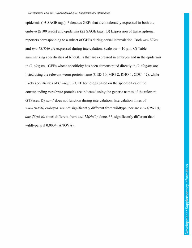
Development 142: doi:10.1242/dev.127597: Supplementary information
epidermis (≥3 SAGE tags); * denotes GEFs that are moderately expressed in both the
embryo (≥100 reads) and epidermis (≥2 SAGE tags). B) Expression of transcriptional
reporters corresponding to a subset of GEFs during dorsal intercalation. Both vav-1/Vav
and unc-73/Trio are expressed during intercalation. Scale bar = 10 µm. C) Table
summarizing specificities of RhoGEFs that are expressed in embryos and in the epidermis
in C. elegans. GEFs whose specificity has been demonstrated directly in C. elegans are
listed using the relevant worm protein name (CED-10, MIG-2, RHO-1, CDC- 42), while
likely specificities of C. elegans GEF homologs based on the specificities of the
corresponding vertebrate proteins are indicated using the generic names of the relevant
GTPases. D) vav-1 does not function during intercalation. Intercalation times of
vav-1(RNAi) embryos are not significantly different from wildtype, nor are vav-1(RNAi);
unc-73(rh40) times different from unc-73(rh40) alone. **, significantly different than
wildtype, p ≤ 0.0004 (ANOVA).
Dev
elo
pmen
t • S
uppl
emen
tary
info
rmat
ion

Development 142: doi:10.1242/dev.127597: Supplementary information
Figure S9: CRML-1 regulates intercalate time and is required in epidermal cells. A)
Intercalation time in wildtype and crml-1(n1962) based on DIC images. **, p < 0.001,
Student’s T-test. B) Extra protrusions seen in crml-1(gm326) mutants are rescued when
CRML-1 is expressed from the lbp-1 promoter. crml-1(gm326) has significantly more
numerous (top) and significantly larger (bottom) protrusions than Plbp-1::crml-1::gfp;
crml-1(gm326) or wildtype (WT). **, p<0.007 (ANOVA). However, protrusion number
and area were not significantly different (N.S.) when comparing Plbp-1::crml-1::gfp; crml-
1(gm326) and WT (ANOVA). Error bars = s.e.m. Sample size (number of cells) is at the
bottom of each bar. C) Representative micrographs of mosaic F-actin reporter expression
in crml-1(gm326), Plbp-1::crml-1::gfp; crml-1(gm326), and WT. Yellow arrows point to
extra protrusions. Scale bar = 5 µm.
Dev
elo
pmen
t • S
uppl
emen
tary
info
rmat
ion

Development 142: doi:10.1242/dev.127597: Supplementary information
Figure S10: UNC-73B::GFP localization is unchanged in crml-1(gm326) mutants.
UNC-73B::GFP localizes laterally (yellow arrowheads) in both wild type and crml-
1(gm326) backgrobackgrounds. Scale bar = 10 µm.
Dev
elo
pmen
t • S
uppl
emen
tary
info
rmat
ion

Development 142: doi:10.1242/dev.127597: Supplementary information
Supplemental Movie 1: Protrusions in wildtype (WT) and epidermally-expressed
CED-10(DN) viewed with an F-actin reporter. Minutes denoted in top left.
Supplementary Movies
Supplemental Movie 2: Protrusions in wildtype (WT) and epidermally-expressed
CED-10(CA) viewed with an F-actin reporter. Minutes denoted in top left. As noted
on the first frame, the ced-10(CA) embryo has an extra contralateral cell labeled with the
F-actin reporter relative to the wild-type movie.
Dev
elo
pmen
t • S
uppl
emen
tary
info
rmat
ion

Development 142: doi:10.1242/dev.127597: Supplementary information
Supplemental Movie 3: Protrusions in wildtype (WT) and unc-73(gm40/GEF1)
viewed with an F-actin reporter. Minutes denoted in top left.
Supplemental Movie 4: Protrusions in wildtype (WT) and crml-1(RNAi) viewed with
an F-actin reporter. Minutes denoted in top left. D
evel
opm
ent •
Sup
plem
enta
ry in
form
atio
n