Polarised cell migration during cell-to-cell transmission of herpes simplex virus in human skin
12
Polarized Cell Migration during Cell-to-Cell Transmission of Herpes Simplex Virus in Human Skin Keratinocytes Fernando Abaitua, F. Rabiya Zia, Michael Hollinshead, Peter O’Hare Section of Virology, Faculty of Medicine, Imperial College, London, United Kingdom In addition to transmission involving extracellular free particles, a generally accepted model of virus propagation is one wherein virus replicates in one cell, producing infectious particles that transmit to the next cell via cell junctions or induced polarized contacts. This mechanism of spread is especially important in the presence of neutralizing antibody, and the concept underpins analysis of virus spread, plaque size, viral and host functions, and general mechanisms of virus propagation. Here, we demon- strate a novel process involved in cell-to-cell transmission of herpes simplex virus (HSV) in human skin cells that has not previ- ously been appreciated. Using time-lapse microscopy of fluorescent viruses, we show that HSV infection induces the polarized migration of skin cells into the site of infection. In the presence of neutralizing antibody, uninfected skin cells migrate to the ini- tial site of infection and spread over infected cells to become infected in a spatially confined cluster containing hundreds of cells. The cells in this cluster do not undergo cytocidal cell lysis but harbor abundant enveloped particles within cells and cell-free vi- rus within interstitial regions below the cluster surface. Cells at the base and outside the cluster were generally negative for virus immediate-early expression. We further show, using spatially separated monolayer assays, that at least one component of this induced migration is the paracrine stimulation of a cytotactic response from infected cells to uninfected cells. The existence of this process changes our concept of virus transmission and the potential functions, virus, and host factors involved. T he mechanisms involved in the transmission of infectious vi- ruses between cells are of fundamental importance for our overall understanding of virus replication, virulence, and patho- genesis and for long-term objectives in combating infection (1). Much of our understanding of virus cell-to-cell transmission has been underpinned by analysis of plaque formation in culture, which demonstrates for many viruses two main routes of trans- mission, i.e., the production of extracellular virions which infect new cells from without and intercellular infection where infec- tious particles transmit between cells via cell-to-cell contacts or specialized, sometimes induced connections, e.g., viral synapses (2, 3). Many viruses, including herpesviruses, spread by direct cell-to-cell transmission and, where examined, indeed as part of the definition, cell-to-cell spreading is generally resistant to neu- tralizing antibody that would otherwise block transmission by ex- tracellular virus (3–6). Plaque formation is then widely analyzed, e.g., to determine the effect of mutation in virus-encoded genes on overall transmission efficiency, to identify distinct requirements for specific virus proteins during cell-to-cell transmission com- pared to those in extracellular entry (7–11), to analyze receptor usage and relocalization at cell-to-cell junctions (12), and to dem- onstrate host responses and interactions with incoming genomes (13), among many other types of investigation. However, while certain basic observations on cell-to-cell transmission have been established, novel features continue to emerge. For example, in HIV and human T-cell leukemia virus (HTLV) it has been dem- onstrated that infection induces polarized contacts, termed viral synapses, via which virus cell-to-cell transmission then occurs (14–16). In vaccinia virus, it appears that the transmission rate during plaque formation (in the absence of extracellular virus neutralization) is accelerated by virtue of a surfing mechanism, whereby virus emerging from an infected cell skips over and is specifically repelled by immediately adjacent cells, so that the virus infects naive cells further away, thus promoting a more rapid over- all transmission and larger plaques (17). In herpes simplex virus (HSV), the main route of transmission in human tissues is cell-to-cell spread, occurring during primary infection when progeny virus spreads from the primary infected cell to adjacent cells in the mucocutaneous tissue and then to axonal termini of sensory neurons (18). Cell-to-cell transmission also occurs upon reactivation from latency, when newly replicated virus spreads from the sensory neuron to the mucocutaneous tis- sue. The simplest model of cell-to-cell transmission is plaque for- mation in cell culture, which has been widely studied in monkey or rodent cell lines such as Vero and BHK cells, and results from a generally cytocidal infection producing progeny viruses which ef- ficiently spread across the monolayer. While infection produces extracellular virus, the majority of HSV remains cell associated and transmits across cell junctions despite the presence of extra- cellular virus-neutralizing antibody. The precise mechanisms of intercellular transmission remain to be fully understood. It is gen- erally assumed that transmission parameters, including, e.g., plaque diameter, represent many multiple cumulative processes, including, e.g., efficiency of virus replication and assembly of in- fectious particles, sorting to and potential reorganization of ap- propriate junctions, intercellular virus transport, and possible in- trinsic immune effectors that may resist infection (1, 8, 10, 19). We have now found that in human skin keratinocytes, an epi- thelial cell type of more physiological relevance for HSV infection processes, the outcome of infection was significantly different than in the other cells. In skin cells in the presence of neutralizing antibody, HSV infection did propagate from cell-to-cell but did Received 1 May 2013 Accepted 1 May 2013 Published ahead of print 8 May 2013 Address correspondence to Peter O’Hare, [email protected]. Copyright © 2013, American Society for Microbiology. All Rights Reserved. doi:10.1128/JVI.01172-13 July 2013 Volume 87 Number 14 Journal of Virology p. 7921–7932 jvi.asm.org 7921 Downloaded from https://journals.asm.org/journal/jvi on 15 February 2022 by 2406:3003:2002:4ad4:f5d3:f20c:ff3d:ea94.
Transcript of Polarised cell migration during cell-to-cell transmission of herpes simplex virus in human skin

Polarized Cell Migration during Cell-to-Cell Transmission of HerpesSimplex Virus in Human Skin Keratinocytes
Fernando Abaitua, F. Rabiya Zia, Michael Hollinshead, Peter O’Hare
Section of Virology, Faculty of Medicine, Imperial College, London, United Kingdom
In addition to transmission involving extracellular free particles, a generally accepted model of virus propagation is one whereinvirus replicates in one cell, producing infectious particles that transmit to the next cell via cell junctions or induced polarizedcontacts. This mechanism of spread is especially important in the presence of neutralizing antibody, and the concept underpinsanalysis of virus spread, plaque size, viral and host functions, and general mechanisms of virus propagation. Here, we demon-strate a novel process involved in cell-to-cell transmission of herpes simplex virus (HSV) in human skin cells that has not previ-ously been appreciated. Using time-lapse microscopy of fluorescent viruses, we show that HSV infection induces the polarizedmigration of skin cells into the site of infection. In the presence of neutralizing antibody, uninfected skin cells migrate to the ini-tial site of infection and spread over infected cells to become infected in a spatially confined cluster containing hundreds of cells.The cells in this cluster do not undergo cytocidal cell lysis but harbor abundant enveloped particles within cells and cell-free vi-rus within interstitial regions below the cluster surface. Cells at the base and outside the cluster were generally negative for virusimmediate-early expression. We further show, using spatially separated monolayer assays, that at least one component of thisinduced migration is the paracrine stimulation of a cytotactic response from infected cells to uninfected cells. The existence ofthis process changes our concept of virus transmission and the potential functions, virus, and host factors involved.
The mechanisms involved in the transmission of infectious vi-ruses between cells are of fundamental importance for our
overall understanding of virus replication, virulence, and patho-genesis and for long-term objectives in combating infection (1).Much of our understanding of virus cell-to-cell transmission hasbeen underpinned by analysis of plaque formation in culture,which demonstrates for many viruses two main routes of trans-mission, i.e., the production of extracellular virions which infectnew cells from without and intercellular infection where infec-tious particles transmit between cells via cell-to-cell contacts orspecialized, sometimes induced connections, e.g., viral synapses(2, 3). Many viruses, including herpesviruses, spread by directcell-to-cell transmission and, where examined, indeed as part ofthe definition, cell-to-cell spreading is generally resistant to neu-tralizing antibody that would otherwise block transmission by ex-tracellular virus (3–6). Plaque formation is then widely analyzed,e.g., to determine the effect of mutation in virus-encoded genes onoverall transmission efficiency, to identify distinct requirementsfor specific virus proteins during cell-to-cell transmission com-pared to those in extracellular entry (7–11), to analyze receptorusage and relocalization at cell-to-cell junctions (12), and to dem-onstrate host responses and interactions with incoming genomes(13), among many other types of investigation. However, whilecertain basic observations on cell-to-cell transmission have beenestablished, novel features continue to emerge. For example, inHIV and human T-cell leukemia virus (HTLV) it has been dem-onstrated that infection induces polarized contacts, termed viralsynapses, via which virus cell-to-cell transmission then occurs(14–16). In vaccinia virus, it appears that the transmission rateduring plaque formation (in the absence of extracellular virusneutralization) is accelerated by virtue of a surfing mechanism,whereby virus emerging from an infected cell skips over and isspecifically repelled by immediately adjacent cells, so that the virusinfects naive cells further away, thus promoting a more rapid over-all transmission and larger plaques (17).
In herpes simplex virus (HSV), the main route of transmissionin human tissues is cell-to-cell spread, occurring during primaryinfection when progeny virus spreads from the primary infectedcell to adjacent cells in the mucocutaneous tissue and then toaxonal termini of sensory neurons (18). Cell-to-cell transmissionalso occurs upon reactivation from latency, when newly replicatedvirus spreads from the sensory neuron to the mucocutaneous tis-sue. The simplest model of cell-to-cell transmission is plaque for-mation in cell culture, which has been widely studied in monkeyor rodent cell lines such as Vero and BHK cells, and results from agenerally cytocidal infection producing progeny viruses which ef-ficiently spread across the monolayer. While infection producesextracellular virus, the majority of HSV remains cell associatedand transmits across cell junctions despite the presence of extra-cellular virus-neutralizing antibody. The precise mechanisms ofintercellular transmission remain to be fully understood. It is gen-erally assumed that transmission parameters, including, e.g.,plaque diameter, represent many multiple cumulative processes,including, e.g., efficiency of virus replication and assembly of in-fectious particles, sorting to and potential reorganization of ap-propriate junctions, intercellular virus transport, and possible in-trinsic immune effectors that may resist infection (1, 8, 10, 19).
We have now found that in human skin keratinocytes, an epi-thelial cell type of more physiological relevance for HSV infectionprocesses, the outcome of infection was significantly differentthan in the other cells. In skin cells in the presence of neutralizingantibody, HSV infection did propagate from cell-to-cell but did
Received 1 May 2013 Accepted 1 May 2013
Published ahead of print 8 May 2013
Address correspondence to Peter O’Hare, [email protected].
Copyright © 2013, American Society for Microbiology. All Rights Reserved.
doi:10.1128/JVI.01172-13
July 2013 Volume 87 Number 14 Journal of Virology p. 7921–7932 jvi.asm.org 7921
Dow
nloa
ded
from
http
s://j
ourn
als.
asm
.org
/jour
nal/j
vi o
n 15
Feb
ruar
y 20
22 b
y 24
06:3
003:
2002
:4ad
4:f5
d3:f
20c:
ff3d
:ea9
4.

not show the typical cytocidal plaque formation seen in other cells.Instead, infected cells formed distinct bunched clusters with a dis-tinctive surrounding area in which uninfected cells aligned in anoriented fashion. The diameter of the plaques was smaller in skinkeratinocytes than, for example, in Vero cells, but this did notreflect lower success of transmission. Rather, we show abundantvirus cell-to-cell transmission. However, in contrast to the typicalperception of HSV plaque propagation, the outcome of infectionwas due to an induced polarized migration of cells to the site ofinfection. In additional analysis, we show that was a consequenceat least in part of paracrine signaling from the infected cells. Al-though relatively simple in concept, and with many aspects re-quiring further investigation, this work has potentially profoundramifications, revealing a new process that has not been previouslyappreciated, changing our perception of virus spread and of thepotential mechanisms involved.
MATERIALS AND METHODSCells, virus, and infection. Vero were grown in Dulbecco modified min-imal essential medium (DMEM; Gibco) containing 10% newborn calfserum (NCS) and penicillin-streptomycin. RPE-1 cells, a human telome-rase immortalized retinal pigment epithelial line, were grown in DMEM-F12 (Sigma) supplemented with 200 mM glutamine, 10% fetal bovineserum (FBS), and penicillin-streptomycin. Human epithelial keratino-cytes were the HaCaT cell line (20) grown in DMEM containing 10% FBSand penicillin-streptomycin or primary keratinocytes which had been im-mortalized with telomerase (21). We have previously show that in single-step growth curves HSV normally replicates, as well in HaCaT cells as, e.g.,Vero cells (22). The telomerase-immortalized keratinocytes (hTert-kera-tinocytes) were grown in Ham’s/F-12 (PAA) supplemented with 10%FBS, penicillin-streptomycin, 10 ng of mouse epidermal growth factor/ml, 1 ng of cholera toxin/ml, 400 ng of hydrocortisone/ml, 5 �g of insulin/ml, 5 �g of transferrin/ml, and 13 ng of liothyronine/ml. All supplementswere from Sigma except mouse epidermal growth factor which was fromAbD Serotec, Oxford, United Kingdom. The virus strains were HSV-1[17]or HSV-1[17].ICP0-YFP, a derivative of HSV-1[17] that expresses theimmediate-early protein ICP0 fused to YFP and has been previously char-acterized (23). Pooled neutralizing human serum (HS; Sigma) was used at1%. Clinical-grade purified neutralizing human immunoglobulin (IVIg,Carimune NF, nanofiltered, human immune globulin; CSL Behring) wasused at 5 mg/ml, having demonstrated complete neutralization of extra-cellular virus at this dose (�6-log reduction in virus titer).
Immunofluorescence studies. Immunofluorescence analysis was per-formed exactly as described previously (24). Anti-ICP4 antibody (Viru-sys) was used at 1:400. For fluorescence readout of total cells, alongside theyellow fluorescent protein (YFP) fluorescence to identify infected cells,monolayers were stained with CellMask Orange (Invitrogen), a generalprobe for plasma membrane staining which can be used in live cells. ForCellMask staining of live cells, the medium was replaced with fresh me-dium containing the probe (5 �g/ml) for 5 min, the cells then washed infresh medium and then imaged. For fixed cells, samples were washed withphosphate-buffered saline (PBS), fixed with 3.7% paraformaldehyde inPBS, and blocked with PBS containing 10% NCS. Images were collectedusing Zeiss Axiovert 135TV microscope and Zeiss �10 (NA 0.5) or �40(NA 0.6) LD lenses. Images were taken using a filter set XF66 (Glen Spec-tra, United Kingdom) using the green fluorescent protein (GFP) setting toimage YFP-ICP0. Each channel was captured sequentially with a Retiga2000R camera using Image Pro plus software. Composite illustrationswere prepared using Adobe software. For more detailed comparative anal-ysis of infection in HaCaT cells samples were examined by confocal mi-croscopy using a Zeiss LSM510 confocal microscope. Images were col-lected using a �10 objective (NA 0.6) and compiled using the Z sectioningmodule. The resulting images were analyzed using Axiovision Le softwareto construct orthogonal axial images for each channel. Example images
are representative of numerous images gathered for each virus and con-dition.
Time-lapse microscopy. Cells plated in 35-mm dishes were infectedwith 50 to 100 PFU per dish with either HSV-1[17] or HSV-1[17].ICP0-YFP. Infections were performed in the absence or presence of 1% pooledHS or clinical grade purified immunoglobulin (IVIg, 5 mg/ml) in mediacontaining 2% NCS. Dishes were placed on a heated chamber at 37°C inan environmentally controlled mini-incubator (PeCon GmBH) main-tained at 5% CO2. Images were captured on a Zeiss Axiovert 135TV mi-croscope using �10 or �40LD lenses and Image Pro plus software capturesuit for sequential collection of fluorescence and phase channels for eachtime point. Routinely single isolated small clusters of infected cells wereidentified by the positive YFP signal (ca. 8 to 12 h postinfection) and timelapse collection then initiated using the AFA module of the software.Animation of the time series was carried out after merging the fluores-cence and phase channels using Image Pro Plus and compressed withImageJ software. Image Pro Plus cell tracking module was performed bymanually tracking individual cells on zoomed versions of each image se-ries and overlaying each track on the video animation. Total distance fromthe origin of the track was then calculated by the automated software andplotted.
Cell migration in gap-filling assays. Cells were plated within 35-mm�-Dishes (Ibidi) that contain two chambers separated by a removablesilicone insert with a specific gap size of 500 �m. Cells were plated in theadjoining chambers and once the monolayers were formed (24 h), thesilicone inserts were removed leaving the defined gap. Fresh medium con-taining 2% NBS and 1% pooled HS was added, and the chambers wereplaced in an environmentally controlled mini-incubator. Time-lapse ex-amination of gap closure and cell migration was then initiated, capturingphase images every 5 min over the subsequent 18-h period. Images werecompiled and compressed as described above.
Cell migration in response to paracrine signaling. Cells were platedon the bottom wells (2 � 105 cells/well) of 24 multiwell plates (Falcon)into which FluoroBlock cell culture inserts (BD Biosciences) were to beinserted (see Fig. 5). These inserts are designed for the plating of cells on amembrane which contains pores of defined size. The base of the mem-brane blocks all fluorescence transmission, such that when using live cellfluorescence analysis with an inverted microscope, any fluorescence signaloriginates only from cells that migrated through the pores onto the bot-tom side of the membrane. Pores of different diameter (3 and 8 �m) wereexamined. The bottom cell monolayers in the 24-well plates were mockinfected or infected with 100 PFU or 1,000 PFU of HSV-1[17].ICP0-YFP/well. After 1 h of adsorption in serum-free media, the inoculum was re-moved, and medium containing 2% FBS and 1% pooled neutralizing HSwas added. The inserts, which had been separately plated with HaCaT cellsat a density of 2 � 104 cells/insert, were then placed into the 24-well platescontaining the mock-infected or infected monolayers. At different timesafter initial infection, the inserts were removed, incubated in a fresh dishin medium with 2 �M Calcein AM (Invitrogen) for 30 min at roomtemperature prior to live imaging microscopy. In separate assays, we con-firmed that the test migratory cells from the upper inserts remained un-infected, as expected from the presence of the neutralizing antibody. Im-ages of cell migration through the pores in response to medium fromuninfected or infected lower chambers were then collected. At least threerandom fields using a low-power �10 objective lens were obtained foreach condition. Representative images are shown and were quantitatedbased on the total pixel density (Calcein AM staining) of the test cells foreach condition.
Transmission electron microscopy (TEM). Cells were fixed in 0.5%glutaraldehyde in 200 mM sodium cacodylate buffer for 30 min, washedin buffer, and secondarily fixed in reduced 1% osmium tetroxide–1.5%potassium ferricyanide for 60 min. The samples were washed in distilledwater and stained overnight at 4°C in 0.5% magnesium uranyl acetate,washed in distilled water, and dehydrated in graded ethanol. The sampleswere then embedded flat in the dish in Epon resin. Resin filled stubs were
Abaitua et al.
7922 jvi.asm.org Journal of Virology
Dow
nloa
ded
from
http
s://j
ourn
als.
asm
.org
/jour
nal/j
vi o
n 15
Feb
ruar
y 20
22 b
y 24
06:3
003:
2002
:4ad
4:f5
d3:f
20c:
ff3d
:ea9
4.

placed on embedded cell monolayers and polymerized. The dish was re-moved, and the sample was reorientated for transverse vertical sectioning.Semithin survey sections 500 to 1,000 nm were first cut and collected ontoglass slides and then stained with crystal violet. Images were collected on aZeiss Axiovert 200M using Axiovision software. The polished block faceand embedded sample were observed through a stereo microscope with acharge-coupled device (CCD) camera attached to the eyepiece, and im-ages were collected. The sample was sectioned again when appropriatetransverse sections were observed, and ultrathin sections (typically 50 to70 nm) were cut and collected onto slot grids, stained with Reynold’s leadcitrate examined in a FEI Tecnai electron microscope with CCD cameraimage acquisition.
RESULTSAltered plaque formation by HSV in human skin cells. We com-pared plaque formation in Vero cells, a monkey kidney line widelyused for study of growth and replication of HSV, to that in ahuman skin keratinocyte line (HaCaT) and a human retinal pig-ment cell line (RPE). Monolayers in 35-mm dishes were infectedwith �200 PFU of HSV-1[17] and then incubated in the presenceof 1% HS to neutralize extracellular virus and limit plaque forma-tion to primary plaques formed by cell-to-cell transmission.Plaques were observed after 60 h by phase-contrast microscopy orcrystal violet staining (Fig. 1). Typical plaques 2 to 3 mm in diam-eter and with a central clear area resulting from cytocidal cell lysiswere observed in Vero cells, with generally similar results andsomewhat larger plaques in RPE cells (Fig. 1a). In contrast, inHaCaT cells, while overall plaque numbers were the same (datanot shown), plaque morphology was distinctly different, lacking aclear central area with, instead, clustered, piled-up cells and amore uniform circular appearance of the plaque. Of note was theobservation of a distinct zone around the plaques in HaCaT cells.This reflected the reorientation of the cells in the zone from a morerandom shape and organization within the body of the monolay-ers to a more elongated shape and uniform alignment orientedtoward the plaque center (Fig. 1a, see also Fig. 1b and c and Fig. 4).This type of plaque morphology was surprisingly long-lived, withregular piled up clusters being observed for at least 3 to 4 days afterinitiation without overt central lysis.
To investigate the altered plaque formation in HaCaT cells, weused HSV-1[17].ICP0-YFP, a version of HSV-1[17] that expressesthe immediate-early protein ICP0 fused to the autofluorescentprotein YFP. After infection with �100 PFU per dish, individualdeveloping plaques could be identified, and infected and unin-fected surrounding cells were simultaneously imaged. Typical re-sults again demonstrated the absence of a clear plaque and insteada turbid plaque with numerous cells in a piled-up cluster sur-rounded by a zone (indicated by a dotted black circle) whereinuninfected cells could be seen to reorient in a more uniform man-ner (Fig. 1b). The boundary of this zone from the more proximaloriented cells to the more distant randomly organized cells wasreadily observed (Fig. 1b, circle). The cells within the surroundingzone did not exhibit any detectable immediate-early ICP0-YFPexpression (see the example with separate channels; there is a zoneindicated by a black circle in the top phase panel, and the samezone is indicated by a white circle in the GFP channel). Althoughthese analyses were performed in live cells, we also did not detectimmediate-early proteins (or indeed any of a number of antigenstested) by immunostaining (data not shown), indicating thatthese surrounding cells were as yet uninfected.
We further compared the zones around the boundaries of live
plaques in the three cell types using YFP in one channel (Fig. 1c,panels i to iii) and a total plasma membrane stain (Cellmask) inthe second channel (Fig. 1c, panels iv to vi). For both Vero andRPE cells, cells beyond the YFP boundary did not exhibit anyspecific realignment, and no distinct differences from the generalmock-infected picture could be observed (panels vii and ix). In
FIG 1 HSV spread comparison in different cell lines. Monolayers of Vero,HaCaT, and RPE cells were infected with �100 PFU of HSV-1[17]/well in thepresence of HS. (a) Bright-field images (�10 objective lens) of plaques at 72 hpostinfection by live microscopy (upper panels) prior to fixation and stainingwith crystal violet (lower panel). (b) Typical plaque formed in HaCaT cells byHSV-1[17].ICP0-YFP, showing a merged image of phase and YFP (48 hpostinfection, �10 objective lens). The dashed black circle indicates the area ofYFP� cells showing an elongated and oriented morphology compared to thesurrounding cells (outside the circle). The images on the right show eachchannel independently, with the corresponding circles indicating the area ofmorphologically altered, YFP� cells. (c) HSV-1[17].ICP0-YFP plaques in eachof the cell types indicated (48 h postinfection, �40 objective lens) showingplaque perimeters (panels i to vi) compared to mock monolayers (panels vii toix). Live monolayers were stained with general plasma membrane stain Cell-Mask (panels iv to ix) compared to the ICP0-YFP signal for virus infection(panels i to iii). The area of morphologically elongated GFP� cells is indicatedwith a white bracket in the CellMask image (panel v).
Cell Migration during HSV Spread in Keratinocytes
July 2013 Volume 87 Number 14 jvi.asm.org 7923
Dow
nloa
ded
from
http
s://j
ourn
als.
asm
.org
/jour
nal/j
vi o
n 15
Feb
ruar
y 20
22 b
y 24
06:3
003:
2002
:4ad
4:f5
d3:f
20c:
ff3d
:ea9
4.
contrast, in HaCaT cells, a distinct zone of more elongated andaligned cells could be seen outside the YFP� boundary ((Fig. 1c,panel v, indicated by a white bracket). Similar results were ob-tained with other HSV-1 strains, e.g., KOS, and with HSV-2 (datanot shown).
We interpret these results to indicate a qualitative difference inthe response to HSV infection in HaCaT keratinocyte cells sur-rounding a developing plaque. One possible explanation was thatthis reflected not just a morphological reorganization but ratherwas a consequence of an active directional migration of theHaCaT cells with the consequential appearance of aligned, elon-gated cells. To pursue this, we first investigated basic growth fea-tures and motility properties in uninfected cells of the three types.
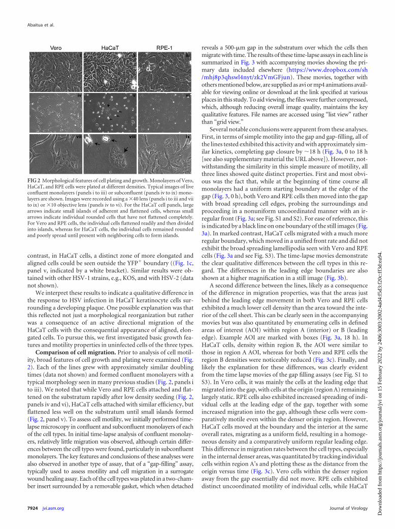
Comparison of cell migration. Prior to analysis of cell motil-ity, broad features of cell growth and plating were examined (Fig.2). Each of the lines grew with approximately similar doublingtimes (data not shown) and formed confluent monolayers with atypical morphology seen in many previous studies (Fig. 2, panels ito iii). We noted that while Vero and RPE cells attached and flat-tened on the substratum rapidly after low density seeding (Fig. 2,panels iv and vi), HaCaT cells attached with similar efficiency, butflattened less well on the substratum until small islands formed(Fig. 2, panel v). To assess cell motility, we initially performed time-lapse microscopy in confluent and subconfluent monolayers of eachof the cell types. In initial time-lapse analysis of confluent monolay-ers, relatively little migration was observed, although certain differ-ences between the cell types were found, particularly in subconfluentmonolayers. The key features and conclusions of these analyses werealso observed in another type of assay, that of a “gap-filling” assay,typically used to assess motility and cell migration in a surrogatewound healing assay. Each of the cell types was plated in a two-cham-ber insert surrounded by a removable gasket, which when detached
reveals a 500-�m gap in the substratum over which the cells thenmigrate with time. The results of these time-lapse assays in each line issummarized in Fig. 3 with accompanying movies showing the pri-mary data included elsewhere (https://www.dropbox.com/sh/mhj8p3qhswl4nyt/zk2VmGFjun). These movies, together withothers mentioned below, are supplied as avi or mp4 animations avail-able for viewing online or download at the link specified at variousplaces in this study. To aid viewing, the files were further compressed,which, although reducing overall image quality, maintains the keyqualitative features. File names are accessed using “list view” ratherthan “grid view.”
Several notable conclusions were apparent from these analyses.First, in terms of simple motility into the gap and gap-filling, all ofthe lines tested exhibited this activity and with approximately sim-ilar kinetics, completing gap closure by �18 h (Fig. 3a, 0 to 18 h[see also supplementary material the URL above]). However, not-withstanding the similarity in this simple measure of motility, allthree lines showed quite distinct properties. First and most obvi-ous was the fact that, while at the beginning of time course allmonolayers had a uniform starting boundary at the edge of thegap (Fig. 3, 0 h), both Vero and RPE cells then moved into the gapwith broad spreading cell edges, probing the surroundings andproceeding in a nonuniform uncoordinated manner with an ir-regular front (Fig. 3a; see Fig. S1 and S2). For ease of reference, thisis indicated by a black line on one boundary of the still images (Fig.3a). In marked contrast, HaCaT cells migrated with a much moreregular boundary, which moved in a unified front rate and did notexhibit the broad spreading lamellipodia seen with Vero and RPEcells (Fig. 3a and see Fig. S3). The time-lapse movies demonstratethe clear qualitative differences between the cell types in this re-gard. The differences in the leading edge boundaries are alsoshown at a higher magnification in a still image (Fig. 3b).
A second difference between the lines, likely as a consequenceof the difference in migration properties, was that the areas justbehind the leading edge movement in both Vero and RPE cellsexhibited a much lower cell density than the area toward the inte-rior of the cell sheet. This can be clearly seen in the accompanyingmovies but was also quantitated by enumerating cells in definedareas of interest (AOI) within region A (interior) or B (leadingedge). Example AOI are marked with boxes (Fig. 3a, 18 h). InHaCaT cells, density within region B, the AOI were similar tothose in region A AOI, whereas for both Vero and RPE cells theregion B densities were noticeably reduced (Fig. 3c). Finally, andlikely the explanation for these differences, was clearly evidentfrom the time lapse movies of the gap filling assays (see Fig. S1 toS3). In Vero cells, it was mainly the cells at the leading edge thatmigrated into the gap, with cells at the origin (region A) remaininglargely static. RPE cells also exhibited increased spreading of indi-vidual cells at the leading edge of the gap, together with someincreased migration into the gap, although these cells were com-paratively motile even within the denser origin region. However,HaCaT cells moved at the boundary and the interior at the sameoverall rates, migrating as a uniform field, resulting in a homoge-neous density and a comparatively uniform regular leading edge.This difference in migration rates between the cell types, especiallyin the internal denser areas, was quantitated by tracking individualcells within region A’s and plotting these as the distance from theorigin versus time (Fig. 3c). Vero cells within the denser regionaway from the gap essentially did not move. RPE cells exhibiteddistinct uncoordinated motility of individual cells, while HaCaT
FIG 2 Morphological features of cell plating and growth. Monolayers of Vero,HaCaT, and RPE cells were plated at different densities. Typical images of liveconfluent monolayers (panels i to iii) or subconfluent (panels iv to ix) mono-layers are shown. Images were recorded using a �40 lens (panels i to iii and viito ix) or �10 objective lens (panels iv to vi). For the HaCaT cell panels, largearrows indicate small islands of adherent and flattened cells, whereas smallarrows indicate individual rounded cells that have not flattened completely.For Vero and RPE cells, the individual cells flattened readily and then dividedinto islands, whereas for HaCaT cells, the individual cells remained roundedand poorly spread until present with neighboring cells to form islands.
Abaitua et al.
7924 jvi.asm.org Journal of Virology
Dow
nloa
ded
from
http
s://j
ourn
als.
asm
.org
/jour
nal/j
vi o
n 15
Feb
ruar
y 20
22 b
y 24
06:3
003:
2002
:4ad
4:f5
d3:f
20c:
ff3d
:ea9
4.

FIG 3 Motility characteristics of the cell lines. Vero, HaCaT, or RPE cell monolayers were plated in a two-chamber system with a removable gasket. As describedin Materials and Methods, the subsequent gap filling assay was analyzed by time-lapse microscopy after removal of the gasket. (a) Individual snapshots at differenttimes (0 to 18 h) after start of the assay showing gap filling by each of the cell lines. The frontline boundary of moving cells is outlined (black) on one boundary.Regions termed A within the denser internal area and B at the cell boundary are indicated by brackets. Boxes within the 18-h time point indicate typical AOI usedto calculate cell density as described in the text. (b) Zoomed image (12-h time point) of the boundaries showing the distinct morphology of the migrating cellsat the front. (c) Three independent AOI (black squares) within the defined regions A (original plated area) and B (just behind boundary) were quantified. Theaverage cell density of an AOI in region A was standardized as 100% and then compared to that in the average AOI from just at the boundary. (d) Quantificationof migration for 10 independent cells within area A for each cell line. Each cell was identified at the start of the time lapse, tracked (one point/5 min) for 18 h, andpositioned from the origin plotted against time. The scales are identical for all cells.
Cell Migration during HSV Spread in Keratinocytes
July 2013 Volume 87 Number 14 jvi.asm.org 7925
Dow
nloa
ded
from
http
s://j
ourn
als.
asm
.org
/jour
nal/j
vi o
n 15
Feb
ruar
y 20
22 b
y 24
06:3
003:
2002
:4ad
4:f5
d3:f
20c:
ff3d
:ea9
4.

cells were clearly more motile, independent of density. Together,these data indicated that in Vero cells, gap filling was mainly due tosignificant cell spreading (and cell enlargement), together withsome selective migration of leading edge cells. RPE cells spreadinto the gap but were clearly more motile, migrating as individualcells at the gap edge but also in denser interior areas. HaCaT cellswere distinct, and yet again in that gap filling was less due to cellspreading and enlargement but rather reflected migration in auniform coordinated manner both at the leading edge and in theinterior, more dense areas of the monolayer.
HSV infection induces polarized migration of keratinocytes.We next examined the potential for cell migration during plaquedevelopment in HaCaT cells and compared this to Vero cells. Cellmonolayers were infected with �100 PFU of HSV-1[17].ICP0-
YFP/dish to allow the identification and monitoring of isolateddeveloping plaques. Plaque progression was then recorded bytime-lapse microscopy for both fluorescence and phase imagesand the merged channels animated as movies (Fig. 4 and see Fig.S4 and S5 at https://www.dropbox.com/sh/mhj8p3qhswl4nyt/zk2VmGFjun). At the initiation of the animations (24 h), therewas a clear contrast in plaque morphology. In Vero cells (Fig. 4aand b and see also the accompanying movie in Fig. S4), the patternof infection (YFP fluorescence) spread out from the center, whichalready showed a central cytolytic effect, with no overt reorgani-zation of surrounding cells. In contrast, in HaCaT cells, infectionwas in a distinct central focus with a much more defined bound-ary, surrounded by a zone of cells aligned toward the focus (Fig.4c). The accompanying movie (see Fig. S5) shows the striking
FIG 4 Polarized migration of HaCaT during plaque progression. Monolayers of HaCaT cells (a and b) or Vero cells (c and d) in 35-mm dishes were infected with�100 PFU of HSV-1[17].ICP0-YFP/dish in the presence of neutralizing antibody (1% HS) and incubated in an environmentally controlled microscope stage.Developing plaques were identified between 16 and 24 h and then recorded for both phase and YFP fluorescence for �18 h (1 frame every 5 min). The still images(a and c) show the last frame, merged for phase and YFP, of each time-lapse series. The time-lapse series upon which the quantitation was based (1 frame/25 min)are shown in accompanying movies (Fig. S4 and S5 in the supplemental material [https://www.dropbox.com/sh/mhj8p3qhswl4nyt/zk2VmGFjun]). The alteredzone of cells surrounding the clustered plaque in HaCaT cells was readily observed. Random cells from outside this zone (blue tracks) and inside (red tracks) wereindividually tracked during the time lapse, and the distance from origin was plotted as a function of time (b and d).
Abaitua et al.
7926 jvi.asm.org Journal of Virology
Dow
nloa
ded
from
http
s://j
ourn
als.
asm
.org
/jour
nal/j
vi o
n 15
Feb
ruar
y 20
22 b
y 24
06:3
003:
2002
:4ad
4:f5
d3:f
20c:
ff3d
:ea9
4.

result wherein this surrounding area reflects a zone of very activecell migration into the developing focus, with cells becomingYFP� only within the cluster. Individual cells were trackedthroughout the animated series and the results plotted in Fig. 4d.Cells exhibiting the realigned morphology (red tracks) showed adistinct directional migration into the focus (see Fig. S4 in theonline material, and this is also plotted Fig. 4d), whereas moredistant randomly ordered cells in the monolayer exhibited verylittle migration. In a further series of experiments, we examinedplaque migration by time lapse microscopy in a different humanskin system, using telomerase-immortalized keratinocytes (21)and observed virtually identical results (data not shown). In con-trast, in infected Vero cells, although some individual cell migra-tion appeared, this was then rapidly obscured due to the out-wardly progressing plaque (Fig. 4a and b). By comparison of boththe visual animations (see Fig. S4 and S5 in the online material[https://www.dropbox.com/sh/mhj8p3qhswl4nyt/zk2VmGFjun]) andthe behavior and lack of orientation of surrounding uninfectedVero cells, it was clear that infection progressed in a qualitativelydistinct manner in the two cell types.
The observations of induced cell migration were made underconditions which restrict transmission by extracellular virus, i.e.,the presence of neutralizing antibody. In additional studies (datanot shown), we considered whether the induced migration wasdependent upon the presence of neutralizing antibody by con-ducting time-lapse analysis of plaque progression in semisolidmedia. The results demonstrated cell migration independent ofthe presence of antibody. However, in the complete absence ofantibody and restricted diffusion, extensive plaque progressionoccurred, generally also with increased cytolysis at the center (seediscussion).
Structural analysis of infection in skin cells. We next pursuedthe distinct morphological aspects of the focus of infected HaCaT
cells. Live developing plaques were stained with Cellmask at 24 hin each of the cell types, the plaques identified in phase and thenimaged through the z dimension (Fig. 5). The resulting stacks(Cellmask, middle panel) show the vertical perspective throughthe center of the plaque (green line, top panels). In contrast toVero and RPE cells, in HaCaT cells even by 24 h, the distinct pilingof the cells in the vertical dimension could be observed. To in-crease the resolution and gain additional insight into the process,we next performed TEM through the z-dimension of the focus, asdescribed in Materials and Methods. By first imaging the embed-ded block (before sectioning) at low magnification on a tiltingstage, the vertical clusters of cells could be readily observed (Fig.6a). Sequential sections were then cut, stained, and aligned, allow-ing a higher-level resolution across a single focus (Fig. 6b). Theextremity of the image represents the surrounding zone of cells,just a single layer deep, that are migrating into the focus. Proceed-ing to the interior, e.g., the boxed areas in Fig. 6c, panels 3 and 4,cells crawling over the bottom layers can be now readily seen, withmany cells at the peak in the center (probably under-representingthe total due to processing). Finally, aligning the sequential sec-tions from the block, the exact areas corresponding to those inpanel B (numbers 1 to 4) could be identified and analyzed at highresolution (Fig. 6c, panels 1 to 4 [the lower panels display enlarge-ments of the panel insets]). This analysis revealed several featuresof virus transmission in these cells.
In the central “older” areas (Fig. 6c, regions 1 and 2), whilethere was a clear cytopathic effect, including marginated chroma-tin, the cells remained relatively intact, with numerous envelopedvirions in the interstitial space between cells (panel 1 [lower panel,white outlined arrows]). It is known that skin cells form tightjunctions, and numerous tight junctions could be observed in thevertical section between cells that had piled up over one another(Fig. 6c, panel 2 [lower panel, white arrow]). Considering the cells
FIG 5 Infected HaCaT cell clustering. Monolayers of Vero, HaCaT, and RPE cells in 35-mm dishes were infected with 100 PFU of HSV-1[17]/well, stained at 24h postinfection with CellMask, and then fixed with paraformaldehyde. Phase images (lower panels) are included to illustrate the morphological changes used toidentify the viral plaques. Entire plaques were then imaged by z-sectioning (a total of �116 �m, with intervals of 7 �m/section) from the lower part of the plaqueat the substratum to the most elevated point. The upper panels show the staining in a conventional x,y perspective, with green lines indicating the orthogonal axisapplied for the z-sectioning. The resulting stacks (middle panel) shows the vertical perspective through the center of the plaque for each cell type.
Cell Migration during HSV Spread in Keratinocytes
July 2013 Volume 87 Number 14 jvi.asm.org 7927
Dow
nloa
ded
from
http
s://j
ourn
als.
asm
.org
/jour
nal/j
vi o
n 15
Feb
ruar
y 20
22 b
y 24
06:3
003:
2002
:4ad
4:f5
d3:f
20c:
ff3d
:ea9
4.

have moved over one another, these junctions may somehow bemaintained or continuously formed as the cells migrate in thecluster. Numerous assembled virions were present between theseconnections (Fig. 6c, panel 2 [lower inset]). Few extracellular vi-rions were present on the outermost surface to the focus. How-ever, we cannot conclude (and in our view less likely) that theremay be selective virion release on the internal aspect of the clus-ters, since surface material may have been lost during processing.Toward the perimeter to the focus, cells just beginning to migrateover the underlying cells could be observed (Fig. 6c, panels 3 and4). The cell in panel 3, shows a migrating elongated cell with a tightjunction (lower inset, white arrow) between the uppermost cell(which did not yet show overt chromatin margination) and un-derlying infected cells (upper panel, arrowed capsids in the nu-cleus). At the extreme perimeter of the focus, the monolayer com-prised single flat cells with, as expected, no overt cytopathic effect.
HSV infection induces a paracrine mediator of skin cell mi-gration. Using both time-lapse microscopy and TEM analysis ofvertical sections, we provide robust evidence for the induction ofHaCaT cell migration and crawling over underlying cells, result-ing in the focus of infection. Using a well-established cell migra-
tion assay system (see Materials and Methods), we next addressedwhether there was evidence that these effects could be due to aparacrine mediator (Fig. 7a). Target HaCaT cells were plated inthe upper chamber insert on a membrane support with defined8-�m pores (Fig. 7b). The insert was then placed in a dish of testcells (lower chamber) that were either mock infected or infectedwith 100 or 1,000 PFU/dish. All cultures were in the presence ofneutralizing antibody to prevent infectious extracellular virus. Weconfirmed that the upper target cells remained uninfected. Cellmigration was then monitored by the presence of cells on thelower side of the membrane support by live cell staining. Only cellson the lower side are detected because the membrane is impervi-ous to the excitation wavelengths. The results demonstrated a veryclear increase in target cell migration with infected test cells com-pared to uninfected test cells and that this increase showed a doseresponse with increasing virus dose. This could be readily seen inthe images of target cell migration taken at low magnification toavoid sample bias (Fig. 7c) and in the quantitation of the data (Fig.7d). Identical results were obtained in repeat experiments, and inexperiments using inserts with a 3-�m pore size instead of 8 �m.In some ways, this system is a more stringent test for paracrine
FIG 6 Ultrastructural characterization of HSV plaques in HaCaT cells. Monolayers of HaCaT cells in 35-mm dishes were infected with 100 PFU of HSV-1[17]/well and processed at 24 h postinfection for TEM as described in Materials and Methods. (a) Low-power binocular image of the vertical face of the embedded resinblock. This image gives a three-dimensional view along the horizontal surface of the plastic support. Arrows indicate the plaques of clustered piled-up cells, withthe size of the arrow indicating the distance from the block front. (b) The block was then sectioned into semithin survey sections (500 to 1,000 nm) and stainedwith crystal violet. Images were collected on a Zeiss Axiovert 200M using a �40 objective lens, and the panels were assembled using Adobe Photoshop. (c)Sequential ultrathin sections (50 to 70 nm) of the same sample were then obtained and collected onto slot grids, stained, and examined by TEM. Because thesections analyzed are immediately adjacent to the section stained with crystal violet, corresponding areas could be matched. The TEM images of the sections inpanel c, sections 1 to 4, therefore correspond to the boxes in panel b, sections 1 to 4. Upper panels (lower magnification) and lower panels (higher magnificationon inset area) show different features of infection within the plaque as discussed in the text. In the lower panels, the features include extracellular virions (blackarrows with white outline), nucleocapsids (short black arrows), and cell tight junctions (white arrows). In the upper part of panels 3 and 4, cells with no distinctfeatures of infection (e.g., no early chromatin marginalization) are indicated by long black arrows. Box 5 in panel b shows flat uninfected cells that correspondto the elongated cells (TEM data not shown).
Abaitua et al.
7928 jvi.asm.org Journal of Virology
Dow
nloa
ded
from
http
s://j
ourn
als.
asm
.org
/jour
nal/j
vi o
n 15
Feb
ruar
y 20
22 b
y 24
06:3
003:
2002
:4ad
4:f5
d3:f
20c:
ff3d
:ea9
4.

effects than that occurring during focus formation, since the cellsare not in direct contact with one another. We believe that theresults indicate that HSV-infected skin cells release a soluble me-diator that can stimulate cell migration. This conclusion is consis-tent with the observations during focus formation and indicatesthat a paracrine effect, at least in part, accounts for the inducedmigration of skin cells during virus infection and transmission.
DISCUSSION
Analysis of plaque formation in tissue culture models of infectionrepresents one of the most fundamental methods used to studyvirus replication and cell-to-cell transmission. Notwithstandingthe relatively straightforward concept, the precise mechanisms ofintercellular transmission during plaque formation remain to befully understood. Furthermore, recent results show that there areprocesses involved in cell-to-cell transmission that had previouslybeen unrecognized, such as virus surfing in vaccinia virus plaqueformation (17), or the induction of polarized cell-to-cell connec-tions (14–16), or possible intercellular transmission via nanotubes(25). HSV plaque formation and cell-to-cell transmission hasbeen widely studied in several types of cell lines, generally resultingin a cytocidal infection producing progeny viruses that efficiently
spread across the monolayer. Infection produces extracellular vi-rus which disseminates to surrounding cells, but the majority ofHSV remains cell associated and transmits across cell junctionsdespite the presence of extracellular virus-neutralizing antibody,which otherwise neutralizes extracellular virus. Cell-to-cell trans-mission clearly represents many cumulative processes, including,for example, efficiency of virus replication and assembly of infec-tious particles, transport to the cell surface and potential reorga-nization of cell-to-cell junctions, intercellular virus transport, andpossible restriction mechanisms that may resist infection (1, 8, 10,19). However, it has been generally assumed that such transmis-sion involves the propagation outward from the initially infectedsingle cell to adjacent surrounding cells and progressively onwardto form the resulting plaque in any particular situation. The re-sults of the findings reported here indicate that we must add an-other hitherto-unrecognized factor in describing and accountingfor the overall mechanisms of virus transmission, namely, the in-duced migration of uninfected cells to the site of infection. Al-though relatively simple in concept, our observations have signif-icant implications (Fig. 8).
First, we noted an obvious difference in plaque formation thatgenerally results in a pronounced cytopathic response, cell lysis,
FIG 7 Paracrine-induced migration of skin cells by HSV infection. (a) Schematic diagram of culture system used to analyze HaCaT cell migration. The upperchamber (the insert) contains target cells plated onto a support with defined 8-�m pores. The support is impervious to light transmission. The lower chamber(a conventional 35-mm dish) contains a monolayer of test cells that would be mock infected or infected with 100 or 1,000 PFU of HSV-1[17].ICP0-YFP/dish. Thecluster of red cells represents a developing plaque. The shading of the media indicates potential secretion of components that might induce cellular migrationthrough the pores onto the bottom side of the membrane, where they can be detected by live-cell fluorescence staining (Calcein AM). (b) Image of the insert basesupport system showing Calcein AM signal through the pores of the membrane of the upper chamber. This represents the background signal. (c) Lower-chambercells were mock infected or HSV infected as indicated and incubated with the upper-chamber cells for 72 h; representative low-power images were then taken ofthe bottom side of the upper-chamber membrane after staining with Calcein AM. The results show images of the target cells (I to III) for each of the test conditions(indicated on the left-hand side). (d) Average total Calcein AM staining (pixel density) of target cells after thresholding to remove the defined background signal(image in panel b). Values were obtained from three random low-power fields for each condition in each of the duplicated samples. The experiment was repeatedthree times with similar results.
Cell Migration during HSV Spread in Keratinocytes
July 2013 Volume 87 Number 14 jvi.asm.org 7929
Dow
nloa
ded
from
http
s://j
ourn
als.
asm
.org
/jour
nal/j
vi o
n 15
Feb
ruar
y 20
22 b
y 24
06:3
003:
2002
:4ad
4:f5
d3:f
20c:
ff3d
:ea9
4.

and a clear plaque center. In contrast, in HaCaT cells (and in anindependent telomerase-immortalized keratinocyte line, data notshown), migrating cells crawled over the developing plaque, re-sulting in a piled cluster and a “turbid” plaque. Although thetransverse electron miscroscopic analysis demonstrated obviouscytopathic effects, the cells retained structure without overt lysisand with abundant virus particles between the cells. It is notewor-thy that previous studies have shown that with vaccinia virus, incertain cell types, productively infected cells exhibit a general in-crease in motility (26). Our results pertain to a paracrine stimula-tion by infected cells of migration in uninfected cells to the site ofinfection, although certain features of the underlying mechanismcould be similar. based on the time-lapse microscopy findings, itwould be generally expected that the base and outside layers of theplaque would be cells in earlier stages of infection as the unin-fected cells migrated inward. HSV employs several mechanisms inthe attempt to combat apoptosis pathways that are initiated by thecell (27–31), although, notwithstanding such virus countermea-sures, a cytocidal lysed plaque phenotype normally results. Theinduced migration of skin cells that we observed here may beunrelated to the relative longevity of the infected cells and forma-tion of cell clusters, being more related to intrinsic properties ofthe cells, e.g., lower capacity for apoptosis or more efficient anti-apoptosis mechanisms effected by HSV. However, it is temptingto speculate that the progressive migration of uninfected cells intoand over the infected cells could in some way be directly linked toprolonged survival (see below). It could be that the associationbetween incoming uninfected skin cells migrating over infectedcells signals events which reduce cytopathic effects or promotesurvival. Using markers for cell death that measure membranepermeability such as Calcein AM/EthD-1 or trypan blue, we de-tected little evidence of overt death in the HaCaT cell infectedclusters (data not shown). Clearly, there may be several explana-tions, and the precise nature of the outcome of infection and re-lationship to cell migration will be the subject of further study.
Second, the observations of induced cell migration were madeunder conditions which restrict transmission by extracellular vi-rus, i.e., the presence of neutralizing antibody. In additional work(data not shown), we demonstrated that induced migration oc-curred independently of the presence of antibody. We also dem-onstrated in the assay for a paracrine mediator in the two-chamber system that infected cells, in the presence of purifiedimmunoglobulin, released a mediator of migration, whereas un-infected cells did not. Nevertheless, in the absence of antibody andwithout semisolid media, the production and diffusion of extra-cellular infectious virus overran inward cell migration, resulting inextensive primary plaque formation generally with increased de-struction in the center. Thus, while neither neutralizing antibody
(or other serum components) are required for cell migration, thepresence of neutralizing antibody restricting transmission to cell-to-cell spread revealed the process of migration. However, we be-lieve that antibody could also somehow contribute to the moreprolonged survival or the reduced cytolytic events that we ob-served in the skin cell clusters. Keratinocytes are migratory cellsand important players in immune mechanisms, secrete a largenumber of other proinflammatory cytokines and chemokines,and are involved in skin dendritic cell maturation (32–34). It ispossible therefore that skin cells carry out immune effector mech-anisms or interact with antibody or antibody-virus complexes indifferent ways from cells such as Vero or BHK cells. Indeed, it hasrecently been shown that antibody-virus complexes can be takenup by cells and effect intracellular antiviral responses (35, 36). Itwill be interesting in future work to examine the contribution ofantibody in skin cells to HSV restriction and whether there is anycontribution by mechanisms other than neutralization of extra-cellular virus to the phenotype we now observe in infected skincells, cell-to-cell transmission, and cell migration.
Finally, we consider two of the most important questionswhich these findings raise, i.e., the mechanisms involved and theimportance of the process to virus transmission. With regard tothe mechanism, the two most straightforward explanations con-cern either (i) a paracrine effect, wherein a soluble mediator se-creted from infected cells signaled migration toward the highestconcentration at the developing plaque center, or (ii) some formof structural or mechanochemical signal transmitted via intrinsiccell-to-cell communication. We certainly do not rule out this lat-ter possibility, but its investigation is currently beyond the scopeof this work. However, using a recognized method to exploreparacrine stimulation of migration, our results provide evidencethat such a mechanism contributes to the HSV-induced cell mi-gration observed in this work. Several paracrine mediators, in-cluding epidermal growth factor, transforming growth factor �,insulin-like growth factor 1, and platelet-derived growth factor,have each been shown to induce chemotaxis in keratinocytes (37–40). Another mediator, keratinocyte growth factor (KGF orFGF7), a member of the fibroblast growth factors family, is also aknown mediator of keratinocyte migration (41–43) and is a goodcandidate for playing a role in orchestrating the activation of pro-teins involved in keratinocyte cell migration. In terms of othercandidates, we have found no evidence for interferon in condi-tioned medium from infected HaCaT cells (data not shown).However, it may also be that HSV-encoded proteins are involved.Other herpesviruses of the beta- and gammaherpesvirus classesare known to encode soluble excreted proteins that bind to cellu-lar chemokines to reduce chemotaxis (44, 45). However, they alsoencode chemokine homologues, which, while generally proin-
FIG 8 Model of plaque progression and cell-to-cell transmission in HaCaT cells. The panels a to c represent the temporal development of the plaque. After initialinfection, gray cell (circled 1), virus spreads intercellularly (circled 2). Additional signaling events, including a paracrine mechanism (circled 3), induce cellmigration (circled 4) to the developing focus of infection. It is possible that intercellular communication (circled 5) contributes to migration, in addition to theparacrine mechanism. Uninfected cells migrate into and over uninfected cells, and cell-to-cell transmission occurs in reorganized clusters (circled 6 and 7) and,at least initially, continue to amplify (circled 6) the paracrine signal. The duration of migration (circled 8) and of the paracrine stimulus (circled 9) may betemporally regulated. This and further aspects of this process are discussed more fully in the text.
Abaitua et al.
7930 jvi.asm.org Journal of Virology
Dow
nloa
ded
from
http
s://j
ourn
als.
asm
.org
/jour
nal/j
vi o
n 15
Feb
ruar
y 20
22 b
y 24
06:3
003:
2002
:4ad
4:f5
d3:f
20c:
ff3d
:ea9
4.

flammatory, are thought to act to aid dissemination via hemopoi-etic cell recruitment (44, 46–48). Moreover, very recent study hasdemonstrated that HSV gG (US4), either on the cell surface or as asecreted product, can act as a potent inducer of chemokine activity(49). It is therefore possible that HSV-encoded proteins are in-volved in the skin cell migration that we observed here. Futureinvestigation will include a candidate approach to examine stim-ulation of cell and virus factors, as well as an unbiased approach tocharacterize the secretome of HSV-infected keratinocytes to iden-tify and ultimately isolate the factor(s) responsible.
The most straightforward explanation for our results is thatinduced cell migration represents one of several countermeasurespromoted by the cells to combat infection. It is possible that theparacrine effectors observed here act on additional cells in the skinin vivo, e.g., dendritic cells and that combined extrinsic and cell-intrinsic mechanisms related to migration act to restrict infection.However, it is possible, although less likely in our view, that virusinduced migration represents some way to increase the pool ofsusceptible cells in the surroundings of neutralizing antibody thatotherwise effectively limits extracellular spread. In conclusion,just as with many processes elucidated from tissue culture studies,especially since our observations were obtained in cells of directphysiological relevance, we propose that induced cell migrationwill play an important role in in vivo virus transmission and virus-host interactions. Although our results raise many questions, theyreveal a hitherto-unrecognized process that must now be takeninto account and investigated.
ACKNOWLEDGMENTS
We are grateful to Roger Everett for the supply of HSV-1[17].ICP0-YFP.We thank Marianne Bolstad, Sonia Barbosa, and Thomas Hennig forcomments and assistance. We are grateful to Gill Elliott and JuliannaStylianos for the hTert-keratinocyte cells and for assistance.
This study was funded by Marie Curie Cancer Care.
REFERENCES1. Flint SJ, Enquist LW, Krug RM, Racaniello VR, Skalka AM. 2009.
Principles of virology. ASM Press, Washington, DC.2. Mothes W, Sherer NM, Jin J, Zhong P. 2010. Virus cell-to-cell trans-
mission. J. Virol. 84:8360 – 8368.3. Sattentau Q. 2008. Avoiding the void: cell-to-cell spread of human vi-
ruses. Nat. Rev. Microbiol. 6:815– 826.4. Black FL, Melnick JL. 1955. Microepidemiology of poliomyelitis and
herpes-B infections: spread of the viruses within tissue cultures. J. Immu-nol. 74:236 –242.
5. Christian RT, Ludovici PP, Jeter WS. 1971. Cell-to-cell transmission ofherpes simplex virus in primary human amnion cells. Proc. Soc. Exp. Biol.Med. 138:1109 –1115.
6. Wheeler CE. 1960. Further studies on the effect of neutralizing antibodyupon the course of herpes simplex infections in tissue culture. J. Immunol.84:394 – 403.
7. Campadelli-Fiume G, Roizman B. 2006. The egress of herpesviruses fromcells: the unanswered questions. J. Virol. 80:6716 – 6719.
8. Dingwell KS, Brunetti CR, Hendricks RL, Tang Q, Tang M, RainbowAJ, Johnson DC. 1994. Herpes simplex virus glycoproteins E and I facil-itate cell-to-cell spread in vivo and across junctions of cultured cells. J.Virol. 68:834 – 845.
9. Farnsworth A, Johnson DC. 2006. Herpes simplex virus gE/gI mustaccumulate in the trans-Golgi network at early times and then redistributeto cell junctions to promote cell-cell spread. J. Virol. 80:3167–3179.
10. Johnson DC, Webb M, Wisner TW, Brunetti C. 2001. Herpes simplexvirus gE/gI sorts nascent virions to epithelial cell junctions, promotingvirus spread. J. Virol. 75:821– 833.
11. Klupp B, Altenschmidt J, Granzow H, Fuchs W, Mettenleiter TC. 2008.Glycoproteins required for entry are not necessary for egress of pseudora-bies virus. J. Virol. 82:6299 – 6309.
12. Krummenacher C, Baribaud I, Eisenberg RJ, Cohen GH. 2003. Cellularlocalization of nectin-1 and glycoprotein D during herpes simplex virusinfection. J. Virol. 77:8985– 8999.
13. Sourvinos G, Everett RD. 2002. Visualization of parental HSV-1 genomesand replication compartments in association with ND10 in live infectedcells. EMBO J. 21:4989 – 4997.
14. Igakura T, Stinchcombe JC, Goon PK, Taylor GP, Weber JN, GriffithsGM, Tanaka Y, Osame M, Bangham CR. 2003. Spread of HTLV-1between lymphocytes by virus-induced polarization of the cytoskeleton.Science 299:1713–1716.
15. Jolly C, Sattentau QJ. 2004. Retroviral spread by induction of virologicalsynapses. Traffic 5:643– 650.
16. McDonald D, Wu L, Bohks SM, KewalRamani VN, Unutmaz D, HopeTJ. 2003. Recruitment of HIV and its receptors to dendritic cell-T celljunctions. Science 300:1295–1297.
17. Doceul V, Hollinshead M, van der Linden L, Smith GL. 2010. Repulsionof superinfecting virions: a mechanism for rapid virus spread. Science327:873– 876.
18. Arvin A, Campadelli-Fiume G, Mocarski E, Moore PS, Roizman B,Whitley R, Yamanishi K. 2007. Human herpesviruses: biology, therapy,and immunoprophylaxis. Cambridge University Press, Cambridge,United Kingdom.
19. Mettenleiter TC, Klupp BG, Granzow H. 2009. Herpesvirus assembly: anupdate. Virus Res. 143:222–234.
20. Boukamp P, Petrussevska RT, Breitkreutz D, Hornung J, Markham A,Fusenig NE. 1988. Normal keratinization in a spontaneously immortal-ized aneuploid human keratinocyte cell line. J. Cell Biol. 106:761–771.
21. Dickson MA, Hahn WC, Ino Y, Ronfard V, Wu JY, Weinberg RA, LouisDN, Li FP, Rheinwald JG. 2000. Human keratinocytes that expresshTERT and also bypass a p16INK4a-enforced mechanism that limits lifespan become immortal yet retain normal growth and differentiation char-acteristics. Mol. Cell. Biol. 20:1436 –1447.
22. Abaitua F, Hollinshead M, Bolstad M, Crump CM, O’Hare P. 2012. ANuclear localization signal in herpesvirus protein VP1-2 is essential forinfection via capsid routing to the nuclear pore. J. Virol. 86:8998 –9014.
23. Everett RD, Sourvinos G, Orr A. 2003. Recruitment of herpes simplexvirus type 1 transcriptional regulatory protein ICP4 into foci juxtaposed toND10 in live, infected cells. J. Virol. 77:3680 –3689.
24. Abaitua F, Souto RN, Browne H, Daikoku T, O’Hare P. 2009. Charac-terization of the herpes simplex virus (HSV)-1 tegument protein VP1-2during infection with the HSV temperature-sensitive mutant tsB7. J. Gen.Virol. 90:2353–2363.
25. Eugenin EA, Gaskill PJ, Berman JW. 2009. Tunneling nanotubes (TNT)are induced by HIV-infection of macrophages: a potential mechanism forintercellular HIV trafficking. Cell. Immunol. 254:142–148.
26. Sanderson CM, Way M, Smith GL. 1998. Virus-induced cell motility. J.Virol. 72:1235–1243.
27. Aubert M, Blaho JA. 1999. The herpes simplex virus type 1 regulatoryprotein ICP27 is required for the prevention of apoptosis in infected hu-man cells. J. Virol. 73:2803–2813.
28. Leopardi R, Van Sant C, Roizman B. 1997. The herpes simplex virus 1protein kinase US3 is required for protection from apoptosis induced bythe virus. Proc. Natl. Acad. Sci. U. S. A. 94:7891–7896.
29. Nguyen ML, Blaho JA. 2009. Cellular players in the herpes simplex virus-dependent apoptosis balancing act. Viruses 1:965–978.
30. Nishiyama Y, Murata T. 2002. Anti-apoptotic protein kinase of herpessimplex virus. Trends Microbiol. 10:105–107.
31. Ogg PD, McDonell PJ, Ryckman BJ, Knudson CM, Roller RJ. 2004. TheHSV-1 Us3 protein kinase is sufficient to block apoptosis induced by over-expression of a variety of Bcl-2 family members. Virology 319:212–224.
32. Kaplan DH, Igyarto BZ, Gaspari AA. 2012. Early immune events inthe induction of allergic contact dermatitis. Nat. Rev. Immunol. 12:114 –124.
33. Li W, Henry G, Fan J, Bandyopadhyay B, Pang K, Garner W, Chen M,Woodley DT. 2004. Signals that initiate, augment, and provide direction-ality for human keratinocyte motility. J. Investig. Dermatol. 123:622– 633.
34. Mutou Y, Tsukimoto M, Homma T, Kojima S. 2010. Immune responsepathways in human keratinocyte (HaCaT) cells are induced by ultravioletB via p38 mitogen-activated protein kinase activation. J. Health Sci. 56:675– 683.
35. Mallery DL, McEwan WA, Bidgood SR, Towers GJ, Johnson CM, JamesLC. 2010. Antibodies mediate intracellular immunity through tripartite
Cell Migration during HSV Spread in Keratinocytes
July 2013 Volume 87 Number 14 jvi.asm.org 7931
Dow
nloa
ded
from
http
s://j
ourn
als.
asm
.org
/jour
nal/j
vi o
n 15
Feb
ruar
y 20
22 b
y 24
06:3
003:
2002
:4ad
4:f5
d3:f
20c:
ff3d
:ea9
4.

motif-containing 21 (TRIM21). Proc. Natl. Acad. Sci. U. S. A. 107:19985–19990.
36. McEwan WA, Tam JC, Watkinson RE, Bidgood SR, Mallery DL, JamesLC. 2013. Intracellular antibody-bound pathogens stimulate immune sig-naling via the Fc receptor TRIM21. Nat. Immunol. 14:327–336.
37. Ando Y, Jensen PJ. 1993. Epidermal growth factor and insulin-likegrowth factor I enhance keratinocyte migration. J. Investig. Dermatol.100:633– 639.
38. Barrandon Y, Green H. 1987. Cell migration is essential for sustainedgrowth of keratinocyte colonies: the roles of transforming growth factor-alpha and epidermal growth factor. Cell 50:1131–1137.
39. Cha D, O’Brien P, O’Toole EA, Woodley DT, Hudson LG. 1996.Enhanced modulation of keratinocyte motility by transforming growthfactor-alpha (TGF-�) relative to epidermal growth factor (EGF). J. Inves-tig. Dermatol. 106:590 –597.
40. Schultz G, Rotatori DS, Clark W. 1991. EGF and TGF-alpha in woundhealing and repair. J. Cell. Biochem. 45:346 –352.
41. Ceccarelli S, Cardinali G, Aspite N, Picardo M, Marchese C, TorrisiMR, Mancini P. 2007. Cortactin involvement in the keratinocyte growthfactor and fibroblast growth factor 10 promotion of migration and corticalactin assembly in human keratinocytes. Exp. Cell Res. 313:1758 –1777.
42. Sato C, Tsuboi R, Shi CM, Rubin JS, Ogawa H. 1995. Comparative studyof hepatocyte growth factor/scatter factor and keratinocyte growth factoreffects on human keratinocytes. J. Investig. Dermatol. 104:958 –963.
43. Tsuboi R, Sato C, Kurita Y, Ron D, Rubin JS, Ogawa H. 1993. Kerati-
nocyte growth factor (FGF-7) stimulates migration and plasminogen ac-tivator activity of normal human keratinocytes. J. Investig. Dermatol. 101:49 –53.
44. Boomker JM, de Leij LF, The TH, Harmsen MC. 2005. Viral chemokine-modulatory proteins: tools and targets. Cytokine Growth Factor Rev. 16:91–103.
45. Vink C, Smit MJ, Leurs R, Bruggeman CA. 2001. The role of cytomeg-alovirus-encoded homologs of G protein-coupled receptors and chemo-kines in manipulation of and evasion from the immune system. J. Clin.Virol. 23:43–55.
46. Penfold ME, Dairaghi DJ, Duke GM, Saederup N, Mocarski ES, KembleGW, Schall TJ. 1999. Cytomegalovirus encodes a potent alpha chemo-kine. Proc. Natl. Acad. Sci. U. S. A. 96:9839 –9844.
47. Saederup N, Lin YC, Dairaghi DJ, Schall TJ, Mocarski ES. 1999. Cytomeg-alovirus-encoded beta chemokine promotes monocyte-associated viremia inthe host. Proc. Natl. Acad. Sci. U. S. A. 96:10881–10886.
48. Wang D, Bresnahan W, Shenk T. 2004. Human cytomegalovirus en-codes a highly specific RANTES decoy receptor. Proc. Natl. Acad. Sci.U. S. A. 101:16642–16647.
49. Viejo-Borbolla A, Martinez-Martin N, Nel HJ, Rueda P, Martin R,Blanco S, Arenzana-Seisdedos F, Thelen M, Fallon PG, Alcami A. 2012.Enhancement of chemokine function as an immunomodulatory strategyemployed by human herpesviruses. PLoS Pathog. 8:e1002497. doi:10.1371/journal.ppat.1002497.
Abaitua et al.
7932 jvi.asm.org Journal of Virology
Dow
nloa
ded
from
http
s://j
ourn
als.
asm
.org
/jour
nal/j
vi o
n 15
Feb
ruar
y 20
22 b
y 24
06:3
003:
2002
:4ad
4:f5
d3:f
20c:
ff3d
:ea9
4.