Point-of-Need Disposable ELISA System for COVID-19 ...
17
doi.org/10.26434/chemrxiv.13050539.v2 Point-of-Need Disposable ELISA System for COVID-19 Serology Testing Cody Carrell, Jeremy Link, Ilhoon Jang, James Terry, Michael Scherman, Zachary Call, Yosita Panraksa, David S. Dandy, Brian J. Geiss, Charles Henry Submitted date: 27/10/2020 • Posted date: 28/10/2020 Licence: CC BY-NC-ND 4.0 Citation information: Carrell, Cody; Link, Jeremy; Jang, Ilhoon; Terry, James; Scherman, Michael; Call, Zachary; et al. (2020): Point-of-Need Disposable ELISA System for COVID-19 Serology Testing. ChemRxiv. Preprint. https://doi.org/10.26434/chemrxiv.13050539.v2 A disposable enzyme-linked immunosorbent assay (dELISA) device for ate-home or doctor’s office use was developed to detect SARS-CoV-2 antibodies. Serology testing for SARS-CoV-2 antibodies is currently run using well-plate ELISAs in centralized laboratories. However, the scale of serology testing needed for epidemiological and clinical screening studies will overwhelm existing clinical laboratory resources. Instead, a point-of-need device that can be used at home or in doctor’s offices for COVID-19 serology testing must be developed and is one of four target products prioritized by the World Health Organization. Lateral flow assays are common and easy to use, but lack the sensitivity needed to reliably detect SARS-CoV-2 antibodies in clinical samples. This work describes a disposable ELISA device that is as simple to use as a lateral flow assay, but as sensitive as a well-plate ELISA. The device utilizes capillary-driven flow channels made of transparency films and double-sided adhesive combined with paper pumps to drive flow. The geometry of the channels and storage pads enables automated sequential washing and reagent addition steps with two simple end-user steps. An enzyme label is used to produce a colorimetric signal instead of a nanoparticle label in order to amplify signal and increase sensitivity, while the integrated washing steps decrease false positives and increase reproducibility. Naked-eye detection can be used for qualitative results or a smartphone camera for quantitative analysis. The device can detect antibodies at 2.8 ng/mL from whole blood, which was very close the concentration of detectable target in a well-plate ELISA (1.2 ng/mL). In this study the dELISA system was used to detect SARS-CoV-2 antibodies, but we believe that the device represents a fundamental step forward in point-of-care technology that will enable sensitive detection of many other analytes outside of a centralized laboratory. File list (3) download file view on ChemRxiv Final ChemRXIV Manuscript.pdf (738.66 KiB) download file view on ChemRxiv Video_One_Dye_Simulation.mp4 (18.77 MiB)
Transcript of Point-of-Need Disposable ELISA System for COVID-19 ...
doi.org/10.26434/chemrxiv.13050539.v2
Point-of-Need Disposable ELISA System for COVID-19 Serology TestingCody Carrell, Jeremy Link, Ilhoon Jang, James Terry, Michael Scherman, Zachary Call, Yosita Panraksa,David S. Dandy, Brian J. Geiss, Charles Henry
Submitted date: 27/10/2020 • Posted date: 28/10/2020Licence: CC BY-NC-ND 4.0Citation information: Carrell, Cody; Link, Jeremy; Jang, Ilhoon; Terry, James; Scherman, Michael; Call,Zachary; et al. (2020): Point-of-Need Disposable ELISA System for COVID-19 Serology Testing. ChemRxiv.Preprint. https://doi.org/10.26434/chemrxiv.13050539.v2
A disposable enzyme-linked immunosorbent assay (dELISA) device for ate-home or doctor’s office use wasdeveloped to detect SARS-CoV-2 antibodies. Serology testing for SARS-CoV-2 antibodies is currently runusing well-plate ELISAs in centralized laboratories. However, the scale of serology testing needed forepidemiological and clinical screening studies will overwhelm existing clinical laboratory resources. Instead, apoint-of-need device that can be used at home or in doctor’s offices for COVID-19 serology testing must bedeveloped and is one of four target products prioritized by the World Health Organization. Lateral flow assaysare common and easy to use, but lack the sensitivity needed to reliably detect SARS-CoV-2 antibodies inclinical samples. This work describes a disposable ELISA device that is as simple to use as a lateral flowassay, but as sensitive as a well-plate ELISA. The device utilizes capillary-driven flow channels made oftransparency films and double-sided adhesive combined with paper pumps to drive flow. The geometry of thechannels and storage pads enables automated sequential washing and reagent addition steps with two simpleend-user steps. An enzyme label is used to produce a colorimetric signal instead of a nanoparticle label inorder to amplify signal and increase sensitivity, while the integrated washing steps decrease false positivesand increase reproducibility. Naked-eye detection can be used for qualitative results or a smartphone camerafor quantitative analysis. The device can detect antibodies at 2.8 ng/mL from whole blood, which was veryclose the concentration of detectable target in a well-plate ELISA (1.2 ng/mL). In this study the dELISA systemwas used to detect SARS-CoV-2 antibodies, but we believe that the device represents a fundamental stepforward in point-of-care technology that will enable sensitive detection of many other analytes outside of acentralized laboratory.
File list (3)
download fileview on ChemRxivFinal ChemRXIV Manuscript.pdf (738.66 KiB)
download fileview on ChemRxivVideo_One_Dye_Simulation.mp4 (18.77 MiB)

download fileview on ChemRxivVideo_Two_Blood_Assay.mp4 (3.18 MiB)
1
Point-of-Need Disposable ELISA System for COVID-19 Serology Testing
Cody Carrell,a Ilhoon Jang,a,b Jeremy Link,a James S. Terryd, Michael S. Schermand, Zachary Call,a Yosita
Panraksae, David S. Dandyc,f, Brian J. Geissd,g, Charles S. Henrya,c,f*
aDepartment of Chemistry, Colorado State University, CO, USA, 80523 bInstitute of Nano Science and Technology, Hanyang University, Seoul, Korea, 04763 cDepartment of Chemical and Biological Engineering, Colorado State University, CO, USA, 80523 dDepartment of Microbiology, Immunology and Pathology, Colorado State University, CO, USA, 80523 eProgram in Biotechnology, Faculty of Science, Chulalongkorn University, Bangkok, Thailand, 10330 fSchool of Biomedical Engineering, Colorado State University, CO USA 80523
* Corresponding author: Charles S. Henry
Email: [email protected] Phone: (970) 491-2852
Abstract
A disposable enzyme-linked immunosorbent assay (dELISA) device for at-home or doctor’s office
use was developed to detect SARS-CoV-2 antibodies. Serology testing for SARS-CoV-2 antibodies is
currently run using well-plate ELISAs in centralized laboratories. However, the scale of serology testing
needed for epidemiological and clinical screening studies will overwhelm existing clinical laboratory
resources. Instead, a point-of-need device that can be used at home or in doctor’s offices for COVID-19
serology testing must be developed and is one of four target products prioritized by the World Health
Organization. Lateral flow assays are common and easy to use, but lack the sensitivity needed to reliably
detect SARS-CoV-2 antibodies in clinical samples. This work describes a disposable ELISA device that
is as simple to use as a lateral flow assay, but as sensitive as a well-plate ELISA. The device utilizes
capillary-driven flow channels made of transparency films and double-sided adhesive combined with
paper pumps to drive flow. The geometry of the channels and storage pads enables automated sequential
washing and reagent addition steps with two simple end-user steps. An enzyme label is used to produce a
colorimetric signal instead of a nanoparticle label in order to amplify signal and increase sensitivity, while
the integrated washing steps decrease false positives and increase reproducibility. Naked-eye detection
can be used for qualitative results or a smartphone camera for quantitative analysis. The device can detect
antibodies at 2.8 ng/mL from whole blood, which was very close the concentration of detectable target in
a well-plate ELISA (1.2 ng/mL). In this study the dELISA system was used to detect SARS-CoV-2
antibodies, but we believe that the device represents a fundamental step forward in point-of-care
technology that will enable sensitive detection of many other analytes outside of a centralized laboratory.
1. Introduction
SARS-CoV-2, the causative agent of COVID-19, has infected at least 32 M people and caused the
deaths of over 980,000 worldwide (September 25, 2020).1 The SARS-CoV-2 virus represents a dire threat
to public health and the global economy. SARS-CoV-2 is highly infectious, making isolation one of the
most effective methods of slowing disease spread. As societies attempt to return to normal, the question
of how to determine who can leave isolation and return to work safely must be addressed. This question
2
is difficult to answer because it is unclear who has been infected and recovered and is therefore immune,
given the high prevalence of asymptomatic individuals. Serology assays that detect the presence of anti-
SARS-CoV-2 antibodies can be used to help answer these questions.2
To detect active infections, the approved SARS-CoV-2 detection methods rely on nucleic acid
amplification technologies like PCR and LAMP to detect viral RNA.3 While these methods are sensitive
and specific, they are also expensive and complicated. The Medicare reimbursement cost for the CDC
approved RT-qPCR assay is $100 and many labs report that this rate is insufficient.4 More importantly,
nucleic acid methods detect active infections and cannot determine who has been infected previously.
Serological assays that detect antibodies are required to determine prior infection status and are
traditionally done in laboratory settings using enzyme-linked immunosorbent assays (ELISA) or at the
point of care using lateral flow assays (LFAs). These assays can also determine candidates for
convalescent plasma donation, an increasingly important treatment strategy.2 While traditional ELISAs
have superior analytical performance, they require expensive equipment and samples must be shipped to
a centralized laboratory for testing. The volume of serology tests needed in the US alone for a robust
serosurvey of different populations will overwhelm clinical laboratory resources and samples may take
days or weeks to process. Additionally, at ~$60/sample the cost of a test would be a significant financial
burden to individuals, insurance companies and/or governments.5 The World Health Organization has
outlined four key target product profiles in the effort to fight the pandemic, and one is a point of care
serology assay that takes < 20 min to perform, can be used by an untrained person, and costs < $12.6
Traditional LFAs meet these requirements, but are lacking the analytical performance needed for such
critically important testing.
LFAs use a series of porous membranes to wick sample across a detection zone. Target analyte is
captured on a nitrocellulose membrane using a capture agent and is typically labeled with nanoparticles
that form a colored line for naked-eye detection.7 LFAs can be performed in a matter of minutes, require
no external instrumentation, cost ~$5 per device, and can be used outside of a centralized laboratory.
Several LFAs have been developed to detect SARS-CoV-2 antibodies but results from these devices are
not trusted in large scale surveys because of poor clinical sensitivity and specificity.8 Commercially
available LFAs for SARS-CoV-2 serology testing report sensitivities of 80-96% and specificities of 80-
99%, which are lower than the desired sensitivities and specificities outlined by the WHO (>95% and
>99% respectively).9-11 Further, in a study comparing six commercial rapid LFAs to an ELISA for
COVID-19 serology testing, the ELISA outperformed all six LFAs for clinical sensitivity and specificity.12
In that study the most sensitive LFA reported a sensitivity of 60% while the ELISA reported 79%
sensitivity for the same samples. LFAs will inherently perform more poorly than ELISAs for two reasons:
1) They cannot wash excess reagent and/or sample from the detection zone to mitigate non-specific
adsorption, and 2) nanoparticle labels cannot amplify colorimetric signal like enzymatic reporters.
Previous attempts to create simplified ELISAs for use at the point-of-need have been successful, but
require multiple timed steps,13, 14 have inefficient washing,15 are inconsistent because of end-user
intervention, and can take over one hour,16 limiting their practicality in at-home settings.13, 17-19 There is a
pressing need for a SARS-CoV-2 serology test that is as easy to use as a one-step LFA with the sensitivity
and specificity of an ELISA.

3
This article describes a disposable ELISA system (dELISA) that automatically performs sequential
washing and reagent addition steps using capillary-driven flow in hydrophilic channels and paper pumps
for colorimetric detection of SARS-CoV-2 antibodies. The dELISA uses a recently developed pump-free
microfluidic device to achieve automated and sequential sample transport, plasma separation from whole-
blood, reagent delivery, and washing steps.20 Individual steps of the assay are controlled through simple
but highly effective valving and flow control structures in a laminate device with capillary-driven channels
for fluid transport previously reported by our group.20, 21 The channel geometry in the laminate device
enables programmable flow of sample, washing buffer, and reagents for sequential delivery to a detection
zone on a nitrocellulose membrane. These processes are performed with only two end-user steps: sample
addition followed by buffer addition. Sequential flow of reagents and washing buffer stands in contrast to
an LFA where all steps occur within the sample matrix without washing. The device is made of a
combination of PET films, pressure sensitive adhesive, and nitrocellulose. As a result, devices are
inexpensive (~$1) and can be made in large quantities. This manuscript discusses the flow control methods
embedded in the dELISA along with the colorimetric assay that was designed specifically for use in the
device. Anti-SARS-CoV-2 IgG nucleocapsid protein (anti-N protein) was detected using the N protein as
a capture probe and a horseradish peroxidase secondary antibody (HRP-Ab) as the label. The chromogenic
substrate 3,3’-Diaminobenzidine (DAB) was used for colorimetric detection. The performance of the
disposable ELISA assay was compared to standard well-plate ELISA and antibodies were detected from
whole blood, a common matrix for serology testing. Although the application of the device in this
manuscript is serology testing for Covid-19, the technology will open the possibility of at-home ELISA
testing for many more targets.
2. Materials and Methods
2.1 Device Construction
The fluidic channels in the device are constructed from 3M™ 9962 polyester film, which is 99 µm
thick and coated with a proprietary hydrophilic coating on both sides. Four layers of the 9962 film are cut
using a CO2 laser cutter (Epilog, Zing 1000) to form channels for flow. These layers were designed in
CorelDRAW X4. 3M™ MP467 double-sided adhesive is used between each piece of 9962 film to bind
the film together and to form a gap between the layers. Each piece of double-sided adhesive is 50 µm
thick and are patterned with the same CO2 laser cutter. Three layers of double-sided adhesive are laminated
between the layers of 9962 film to create the final device as shown in Figure 1. Layers are shown in more
detail in Figure S1. 35 mm2 glass fiber pads (Millipore Sigma, GXDX203000) were used as conjugate
release membranes and the secondary antibody and substrate were dried and stored on these pads. The
pads are inserted into the device before the final 9962 film layer is sealed on top. A plasma separation
membrane (Vivid GX Membrane, Pall Corporation) was integrated above the main channel between the
buffer inlet and the nitrocellulose. Finally, a 315 mm2 nitrocellulose membrane (GE FF120) striped with
capture antigen is inserted into the end of the channel and a waste pad made of GE CF4 membrane is
placed at the end of the nitrocellulose. To demonstrate sequential delivery and washing in the
device tartrazine (yellow dye, 1870 µM) and erioglaucine (blue dye, 800 µM) were used.21
4
2.2 Anti-SARS-CoV-2 Assay
The anti-SARS-CoV-2 immunoassay used SARS-CoV-2 nucleocapsid protein (N protein AA133-
419) as the capture antigen. Recombinant SARS-CoV-2 N protein was produced as previously described.22
Briefly, a bacteria-codon optimized gBlock was cloned into a pET28a bacterial expression with a C-
terminal 6xHis tag. Recombinant protein was expressed in BL21(DE3) pLysS E. coli and purified by
nickel affinity and size exclusion chromatography in 50 mM HEPES buffer (pH 7.4) and 500mM NaCl
throughout purification to reduce aggregation. Protein purity and quality was verified by SDS-PAGE gel
electrophoresis. N protein was striped onto the nitrocellulose strip with a reagent dispenser (Claremont
Bio). The striping solution contained 45 mM trehalose, 4.5% glycerol, and 0.5 mg/mL N protein. The
trehalose and glycerol were used to improve storage capability. Roughly 120 ng of N protein was added
to each 3 mm nitrocellulose strip. The secondary antibody was an anti-mouse-IgG conjugated to horse
radish peroxidase (HRP) (Abcam ab97040, Lot no. 3327554). The antibody was diluted to 5 µg/mL in a
solution of 0.01 M FeSO4-EDTA, 4% Trehalose, and 0.1% BSA to improve long term storage.23 Two 5
µL aliquots of the secondary antibody solution (50 ng) were sequentially dried onto a 3x5 glass fiber pad.
The colorimetric substrate used was 3,3’-Diaminobenzidine (DAB). Pierce™ DAB Substrate kit from
ThermoFisher, which included a 10x solution of DAB and a peroxide buffer, was used for the substrate
and washing buffer. 15µL of the 10x DAB solution was added to a 3x5 glass fiber pad in three 5µL aliquots
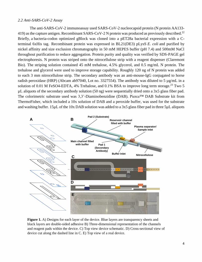
Figure 1. A) Designs for each layer of the device. Blue layers are transparency sheets and
black layers are double-sided adhesive B) Three-dimensional representation of the channels
and reagent pads within the device. C) Top view device schematic. D) Cross-sectional view of
device cut along the dashed line in C. E) Top view of a real device.
A B
C
D
Nitrocellulose
Pad 2 (Substrate)
Reservoir channel
filled with buffer
Pad 1
(Secondary
Antibody)
Plasma separator/
Sample Inlet
Main channel filled
with buffer
E
Buffer Inlet
5
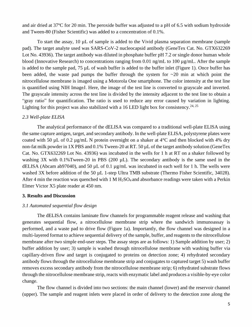
and air dried at 37ºC for 20 min. The peroxide buffer was adjusted to a pH of 6.5 with sodium hydroxide
and Tween-80 (Fisher Scientific) was added to a concentration of 0.1%.
To start the assay, 10 µL of sample is added to the Vivid plasma separation membrane (sample
pad). The target analyte used was SARS-CoV-2 nucleocapsid antibody (GeneTex Cat. No. GTX632269
Lot No. 43936). The target antibody was diluted in phosphate buffer pH 7.2 or single donor human whole
blood (Innovative Research) to concentrations ranging from 0.01 ng/mL to 100 µg/mL. After the sample
is added to the sample pad, 75 µL of wash buffer is added to the buffer inlet (Figure 1). Once buffer has
been added, the waste pad pumps the buffer through the system for ~20 min at which point the
nitrocellulose membrane is imaged using a Motorola One smartphone. The color intensity at the test line
is quantified using NIH ImageJ. Here, the image of the test line is converted to grayscale and inverted.
The grayscale intensity across the test line is divided by the intensity adjacent to the test line to obtain a
“gray ratio” for quantification. The ratio is used to reduce any error caused by variation in lighting.
Lighting for this project was also stabilized with a 16 LED light box for consistency.24, 25
2.3 Well-plate ELISA
The analytical performance of the dELISA was compared to a traditional well-plate ELISA using
the same capture antigen, target, and secondary antibody. In the well-plate ELISA, polystyrene plates were
coated with 50 µL of 0.2 µg/mL N protein overnight on a shaker at 4°C and then blocked with 4% dry
non-fat milk powder in 1X PBS and 0.1% Tween-20 at RT. 50 µL of the target antibody solution (GeneTex
Cat. No. GTX632269 Lot No. 43936) was incubated in the wells for 1 h at RT on a shaker followed by
washing 3X with 0.1%Tween-20 in PBS (200 µL). The secondary antibody is the same used in the
dELISA (Abcam ab97040), and 50 µL of 0.1 µg/mL was incubated in each well for 1 h. The wells were
washed 3X before addition of the 50 µL 1-step Ultra TMB substrate (Thermo Fisher Scientific, 34028).
After 4 min the reaction was quenched with 1 M H2SO4 and absorbance readings were taken with a Perkin
Elmer Victor X5 plate reader at 450 nm.
3. Results and Discussion
3.1 Automated sequential flow design
The dELISA contains laminate flow channels for programmable reagent release and washing that
generates sequential flow, a nitrocellulose membrane strip where the sandwich immunoassay is
performed, and a waste pad to drive flow (Figure 1a). Importantly, the flow channel was designed in a
multi-layered format to achieve sequential delivery of the sample, buffer, and reagents to the nitrocellulose
membrane after two simple end-user steps. The assay steps are as follows: 1) Sample addition by user; 2)
buffer addition by user; 3) sample is washed through nitrocellulose membrane with washing buffer via
capillary-driven flow and target is conjugated to proteins on detection zone; 4) rehydrated secondary
antibody flows through the nitrocellulose membrane strip and conjugates to captured target 5) wash buffer
removes excess secondary antibody from the nitrocellulose membrane strip; 6) rehydrated substrate flows
through the nitrocellulose membrane strip, reacts with enzymatic label and produces a visible-by-eye color
change.
The flow channel is divided into two sections: the main channel (lower) and the reservoir channel
(upper). The sample and reagent inlets were placed in order of delivery to the detection zone along the
6
main straight channel (Figure 1b). The sample is added to the main channel through the plasma separation
membrane while the reagents dried in the glass pad are rehydrated with buffer for delivery through the
main channel. The timing of the sample and reagent delivery was controlled by adjusting the distance
between the nitrocellulose and each reagent inlet. The opening above the glass fiber pads prevents air
bubbles from forming in the main channel and helps maintain consistent flow. The reservoir channel,
which connects upstream of each reagent pad, supplies the buffer solution to rehydrate reagents and
generate flow in the main channel. Although the buffer channels are connected upstream of the glass pads,
the buffer inlet is placed downstream of the glass pads so the buffer can fill both the main channel and the
reservoir channel upon buffer addition. This channel geometry also makes the device more compact. Since
the two reagent channels have different vertical positions, buffer can fill the main channel without being
affected by the glass pads (Figure 1c). Finally, a burst valve was implemented under the separation
membrane, which releases the sample and buffer into the main channel at the same time without air
bubbles.21 The burst valve initiates flow in a channel only after two separate channels meet to ensure
proper flow timing.
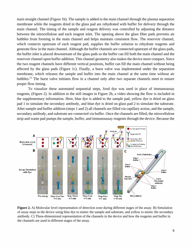
To visualize these automated sequential steps, food dye was used in place of immunoassay
reagents, (Figure 2). In addition to the still images in Figure 2b, a video showing the flow is included in
the supplementary information. Here, blue dye is added to the sample pad, yellow dye is dried on glass
pad 1 to simulate the secondary antibody, and blue dye is dried on glass pad 2 to simulate the substrate.
After sample and buffer addition (steps 1 and 2) all channels are filled via capillary action, and the sample,
secondary antibody, and substrate are connected via buffer. Once the channels are filled, the nitrocellulose
strip and waste pad pumps the sample, buffer, and immunoassay reagents through the device. Because the
Figure 2. A) Molecular level representation of detection zone during different stages of the assay. B) Simulation
of assay steps in the device using blue dye to mimic the sample and substrate, and yellow to mimic the secondary
antibody. C) Three-dimensional representation of the channels in the device and how the reagents and buffer in
the channels are used in different stages of the assay.
7
sample is placed at the front of the device it is programmed to pass through the nitrocellulose
membrane first and the target is captured on the test line. Next, the buffer between the sample inlet and pad
1 (yellow) will flow through the nitrocellulose membrane and wash away excess sample constituents that
might interfere with the remaining assay (step 3). The pressure difference from pad one to
the nitrocellulose membrane is larger than that of the difference between the pad two (blue) and
the nitrocellulose membrane, so the rehydrated enzyme label from the yellow pad flows through next and
any target analyte on the test line will capture the enzyme label (step 4). Once the buffer above pad one is
depleted, the flow from the blue pad to the nitrocellulose begins. The rehydrated substrate stored on pad
two will be preceded by a slug of buffer between pads 1 and 2 in the main channel (step 5). Once the
buffer has washed away excess label, the substrate reaches the test line and reacts with the enzyme label to
produce a visible color change (step 6). After flow stops (~20 min) the color change is detected with the
naked eye for qualitative detection, or imaged (smartphone camera) for quantitative information.
The volume of buffer used to wash the sample or reagents through the nitrocellulose membrane can be
controlled by changing the length of channels for any given assay. In the current device design, the total
volume of washing buffer was 75 µL and the assay takes 20 min to run.
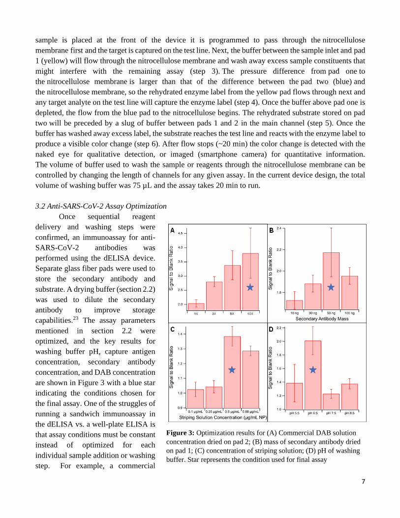
3.2 Anti-SARS-CoV-2 Assay Optimization
Once sequential reagent
delivery and washing steps were
confirmed, an immunoassay for anti-
SARS-CoV-2 antibodies was
performed using the dELISA device.
Separate glass fiber pads were used to
store the secondary antibody and
substrate. A drying buffer (section 2.2)
was used to dilute the secondary
antibody to improve storage
capabilities.23 The assay parameters
mentioned in section 2.2 were
optimized, and the key results for
washing buffer pH, capture antigen
concentration, secondary antibody
concentration, and DAB concentration
are shown in Figure 3 with a blue star
indicating the conditions chosen for
the final assay. One of the struggles of
running a sandwich immunoassay in
the dELISA vs. a well-plate ELISA is
that assay conditions must be constant
instead of optimized for each
individual sample addition or washing
step. For example, a commercial
Figure 3: Optimization results for (A) Commercial DAB solution
concentration dried on pad 2; (B) mass of secondary antibody dried
on pad 1; (C) concentration of striping solution; (D) pH of washing
buffer. Star represents the condition used for final assay
8
peroxide buffer was used as the washing buffer to improve the activity of HRP with DAB, but the buffer
was adjusted from a pH of 5.5 to 6.5 for the final assay. While changing from 5.5 to 6.5 decreased the
HRP activity, it improved the antibody-antigen binding, which had a larger impact on assay sensitivity. 1
µg/mL target antibody was used during the optimization experiments as that concentration falls within the
linear range of the assay.
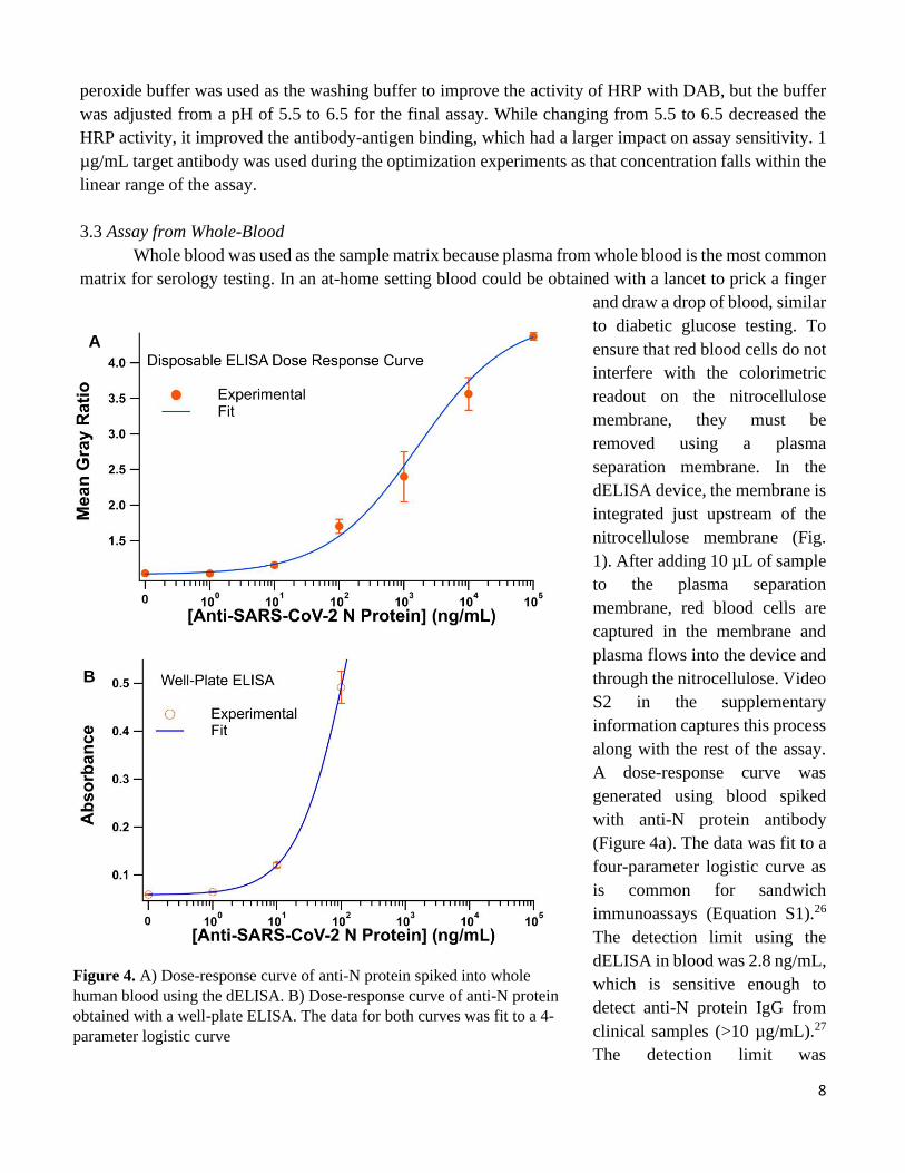
3.3 Assay from Whole-Blood
Whole blood was used as the sample matrix because plasma from whole blood is the most common
matrix for serology testing. In an at-home setting blood could be obtained with a lancet to prick a finger
and draw a drop of blood, similar
to diabetic glucose testing. To
ensure that red blood cells do not
interfere with the colorimetric
readout on the nitrocellulose
membrane, they must be
removed using a plasma
separation membrane. In the
dELISA device, the membrane is
integrated just upstream of the
nitrocellulose membrane (Fig.
1). After adding 10 µL of sample
to the plasma separation
membrane, red blood cells are
captured in the membrane and
plasma flows into the device and
through the nitrocellulose. Video
S2 in the supplementary
information captures this process
along with the rest of the assay.
A dose-response curve was
generated using blood spiked
with anti-N protein antibody
(Figure 4a). The data was fit to a
four-parameter logistic curve as
is common for sandwich
immunoassays (Equation S1).26
The detection limit using the
dELISA in blood was 2.8 ng/mL,
which is sensitive enough to
detect anti-N protein IgG from
clinical samples (>10 µg/mL).27
The detection limit was
Figure 4. A) Dose-response curve of anti-N protein spiked into whole
human blood using the dELISA. B) Dose-response curve of anti-N protein
obtained with a well-plate ELISA. The data for both curves was fit to a 4-
parameter logistic curve
0
A
B
0
9
calculated using Eq S2. Future studies will focus on using the dELISA with real patient samples to
establish a clinical sensitivity and specificity for the device.
In upcoming clinical studies, the results will be validated with a well-plate ELISA, so in this work
results were also compared to a well-plate ELISA using the same antigen and secondary antibody. The
results for the well-plate ELISA are shown in Figure 4b. The well-plate ELISA dose-response curve was
once again fit with a 4-parameter logistic curve and a detection limit of 1.2 ng/mL was calculated. The
detection limit of the dELISA (2.8 ng/mL) is roughly the same as the detection limit in the dELISA (1.2
ng/mL). These results exceeded expectations for the dELISA considering that a well-plate ELISA uses
absorbance values from a plate reader, while our device quantifies results using images from a smartphone.
The advantages of the dELISA over a traditional well-plate assay include faster assay time (2.5 h versus
20 min), decreased end-user steps (13 vs 2), increased portability, decreased cost, and increased ease of
use. Hands-on time and preparation time are also significantly less in the dELISA than the well-plate
ELISA. Most importantly, the dELISA is a true at-home test that can be run outside of a centralized
laboratory by an untrained end-user. These advantages are common for POC assays but are typically offset
by sacrificing analytical performance. This is not the case for the dELISA, which has the sensitivity of a
well-plate ELISA and is as easy to use as an LFA.
The question of whether the dELISA could be used to detect Anti-SARS-CoV-2 antibodies from
real blood or nasopharyngeal samples is yet to be answered, but the detection limits presented here are
promising as well-plate ELISAs have been used to detect actual SARS-CoV-2 antibodies types at higher
concentrations.27 Well-plate ELISAs and even LFAs have detected lower concentrations of antibodies
previously, but we believe the limiting factor in the current dELISA setup is the capture antigen and
secondary antibody and their specificity toward the particular commercial Anti-NP used. To improve
results further a thorough screening of suitable secondary antibody for clinical anti-NP similar to the work
presented by Cate et al. would need to be conducted.28 Additionally, the drying procedure for the
secondary antibody could be improved through lyophilization, and increasing the sample volume would
increase sensitivity, but also increase assay time.
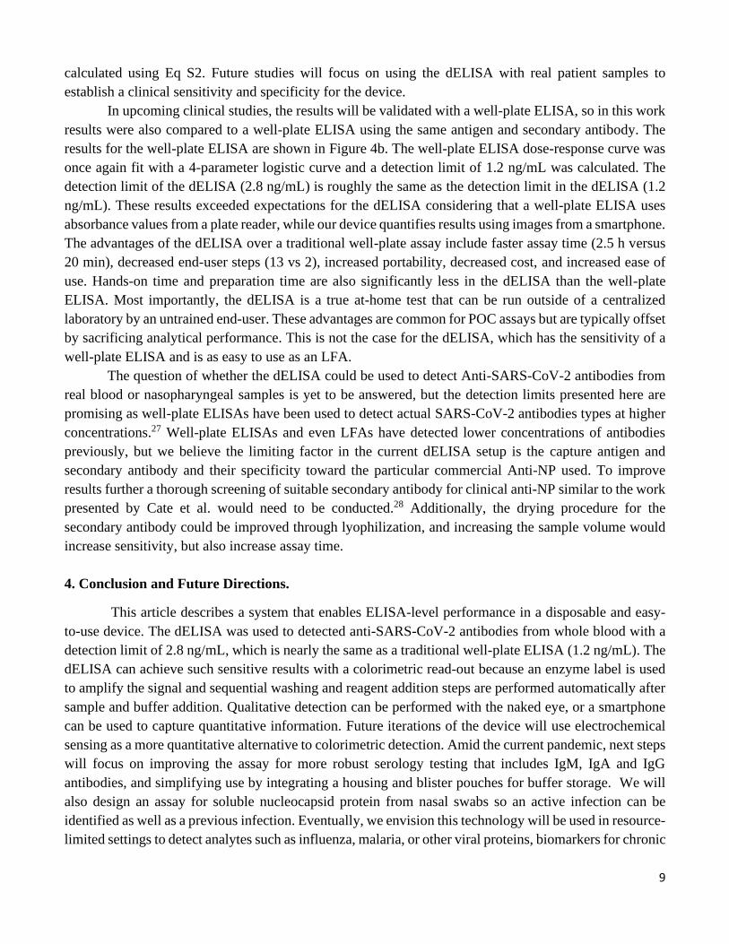
4. Conclusion and Future Directions.
This article describes a system that enables ELISA-level performance in a disposable and easy-
to-use device. The dELISA was used to detected anti-SARS-CoV-2 antibodies from whole blood with a
detection limit of 2.8 ng/mL, which is nearly the same as a traditional well-plate ELISA (1.2 ng/mL). The
dELISA can achieve such sensitive results with a colorimetric read-out because an enzyme label is used
to amplify the signal and sequential washing and reagent addition steps are performed automatically after
sample and buffer addition. Qualitative detection can be performed with the naked eye, or a smartphone
can be used to capture quantitative information. Future iterations of the device will use electrochemical
sensing as a more quantitative alternative to colorimetric detection. Amid the current pandemic, next steps
will focus on improving the assay for more robust serology testing that includes IgM, IgA and IgG
antibodies, and simplifying use by integrating a housing and blister pouches for buffer storage. We will
also design an assay for soluble nucleocapsid protein from nasal swabs so an active infection can be
identified as well as a previous infection. Eventually, we envision this technology will be used in resource-
limited settings to detect analytes such as influenza, malaria, or other viral proteins, biomarkers for chronic

10
illness such as heart failure or cancer, and/or other antibodies at levels that were previously undetectable
outside a centralized laboratory.
11
References
1. COVID-19 Dashboard. John's Hopkins University Center for Systems Science and Engineering (CSSE): 2020. 2. Shen, C.; Wang, Z.; Zhao, F.; Yang, Y.; Li, J.; Yuan, J.; Wang, F.; Li, D.; Yang, M.; Xing, L., Treatment of 5 critically ill patients with COVID-19 with convalescent plasma. Jama 2020, 323 (16), 1582-1589. 3. CDC Diagnostic Tests for COVID-19. https://www.cdc.gov/coronavirus/2019-ncov/lab/testing.html (accessed 27 July, 2020). 4. CMS Increases Medicare Payment for High-Production Coronavirus Lab Tests. https://www.cms.gov/newsroom/press-releases/cms-increases-medicare-payment-high-production-coronavirus-lab-tests-0#:~:text=Medicare%20will%20pay%20laboratories%20for,the%20spread%20of%20COVID%2D19. (accessed 28 July, 2020). 5. Dalvie, M. A.; Sinanovic, E.; London, L.; Cairncross, E.; Solomon, A.; Adam, H., Cost analysis of ELISA, solid-phase extraction, and solid-phase microextraction for the monitoring of pesticides in water. Environ Res 2005, 98 (1), 143-50. 6. COVID-19 Target product profiles for priority diagnostics to support response to the COVID-19 pandemic v.0.1. Organization, W. H., Ed. 2020. 7. Posthuma-Trumpie, G. A.; Korf, J.; van Amerongen, A., Lateral flow (immuno) assay: its strengths, weaknesses, opportunities and threats. A literature survey. Anal. Bioanal. Chem. 2009, 393 (2), 569-582. 8. Krammer, F.; Simon, V., Serology assays to manage COVID-19. Science 2020, 368 (6495), 1060-1061. 9. Li, Z.; Yi, Y.; Luo, X.; Xiong, N.; Liu, Y.; Li, S.; Sun, R.; Wang, Y.; Hu, B.; Chen, W., Development and clinical application of a rapid IgM‐IgG combined antibody test for SARS‐CoV‐2 infection diagnosis. Journal of medical virology 2020. 10. Lou, B.; Li, T.-D.; Zheng, S.-F.; Su, Y.-Y.; Li, Z.-Y.; Liu, W.; Yu, F.; Ge, S.-X.; Zou, Q.-D.; Yuan, Q., Serology characteristics of SARS-CoV-2 infection since exposure and post symptom onset. European Respiratory Journal 2020. 11. Lassaunière, R.; Frische, A.; Harboe, Z. B.; Nielsen, A. C.; Fomsgaard, A.; Krogfelt, K. A.; Jørgensen, C. S., Evaluation of nine commercial SARS-CoV-2 immunoassays. Medrxiv 2020. 12. Ong, D. S. Y.; de Man, S. J.; Lindeboom, F. A.; Koeleman, J. G. M., Comparison of diagnostic accuracies of rapid serological tests and ELISA to molecular diagnostics in patients with suspected coronavirus disease 2019 presenting to the hospital. Clin Microbiol Infect 2020, 26 (8), 1094.e7-1094.e10. 13. Carrell, C. S.; Wydallis, R. M.; Bontha, M.; Boehle, K. E.; Beveridge, J. R.; Geiss, B. J.; Henry, C. S., Rotary manifold for automating a paper-based Salmonella immunoassay. RSC Adv. 2019, 9 (50), 29078-29086. 14. Verma, M. S.; Tsaloglou, M.-N.; Sisley, T.; Christodouleas, D.; Chen, A.; Milette, J.; Whitesides, G. M., Sliding-strip microfluidic device enables ELISA on paper. Biosensors and Bioelectronics 2018, 99, 77-84. 15. Apilux, A.; Ukita, Y.; Chikae, M.; Chailapakul, O.; Takamura, Y., Development of automated paper-based devices for sequential multistep sandwich enzyme-linked immunosorbent assays using inkjet printing. Lab Chip 2013, 13 (1), 126-135. 16. Fridley, G. E.; Le, H.; Yager, P., Highly sensitive immunoassay based on controlled rehydration of patterned reagents in a 2-dimensional paper network. Analytical chemistry 2014, 86 (13), 6447-6453. 17. Jeong, S.-G.; Kim, J.; Jin, S. H.; Park, K.-S.; Lee, C.-S., Flow control in paper-based microfluidic device for automatic multistep assays: A focused minireview. Korean Journal of Chemical Engineering 2016, 33 (10), 2761-2770. 18. Preechakasedkit, P.; Siangproh, W.; Khongchareonporn, N.; Ngamrojanavanich, N.; Chailapakul, O., Development of an automated wax-printed paper-based lateral flow device for alpha-fetoprotein enzyme-linked immunosorbent assay. Biosensors and Bioelectronics 2018, 102, 27-32. 19. Yakoh, A.; Chaiyo, S.; Siangproh, W.; Chailapakul, O., 3D Capillary-Driven Paper-Based Sequential Microfluidic Device for Electrochemical Sensing Applications. ACS Sensors 2019, 4 (5), 1211-1221.
12
20. Jang, I.; Carrão, D. B.; Menger, R. F.; Moraes de Oliveira, A. R.; Henry, C. S., Pump-Free Microfluidic Rapid Mixer Combined with a Paper-Based Channel. ACS Sensors 2020, 5 (7), 2230-2238. 21. Ilhoon, J.; David S., D.; Brian J., G.; Charles, H.; Hyunwoong, K.; Simon, S., Flow Control in a Laminate Capillary-Driven Microfluidic Device. 2020. 22. Fagre, A.; Lewis, J.; Eckley, M.; Zhan, S.; Rocha, S. M.; Sexton, N. R.; Burke, B.; Geiss, B.; Peersen, O.; Kading, R.; Rovnak, J.; Ebel, G. D.; Tjalkens, R. B.; Aboellail, T.; Schountz, T., SARS-CoV-2 infection, neuropathogenesis and transmission among deer mice: Implications for reverse zoonosis to New World rodents. bioRxiv 2020, 2020.08.07.241810. 23. Ramachandran, S.; Fu, E.; Lutz, B.; Yager, P., Long-term dry storage of an enzyme-based reagent system for ELISA in point-of-care devices. Analyst 2014, 139 (6), 1456-1462. 24. Henry, C. S.; Feeny, R.; Franklin, A. B.; Carrell, C., Rotary manifold for paper-based immunoassays. Google Patents: 2020. 25. Boehle, K. E.; Doan, E.; Henry, S.; Beveridge, J. R.; Pallickara, Sangmi L.; Henry, C. S., Single board computing system for automated colorimetric analysis on low-cost analytical devices. Analytical Methods 2018, 10 (44), 5282-5290. 26. Christopoulos, T. K.; Diamandis, E. P., Theory of immunoassays. Immunoassay 1996, 25-50. 27. Ma, H.; Zeng, W.; He, H.; Zhao, D.; Yang, Y.; Jiang, D.; Zhou, P.; Qi, Y.; He, W.; Zhao, C.; Yi, R.; Wang, X.; Wang, B.; Xu, Y.; Yang, Y.; Kombe Kombe, A. J.; Ding, C.; Xie, J.; Gao, Y.; Cheng, L.; Li, Y.; Ma, X.; Jin, T., COVID-19 diagnosis and study of serum SARS-CoV-2 specific IgA, IgM and IgG by chemiluminescence immunoanalysis. medRxiv 2020, 2020.04.17.20064907. 28. David, C.; Helen, H.; Veronika, G.; Joshua D, B.; H Gleda, H.; Brianda, B.-L.; Ben D, G.; Caitlin E, A.; Ethan, S.; Samantha, K.; Ryan, G.; Rafael, R.; Crissa, B.; Sam A, B.; John T, C.; Puneet K, D.; David, S. B.; Bernhard H, W.; Kevin P, N., Antibody Screening Results for Anti-Nucleocapsid Antibodies Towards the Development of a SARS-CoV-2 Nucleocapsid Protein Antigen Detecting Lateral Flow Assay. 2020.
13
Supplementary Information
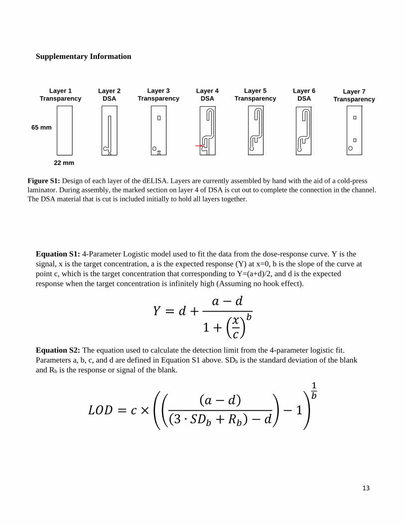
Equation S1: 4-Parameter Logistic model used to fit the data from the dose-response curve. Y is the
signal, x is the target concentration, a is the expected response (Y) at x=0, b is the slope of the curve at
point c, which is the target concentration that corresponding to Y=(a+d)/2, and d is the expected
response when the target concentration is infinitely high (Assuming no hook effect).
𝑌 = 𝑑 +𝑎 − 𝑑
1 + (𝑥𝑐)
𝑏
Equation S2: The equation used to calculate the detection limit from the 4-parameter logistic fit.
Parameters a, b, c, and d are defined in Equation S1 above. SDb is the standard deviation of the blank
and Rb is the response or signal of the blank.
𝐿𝑂𝐷 = 𝑐 × (((𝑎 − 𝑑)
(3 ∙ 𝑆𝐷𝑏 + 𝑅𝑏) − 𝑑) − 1)
1𝑏
Figure S1: Design of each layer of the dELISA. Layers are currently assembled by hand with the aid of a cold-press
laminator. During assembly, the marked section on layer 4 of DSA is cut out to complete the connection in the channel.
The DSA material that is cut is included initially to hold all layers together.
Layer 1
Transparency
Layer 2
DSA
Layer 3
Transparency
Layer 4
DSA
Layer 5
Transparency
Layer 6
DSALayer 7
Transparency
65 mm
22 mm

download fileview on ChemRxivFinal ChemRXIV Manuscript.pdf (738.66 KiB)

Other files
download fileview on ChemRxivVideo_One_Dye_Simulation.mp4 (18.77 MiB)
download fileview on ChemRxivVideo_Two_Blood_Assay.mp4 (3.18 MiB)