Pleural effusion(X-ray Findings)
20
Pleural effusion X-ray findings Jeetendra
-
Upload
z2jeetendra -
Category
Health & Medicine
-
view
105 -
download
0
Transcript of Pleural effusion(X-ray Findings)

Pleural effusion X-ray findings
Jeetendra
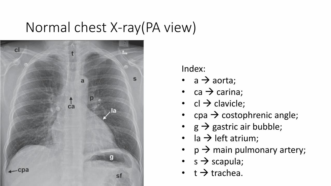
Normal chest X-ray(PA view)
Index:• a aorta; • ca carina;• cl clavicle;• cpa costophrenic angle; • g gastric air bubble; • la left atrium;• pmain pulmonary artery; • s scapula; • t trachea.

Normal chest X-ray(PA view)
Index:• Aaorta; • Apw aortopulmonary window; • Cap cardiophrenic angle;• g gastric air bubble; • ip interlobar (or descending)
pulmonary artery; • L liver; • lv left ventricle; • rts right tracheal (or paratracheal)
stripe; • sp spleen;

Normal chest X-ray(PA view)
Index
• A aorta• Ajl anterior junction line• Apw aortopulmonary window • Ip interlobar (or descending)
pulmonary artery• Pjl posterior junction line

Normal Chest X-ray (lateral view)Index:• a aorta• bi bronchus intermedius• cpa costophrenic angle• d diaphragm • e esophagus• ivc inferior vena cava• lpa left pulmonary artery• lul left upper lobe bronchus• lv left ventricle• m manubrium• mf minor fissure• MF major fissure• rpa right pulmonary artery • rul right upper lobe bronchus• rv right ventricle • st sternum • svc superior vena cava • t trachea• v vertebral body

Introduction to pleural space
• Contain 2 layers i.e. Visceral and Parietal pleura
• Visceral pleura is outer lining of the lung
• Parietal pleura is lining of the chest cavity
• Normally, these surfaces are smooth and are separated by a minimal amount of pleural fluid
• Provides nearly friction-free environment for movement of the lung within the thorax
• Normally contains no more than 3 to 5 mL of pleural fluid
Reasons for accumulation of fluid• Increase pulmonary capillary pressure (transudate)
• congestive heart failure • hypoproteinemia• fluid overload• liver failure• nephrosis
• Alter thoracic vascular or lymphatic pathways
• Alter pleural capillary or lymphatic permeability(Exudate)• Infection or inflammation• pulmonary embolism• neoplasms
• Affect diaphragmatic peritoneal and pleural surfaces• pancreatitis• subphrenic abscesses• liver abscesses • ovarian tumors • peritonitis• ascites

Pleural effusion
• Most common radiographic sign is pleural meniscus
• Volume of fluid to produce pleural meniscus within costophrenicangle varies in individual

Clinical findings
• Decreased breath sounds
• Dullness to percussion
• Decreased tactile fremitus
• Egophony
• Pleural friction rub

Pleural effusion(lateral view)
• Approx 100 mL of pleural fluid will cause appreciable blunting of the posterior costophrenic angle on the lateral view
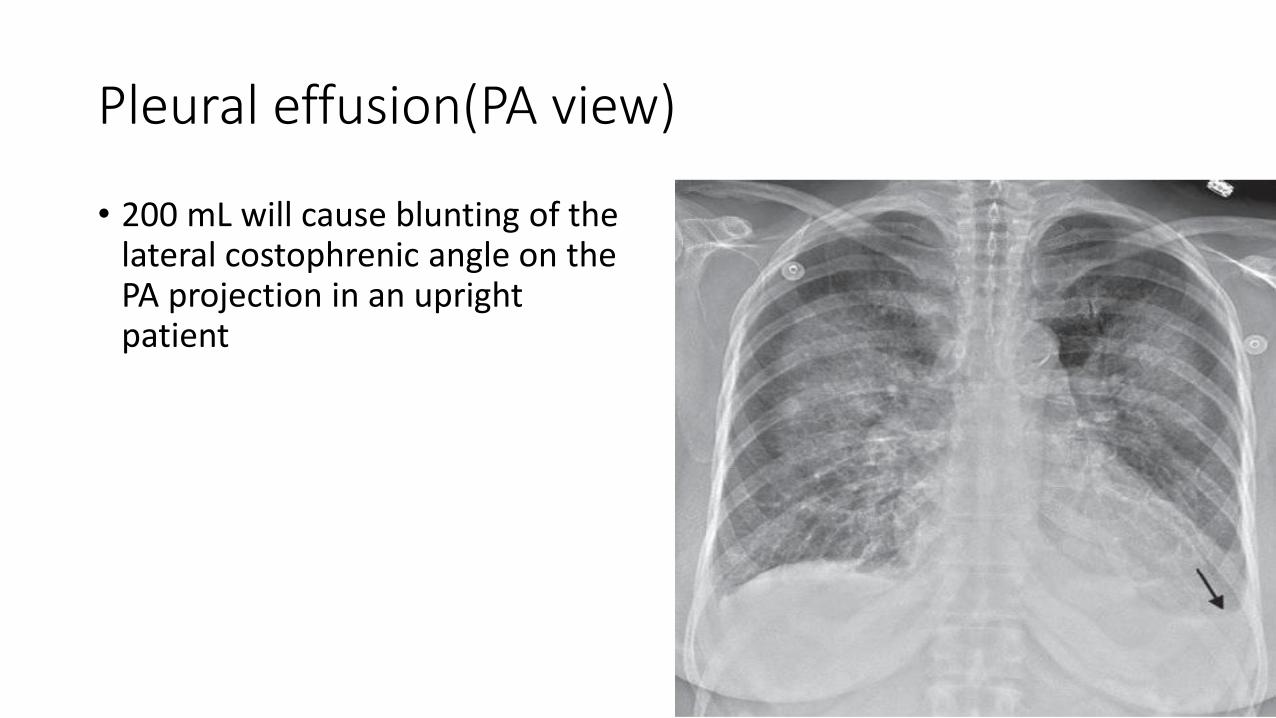
Pleural effusion(PA view)
• 200 mL will cause blunting of the lateral costophrenic angle on the PA projection in an upright patient

Pleural effusion(lateral decubitus view)
• A lateral decubitus chest radiograph, with the side containing the pleural effusion placed down (dependent), demonstrate smaller amounts of free-flowing pleural effusions
• 1 millimeter of thickness of pleural fluid in the lateral decubitus = approx 20 mL of pleural fluid

Sub-pulmonic pleural effusions
• Sub-pulmonic pleural effusions elevate the lung base
• Mimics an elevated diaphragmatic leaflet
• On the left side, a marked separation (>2 cm) of the lung from the stomach bubble suggests a sub-pulmonic effusion. Right Sub-pulmonic effusion
with elevated right hemi-diaphargm

The Same thing.. Rt. Sub-pulmoniceffusion

The apex of the curvature at the lung base is shifted laterally, and its slope slants sharply towards the lateral costophrenicsulcus
The rock of Gibraltar sign
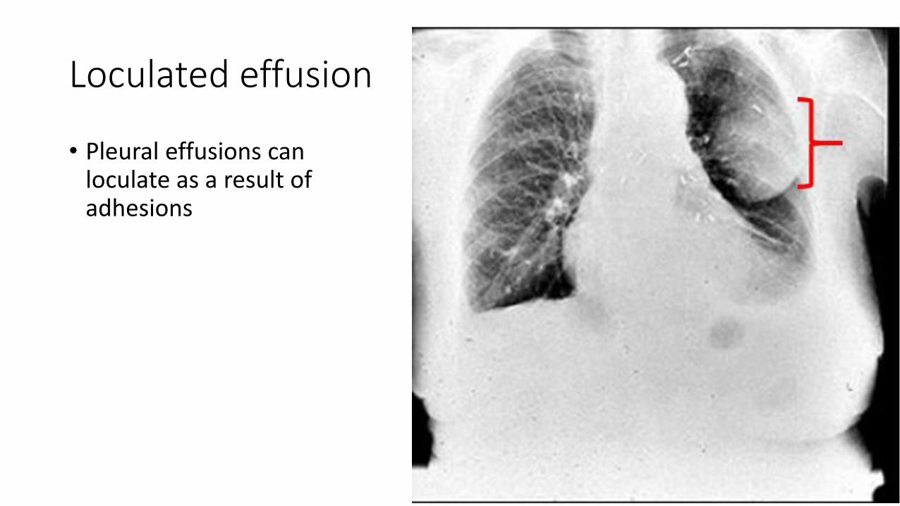
Loculated effusion
• Pleural effusions can loculate as a result of adhesions


Features
• Typical configuration of a loculation along the chest wall, often described as pleural or extrapleural sign
• Angles of interface between the pleural “mass” and the chest wall are obtuse, and the mass displays tapered borders
• Surface of the “mass” is usually smooth, poorly marginated when seen PA, and only partially visualized when displayed in an oblique projection (“incomplete margin sign”)
• Homogeneous content
• “mass” droops on upright images owing to its liquid content

References
• Michael Y. M. Chen, Thomas L. Pope, David J. Ott. Basic Radiology. 2nd ed. Mc. Grow hill. P-115-9.
• Cochard, Larry R.,Netter, Frank H. Netter's Introduction to Imaging. Elseiver. P-37-9.

Thank you!!