Pleomorphic adenoma of palate: differential diagnosis … october 2015 PDF 12-18.pdfPleomorphic...
7
Journal of Government Dental College and Hospital, October 2015, Vol.-02, Issue- 01, P. 12-18 12 www.jgdch.com, PISSN: 2394- 8701, E ISSN: 2394 – 871X Case report Pleomorphic adenoma of palate: differential diagnosis and case report 1 Dr. Jigna S. Shah, 2 Dr. MonaliPrajapati, 3 Dr. Utsav Bhatt 1 Professor & Head, Dept of Oral Medicine & Radiology, Govt. Dental College & Hospital, Ahmedabad, Gujarat, India 2 Postgraduate Student, Dept of Oral Medicine & Radiology, Govt. Dental College & Hospital, Ahmedabad, Gujarat, India 3 Assistant Professor, Dept of Oral Surgery, Govt. Dental College & Hospital, Ahmedabad, Gujarat, India Corresponding author: Dr. Jigna S. Shah Abstract: Pleomorphic adenoma of minor salivary gland is most common in palate (10%), followed by lip (4%).Approximately 34.7-67.1% of salivary gland tumors arising from an intraoral site are benign. The smaller the salivary gland that is affected, the more likely it is to trigger a malignant tumor. The propensity of malignant transformation is documented to be 1.9-23.3%,hence any suspected minor salivary gland tumor should be keenly scrutinized. A case report of 28- year old male patient diagnosed of pleomorphic adenoma of palate, is presented here. Key words: differential diagnosis, palate, pleomorphic adenoma Introduction Salivary gland tumors account for 3% of the head and neck tumors. Pleomorphic adenoma is the most common salivary gland tumor, accounting for about 40–70 % of all major and minor salivary gland tumors. 1 It accounts for 53% to 77% of parotid tumors, 44% to 68% of submandibular tumors and 33% to 43% of minor gland tumors. 2 Pleomorphic adenoma can be defined as a benign mixed tumor composed of epithelial and myoepithelial cells arranged with various morphological patterns, demarcated from surrounding tissues by fibrous capsule. 3 They have dual origin from epithelial and myoepithelial elements. 1,4 It derives its name from the architectural pleomorphism seen by light microscopy and is also known as “mixed tumor, salivary gland type”. 4 The most common site of this tumor in the oral cavity is the palatal area followed by the lip, buccal mucosa, floor of the mouth, tongue, tonsil, pharynx, and retro molar area. 1 The diagnosis of pleomorphic adenoma is established on the basis of history, physical examination, cytology and histopathology. Computed tomographic scan and MRI can provide information on the location, size of the tumor and extension to surrounding superficial and deep structures. The treatment is strictly wide local excision with the removal of periosteum or bone if they are involved. 1,4 Case report A 28-year-old male presented with a slow growing swelling, of approximately 6month duration involving his hard and soft palate junction on the left side. An asymptomatic peanut sized swelling was noticed by the patient before 6months which gradually increased in sizecausing difficulty in mastication, speech and swallowing which raised concern to the patient so he seeked medical advice. The patient’s medical history was noncontributory. General physical examination revealed a well
-
Upload
phungtuyen -
Category
Documents
-
view
219 -
download
0
Transcript of Pleomorphic adenoma of palate: differential diagnosis … october 2015 PDF 12-18.pdfPleomorphic...
Journal of Government Dental College and Hospital, October 2015, Vol.-02, Issue- 01, P. 12-18
12
www.jgdch.com, PISSN: 2394- 8701, E ISSN: 2394 – 871X
Case report
Pleomorphic adenoma of palate: differential diagnosis and case report
1Dr. Jigna S. Shah, 2Dr. MonaliPrajapati, 3Dr. Utsav Bhatt
1Professor & Head, Dept of Oral Medicine & Radiology, Govt. Dental College & Hospital, Ahmedabad, Gujarat, India
2Postgraduate Student, Dept of Oral Medicine & Radiology, Govt. Dental College & Hospital, Ahmedabad, Gujarat, India
3Assistant Professor, Dept of Oral Surgery, Govt. Dental College & Hospital, Ahmedabad, Gujarat, India
Corresponding author: Dr. Jigna S. Shah
Abstract:
Pleomorphic adenoma of minor salivary gland is most common in palate (10%), followed by lip (4%).Approximately 34.7-67.1%
of salivary gland tumors arising from an intraoral site are benign. The smaller the salivary gland that is affected, the more likely it
is to trigger a malignant tumor. The propensity of malignant transformation is documented to be 1.9-23.3%,hence any suspected
minor salivary gland tumor should be keenly scrutinized. A case report of 28- year old male patient diagnosed of pleomorphic
adenoma of palate, is presented here.
Key words: differential diagnosis, palate, pleomorphic adenoma
Introduction
Salivary gland tumors account for 3% of the head and
neck tumors. Pleomorphic adenoma is the most
common salivary gland tumor, accounting for about
40–70 % of all major and minor salivary gland
tumors.1 It accounts for 53% to 77% of parotid
tumors, 44% to 68% of submandibular tumors and
33% to 43% of minor gland tumors.2Pleomorphic
adenoma can be defined as a benign mixed tumor
composed of epithelial and myoepithelial cells
arranged with various morphological patterns,
demarcated from surrounding tissues by fibrous
capsule.3They have dual origin from epithelial and
myoepithelial elements.1,4 It derives its name from
the architectural pleomorphism seen by light
microscopy and is also known as “mixed tumor,
salivary gland type”.4The most common site of this
tumor in the oral cavity is the palatal area followed
by the lip, buccal mucosa, floor of the mouth, tongue,
tonsil, pharynx, and retro molar area.1
The diagnosis of pleomorphic adenoma is established
on the basis of history, physical examination,
cytology and histopathology. Computed tomographic
scan and MRI can provide information on the
location, size of the tumor and extension to
surrounding superficial and deep structures. The
treatment is strictly wide local excision with the
removal of periosteum or bone if they are involved.1,4
Case report
A 28-year-old male presented with a slow growing
swelling, of approximately 6month duration
involving his hard and soft palate junction on the left
side. An asymptomatic peanut sized swelling was
noticed by the patient before 6months which
gradually increased in sizecausing difficulty in
mastication, speech and swallowing which raised
concern to the patient so he seeked medical advice.
The patient’s medical history was noncontributory.
General physical examination revealed a well
Journal of Government Dental College and Hospital, October 2015, Vol.-02, Issue- 01, P. 12-18
13
www.jgdch.com, PISSN: 2394- 8701, E ISSN: 2394 – 871X
oriented and moderately built individual with no
signs of any systemic illness.
The intraoral examination revealeda smooth,
nonulcerated, dome-shaped, palatal swelling of size
approximately 3.5x3x3cm on his hard palate,
encroaching the midline. On palpation, the swelling
was firm, non-tender, non-fluctuant, non-
compressible and non-reducible. Clinically all teeth
were present except third molars in all quadrant. No
displacement or mobility of teeth was found and all
teeth were vital. (Figure 1)The clinical findings were
suggestive of minor salivary gland tumor. To
conclude the diagnosis various investigations were
done.
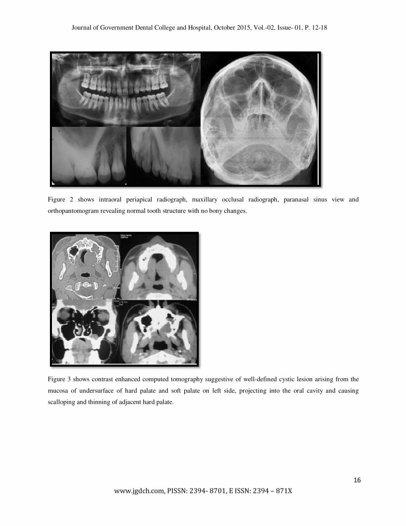
Intraoral periapical radiograph, maxillary occlusal
radiograph, paranasal sinus view and
orthopantomogram revealed normal tooth structure
with no bony changes ruling out the possibility of
odontogenic cause. (Figure 2) Contrast enhanced
computed tomography report revealed a well-defined
cystic lesion arising from the mucosa of undersurface
of hard palate and soft palate on left side, projecting
into the oral cavity and causing scalloping and
thinning of adjacent hard palate. (Figure 3) On the
basis of clinical and radiographic findings, benign
salivary gland tumor was considered as the
provisional diagnosis. Fine needle aspiration
cytology was performed, which was suggestive of
pleomorphic adenoma. All preoperative blood and
urine investigations were done, which were within
normal limits. Wide local excision of the mass was
done (Figure 4A, B). The excised mass was sent for
histopathological examination, which revealed
biphasic appearance of tumor with epithelial and
stromal component. Epithelial component showed
well differentiated ducts and ductules lined by
flattened and cuboidal cells. Stroma showed
chondromyxoid appearance. At places squamous
metaplasia was evident. (Figure 5) This confirmed
the diagnosis as pleomorphic adenoma.Post-
operative healing after 10 days was normal (Figure
6). No recurrence of the lesion was noted on 1 year
follow up.
Discussion
Pleomorphic adenoma of minor salivary gland is
most common in palate (10%), followed by lip
(4%).Approximately 34.7-67.1% of salivary gland
tumors arising from an intraoral site are benign.5The
smaller the salivary gland that is affected, the more
likely it is to trigger a malignant tumor. The
propensity of malignant transformation is
documented to be 1.9-23.3%,5 hence any suspected
minor salivary gland tumor should be keenly
scrutinized.
The tumor can occur at any age but it mainly affects
patients in the fourth, fifth and sixth decade, with a
slight female predilection2,4with a ratio of 2:13.
Clinically, pleomorphic adenoma of palatal minor
salivary glands presents as a painless, slowly
growing, dome shaped, firm, non-tender swelling,
commonly seen on the posterior lateral aspect of the
palate.3,4,5Because of tightly bound nature of the hard
palate mucosa, it appears to be fixed. While in cases
of lips and buccal mucosa, it is freely movable. The
more common palatal mixed tumors are located
laterally and rarely cross the midline.6 This
presentation is due to the highest concentration of
salivary glands there.4 The patient in the presented
case was of 28 year old male having typical clinical
presentation of pleomorphic adenoma.
The differential diagnosis for this case
includes chronic abscess, benign tumors such as soft
tissue tumors (fibroma, lipoma, neurofibroma,
neurilemmoma, lymphoma, and otherbenign and
Journal of Government Dental College and Hospital, October 2015, Vol.-02, Issue- 01, P. 12-18
14
www.jgdch.com, PISSN: 2394- 8701, E ISSN: 2394 – 871X
malignant salivary gland tumors), fibro-osseous
lesion (ossifying fibroma.) . Palatal abscess could be
ruled out by clinical examination since the source of
a palatal abscess, which is typically a non- vital tooth
in the vicinity or a localized periodontal defect, was
not found in our case. Owing to absence of history of
constant trauma and the size of the lesion, fibroma
could be ruled out. Most oral lipomasexhibit the
characteristic yellow color of adipose tissue, which is
visible through the thin overlying epithelium,
consistency varying from soft to firm depending on
the quantity of fibrous tissue in it,7 hence ruling out
the possibility of lipoma in this case. Neurilemoma,
neurofibroma, extranodal lymphoma and other
benign and malignant salivary gland tumors may
present as swelling identical to that of pleomorphic
adenoma, hence importance of histopathological
evaluation must not be neglected. Also, incidence of
neurilemmomain palate is only 7-9%8and
neurofibroma of oral cavity is more prevalent in
posterior mandible.9 Ossifying fibroma is a fibro-
osseous lesion having concentric growth pattern
which causes expansion of buccal as well as palatal
cortical plate whereas there was only palatal
expansion in the presented case.
Plain X-rays play insignificant part in the diagnosis
of salivary gland tumor of the palate.1In the present
case also, the conventional radiographs failed to
show any bony changes whereas the lesion was well
appreciated in the CT images as it shows scalloping
and thinning of hard palate favouring benign salivary
gland tumor. FNAC can help determine severity of
neoplasm, identify histological subtype5 and
determine whether the tumor is malignant in nature
with 90 % sensitivity.1,10 FNAC in the reported case
was diagnostic of pleomorphic adenoma with no
evidence of malignant changes. To confirm the
diagnosis, histopathological analysis was done and it
showed no evidence of any malignant change. The
treatment of pleomorphic adenoma is strictly wide
local excision with the removal of periosteum or
bone if they are involved.1,4 Pleomorphic adenomas
of the minor glands have little propensity for
recurrence as compared to that of parotid gland.4
However, recurrence if at all occurs can be
attributable to inadequate surgical techniques such as
simple enucleation leaving behind microscopic
pseudopod-like extensions, capsular penetration, and
tumor rupture with spillage of tumor cells.1,3,4 No
signs of recurrence were evident in the present case.
Conclusion
Pleomorphic adenoma, though a common entity
should be thoroughly evaluated owing to its diverse
histological property and increased risk of malignant
transformation. Definitive diagnosis lies on
histopathological examination. CT is necessary for
ruling out bony erosions and treatment by wide local
excision with removal of periosteum and curettage of
bone lowers the risk of recurrence.
References
1. Patigaroo SA, Patigaroo FA, Ashraf J, et al. Pleomorphic adenoma of hard palate: an experience. J
Maxillofac Oral Surg. 2012;13:36-41.
2. Lenka SP, Padhiary SK, Subudhi SK, Pathak H, Sahoo S.Pleomorphic adenoma of hard palate: a case
report. Egypt Dent J. 1971;17:243-46.
3. Debnath SC,Saikia AK, Debnath A. Pleomorphic adenoma of the palate.J Maxillofac Oral

Journal of Government Dental College and Hospital, October 2015, Vol.-02, Issue- 01, P. 12-18
13
www.jgdch.com, PISSN: 2394- 8701, E ISSN: 2394 – 871X
Surg. 2010;9:420-3.
4. Rahnama M, Orzędała-koszel U, Czupkałło Ł, Łobacz M. Pleomorphic adenoma of the palate : a case report
and review of the literature. Contemp Oncol (Pozn).2013;17:103-6.
5. Sahoo NK, Rangan MN, Gadad RD. Pleomorphic adenoma palate: Major tumor in a minor gland. Ann
Maxillofac Surg. 2013;3:195-7.
6. Satpathy Y, Spadigam AE, Dhupar A, Syed S. Epithelial and stromal patterns of pleomorphic adenoma of
minor salivary glands: A histopathological and histochemical study.J Oral Maxillofac Pathol. 2014;18:379-
85.
7. Pattipati S, Kumar MN, Ramadevi, Kumar BP. Palatal Lipoma: a case report. J Clin Diagnostic Res.
2013;7:3105-6.
8. Kudoh M, Harada H, Matsumoto K, Sato Y, Omura K, Ishii Y. Massive neurilemoma of the hard plate in
which preoperative diagnosis was difficult. Case Rep Surg.2015:1-9.
9. Bharath TS, Krishna YR, Nalabolu GR, Pasupuleti S, Surapaneni S, Ganta SB. Neurofibroma of the Palate.
2014:1-5.
10. Fernandes H, D'souza CR, Khosla C, George L, Katte NH. Role of FNAC in the Preoperative diagnosis of
salivary gland lesions. J Clin Diagnostic Res. 2014;8:4-6.
Figure 1 shows a smooth,nonulcerated, dome-shaped, palatal swelling of size approximately 3.5x3x3cm on his hard
palate, encroaching the midline.
15
Journal of Government Dental College and Hospital, October 2015, Vol.-02, Issue- 01, P. 12-18
14
www.jgdch.com, PISSN: 2394- 8701, E ISSN: 2394 – 871X
Figure 2 shows intraoral periapical radiograph, maxillary occlusal radiograph, paranasal sinus view and
orthopantomogram revealing normal tooth structure with no bony changes.
Figure 3 shows contrast enhanced computed tomography suggestive of well-defined cystic lesion arising from the
mucosa of undersurface of hard palate and soft palate on left side, projecting into the oral cavity and causing
scalloping and thinning of adjacent hard palate.
16

Journal of Government Dental College and Hospital, October 2015, Vol.-02, Issue- 01, P. 12-18
15
www.jgdch.com, PISSN: 2394- 8701, E ISSN: 2394 – 871X
Figure 4 shows operative procedure of enucleation of pleomorphic adenoma (A) and enucleated lobulated tumor
mass (B).
Figure 5 shows biphasic appearance of tumor with epithelial and stromal component. (H & E stain)
17
Journal of Government Dental College and Hospital, October 2015, Vol.-02, Issue- 01, P. 12-18
16
www.jgdch.com, PISSN: 2394- 8701, E ISSN: 2394 – 871X
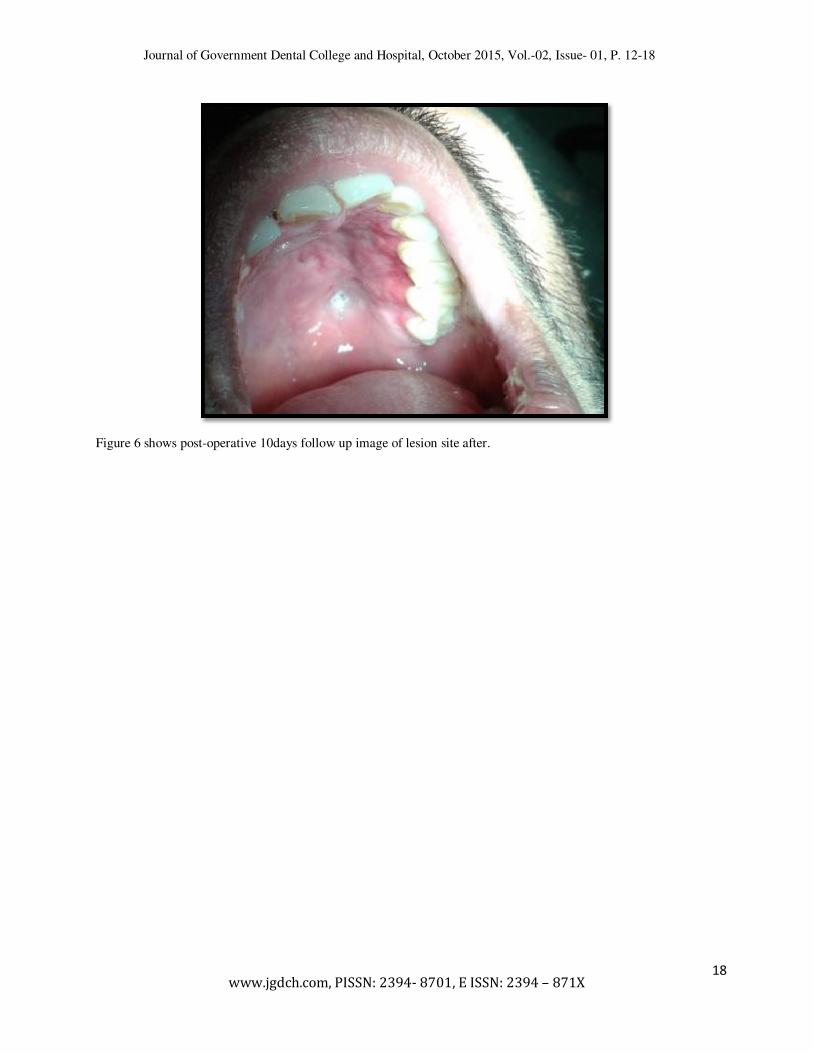
Figure 6 shows post-operative 10days follow up image of lesion site after.
18