
Platelet Rich Plasma—A Healing Aid and Perfect …
7
International Journal of Clinical Pediatric Dentistry, January-April 2011;4(1):69-75 69 IJCPD Platelet Rich Plasma—A Healing Aid and Perfect Enhancement Factor: Review and Case Report 1 Rani Somani, 2 Iram Zaidi, 3 Shipra Jaidka 1 Professor, Department of Pediatric and Preventive Dentistry, DJ College of Dental Sciences and Research Modinagar, Ghaziabad, Uttar Pradesh, India 2 Postgraduate Student, Department of Pediatric and Preventive Dentistry, DJ College of Dental Sciences and Research Modinagar, Ghaziabad, Uttar Pradesh, India 3 Associate Professor, Department of Pediatric and Preventive Dentistry, DJ College of Dental Sciences and Research Modinagar, Ghaziabad, Uttar Pradesh, India Correspondence: Rani Somani, Professor, Department of Pediatric and Preventive Dentistry, DJ College of Dental Sciences and Research, Modinagar, 617, G-3, Sector-1, Vaishali, Ghaziabad, Uttar Pradesh, India, e-mail: [email protected] CASE REPORT INTRODUCTION Platelet rich plasma, a novel based biologically active tool, is a new approach in pediatric dentistry. Platelet rich plasma brings the power of modern biological, chemical and physical science to solve the real clinical problems in pediatric dentistry. Marx in 1998 introduces PRP for reconstruction of mandibular defects, and it represents a relatively new biotechnology that is part of the growing interest in tissue engineering and cellular therapy. Gibble and Ness in 1990 introduced fibrin glue, alternatively referred to as fibrin sealant or fibrin gel, a biomaterial that was developed in response to the necessity for improved hemostatic agents with adhesive properties. Platelet rich plasma gel (PRP gel) is an autologous modification of fibrin glue obtained from autologous blood used to deliver growth factors in high concentrations. It is an autologous concentration of human platelets in a small volume of plasma, mimics coagulation cascade, leading to formation of fibrin clot, which consolidates and adheres to application site. Its biocompatible and biodegradable properties prevent tissue necrosis, extensive fibrosis and promote healing. Availability of PRP It is an autologous source of growth factors obtained by sequestrating and concentrating platelets by gradient density centrifugation. ABSTRACT Platelet rich plasma (PRP) has been a breakthrough in the stimulation and acceleration of tissue healing. It represents a relatively new approach in regenerative procedures and is a developing area in pediatric dentistry. It is an autologous source of growth factors obtained by sequestrating and concentrating platelets by gradient density centrifugation. This novel and potentially promising technique enhances body’s natural wound healing mechanism. This article goes on to describe preparation and clinical benefits of PRP in pediatric dentistry. Keywords: Growth factor, PRP, Regeneration, Wound healing. Constituents of PRP PRP contains high concentration of platelets and native concentration of fibrinogen. The alpha granules of platelets include a high concentration of factors, which are released on activation of platelets by adding calcium chloride and thrombin to PRP. The growth factors are diverse group of polypeptides that have important roles in the regulation of growth and development of a variety of tissues. Various growth factors released from PRP are listed in Table 1. PREPARATION OF PRP Platelet Rich Plasma Procurement Techniques It can be done by using various techniques: • Gradient density cell separators • Concentrating cell separators. 1. Using gradient density cell separators: These are general purpose cell separators like ELMD-500 (Medtronic Electromedic, Autotransfusion System, Parker, CO) require large quantity of blood (450 ml) and need to be operated in hospital setting. Blood is drawn into a bag containing citrate-phosphate-dextrose anticoagulant. First, the blood is centrifuged in a general purpose cell separator, at 5,600 rpm to separate the platelet poor plasma (PPP) from the red blood cells (RBC) and the 10.5005/jp-journals-10005-1085
Transcript of Platelet Rich Plasma—A Healing Aid and Perfect …

Platelet Rich Plasma—A Healing Aid and Perfect Enhancement Factor: Review and Case Report
International Journal of Clinical Pediatric Dentistry, January-April 2011;4(1):69-75 69
IJCPD
Platelet Rich Plasma—A Healing Aid andPerfect Enhancement Factor:
Review and Case Report1Rani Somani, 2Iram Zaidi, 3Shipra Jaidka
1Professor, Department of Pediatric and Preventive Dentistry, DJ College of Dental Sciences and ResearchModinagar, Ghaziabad, Uttar Pradesh, India
2Postgraduate Student, Department of Pediatric and Preventive Dentistry, DJ College of Dental Sciences and ResearchModinagar, Ghaziabad, Uttar Pradesh, India
3Associate Professor, Department of Pediatric and Preventive Dentistry, DJ College of Dental Sciences and ResearchModinagar, Ghaziabad, Uttar Pradesh, India
Correspondence: Rani Somani, Professor, Department of Pediatric and Preventive Dentistry, DJ College of Dental Sciencesand Research, Modinagar, 617, G-3, Sector-1, Vaishali, Ghaziabad, Uttar Pradesh, India, e-mail: [email protected]
CASE REPORT
INTRODUCTIONPlatelet rich plasma, a novel based biologically active tool,is a new approach in pediatric dentistry. Platelet rich plasmabrings the power of modern biological, chemical and physicalscience to solve the real clinical problems in pediatricdentistry. Marx in 1998 introduces PRP for reconstructionof mandibular defects, and it represents a relatively newbiotechnology that is part of the growing interest in tissueengineering and cellular therapy. Gibble and Ness in 1990introduced fibrin glue, alternatively referred to as fibrinsealant or fibrin gel, a biomaterial that was developed inresponse to the necessity for improved hemostatic agentswith adhesive properties. Platelet rich plasma gel (PRPgel) is an autologous modification of fibrin glue obtainedfrom autologous blood used to deliver growth factors inhigh concentrations. It is an autologous concentration ofhuman platelets in a small volume of plasma, mimicscoagulation cascade, leading to formation of fibrin clot,which consolidates and adheres to application site. Itsbiocompatible and biodegradable properties prevent tissuenecrosis, extensive fibrosis and promote healing.
Availability of PRP
It is an autologous source of growth factors obtained bysequestrating and concentrating platelets by gradient densitycentrifugation.
ABSTRACTPlatelet rich plasma (PRP) has been a breakthrough in the stimulation and acceleration of tissue healing. It represents a relatively newapproach in regenerative procedures and is a developing area in pediatric dentistry. It is an autologous source of growth factors obtained bysequestrating and concentrating platelets by gradient density centrifugation. This novel and potentially promising technique enhances body’snatural wound healing mechanism. This article goes on to describe preparation and clinical benefits of PRP in pediatric dentistry.Keywords: Growth factor, PRP, Regeneration, Wound healing.
Constituents of PRP
PRP contains high concentration of platelets and nativeconcentration of fibrinogen. The alpha granules of plateletsinclude a high concentration of factors, which are releasedon activation of platelets by adding calcium chloride andthrombin to PRP. The growth factors are diverse group ofpolypeptides that have important roles in the regulation ofgrowth and development of a variety of tissues. Variousgrowth factors released from PRP are listed in Table 1.
PREPARATION OF PRP
Platelet Rich Plasma Procurement Techniques
It can be done by using various techniques:• Gradient density cell separators• Concentrating cell separators.1. Using gradient density cell separators: These are general
purpose cell separators like ELMD-500 (MedtronicElectromedic, Autotransfusion System, Parker, CO)require large quantity of blood (450 ml) and need to beoperated in hospital setting. Blood is drawn into a bagcontaining citrate-phosphate-dextrose anticoagulant.First, the blood is centrifuged in a general purpose cellseparator, at 5,600 rpm to separate the platelet poorplasma (PPP) from the red blood cells (RBC) and the
10.5005/jp-journals-10005-1085

Rani Somani et al
70JAYPEE
PRP (also termed “buffy coat,” which contains theplatelets and the leukocytes). Then centrifuged at2,400 rpm to obtain PRP from the slurry of RBC andPRP. The procurement of PRP with this technique canbe accomplished in 30 minutes, and use of the obtainedPRP is recommended within 6 hours after being drawnfrom the patient. Platelet counts of 500,000 to 1,000,000in the PRP are usually obtained with this plasmapheresistechnique. With this processing technique, the remainingerythrocytes and PPP can be returned to the circulationor discarded.
Advantage
RBCs can be returned back to patient venous blood.
Disadvantages
• Need large volume of blood• Need large hospital setup.
2. Using concentrating cell separators: Platelet concentratingcell separators are more widely used since equipmentcan be accommodated in dental clinic setup.Thetechnology permits procurement of PRP using smaller
PDGF – platelet-derived growth factor; TFG-b – transforming growth factor beta; PDEGF – platelet-derived epidermal growth factor;PDAF – platelet-derived angiogenesis factor; IGF-1 – insulin-like growth factor 1; PF-4 – platelet factor 4; bFGF – basic fibroblastgrowth factor.
Table 1: Summary of growth factors released from platelets
Growth Molecular Source cells Target Actionfactor properties
PDGF
TGF-b
PDEGF
PDAF
IGF-1
PF-4
Cationic polypeptide(Mr = 30 kda)
2-chain polypeptide(Mr = 25 kda); 3different gene productsin humans: TGF-β1,TGF-β2, TGF-β3
53-amino acidpolypeptide (Mr = 6kda)
Acidic polypeptide(Mr = 45 kda)
Single-chainpolypeptide (Mr = 47kda) 47% homologywith insulin.
Homotetramer(Mr = 29 kda)
Platelets, macrophages,monocytes, endothelialcells, smooth musclecells.
Platelets, T-lymphocytes,macrophages/monocytes,neutrophils.
Platelets, macrophages,monocytes.
Platelets, endothelialcells.
Osteoblasts,macrophages, monocytes,chondrocytes.
Platelets
Fibroblasts, smooth musclecells, glial cells,macrophages/neutrophils.
Fibroblasts, marrow stemcells, endothelial cells,epithelial cells,preosteoblasts.
Fibroblasts, endothelialcells, epithelial cells.
Endothelial cells
Fibroblasts, osteoblasts,chondrocytes.
Fibroblasts, neutrophils
Stimulates chemotaxis/mitogenesis in fibroblast/glial/smooth muscle cells; regulatescollagenase secretion/collagensynthesis; stimulates macrophage/neutrophil chemotaxis.
Stimulates/inhibits endothelial,fibroblastic, and osteoblasticmitogenesis; regulates collagensynthesis/collagenase secretion;regulates mitogenic effects ofother growth factors; stimulatesendothelial chemotaxis andangiogenesis.
Stimulates endothelialchemotaxis/angiogenesis;regulates collagenase secretion;stimulates epithelial/mesenchymalmitogenesis.
Increases angiogenesis and vesselpermeability; stimulatesmitogenesis for endothelial cellsby direct or indirect actions;several cytokines and growthfactors up-regulate PDAF,including IGF-1, TGF-alpha andbeta, PDGF, bFGF, PDEGF, andIL-1 beta.
Stimulates cartilage growth, bonematrix formation, and replicationof preosteoblasts and osteoblasts;acts as an autocrine and paracrinefactor; in combination with PDGFcan enhance the rate and qualityof wound healing.
Chemoattractant for neutrophils andfibroblasts; potent antiheparinagent.

Platelet Rich Plasma—A Healing Aid and Perfect Enhancement Factor: Review and Case Report
International Journal of Clinical Pediatric Dentistry, January-April 2011;4(1):69-75 71
IJCPD
volumes of blood, increasing the platelet concentrationand avoiding need of RBC and PPP reinfusion. Twosuch separators commercially available are HarvestSmartPrep Platelet Concentrate System (HSPCS)(Harvest Technologies, Plymouth, MA) and the 3iPlatelet Concentrate Collection System (3i PCCS) (3iImplant Innovations, Palm Beach Gardens, FL). Bothplatelet-concentrating cell separators are similar inperformance and simplicity, and they represent asignificant advantage that they requires less time toproduce the PRP (15 minutes versus 20 minutes) andless operator intervention and training.
Advantages
• Do not need large hospital setup• Need small volume of blood.
Disadvantage
RBCs cannot be returned back to patients venous blood.
Properties of Platelet Rich Plasma
• Increase tissue vascularity through increasedangiogenesis.
• Enhancing collagen synthesis• Enhancing osteogenesis• Increasing the rate of epithetlial, and granulation tissue
production.• Antimicrobial effect• Reaction with other material: PRP does not react or
interfere with any other restorative material glassionomer cements or composite resin used as fillingmaterial are not affected by it.
• Biocompatibility: Any material that is identified to beused in humans or animals should be biocompatiblewithout having any toxic or injurious effects on biologictissues and its functions. PRP offers a biologically activesubstance with the release of growth factor.
• Tissue regeneration: PRP allows regeneration of tissuewith the release of growth factors.The properties of PRP are based on the production and
release of multiple growth and differentiation factors uponplatelet activation. These factors are critical in the regulationand stimulation of the wound healing process, and they playan important role in regulating cellular processes such asmitogenesis, chemotaxis, differentiation and metabolism.Growth factors interact one with another, consequentlyforming a cascade of different signal proteins with multiplepathways, ultimately leading to the activation of geneexpression and then protein production. Recent reports havesuggested that PRP leads to more rapid epithelialization,
more dense and mature bone with better organizedtrabeculae, and greater bone regeneration.
MECHANISM OF ACTION OF PLATELETRICH PLASMA
Platelet rich plasma has been found to work via threemechanisms:1. Increase in local cell division (producing more cells):
According to Nathan E Carlson 2002 after the injury,platelets begin to stick to exposed collagen proteins andrelease granules containing adenosine diphosphate,serotonin and thromboxane, all of which contribute tothe hemostatic mechanism and the clotting cascade.
2. Inhibition of excess inflammation by decreasing earlymacrophage proliferation.
3. Degranulation of the agranules in platelets, whichcontain the synthesized and prepackaged growth factors.The active secretion of these growth factors is initiated
by the clotting process of blood and begins within 10minutes after clotting. More than 95% of the presynthesizedgrowth factors are secreted within 1 hour (Kevy andJacobson). PRP has been shown to remain sterile and theconcentrated platelets viable for up to 8 hours oncedeveloped in the anticoagulated state.
ROLE OF PLATELET RICH PLASMAIN THE PROCESS OF WOUND HEALING
The process of wound healing can be divided into threedifferent stages:• Biochemical activation• Cellular activation• Cellular response.
1. Biochemical activation involves the translation ofmechanical injury into biochemical signals that can beunderstood by the body. The trigger that starts thecascades is the Hagemann factor found in serum. Wheninjury causes disruption of the microcirculation, plasmacomes in contact with tissue proteins and the basementmembrane. This activates the Hagemann factor andcirculating platelets. The activated Hagemann factoractivates the clotting cascade and produces fibrin to helpin hemostasis and thrombin formation that causes themaximal release of platelet alpha granules.
2. The cellular activation stage results in the influx of cellsinto the wound. The first cellular response involvesneutrophils, monocytes, and platelets. Plateletsaccumulate at the wound site in response to the initialinjury, in response to thrombin, platelets release theirgranules that contain locally acting growth factors. Thesefactors signal the local mesenchymal and epidermal cells

Rani Somani et al
72JAYPEE
to migrate, divide, and increase their collagen andglycosaminoglycan synthesis. This initial release isthought to accentuate the reparative response.
3. The monocytes transformed into macrophages areinvolved in the final cellular response. These cells assistthe neutrophils in host defense and produce many ofthe growth factors, which direct repair until the woundis healed.Platelet rich plasma accentuates all these processes.The
active secretion of these growth factors is initiated by theclotting process of blood and begins within 10 minutes afterclotting. More than 95% of the presynthesized growthfactors are secreted within 1 hour (Kevy and Jacobson).PRP has been shown to remain sterile and the concentratedplatelets viable for up to 8 hours once developed in theanticoagulated state. Thus, activated autologous plateletrich plasma releases growth factors that increase collagencontent, accelerate epithelial and epidermal regeneration,promote angiogenesis, enhance wound strength, hastenhemostasis, improve tissue regeneration, hasten remodeling,reduce pain, and reduce infection, which ultimately leadsto regeneration.
THERAPEUTIC POTENTIAL OF PRP INPEDIATRIC DENTISTRY
Platelet rich plasma has been a breakthrough in thestimulation and acceleration of tissue healing. It representsa relatively new approach in regenerative procedures.
Following are the clinical applications of PRP inpediatric dentistry:1. Pulp Capping: PRP has been proposed as a potential
medicament for capping of pulps with reversible pulpitisbecause of its excellent tissue compatibility. It is muchsuperior to routinely used calcium hydroxide based ontissue reaction between these material.
2. Pulpotomy: Formocresol has been routinely used as apulpotomy agent for deciduous teeth. But this materialhas been criticized for its tissue irritating, cytotoxic andmutagenic effects. PRP was found to be an ideal materialwith low toxic effect, increased tissue regeneratingproperties and good clinical results. Damle et al in 2004compared PRP and calcium hydroxide as pulpotomyagent and found that PRP gives 100% success rate ascompared to 60% of calcium hydroxide.
Nakashaki et al in 2007 compared PRP andhydroxipatite and found that PRP is a better pulptotomyagent.
3. Extraction Socket: Extraction has always been a sourceof trauma, anxiety and fear in children. Therefore in anattempt to promote rapid healing application of PRP was
done in extraction socket. Following tooth removal boneformation normally takes 16 weeks and may result inless than adequate volume for the necessaryreconstruction. Platelet rich plasma (PRP) has beenpromoted as an effective method for improving boneformation. Its use is often expensive, time consuming,or not clinically convenient for the patient and/orclinician. PRP initiates healing by sequestrating plateletsand enriching natural clot which initiates a rapid andcomplete healing process. It also accelerates healing bypromoting rapid revascularization and re-epithelizationof flaps and cell proliferation. Ratushki et al in 2008has done a study in which they examine a simple methodfor obtaining a “Buffy Coat”-PRP (BC-PRP) and itseffect on bone healing following the removal of bilateralmandibular 3rd molars and found that after applicationof PRP there was better bone regeneration and betterhealing.
CASE REPORTS
Platelet rich plasma has been a breakthrough in thestimulation and acceleration of tissue healing. It representsa relatively new approach in regenerative procedures. Basedon this, application of PRP in pulpotomy and extractionsocket was done in the Department of Pediatric Dentistry,DJ Dental College, Modinagar. The patients were selectedfrom the outpatient department with good general health,no history of systemic illness or hospitalization and withno history of antibiotic intake in the past six months. Theparents and guardians of the child were informed about thestatus of the child’s dentition and written consent wasobtained from them.
CASE 1
Pulpotomy with PRP
Criteria for case selection:The criteria given by Hellig J et al in 1984 and Waterhouseet al in 2000 were followed:• Teeth with deep carious lesion (radiographically the
caries should be approximating to the pulp)• Teeth should be restorable after completion of the
procedure• Absence of symptoms indicative of advanced pulpal
inflammation, such as spontaneous pain or history ofnocturnal pain.
• Absence of clinical sign or symptoms suggesting a non-vital tooth such as suppurating sinus soft tissue swelling.
• Absence of clinical radiographic signs of pulpal necrosis,i.e. furcation involvement, periapical pathology, internalresorption, calcification in canal.
Platelet Rich Plasma—A Healing Aid and Perfect Enhancement Factor: Review and Case Report
International Journal of Clinical Pediatric Dentistry, January-April 2011;4(1):69-75 73
IJCPD
• Hemorrhage should stop within five minutes fromamputated pulp stumps using a sterile pledget of moistcotton. After assessment of clinical and radiographiccriteria, single visit pulpotomy was performed onselected teeth.
PREPARATION OF PRP
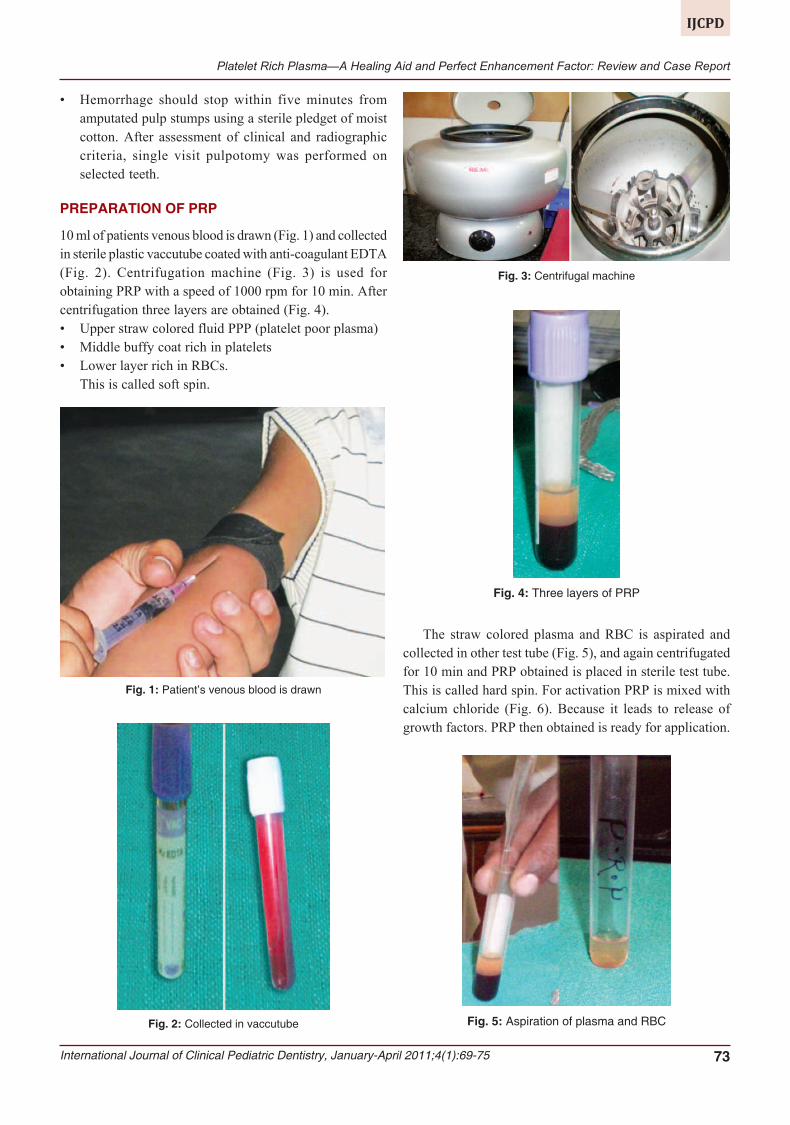
10 ml of patients venous blood is drawn (Fig. 1) and collectedin sterile plastic vaccutube coated with anti-coagulant EDTA(Fig. 2). Centrifugation machine (Fig. 3) is used forobtaining PRP with a speed of 1000 rpm for 10 min. Aftercentrifugation three layers are obtained (Fig. 4).• Upper straw colored fluid PPP (platelet poor plasma)• Middle buffy coat rich in platelets• Lower layer rich in RBCs.
This is called soft spin.
Fig. 1: Patient’s venous blood is drawn
Fig. 2: Collected in vaccutube
Fig. 3: Centrifugal machine
Fig. 4: Three layers of PRP
The straw colored plasma and RBC is aspirated andcollected in other test tube (Fig. 5), and again centrifugatedfor 10 min and PRP obtained is placed in sterile test tube.This is called hard spin. For activation PRP is mixed withcalcium chloride (Fig. 6). Because it leads to release ofgrowth factors. PRP then obtained is ready for application.
Fig. 5: Aspiration of plasma and RBC

Rani Somani et al
74JAYPEE
Fig. 6: Mixed with calcium chloride
Fig. 7: Pulpotomy with PRP
Fig. 8: Postoperative after 15 days
Fig. 9: Postoperative after one month
PROCEDURE
Preoperative radiograph was taken. Local anesthesia wasachieved using 2% xylocaine with 1:80,000 adrenaline. Theteeth were isolated using rubber dam, cavity outline wasestablished with high-speed round bur with water coolent.Caries was excavated with a spoon excavater. The pulpchamber was entered and roof was removed. After coronalamputation of pulp freshly prepared PRP was placed overthe pulp stump and gently packed with the sterile pledgetof moist cotton. A thick mix of ZOE was placed to seal thecoronal pulp chamber (Fig. 7). Clinical examination wasundertaken at 15 days (Fig. 8), 1st and 3rd month intervals,where as radiographic evaluation of the treated teeth werecarried out at 1st and 3rd month interval (Fig. 9).
Teeth were evaluated for the presence and absence offollowing findings:
Clinical findings:• Spontaneous pain or pain initiated by stimuli.• Signs of sinus formation, tenderness to percussion, soft
tissue swelling and mobility.• Signs of defective restoration or recurrent caries.
Radiographic findings:• Signs of pulpal degeneration, such as periapical or furcal
radiolucency, canal calcification, internal resorption.• Defective restoration or recurrent caries.
RESULTS
Preoperatively, the incidence of pain was present in all thepatients. No signs of pulpal degeneration noticed clinicallyand radiographically.
Clinical examination after one and three months oftreatment revealed absence of pain, swelling and mobility.
Radiographic examination revealed no periapical orfurcation involvement. None of teeth showed calcific barrierformation at the mesiodistal width of the root canal. Thus,PRP showed a 100% success rate, as all teeth wereasymptomatic in a three months evaluation.
CASE 2
PRP in Extraction Socket
In an attempt to promote rapid healing, freshly preparedPRP was placed in extraction socket immediately afterextraction and betadiene pack was given (Figs 10 and 11).Post extraction instructions were given and patient wasrecalled after 3 days. Clinical examination was done and itshows rapid healing of socket (Fig. 12). PRP initiates healingby sequestrating platelets and enriching natural clot, whichinitiates a rapid and complete healing process. It alsoaccelerates healing by promoting rapid revascularizationand re-epithelization of flaps and cell proliferation.

Platelet Rich Plasma—A Healing Aid and Perfect Enhancement Factor: Review and Case Report
International Journal of Clinical Pediatric Dentistry, January-April 2011;4(1):69-75 75
IJCPD
Fig. 10: Extraction socket
Fig. 11: Application of PRP in extraction socket
Fig. 12: After 3 days (approximation of flaps)
CONCLUSION
PRP prepared by sequestration and concentration of plateletsis an autologous, safe and user friendly source of growthfactors, is an innovative biological tool in the field ofpediatric dentistry for pulpotomy and rapid healing ofextraction wound.
REFERENCES
1. Earl G Freymiller, Tara L. Aghaloo. Platelet rich plasma: Readyor not? J Oral Maxillofac Surg 2004;62:484-88.
2. Robert E Marx. Platelet rich plasma: Evidence to supportits use. J Oral Maxillofac Surg 2004;62:489-96.
3. Andrés R Sánchez, Phillip J SheridanMS2/Leo I Kupp.Is platelet rich plasma the perfect enhancement factor?A current review. The International Journal of Oral andMaxillofacial Implants.
4. Born GVR, Cross MJ. The aggregation of blood platelets. JPhysiol 1963;168:178-95.
5. Gernot Weibrich, Wilfried KG Kleis Curasan. PRP kit vs PCCSPRP system collection efficiency and platelet counts of twodifferent methods for the preparation of platelet rich plasma.Clin Oral Impl Res 2002;13:437-43.
6. Federico Luengo Gimeno, Silvia Gatto, José Ferro, Juan OscarCroxatto, Juan Eduardo Gallo. Preparation of platelet rich plasmaas a tissue adhesive for experimental transplantation in rabbits.Thrombosis Journal 2006;4:18.
7. Juliette Van Den Dolder. Platelet rich plasma: Quantificationof growth factor levels and the effect on growth anddifferentiation tissue engineering 2006;12(11).
8. Nathan E Carloson, Robert B Roach. Platelet rich plasma clinicalapplications indentistry JADA, October 2002, Vol. 133.
9. Giuseppe Intin, et al. Calcium sulfate and platelet rich plasmamake a novel osteoinductive biomaterial for bone regeneration.Journal of Translational Medicine 2007;5:13.


