Plasticity of the Visual Pathway and Neuroimaging · Chapter 13 Plasticity of the Visual Pathway...
20
Chapter 13 Plasticity of the Visual Pathway and Neuroimaging M. Gallegos-Duarte, S. Moguel-Ancheita, J.D. Mendiola- Santibañez, V. Morales-Tlalpan and C. Saldaña Additional information is available at the end of the chapter http://dx.doi.org/10.5772/53013 1. Introduction Once the formation of cerebral and ocular structures has been completed, the simple light stimulus is enough to activate the necessary neural circuits that integrate the visual tract. It has been estimated that 50% of the cerebral cortex participates in the integration of the hu‐ man binocular visual system. This system is practically distributed throughout the brain. Therefore, its study has captured the interest of various scientific fields. The neuronal plasticity of the visual system is the theme of several current works [1, 2, 3]. Nevertheless, very little is known about the adaptive changes and about neuronal plasticity in children who have congenital strabismus (CS), main subject of the current chapter. The vision is a brain function highly specialized and complex [4]. The structures that per‐ form it are developed by means of division processes, migration, functional integration and regionalization during the corticogenesis [5]. An incorrect development or a bad functioning of the molecular, cellular or physiological mechanisms can induce an altered vision as in the case of strabismus. The visual process is the result of the elaboration of images generated in the cortical net‐ work. This process comprises from the moment the light stimulus reaches the retina until the evocation of visual memories imbued with an affective component generated by the thalamus. In a simple way, we will say that the visual system is integrated by the visual via and cortical integrator, but in this chapter we will specifically refer to the last one [6, 7]. The visual via takes the information in one direction in an antero-posterior direction. It ex‐ tends from the eyes as sensorial receptors, arrives to the optical nerves, whose monocular © 2013 Gallegos-Duarte et al.; licensee InTech. This is an open access article distributed under the terms of the Creative Commons Attribution License (http://creativecommons.org/licenses/by/3.0), which permits unrestricted use, distribution, and reproduction in any medium, provided the original work is properly cited.
Transcript of Plasticity of the Visual Pathway and Neuroimaging · Chapter 13 Plasticity of the Visual Pathway...

Chapter 13
Plasticity of the Visual Pathway and Neuroimaging
M. Gallegos-Duarte, S. Moguel-Ancheita,J.D. Mendiola- Santibañez, V. Morales-Tlalpan andC. Saldaña
Additional information is available at the end of the chapter
http://dx.doi.org/10.5772/53013
1. Introduction
Once the formation of cerebral and ocular structures has been completed, the simple lightstimulus is enough to activate the necessary neural circuits that integrate the visual tract. Ithas been estimated that 50% of the cerebral cortex participates in the integration of the hu‐man binocular visual system. This system is practically distributed throughout the brain.Therefore, its study has captured the interest of various scientific fields.
The neuronal plasticity of the visual system is the theme of several current works [1, 2, 3].Nevertheless, very little is known about the adaptive changes and about neuronal plasticityin children who have congenital strabismus (CS), main subject of the current chapter.
The vision is a brain function highly specialized and complex [4]. The structures that per‐form it are developed by means of division processes, migration, functional integration andregionalization during the corticogenesis [5]. An incorrect development or a bad functioningof the molecular, cellular or physiological mechanisms can induce an altered vision as in thecase of strabismus.
The visual process is the result of the elaboration of images generated in the cortical net‐work. This process comprises from the moment the light stimulus reaches the retina untilthe evocation of visual memories imbued with an affective component generated by thethalamus.
In a simple way, we will say that the visual system is integrated by the visual via andcortical integrator, but in this chapter we will specifically refer to the last one [6, 7]. Thevisual via takes the information in one direction in an antero-posterior direction. It ex‐tends from the eyes as sensorial receptors, arrives to the optical nerves, whose monocular
© 2013 Gallegos-Duarte et al.; licensee InTech. This is an open access article distributed under the terms ofthe Creative Commons Attribution License (http://creativecommons.org/licenses/by/3.0), which permitsunrestricted use, distribution, and reproduction in any medium, provided the original work is properly cited.

information influences the chiasma, where the nasal sector of the monocular informationwill perform a crossing of fibers, remaining the temporal fibers without crossing, fromthis point, the information shared by both eyes continues through the optic bands to theprimary visual cortex or striate cortex to systematize the information and generate hyper‐complex images [4, 8]. All the steps described, up to now, are considered to form part ofthe first afferent visual system.
Once the images that have been previously decoded by the striate cortex, in accordance withits temporal frequency space, perceptual-visual process initiates [1, 9]; for that, the visual in‐formation must be included in the intern dialog of the brain by means of the cortical net‐work for the purpose of giving it a sense. For that, the information coming from the striatecortex travels through the anterior portions of the brain to be processed in multiple neuronalcenters and circuits that shape the cortical integrator [4, 8].
Although the striate cortex is in charge of capturing a great amount of visual elements, itlacks an adequate working memory, for that situation, it results impossible to process thelogic of the hyper complex images and give them a ludic sense, to identify and give sense tothe environment, it is necessary to resend the information to the working memory sited inthe thalamic cortex that adjoins the temporal lobes [4, 8].
Part of the information coming from the grooved area is directed towards the temporallobes to give a logic sense to answer the question “what am I seeing?” The temporal cortexshares its proximity with the thalamus and auditory areas, because besides de affectivesense (like, dislike, danger, repulsion, attraction, etc.) the temporal area processes the visualinformation to give it directionality [10, 11].
The temporal lobes cooperate with their counterpart of Magno-cellular elements that con‐form the occipital-parietal via, this last one is the one in charge of assertively answering thequestion “where is what I am seeing?” the four pathways: left and right occipital temporalas well as the left and right occipital-parietal, give a meaning to the visual information com‐ing from the striate cortex [2, 9].
These associative areas conduct the information that must transform in complex pre-logic el‐ements such as the early recognition of facial expressions or manual attitudes of others(threaten, friendship, indifference), for example, and they are the base for the realization ofsome specific visual-motor functions such as reading-writing. Once the limbic system adja‐cent to the temporal lobes, gives affective sense, motivation, and nuance to the visual infor‐mation, all the observed can be evaluated in relation with other experiences kept in thememory [4, 8, 12].
The cortical integrator works in parallel in an interactive form, as if it were a close cyberneticnegative feedback system; its most elaborated product is the binocular vision. It is to be ex‐pected that the correct operation and integration of the neuronal visual network depend onan adequate connectivity among the parts that conform the cortical network. In this context,the visual perception is the result of the process of the information that is generated in theneuronal circuits [1, 13, 14].
Medical Imaging in Clinical Practice308

From this point, all the information is distributed through the cortical network [1, 9] and thisis important to consider before analyzing the studies of neuroimaging, by means of a neuro‐metric study [15], electroencephalography [16, 17], or visual perceptual study [18].
Although the CE does not initially affect the superior functions such as judgment or intel‐lect, frequently patients who have this disease may get confused with letters, they requiremore time to learn to read, are easily distracted, and in general, the development of theiractivities take more time respecting the individuals with a normal vision. In fact, the chil‐dren with strabismus present different grades of visual perceptual deficit, which we believeis related with the alterations in the pathway of neuronal interconnection sited in the striatecortex and the cortico-cortical and interhemispheric zones that participate in the processingof the image [6, 15, 17, 19] as it will be forwardly described in the perceptual-visual analysis(Table 1).
Visual perceptual ability Before the treatment After the treatment
Fusion Amplitude -0.4/-0.7 +3 /-1
Stereopsis 0% 0%
Fixation disparity H: -1.1 V: -0.4 H:-1.1 V: -0.7
Perception Speed 43% 50%
Main elements 88% 95%
Visual memory 87% 93%
Saccadic Movements 72 % 75 %
Shapes and Sizes 42% 80%
Following movements 95% 97%
Peripheral vision 42% 50%
Spatial vision 82% 85%
Table 1. Percentages before and three months after surgery for correction of congenital strabismus H: horizontal, V:vertical.
The Computerized Visual Perceptual Analysis (CVPA) is used to meet the efficiency of visu‐al perceptual system. The CVPA basically estimates the efficiency of the integrator in theposterior portions of the brain, that is, the development of the occipito-temporal and occipi‐to-parietal pathway.
Plasticity of the Visual Pathway and Neuroimaginghttp://dx.doi.org/10.5772/53013
309
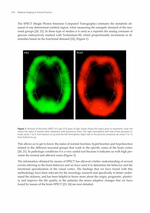
The SPECT (Single Photon Emission Computed Tomography) estimates the metabolic de‐mand of one determined cerebral region, when measuring the energetic demand of the neu‐ronal groups [20, 21]. In these type of studies it is used as a reporter the analog consume ofglucose radioactively marked with Technetium-99, which proportionally increments or di‐minishes based on the functional demand [22], (Figure 1).
Figure 1. Pictures of the brain SPECT of a girl of 6 years of age. Green shows the status prior to treatment, color redshows the status 4 months after treatment with botulinum toxin. The right hemisphere (left side of the pictures) in‐clude zones 1 to 6, from bottom to up and the left hemisphere (right side of the picture) comprises de zones 7 to 12from bottom to up.
This allows us to get to know the states of normal function, hyperfunction and hypofunctionrelated to the different neuronal groups that work in the specific zones of the brain cortex[20, 21]. In pathologic conditions it is a very useful tool because it indicates us with high pre‐cision the normal and affected zones (Figure 2).
The information obtained by means of SPECT has allowed a better understanding of severalevents referring to the brain behavior and we have used it to determine the behavior and thefunctional specialization of the visual cortex. The findings that we have found with thismethodology have been relevant for the neurology research and specifically to better under‐stand the sickness, and has been helpful to know more about the origin, prognostic, plastici‐ty and improve the life quality in the patients; the neuro adaptive changes that we havefound by means of the brain SPECT [23, 24] are next detailed.
Medical Imaging in Clinical Practice310

The neuronal network generates electric impulses, and this electric activity can be registeredby means of the use of the Digitalized Brain Mapping (DBM), (Figure 3), as well as the anal‐ysis of the coherence of electroencephalography [25]. These methods provide an objectiveand noninvasive index of the functional relations that exist between the areas of the brainsurface.
Figure 2. Above, to the right, the picture shows the combination of both studies (MERGE): yellow are those areas thatdid not suffer many changes, red highlights areas that showed an important metabolic increase (zones 1 and 12), andin shades of green (zones 5 and 9) are those areas that modulated their metabolic activity. At the bottom of the fig‐ure, the graphs show uptake of Technetium-99. The black line shows the previous state to the eye alignment by theuse of botulinum toxin (Pre), the gray shows the reception levels of the glucose analog 3 months after the treatment(Post). The increase of the uptake after the treatment indicates a greater metabolic activity in V1 and V2. Reception ofTechnetium 99, the axis “X” shows the intensity level while the axis “Y” indicates the number of times that such inten‐sity is registered
Plasticity of the Visual Pathway and Neuroimaginghttp://dx.doi.org/10.5772/53013
311

Figure 3. The Brain Mapping shows an increase in the absolute power and hypersimetry in the posterior regions ofthe brain after the surgery treatment of an 11 years old boy with congenital strabismus. Previous to the treatment hepresented slow activity and asymmetry of the background activity in occipital regions.
In a previous study, we reported the results for 193 DBM obtained from children with con‐genital strabismus [26]. We found that 57% of the patient’s studies without treatment pre‐sented alterations in the electric behavior that went from mild, such as intertemporal hypocoherence, up to important such as paroxysms and epilepsy, but 6 months after the surgeryonly 29% of the patients presented alterations in the electric activity (Figure 4). This motivat‐ed to perform a more meticulous analysis of the changes of the electric behavior that happenafter the surgery by means of Neurometry [15].
Medical Imaging in Clinical Practice312

Using digitized brain mapping, the authors have discovered morphometric and neurofunc‐tional alterations in the cerebral cortex of patients with essential strabismus, such as inter-temporal hypocoherence, cortico-subcortical dysfunction, slowing-down and asymmetry inthe frequency, among others (Figures 3 and 4).
Figure 4. Summary of the reports of the Digitalized Brain Mapping obtained from 193 children with congenital stra‐bismus. The dotted lines separate the sample in 4 levels: 43% were located with a normal electric behavior, 43% inlevel 1 with low electroencephalographic alterations and 14% presented important alterations
Plasticity of the Visual Pathway and Neuroimaginghttp://dx.doi.org/10.5772/53013
313

Neurometry allows us to get to know the coherence, which is determined by the activity ofthe short and large interconnection fibers, intra and interhemispheric, and expresses the syn‐chrony among neuronal groups [15, 27] (Figures 6 and 7). So, meanwhile the hypo coher‐ence expresses the lack of capacity of connection of neuronal groups, the hyper coherenceindicates that two or more areas are over connected and work in excessive form. Besides, itallows comparing the obtained results with normative values of the asymptomatic popula‐tion acquiring a statistical meaning of ±2 standard deviation [26, 15], (Figures 5 and 6).
Figure 5. Neurometric report before the treatment of the intrahemispheric (up) and interhemispheric (down) coher‐ence analysis of a 7 years old boy with congenital strabismus. The excessive hypocoherence is marked in blue color,the excessive hypercoherence is marked in red. In abbreviation is marked by pairs the zones of interest analyzed ( O1=left occipital, T4 = right temporal, as an example).
The Neurometry analysis has enabled us to determine that the following functional relationsare altered: occipito-temporal, occipito-parietal and, especially, inter-temporal; the latter in‐cludes intra- and inter-hemispheric relations [10, 15, 28]. Similarly, we have employed thebrain SPECT technique to define those areas with functional deficits [29, 30].
Medical Imaging in Clinical Practice314

n a study performed by neurometric analysis we found previous to the surgery treatment ofcongenital strabismus that 15 of the 16 patients showed everlasting hypocoherence [28]; butafter the surgery we found an improvement in this parameter. (Figure 6).
Figure 6. Neurometric report of the same boy as in figure 9 but 6 months after the surgical treatment. A diminish ofthe hypocoherence for Delta in frontal regions, an increase of the intertemporal coherences for Alfa and the modula‐tion of the occipito-occipital coherence are observed.
2. Statement of problem
The CS is a neurological disease that manifests as a pathological deviation of the eyes [6, 31].It represents the maximum perturbation of the binocular visual system and affects 3% of hu‐man beings [32, 33]. Due to the sensorial and motor alterations that characterize this diseasemay be systemized in an accurate manner, and results very simple to correlate the clinicwith the neuroimage studies [26].
Plasticity of the Visual Pathway and Neuroimaginghttp://dx.doi.org/10.5772/53013
315

Given its anatomic and functional repercussions together with its esthetic and social impact,the predominance of CS in humans is patent [34]. In Mexico, where the majority of the pop‐ulation has a mixture of Spanish and Native American origins, this condition is one of themost frequent congenital disabilities [35].
However, one thing is to study the visual tract in healthy subjects and other very differentmatter is the study of the cerebral cortex of individuals with functional and/or structuralasymmetries in their visual system, as happens in patients with essential strabismus. In par‐ticular, among our main concerns are the results of surgical and pharmacological manipula‐tions of the extra ocular muscles, which will be addressed in this chapter.
In this manner, strabismus taken as a model of study offers researchers a unique opportuni‐ty to gain more insight into the mechanisms the brain uses to compensate the disequilibriumin the cortical network. This network, given the nature of the disease, instead of working inparallel presents peculiarities that should be considered in the study of brain plasticity.
The analysis of the cortical alterations inherent to strabismus has been possible due to neu‐roimaging techniques. As a result, it has been possible to ascertain, for example, that the finemorphometry in the brain of strabismic children is different from that in healthy children.This is particularly patent in the posterior portions of the brain.
3. Method used
To inquire about the changes that occur in the binocular visual system are analyzed theadaptive changes and brain plasticity before and after the ocular alignment, using the neu‐roimage studies [36, 37]: SPECT, CVPA, DBM and Neurometry.
To identify these changes, the authors have carried out studies to determine the "before" and"after" in the treatment of strabismus by combining various neuroimaging approaches. As aresult, have been identified positive and objective signs of brain plasticity.
To clinically study the perceptual system of the cortical network we use besides the visualacuteness, the CVPA, which examines the 10 basic visual abilities that are detailed in thetable 1; these abilities are a reference of the functionality of the visual perceptual system [38].
To get to know the adaptive changes in the perceptual area, we carried out the CVPA on 22children from 6 to 7 years old before and after the strabismus surgery. These children didn´treceived any additional treatment besides the surgery treatment (Table 1).
We have obtained SPECT images previous to the surgery or pharmacological treatment and4 months after the treatment (Figures 7 and 8). The images were compared to determine themodified area and with that we could localize specifically the metabolic modifications in theseveral neuronal groups and, afterwards associate them to the neuronal funciton changes inthe patients [36].
Medical Imaging in Clinical Practice316

Figure 7. Zone 1 analysis of the SPECT (right occipital) of a 5 years old girl with congenital strabismus. The graphicsand photos show the uptake of Technetium-99, the axis “X” shows the intensity level while the “Y” axis indicates thenumber of times that such intensity is registered. The black line shows the state previous to the strabismus treatmentof the first case, and the gray line shows the state after the treatment. This metabolic modulation obeys the neuroa‐daptative change and the brain plasticity. The pictures show the different zones of interest that that were analyzed(marked in red arrows, has shown in the upper graphic)
Figure 8. Behavior of the Technetium 99 before and after the treatment with botulinum toxin in a patient of 7 yearsold with congenital strabismus. The “X” axis shows the intensity level and the “Y” axis shows the number of times thatsuch intensity is registered, the black line shows the previous behavior and the gray line shows the behavior after thetreatment. Before the treatment, the zone 1 shows the increase of the density from 183 to 250, after the treatment animportant increase is observed in 284, possibly related to the inter-hemispheric connectivity because the patient couldobtain a binocular vision
Plasticity of the Visual Pathway and Neuroimaginghttp://dx.doi.org/10.5772/53013
317

To quantify the neurological adaptative changes related to the CS treatment, it was analyzedand graphed the capturing of the Technetium-99 for the purpose of establishing the grade ofhyper and hypo function of the 12 zones of interest. In this communication it is showed thevalues before and after treatment in the gray scale (Figures 2, 7 and 8).
4. Results
In this study, after the surgery, it was demonstrated a certain recovery in CVPA, some ofthese changes as is the case of the saccadic movements and the disparity of the fixation werediscrete, nevertheless other parameters such as perception of forms and sizes, the magni‐tude of the fusion or the peripheral vision showed important changes. Up to here, thechanges clinically detected by CVPA indicate that there were favorable adaptive changes(Table 1).
From a metabolic point of view, the alterations previous to the strabismus treatment includethe presence of hypometabolic regions; these regions show a substantial improvement afterthe pharmacological treatment with the botulinum toxin (Figures 1, 2, 7 and 8).
The pictures of the brain cortex showed a representative example of the change observedin several patients, in those pictures it is showed the metabolic change in special form, inthe same individual before and after the correction of strabismus [4, 36, 37]. The imagewas divided in 12 sections to locate the prompt changes and to make the analysis easy(Figures 1 and 2).
From a bioelectric perspective, one of the most significant neuro-adaptive changes we havefound after the strabismus treatment is an improvement in inter-temporal and inter-parietalcoherences (Figure 9 and 10). On the other hand, we have shown the presence of hyper-sym‐metry and higher efficacy in the cortical input of the primary visual cortex using the Neuro‐metry technique (Figure 11). These findings are in accordance with brain SPECT results(Figures 2 and 8).
When comparing the previous and posterior neurometries to the surgery treatment of 9patients of strabismus, we found significant changes: improvement in the intertemporalcoherences, an occipito-occipital hypersimetry (Figure 11), as well as a diminish in the oc‐cipital hyper-coherence, these data suggest that the surgery modifies substantially the con‐nectivity cortico-cotical interhemispheric, as well as an increase in the activity of thestriate cortex.
The findings found are congruent with the observed in the brain SPEC, in the sense that af‐ter the orientation of the visual axes, it was evident an exponential increase of the metabo‐lism in the cortical areas V1 and V2 responsible of the elaboration of the hyper compleximages with the depth sense, and these favorable adaptive changes indicate the presence ofneuroplasticity in the binocular visual system in a punctual manner.
Medical Imaging in Clinical Practice318

Delta Theta Alpha Beta
P4-O2 Right pre -0.71 -0.87 -0.82 -0.31
P4-O2 Right post 0.03 -0.66 0.05 -0.62
Sta
nd
ard
de
via
tio
n
Right occipito-parietal coherence
Figure 9. Posterior neuroelectric changes to the surgical correction of strabismus in a 5 years old child. A marked hy‐pocoherence in the occipito-parietal via before the treatment is represented with the blue line while the red lineshows an improvement for Alfa and Delta after de surgery.
Delta Theta Alpha Beta
F3-C3 Left pre 1.68 1.76 1.95 2.17
F3-C3 Left post -0.35 -0.2 -0.45 0.65
Sta
nd
ard
de
via
tio
n
Left fronto-central coherence
Figure 10. Neurometric analysis of a 5 years old child with congenital strabismus. The blue line showed the previousstate of hypercoherence and the green line shows the normofuntion 3 months after the treatment.
Plasticity of the Visual Pathway and Neuroimaginghttp://dx.doi.org/10.5772/53013
319

DELTA THETA ALPHA BETA
O1-O2 PRE 0.17 -0.91 -1.17 -0.94
O1-O2 POST -0.53 0.36 0.2 0.38
-1.4
-1.2
-1
-0.8
-0.6
-0.4
-0.2
0
0.20.4
0.6S
tan
da
rd d
ev
iati
on
Occipito-occipital symmetry
Figure 11. Neurometric analysis of a 5 years old boy with congenital strabismus before and 3 months after the strabis‐mus surgery- the blue line shows important occipito-occipital asymmetry before the surgery. The red line shows occi‐pital hypersimetry after the surgery; this hypersimetry is a finding frequently found after the surgical correction ofcongenital strabismus and indicates an increase in the function of the striate cortex.
5. Discussion
It is highlighted the fact that the human brain has plasticity or capacity to minimize the ef‐fects of the injuries through the structural and functional changes [39, 40], and in the bestway to evaluate the plasticity, is by means of the clinic situation analysis with respect theprevious and posteriors state to the treatment, or more simple, determining a “before” and“after” in the most objective possible mode, and what a better way to do it than by means ofa neuro image [36, 37, 41].
To know with more acuteness what happens with the brain plasticity, we have used differ‐ent functional neuro-image methods such as brain SPECT, the DBM and the Neurometry aswe will next see [29, 30, 42].
Although the data obtained through the SPECT are a reflex of the cortical metabolic activity,they don´t measure neither the activity nor the electric connectivity, for such motive it is im‐portant to study them by means of other techniques additional to this activity. The combina‐tion of both techniques of complementary form allows us to establish with exactness if thisevents area correlated.
The authors decided to use a combination of three different neuroimaging methods beforeand after the treatment of strabismus to determine some neuroadaptatives changes in pa‐tients with strabismus and have encountered significant neuro-adaptive changes, whichwe want to share with the reader. In this chapter, we want to share our neuroimaging
Medical Imaging in Clinical Practice320

findings with the international scientific community, as well as our conclusions with re‐gard to this subject.
The idea arose due to evidence that after the correction of the strabismus, the parents re‐ported an improvement in the performance of homework such as reading, playing orwriting, without any other different treatment than the surgery or the application of botu‐linum toxin.
Based on these studies, we have identified metabolic and neurofunctional changes presentafter the strabismus treatment. This information has shed some light in those neuro-adap‐tive changes prompted in the cortical integrator as a consequence of the manipulation of theextra-ocular muscles. This evidence has been obtained from neuroimaging techniques.
Regarding this long waited improvement related to strabismus, it is curious that in 1887,George Thomas Stevens had proposed the rehabilitation of an epileptic event and chorea bymeans of the optical and surgical correction of strabismus. The unusual idea of Stevens totreat in this mode an epileptic event more than one hundred years ago motivated, and notwithout reason, a scrupulous following for two and a half years to establish a commission ofdistinguished neurologists pertaining to the incipient neurological society of York. Thiscommission in the middle of a controversial resolution dictated as unjustifiable the offer ofthe treatment of that doctor [16, 43].
More than once century later, while watching our results, we found three interesting factsrelated with that controversial proposal of Stevens: a) first of all ,the epilepsy is presented ina relative higer proportion that the general population in children with congenital strabis‐mus, in the same way that some neurological diseases that manifest epilepsy have besidessatrabismus [15], b) after the ocular alignement the metabolic and neuroelectrical cortical ac‐tivity changes positively, c) and finally, the fact that some strabismus manifestations such asthe variability or inestability in the deviation angle worsen when a patient with strabismusand epilepsy does not take its medicines but improve when they are under control.
Maybe, Stevens was not so wild as it seem to be on that time; of course that keeping the duedistances with what was expected to obtain. We do not suggest that by treating strabismus,the epilepsy is corrected or improved, but now we know that the cortical network can makeimportant adjustments with the purpose to adapt to the new state.
It is now known that patients with congenital strabismus have a greater incidence of pre‐senting depression, suicide, epilepsy, and attention deficit than the general population [4,6, 15-17]. All indicates that the cortical network is implicated in the origin of strabismusand that the correction of this disease can improve in some way the efficiency of the corti‐cal network.
6. Conclusions
The brain plasticity obeys the brain capacity to diminish the effects of the neuronal damage,being of genetic origin or produced by an injury [44], In spite that the cortical integrator has
Plasticity of the Visual Pathway and Neuroimaginghttp://dx.doi.org/10.5772/53013
321

a specialization level and maximum sophistication, it remains in an invariable state; on thecontrary, the studies here shown mark that the plasticity of the visual system to compensatethe binocular privation continually present.
The funcitonal observed changes indicate that the simple fact of relocating the eyes in a waythat the corresponding areas of the retina are stimulated, allows the cortical network to be incharge of the visual perception.
Effectively, the reactivation of areas relatively silent previous to the correction of the diseaseindicate that exists a residual capacity of the binocular system to reorganize, improve itsconnectivity, and the neuroconduction not only through the short and long intra and interhemispherical interconnection via, but also through zones that comprise great cellulargroups and that are capable of reactivating in a relative short time.
The combined use of SPECT, CVPA, DBM and Neurometry allow to best understand theconcepts of regional plasticity, distinguishing that at least in the treatment of the strabismus,the occipital symmetry is increased, the metabolism is increased in V1 and V2, diminishesthe intertemporal hypocoherence, improves the occipito-temporal coherence and eventuallydiminished the paroxysms. Clinically diminishes the angular variability, improves the per‐ceptual parameters and in some way we believe that this helps us to contribute to a bettervisual performance.
As we can see it, the cortical plasticity does not refer simply to repair the damage, but it is acomplex strategy of the cortical integrator guided to optimize the resources of the entire net‐work, increasing the efficiency of all the visual system.
Based on all mentioned, we belive that the strabismus instead of being a merely cosmeticproblem represents a neurological alteration, and when the treatment is applied involvesneuroadaptative changes very favorable for the patient.
Nomenclatures
DBM Digitized brain mapping
SPECT Single Photon Emission Computed Tomography
CVPA Computerized Visual Perceptual Analysis
MERGE combining images computer system
Acknowledgements
The authors wish to thank the Mario Moreno Reyes foundation for the financial support.Jorge D. Mendiola-Santibañez thanks to CONACyT for the financial support, and CarlosSaldaña to PROMEP.
Medical Imaging in Clinical Practice322

Author details
M. Gallegos-Duarte1*, S. Moguel-Ancheita2, J.D. Mendiola- Santibañez3,V. Morales-Tlalpan4 and C. Saldaña1
*Address all correspondence to: [email protected]
1 Laboratory of Biophysics of Membranes and Nanotechnology, Department of BiomedicalResearch, Faculty of Medicine, Universidad Autónoma de Querétaro, Mexico
2 National Medical Center “20 de Noviembre” ISSSTE, Col. del Valle, México
3 Engineering Faculty, Universidad Autónoma de Querétaro, Querétaro, Qro, México
4 Regional Hospital of high specialty of the Bajío, San Carlos La Roncha. León, Guanajuato,México
References
[1] Kusonoki M, Goldberg M.E. The time course of perisaccadic recceptive fields shiftsin the lateral intraparietal area of the monkey. J Neurophhysiol 2002; 89:1519-1527.
[2] Ross J, Morrone M.C,Goldberg M.E,Burr D.C.Changes in visual perception at thetime of saccades. Trens Neurosci 2001; 24: 113-121.
[3] McCoy PA, Huang HS, Philpot BD. Advances in understanding visual cortex plasti‐city. Curr Opin Neurobiol 2009;19(3):298-304.
[4] Moguel-Ancheita S, Orozco-Gómez LP. Disfuncionalidad neuronal y psicomotora co‐mo retraso en el tratamiento de la ambliopía. Cir Ciruj 2007; 75:481-489.
[5] Bystron I, Blakemore C, Rakic P. Development of the human cerebral cortex: BoulderCommittee revisited. Neuroscience. 2008;9: 110-122.
[6] Gallegos-Duarte M, Mendiola-Santibáñez J, Saldaña C. Alteraciones de la sustanciablanca en el estrabismo congénito esencial. Estudio neurofuncional y morfométrico.Acta Estrabológica 2012; 51 (1): 13-40.
[7] Nishitani N, Uutela K, Shibasaki H, Hari1 R. Cortical visuomotor integration duringeye pursuit and eye–finger pursuit. J. Neurosci 1999; 19 (7): 2647–2657.
[8] Wurtz R, Kandel ER. Vías visuales centrales. En: Kandel ER, Schwartz JH, Jessell TM,editores. Principios de neurociencia. 4ta ed. España: McGraw Hill;2000. pp. 524-545.
[9] Merriam E.P, Genovese C.R, Colby C.L. Spatial updating in human parietal cortex.Neuron 2003; 39: 361-373.
Plasticity of the Visual Pathway and Neuroimaginghttp://dx.doi.org/10.5772/53013
323

[10] Farivar R. Dorsal-ventral integration in object recognition. Brain Res Rev 2009; 61 (2):144–53.
[11] Bachevalier J, Mishkin M. Visual recognition impairment follows ventromedial butnot dorsolateral prefrontal lesions in monkeys. Behav Brain Res 1986; 20: 249–261.
[12] Alvarez P, Squire LR. Memory consolidation and the medial temporal lobe: A sim‐plenetworkmodel. Neurobiology 1994; (91): 7041-7045.
[13] Hall NJ, Colby CL. Remapping for visual stability. Philos Trans R Soc Lond B BiolSci. 2011; 366 (1564):528-39. Review.
[14] El proceso Cognitivo Cap 2: Publicaciones de la facultad de Medicina de la UNAM.On line: http://www.facmed.unam.mx/publicaciones/otraspub/gea-concurso/Cap2.PDF (accessed 22 mars 2008).
[15] Gallegos-Duarte M, Mendiola-Santibáñez JD, Ortiz-Retana J J; Rubín de Celis B, Vi‐dal-Pineda R, Sigala-Zamora A. Desviación disociada. Un estrabismo de origen corti‐cal. Cir Ciruj 2007; 75 (4): 237-243.
[16] Gallegos-Duarte M, Moguel-Ancheita S. Participación y neuromodulación de la cor‐teza en un caso de estrabismo disociado y epilepsia. Arch Chil Oftalmol. 2006; 63 (2):199-209.
[17] Gallegos-Duarte M, Moguel-Ancheita S, Rubin de Celis B. Alteraciones en el mapeco‐cerebral en la endotropia congénita variable. Rev Mex Oftalmol 2004; 78 (3): 122-126
[18] Visual perceptual learning. Lu ZL, Hua T, Huang CB, Zhou Y, Dosher BA. NeurobiolLearn Mem. 2011; 95 (2):145-51. Review.
[19] Mendiola-Santibáñez JD, Gallegos-Duarte M. Segmentation of the brain and Whitematter on MRI using morphological connected transformation for the strabismusstudy. In: Brain Imaging. INTECH Open Access Publishers. p 171-194.
[20] Mullan BP, Oconnor MK, Hung JC, Single photon emission computed tomographybrain imaging. Neurosurg Clin N Am 1996; 7(4): 617-651.
[21] Mizoguchi S, Suzuky Y, Kiyosawa M, Mochizuki M, Kacasaki T, Is K, et al. Detectionof visual activation of lateral geniculate nucleus by positron emission tomography.Graef Arch Clin Exp Ophthalmol 2003; 241 (1): 8-12.
[22] Ochoa-Madrigal MG, Ortega-Soto H, Valencia-Granados FJ, Cortés-Marmolejo F,Gutiérrez-Trejo MA, Galicia-Tapia et al. Perfusión sanguínea cerebral medianteSPECT en niños con trastorno con déficit de atención con hiperactividad. NeurolNeurocir Psiquiat 2004; 37 (4): 145-155.
[23] Moguel-Ancheita S. Aplicaciones de Toxina botulínica en Estrabismo. Rev Mex Oftal‐mol 1997; 71(5):194-200.
[24] Moguel-Ancheita S. Tratamiento del Estrabismo con toxina botulínica. Rev Mex Pe‐diatr 2000; 67(4):166-171.
Medical Imaging in Clinical Practice324

[25] Urretstarazu E, Iriarte J. Análisis matemáticos en el estudio de señales electroencefa‐lográficas. Rev Neurol 2005; 41 (7): 223-434. [35].- Hoyt CS, Good W V. Infantile stra‐bismus: What is it? Where is it? Br J Ophthalmol 1994; 78: 325-6.
[26] Gallegos-Duarte M, Rubio-Chevannier HF, Mendiola-Santibáñez J; Brain MappingAlterations in Strabismus. Brain Research Journal 2007; 1 (4): 287-337.
[27] Calderón-González PL, Parra-Rodríguez M A, Libre-Rodríguez JJ, Gutiérrez J.V.Análisis espectral de la coherencia cerebral en la enfermedad de Alzheimer. RevNeurol 2004; 38 (5): 422-427.
[28] Gallegos-Duarte M. Neuroelectic alterations in strabismus. Cir Ciruj 2010; 78 (3):215-220.
[29] Zeki S, Watson JDG, Lueck CJ, Fristch KJ, Kennard C, Frackowiak RSJ. A direct dem‐onstration of functional specialization in human visual cortex. J Neurosc 1991; 11(3):641-649.
[30] Marg E. Imaging visual function of the human brain. Am J Optom & Physiol Optics1988; 65(10): 828-851.
[31] Gallegos-Duarte, M; Estigma y origen de la endotropia congénita. Rev Mex Oftalmol2005; 79 (1): 10-16.
[32] Engle EC Genetic basis of congenital strabismus. Arch Ophtalmol 2007; 125: 189-195.
[33] A study of heredity as a risk factor in strabismus. Ziakas NG, Woodruff G, Smith LK,Thompson JR. Eye 2002; 16. 519-521.
[34] Michaelides M, Moore AT. The genetics of strabismus. J Med Genet 2004; 41: 641-646.
[35] Hoyt CS, Good WV.: Infantile strabismus: What is it? Where is it? Br J Ophthalmol1994; 78:325-6.
[36] Moguel-Ancheita S, Orozco-Gómez LP, Gallego-Duarte M, Alvarado I, Montes C.Cambios metabólicos en la corteza cerebral relacionados con el tratamiento de estra‐bismo. Resultados preliminares con SPECT. Cir Ciruj 2004; 72: 165-17.
[37] Gallegos-Duarte M 2004: Adaptative neurological modifications after medical andsurgical treatment of strabismic syndrome with variability in the angle of presenta‐tion, may 10, 2004, Spring meeting. French Association of Ophthalmology. Palais duCongrès, Paris, France.
[38] Kavale K. Meta-analysis of the relationship between visual perceptual skills andReading achievement. J Learn Disabil 1982; (15):1 42-51.
[39] Aguilar-Rebolledo F. ¿Es posible la restauración cerebral? Mecanismos biológicos dela plasticidad neuronal. Plas Rest Neurol 2003; 2 (2): 143-152.
[40] Lobato RD. Historical vignette of Cajal´s work “Degeneration and regeneration of thenervous system” with a reflection of the author. Neurocirugía 2008; 19:456-468.
Plasticity of the Visual Pathway and Neuroimaginghttp://dx.doi.org/10.5772/53013
325

[41] Pascual-Castroviejo. Plasticidad cerebral. Rev Neurol (Barc) 1996; 24 (135): 1361-1366.
[42] Milner AD, Goodale MA. The visual brain in action. Psyche 1998; 4(12):1-11.
[43] Keane JR. Strabismus surgery for neurological illness. The Stevens commission1887-1889. Arch Neurol 1989; 46 (3) : 323-4.
[44] Bergado-Rosado JA, Almaguer-Melian W. Mecanismos celulares de la neuroplastici‐dad. Rev Neurol 2002; 31 (11): 1074-1095.
Medical Imaging in Clinical Practice326