Plant lipids: Key players of plasma membrane organization ...
76
HAL Id: hal-02997549 https://hal.archives-ouvertes.fr/hal-02997549 Submitted on 10 Nov 2020 HAL is a multi-disciplinary open access archive for the deposit and dissemination of sci- entific research documents, whether they are pub- lished or not. The documents may come from teaching and research institutions in France or abroad, or from public or private research centers. L’archive ouverte pluridisciplinaire HAL, est destinée au dépôt et à la diffusion de documents scientifiques de niveau recherche, publiés ou non, émanant des établissements d’enseignement et de recherche français ou étrangers, des laboratoires publics ou privés. Plant lipids: Key players of plasma membrane organization and function Adiilah Cassim, Paul Gouguet, Julien Gronnier, Nelson Laurent, Véronique Germain, Magali Grison, Yohann Boutté, Patricia Gerbeau-Pissot, Françoise Simon-Plas, Sebastien Mongrand, et al. To cite this version: Adiilah Cassim, Paul Gouguet, Julien Gronnier, Nelson Laurent, Véronique Germain, et al.. Plant lipids: Key players of plasma membrane organization and function. Progress in Lipid Research, Elsevier, 2018. hal-02997549
Transcript of Plant lipids: Key players of plasma membrane organization ...

HAL Id: hal-02997549https://hal.archives-ouvertes.fr/hal-02997549
Submitted on 10 Nov 2020
HAL is a multi-disciplinary open accessarchive for the deposit and dissemination of sci-entific research documents, whether they are pub-lished or not. The documents may come fromteaching and research institutions in France orabroad, or from public or private research centers.
L’archive ouverte pluridisciplinaire HAL, estdestinée au dépôt et à la diffusion de documentsscientifiques de niveau recherche, publiés ou non,émanant des établissements d’enseignement et derecherche français ou étrangers, des laboratoirespublics ou privés.
Plant lipids: Key players of plasma membraneorganization and function
Adiilah Cassim, Paul Gouguet, Julien Gronnier, Nelson Laurent, VéroniqueGermain, Magali Grison, Yohann Boutté, Patricia Gerbeau-Pissot, Françoise
Simon-Plas, Sebastien Mongrand, et al.
To cite this version:Adiilah Cassim, Paul Gouguet, Julien Gronnier, Nelson Laurent, Véronique Germain, et al.. Plantlipids: Key players of plasma membrane organization and function. Progress in Lipid Research,Elsevier, 2018. �hal-02997549�

1
1 Plant lipids: key players of plasma membrane organization and function23456 Adiilah Mamode Cassim1#, Paul Gouguet1#, Julien Gronnier1$, Nelson Laurent2, Véronique Germain1, 7 Magali Grison1, Yohann Boutté1, Patricia Gerbeau-Pissot2, Françoise Simon-Plas2*, Sébastien 8 Mongrand1*
91011 1, Laboratoire de Biogenèse Membranaire (LBM), CNRS, University of Bordeaux, UMR 5200, F-33882 12 Villenave d’Ornon, FRANCE13 2, Agroécologie, AgroSup Dijon, INRA, University of Bourgogne Franche-Comté, F-21000 Dijon, ERL 14 6003 CNRS, Dijon, FRANCE1516171819202122 #, AMC and PG must be considered as co-first authors23 *, FSP and SM must be considered as co-corresponding and last authors24 $, Present address: Cyril Zipfel’s laboratory. Institute of Plant Biology, University of Zurich, 25 Zollikerstrasse 107. 8008 Zurich Switzerland262728293031 For correspondence: Sébastien Mongrand. Laboratoire de Biogenèse Membranaire (LBM), Unité 32 Mixte de Recherche UMR 5200, CNRS, University of Bordeaux, F-33882 Villenave d’Ornon. FRANCE; 33 [email protected]
123456789101112131415161718192021222324252627282930313233343536373839404142434445464748495051525354555657585960

2
1 Abstract2 The plasma membrane (PM) is the biological membrane that separates the interior of all cells from 3 the outside. The PM is constituted of a huge diversity of proteins and lipids. In this review, we will 4 update the diversity of molecular species of lipids found in plant PM. We will further discuss how 5 lipids govern global properties of the plant PM, explaining that plant lipids are unevenly distributed 6 and are able to organize PM in domains. From that observation, it emerges a complex picture 7 showing a spatial and multiscale segregation of PM components. Finally, we will discuss how lipids 8 are key players in the function of PM in plants, with a particular focus on plant-microbe interaction, 9 transport and hormone signaling, abiotic stress responses, plasmodesmata function. The last chapter
10 is dedicated to the methods that the plant membrane biology community needs to develop to get a 11 comprehensive membrane organization in plants.121314151617181920 Keywords21 Plant, plasma membrane, sphingolipids, sterols, phospholipids, signaling, domain, raft, 22 plasmodesmata, liposome, model membrane, detergent, biophysics, registration, interdigitation, 23 pinning24
616263646566676869707172737475767778798081828384858687888990919293949596979899100101102103104105106107108109110111112113114115116117118119120

3
1 Abbreviations:2 ASG, acylated steryl glycosides ; CER, ceramides ; DRM, detergent-resistant membrane fraction ; 3 DRP1A, DYNAMIN-RELATED PROTEIN1A ; EVs, Extracellular vesicles ; GIPC, Glycosyl Inositol 4 Phosphoryl Ceramides ; GIPCs, Glycosyl Inositol Phosphoryl Ceramides ; gluCER, glucosylceramide ; 5 gluCER, glucosylceramide ; GUV, giant unilamellar vesicles ; hVLCFA ,2-hydroxylated Very Long Chain 6 Fatty Acid ; ISO, Inside Out vesicles ; Ld, liquid-disordered ; Lo, liquid-ordered ; MCS, Membrane 7 contact site ; MSC, Membrane Surface charge ; PAMPs, Pathogen-associated Molecular Patterns ; 8 PDCB1, Plasmodesmata Callose Binding 1 ; PDs, plasmodesmata ; PIN, PINFORMED ; Plasmodesmata, 9 PdBG2, beta-1,3-glucase ; REM, Remorin ; RSO, Right Side Out vesicles ; SG, Steryl glycosides ; So,
10 solid-ordered ; VLCFA, Very Long Chain Fatty Acid.11
121122123124125126127128129130131132133134135136137138139140141142143144145146147148149150151152153154155156157158159160161162163164165166167168169170171172173174175176177178179180

4
1 Table of content2 1 Update on the lipid content of plant PM: how to visualize them?3 1.1 Glycerolipids: galactolipids, phospholipids and phosphoinositides4 1.2 Sphingolipids5 1.3 Free and esterified phytosterols67 2 Lipids govern global properties of the plant PM8 2.1 Fluidity of PM9 2.2 Phytosterols are crucial regulators of membrane order
10 2.3. Involvement of sphingolipids in PM membrane order11 2.4. Electrostatic charge and pH domains of the PM
12 3 Plant lipids are unevenly distributed within the PM and able to organize into domains13 3.1 Asymmetric composition of inner and outer leaflets14 3.2 Membrane phases in model and biological membranes 15 3.2.1 Membrane phases, dyes and modeling approaches16 3.2.2 Solubilization by detergents: evidences from model membranes17 3.2.3 Isolation of Detergent Resistant Membranes from PM, biochemical fractions with a 18 specific lipid composition.19 3.2.3.1. Glycerolipids in plant DRMs20 3.2.3.2 Sterols in plant DRMs21 3.2.3.3 Sphingolipids in plant DRMs22 3.2.4 The use of DRMs to study the segregation of lipids in plant PM; some limits but 23 significant contributions.2425 4. Spatial and multiscale segregation of lipids and proteins: a complex picture emerging from 26 the combined use of various imaging techniques27 4.1. Micro- and nano-domains coexist in the plant PM 28 4.2 PM lipids are critical regulators of plant PM organization at a nanometer scale 29 4.3. PM heterogeneity might originate from a tight control along the secretory pathway30 4.3. First models of plant PM organization, interdigitation, pinning and registration31 4.3.1. A model for plant PM organization: mechanisms at work32 4.3.2. PM asymmetry, interdigitation, pinning and registration3334 5. Lipids are key players in plant PM function35 5.1 Plant-microbe interactions36 5.1.1 Membrane lipids in plant microbe interaction37 5.1.2 Sphingolipids as receptors of necrotrophic toxins and plant-pathogen elicitors38 5.1.3. Lipid domain-associated proteins in plant microbe interaction39 5.1.4 GPI-anchored proteins & outer-leaflet PM domains40 5.1.5 Extracellular vesicles & host-induced gene silencing41 5.2 Hormone signalling and transport42 5.3 Abiotic stress43 5.4 Plasmodesmata function4445 6. Conclusions and perspectives: How to get a comprehensive membrane organization in 46 plants?47
181182183184185186187188189190191192193194195196197198199200201202203204205206207208209210211212213214215216217218219220221222223224225226227228229230231232233234235236237238239240

5
1 Introduction2 The Plasma Membrane (PM) is a key structure protecting the cell, regulating nutrient exchanges 3 and acting as a control tower allowing the cell to perceive signals. Plasma comes from the greek 4 πλάσμα meaning “which molds”, meaning that the PM takes the shape of the cell by delimitating it. 5 The PM harbors the appropriate signaling cascades allowing adaptive responses ensuring proper cell 6 functions in a continuously fluctuating environment, crucial for cell survival. To address this 7 challenge, the PM needs to be both stable and robust yet incredibly fluid and adaptable. This 8 amazing combination of long-term stability and short-term dynamics in order to adapt to signals 9 relies on its fascinating molecular organization. PMs are extremely complex systems, harboring many
10 different molecular species of lipids in which heterogeneity is more likely to occur than homogeneity. 11 In plants as in animals, the recent development of proteomics, lipidomics and methods to visualize 12 lipids and proteins in vivo has greatly increased our knowledge of the PM. 13 The combination of biophysical, biochemical, and cell biology approaches, recently including super-14 resolution imaging both of the PM’s physical state and of the nanometric distribution of its 15 constituents has significantly broadened our vision of PM organization. In this update review, we will 16 present the current state of knowledge of the plant-PM lipid composition, then we will examine how 17 lipids govern the nano- and micro-scopic properties and organization of the plant PM. We will 18 illustrate how the available data show that lipids are not distributed homogeneously within and 19 between each leaflet of the PM. The role of interdigitation and registration between these two 20 leaflets will be also discussed. Finally, we will show how lipids contribute to the organization of the 21 PM, and how this organization plays a decisive role in a certain number of essential processes of 22 plant cell physiology including immunity, abiotic stress and cell-to-cell communication through 23 plasmodesmata. Note that the involvement of lipids as signaling second messenger molecules are 24 not reviewed in details here, except for plant microbe interactions, and we prompted the readers to 25 refer to reviews [1].26
241242243244245246247248249250251252253254255256257258259260261262263264265266267268269270271272273274275276277278279280281282283284285286287288289290291292293294295296297298299300

6
1 1. Update on the lipid content of plant PM: How to visualize them?2 The PM is an asymmetric proteo-lipidic matrix. The lipid-to-protein ratio (mass/mass) was 3 experimentally determined to be close to 1.3 in tobacco PM [2]. Therefore, one can estimate a molar 4 ratio of 1 protein for 50-100 molecules of lipids. Proteomic data on purified plant PM identified ca. 5 500-1000 proteins in the PM, and the lipidome is theoretically made up of thousands molecular 6 species of glycerophospholipids, sphingolipids and sterol-based structures [3]. A conserved feature of 7 cellular organelles is the distinct lipid composition of their membranes, essential to specify their 8 identity and function.9 Highly purified RSO (right side out) PM vesicles are easily obtained using a two-phase aqueous
10 polymer partition system from various plant material [4]. Enzymatic reactions or western blotting are 11 generally used to address the purity of the PM fractions and the absence of contaminants. In parallel, 12 development of high-throughput lipidomic methods by LC-MS allow the complete characterization of 13 the main class of lipids present in the plant PM [5, 6]: phospholipids [7], phosphoinositides [8], 14 sphingolipids [9, 10] and sterols [11, 12]. Such procedures allow the characterization of the molecular 15 species of each lipid class at a level of detail including the fatty acid position for glycerolipids, the 16 nature of long-chain bases for sphingolipids and the many classes of phytosterols [13]. Besides these 17 biochemical tools, strategies have been developed to visualize lipids in vivo using biosensors showing 18 affinity for lipids. Imaging lipidomics have also been developed, particularly in seeds [14, 15] but the 19 resolution is not yet high enough to allow the characterization of lipids inside a given membrane. 20 Recently, “Imaging lipidomics: automated MS imaging of tissue with lipid structure identification” by 21 Ellis et al. (Nature Methods) reported a method that enables the acquisition of lipid tandem mass 22 spectrometry data in parallel with a high-resolution mass spectrometry imaging experiment. Authors 23 developed a lipidome-per-pixel approach able to identify in rat cerebellar tissue hundreds of lipid 24 molecule species and their spatial locations [16, 17]. Nano-SIMS (Secondary-ion mass spectrometry) 25 has also been developed with labeled lipids allowing the deciphering of lipid segregation in the plane 26 of the PM in animal cell culture with a lateral resolution of 90nm [18]. This high-resolution method is 27 yet to be introduced in plants as the cell wall could strongly impair access to the PM.2829 1.1 Glycerolipids: galactolipids, phospholipids and phosphoinositides30 Phospholipids represent ca. 30% of tobacco PM lipids [2]. As can be expected, 31 Phosphatidylcholine (PC) and Phosphatidylethanolamine (PE) are the major phospholipids of plant 32 PM with palmitic and linoleic acids as main acyl chains [7, 19-24]. Phosphatidylglycerol (PG), 33 phosphatidylinositol (PI), phosphatidylserine (PS) are minor phospholipids. Among these 34 phospholipids, only PS is associated with a high proportion of very long chain fatty acids e.g., behenic 35 C22 and lignoceric C24 acid. 36 Polyphosphoinositides or phosphatidylinositol-phosphates (PIPs) represent a minor fraction of 37 total phospholipids; they are composed of a PI backbone with up to 3 phosphorylations on the 38 inositol moiety. PIPs are involved in many regulatory processes, such as cell signaling and 39 intracellular trafficking. Membrane compartments are enriched or depleted in specific PIPs, providing 40 a unique signature for these compartments. The precise subcellular localizations and dynamics of 41 PIPs were revealed in plants thanks to the design of genetically encoded biosensors with distinct 42 relative affinities [25, 26]. Recently, a full set of phospholipid biosensors was generated in 43 Arabidopsis thaliana called “PIP-lines” [27]. This library extended the range of available PIP 44 biosensors and allowed rapid progress in the understanding of PIP dynamics in plants as well as its 45 monitoring in vivo, see below. Hence, not only phosphatidylinositol-4-phosphate (PI4P),
301302303304305306307308309310311312313314315316317318319320321322323324325326327328329330331332333334335336337338339340341342343344345346347348349350351352353354355356357358359360

7
1 phosphatidylinositol-3-phosphate (PI3P), phosphatidylinositol-4,5-bisphosphate (PI4,5P2), 2 phosphatidylinositol-3,5-bisphosphate (PI3,5P2), phosphatidylinositol-3,4,5-bisphosphate (PI3,4,5P3), 3 but also phosphatidylserine (PS) and phosphatidic acid (PA) can be visualized via these biosensors 4 [27-30]. Quantitative imaging analysis revealed that there is a gradient of PI4P throughout the cell, 5 with the highest concentration at the PM, intermediate concentration in post-Golgi/endosomal 6 compartments, and the lowest concentration in the Golgi apparatus. A similar gradient of PI3P was 7 observed from high concentrations in late endosomes to low concentrations in the tonoplast. Inside 8 the PM, polyphosphoinositides (PI4P and PI4,5P2) were enriched in DRMs compared with the whole 9 PM, suggesting that PIPs could be present inside domains at the PM [31]. This hypothesis was further
10 supported by the visualization of nanodomain-like clustering by immunogold labeling [31]. 11 Importantly, PIPs and PS influence membrane biophysical properties, which emerge as important 12 features in specifying cellular territories; this is discussed in the chapter 2. 13 Note that Digalactosyldiacylglycerol (DGDG), present in plastids, is also found in the plant PM 14 particularly in response to phosphate deprivation [32, 33]. Neutral lipids like diacylglycerol (DAG) 15 were also visualized in vivo [34] and showed to be present at the PM of root epidermal cells in the 16 transition zone, at the trans-Golgi network, the cell plate during cytokinesis, and the apex of growing 17 root hairs. 1819 1.2 Sphingolipids20 Sphingolipids are ubiquitous in eukaryotes with a sphingoid backbone called the long-chain 21 amino-alcohol base (LCB). They are abundant and essential components of biological membranes 22 and they can represent up to 10 % of total lipids in plants [35]. Detected for the first time in 1870 in 23 brain samples, their name comes from the greek Σφίγξ “to squeeze, to strangle” related to the strong 24 amide bond that composes the link between their two lipophilic moieties and with an allusion to the 25 Sphinx for the cryptic nature of these lipids at the time of their discovery. In animal PMs, the main 26 sphingolipid class is sphingomyelin, which is not present in plants. Minor sphingolipids called 27 gangliosides are a class of acidic glycolipids that play an important role in immunity and modulate 28 cellular signal transduction events [36]29 Plant sphingolipids are of four major classes: ceramides (CER), glucosylceramide (gluCER), Glycosyl 30 Inositol Phosphoryl Ceramides (GIPCs) and free Long Chain Bases (LCBs), representing ca., 2%, 34%, 31 64% and 0.5 % of total sphingolipids, respectively in Arabidopsis thaliana [10]. In addition to the PM, 32 sphingolipids are enriched in endosomes and tonoplasts, representing around 10 to 20% of total 33 membrane lipids [37]. The complex structural diversity of plant sphingolipids arises from the possible 34 occurrence of three very diverse building blocks: the polar head, the fatty acyl chain linked by an 35 amide bond (forming a ceramide) to the LCB [38]. 36 In this review, we will mostly focus on GIPCs because recent discoveries on the role of these lipids 37 in the organization of the PM [39] and as toxin receptors [40] swung them into the spotlight. GIPCs 38 are representatives of a class of acidic glycolipids from plants, possibly analogous to the acidic 39 gangliosides found in animal cell membranes. They have been discovered during the late 1950’s by 40 Edward Carter [41] and have been forgotten until the beginning of the 2000’s. GIPCs are composed 41 of a ceramide and a glycan polar head group. The diversity of GIPCs resides in: 1/ the length, the 42 number and position of hydroxylations and unsaturations in the FA chain; 2/ the hydroxylation 43 degree, saturation and position of double bond(s) in the LCB; 3/ the nature and the number of 44 glycans, and the type of glycosidic links between the glycans that compose the polar head group [38] 45 [42]. In general, the ceramide moiety of plant GIPCs consists mainly of a t18:0 (trihydroxylated LCB of
361362363364365366367368369370371372373374375376377378379380381382383384385386387388389390391392393394395396397398399400401402403404405406407408409410411412413414415416417418419420

8
1 18 carbon atoms) or a t18:1 (trihydroxylated LCB of 18 carbon atoms and an unsaturation) as LCB, 2 amidified to a Very Long Chain Fatty Acid (VLCFA) or 2-hydroxylated VLCFA (hVLCFA). Hence, 95 3 mol% of PM VLCFA and hVLCFA are amidified in GIPCs [39]. The polar head of GIPCs is made up of a 4 phosphate linked to an inositol to which glycan moieties are bound. The degree of glycosylation of 5 the GIPC polar head groups defines the different GIPC series.6 The basic structure of the GIPC polar head is an inositol phosphoryl ceramide (IPC) backbone 7 linked to a glucuronic acid (GlcA). A sugar unit bound to GlcA-IPC forms the series A GIPCs. Only a few 8 structures have been fully resolved with the exact sugars and the nature of the sugar bond: tobacco 9 series A GIPCs has the most basic known structure: GlcNAc(α1->4)GlcA(α1->2)inositol-1-O
10 phosphorylceramide [43]. Additional sugar moieties such as glucosamine (GlcN), N-acetyl-11 glucosamine (GlcNAc), arabinose (Ara), galactose (Gal) and mannose (Man) may lead to glycan 12 patterns of three to seven sugars, so-called series B to F GIPCs, see Figure 1. GIPCs found in corn 13 seeds display branched polar heads, see for review [44]. These series are species- and tissue-specific 14 [10, 45-48]. In Arabidopsis, series A Man-GlcA-IPC is predominant in leaves [45, 49], and GlcN(Ac)-15 GlcA-IPC is mainly in seeds and pollen [50], as well as in vegetative tissues of rice [51] and tobacco 16 [45]. The core structure of series B, predominant in monocots is yet to be deciphered. A broad study 17 of the GIPC polar head of 23 plant species from algae to monocots [46] further showed that polar 18 head structures are largely unknown and versatile for the different biological taxa and may contain 19 up to 19 sugars [52]. 20 The polar head also accounts for the high polarity of the GIPCs and its subsequent insolubility in 21 traditional lipid extraction solvents, such as chloroform/methanol (2/1, v/v). Hence, even 50 years 22 after their discovery, the structure and character of GIPC remain elusive. GIPCs are not commercially 23 available but different purification procedures have been published [45, 49, 52-54]. With the 24 emergence of more comprehensive extraction techniques and technological advances in the field of 25 sphingolipidomics over that past decade, more accurate quantification of sphingolipids and the 26 discovery of novel structures are underway. 27 While the synthesis pathway of gangliosides, their animal homologue, are well studied, that of 28 plant GIPCs remain uncharacterized. The biosynthesis of sphingolipids starts with the condensation 29 of serine and palmitoyl-CoA in the Endoplasmic Reticulum (ER), catalyzed by serine palmitoyl 30 transferase (SPT) forming the 3-ketosphinganine [38]. The second step is the reduction of 3-31 ketosphinganine by the enzyme 3-ketosphinganine reductase (KSR) generating sphinganine (d18:0), 32 the most basic LCB. The next steps are the modifications of LCBs by the LCB C-4 hydroxylase, ∆4 33 desaturase, and ∆8 desaturase arising up to nine different LCB structures [9]. The condensation of an 34 LCB with a fatty acyl chain (in the case of GIPC a VLCFA) by ceramide synthases also known as Lag 1 35 Homolog or LOH 1,2 and 3 produce a ceramide. The specificity of these enzymes relies on the length 36 of the acyl chain and the hydroxylation degree (di- or tri-hydroxylation) of the LCB [55]. Ceramides 37 can also be phosphorylated in the ER by ceramide kinases CERK or ACD5 [56]. There can also be a 38 hydroxylation of the alpha-carbon of the fatty acyl chain [35] [45] yielding hydroxyl-ceramide. The 39 hydroxylation of sphingolipids likely plays a role in the interaction of the hydroxyl group between 40 GIPCs and with sterols in the PM [57] [39]. The enzymes responsible for the hydroxylation have been 41 identified in Arabidopsis, named FAH 1 and FAH2. As biosynthetic intermediates, ceramides are used 42 in the synthesis of the two major PM sphingolipids: GluCer and GIPC accounting for 5-10% and 40 43 mol% of PM lipids, respectively [58]. By contrast, the synthesis of GluCer is located in the ER and is 44 catalyzed by plant a glucosylceramide synthase (GCS) with sterol glucoside (SG) acting as a glucosyl 45 donor [59]. The ceramides are converted to GIPCs by several glycosylation steps in the Golgi
421422423424425426427428429430431432433434435436437438439440441442443444445446447448449450451452453454455456457458459460461462463464465466467468469470471472473474475476477478479480

9
1 apparatus. The first enzyme involved in the synthesis of GIPCs is the inositol phosphorylceramide 2 synthase (IPCS) converting ceramide into inositol phosphorylceramide (IPC). This enzyme first 3 identified as ERH1 (enhance RPW8-mediated Hypersensitive response-like cell death) in plant holds a 4 key role in modulating plant programmed cell death associated with defense [60]. The pool of 5 ceramide for GluCer or GIPC synthesis is determined by the hydroxylation state of the LCB and acyl 6 chain length. In Arabidopsis seedlings, trihydroxy-LCBs (mostly t18:1) are predominant in both GIPCs 7 and GlcCers. GIPC are characterized by the presence of t18:0 largely associated with VLCFAs while 8 GlcCer are composed of dihydroxy-LCBs (d18:1 Δ8) in association with LCFAs (C16) [9]. In both cases, 9 the KO mutation of GCS or IPCS leads to dramatic functional and developmental impairments.
10 The second enzyme of the GIPC synthesis pathway is the inositol phosphoceramide 11 glucosyltransferase (IPUT1). IPUT1 encodes an IPC glucuronosyltransferase activity, transferring an 12 alpha-glucuronic acid (GlcA) residue onto the IPC backbone. It is the first GIPC glycosylation enzyme 13 to be characterized. The silencing of IPUT1 triggers the accumulation of IPC in Nicotiana 14 benthamiana, as well as ceramides and GluCer. Its overexpression increases GIPC content. In 15 Arabidopsis, IPUT1 is essential for pollen tube viability. The major defect of the iput1 mutant pollen is 16 a disfunction in tube guidance and ovule fertilization [61]. Further glycosylation patterns of GIPCs 17 and glycosyltransferases involved are still not well documented. So far only three more biosynthetic 18 enzymes involved in the glycosylation process have been characterized. Understanding the diversity 19 of sugar moieties of the polar head and all the biosynthetic pathways involved remain a challenge. 20 Golgi-localized nucleotide sugar transporter (GONST1) was shown to be indirectly involved in GIPC 21 synthesis by specifically supplying GDP-mannose to the Golgi lumen for GIPC glycosylation. 22 Interestingly, in gonst1 mutants, only mannosylation of GIPC is defective, while that of the cell wall 23 polysaccharides remain unchanged [47]. The mutants also have a dwarfed phenotype and display 24 spontaneous Hypersensitive Response highlighting the importance of GIPC sugar head groups in 25 different plant functions such as defense signaling. Alongside GONST1, Glucosyl-mannosyl 26 transferase (GMT1) of the CAZy GT family, located in the Golgi and specifically targeting GIPCs has 27 recently been reported to transfer a mannose (Man) onto the GIPC head group by Mortimer team 28 [49]. The phenotype of gmt1 mutant is fairly similar to that of gonst1 affecting GIPC mannosylation 29 level, displaying a constitutive plant immune response and reducing cellulose content. In plants, 30 GIPCs are highly glycosylated with the most common pattern being a GlcA-IPC to which additional 31 glycan moieties such as Man but also glucosamine (GlcN), N-acetyl glucosamine (GlcNac) and 32 arabinose (Ara) can be attached [42, 45]. The most recent GT identified is glucosamine 33 inositolphosphorylceramide transferase1 (GINT1). It is involved in GIPC glycosylation in seeds and 34 pollen yielding GIPC containing GlcNac and/or GlcN. The study also showed the importance of 35 GlcN(Ac) GIPC in Arabidopsis seedling survival but not in its vegetative growth suggesting once again 36 the importance of GIPC glycan patterns in essential and specific plant functions [62].3738 Beside biochemical methods (i.e. purification of PM coupled with lipidomic analyses), plant 39 sphingolipids are hardly located in vivo because of lack of appropriate biosensors and fluorescently-40 labeled lipid-probes. Only one publication reported lipid staining protocols and the use of several 41 fluorescent lipid analogues in Arabidopsis leaf tissue and protoplasts [63]. As stated earlier, tobacco 42 PM contains GIPCs representing up to 40 mol% of total tobacco lipids, enriched in the outer-leaflet 43 and interacting with sterols in the formation of microdomains of ca. 35 nm. GIPCs are further 44 enriched in the DRMs. GIPCs in DRMs are in their polyglycosylated forms [39]. PM biophysical 45 modeling approaches propose that acyl terminal ends (six to seven carbon atoms) of the apoplastic
481482483484485486487488489490491492493494495496497498499500501502503504505506507508509510511512513514515516517518519520521522523524525526527528529530531532533534535536537538539540

10
1 leaflet (h)VLCFA of GIPCs penetrate within the inner-leaflet and interdigitate with carbon chains of 2 the inner-leaflet phospholipids [39]. This remains to be fully determined in vivo. Interdigitation is an 3 interesting phenomenon which could explain the limited diffusion of proteins in the PM and thermal 4 adaptation [64]. All these aspects of GIPC function will be further discussed in this review.56 1.3.Free and esterified phytosterols7 The amount of sterols is relatively stable among plant species i.e. 2–3 mg of total sterols per dry 8 gram plant. Synthesized in the ER, sterols accumulate in the PM and reach up to 30mol% of PM lipids 9 [58]. Higher-plant cells contain a vast array of sterols: e.g. 61 sterols and pentacyclic triterpenes have
10 been identified in maize seedlings [65]. Phytosterols are C27-sterol consisting of the steroid nucleus 11 made up of four carbon rings (three with six-carbon and one with five-carbon) and an eight-carbon 12 side chain. In most cases, the second ring has a double bond between carbon C5 and C6. Phytosterols 13 mainly differ from mammalian cholesterol on the side chain by an extra alkyl group in the C24 14 position (Figure 2). For example, campesterol is the phytosterol whose chemical structure is the most 15 similar to that of cholesterol, with only an additional methyl group. In contrast, the 5-sterols, with 16 an ethyl group, are represented by -sitosterol and stigmasterol. Stigmasterol contains an additional 17 double bond in the C22 position and is by far the most abundant of plant sterols. Proportions of 18 other pathway end-products such as fucosterol are genetically defined in higher plants [66]. In 19 comparison with cholesterol, the interesting fucosterol that is the major sterol in algae exhibits 20 modifications at the hydrocarbon tail with a branched chain and a double bond at position C24. 21 Biosynthesis of phytosterols is described in recent reviews [67]. 22 Steryl glycosides (SGs) and acylated steryl glycosides (ASGs) are derivatives of a typical 23 membrane-bound sterol molecule. The composition of sterols in SG reflects usually the free sterol 24 composition of the plant. The sugar moiety, the number of sugar and the configuration of its linkage 25 to the sterol may vary. The sugar moiety, most common being the pyranose form of D-glucose, is 26 attached to the 3-hydroxy group at the C3-atom of a sterol characterized by a planar sterol backbone 27 made up of four condensed aliphatic rings and a hydrocarbon side chain at C17. SGs generally carry 28 one or more sugar residues, the steryl D-monoglucopyranoside being the most abundant SG in 29 plants. Finally, an acylation of the sugar moiety could increase SG diversity producing ASG forms. 30 Indeed, SGs may be acylated, usually at the C60-atom of the sugar moiety with palmitic, oleic and 31 less frequently with stearic, linoleic, and linolenic acid. Interestingly, the proportions of SG and ASG 32 in PM differ extremely depending on the plant species and the growth conditions [22, 68]. The 33 biological role of conjugated sterols has been discussed recently in [67, 69].34 Elucidation of sterol function relies on development of tools for in situ visualization. Several 35 methods to visualized sterols has been developed, for review [70]. Filipin has been extensively used 36 as a specific probe for detection of fluorescent filipin-sterol complexes, including on fixed samples. It 37 is the only established tool for sterol visualization in plants [71-74]. Although powerful to visualize 38 domains enriched or deprived in sterol, filipin has also been used to measure the asymmetrical 39 distribution of sterol using purified Right Side Out (RSO) vs. Inside Out (ISO) PM oat vesicles [75]. 40 Recently, imaging method using tunable orthogonal cholesterol sensors allowed simultaneous in situ 41 quantification of cholesterol in two leaflets of various mammalian cell PM [76]. This study revealed a 42 marked transbilayer asymmetry of PM cholesterol, with the concentration in the inner leaflet being 43 12-fold lower than that in the outer leaflet. The asymmetry was maintained by active transport of 44 cholesterol and its chemical retention in the outer leaflet [76]. Development of such sensors for
541542543544545546547548549550551552553554555556557558559560561562563564565566567568569570571572573574575576577578579580581582583584585586587588589590591592593594595596597598599600

11
1 phytosterols (free and conjugated) is of great importance in order to address the role of these lipids 2 in plant biology.3
601602603604605606607608609610611612613614615616617618619620621622623624625626627628629630631632633634635636637638639640641642643644645646647648649650651652653654655656657658659660

12
1 2. Lipids govern global properties of the plant PM2 2.1 Fluidity of PM3 Confined in a restricted two-dimensional space, PM constituents are mobile and animated with 4 membrane fluidity reflecting the dynamic organization of biological membranes [77]. Hydrocarbon 5 chains perform balance and bending movements, giving elasticity to the PM. These undulations are 6 sources of fluidity and can be measured by atomic force microscopy that regrettably shows its real 7 limits of use in plants due to the presence of the cell wall. Lipids and proteins by rotating around 8 their axis or moving in the plane of the PM, lead to increase fluidity. Notably, lateral diffusion within 9 plant PM was firstly evaluated around the order of 0.02 μm2.sec-1 for the agglutinin receptor of the
10 PM of wheat grain cells [78], whereas more recent data gives a more than ten-fold higher value of 11 0.34 μm2.sec-1 for the flagellin receptor FLS2 in Arabidopsis protoplast [79]. Indeed, PM proteins 12 exhibit distinct relatively low short-distance lateral mobility within plant PM [80, 81]. Depending on 13 the lipid environment, the diffusion of labeled tracer molecule also varies from 0.1 to 6 μm2.sec-1 in 14 model membranes [82] highlighting the effect on lipid dynamics of unsaturated and saturated PC and 15 cholesterol. Thus, several factors affect PM fluidity, notably the steric hindrance and the interactions 16 of its constituents. The huge diversity of plant lipids, many of which deviate from the canonical 17 cylindrical form, would thus imply that the PM is bound to be very heterogenous in its geometrical 18 arrangement [83]. For example, PC occupies similar volumes at both its extremities i.e. its polar head 19 and its two acyl chains, corresponding to a cylinder, this geometry generates a spontaneous 20 organization in lamellar phase [84]. Furthermore, the level of unsaturation results in a larger steric 21 hindrance of the carbon chain, and therefore a greater disorder in the arrangement of the lipids.22 Alterations in lipid composition during cold acclimation, such as the increase at the PM of both 23 unsaturated fatty acids and phospholipids have been known to be associated with increase in 24 tolerance of plants to cold stress [85]. In particular, accumulation of N-25 acylphosphatidylethanolamines (NAPEs) that is related to high lipid unsaturation degree is critical to 26 maintain membrane fluidity. Indeed, changes in lipid composition regulate cryobehavior of the PM 27 [86] by contributing to maintain the membrane phase transition temperature below the chilling 28 temperature [87]. In cold conditions, plant cell PMs accumulate unsaturated fatty acids to decrease 29 membrane viscosity [88]. A similar positive effect on membrane stability is achieved by a decrease in 30 the unsaturation level of individual phospholipids and total lipids during water deprivation [23].3132 2.2 Phytosterols are crucial regulators of membrane order33 Sterols are known to favor the packing effect in the membrane bilayer as firstly described for 34 cholesterol in animal membranes [89]. Phytosterols are the major component contributing to plant 35 PM rigidity [90-92], Interestingly, major free phytosterols differentially modulate the level of 36 membrane order [93-95]. Indeed, campesterol shows a high potency to organize lipid bilayers [93, 37 96, 97] which could be attributed to its short hydrocarbon tail. Stigmasterol exhibits a much weaker 38 ordering effect than other sterols [93], even if it is a somewhat controversial question [98]. This 39 phytosterol carries an extra carbon-carbon double-bond on the side chain in the C22 position, 40 similarly with -spinasterol [99] and brassicasterol [94] that both also display a feeble ordering 41 capacity. Langmuir monolayer experiments (Su et al., 2007), ultrasound velocimetry studies [100] 42 and thermodynamic analysis [101] [102] propose a better condensing efficiency for -sitosterol than 43 for stigmasterol. Such variable ability to pack lipid bilayer has been explained by differential 44 interactions between plant sterols and unsaturated or saturated lipids [103] [104]. It is worth noting 45 that phytosterols exist as a mixture within plant PM, and that such ratio of phytosterol relative
661662663664665666667668669670671672673674675676677678679680681682683684685686687688689690691692693694695696697698699700701702703704705706707708709710711712713714715716717718719720

13
1 amounts allows to finely control the level of membrane order in artificial membrane [93], in model 2 PM of soybean [104] and in native PM of Arabidopsis mutants [105].3 Properties of the conjugated forms of phytosterols, SG and ASG present in plant PM have also 4 been shown to have a strong ability to order membranes [93, 106]. Furthermore, free and 5 conjugated phytosterols work in synergy to order the membrane [93]. Regardless of the distribution 6 of SG and ASG between different phases of the PM, it seems very likely that the proportion of these 7 lipids in certain membrane domains clearly exceeds those of phospholipids and thus could locally 8 participate in the control of the biophysical properties of membrane domains. Regarding the scarcely 9 documented properties of phytostanols i.e. the saturated analogues of sterols reduced in the double
10 bond in a ring skeleton, Langmuir monolayer studies have evidenced that -sitostanol exhibited a 11 similar ability to -sitosterol in strongly interacting with saturated phospholipids [107]. Accordingly, 12 incorporation of -sitostanol into artificial membranes is able to modify their packing level as well as 13 their behavior [108]. Overall, the multiple phytosterols are essential regulators of membrane order 14 and therefore play a significant role in the partitioning of the PM in order to ensure homeostasis and 15 dynamic functions.1617 2.3. Involvement of sphingolipids in PM membrane order18 Plant sphingolipids also interact with phytosterols to increase the level of plant PM order [90-19 93, 109]. In mammalian models, cholesterol appears to interact preferentially with sphingolipids over 20 phospholipids [110]. Several parameters have been proposed to explain such affinity: 1/ sterols 21 emphasize a better shielding from water by the bulky sphingolipid head group; 2/ a pairing between 22 the two lipids i.e. hydrogen bonding between these lipid species, with the low amount of water at 23 the PM interface increasing the stability of these bonds. The interface within the region of the 24 membrane of the amide bond of the sphingolipid LCB can both donate and accept a hydrogen bond 25 as well as the hydroxyl groups of the LCB and Fatty acids; 3/ the saturation of sphingolipid 26 hydrophobic tails which increases the order level [111]. 27 In plants, GIPCs also show an ability to increase, in a sterol-dependent manner, the lipid 28 packing of the PM [93] and both mechanisms could be similarly proposed (Figure 3). First, the major 29 GIPC polar head is composed of Hexose–glucuronic acid-inositol-phosphate, and up to seven sugar 30 moieties can be added [45]. Thus, the volume occupied by the head group of GIPC is far much bulkier 31 than phospholipid head groups, and, as a general trend the volume occupied by the phospho-32 inositol-sugar head group increases with the complexity of the oligosaccharide chain. Predictions 33 based on the geometrical properties of glycosphingolipid molecules indicated accordingly that local 34 enrichment of such bulkier head group strongly favors phase separation and is concomitantly 35 accompanied by spontaneous acquisition of a positive membrane curvature (for review [36]). 36 Moreover, GIPC LCB profiles are abundant in tri-hydroxylated LCB species in widely varying 37 proportions (for review, see [38]), and one hydroxyl residue is very often present at the 2 position of 38 the fatty acid. One may hypothesize that the presence of these three hydroxyl and the amide groups 39 at the interface between the polar phase and hydrophobic phase of the bilayer may be of 40 importance for sphingolipid/phytosterol interactions, see Figure 3, but also sphingolipid/sphingolipid 41 interactions [57]. Similar mechanisms have been experimentally confirmed, showing strong 42 interactions between phytoceramides and POPC (palmitoyl-oleoyl-PC) into a highly packed gel phase 43 (32.1 Å2/molecule) [112], and between GIPC and sitosterols [58].4445 2.4. Electrostatic charge and pH domains of the PM
721722723724725726727728729730731732733734735736737738739740741742743744745746747748749750751752753754755756757758759760761762763764765766767768769770771772773774775776777778779780

14
1 In all eukaryotes, the PM cytosolic-leaflet is the most electronegative compartment of the cell 2 [113]. Electrostatic territories are controlled by a combination of negatively charged lipids that are 3 organized as a gradient along the endocytic pathway. Membrane surface charge (MSC) is critical for 4 the specific recruitment to membranes of proteins with polybasic regions. Thus, PM electrostatics is 5 fundamental parameter in signaling, intracellular trafficking and polarity. For example, MSC controls 6 the PM localization and function of the polar auxin transport regulator PINOID as well as proteins 7 from the BRI1 kinase inhibitor 1 (BKI1)/Membrane-associated kinase regulator (MAKR) family, which 8 are involved in brassinosteroid and receptor-like kinase signaling [114]. MSC can be probed by 9 biosensors constituted of a fluorescent protein fused to an unstructured peptide of varying net
10 positive charges [114]. Negatively charged lipids regulate the MSC in plant PM. By contrast to yeast 11 and animals, PI4P strongly accumulates at the PM establishing a negative inner surface potential of 12 this membrane [114]. In addition, it was recently shown that PM surface potential varies according to 13 other negatively charged PM lipids such as PA and PS which are separately required to generate the 14 electrostatic signature of the plant PM [115]. Therefore, the combinatorial lipid composition of the 15 cytosolic leaflet of PM not only defines electrostatic territory but also distinguishes different 16 compartments within this territory by specifying their MSC. How the spatiotemporal pattern of PIPs 17 is established and maintained within plant cell is one of the many future challenges to tackle.1819 A recent study showed that the pH on both sides of the plant PM is different in vivo. Genetically 20 encoded fluorescent pH sensors enable access to membrane-associated pH and transmembrane 21 differential pH values from the surface of the root to the deepest cell layers beyond the Casparian 22 strip barrier [116]. This study demonstrated that the apoplastic pH close to the PM was maintained 23 at values ranging from 6.0 to 6.4 in mature root cells despite direct contact with the soil. By contrast, 24 the overall pH in the apoplastic space is far more acidic [116]. The role of lipids in this observation 25 remains to be determined. 26
781782783784785786787788789790791792793794795796797798799800801802803804805806807808809810811812813814815816817818819820821822823824825826827828829830831832833834835836837838839840
15
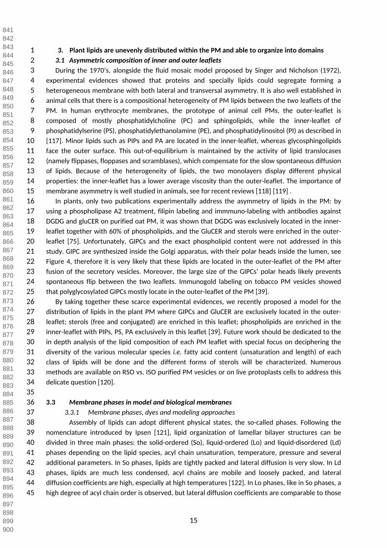
1 3. Plant lipids are unevenly distributed within the PM and able to organize into domains2 3.1 Asymmetric composition of inner and outer leaflets3 During the 1970’s, alongside the fluid mosaic model proposed by Singer and Nicholson (1972), 4 experimental evidences showed that proteins and specially lipids could segregate forming a 5 heterogeneous membrane with both lateral and transversal asymmetry. It is also well established in 6 animal cells that there is a compositional heterogeneity of PM lipids between the two leaflets of the 7 PM. In human erythrocyte membranes, the prototype of animal cell PMs, the outer-leaflet is 8 composed of mostly phosphatidylcholine (PC) and sphingolipids, while the inner-leaflet of 9 phosphatidylserine (PS), phosphatidylethanolamine (PE), and phosphatidylinositol (PI) as described in
10 [117]. Minor lipids such as PIPs and PA are located in the inner-leaflet, whereas glycosphingolipids 11 face the outer surface. This out-of-equilibrium is maintained by the activity of lipid translocases 12 (namely flippases, floppases and scramblases), which compensate for the slow spontaneous diffusion 13 of lipids. Because of the heterogeneity of lipids, the two monolayers display different physical 14 properties: the inner-leaflet has a lower average viscosity than the outer-leaflet. The importance of 15 membrane asymmetry is well studied in animals, see for recent reviews [118] [119] .16 In plants, only two publications experimentally address the asymmetry of lipids in the PM: by 17 using a phospholipase A2 treatment, filipin labeling and immmuno-labeling with antibodies against 18 DGDG and gluCER on purified oat PM, it was shown that DGDG was exclusively located in the inner-19 leaflet together with 60% of phospholipids, and the GluCER and sterols were enriched in the outer-20 leaflet [75]. Unfortunately, GIPCs and the exact phospholipid content were not addressed in this 21 study. GIPC are synthesized inside the Golgi apparatus, with their polar heads inside the lumen, see 22 Figure 4, therefore it is very likely that these lipids are located in the outer-leaflet of the PM after 23 fusion of the secretory vesicles. Moreover, the large size of the GIPCs’ polar heads likely prevents 24 spontaneous flip between the two leaflets. Immunogold labeling on tobacco PM vesicles showed 25 that polyglycosylated GIPCs mostly locate in the outer-leaflet of the PM [39]. 26 By taking together these scarce experimental evidences, we recently proposed a model for the 27 distribution of lipids in the plant PM where GIPCs and GluCER are exclusively located in the outer-28 leaflet; sterols (free and conjugated) are enriched in this leaflet; phospholipids are enriched in the 29 inner-leaflet with PIPs, PS, PA exclusively in this leaflet [39]. Future work should be dedicated to the 30 in depth analysis of the lipid composition of each PM leaflet with special focus on deciphering the 31 diversity of the various molecular species i.e. fatty acid content (unsaturation and length) of each 32 class of lipids will be done and the different forms of sterols will be characterized. Numerous 33 methods are available on RSO vs. ISO purified PM vesicles or on live protoplasts cells to address this 34 delicate question [120].3536 3.3 Membrane phases in model and biological membranes 37 3.3.1 Membrane phases, dyes and modeling approaches38 Assembly of lipids can adopt different physical states, the so-called phases. Following the 39 nomenclature introduced by Ipsen [121], lipid organization of lamellar bilayer structures can be 40 divided in three main phases: the solid-ordered (So), liquid-ordered (Lo) and liquid-disordered (Ld) 41 phases depending on the lipid species, acyl chain unsaturation, temperature, pressure and several 42 additional parameters. In So phases, lipids are tightly packed and lateral diffusion is very slow. In Ld 43 phases, lipids are much less condensed, acyl chains are mobile and loosely packed, and lateral 44 diffusion coefficients are high, especially at high temperatures [122]. In Lo phases, like in So phases, a 45 high degree of acyl chain order is observed, but lateral diffusion coefficients are comparable to those
841842843844845846847848849850851852853854855856857858859860861862863864865866867868869870871872873874875876877878879880881882883884885886887888889890891892893894895896897898899900

16
1 of Ld phases. Phase formation in lipid mixtures has been extensively studied in vitro with liposomes 2 and giant unilamellar vesicles (GUV) as models. In most reports, GUV membranes exhibit 3 micrometer-size liquid immiscibility over a wide range of temperatures. GUVs with such properties 4 contained a minimum of three components: high-melting temperature lipids (e.g. saturated VLCFA-5 containing sphingolipids), low melting temperature lipids (e.g. not necessarily but often unsaturated 6 phospholipids), and sterols (for more details see review [123]). Lo phases are also referred to as 7 cholesterol-dependent phases because cholesterol was used in most studies on the subject, [124, 8 125] for reviews. In the mammalian model, lateral partitioning of Lo and Ld phases is thus explained 9 by a preferential interaction of sphingolipids with cholesterol over phospholipids, likely due to better
10 shielding from water by the sphingolipid headgroup [93].11 Lo phases are also observed in the presence of various free- and conjugated-phytosterols such as 12 SG and ASG [93, 126, 127]. Use of environmental fluorescent probes sensitive to membrane order 13 such as di-4-ANEPPDHQ and Laurdan [128] allow the analysis of phase separation on GUVs made of 14 various mixtures of plant lipids. This reveal contrasted abilities of free-phytosterols to control phase 15 separation on model membranes. Although stigmasterol added to DOPC/DPPC vesicles fail to induce 16 the lateral segregation of lipids into domains of different order levels, GUVs containing sitosterol and 17 campesterol promote the formation of Lo domains at the surface of model membranes [93]. 18 Noteworthy, SG and ASG added separately exhibit the same ability as the corresponding free sterols, 19 increasing the amount of Lo phase. This effect is reinforced when used in combination and increases 20 strikingly when free and conjugated sterols are present in the mixture [93]. The same study indicates 21 that GIPCs, the major plant sphingolipids [2] are not able alone to promote the formation of a liquid-22 ordered phase within a phospholipid bilayer revealing no extensive phase separation in the binary 23 sphingolipid/phospholipid system. By contrast, this study shows the ability of GIPCs to increase the 24 amount of Lo phase of the membrane in presence of phytosterol and interestingly this remains still 25 the case even in the absence of saturated phospholipids such as DPPC. These in vitro studies expose 26 the complex association of different classes of lipids necessary to form distinct phases that are in vivo 27 linked to various membrane functions such as cell signaling or development. Yet there is a real 28 benefit to be able to observe this partitioning in vivo via fluorescent probes and dyes.2930 The partitioning of lipid fluorophores between coexisting Lo and Ld phases for different ternary 31 lipid mixtures has been extensively performed by comparing fluorescence intensities in coexisting 32 domains. These labeled lipids have a fluorophore (e.g. NBD, Texas Red, Bodipy, etc…) attached either 33 to the head group or to the hydrocarbon chain. Studies using fluorescently labeled lipid analogues in 34 different mixtures must be analyzed cautiously for several reasons: 1/ the fluorophore might alter 35 the distribution of the lipids on which it is grafted i.e. a large fluorophore attached to an acyl chain 36 might hamper the incorporation of the labeled lipids into the Lo domains, as found in the case of 37 fluorescent ganglioside probes [129]; 2/ It has been shown that the same fluorescent probe might 38 have different partitioning preferences depending on the chosen lipid mixture [130]. Nevertheless, 39 an important finding from this body of research is that partitioning in ordered-phases is increased for 40 fluorophores with saturated chains that approximately match the thickness of one leaflet of the host 41 membrane [131]. Fluorescent lipid probes with unsaturated chains are found to partition into the Ld 42 phase. These studies indicate the ability of different molecular species of lipids to partition 43 selectively in the different phases of a complex model membrane, according their structure [132] 44 [133].45 There is a current lack of such fluorescent probes designed from typical structures of plant PM
901902903904905906907908909910911912913914915916917918919920921922923924925926927928929930931932933934935936937938939940941942943944945946947948949950951952953954955956957958959960

17
1 lipids. This prevents unambiguous assessing of specific lipid behaviors in different complex mixtures. 2 Non-perturbing specific-labeling of PM nanodomains in plant cells has been, and remains, one of the 3 foremost challenges in the field.45 Modeling approaches based on simulation can also bring grist to the mill of such 6 experimental evidence. For example, very recent work by Ingolffson et al., in a pioneering in silico 7 study of PM-lipid assembly mimicking the complexity of the animal PM, confirmed the non-ideal 8 lateral mixing of the different lipid species [134]. Based on large-scale molecular 9 dynamicssimulation, this study provided a high-resolution view of the lipid organizationof the PM
10 at an unprecedented level of complexity since the model consists of 63 different lipid species, 14 11 types of head groups and 11 types of aliphatic moieties asymmetricallydistributed across the two 12 leaflets. This closely mimics an idealizedmammalian PM. A general non-ideal lateral mixingof the 13 different lipid species was observed together with the formation and disappearance on the 14 microsecond time scale of transient domains with liquid-orderedcharacteristics whereas distinct 15 perennial nanodomains consisting of gangliosides were observed. Nonetheless, the lack of 16 biophysical parameters for plant lipids necessary for the calculation of molecular dynamics impairs 17 the use of these approaches to modelize plant PM (see section “Conclusions”).1819 3.3.2 Solubilization by detergents: evidences from model membranes20 Detergents are amphiphilic molecules, most of them consisting of a polar head and a hydrophobic 21 chain. These molecules have a conical shape and spontaneously form micellar structures displaying a 22 positive curvature in aqueous solution. Detergents have thus the ability of incorporating themselves 23 into membranes and of solubilizing proteins by replacing their lipid environment. Pioneering work 24 evidencing correlations between resistance to detergent solubilization of a fraction of the PM and its 25 peculiar lipid and protein composition suggested the possible existence of lipid domains in the PM of 26 mammalian cells [135]. 27 Detergent-resistant membrane fractions (DRMs) could be isolated from a variety of eukaryotic 28 cells and gave birth to the hypothesis that such fractions are present within native PM as a distinct 29 phase within the bilayer. DRMs are rich in saturated phospholipids, sphingolipids and sterols, and 30 display the properties of the Lo phase previously described in model membranes. Such a hypothesis 31 received strong support from parallel studies on lipid vesicles constructed to mimic the lipid 32 composition of these membranes [136]. In particular, [137] demonstrated that when mixtures of 33 sphingolipids, unsaturated phospholipids and cholesterol were treated in the cold with nonionic 34 detergents such as Triton X-100, the lower-melting phospholipids were readily solubilized while the 35 higher-melting sphingolipid species, and to a lesser extent cholesterol, were largely recovered in an 36 insolubilized and sedimentable fraction. Similar results were obtained using analogous lipid mixtures 37 without cholesterol, or in which long-chain saturated phospholipids replaced the sphingolipid 38 component. Measurements of diphenylhexatriene fluorescence polarization have suggested that the 39 existence of a DRM fraction was correlated with the presence of Lo phases in the original bilayers. 40 Since that, numerous studies have addressed the differential sensitivity of Lo and Ld domains to 41 detergent solubilization. Nevertheless, only a few reports, like that of [138] compared the effect of 42 distinct detergents on Ld and Lo domain solubilization within model bilayers. Most works indeed 43 focused solely on the impact of Triton X-100. Atomic Force Microscopy (AFM) observations on 44 vesicles containing dioleoyl-PC/sphingomyelin/cholesterol provided evidence for both Triton X- 100-45 insoluble domains composed of sphingomyelin and cholesterol, and Triton X-100-soluble areas
961962963964965966967968969970971972973974975976977978979980981982983984985986987988989990991992993994995996997998999100010011002100310041005100610071008100910101011101210131014101510161017101810191020

18
1 surrounding them [139]. Similarly, using real-time AFM imaging of dioleoyl-2 PC/sphingomyelin/cholesterol mixtures, [140] found that detergent did not affect 3 sphingomyelin/cholesterol Lo phases, while dioleoyl-PC Ld phases were completely solubilized. 4 Developing the same experimental approach on dioleoyl-PC/dipalmitoyl-PC vesicles, authors showed 5 that Triton X-l00 concentrations right above the critical micellar concentration (CMC) enabled the 6 solubilization of the dioleoyl-PC matrix, but prevented dipalmitoyl-PC domains from solubilization 7 [141]. [142] proposed a model based on equilibrium thermodynamics showing that resistance to 8 solubilization only depends on the target lipid affinity with the micellar phase. The effects of 9 cholesterol on the resistance of lipid mixtures to solubilization have also been investigated.
10 Ahyayauch et al. demonstrated that cholesterol facilitates PC solubilization better than 11 sphingomyelin [143]. Furthermore, cholesterol was found to induce higher resistance to 12 solubilization of dipalmitoyl-PC vesicles, with a notable exception at 4°C. Interestingly, cholesterol 13 also induced higher resistance of palmitoyl-oleoyl-PC bilayers to detergent solubilization on a 14 broader range of temperatures (from 4 to 15 °C) [144]. In addition, sterols are not the only lipid 15 family determining detergent insolubility. For example, it was shown that addition of 5–30 mol% 16 ceramides prevented Triton X-100 from completely solubilizing sphingomyelin-containing bilayers 17 [145]. It is noteworthy that microscopic observations carried out by Staneva et al. [144] revealed 18 neither domain formation, nor domain coalescence in response to Triton X-100 treatment in 19 heterogeneous GUV systems.20 Cholesterol is not the only sterol to induce resistance to detergent solubilization. Plant sterols, 21 like campesterol, sitosterol, stigmasterol have a similar effect to cholesterol, when incorporated in 22 palmitoyl-oleoyl-PC vesicles [98]. All these results, based on biophysical analysis performed on model 23 membranes indicate: 1/ that animal and plant PM-mimicking lipid mixtures undergo a segregation 24 between different phases corresponding to different physical states; 2/ that the different lipids 25 present within the bilayer partition differentially in these phases according to their chemical 26 structure; 3/ that there is a close correlation within the composition and physical characteristics of 27 the DRM fraction isolated from model membranes and the ones displayed by the Lo phase. 2829 3.3.3 Isolation of Detergent Resistant Membranes from PM, biochemical fractions with a specific 30 lipid composition.31 Based on the conceptual framework exposed in the previous sections, a tremendously high 32 number of publications (about 2000 in the last 40 years) reported the isolation and characterization 33 of DRMs from biological membranes from a wide variety of animals, plants and microorganisms 34 [146]. Virtually all protocols rely on a similar experimental procedure: treatment of either intact cells 35 or purified membranes with a nonionic detergent (most frequently Triton X-100, but Triton X-114, 36 Brij or Lubrol has also been used), generally at low temperature (4°C) followed by ultra-37 centrifugation on a sucrose (or Ficoll) gradient to recover the insoluble fraction [147]. Parameters 38 which have been proven to be crucial and were carefully adapted on each material concern mainly 39 the concentration of detergent and the detergent-to-membrane ratio used. The protein yield 40 recovered in the insoluble fraction may vary between 5 and 20% of the initial amount of membrane 41 proteins, depending on the biological material and the experimental conditions. In animal cells, 42 extensive characterizations of lipids associated to DRMs consistently revealed a 3- to 5-fold 43 enrichment in lipids associated to the Lo phase of model membrane, in particular cholesterol, 44 saturated phospholipids, gangliosides and sphingomyelin [148] [149]. Analyses of the phospholipids 45 content of DRMs classically exhibit a decrease in anionic phospholipids compared to the whole
102110221023102410251026102710281029103010311032103310341035103610371038103910401041104210431044104510461047104810491050105110521053105410551056105710581059106010611062106310641065106610671068106910701071107210731074107510761077107810791080

19
1 membrane [150], an increase of the proportion of saturated fatty acids [151], and a typical 2 enrichment in GM1 gangliosides [152]. The biochemical analysis of such DRMs extracted from animal 3 cell membranes have been extensively performed using proteomics approaches (see for review 4 [153]). It emerged from this huge amount of data that some typical proteins, such as caveolin, or 5 protein families, such as kinases of the Src family or small G-proteins, are in a reproducibly enriched 6 in such fractions, together with some proteins harboring particular post-translational modifications 7 such as GPI-anchored proteins. We can note here that some free detergent methods to isolate sub-8 fraction of the PM have also been developed. [154] [155] [156].9 In plants, DRMs were first purified in tobacco, with the first isolation reported by [157] and
10 the characterization of DRM-associated proteins and lipids provided by [19]. Similar studies revealing 11 by mass spectrometry the catalogue of proteins were then performed on different species such as 12 Arabidopsis thaliana [158] [159] [160] [161] [162], Medicago truncatula [163] [164], Oryza sativa 13 [165], Avena sativa and Secale cereale [166] or more recently Beta vulgaris [167]. According to the 14 methodology used, the amount of proteins may differ between the studies but the continuous 15 improvement of performance and sensitivity of the mass spectrometry approaches, led to an 16 increase of the number of proteins identified which reached more than 300 proteins in the more 17 recent studies [160] [161] [168]. The latest extensive analysis published on plant PM proteome, 18 performed in rice, allowed the identification of more than 3900 proteins, which is quite consistent 19 with the yield of PM-derived DRM proteins which is typically around 10% of total proteins in most 20 studies [19] [169]. The family of proteins identified in the different studies is also quite consistent, 21 with a high proportion of proteins involved in signal transduction, responses to different stress, and 22 plant-microorganism interactions. Accordingly, a few studies implementing quantitative proteomics 23 approaches based on different methodologies, clearly revealed a qualitative and/or quantitative 24 modification of the proteins associated to DRMs upon environmental modifications, for instance in 25 the early steps of plant defense signaling [168] [161] [165], or following abiotic stress [170] [167]. 26 Note that by contrast with proteins where genetically encoded fluorescent tags or specific antibodies 27 are available, generating DRMs is the most used technique in order to study the potential 28 segregation of lipids. In the next chapters, we will discuss the lipids found in DRMs purified from 29 plant PMs.3031 3.2.3.1. Glycerolipids in plant DRMs32 As expected from biophysical work, major structural phospholipids, i.e. PC, PE, PS, PA are 33 markedly depleted in plant DRMs when compared to the total PM [19] [159]. The case of 34 phosphoinositides (PIPs) deserves a particular attention. Despite the real challenge related to the 35 detection of such a minor class of lipids in very reduced biological sample such as DRMs, the 36 combined use of Thin-Layer Chromatography to separate the different classes of phosphoinositides 37 prior to quantitative Gas Chromatography-Mass Spectrometry analysis [171] allows to quantify PI4P 38 and PI(4,5)P2 in DRMs isolated from tobacco leaves and BY-2 cells [31]. It was shown that both PIPs 39 isomers represent less than 5 mol% of the total lipids of tobacco PM. However, their relative amount 40 is 11-fold higher in DRMs compared to PM from which they originate. Hence, it is estimated that 50 41 mol% of PM phosphoinositides likely segregate in BY-2 cell PM-domains. A lower increase in PI4P and 42 PI(4,5)P2 was also observed in tobacco leaf DRMs: 43 and 31 mol% of the isomers were found to 43 concentrate in this fraction respectively. Moreover, PIPs display highly saturated fatty acids in both 44 DRMs and PM, with 16:0, 18:0 and 18:1 being the major fatty acids, which is all the more consistent 45 with the packed lipid environment or liquid-ordered phase characteristic of this fraction. This work
108110821083108410851086108710881089109010911092109310941095109610971098109911001101110211031104110511061107110811091110111111121113111411151116111711181119112011211122112311241125112611271128112911301131113211331134113511361137113811391140

20
1 has demonstrated that PIPs are the only glycerolipids enriched in plant DRMs, which is in agreement 2 with biochemical studies performed on animal cells. In good agreement with this result, the 3 nanodomain-protein marker from the potato REM group 1 isoform 3 (StREM1.3 or REM) was shown 4 to cluster in the PM inner-leaflet nanodomain by specific binding to PI4P [172]. The question arises 5 whether PI4P is clustered in PM-nanodomains before the anchoring of REMORIN, or whether REM’s 6 C-terminal anchor promotes PI4P clustering. In line with the results obtained for PIPs, striking 7 evidence related to the characteristics of the fatty acyl chains associated to DRM glycerolipids have 8 been obtained. Indeed, the comparative analysis of polar lipids from tobacco leaves or BY-2 cells 9 revealed a very significant enrichment in saturated fatty acids (C16:0 and C18:0), in agreement with a
10 DRM phospholipids double-bond index lower than that of the overall PM [19]. This was consistently 11 observed in maize embryos and bean leaves with the total amount of saturated long-chain fatty acids 12 being higher in the PM than in DRMs, and with the saturated/unsaturated ratio rising from 1.4 in PM 13 to 6.5 in the DRMs from bean leaves and from 0.3 in the PM to 1.0 of maize embryo [173]. These 14 characteristics perfectly fit with the direct relationship classically observed in model membranes 15 between the proportion of lipids with saturated fatty acyl chains and the global order of the bilayer 16 which has been confirmed using lipids from plant PMs [93].1718 3.2.3.2 Sterols in plant DRMs19 In all plant tissues tested, free sterols are major components of isolated DRMs. Quantitative 20 analyses showed a clear increase in the sterol-to-protein ratio to around 1.7-fold in DRMs prepared 21 from both tobacco leaves and Medicago truncatula roots [19] [163] or even 2.7- and 4-fold 22 enrichment in DRM fractions from bean leaves and Arabidopsis cell cultures [158] [174]. On the 23 other hand, maize embryo [174] and Arabidopsis seedlings [170] exhibited a smaller free-sterol 24 accumulation with only an 1.3-fold enrichment in DRM fractions indicating a range of plant sterol 25 enrichment factors as broad as that observed in animal cells. In general, relative abundances of 26 individual free-sterol species such as stigmasterol, campesterol and sitosterol in DRM and PM 27 fractions are similar [19] [158] [159] with the noticeable exception of maize embryo membranes 28 [174]. Moreover, additional results reinforced the possible role of phytosterol in the structuration of 29 plant PM domains suggested by their enrichment in DRMs. They were essentially provided by the use 30 of the pharmacological compound methyl-β -cyclodextrin. This cyclic oligosaccharide able to trap
31 sterols from artificial and biological membranes has been widely used to lower the content of 32 membrane cholesterol in various types of animal cells and to assess sterol-associated membrane 33 structuring, see for review [175] to read about the specific and nonspecific effects of cyclodextrins. 34 This molecule has been proven to remove from isolated plant PM, with a comparable efficiency to 35 that associated with cholesterol, the major free phytosterols (campesterol, stigmasterol, sitosterol 36 and isofucosterol) [91]. Such a treatment resulted in a decrease by about 50% of BY-2 cell PM sterols, 37 without affecting PM-content in conjugated sterols, phospholipids, sphingolipids and proteins. 38 Importantly, methyl-β -cyclodextrin treatment totally abolished the recovery of any DRM fraction
39 after PM solubilization at 4°C with Triton X-100 [91]. Moreover, the use of environment sensitive 40 fluorescent probes allowed to associate this depletion in free-sterols with a decrease in liquid phase 41 heterogeneities, and particularly in Lo phases [91]. This work on isolated PM has been further 42 corroborated by similar results obtained on living BY-2 cells showing a clear decrease of the 43 proportion of ordered PM-domains by cyclodextrin treatment that reduced by ca. 20% the amount 44 of PM sterols [90]. Finally, the combination of cyclodextrin and extensive quantitative proteomic 45 characterization of DRMs isolated from Arabidopsis PM identified a subset of proteins, whose
114111421143114411451146114711481149115011511152115311541155115611571158115911601161116211631164116511661167116811691170117111721173117411751176117711781179118011811182118311841185118611871188118911901191119211931194119511961197119811991200

21
1 association to DRMs is sterol-dependent [160] [162]. 2 As exposed in Section 1.3, phytosterols can be conjugated with sugars, which in turn can be 3 acylated to form SG and ASG (for a review see [69]). Lipidomic analyses have shown varying amounts 4 of SG and ASG in plant DRMs. In M. truncatula roots, the same enrichments in SG, ASG and free-5 sterols were observed in DRMs compared to PM fractions [163]. Conversely, while free-sterols did 6 not show any significant enrichments in DRMs from Arabidopsis thaliana seedlings and plants, SG 7 and ASG were found considerably enriched (more than 4-fold) in these fractions [159] [170]. In oat 8 roots, conjugated-sterols were observed in similar proportions in PM and DRMs whereas a clear 9 enrichment of free-sterols was reported [75]. Furthermore, total sterol amounts were markedly
10 increased in tobacco leaf DRMs, mainly because of the high-enrichments in free-sterols and ASG [31]. 11 As exposed in section 2.1, conjugated-sterols in combination with free-phytosterols are potent 12 modulators of the order level of model membranes, suggesting again a close relationship between 13 the composition of the DRM fraction and the presence of a Lo phase in plant PM.1415 3.2.3.3 Sphingolipids in plant DRMs16 By contrast with DRMs extracted from animal cells, plant DRM sphingolipid content has only been 17 investigated in very few studies. A first line of evidence of the enrichment of sphingolipids in DRMs 18 relies on the characterization of their VLCFA content. By analyzing highly-purified PM from bean 19 leaves and germinating maize embryos, a 3- to 4-fold increase of VLCFA relative amount in DRMs 20 compared to PM was evidenced [173]. Moreover in the two plant species considered, VLCFAs 21 harboring 20- to 32-carbon chains were present in DRMs. A significant enrichment of VLCFAs (from 22 C20:0 to C26:0) in DRMs compared to PM was also observed in tobacco leaves and BY-2 cells [58]. 23 Among the hundreds of sphingolipid species that exist in plants many possess 2-hydroxy fatty acids, 24 containing a hydroxylated C-2 position [9] which contributes to the rigid binding of sphingolipids 25 between themselves and other lipids through hydrogen-bonding between hydroxyl groups, 26 evidenced in artificial membranes [176, 177]. It is thus likely that 2-hydroxy sphingolipids contribute 27 to the ordered structure of PM domains. Using mutant rice lines in which the levels of sphingolipids 28 containing 2-hydroxy fatty acids were decreased by knocking-down two genes encoding fatty acid 2-29 hydroxylases (FAH1 and FAH2), [51] demonstrated that the DRM/PM ratio was altered in these lines. 30 This result suggested a role for such lipids in structuring Lo phases within the PM, which was further 31 confirmed by the observation using the environmental probe ANEPPDHQ that the PM in OsFAH1/2-32 KD1 was significantly more disordered than in the wild type.33 GluCer belongs to the monosaccharidic cerebroside family, and many GluCer species have been 34 reported in plants, since lipidomics approaches showed that GluCer can exhibit a wide range of LCB 35 and fatty acid composition [38]. Their relative abundance in plant PMs has not been unequivocally 36 determined, varying significantly form one species to another and also according to the 37 photosynthetic activity of the tissue considered [178]. In line with those observations, various 38 quantitative data have been reported concerning the enrichment of GluCer in DRMs. In DRMs 39 isolated from tobacco leaves and BY-2 cells [19] or Medicago truncatula roots [163], GluCer was only 40 slightly enriched. On the other hand, GluCer was found significantly enriched in Arabidopsis and leek 41 DRMs prepared from PM and microsomal membranes [158] [170]. DRMs isolated from leek Golgi 42 membranes also showed 4-, 5-fold GluCer enrichment [159] and DRMS isolated from tobacco pollen 43 tubes harbor a percentage of GluCer wich increased up to two fold with the detergent/protein ratio 44 [179]. Taking into account these data, it is difficult to conclude whether or not GluCer enrichment 45 can be considered as an essential component of plant DRMs, even if its relative proportion increased
120112021203120412051206120712081209121012111212121312141215121612171218121912201221122212231224122512261227122812291230123112321233123412351236123712381239124012411242124312441245124612471248124912501251125212531254125512561257125812591260

22
1 in this fraction compared to the PM, in most studies performed. 2 The first indication of an enrichment of GIPCs in plant DRMs was reported by [158], showing an 3 approximately 5-fold higher LCB-to-protein ratio in DRMs extracted from Arabidopsis microsomal 4 fractions. The relative decrease of (8Z)-4-hydroxy-8-sphingenine (abbreviated t18:1c) in the DRMs 5 compared with microsomes and the increase of the ratio of this LCB compared with its stereoisomer 6 (8E)-4-hydroxy-8-sphingenine (t18:1t) in the DRMs suggested that DRMs might contain a high 7 proportion of GIPCs, which have a greater 8Z:8E ratio than cerebrosides [158]. However, although 8 GIPCs belong to one of the earliest classes of plant sphingolipids that were identified [41], their study 9 has for long been impaired by their limited solubility in typical lipid extraction solvents, and very
10 recent progress concerning their structural characterization and role in membrane organization relies 11 on the development of efficient protocols of purification [42] [53]. By taking advantage of such 12 methodological developments, [39] showed that the hVLCFA and VLCFA contents were highly 13 comparable between DRMs and purified GIPCs, with an even higher proportion of hVLCFAs in DRMs 14 purified from BY-2 cells, suggesting that hVLCFA-containing GIPCs are most likely present in this 15 fraction. Moreover, levels of the two LCBs t18:0 and t18:1, which are mostly present in GIPCs [45], 16 strongly increase in DRMs when compared with PM, reaching 80% of total LCBs in DRMs. A further 17 characterization indicated that series A GIPCs were found in both PM and DRM fractions of tobacco 18 leaves, whereas for BY-2 series B GIPCs were 3-fold enriched in DRMs when compared with the PM, 19 reaching 17% of total GIPCs in BY-2 DRMs [39]. When the global lipid composition of the PM and 20 DRM fractions was recalculated taking into account these data, it appeared that GIPCs represent 45 21 and 30 mol% of total PM lipids isolated from leaves and BY-2 cell suspensions, respectively, and up to 22 60 mol% of the DRM fraction, suggesting that the contribution to sphingolipid-enrichment in PM Lo 23 phases is mainly due to GIPCs [39]. 24
25 3.2.3 The use of DRMs to study the segregation of lipids in plant PM; some limits but 26 significant contributions.
27 The “raft hypothesis” states that specific PM-lipids, mainly sterols and saturated sphingolipids, 28 interact together to form dynamic nano-scale clusters by recruiting lipids and proteins that are 29 present in signaling and trafficking platforms in the PM [111]. Experimentally, the nonionic detergent 30 Triton X-100 is used to separate Lo phases from the rest of membrane preparation by isolation of 31 DRMs isolated in the upper-phases of a sucrose density gradient after ultracentrifugation. DRMs are 32 considered by many as in vitro counterparts of membrane rafts [147]. The characterization of lipids 33 found in DRMs isolated from PM fractions of various plant species gave rise to a overall features such 34 as a global decrease of glycerolipids content, with the noticeable exception of phosphoinositides; a 35 strong enrichment in lipids containing saturated fatty acyl chains; an increase in free- and 36 conjugated-sterols; and a strong enrichment in sphingolipids, and in particular in GIPCs. These 37 characteristics are consistent with the canonical description of DRMs isolated from a plethora of 38 animal PMs whilst taking into account the specificities of plant PM lipids [180]. Moreover, it appears 39 that such a composition is typical of the Lo phase in model membranes, as detailed in Section 3.2.1. 40 Yet, as stated in several publications (e.g. [181] [182] [183] [184]) the use of DRMs to evidence PM-41 associated dynamics should be accompanied with great precaution as DRM fractions should not be 42 considered as direct equivalents to PM-domains. However, the numerous convergent correlations 43 obtained on many different biological materials and model membranes have indicated for instance
126112621263126412651266126712681269127012711272127312741275127612771278127912801281128212831284128512861287128812891290129112921293129412951296129712981299130013011302130313041305130613071308130913101311131213131314131513161317131813191320

23
1 that 1/ a consistency between the composition of the Lo phase in situ and the DRM lipid content; 2/ 2 a relationship between the presence and abundance of DRMs and the order of biological 3 membranes; 3/ the association of particular proteins to DRMs and their clustered distribution within 4 the PM make them valuable tools to progress towards a better understanding of plant PM 5 organization. An example of this last point is the PM-associated NADPH-oxidase RbohD, which was 6 demonstrated to be responsible for the oxidative burst observed in the very early steps of the plant 7 immune signaling cascade, was proved to be exclusively associated to DRMs in tobacco [19]. This 8 characteristic could be related to the, immunoelectron microscopy observation that this protein is 9 organized within the PM in clusters of about 20nm in diameter [185]. Upon activation, NADPH-
10 oxidase products (the Reactive Oxygen Species) were also present as discrete nanometer sized 11 patches along the PM [186]. Similarly, the presence of the flagellin receptor FLS2 in DRMs and its 12 enrichment in this fraction observed a few minutes after treatment of Arabidopsis cells with the 13 bacterial elicitor flagellin is fully consistent with recent results observed using super-resolution 14 microscopy indicating its clustered distribution in the membrane, a significant modification of its 15 dynamics within PM, namely an increased population of long-lived receptor clusters and a reduction 16 of its lateral displacement a few minutes after elicitor treatment [187]. Moreover, group 1 Remorin 17 proteins were the first biochemical markers of plant DRMs and observed as forming PM-associated 18 clusters of about 75 to 100nm in diameter [188] [189]. Such nanodomain-organization was shown to 19 be sterol-dependent as it was strongly impaired by the use of sterol-chelator methyl-β-cyclodextrin 20 [188] or by inhibitors of sterol biosynthesis, see Table 1 [172]. More recent data have confirmed the 21 confinement this protein in PM-nanodomains to be sterol- and PI4P-dependent and by using in vivo 22 single-particle tracking microscopy that the size these Remorin-associated domains were of ca. 80nm 23 in diameter [172]. 24 One must note that the colocalization studies of Remorins of different phylogenetic groups, 25 namely groups 1, 3, 4 and 6 have shown the coexistence of highly-distinct membrane domains in the 26 plane of the PM [190]. These results have demonstrated that the use of biochemical approaches such 27 as DRMs cannot be sensitive enough to accurately represent the biological complexity of membrane-28 compartmentalization in vivo. A decisive milestone will be the ability to describe, in an extensive and 29 comprehensive manner, the distribution of the various lipids together with proteins within PM, and 30 to identify key causal mechanisms underlying such an organization. To do so, super-resolution 31 microscopy with one or more fluorophores can be used for proteins to visualize in vivo whether the 32 protein of interest is enriched in nanodomains; this kind of method is currently at its infancy for lipids 33 due to the few fluorescently-labeled lipids available and because it is hard to insure a proper intake 34 of such dyes in plant cells (see below). The use of chelator of lipids, fluorescent lipid probes or lipid-35 biosynthesis inhibitors (table 1) are the next steps to address the role of lipids in PM-nanodomain 36 formation and maintenance.37
132113221323132413251326132713281329133013311332133313341335133613371338133913401341134213431344134513461347134813491350135113521353135413551356135713581359136013611362136313641365136613671368136913701371137213731374137513761377137813791380

24
1 4. Spatial and multiscale segregation of lipids and proteins: a complex picture emerging from 2 the combined use of various imaging techniques34 Since the publication of the fluid mosaic model for biological membranes in 1972, a lot of 5 experimental evidences revealed the outstanding complexity of the PM. Rafts characterized by tight 6 lipid-packing are involved in a wide variety of cellular processes: regulation of endo- and exocytosis, 7 hormone signaling, membrane trafficking in polarized epithelial cells, T-cell activation, cell migration, 8 life cycle of influenza and HIV viruses [191]. As expected for biology as an experimental science, 9 understanding the organization of the PM strongly relies on the evolution of microscopic methods.
10 Thanks to the development of new efficient methodologies, among them imaging lipids and super-11 resolution microscopy [111], very important results have been published refining this organization 12 but in the same time raising new questions. Currently, we can clearly state that the PM is organized 13 in domains that differ in the nature of their components, their stability and their size from the 14 nanometer to the micrometer scale. This complex remodeling is highly-dynamic and respond to 15 various abiotic and biotic stresses. Particular interest of the scientific community for cell surface 16 signaling processes in the past decades has led to an improved vision of the PM’s membrane 17 organization: a complex multi-component and multi-scale heterogeneity with a high degree of 18 subcompartmentilization into micro- to nano-domains have been evidenced in vivo and deserve to 19 be clearly qualified. Domains were originally referred as to “lipid-rafts” but the designation has very 20 much evolved since. It is now known that lipid-rafts do not cover a single type of domain but rather 21 include a collection of domains differing in their protein and lipid composition and their resident time 22 (aggregation/disaggregation).2324 4.1. Micro- and nano-domains coexist in the plant PM 25 4.1.1 Microdomains in plant cells 26 Cell polarization-induced PM-microdomains (above 1µm) are easily observed by classical confocal 27 fluorescence microscopy in leaf, root and pollen tube cells [182, 192]. The accumulation of proteins 28 and lipids into microdomains is involved in defining cells’ fate, functional specialization for cell 29 polarity and specialization of host membranes for defense [193] [194]. These include the polar 30 distribution of PINFORMED (PIN), AUX1–Auxin transporter protein 1, ABCB (ATP-binding cassette 31 protein subfamily B)/P-glycoprotein, Auxin binding protein 1 (ABP1) or RAC/Rho of plants GTPases 32 (ROP) that localize to the apical or basal pole of a cell [195] and the lateral and equatorial domains in 33 plant endodermal cells populated by DYNAMIN-RELATED PROTEIN1A (DR1PA) or CASPARIAN STRIP 34 MEMBRANE DOMAIN PROTEINs (CASPs) [182] [196]. Similarly, tip-growing cells like pollen tubes and 35 root hairs are also of particular interest for the study of membrane microdomains. For instance, the 36 pollen-specific H+-ATPase is located in the shank whereas Phospholipase C, G proteins and 37 phosphoinositide kinases are located at the apex of the pollen tube (reviewed in see for review [197]. 38 Lipids are also segregated into microdomains i.e. sterol-, phosphoinositide-, PI4,5P2 and 39 diacylglycerol-rich microdomains are shown to be especially concentrated in the apex of the pollen 40 tube [179]. The presence of microdomains enriched in phosphoinositides (PIPs) plays an important 41 role in polar tip growth by regulating the machinery maintaining polarity and by controling 42 cytoskeletal dynamics and the remodeling of vesicle trafficking [198]. In root endodermal cells, 43 EXO70A1 exocyst subunits colocalize with PI4,5P2 [199]. Finally, in plants, plasmodesmata (PDs), 44 which are channels characterized by the apposition of the endoplasmic reticulum and the PM 45 possess a specific lipid composition [200]. These PM-lined PD (PD-PM) have been shown to contain
138113821383138413851386138713881389139013911392139313941395139613971398139914001401140214031404140514061407140814091410141114121413141414151416141714181419142014211422142314241425142614271428142914301431143214331434143514361437143814391440

25
1 definite microdomains where not only proteins such as Plasmodesmata-located protein 1 (PDLP1) 2 and PD callose binding proteins (PDCBs), but also lipids such as sterols and sphingolipids are enriched 3 [201] [202] [7]. 4 Importantly, besides local enrichment of specific lipids and proteins within microdomains, the 5 characterization of the biophysical state of pollen tube microdomains has shown that they are highly-6 segregated in the cell, i.e. they are especially concentrated at the PM of the cellular apex but also 7 present as a ring-like distribution around the tube [72, 179]. Similarly, the cell plate of Arabidopsis 8 contains highly-ordered membrane microdomains which rely on sterols and DRP1A-dependent 9 endocytosis [203]. Yet, how the localization of proteins and lipids in microdomains relies on the
10 cooperativity of multiple mechanisms is not yet understood [204] [205] [197].1112 4.1.2 Nanodomains in plant PM 13 The development of new methods of high- and super-resolution imaging has provided the ability 14 to observe membrane domains at the nanoscale level, termed nanodomains and defined by a size 15 below 1µm [206, 207]. These methods include mainly stimulated emission depletion microscopy 16 (STED), structured illumination microscopy (SIM) and single-molecule localization microscopy (SMLM, 17 including methods such as Photo-Activated Localization Microscopy (PALM) and Stochastic Optical 18 Reconstruction Microscopy (STORM)). These techniques have been routinely used in animal cells but 19 have only recently emerged in plant studies. Super-resolution microscopy techniques allow the 20 acquisition of high-density super-resolved nanoscale maps of individual fusion-protein localizations 21 and trajectories in the PM [208]. Single-molecule tracking combined with photoactivated localization 22 microscopy (spt-PALM) allows not only the description of the supra-molecular organization of 23 proteins at the PM level far below the resolution limit of confocal microscopy but also allows the 24 determination of the mobility dynamics of single molecules or particles in the PM. 25 Super-resolution microscopy methods have shown that PM-associated proteins are sub-26 compartmentalized within nanodomains, to only name a few: Hypersensitive Induced Response HIR1 27 [209] PIN2 [204], Borate efflux transporter (BOR1) [210], Dynamin-related protein 1a (DRP1A), 28 Cellulose synthase A6 CESA6 [211], IDQ family of calmodulin–binding proteins [204] [210] [212] S-29 type anion efflux channel/ Calcium protein kinase SLAH3/CPK21 and REM1.3 [189] [172], flagellin 30 receptor FLS2 [187], Brassinosteroid insensitive 1/ Somatic embryogenesis receptor kinase 1- BRI1-31 associated receptor kinase 1 BRI1/SERK1-BAK1 [213], Flotilin [214] and the NADPH oxidase [215]. 32 Notably, the study of group 1 REM mutants has revealed that the protein mobility rate (measured by 33 the Mean Square Displacement) and protein supramolecular organization are not necessarily 34 coupled. These results have shown that proteins displaying the same mobility rate can however 35 assemble into clusters of different sizes [172]. Differential combinations of multiscale organizations 36 have been evidenced as for instance PIN2, which is preferentially targeted in a polar fashion to PM-37 microdomains in Arabidopsis roots, locates at a lower scale into PM-nanodomains as shown by STED 38 microscopy [204]. By contrast, REMORIN StREM1.3 localizes in nanodomains of ca. 80 nm observed 39 by SPT-PLAM [172], but without a particular polar localization in N. benthamiana leaf epidermal cells. 40 Lipids can also be found in nanodomains both in the inner-leaflet and in the outer-leaflet. 41 Immunogold-electron microscopy has shown that PI(4,5)P2 is found clustered in the inner-leaflet of 42 the PM [31]. Interestingly this cluster formation was not significantly sensitive to sterol depletion 43 [31]. More recently, immunogold-electron microscopy strategy has revealed that the distribution of 44 polyglycosylated GIPCs, likely in interaction with phytosterols, form nanodomains of ca. 40 nm in the 45 outer-leaflet of tobacco PM [39]. These two results strengthen the idea that lateral nano-segregation
144114421443144414451446144714481449145014511452145314541455145614571458145914601461146214631464146514661467146814691470147114721473147414751476147714781479148014811482148314841485148614871488148914901491149214931494149514961497149814991500

26
1 of lipids also takes place at the PM in plants. Yet, tools dedicated to the study the dynamic of plant 2 lipids at the nanoscale level are still lacking, impairing progress in understanding their molecular 3 distribution, behavior and dynamics of PM lipids.45 Therefore, the plant PM must be acknowledged as a fluid yet highly-compartmentalized mosaic 6 wherein numerous membrane domains with different compositions and biophysical properties co-7 exist at different scales [192] [216]. The challenging questions now reside in clearly defining the 8 essential mechanisms governing specific interactions between the different molecular species of PM 9 intrinsic components. Currently microcopy methods are able to localize and track single molecules
10 with a resolution of 1 nm achieving an ultimate resolution limit in fluorescence microscopy: MINFLUX 11 [217] [218], subdiffusive motion at the single trajectory level [219] or motion transition state [220]. 12 Such methods must be adapted to plant cells to address the specific question of plant PM biology 13 and will pave the way to a better understanding of the PM’s dynamic organization.1415 4.2 PM lipids are critical regulators of plant PM organization at the nanometer scale 16 Lipid-lipid interactions and protein-lipid interactions are believed to be key regulation parameters 17 governing plant PM organization. In pollen tubes, various isoforms of the exocyst complex colocalize 18 with either PI4,5P2 or PA, resulting in the formation of PM domains [221]. The localization of 19 EXO70A1 not only coincides with, but is also required for the accumulation of PI4,5P2 [199]. The 20 targeting of REMs to inner-leaflet PM nanodomains is independent of the secretory pathway, 21 although it is still mediated by direct interactions with PI4P in a sterol-dependent manner [172]. This 22 understanding of the anchoring mechanisms of REMs confirms the impairment of clustered 23 distribution of REMs by phytosterol depletion [188] [222]. The use of raster image correlation 24 spectroscopy (RICS) has shown that the lateral mobility of auxin transporters PIN is dependent on the 25 amount of sterols in tobacco cell PM, arguing in favor of a sterol-dependent protein organization 26 within the plant PM [80].27 In plasmodesmata (PD), modulations of the sterol composition alter callose-mediated PD 28 permeability and reversibly impaired the PD localization of the glycosylphosphatidylinositol-anchored 29 proteins Plasmodesmata Callose Binding 1 (PDCB1) and the Plasmodesmata beta-1,3-glucanase 30 (PdBG2). This study emphasizes the importance of lipids in defining PD membrane microdomains and 31 is in line with the lipid-raft model postulating the existence of nanoscopic assemblies of sphingolipids 32 and sterols in the outer-leaflet of the PM. Finally, it is important to acknowledge that nanodomains 33 exist in both leaflets and the lipid content of each of them regulates the clustering of proteins and 34 lipids. The possible interaction between nanodomains across the two leaflets (a process called 35 pinning or registration) will be discussed in the following section.3637 4.3. PM heterogeneity might originate from a tight control along the secretory pathway38 The neo-synthesis of lipids found at the PM results from intricate pathways that originate at the 39 endoplasmic reticulum (ER), which is certainly the most ancient eukaryotic endomembrane 40 compartment. From the ER, lipids are transported to the Golgi apparatus where they are further 41 assembled and modified before reaching the PM, see Figure 4. At the PM, it is thought that the auto-42 association of glycerolipids, sphingolipids and sterols drives membranes close to a demixing point 43 (phase separation) and induces sorting of lipids to either Lo phases or Ld phases of the membrane 44 [223]. The Lo phase is enriched in sphingolipids and sterols. A higher proportion of free-hydroxyl 45 groups in the long-chain-bases and acyl-chains of sphingolipids as compared to glycerophospholipids,
150115021503150415051506150715081509151015111512151315141515151615171518151915201521152215231524152515261527152815291530153115321533153415351536153715381539154015411542154315441545154615471548154915501551155215531554155515561557155815591560

27
1 allows more interactions with sterols and thereby ensuring the stabilization of sphingolipid-enriched 2 membrane domains (see Section 3). The characteristic length of sphingolipid acyl-chains is a 3 particularity amongst lipids that confer special physical properties to biological membranes. This 4 particularity increases the melting-point of sphingolipids as compared to other lipids and causes 5 strong hydrophobic mismatches between sphingolipid acyl-chains and the polar heads of other lipids 6 with smaller acyl-chains. Hence, sphingolipids are segregated and more physically-ordered 7 microdomains are created within the membrane [224]. Acyl-chain length also induces the formation 8 of interdigitated phases (interdigitated lipid-leaflets) and plays a role in membrane stiffness and 9 thickness [224] [64].
10 Considering that these different phases are observed at the PM, one might ask how this 11 complexity is implemented. Does this PM-lipid heterogeneity already occur within secretory 12 pathways that lead to the PM and does it have a role in the secretion of proteins? In mammalian 13 cells, a protein secretion model called the rapid-partitioning model, proposes a cis-to-trans gradient 14 of the sphingolipid/glycerophospholipid ratio that would account for a partitioning of 15 transmembrane cargos and enzymes into distinct domains of the Golgi: domains enriched in Golgi 16 resident enzymes (low sphingolipid/glycerophospholipid ratio) and domains where transmembrane 17 cargos are progressively enriched (high sphingolipid/glycerophospholipid ratio) at the trans-most 18 cisterna of the Golgi until their loading into post-Golgi vesicles [225]. This model is in agreement with 19 the observation that newly arrived cargos exit the Golgi with mono-exponential export kinetics. 20 Moreover, alteration of sphingolipid homeostasis by treating mammalian cells with short acyl-chain 21 ceramides (for further incorporation in sphingolipids at the Golgi) impacts the export of protein 22 cargos from the Golgi, reduces the lipid order in Golgi membranes, and alters the ultrastructure of 23 Golgi cisternae from flat to highly-curled membrane sacs, further supporting the role of very-long-24 chain sphingolipids in Golgi morpho-dynamics and sorting [226] [227].25 In plants, inhibition of the condensation of glucose with ceramides (produces GluCer) results in 26 the disaggregation of Golgi cisternae into vesicular structures and in the inhibition of secretion [228]. 27 In animal and plant cells, the trans-most cisterna of the Golgi apparatus is continuous with a tubular, 28 branching and reticulated Golgi structure called the trans-Golgi Network (TGN). In yeast, it has been 29 observed that secretory vesicles budding-off of the TGN are enriched in sterols and sphingolipids and 30 possess a high proportion of Lo phases [229]. In mammalian cells, a genetically encoded probe that 31 labels sphingomyelin has revealed that sphingomyelin synthesis at the Golgi promotes sphingomyelin 32 enrichment in a subset of TGN-derived secretory vesicles, where the sorting of a 33 glycosylphosphatidylinositol-anchored protein is in turn also promoted [230]. In plants, extraction of 34 DRMs has revealed that both PM and Golgi yield DRMs suggesting that they can be enriched in 35 sterols and sphingolipids [159]. However, as stated before, this method can generate artificial 36 segregations of lipids within membranes. Therefore, a combination of subcellular remodeling of 37 sterols using the fluorescent probe filipin, and a novel extraction approach to specifically immuno-38 purify TGN sub-domains and Golgi apparatus without any detergents coupled with quantitative mass 39 spectrometry, has opened new perspectives in defining which lipids are present in these 40 compartments [231] [232]. The in situ subcellular remodeling of sterols by filipin has revealed that 41 sterols are the most present at the PM and in sub-populations of TGN vesicles [231]. Further on, 42 purification fractions of TGN sub-domains and Golgi have identified an enrichment of sterols and α-43 hydroxylated VLCFAs, a specific signature of plant sphingolipids. Moreover, this signature was 44 specifically stronger in a sub-domain of TGN: Secretory Vesicles [232]. Not only an enrichment of 45 sphingolipids is observed in TGN-derived vesicles, but the length of the sphingolipid acyl-chains is
156115621563156415651566156715681569157015711572157315741575157615771578157915801581158215831584158515861587158815891590159115921593159415951596159715981599160016011602160316041605160616071608160916101611161216131614161516161617161816191620

28
1 found to be a critical factor for the correct polarized secretory sorting of the auxin-carrier protein 2 PIN2 in root epidermal cells [232]. Altogether, the enrichment of sterols and sphingolipids at TGN-3 derived secretory vesicles seems to be a conserved feature in eukaryotic cells, and appears to be 4 required for protein sorting and vesicle budding. This enrichment is favorable to Lo phase lipid-5 segregation at TGN and suggests that a gradient of lipids along the secretory pathway is established 6 from the ER where no enrichment of sterols and sphingolipids is observed through the TGN to the 7 PM. This membrane heterogeneity is not only a structuration of pre-PM lipids but it has as well an 8 important role to play in many intracellular trafficking pathways, see Figure 4. 9 The lipid heterogeneity within the secretory pathway also exists for lipids other than sterols and
10 sphingolipids. In plants, the TGN-localized choline transporter like1 (CTL1) is involved in PM-recycling 11 of the ion transporter NRAMP1 and the auxin efflux carrier PIN1 [233]. An interesting observation is 12 that free-choline, but not phosphatidylcholine (PC), can inhibit phospholipase D (PLD) activity [233]. 13 PLD hydrolyses PC and phosphatidylethanolamine (PE) to produce phosphatidic acid (PA), a 14 phospholipid that favors the fission of vesicles [234]. Hence, one hypothesis is that CTL1 transports 15 choline inside the TGN lumen to maintain a low choline concentration on the cytoplasmic side of 16 TGN in order to conserve high PLD activity converting PC and PE into PA. This mechanism would 17 require further characterization, but it could be a possible way to establish membrane heterogeneity 18 between the lumenal and cytoplasmic leaflets of TGN membranes. Another example of phospholipid 19 membrane heterogeneity is with phosphatidylserine (PS) in yeast where it has been suggested that 20 PS resides primarily in the lumenal leaflet of the Golgi and is flipped to the cytosolic leaflet in the 21 TGN [235]. This leaflet translocation of PS is operated by PS flippases at the TGN and is thought to 22 control oxysterol-binding proteins (OSBP), which exchange ER-associated sterols with TGN-associated 23 PI4P in unidirectional fashion [235]. The exchange of lipids participates in creating sterol enrichment 24 and membrane lipid order at the TGN [236]. An elegant model has proposed that this exchange of 25 sterols for PI4P occurs at ER-TGN membrane contact sites, where PI4P is generated at the TGN by PI4 26 kinases (PI4KIIα) which are themselves regulated in an oscillatory (waves of PI4P consumption by 27 OSBPs) fashion by sterols [236]. These studies have revealed a crucial characteristic of membrane 28 heterogeneity at the TGN: its highly-dynamic and oscillatory nature. 29 In plants, PIPs are localized in a gradient throughout the endomembrane system, PI4P being 30 mainly located at the PM with a secondary pool at the TGN while PI3P is mainly located in late 31 endosomal compartments, see Section 1 [27]. The function of PI4P at the TGN and its relationship 32 with other lipids have not yet been addressed in plants and will definitely be an exciting field to 33 explore in respect to plant trafficking specificity. In plants, unlike animal TGNs, at least two 34 populations of TGNs are observed, one is associated to the Golgi apparatus and one is independent 35 from it, as TGNs detach from the Golgi apparatus to form a highly-dynamic Golgi-independent 36 structure [237] [197] [238] [239] [240]. This highly-dynamic TGN can undergo homotypic fusion and 37 can associate transiently with the Golgi apparatus similarly to what is found for early endosomes and 38 TGN in mammalian cells. In addition, it has been observed that the plant TGN can integrate the 39 endocytic tracer FM4-64 relatively fast (a couple of minutes) during endocytosis before reaching Late 40 endosomes/MVBs [241] [239]. Hence, plants have no early endosomes as described in animals, and 41 endocytic vesicles converge directly to the TGN, where endocytic cargoes are sorted for recycling 42 and/or degradation [239] [237] [197]. As such, plant TGNs can be viewed as a functional equivalent 43 of mammalian early endosomes. Hence, it will be interesting in the future to see how the two 44 systems evolve in respect to lipid heterogeneity regulation at the plant TGN. 45
162116221623162416251626162716281629163016311632163316341635163616371638163916401641164216431644164516461647164816491650165116521653165416551656165716581659166016611662166316641665166616671668166916701671167216731674167516761677167816791680

29
1 4.3. A model for plant PM organization, interdigitation, pinning and registration2 4.3.1. A model for plant PM organization: mechanisms at work3 All these observations led to a model of plant PM organization that supposes both lipid-driven 4 phase segregation and protein-dependent protein localization. This model is based on experiments 5 on artificial membranes, composed of lipids mimicking plant PMs (lipids and/or proteins), indicating 6 that lipid-lipid interactions strongly order plant membranes [93], whereas protein-lipid interactions 7 could untighten plant PM organization [242]. Several models could explain lipid-lipid interactions. 8 The “condensed lipid complex” model has been stated following the visualization of low-energy free 9 stoichiometric cholesterol-lipid complexes occupying smaller molecular lateral zones than those
10 occupied by each lipid alone [243]. Sphingolipids, originally proposed as preferred partners of 11 cholesterol, cannot form this type of complex. The existence of cholesterol superlattice in lipid 12 bilayers highlights a parallel model proposing long-distance repulsion forces between cholesterol 13 molecules as the source for sterol-lipid interactions [244]. The “umbrella” model states that the shift 14 between the small polar head of cholesterol and its large apolar body determines its preferential 15 association with some adjacent molecules of the membrane [245]. In this model, cholesterol is 16 covered by the polar heads of neighboring phospholipids to limit the unfavorable free energy due to 17 the exposure of the apolar portion of cholesterol to water molecules. Such interactions between 18 cholesterol and “large-headed” lipids provide increased protection. In addition, the free energy 19 needed to cover a cholesterol cluster is larger than the energy required to cover a single cholesterol. 20 An essential property thus emerges from these models, namely the strong tendency of cholesterol 21 molecules not to regroup, which has been accordingly demonstrated by Monte Carlo simulations 22 [246]. By sailing between lipid molecules, proteins increase membrane line tension and modify the 23 mean size of the ordered domains as reported in Lung Surfactant Monolayers [247]. In agreement, it 24 has been demonstrated that short hydrophobic transmembrane peptides decrease the affinity of 25 sterols for neighboring phospholipids. 26 In animal, all these data led to a model that incorporates the importance of hydrophobic 27 matching between integral membrane proteins and the lipid bilayer thickness considered in the so-28 called “mattress” model [248]. This thermodynamic model includes the elastic properties of lipids 29 and proteins, as well as indirect and direct lipid-protein interactions expressed in terms of the 30 geometrical variables. The notion of hydrophobic mismatch regions between lipids and proteins is 31 also an important component of the model. This proposal remains speculative in plants and calls for 32 further investigations.3334 4.3.2. PM asymmetry, interdigitation, pinning and registration35 As stated before, the animal cell PM has a highly asymmetric distribution of lipids with PIPs, PE 36 and PS mostly confined to the inner-leaflet, and sterols and sphingolipids to the outer-leaflet [249]. 37 The same observations seem to be true in plant PM [75] [58]. In addition, the PM contains dynamic 38 nanodomains involved in a continuous repartitioning of components between different domains. 39 Recent experimental data in the animal field have shown that transient links between lipids and 40 proteins involving both the extracellular matrix and cytoplasmic components may temporarily pin 41 membrane domains, see below. It is becoming increasingly clear that asymmetry and pinning 42 processes, also called registration, play important roles in PM nanodomain formation and coupling 43 between the two PM leaflets [250]. 44 For example, a direct interaction between outer-leaflet sphingolipids Lactosylceramides 45 containing VLCFAs, and inner-leaflet nanodomain acylated-protein kinase has been shown. This
168116821683168416851686168716881689169016911692169316941695169616971698169917001701170217031704170517061707170817091710171117121713171417151716171717181719172017211722172317241725172617271728172917301731173217331734173517361737173817391740

30
1 interleaflet pinning has been shown to specifically modulate neutrophil activity [251]. Similarly, 2 transbilayer pinning between outer-leaflet long-acyl-chain-GPI-anchored proteins and inner-leaflet 3 PSare demonstrated to be pivotal in generating actin-dependent nanoclusters of PM lipid-anchored 4 proteins [252]. These interactions may provide clues to the underlying mechanisms for the 5 registration of functional lipid domains between both leaflets of the PM. Yet, cross leaflet lipid-lipid 6 interactions seem to be the main driving force behind the formation of ordered membrane domains 7 in vivo [253]. 8 How asymmetry, pinning, and interdigitation contribute to PM organization is only beginning to 9 be unraveled in animals. Currently, very little is known in plants but this area of research will surely
10 be developed in the next few years of membrane biology. The pending questions are of a 11 fundamentally compelling nature. One may ask whether VLCFA-containing GIPCs in PD could register 12 with acylated-proteins across the plant PM, or whether phytosterols in one leaflet influence the 13 fluidity of the other leaflet. This makes PM domains exceptionally challenging to study and even 14 then, much of what is known about membrane domains has been deduced from studies on model 15 membranes at equilibrium. However, living cells are by definition not at equilibrium, PM-lipids are 16 still distributed asymmetrically in vivo so model membranes may not be as biased as can be 17 expected. Moreover, each phospholipid group encompasses a wealth of species that vary according 18 to their different acyl-chain combinations, and consequently their lateral distribution is 19 heterogeneous and modulated in vivo. It is therefore with a combination of in vivo and in vitro 20 analyses that these questions clearly need to be tackled in plant membrane biology.21
174117421743174417451746174717481749175017511752175317541755175617571758175917601761176217631764176517661767176817691770177117721773177417751776177717781779178017811782178317841785178617871788178917901791179217931794179517961797179817991800
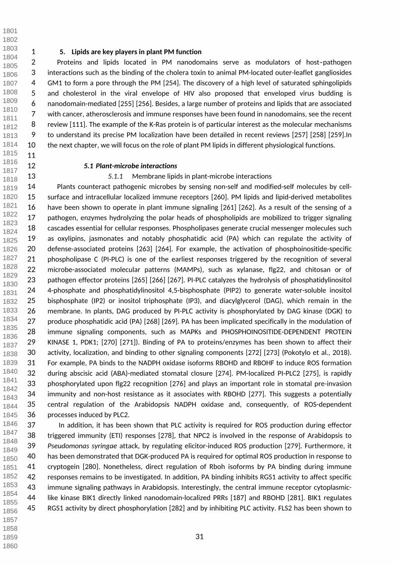
31
1 5. Lipids are key players in plant PM function 2 Proteins and lipids located in PM nanodomains serve as modulators of host–pathogen 3 interactions such as the binding of the cholera toxin to animal PM-located outer-leaflet gangliosides 4 GM1 to form a pore through the PM [254]. The discovery of a high level of saturated sphingolipids 5 and cholesterol in the viral envelope of HIV also proposed that enveloped virus budding is 6 nanodomain-mediated [255] [256]. Besides, a large number of proteins and lipids that are associated 7 with cancer, atherosclerosis and immune responses have been found in nanodomains, see the recent 8 review [111]. The example of the K-Ras protein is of particular interest as the molecular mechanisms 9 to understand its precise PM localization have been detailed in recent reviews [257] [258] [259].In
10 the next chapter, we will focus on the role of plant PM lipids in different physiological functions.1112 5.1 Plant-microbe interactions13 5.1.1 Membrane lipids in plant-microbe interactions14 Plants counteract pathogenic microbes by sensing non-self and modified-self molecules by cell-15 surface and intracellular localized immune receptors [260]. PM lipids and lipid-derived metabolites 16 have been shown to operate in plant immune signaling [261] [262]. As a result of the sensing of a 17 pathogen, enzymes hydrolyzing the polar heads of phospholipids are mobilized to trigger signaling 18 cascades essential for cellular responses. Phospholipases generate crucial messenger molecules such 19 as oxylipins, jasmonates and notably phosphatidic acid (PA) which can regulate the activity of 20 defense-associated proteins [263] [264]. For example, the activation of phosphoinositide-specific 21 phospholipase C (PI-PLC) is one of the earliest responses triggered by the recognition of several 22 microbe-associated molecular patterns (MAMPs), such as xylanase, flg22, and chitosan or of 23 pathogen effector proteins [265] [266] [267]. PI-PLC catalyzes the hydrolysis of phosphatidylinositol 24 4-phosphate and phosphatidylinositol 4,5-bisphosphate (PIP2) to generate water-soluble inositol 25 bisphosphate (IP2) or inositol triphosphate (IP3), and diacylglycerol (DAG), which remain in the 26 membrane. In plants, DAG produced by PI-PLC activity is phosphorylated by DAG kinase (DGK) to 27 produce phosphatidic acid (PA) [268] [269]. PA has been implicated specifically in the modulation of 28 immune signaling components, such as MAPKs and PHOSPHOINOSITIDE-DEPENDENT PROTEIN 29 KINASE 1, PDK1; [270] [271]). Binding of PA to proteins/enzymes has been shown to affect their 30 activity, localization, and binding to other signaling components [272] [273] (Pokotylo et al., 2018). 31 For example, PA binds to the NADPH oxidase isoforms RBOHD and RBOHF to induce ROS formation 32 during abscisic acid (ABA)-mediated stomatal closure [274]. PM-localized PI-PLC2 [275], is rapidly 33 phosphorylated upon flg22 recognition [276] and plays an important role in stomatal pre-invasion 34 immunity and non-host resistance as it associates with RBOHD [277]. This suggests a potentially 35 central regulation of the Arabidopsis NADPH oxidase and, consequently, of ROS-dependent 36 processes induced by PLC2.37 In addition, it has been shown that PLC activity is required for ROS production during effector 38 triggered immunity (ETI) responses [278], that NPC2 is involved in the response of Arabidopsis to 39 Pseudomonas syringae attack, by regulating elicitor-induced ROS production [279]. Furthermore, it 40 has been demonstrated that DGK-produced PA is required for optimal ROS production in response to 41 cryptogein [280]. Nonetheless, direct regulation of Rboh isoforms by PA binding during immune 42 responses remains to be investigated. In addition, PA binding inhibits RGS1 activity to affect specific 43 immune signaling pathways in Arabidopsis. Interestingly, the central immune receptor cytoplasmic-44 like kinase BIK1 directly linked nanodomain-localized PRRs [187] and RBOHD [281]. BIK1 regulates 45 RGS1 activity by direct phosphorylation [282] and by inhibiting PLC activity. FLS2 has been shown to
180118021803180418051806180718081809181018111812181318141815181618171818181918201821182218231824182518261827182818291830183118321833183418351836183718381839184018411842184318441845184618471848184918501851185218531854185518561857185818591860

32
1 be no longer endocytosed [283], pointing PA as a core regulatory component of plant receptor 2 kinase-based immunity. Interestingly, remodeling of cortical actin network in response to elicitors is 3 mediated by the negative regulation of CAPPING PROTEIN by PLD-produced PA [284].4 The production of PA by Phospholipase D enzymes (PLD) is involved in ROS production in 5 response to elicitation [285]. This production of PA has been also shown to be essential for 6 phytoalexin biosynthesis [286] yet considering the various subcellular localizations of PLDs this may 7 not be specific to PM-associated PA [287]. PLDδ has been also found to be involved in non-host 8 resistance of A. thaliana epidermis against the barley powdery mildew fungus Blumeria graminis f. 9 sp. Hordei and the pea powdery mildew fungus Erysiphe pisi. PM-localized PLDδ is enriched at the
10 penetration sites and PA is supposedly produced and necessary for resistance considering the 11 observed increase in susceptibility after treatment with n-butanol, a PLD inhibiting drug [288]. PLD-12 produced PA is also involved in plant-virus interactions by promoting the RNA replication of the Red 13 clover necrotic mosaic virus. A viral auxiliary replication protein binds PA in vitro and the exogenous 14 addition of PA increase replication rates. This is consistent with the observed increase of PA levels in 15 infected cells [289]. PA could therefore play a central role in viral replication by tethering protein 16 complexes to each other and to the membrane, thereby putatively modulating catalytic activities 17 [290] and membrane curvature [291]. As PA negatively favours curved membranes [292], a local 18 increase in PA levels is likely to impact membrane structure and charge. Nonetheless, stimuli-19 dependent impact of PA production on membrane organization and the dynamics of plant immune 20 component remain to be studied. What are the molecular function(s) of PA in immunity is still to be 21 further studied.22 In addition to PA, both phosphoinositides and lysophospholipids have been shown to play a role 23 in plant defense. Lysophospholipids are derived from glycerophospholipids by the action of PLAs. 24 Examples of lysophospholipids include L-PA, lysophosphatidylcholine, sphingosylphosphorylcholine, 25 and sphingosine-1-phosphate [293]. The signaling activity or specificity of these compounds is 26 dependent on the length and position of the acyl chain, the degree of saturation, and the presence of 27 the phosphate head group. Acyl chain length and degree of saturation have been shown to influence 28 plant-pathogen interactions. The accumulation of C16:1 and C16:2 fatty acids in tomato and 29 eggplant, due to the overexpression of a yeast delta-9-desaturase, resulted in a heightened 30 resistance to powdery mildew Erysiphe polygoni DC and Verticilium dahliae, respectively. An increase 31 in C18:2 and C18:3 has also been shown to increase resistance to Colletotrichum gloeosporioides and 32 Pseudomonas syringae in avocado and tomato respectively [294] [295]. Moreover, bean resistance to 33 Botrytis cinerea induced by a non-pathogenic strain of Pseudomonas has been correlated with an 34 increase of C18:2 and C18:3 [296].35 As stated in chapter 1, plant sterols are core components of membrane and accumulate in the 36 PM. Conversely, PM sterols are conserved regulators of membrane organization. Mutants altered in 37 sterol biosynthesis and the use of sterol-biosynthesis inhibiting drugs, affect cell wall composition 38 and induce abnormal callose and lignin deposits (cell wall compounds involved in biotic stress) [297]. 39 Cryptogein is able to induce an increase in PM-fluidity via sterol-binding [109]. Highly-hydroxylated 40 sphingolipids increase membrane stability and decrease membrane permeability which are both 41 associated to increased defence against phytopathogenic fungi [35] [298]. Rice fah1/2 knock-down 42 mutants, displaying the lack of an α-hydroxyl group on the fatty-acid moiety of sphingolipids, exhibit 43 a decrease in PM order level [51]. These mutants show reduced resistance to the rice blast fungus 44 Magnaporthe oryzae, with the delocalization of major actors of innate immunity such as NB-LRRs, 45 NADPH oxidases, Small GTPases and Calcium-dependent kinases [51]. On the contrary, Arabidopsis
186118621863186418651866186718681869187018711872187318741875187618771878187918801881188218831884188518861887188818891890189118921893189418951896189718981899190019011902190319041905190619071908190919101911191219131914191519161917191819191920

33
1 fah1/2 knock-out mutants display an increased resistance to obligate biotrophic fungi Golovinomyces 2 cichoracearum potentially due to a consequential increase in intracellular ceramides and salicylate 3 [299]. Interestingly, a sphingosine analogue produced by the fungal pathogen Alternaria alternata f. 4 sp. Lycopersici (AAL), serves as a virulence factor that induces PCD in plants and animals [300].56 5.1.2 Sphingolipids as receptors of necrotrophic toxins and plant-pathogen 7 elicitors 8 Glycosylated lipids are often receptors in insect host binding to microbial toxins [301]. One very 9 important glycosylated lipids of the animal kingdom are gangliosides. Their polar heads act as surface
10 recognition markers and surface receptors for bacterial toxins such as cholera and Bt toxins [254]. 11 They are recognized and used by virus to enter and infect cells [302]. Plant GIPCs bear structural 12 similarities with gangliosides because they contain negatively charged glycan polar heads located in 13 the outer-leaflet of the PM [303].14 A recent study has shown that GIPCs were located in the outer-leaflet of plant PM are receptors 15 to Necrosis and ethylene-inducing peptide 1-like (NLP) proteins [40]. In the study of Lenarčič et al 16 2017, microbial NLP proteins are used for the identification and characterization of NLP receptors. 17 NLPs are part of a superfamily of cytotoxins produced by plant pathogens such as bacteria, fungi and 18 oomycetes [304]. They can be cytolysins, inducing symptoms on eudicot plants but not monocot 19 plants where no necrotic and cytolytic effects are observed [305]. The secretion of NLPs occurs in the 20 extracellular environment of host plants with the toxins targeting the PM outer-leaflet [304] [306].In 21 vitro, NLPs were shown to specifically bind to purified GIPCs from tobacco and Arabidopsis but not to 22 phospholipids, GluCer or sphingomyelin. Moreover, NLPs also bind to all GIPCs, irrespective of the 23 plant clade, with similar affinities [40]. Upon binding to the hexose moieties of the GIPC polar heads, 24 NLPs undergo structural changes triggering the conformational modification of their L3 loop and the 25 incorporation of a Mg2+ ion responsible for its cytotoxicity. This results in the interaction of the W155 26 residue of the L3 loop with the membrane, crucial for cytolysis [40]. The study has also showed that 27 sugar residues exposed on the plant outer membrane surface are important for NLP toxicity, such 28 that Glucosamine, Man/Glucose being GIPC terminal sugars of tobacco and Arabidopsis respectively, 29 induces membrane damage upon binding to NLPs. Plant mutants impaired in the GIPC biosynthetic 30 pathway, are less sensitive to NLPs, implying the importance of intact GIPCs for NLP cytotoxicity [40]. 31 The sensitivity to NLP toxicity occurs only for eudicots but not monocots with the exception of a 32 monocot Phalaenopsis species (an orchid). This intriguing fact could be explained by the presence of 33 different GIPC series in these two plant clades. While both have similar terminal hexose sugars with 34 similar affinity to NLPs; eudicots GIPCs contain two hexoses linked to IPC (series A GIPC) whereas 35 monocot GIPCs contain three hexoses (series A GIPC), with the exception of Phalaenopsis containing 36 both series A and B. Biophysical characterization of GIPC series in monolayer artificial membranes 37 suggests a perpendicular arrangement of the polar head of both series A and B GIPC, such that the 38 terminal hexose in series B GIPC, is located further away from the membrane surface compared to 39 series A GIPC. Hence, NLPs binding to series B GIPC terminal sugars have their L3 loop positioned 40 farther from the plant membrane, preventing NLP contact with the membrane and thereby, 41 cytolysis. Both, the length of GIPC head groups and the structural design of the NLP sugar-binding 42 sites explain the differential sensitivity of host plants to NLP toxins.4344 5.1.3. Lipid domain-associated proteins in plant microbe interaction
192119221923192419251926192719281929193019311932193319341935193619371938193919401941194219431944194519461947194819491950195119521953195419551956195719581959196019611962196319641965196619671968196919701971197219731974197519761977197819791980

34
1 Remorins (REM) are plant-specific nanodomain-organized proteins notably involved in plant-2 microbe interactions. REMs are anchored by their C-terminal domain to the cytosolic leaflet of the 3 PM. The anchoring and lateral segregation in the PM is PI4P- and sterol-mediated [172]. REM1.3 was 4 shown to be delocalized after sterol disrupting treatments, such as methyl-β-D-cyclodextrin [189] 5 [222] [160] [188] [172] or fenpropimorph [172]. Both their presence at the PM and their correct 6 partitioning within their cognate nanodomains, are essential for their cellular function, e.g. 7 StREM1.3’s role in hindering Potato Virus X cell-to-cell movement [172] [307]. Other REM-group 8 proteins have been evidenced as key players in biotic interactions such as SYMREM1 (MtREM2.2) 9 involved in the nodulation process of M. truncatula with Sinorhizobium meliloti [308] Its role was also
10 shown to be essential in the dynamic stabilization of the LYK3-FLOT4-SYMREM1 PM-nanodomains, 11 important for root bacterial symbiosis [309]. Remorins are found in DRM, the closest biochemical 12 counterpart to PM-nanodomains known today, in virtually all clades of land plants: Poplar [310], Oat 13 and Rye [166], Tobacco [169], Arabidopsis [162]. The role of REMs as actors involved in a wide range 14 of biotic interactions has been established throughout the years: ranging from susceptibility factors 15 to viral infections [311], to oomycetes [312] or on the contrary as resistance factors to Potato Virus X 16 [188]. Their capacity to impact PD permeability [172] [313] also demonstrates their implication in 17 innate immunity. Super-resolution microscopy has been used to understand the role of nanodomain 18 dynamics in the context of viral infection, revealing that an optimal partition (i.e. size of 19 nanodomains, number of molecules in and out domains, and mobility) of REM-associated 20 nanodomains was necessary for the function of REMs [172].21 PM receptors involved in the oxidative-burst response to the perception of Pathogen-Associated 22 Molecular Patterns (PAMPs) are key players in plant innate immunity [314]. Their PM localization 23 gives them the role of gatekeepers capable of dynamically associating to co-receptors complexes and 24 triggering signaling pathways to prepare the cell for immediate and short-to-mid-term defense 25 responses [314]. The best example is FLS2, an LRR receptor-like kinase involved in the perception of 26 the bacterial elicitor flagellin (flg22) in Arabidopsis [315]. PM-subcompartmentalization via raft-like 27 domains is an essential phenomenon in the PAMP perception and response processes. Upon 28 cryptogein treatment on tobacco cells, a quantitative proteomics approach has evidenced the 29 delocalization of various Dynamin and 14-3-3 proteins into the DRM fraction, both involved in PM-30 based signaling [168]. The reorganization of the PM in response to elicitors is not only observed via 31 its protein composition but also via the biophysical properties conferred by lipids: upon elicitation 32 with flg22 or cryptogein, PM order and fluidity are altered [90]. As regards to flg22’s cognate 33 receptor FLS2, it was shown to relocate to DRM fractions upon flg22 treatment as well as many other 34 key proteins involved in immunity-associated signaling [161], revealing that PM-remodeling, at both 35 the lipid and protein level, is important for a functional immune response. 36 In good agreement, perception of flg22 in BY-2 cells induces global increase of the order level of 37 the PM. While this modification of the PM properties correlates with signal initiation [316], the 38 potential functional implication and molecular basis of such membrane modification remains to be 39 elucidated. Cryptogein is shown to be able to induce an increase in PM-fluidity via sterol-binding 40 [109]. Sterols and their associated micro-environments appear to be crucial for immune responses at 41 the cellular level to the extent that both mutants for sterol biosynthesis and sterol-biosynthesis 42 inhibiting drugs affect cell wall composition and induce abnormal callose and lignin deposits i.e. cell 43 wall compounds involved in biotic stress [297].4445 5.1.4. GPI-anchored proteins & outer-leaflet PM domains
198119821983198419851986198719881989199019911992199319941995199619971998199920002001200220032004200520062007200820092010201120122013201420152016201720182019202020212022202320242025202620272028202920302031203220332034203520362037203820392040

35
1 The importance of outer-leaflet PM-nanodomains enriched in sphingolipids and sterols is also 2 underscored by the presence of GPI-anchored proteins in these domains, many of which are 3 implicated in host responses to invading microbes. GPI-anchored β-1,3-glucanases (BGs), responsible 4 for callose degradation, are found in DRM fractions alongside callose synthases [310]. Their presence 5 in microdomains around PD enable a turnover of callose deposits when proper signaling occurs. The 6 localization of BGs at PD is regulated by the presence of sterol-enriched domains at the PM, which 7 share a virtually identical lipid composition with the PD-PM interface to the extent that sterol-8 biosynthesis inhibitors abolish this targeting [7]. The PD-enriched GPI-anchored protein LYM2 has 9 been found to impact PD conductance in response to chitin treatments [317]. Several GPI-anchored
10 BGs have been shown to associate to PD thanks to their GPI-anchoring motif [318] in a salicylic acid-11 dependent fashion [319] consolidating the idea that addressing proteins to PM-PD microdomains via 12 GPI-anchoring plays a significant role in host responses to pathogens.1314 5.1.5 Extracellular Vesicles & host-induced gene silencing15 Extracellular vesicles (EVs) are PM-derived vesicles secreted in the extracellular matrix involved in 16 inter-cellular communication and in response to stress, notably biotic stimuli. The existence of these 17 vesicles has been first observed in plants in the 1960’s [320] yet EVs have only recently been better 18 characterized [321] regarding their function during plant-pathogen interactions. They are observed 19 upon xylanase treatment [322] and in response to hormone treatments [323]. EVs are believed to be 20 derived from multi-vesicular bodies (MVB) which accumulate around appressorium and haustoria 21 during defense against fungal pathogens [324] [325]. They have recently been shown to contain a 22 number of biotic stress-related compounds such as phytoalexins like glucosinolates [323], small-23 interfering RNAs that can effectively silence pathogen-associated virulence and/or housekeeping 24 genes [326] [327] [328] and signaling-associated enzymes, such as PLD and PLC [329]. The presence 25 of these phospholipases hints to the possibility of EVs being involved in lipid signaling pathways.26 Plant EVs contain phospholipids such as PI4P [322] [330], PI and PA [323]. The lipid composition 27 may vary upon which organ they are secreted, and upon different stimuli applied to the secreting 28 cells. For example, upon jasmonic acid treatment, sunflower seed-derived EVs will be enriched in 29 PI4P and depleted in PI [323]. The lipidome of plant EVs, in different conditions, has yet to be 30 published and it will surely reveal crucial information on their biogenesis and activity.3132 5.2 Hormone signaling and transport33 Lipid-mediated protein sorting mechanisms at TGN have strong impact on plant development 34 since they are involved in directing the secretion and endosomal recycling pathways of a set of 35 proteins that includes hormone transporters. Higher plants are multi-cellular organisms able to 36 respond and quickly adapt to their environment. In particular, the plant hormone auxin plays a 37 fundamental role in the regulation of a variety of developmental processes enabling plants to adapt 38 to their environment, including directional growth as gravitropism [331] [332] [333] [334] [335] [336] 39 [337]. Auxin mediated control of plant development relies on establishment of concentration 40 gradient of auxin that are generated by the activity of plasma membrane localized auxin carriers 41 [335] [336] [337] [338] [339]. Therefore, the mechanisms that control the remodeling of auxin 42 carriers represent a key control point for signals that control plant development and response to 43 abiotic stress. Several studies have shown that the TGN is involved in auxin-carrier trafficking but the 44 sorting mechanisms are poorly understood [340] [341] [342]. Several elements related to G proteins 45 (guanine nucleotide-binding proteins) are known to be involved in TGN-mediated auxin-carriers’
204120422043204420452046204720482049205020512052205320542055205620572058205920602061206220632064206520662067206820692070207120722073207420752076207720782079208020812082208320842085208620872088208920902091209220932094209520962097209820992100

36
1 trafficking. Our goal in this review is not to create an exhaustive list of all these elements but we can 2 name a few: the small GTPase protein RAB-A1b, the ECHIDNA protein which interacts with YPT/RAB 3 GTPase interacting protein 4a (YIP4a) and YIP4b, the ADP ribosylation factor (ARF) ARF1 as well as 4 the ARF-guanine exchange factors (ARF-GEFs) GNOM and BIG1-5, and finally the ARF-GTPase 5 activating proteins (ARF-GAPs) SCARFACE/VAN3[342] [343] [344] [345] [346] [347]. On the lipid side, 6 sphingolipids represent a class of lipids particularly interesting since VLCFAs, an imprint of 7 sphingolipids, are enriched at TGN [232]. Shortening of the acyl-chain using pharmacological and 8 genetic tools reveal that the length of sphingolipid acyl-chains is involved in the secretory sorting of 9 the efflux auxin carrier PIN2 (but not in PIN1 or AUX1 trafficking), auxin redistribution during root
10 response to gravity (root gravitropism) and root gravitropism per se [232]. Interestingly, it had been 11 shown before that VLCFA-containing sphingolipids are involved in PIN1 and AUX1 trafficking (but not 12 PIN2 trafficking) and lateral root formation [55]. These results are not necessarily contradictory. 13 Indeed, the structural diversity of sphingolipids is wide and has been described in chapter 1 and in 14 several reviews [298] [35]. LCBs are not always found included in a ceramide molecule but can also 15 be freely found at non-negligible proportion inside the cell as sphinganine and phytosphingosines. 16 Finally, aside from sphingolipids, VLCFAs can also be included in some phospholipids. On this topic, it 17 has been shown that the pas2 mutant displays a reduced level of VLCFA-containing 18 phosphatidylethanolamine (PE) and show endocytic trafficking defects [348]. Decrease of VLCFA-19 containing PE in the pas2 mutant targets the RAB-A2a compartment and the plasma membrane 20 endocytic recycling of the auxin efflux-carrier PIN1 but has no effect on PIN2 localization [349] [348].21 Coming back on sphingolipids, it has been shown that the loh1/loh2 double mutant, which is 22 defective in ceramide synthases LOH1 and LOH2, are involved in endocytosis and plasma membrane 23 recycling of the auxin carriers PIN1 and AUX1, but not PIN2, potentially through RAB-A2a 24 compartments [55]. Hence, it is possible that VLCFA-containing sphingolipids are important for 25 endocytic/recycling of certain subset of proteins from plasma membrane through a RAB-A2a-positive 26 subdomain of TGN that would host the recycling pathway. In contrast, the herbicide metazachlor, 27 that drastically modifies both GluCer and GIPCs fatty acids composition by replacing VLCFAs in GluCer 28 and GIPCs pools by LCFAs without modifying the global quantity of either GluCer or GIPCs, alters PIN2 29 polarity at plasma membrane while it neither affects PIN1 polarity nor AUX1 localization [232]. 30 Importantly, metazachlor blocks PIN2 predominantly at SYP61/secretory vesicles (SVs) 31 compartments as compared to RAB-A2a/CCVs [232]. Concomitantly, metazachlor treatment neither 32 alters endocytosis nor plasma membrane recycling of PIN2 but rather blocks the secretion of de novo 33 synthetized PIN2 at SYP61/SVs compartments. From these studies, it can be postulated that TGN-34 associated RAB-A2a/Clathrin-coated vesicles (CCVs) compartments could host the ceramide-35 dependent PM recycling of auxin carriers PIN1 and AUX1 while TGN-associated SYP61/SVs 36 compartments could host VLCFA-containing GluCer and GIPC-dependent secretory sorting of PIN2. 37 However, future challenges on this topic need to address TGN sub-domains’ dynamics, interactions 38 and maturation, which could involve a tight regulation of lipid homeostasis and crosstalk to define 39 the identity of each sub-domain. 40 The VLCFAs of sphingolipids determine a specific physical property of sphingolipids, which is the 41 ability to insert their acyl-chain within the opposing leaflet of the membrane, a phenomenon known 42 as interdigitation, see Figure 4. Interestingly, it has been shown in animal cells that the coupling of 43 membrane leaflets is cholesterol-dependent [64]. In yeast and in plants, it has been shown by 44 immuno-purification of intact compartments coupled with lipid mass spectrometry analyses, that 45 TGN-derived secretory vesicles are enriched in both sphingolipids and sterols [229] [232]. Currently,
210121022103210421052106210721082109211021112112211321142115211621172118211921202121212221232124212521262127212821292130213121322133213421352136213721382139214021412142214321442145214621472148214921502151215221532154215521562157215821592160

37
1 it is not clear whether the pool of sterols at the TGN would play a role in the trafficking of auxin 2 carriers. Previously, it has been shown that sterols are involved in endocytosis and recycling at 3 plasma membrane of the PINs auxin carriers [350] [351] [352]. Sterol-mediated endocytosis is 4 involved in PIN2 polarity establishment after cytokinesis by removing PIN2 from the new basal 5 membrane of daughter cells, while PIN2 at the new apical membrane remains [351]. Sterol-mediated 6 auxin carriers’ sub-cellular localization impacts auxin distribution at the tissue-level, which has 7 repercussions on plant development such as the graviresponse of the root [351]. Interestingly, not 8 only are the major sterols: sitosterol, stigmasterol and campesterol involved in plant development, 9 but also some very rare intermediate-sterol compounds such as derivatives of cycloartanol (4-
10 carboxy-4-methyl-24-methylenecycloartanol) and 4α-methyl sterols (24-ethyl-ionophenol and 24-11 ethylidene-ionophenol) [353] [354]. Future studies will reveal how sterols interplay with other lipids 12 such as sphingolipids and anionic phospholipids to regulate hormone transport. 1314 5.3 Abiotic stress15 Lipids have been involved as second messengers in many responses to abiotic stress, see for 16 review [262, 355-357] [358]. PLD and PLC/DGK-mediated PA formation and it subsequent 17 phosphorylation to form Diacylglycerol Pyrrophosphate (DGPP) are key players in this response 18 [359]. Other lipids participate to the response: oxylipins, PIPs, sphingolipids, fatty acids, 19 lysophospholipids, N-acylethanolamines and galactolipids, see for reviews [1] [360]. Studies have 20 started deciphering the different isoforms of enzymes involved in these transduction pathways as 21 well as their tight regulations, for recent reviews see [361]. For example, PA displays different modes 22 of action, including direct target protein binding and biophysical effects on cell membranes. It is 23 puzzling that PA production can be triggered by opposite stressors, such as cold and heat. How PA 24 regulates this diversity of response is discussed in this recent review [264].25 PLD-derived PA was shown to recruit the ABA-regulated ABI1 phosphatase 2C (PP2C) to the PM 26 [362]. The identification of ABI1 as a direct target of the PA provides a functional link between the 27 two families of important signaling enzymes, PLD and PP2C. Recently, a study on the regulation of 28 ion transport in plants showed that C2-domain ABA-related (CAR) family of small proteins is involved 29 in the Ca2+-dependent recruitment of the pyrabactin resistance 1/PYR1- like (PYR/PYL) ABA receptors 30 to the PM. CARs are peripheral membrane proteins that cluster at the PM and generate strong 31 positive membrane curvature. These features represent a mechanism for the generation, 32 stabilization, and/or specific recognition of PM discontinuities involved in the recruitment of PYR/PYL 33 receptors and other signaling components in cell responses to salt stress [363].3435 5.4 Plasmodesmata function36 Plasmodesmata (PD) are specialized nano-sized membrane-lined channels, which cross the walls 37 of plants and of some algal cells. PDs enable direct, regulated, symplastic transport of small RNAs and 38 molecules between cells. They are also hijacked by phytoviruses to allow their propagation from cell-39 to-cell. Ultrastructure of PD has been deciphered. They are lined by the PM forming what is termed 40 the PD-PM subcompartment (PD-PM) and contain a strand of tubular modified endoplasmic 41 reticulum (ER) called desmotubule, and the space between these two membranes is thought to 42 control PD permeability [200]. A recent study has reconstructed PD three-dimensional ultrastructure 43 with an unprecedented resolution using electron tomography, showing that ER-PM contact sites 44 undergo substantial remodeling events during cell differentiation [364]. Post-cytokinesis PD, called 45 type I PD, present an intimate ER-PM contact along the entire length of the pores whereas during cell
216121622163216421652166216721682169217021712172217321742175217621772178217921802181218221832184218521862187218821892190219121922193219421952196219721982199220022012202220322042205220622072208220922102211221222132214221522162217221822192220

38
1 expansion, the PD pore widens and the two membranes separate, leaving a cytosolic sleeve spanned 2 by tethers whose presence correlates with the appearance of the intermembrane gaps, called type II 3 PD. Surprisingly, the type II PD allow diffusion of macromolecules despite the apparent lack of an 4 open cytoplasmic sleeve, forcing the reassessment of the mechanisms that control plant cell-to-cell 5 communication [364].6 The membrane organization of PDs is therefore characterized by the close apposition of the ER-7 derived desmotubule and the PM with spoke-like structures linking the two membranes likely 8 defining microdomains of the PM, PD-PM and desmotubules has been recently proposed to be 9 membrane contact sites (MCS). MCS control close appositions between two membranes that form
10 microdomains involved in the control of lipid exchanges or in coupling events (review in [365]). MCS-11 subdomains are likely to display specific biophysical properties and may cluster proteins and 12 negatively-charged lipids like phosphoinositides, promoting specific physicochemical membrane 13 properties taking part in shaping local membrane electronegativity gradients [365].14 Firmly anchored within the cell wall, PDs are difficult to purify. A two-step simple purification 15 procedure (consisting in isolating cell wall fragments containing intact PD and an enzymatic 16 degradation of the wall matrix to release of PD) has been successfully design to obtain highly-purified 17 PD preparations [366]. Hence, analyses of PD fractions have provided valuable information on the 18 functional and structural elements that define PD, particularly for lipids. PD membranes display 19 enrichment in sterols and sphingolipids with saturated VLCFAs (likely GIPCs) when compared with 20 the bulk of the PM. This profile is reminiscent of DRMs although the isolation procedure is detergent-21 free and suggest that lipids are laterally segregated the PD-PM cell-to-cell junction in Arabidopsis 22 thaliana [7]. This study identifies a role for sterols in modulating cell-to-cell connectivity, possibly by 23 establishing and maintaining the positional specificity of callose-modifying GPI-anchored proteins at 24 PD and emphasizes the importance of lipids in defining PD-associated nanodomains. The role played 25 by the other major lipid constituents, such as GIPCs and glycerolipids in defining specialized 26 membrane domains in PD, remains to be further studied. This change of paradigm regarding the 27 membrane organization of PD will likely pave the way to a deep understanding of cell-to-cell 28 communication in plants.29
222122222223222422252226222722282229223022312232223322342235223622372238223922402241224222432244224522462247224822492250225122522253225422552256225722582259226022612262226322642265226622672268226922702271227222732274227522762277227822792280

39
1 Conclusions and perspectives: How to get a comprehensive membrane organization in plants? 2 The deep-seated understanding of how the plant PM is organized in terms of the involvement of 3 lipids and proteins clearly necessitates multiple approaches. Our community needs to develop new 4 dedicated methodologies and dare multidisciplinarity even further that what we have been 5 accustomed to. For example, the extensive use of biophysical approaches to study lipid/protein 6 interactions as reviewed in [367], among them: 1/ surface plasmon resonance [40], isothermal 7 titration calorimetry (ITC), Langmuir monolayer tensiometry, liposomes binding and lipid strips and 8 arrays [114] to study the interaction of proteins with lipids; 2/ liposome leakage and lipid-mixing 9 assays [307] to investigate how proteins can destabilize membrane bilayers; 3/ Circular dichroism
10 [307], Fourier-transform infrared spectroscopy [172], solid-state NMR with labeled lipids or proteins 11 [368] to obtain 3D structures of the membrane-bound interaction-complexes; 4/ Atomic force 12 microscopy (AFM) to scan the surface of bilayers and access the topology of different membrane 13 organizations. A deep understanding of the structural aspects of protein/lipid interactions will allow 14 the targeted-mutagenesis of key residues, the biological role of which could be further addressed by 15 reverse genetics approaches.16 In all the methods described above, there is an urgent need for biochemistry to obtain purified 17 proteins and lipids, in order to reconstitute proteoliposomes. Concerning proteins, the problem of 18 insolubility of highly hydrophobic proteins could be solved by using different expression systems 19 [369]. As for lipids, most of plant lipids are commercially available with the notable exception of 20 GIPCs. The lack of commercially-available GIPCs strongly impairs any serious in vitro reconstitutions 21 of true plant PM-like vesicles. One must undertake hardcore preparative biochemistry as described 22 during the 1970’s to obtain milligrams of purified GIPCs from living material [52, 54]. Purification of 23 mg amount of GIPCs will also pave the way to the development of fluorescently labeled sphingolipid 24 analogs by conjugating a hydrophilic fluorophore to the headgroup or a hydrophobic fluorophore to 25 the amidified fatty acids e.g. [370]. Unfortunately, very few plant lipids labeled with a fluorochrome 26 or deuterated are available. Similarly, none of the great diversity of free- and conjugated-27 phytosterols are commercialized in a labeled form, which strongly impairs NMR studies that would 28 enable to further enquire about the role of each molecular species individually. From purified lipids, 29 the preparation of asymmetrical liposomes will also be very challenging. To have access to the 30 properties of asymmetric vesicles that mimic the plant PM would provide means to understand lipid 31 raft formation or transmembrane helix orientation [371],32 Molecular dynamics (MD) is a method that computes the physical movements of atoms and 33 molecules in order to give a view of the dynamic evolution of a virtual chemical system. It can 34 therefore study the interaction dynamics of lipids and proteins, or the conformational changes of 35 molecules, for a fixed period of time. MD computes lipid-lipid and lipid-protein interactions and 36 provides a comprehensive atomistic model of a typical lipid bilayer and gather information on 37 membrane domain formations [372] [373]. MD has been used in plant membrane biology to 38 decipher the structural basis of the unconventional lipid-binding motif of REM that confers 39 nanodomain organization [172]. Such approach of “computational microscopy” captures the 40 molecular interactions within a complex system at a spatiotemporal resolution, unmatched by any 41 other conventional experimental methods e.g. [134, 374]. MD can be of particular interest in 42 grasping the intricacies of plant PM dynamics because the control of the thermodynamic parameters 43 permitted by MD can be used to mimic the constantly varying environmental conditions sustained by 44 plants in their natural habitats, and thereby to understand the biophysical implications of these 45 variations on the PM. Unfortunately, neither plant sphingolipids (GluCER, GIPCs), nor phytosterols
228122822283228422852286228722882289229022912292229322942295229622972298229923002301230223032304230523062307230823092310231123122313231423152316231723182319232023212322232323242325232623272328232923302331233223332334233523362337233823392340

40
1 (free or conjugated) have been, to this day, modelized in the force field, preventing all MD studies 2 with plant PM lipids. The force field refers to the functional form and parameter sets used to 3 calculate the potential energy of a system of atoms or coarse-grained particles; it is the necessary 4 step for MD simulations. The parameters of the energy functions may be derived from experiments 5 in physics (solid-state NMR, Langmuir monolayer, calorimetry…) or chemistry (chemical structure, 6 liquid-state NMR…) [375]. All-atom force fields provide parameters for every type of atom in a 7 system. Coarse-grained potentials, which are often used in long-time simulations of macromolecules 8 and multi-component complexes, provide even cruder representations for higher computing 9 efficiency. Such modelization must be carried for plant sphingolipids and phytosterols as it was
10 described for animal gangliosides in [376]. 11 Other aspects still missing in the plant membrane biology field, are the methods to image lipids in 12 vivo necessary to follow their segregation at the nanoscale level, their dynamics and interactions 13 with proteins. Two reasons can be given: firstly, as stated before, very few fluorescently-labeled 14 lipids are available, and secondly, the cell wall strongly impairs the intake of fluorescently labeled 15 molecules. Nevertheless, the use of in vivo bioorthogonal click chemistry could be an elegant 16 approach to circumvent this problem in plants [377] [378]. Although genetically encoded biosensors 17 for PIPs, PA, DAG and PS have already been developed [27, 114] [115]), biosensors of plant 18 sphingolipids and phytosterols are crucially missing and will surely be developed using lipid-binding 19 domains found in plant proteins. Inhibitors of lipid synthesis (table 1), and genetically-modified lipid-20 using enzymes (phosphoinositides kinase, lipase phosphatase) able to target the PM and specifically 21 modify in vivo pools of lipids, are used to address the question of the role of lipids in nanodomain 22 formation and dynamics [115, 172, 379]. Similarly, membrane surface charge or pH biosensors must 23 be improved as described in animal literature with intramolecular FRET sensors [380, 381]. The 24 development of environmentally-sensitive probes to label the outer- or inner-leaflet and measure 25 independently the fluidity of plant PM leaflets, must also be engaged (see examples of available 26 probes in [382]). Furthermore, super-resolution microscopy will continue to allow a deep 27 understanding of segregation and dynamics of proteins and lipids at the nanometer scale. The 28 development of fluorescent bimolecular tracking in the live-cell PM will reveal whether proteins and 29 lipids may directly or indirectly interact with each other [208]. 30 Finally, state of the art lipidomics [383] and proteomics approaches must be further developed 31 including phosphoproteomics and lipid-modification of proteins (myristoylation, palmitoylation, 32 isoprenylation and GPI-anchoring, review in [384]).3334 The study of K-RAS protein which controls cell proliferation in animal cells should be exemplified 35 for the combinations of approaches mobilized by Hancock’s group to tackle its function in relation to 36 its organization within PM [257-259, 385-387]. K-Ras controls cell proliferation, and when mutated, 37 cells continuously proliferate and often develop into cancer. This group tackled the role of K-Ras in 38 PM nanodomains by using in parallel biochemistry, biophysics, modeling, high-resolution imagery, 39 mutagenesis, structural biology, model membranes, transcriptomics, cancerology and genetics 40 techniques. This form of multidisciplinary approach has led to a deep understanding of the 41 anchoring, the clustering of K-Ras with PM lipids, as well as the integration of these molecular 42 mechanisms intohigher levels of cell biology, hence determining their consequences on the fate of 43 the cell. For example, a recent paper of this group has shown that K-Ras anchoring sequences can 44 create lipid nanodomains with a remarkable specificity [258], and that lipid nanodomains are not 45 preexisting. A matter of the chicken or the egg causality dilemma! Similarly the works of Katharina
234123422343234423452346234723482349235023512352235323542355235623572358235923602361236223632364236523662367236823692370237123722373237423752376237723782379238023812382238323842385238623872388238923902391239223932394239523962397239823992400

41
1 Gaus’ [380, 381, 388] [389] [390, 391] or Akihiro Kusumi’s labs [129, 392-394] are exemplary to 2 understand, by developing single-molecule imaging methods, how T-cells initiate an immune 3 response and to better fathom the intricate complexity between cytoskeleton and protein/lipid 4 segregation. These two research groups have been remarkable by developing, in collaboration with 5 biophysicists, state of the art technology to follow single-molecules in the PM.67 Taken together, tools and methods must be developed by the plant membrane biology 8 community in the near future to pave the way towards the better understanding of the intimate 9 molecular relationships between lipids and proteins at the basis of domain segregation, dynamics,
10 signaling and function.11
240124022403240424052406240724082409241024112412241324142415241624172418241924202421242224232424242524262427242824292430243124322433243424352436243724382439244024412442244324442445244624472448244924502451245224532454245524562457245824592460

42
1 Acknowledgements2 We apologize to researchers whose work could not be cited here because of space limitations. AMC 3 and PG are supported by the Ministere de l’Enseignement Superieur et de la Recherche, France 4 (MERS, doctoral grants). This work was supported by the Bordeaux Metabolome Facility-MetaboHUB 5 (grant no. ANR–11–INBS–0010). We acknowledge the platform Metabolome-Fluxome-Lipidome of 6 Bordeaux (http://www.biomemb.cnrs.fr/INDEX.html) for contribution to lipid analysis.78 No conflict of interest declared.9
246124622463246424652466246724682469247024712472247324742475247624772478247924802481248224832484248524862487248824892490249124922493249424952496249724982499250025012502250325042505250625072508250925102511251225132514251525162517251825192520
43
1 Figure legends :2 Figure 1: Determined structures of GIPC glycosidic polar head from tobacco and maize. 3 A, tobacco GIPC of series A are major in tobaco leaves (top) with glucuronic acid (GlcA) and either 4 glucosamine (GlcN) or N-acetyl glucosamine (GlcNAc). Other minor polar head of series B and higher 5 glycosylated GIPC with arabinoase (Ara), galactose (Gal) and Manose (Man) have been identified, but 6 the precise structure remains to be determined. Grey part is the conserved glycan moitie of 7 glucuronic-Hex. Cer indicates the ceramide moitie, in tobacco, with t18:0 and t18:1 for LCB, and 8 VLCFA mostly alpha 2-hydroxylated ; B, GIPC found in corn seeds with branched polar head9
10 Figure 2: Structures of specific plasma membrane phytosterols compared with animal cholesterol.11 A, free phytosterols ; B, phytostanol ; C, conjugated phytosterols.1213 Figure 3: Biophysical features involved between a GIPC of series A and three molecules of 14 sitosterols.15 These interactions are important for nanodomain formations in the PM. LCB, Long Chain Base; 16 VLCFA, Very Long Chain Fatty Acid.1718 Figure 4: Formation of GIPC- and sterol-enriched domains along the secretory pathway.19 GIPCs are synthesized in the lumen of the trans Golgi network (TGN) by grafting on the ceramide 20 sequentially inositol-phosphate (IPCS, inositolphosphorylceramide synthase), glucuronic acid (IPUT1, 21 inositol phosphorylceramide glucuronosyltransferase) and mannose (GMT1, GIPC mannosyl-22 transferase1). Golgi-localized nucleotide sugar transporter (GONST1) is responsible for the import of 23 GDP-mannose to fuel GIPC synthesis. After vesicular fusion to the PM, GIPC polar heads face the 24 apoplasm. Polyglycosylated GIPCs form nanodomains in the PM (in red). 2526
252125222523252425252526252725282529253025312532253325342535253625372538253925402541254225432544254525462547254825492550255125522553255425552556255725582559256025612562256325642565256625672568256925702571257225732574257525762577257825792580

44
1 REFERENCES2 [1] Xue HW, Chen X, Mei Y. Function and regulation of phospholipid signalling in plants. The 3 Biochemical journal. 2009;421:145-56.4 [2] Cacas JL BC, Grosjean K, Gerbeau-Pissot P, Lherminier J, Rombouts Y, Maes E, Bossard C, 5 Gronnier J, Furt F, Fouillen L, Germain1, Bayer E, Cluzet S, Robert F, Schmitter JM, Deleu M, 6 Lins M, Simon-Plas F, Mongrand S. Re-visiting plant plasma membrane lipids in tobacco: a 7 focus on sphingolipids. . Plant physiology. 2015;in press.8 [3] Yetukuri L, Ekroos K, Vidal-Puig A, Oresic M. Informatics and computational strategies for 9 the study of lipids. Molecular bioSystems. 2008;4:121-7.
10 [4] Larsson C, Sommarin, M., Widell, S. Isolation of highly purified plant plasma membranes 11 and separation of inside-out and right-side-out vesicles. Methods in Enzymology. 12 1994;228:451-69.13 [5] Samarakoon T, Shiva S, Lowe K, Tamura P, Roth MR, Welti R. Arabidopsis thaliana 14 membrane lipid molecular species and their mass spectral analysis. Methods in molecular 15 biology. 2012;918:179-268.16 [6] Yu D, Rupasinghe TWT, Boughton BA, Natera SHA, Hill CB, Tarazona P, et al. A high-17 resolution HPLC-QqTOF platform using parallel reaction monitoring for in-depth lipid 18 discovery and rapid profiling. Anal Chim Acta. 2018;1026:87-100.19 [7] Grison MS, Brocard L, Fouillen L, Nicolas W, Wewer V, Dormann P, et al. Specific 20 membrane lipid composition is important for plasmodesmata function in Arabidopsis. The 21 Plant cell. 2015;27:1228-50.22 [8] Doignon F, Laquel P, Testet E, Tuphile K, Fouillen L, Bessoule JJ. Requirement of 23 Phosphoinositides Containing Stearic Acid To Control Cell Polarity. Molecular and cellular 24 biology. 2015;36:765-80.25 [9] Markham JE, Jaworski JG. Rapid measurement of sphingolipids from Arabidopsis thaliana 26 by reversed-phase high-performance liquid chromatography coupled to electrospray 27 ionization tandem mass spectrometry. Rapid communications in mass spectrometry : RCM. 28 2007;21:1304-14.29 [10] Markham JE, Li J, Cahoon EB, Jaworski JG. Separation and identification of major plant 30 sphingolipid classes from leaves. The Journal of biological chemistry. 2006;281:22684-94.31 [11] Schrick K, Shiva S, Arpin JC, Delimont N, Isaac G, Tamura P, et al. Steryl glucoside and 32 acyl steryl glucoside analysis of Arabidopsis seeds by electrospray ionization tandem mass 33 spectrometry. Lipids. 2012;47:185-93.34 [12] Wewer V, Dombrink I, vom Dorp K, Dormann P. Quantification of sterol lipids in plants 35 by quadrupole time-of-flight mass spectrometry. Journal of lipid research. 2011;52:1039-54.36 [13] Wenk MR. Lipidomics: new tools and applications. Cell. 2010;143:888-95.37 [14] Horn PJ, Korte AR, Neogi PB, Love E, Fuchs J, Strupat K, et al. Spatial mapping of lipids at 38 cellular resolution in embryos of cotton. The Plant cell. 2012;24:622-36.39 [15] Woodfield HK, Sturtevant D, Borisjuk L, Munz E, Guschina IA, Chapman K, et al. Spatial 40 and Temporal Mapping of Key Lipid Species in Brassica napus Seeds. Plant physiology. 41 2017;173:1998-2009.42 [16] Ellis SR, Paine MRL, Eijkel GB, Pauling JK, Husen P, Jervelund MW, et al. Automated, 43 parallel mass spectrometry imaging and structural identification of lipids. Nat Methods. 44 2018.45 [17] Frisz JF, Klitzing HA, Lou K, Hutcheon ID, Weber PK, Zimmerberg J, et al. Sphingolipid 46 domains in the plasma membranes of fibroblasts are not enriched with cholesterol. The 47 Journal of biological chemistry. 2013;288:16855-61.
258125822583258425852586258725882589259025912592259325942595259625972598259926002601260226032604260526062607260826092610261126122613261426152616261726182619262026212622262326242625262626272628262926302631263226332634263526362637263826392640

45
1 [18] Frisz JF, Lou K, Klitzing HA, Hanafin WP, Lizunov V, Wilson RL, et al. Direct chemical 2 evidence for sphingolipid domains in the plasma membranes of fibroblasts. Proceedings of 3 the National Academy of Sciences of the United States of America. 2013;110:E613-22.4 [19] Mongrand S, Morel J, Laroche J, Claverol S, Carde JP, Hartmann MA, et al. Lipid rafts in 5 higher plant cells: purification and characterization of Triton X-100-insoluble microdomains 6 from tobacco plasma membrane. The Journal of biological chemistry. 2004;279:36277-86.7 [20] Uemura M, Joseph RA, Steponkus PL. Cold Acclimation of Arabidopsis thaliana (Effect on 8 Plasma Membrane Lipid Composition and Freeze-Induced Lesions). Plant physiology. 9 1995;109:15-30.
10 [21] Bohn M, Heinz E, Luthje S. Lipid composition and fluidity of plasma membranes isolated 11 from corn (Zea mays L.) roots. Archives of biochemistry and biophysics. 2001;387:35-40.12 [22] Uemura M, Steponkus PL. A Contrast of the Plasma Membrane Lipid Composition of Oat 13 and Rye Leaves in Relation to Freezing Tolerance. Plant physiology. 1994;104:479-96.14 [23] Quartacci MF, Glisic O, Stevanovic B, Navari-Izzo F. Plasma membrane lipids in the 15 resurrection plant Ramonda serbica following dehydration and rehydration. Journal of 16 experimental botany. 2002;53:2159-66.17 [24] Berglund AH, Quartacci MF, Calucci L, Navari-Izzo F, Pinzino C, Liljenberg C. Alterations 18 of wheat root plasma membrane lipid composition induced by copper stress result in 19 changed physicochemical properties of plasma membrane lipid vesicles. Biochimica et 20 biophysica acta. 2002;1564:466-72.21 [25] Vermeer JE, Munnik T. Using genetically encoded fluorescent reporters to image lipid 22 signalling in living plants. Methods in molecular biology. 2013;1009:283-9.23 [26] Tejos R, Sauer M, Vanneste S, Palacios-Gomez M, Li H, Heilmann M, et al. Bipolar Plasma 24 Membrane Distribution of Phosphoinositides and Their Requirement for Auxin-Mediated 25 Cell Polarity and Patterning in Arabidopsis. The Plant cell. 2014;26:2114-28.26 [27] Simon ML, Platre MP, Assil S, van Wijk R, Chen WY, Chory J, et al. A multi-colour/multi-27 affinity marker set to visualize phosphoinositide dynamics in Arabidopsis. The Plant journal : 28 for cell and molecular biology. 2014;77:322-37.29 [28] Yamaoka Y, Yu Y, Mizoi J, Fujiki Y, Saito K, Nishijima M, et al. PHOSPHATIDYLSERINE 30 SYNTHASE1 is required for microspore development in Arabidopsis thaliana. The Plant 31 journal : for cell and molecular biology. 2011;67:648-61.32 [29] Hirano T, Stecker K, Munnik T, Xu H, Sato MH. Visualization of Phosphatidylinositol 3,5-33 Bisphosphate Dynamics by a Tandem ML1N-Based Fluorescent Protein Probe in Arabidopsis. 34 Plant Cell Physiol. 2017;58:1185-95.35 [30] van Leeuwen W, Vermeer JE, Gadella TW, Jr., Munnik T. Visualization of 36 phosphatidylinositol 4,5-bisphosphate in the plasma membrane of suspension-cultured 37 tobacco BY-2 cells and whole Arabidopsis seedlings. The Plant journal : for cell and molecular 38 biology. 2007;52:1014-26.39 [31] Furt F, Konig S, Bessoule JJ, Sargueil F, Zallot R, Stanislas T, et al. Polyphosphoinositides 40 are enriched in plant membrane rafts and form microdomains in the plasma membrane. 41 Plant physiology. 2010;152:2173-87.42 [32] Andersson MX, Stridh MH, Larsson KE, Liljenberg C, Sandelius AS. Phosphate-deficient 43 oat replaces a major portion of the plasma membrane phospholipids with the galactolipid 44 digalactosyldiacylglycerol. FEBS letters. 2003;537:128-32.45 [33] Andersson MX, Larsson KE, Tjellstrom H, Liljenberg C, Sandelius AS. Phosphate-limited 46 oat. The plasma membrane and the tonoplast as major targets for phospholipid-to-glycolipid
264126422643264426452646264726482649265026512652265326542655265626572658265926602661266226632664266526662667266826692670267126722673267426752676267726782679268026812682268326842685268626872688268926902691269226932694269526962697269826992700

46
1 replacement and stimulation of phospholipases in the plasma membrane. The Journal of 2 biological chemistry. 2005;280:27578-86.3 [34] Vermeer JEM, van Wijk R, Goedhart J, Geldner N, Chory J, Gadella TWJ, Jr., et al. In Vivo 4 Imaging of Diacylglycerol at the Cytoplasmic Leaflet of Plant Membranes. Plant Cell Physiol. 5 2017;58:1196-207.6 [35] Lynch. D DT. An introduction to plant sphingolipids and a review of recent advances in 7 understanding their metabolism and function. New Phytologist. 2004;16:677-702.8 [36] Sonnino S, Prinetti A. Gangliosides as regulators of cell membrane organization and 9 functions. Advances in experimental medicine and biology. 2010;688:165-84.
10 [37] Moreau P, Bessoule JJ, Mongrand S, Testet E, Vincent P, Cassagne C. Lipid trafficking in 11 plant cells. Progress in lipid research. 1998;37:371-91.12 [38] Pata MO, Hannun YA, Ng CK. Plant sphingolipids: decoding the enigma of the Sphinx. 13 The New phytologist. 2010;185:611-30.14 [39] Cacas JL, Bure C, Grosjean K, Gerbeau-Pissot P, Lherminier J, Rombouts Y, et al. 15 Revisiting Plant Plasma Membrane Lipids in Tobacco: A Focus on Sphingolipids. Plant 16 physiology. 2016;170:367-84.17 [40] Lenarčič T AI, Böhm H, Hodnik V, Pirc K, Zavec AP, Podobnik M, Greimel P, Yamaji AH, 18 Kobayashi T, Feussner I, Mortimer JC, Mamode-Cassim A, Mongrand S, Oecking C, Anderluh 19 G, Nürnberger T. Eudicot plant-specific sphingolipids determine host selectivity of microbial 20 NLP cytolysins. 2017.21 [41] Carter HE, Gigg RH, Law JH, Nakayama T, Weber E. Biochemistry of the sphingolipides. 22 XI. Structure of phytoglycolipide. The Journal of biological chemistry. 1958;233:1309-14.23 [42] Bure C, Cacas JL, Mongrand S, Schmitter JM. Characterization of glycosyl inositol 24 phosphoryl ceramides from plants and fungi by mass spectrometry. Analytical and 25 bioanalytical chemistry. 2014;406:995-1010.26 [43] Hsieh TC, Lester RL, Laine RA. Glycophosphoceramides from plants. Purification and 27 characterization of a novel tetrasaccharide derived from tobacco leaf glycolipids. The Journal 28 of biological chemistry. 1981;256:7747-55.29 [44] Sperling P, Heinz E. Plant sphingolipids: structural diversity, biosynthesis, first genes and 30 functions. Biochimica et biophysica acta. 2003;1632:1-15.31 [45] Bure C, Cacas JL, Wang F, Gaudin K, Domergue F, Mongrand S, et al. Fast screening of 32 highly glycosylated plant sphingolipids by tandem mass spectrometry. Rapid 33 communications in mass spectrometry : RCM. 2011;25:3131-45.34 [46] Cacas JL, Bure C, Furt F, Maalouf JP, Badoc A, Cluzet S, et al. Biochemical survey of the 35 polar head of plant glycosylinositolphosphoceramides unravels broad diversity. 36 Phytochemistry. 2013;96:191-200.37 [47] Mortimer JC, Yu X, Albrecht S, Sicilia F, Huichalaf M, Ampuero D, et al. Abnormal 38 glycosphingolipid mannosylation triggers salicylic acid-mediated responses in Arabidopsis. 39 The Plant cell. 2013;25:1881-94.40 [48] Bure C, Cacas JL, Mongrand S, Schmitter JM. Characterization of glycosyl inositol 41 phosphoryl ceramides from plants and fungi by mass spectrometry. Analytical and 42 bioanalytical chemistry. 2013.43 [49] Fang L, Ishikawa T, Rennie EA, Murawska GM, Lao J, Yan J, et al. Loss of Inositol 44 Phosphorylceramide Sphingolipid Mannosylation Induces Plant Immune Responses and 45 Reduces Cellulose Content in Arabidopsis. The Plant cell. 2016;28:2991-3004.
270127022703270427052706270727082709271027112712271327142715271627172718271927202721272227232724272527262727272827292730273127322733273427352736273727382739274027412742274327442745274627472748274927502751275227532754275527562757275827592760

47
1 [50] Luttgeharm KD, Kimberlin AN, Cahoon RE, Cerny RL, Napier JA, Markham JE, et al. 2 Sphingolipid metabolism is strikingly different between pollen and leaf in Arabidopsis as 3 revealed by compositional and gene expression profiling. Phytochemistry. 2015;115:121-9.4 [51] Nagano M, Ishikawa T, Fujiwara M, Fukao Y, Kawano Y, Kawai-Yamada M, et al. Plasma 5 Membrane Microdomains Are Essential for Rac1-RbohB/H-Mediated Immunity in Rice. The 6 Plant cell. 2016;28:1966-83.7 [52] Kaul K, Lester RL. Characterization of Inositol-containing Phosphosphingolipids from 8 Tobacco Leaves: Isolation and Identification of Two Novel, Major Lipids: N-9 Acetylglucosamidoglucuronidoinositol Phosphorylceramide and
10 Glucosamidoglucuronidoinositol Phosphorylceramide. Plant physiology. 1975;55:120-9.11 [53] Blaas N, Humpf HU. Structural profiling and quantitation of glycosyl inositol 12 phosphoceramides in plants with Fourier transform mass spectrometry. Journal of 13 agricultural and food chemistry. 2013;61:4257-69.14 [54] Hsieh TC, Kaul K, Laine RA, Lester RL. Structure of a major glycophosphoceramide from 15 tobacco leaves, PSL-I: 2-deoxy-2-acetamido-D-glucopyranosyl(alpha1 leads to 4)-D-16 glucuronopyranosyl(alpha1 leads to 2)myoinositol-1-O-phosphoceramide. Biochemistry. 17 1978;17:3575-81.18 [55] Markham JE, Molino D, Gissot L, Bellec Y, Hematy K, Marion J, et al. Sphingolipids 19 containing very-long-chain Fatty acids define a secretory pathway for specific polar plasma 20 membrane protein targeting in Arabidopsis. The Plant cell. 2011;23:2362-78.21 [56] Liang H, Yao N, Song JT, Luo S, Lu H, Greenberg JT. Ceramides modulate programmed 22 cell death in plants. Genes & development. 2003;17:2636-41.23 [57] Marques JT, Marinho HS, de Almeida RFM. Sphingolipid hydroxylation in mammals, 24 yeast and plants - An integrated view. Progress in lipid research. 2018;71:18-42.25 [58] Cacas JL, Bure C, Grosjean K, Gerbeau-Pissot P, Lherminier J, Rombouts Y, et al. Re-26 Visiting Plant Plasma Membrane Lipids in Tobacco: A Focus on Sphingolipids. Plant 27 physiology. 2015.28 [59] Hillig I, Leipelt M, Ott C, Zahringer U, Warnecke D, Heinz E. Formation of 29 glucosylceramide and sterol glucoside by a UDP-glucose-dependent glucosylceramide 30 synthase from cotton expressed in Pichia pastoris. FEBS letters. 2003;553:365-9.31 [60] Wang W, Yang X, Tangchaiburana S, Ndeh R, Markham JE, Tsegaye Y, et al. An 32 inositolphosphorylceramide synthase is involved in regulation of plant programmed cell 33 death associated with defense in Arabidopsis. The Plant cell. 2008;20:3163-79.34 [61] Rennie EA, Ebert B, Miles GP, Cahoon RE, Christiansen KM, Stonebloom S, et al. 35 Identification of a sphingolipid alpha-glucuronosyltransferase that is essential for pollen 36 function in Arabidopsis. The Plant cell. 2014;26:3314-25.37 [62] Ishikawa T, Fang L, Rennie EA, Sechet J, Yan J, Jing B, et al. GLUCOSAMINE 38 INOSITOLPHOSPHORYLCERAMIDE TRANSFERASE1 (GINT1) Is a GlcNAc-Containing 39 Glycosylinositol Phosphorylceramide Glycosyltransferase. Plant physiology. 2018;177:938-40 52.41 [63] Blachutzik JO, Demir F, Kreuzer I, Hedrich R, Harms GS. Methods of staining and 42 visualization of sphingolipid enriched and non-enriched plasma membrane regions of 43 Arabidopsis thaliana with fluorescent dyes and lipid analogues. Plant Methods. 2012;8:28.44 [64] Rog T, Orlowski A, Llorente A, Skotland T, Sylvanne T, Kauhanen D, et al. Interdigitation 45 of long-chain sphingomyelin induces coupling of membrane leaflets in a cholesterol 46 dependent manner. Biochimica et biophysica acta. 2016;1858:281-8.
276127622763276427652766276727682769277027712772277327742775277627772778277927802781278227832784278527862787278827892790279127922793279427952796279727982799280028012802280328042805280628072808280928102811281228132814281528162817281828192820

48
1 [65] Guo DA, Venkatramesh M, Nes WD. Developmental regulation of sterol biosynthesis in 2 Zea mays. Lipids. 1995;30:203-19.3 [66] Schaller H. New aspects of sterol biosynthesis in growth and development of higher 4 plants. Plant Physiol Biochem. 2004;42:465-76.5 [67] Moreau RA, Nystrom L, Whitaker BD, Winkler-Moser JK, Baer DJ, Gebauer SK, et al. 6 Phytosterols and their derivatives: Structural diversity, distribution, metabolism, analysis, 7 and health-promoting uses. Progress in lipid research. 2018;70:35-61.8 [68] Webb MS, Uemura M, Steponkus PL. A Comparison of Freezing Injury in Oat and Rye: 9 Two Cereals at the Extremes of Freezing Tolerance. Plant physiology. 1994;104:467-78.
10 [69] Ferrer A, Altabella T, Arro M, Boronat A. Emerging roles for conjugated sterols in plants. 11 Progress in lipid research. 2017;67:27-37.12 [70] Boutte Y, Men S, Grebe M. Fluorescent in situ visualization of sterols in Arabidopsis 13 roots. Nat Protoc. 2011;6:446-56.14 [71] Ovecka M, Berson T, Beck M, Derksen J, Samaj J, Baluska F, et al. Structural sterols are 15 involved in both the initiation and tip growth of root hairs in Arabidopsis thaliana. The Plant 16 cell. 2010;22:2999-3019.17 [72] Liu P, Li RL, Zhang L, Wang QL, Niehaus K, Baluska F, et al. Lipid microdomain 18 polarization is required for NADPH oxidase-dependent ROS signaling in Picea meyeri pollen 19 tube tip growth. The Plant journal : for cell and molecular biology. 2009;60:303-13.20 [73] Bonneau L, Gerbeau-Pissot P, Thomas D, Der C, Lherminier J, Bourque S, et al. Plasma 21 membrane sterol complexation, generated by filipin, triggers signaling responses in tobacco 22 cells. Biochimica et biophysica acta. 2010;1798:2150-9.23 [74] Stanislas T, Grebe M, Boutte Y. Sterol dynamics during endocytic trafficking in 24 Arabidopsis. Methods in molecular biology. 2014;1209:13-29.25 [75] Tjellstrom H, Hellgren LI, Wieslander A, Sandelius AS. Lipid asymmetry in plant plasma 26 membranes: phosphate deficiency-induced phospholipid replacement is restricted to the 27 cytosolic leaflet. FASEB journal : official publication of the Federation of American Societies 28 for Experimental Biology. 2010;24:1128-38.29 [76] Liu SL, Sheng R, Jung JH, Wang L, Stec E, O'Connor MJ, et al. Orthogonal lipid sensors 30 identify transbilayer asymmetry of plasma membrane cholesterol. Nat Chem Biol. 31 2017;13:268-74.32 [77] Hazel JR, Williams EE. The role of alterations in membrane lipid composition in enabling 33 physiological adaptation of organisms to their physical environment. Progress in lipid 34 research. 1990;29:167-227.35 [78] Jacobson K, Derzko Z, Wu ES, Hou Y, Poste G. Measurement of the lateral mobility of cell 36 surface components in single, living cells by fluorescence recovery after photobleaching. J 37 Supramol Struct. 1976;5:565(417)-576(428).38 [79] Ali GS, Prasad KV, Day I, Reddy AS. Ligand-dependent reduction in the membrane 39 mobility of FLAGELLIN SENSITIVE2, an arabidopsis receptor-like kinase. Plant Cell Physiol. 40 2007;48:1601-11.41 [80] Lankova M, Humpolickova J, Vosolsobe S, Cit Z, Lacek J, Covan M, et al. Determination 42 of Dynamics of Plant Plasma Membrane Proteins with Fluorescence Recovery and Raster 43 Image Correlation Spectroscopy. Microsc Microanal. 2016;22:290-9.44 [81] Martiniere A, Lavagi I, Nageswaran G, Rolfe DJ, Maneta-Peyret L, Luu DT, et al. Cell wall 45 constrains lateral diffusion of plant plasma-membrane proteins. Proceedings of the National 46 Academy of Sciences of the United States of America. 2012;109:12805-10.
282128222823282428252826282728282829283028312832283328342835283628372838283928402841284228432844284528462847284828492850285128522853285428552856285728582859286028612862286328642865286628672868286928702871287228732874287528762877287828792880

49
1 [82] Machan R, Hof M. Lipid diffusion in planar membranes investigated by fluorescence 2 correlation spectroscopy. Biochimica et biophysica acta. 2010;1798:1377-91.3 [83] Janmey PA, Kinnunen PK. Biophysical properties of lipids and dynamic membranes. 4 Trends Cell Biol. 2006;16:538-46.5 [84] Lentz BR, Barenholz Y, Thompson TE. Fluorescence depolarization studies of phase 6 transitions and fluidity in phospholipid bilayers. 1. Single component phosphatidylcholine 7 liposomes. Biochemistry. 1976;15:4521-8.8 [85] Moellering ER, Muthan B, Benning C. Freezing tolerance in plants requires lipid 9 remodeling at the outer chloroplast membrane. Science. 2010;330:226-8.
10 [86] Uemura M TY, Nakagawara c, Shigematsu s, Minami A, Kawamura Y. Responses of the 11 plasma membrane to low temperatures. Physiologia Plantarum. 2005;126:81-9.12 [87] Thomashow MF. PLANT COLD ACCLIMATION: Freezing Tolerance Genes and Regulatory 13 Mechanisms. Annu Rev Plant Physiol Plant Mol Biol. 1999;50:571-99.14 [88] Martiniere A, Shvedunova M, Thomson AJ, Evans NH, Penfield S, Runions J, et al. 15 Homeostasis of plasma membrane viscosity in fluctuating temperatures. The New 16 phytologist. 2011;192:328-37.17 [89] Van Blitterswijk WJ, Van Hoeven RP, Emmelot P. On the lipid fluidity of malignant 18 lymphoid cell membranes. Cancer research. 1981;41:3670-1.19 [90] Gerbeau-Pissot P, Der C, Thomas D, Anca IA, Grosjean K, Roche Y, et al. Modification of 20 plasma membrane organization in tobacco cells elicited by cryptogein. Plant physiology. 21 2014;164:273-86.22 [91] Roche Y, Gerbeau-Pissot P, Buhot B, Thomas D, Bonneau L, Gresti J, et al. Depletion of 23 phytosterols from the plant plasma membrane provides evidence for disruption of lipid 24 rafts. FASEB journal : official publication of the Federation of American Societies for 25 Experimental Biology. 2008;22:3980-91.26 [92] Roche Y, Klymchenko AS, Gerbeau-Pissot P, Gervais P, Mely Y, Simon-Plas F, et al. 27 Behavior of plant plasma membranes under hydrostatic pressure as monitored by 28 fluorescent environment-sensitive probes. Biochimica et biophysica acta. 2010;1798:1601-7.29 [93] Grosjean K, Mongrand S, Beney L, Simon-Plas F, Gerbeau-Pissot P. Differential effect of 30 plant lipids on membrane organization: hot features and specificities of phytosphingolipids 31 and phytosterols. The Journal of biological chemistry. 2015.32 [94] Shaghaghi M, Chen MT, Hsueh YW, Zuckermann MJ, Thewalt JL. Effect of Sterol 33 Structure on the Physical Properties of 1-Palmitoyl-2-oleoyl-sn-glycero-3-phosphocholine 34 Membranes Determined Using (2)H Nuclear Magnetic Resonance. Langmuir : the ACS journal 35 of surfaces and colloids. 2016;32:7654-63.36 [95] Mouritsen OG, Bagatolli LA, Duelund L, Garvik O, Ipsen JH, Simonsen AC. Effects of 37 seaweed sterols fucosterol and desmosterol on lipid membranes. Chem Phys Lipids. 38 2017;205:1-10.39 [96] Bruckdorfer KR, Demel RA, De Gier J, van Deenen LL. The effect of partial replacements 40 of membrane cholesterol by other steroids on the osmotic fragility and glycerol permeability 41 of erythrocytes. Biochimica et biophysica acta. 1969;183:334-45.42 [97] Edwards PA, Green C. Incorporation of plant sterols into membranes and its relation to 43 sterol absorption. FEBS letters. 1972;20:97-9.44 [98] Halling KK, Slotte JP. Membrane properties of plant sterols in phospholipid bilayers as 45 determined by differential scanning calorimetry, resonance energy transfer and detergent-46 induced solubilization. Biochimica et biophysica acta. 2004;1664:161-71.
288128822883288428852886288728882889289028912892289328942895289628972898289929002901290229032904290529062907290829092910291129122913291429152916291729182919292029212922292329242925292629272928292929302931293229332934293529362937293829392940

50
1 [99] Haralampiev I, Scheidt HA, Huster D, Muller P. The Potential of alpha-Spinasterol to 2 Mimic the Membrane Properties of Natural Cholesterol. Molecules. 2017;22.3 [100] Hodzic A, Rappolt M, Amenitsch H, Laggner P, Pabst G. Differential modulation of 4 membrane structure and fluctuations by plant sterols and cholesterol. Biophysical journal. 5 2008;94:3935-44.6 [101] Hac-Wydro K, Wydro P, Dynarowicz-Latka P, Paluch M. Cholesterol and phytosterols 7 effect on sphingomyelin/phosphatidylcholine model membranes--thermodynamic analysis 8 of the interactions in ternary monolayers. Journal of colloid and interface science. 9 2009;329:265-72.
10 [102] Mannock DA, Benesch MG, Lewis RN, McElhaney RN. A comparative calorimetric and 11 spectroscopic study of the effects of cholesterol and of the plant sterols beta-sitosterol and 12 stigmasterol on the thermotropic phase behavior and organization of 13 dipalmitoylphosphatidylcholine bilayer membranes. Biochimica et biophysica acta. 14 2015;1848:1629-38.15 [103] Bernsdorff CW, R. Differential Properties of the Sterols Cholesterol, Ergosterol, β-16 Sitosterol, trans-7-Dehydrocholesterol, Stigmasterol and Lanosterol on DPPC Bilayer Order. J 17 Phys Chem B 2003;107:10658-64.18 [104] Zhuang X, Ou A, Klauda JB. Simulations of simple linoleic acid-containing lipid 19 membranes and models for the soybean plasma membranes. J Chem Phys. 20 2017;146:215103.21 [105] Sena F, Sotelo-Silveira M, Astrada S, Botella MA, Malacrida L, Borsani O. Spectral 22 phasor analysis reveals altered membrane order and function of root hair cells in 23 Arabidopsis dry2/sqe1-5 drought hypersensitive mutant. Plant Physiol Biochem. 24 2017;119:224-31.25 [106] Mudd JB, McManus TT. Effect of steryl glycosides on the phase transition of 26 dipalmitoyl lecithin. Plant physiology. 1980;65:78-80.27 [107] Hac-Wydro K, Flasinski M, Broniatowski M, Dynarowicz-Latka P, Majewski J. Properties 28 of beta-sitostanol/DPPC monolayers studied with Grazing Incidence X-ray Diffraction (GIXD) 29 and Brewster Angle Microscopy. Journal of colloid and interface science. 2011;364:133-9.30 [108] Hac-Wydro K, Lenartowicz R, Dynarowicz-Latka P. The influence of plant stanol (beta-31 sitostanol) on inner leaflet of human erythrocytes membrane modeled with the Langmuir 32 monolayer technique. Colloids Surf B Biointerfaces. 2013;102:178-88.33 [109] Sandor R, Der C, Grosjean K, Anca I, Noirot E, Leborgne-Castel N, et al. Plasma 34 membrane order and fluidity are diversely triggered by elicitors of plant defence. Journal of 35 experimental botany. 2016;67:5173-85.36 [110] Lonnfors M, Doux JP, Killian JA, Nyholm TK, Slotte JP. Sterols have higher affinity for 37 sphingomyelin than for phosphatidylcholine bilayers even at equal acyl-chain order. 38 Biophysical journal. 2011;100:2633-41.39 [111] Sezgin E, Levental I, Mayor S, Eggeling C. The mystery of membrane organization: 40 composition, regulation and roles of lipid rafts. Nature reviews Molecular cell biology. 41 2017;18:361-74.42 [112] Marques JT, Cordeiro AM, Viana AS, Herrmann A, Marinho HS, de Almeida RF. 43 Formation and Properties of Membrane-Ordered Domains by Phytoceramide: Role of 44 Sphingoid Base Hydroxylation. Langmuir : the ACS journal of surfaces and colloids. 45 2015;31:9410-21.46 [113] Platre MP, Jaillais Y. Anionic lipids and the maintenance of membrane electrostatics in 47 eukaryotes. Plant signaling & behavior. 2017;12:e1282022.
294129422943294429452946294729482949295029512952295329542955295629572958295929602961296229632964296529662967296829692970297129722973297429752976297729782979298029812982298329842985298629872988298929902991299229932994299529962997299829993000

51
1 [114] Simon ML, Platre MP, Marques-Bueno MM, Armengot L, Stanislas T, Bayle V, et al. A 2 PtdIns(4)P-driven electrostatic field controls cell membrane identity and signalling in plants. 3 Nat Plants. 2016;2:16089.4 [115] Platre MP, Noack LC, Doumane M, Bayle V, Simon MLA, Maneta-Peyret L, et al. A 5 Combinatorial Lipid Code Shapes the Electrostatic Landscape of Plant Endomembranes. Dev 6 Cell. 2018;45:465-80 e11.7 [116] Martiniere A, Gibrat R, Sentenac H, Dumont X, Gaillard I, Paris N. Uncovering pH at 8 both sides of the root plasma membrane interface using noninvasive imaging. Proceedings 9 of the National Academy of Sciences of the United States of America. 2018.
10 [117] Zachowski A. Phospholipids in animal eukaryotic membranes: transverse asymmetry 11 and movement. The Biochemical journal. 1993;294 ( Pt 1):1-14.12 [118] Devaux PF, Morris R. Transmembrane asymmetry and lateral domains in biological 13 membranes. Traffic. 2004;5:241-6.14 [119] Nickels JD, Smith JC, Cheng X. Lateral organization, bilayer asymmetry, and inter-leaflet 15 coupling of biological membranes. Chem Phys Lipids. 2015.16 [120] Murate M, Kobayashi T. Revisiting transbilayer distribution of lipids in the plasma 17 membrane. Chem Phys Lipids. 2016;194:58-71.18 [121] Ipsen JH, Mouritsen OG, Zuckermann MJ. Theory of thermal anomalies in the specific 19 heat of lipid bilayers containing cholesterol. Biophysical journal. 1989;56:661-7.20 [122] Mouritsen OG. Theoretical models of phospholipid phase transitions. Chem Phys 21 Lipids. 1991;57:179-94.22 [123] Veatch SL, Keller SL. Separation of liquid phases in giant vesicles of ternary mixtures of 23 phospholipids and cholesterol. Biophysical journal. 2003;85:3074-83.24 [124] Almeida PF. Thermodynamics of lipid interactions in complex bilayers. Biochimica et 25 biophysica acta. 2009;1788:72-85.26 [125] London E. How principles of domain formation in model membranes may explain 27 ambiguities concerning lipid raft formation in cells. Biochimica et biophysica acta. 28 2005;1746:203-20.29 [126] Beck JG, Mathieu D, Loudet C, Buchoux S, Dufourc EJ. Plant sterols in "rafts": a better 30 way to regulate membrane thermal shocks. FASEB journal : official publication of the 31 Federation of American Societies for Experimental Biology. 2007;21:1714-23.32 [127] Dufourc EJ. Sterols and membrane dynamics. J Chem Biol. 2008;1:63-77.33 [128] Klymchenko AS, Kreder R. Fluorescent probes for lipid rafts: from model membranes to 34 living cells. Chem Biol. 2014;21:97-113.35 [129] Komura N, Suzuki KG, Ando H, Konishi M, Koikeda M, Imamura A, et al. Raft-based 36 interactions of gangliosides with a GPI-anchored receptor. Nat Chem Biol. 2016;12:402-10.37 [130] Fidorra M, Garcia A, Ipsen JH, Hartel S, Bagatolli LA. Lipid domains in giant unilamellar 38 vesicles and their correspondence with equilibrium thermodynamic phases: a quantitative 39 fluorescence microscopy imaging approach. Biochimica et biophysica acta. 2009;1788:2142-40 9.41 [131] Klausner RD, Wolf DE. Selectivity of fluorescent lipid analogues for lipid domains. 42 Biochemistry. 1980;19:6199-203.43 [132] Mesquita RM, Melo E, Thompson TE, Vaz WL. Partitioning of amphiphiles between 44 coexisting ordered and disordered phases in two-phase lipid bilayer membranes. Biophysical 45 journal. 2000;78:3019-25.
300130023003300430053006300730083009301030113012301330143015301630173018301930203021302230233024302530263027302830293030303130323033303430353036303730383039304030413042304330443045304630473048304930503051305230533054305530563057305830593060

52
1 [133] Baumgart T, Hunt G, Farkas ER, Webb WW, Feigenson GW. Fluorescence probe 2 partitioning between Lo/Ld phases in lipid membranes. Biochimica et biophysica acta. 3 2007;1768:2182-94.4 [134] Ingolfsson HI, Melo MN, van Eerden FJ, Arnarez C, Lopez CA, Wassenaar TA, et al. Lipid 5 organization of the plasma membrane. J Am Chem Soc. 2014;136:14554-9.6 [135] Brown DA, Rose JK. Sorting of GPI-anchored proteins to glycolipid-enriched membrane 7 subdomains during transport to the apical cell surface. Cell. 1992;68:533-44.8 [136] London E, Brown DA. Insolubility of lipids in triton X-100: physical origin and 9 relationship to sphingolipid/cholesterol membrane domains (rafts). Biochimica et biophysica
10 acta. 2000;1508:182-95.11 [137] Schroeder R, London E, Brown D. Interactions between saturated acyl chains confer 12 detergent resistance on lipids and glycosylphosphatidylinositol (GPI)-anchored proteins: GPI-13 anchored proteins in liposomes and cells show similar behavior. Proceedings of the National 14 Academy of Sciences of the United States of America. 1994;91:12130-4.15 [138] Garner AE, Smith DA, Hooper NM. Visualization of detergent solubilization of 16 membranes: implications for the isolation of rafts. Biophysical journal. 2008;94:1326-40.17 [139] Rinia HA, de Kruijff B. Imaging domains in model membranes with atomic force 18 microscopy. FEBS letters. 2001;504:194-9.19 [140] El Kirat K, Morandat S. Cholesterol modulation of membrane resistance to Triton X-100 20 explored by atomic force microscopy. Biochimica et biophysica acta. 2007;1768:2300-9.21 [141] Morandat S, El Kirat K. Membrane resistance to Triton X-100 explored by real-time 22 atomic force microscopy. Langmuir : the ACS journal of surfaces and colloids. 2006;22:5786-23 91.24 [142] Keller S, Tsamaloukas A, Heerklotz H. A quantitative model describing the selective 25 solubilization of membrane domains. J Am Chem Soc. 2005;127:11469-76.26 [143] Ahyayauch H, Collado MI, Goni FM, Lichtenberg D. Cholesterol reverts Triton X-100 27 preferential solubilization of sphingomyelin over phosphatidylcholine: a 31P-NMR study. 28 FEBS letters. 2009;583:2859-64.29 [144] Staneva G, Seigneuret M, Koumanov K, Trugnan G, Angelova MI. Detergents induce 30 raft-like domains budding and fission from giant unilamellar heterogeneous vesicles: a direct 31 microscopy observation. Chem Phys Lipids. 2005;136:55-66.32 [145] Sot J, Bagatolli LA, Goni FM, Alonso A. Detergent-resistant, ceramide-enriched domains 33 in sphingomyelin/ceramide bilayers. Biophysical journal. 2006;90:903-14.34 [146] Babiychuk EB, Draeger A. Biochemical characterization of detergent-resistant 35 membranes: a systematic approach. The Biochemical journal. 2006;397:407-16.36 [147] Lingwood D, Simons K. Detergent resistance as a tool in membrane research. Nat 37 Protoc. 2007;2:2159-65.38 [148] Rietveld A, Simons K. The differential miscibility of lipids as the basis for the formation 39 of functional membrane rafts. Biochimica et biophysica acta. 1998;1376:467-79.40 [149] Salzer U, Prohaska R. Stomatin, flotillin-1, and flotillin-2 are major integral proteins of 41 erythrocyte lipid rafts. Blood. 2001;97:1141-3.42 [150] Pike LJ, Han X, Chung KN, Gross RW. Lipid rafts are enriched in arachidonic acid and 43 plasmenylethanolamine and their composition is independent of caveolin-1 expression: a 44 quantitative electrospray ionization/mass spectrometric analysis. Biochemistry. 45 2002;41:2075-88.
306130623063306430653066306730683069307030713072307330743075307630773078307930803081308230833084308530863087308830893090309130923093309430953096309730983099310031013102310331043105310631073108310931103111311231133114311531163117311831193120

53
1 [151] Schuck S, Honsho M, Ekroos K, Shevchenko A, Simons K. Resistance of cell membranes 2 to different detergents. Proceedings of the National Academy of Sciences of the United 3 States of America. 2003;100:5795-800.4 [152] Pinaud F, Michalet X, Iyer G, Margeat E, Moore HP, Weiss S. Dynamic partitioning of a 5 glycosyl-phosphatidylinositol-anchored protein in glycosphingolipid-rich microdomains 6 imaged by single-quantum dot tracking. Traffic. 2009;10:691-712.7 [153] Foster LJ, Chan QW. Lipid raft proteomics: more than just detergent-resistant 8 membranes. Subcell Biochem. 2007;43:35-47.9 [154] Ayuyan AG, Cohen FS. Raft composition at physiological temperature and pH in the
10 absence of detergents. Biophysical journal. 2008;94:2654-66.11 [155] Persaud-Sawin DA, Lightcap S, Harry GJ. Isolation of rafts from mouse brain tissue by a 12 detergent-free method. Journal of lipid research. 2009;50:759-67.13 [156] Lee SC, Knowles TJ, Postis VL, Jamshad M, Parslow RA, Lin YP, et al. A method for 14 detergent-free isolation of membrane proteins in their local lipid environment. Nat Protoc. 15 2016;11:1149-62.16 [157] Peskan T, Westermann M, Oelmuller R. Identification of low-density Triton X-100-17 insoluble plasma membrane microdomains in higher plants. European journal of 18 biochemistry / FEBS. 2000;267:6989-95.19 [158] Borner GH, Sherrier DJ, Weimar T, Michaelson LV, Hawkins ND, Macaskill A, et al. 20 Analysis of detergent-resistant membranes in Arabidopsis. Evidence for plasma membrane 21 lipid rafts. Plant physiology. 2005;137:104-16.22 [159] Laloi M, Perret AM, Chatre L, Melser S, Cantrel C, Vaultier MN, et al. Insights into the 23 role of specific lipids in the formation and delivery of lipid microdomains to the plasma 24 membrane of plant cells. Plant physiology. 2007;143:461-72.25 [160] Kierszniowska S, Seiwert B, Schulze WX. Definition of Arabidopsis sterol-rich 26 membrane microdomains by differential treatment with methyl-beta-cyclodextrin and 27 quantitative proteomics. Molecular & cellular proteomics : MCP. 2009;8:612-23.28 [161] Keinath NF, Kierszniowska S, Lorek J, Bourdais G, Kessler SA, Shimosato-Asano H, et al. 29 PAMP (pathogen-associated molecular pattern)-induced changes in plasma membrane 30 compartmentalization reveal novel components of plant immunity. The Journal of biological 31 chemistry. 2010;285:39140-9.32 [162] Zauber H, Szymanski W, Schulze WX. Unraveling sterol-dependent membrane 33 phenotypes by analysis of protein abundance-ratio distributions in different membrane 34 fractions under biochemical and endogenous sterol depletion. Molecular & cellular 35 proteomics : MCP. 2013;12:3732-43.36 [163] Lefebvre B, Furt F, Hartmann MA, Michaelson LV, Carde JP, Sargueil-Boiron F, et al. 37 Characterization of lipid rafts from Medicago truncatula root plasma membranes: a 38 proteomic study reveals the presence of a raft-associated redox system. Plant physiology. 39 2007;144:402-18.40 [164] Guillier C, Cacas JL, Recorbet G, Depretre N, Mounier A, Mongrand S, et al. Direct 41 purification of detergent-insoluble membranes from Medicago truncatula root microsomes: 42 comparison between floatation and sedimentation. BMC plant biology. 2014;14:255.43 [165] Fujiwara M, Hamada S, Hiratsuka M, Fukao Y, Kawasaki T, Shimamoto K. Proteome 44 analysis of detergent-resistant membranes (DRMs) associated with OsRac1-mediated innate 45 immunity in rice. Plant Cell Physiol. 2009;50:1191-200.
312131223123312431253126312731283129313031313132313331343135313631373138313931403141314231433144314531463147314831493150315131523153315431553156315731583159316031613162316331643165316631673168316931703171317231733174317531763177317831793180

54
1 [166] Takahashi D, Kawamura Y, Uemura M. Detergent-resistant plasma membrane 2 proteome to elucidate microdomain functions in plant cells. Frontiers in plant science. 3 2013;4:27.4 [167] Gutierrez-Carbonell E, Takahashi D, Luthje S, Gonzalez-Reyes JA, Mongrand S, 5 Contreras-Moreira B, et al. A Shotgun Proteomic Approach Reveals That Fe Deficiency 6 Causes Marked Changes in the Protein Profiles of Plasma Membrane and Detergent-7 Resistant Microdomain Preparations from Beta vulgaris Roots. J Proteome Res. 8 2016;15:2510-24.9 [168] Stanislas T, Bouyssie D, Rossignol M, Vesa S, Fromentin J, Morel J, et al. Quantitative
10 proteomics reveals a dynamic association of proteins to detergent-resistant membranes 11 upon elicitor signaling in tobacco. Molecular & cellular proteomics : MCP. 2009;8:2186-98.12 [169] Morel J, Claverol S, Mongrand S, Furt F, Fromentin J, Bessoule JJ, et al. Proteomics of 13 plant detergent-resistant membranes. Molecular & cellular proteomics : MCP. 2006;5:1396-14 411.15 [170] Minami A, Fujiwara M, Furuto A, Fukao Y, Yamashita T, Kamo M, et al. Alterations in 16 detergent-resistant plasma membrane microdomains in Arabidopsis thaliana during cold 17 acclimation. Plant Cell Physiol. 2009;50:341-59.18 [171] Konig S, Hoffmann M, Mosblech A, Heilmann I. Determination of content and fatty acid 19 composition of unlabeled phosphoinositide species by thin-layer chromatography and gas 20 chromatography. Anal Biochem. 2008;378:197-201.21 [172] Gronnier J, Crowet JM, Habenstein B, Nasir MN, Bayle V, Hosy E, et al. Structural basis 22 for plant plasma membrane protein dynamics and organization into functional 23 nanodomains. Elife. 2017;6.24 [173] Carmona-Salazar L, El Hafidi M, Gutierrez-Najera N, Noyola-Martinez L, Gonzalez-Solis 25 A, Gavilanes-Ruiz M. Fatty acid profiles from the plasma membrane and detergent resistant 26 membranes of two plant species. Phytochemistry. 2015;109:25-35.27 [174] Carmona-Salazar L, El Hafidi M, Enriquez-Arredondo C, Vazquez-Vazquez C, Gonzalez 28 de la Vara LE, Gavilanes-Ruiz M. Isolation of detergent-resistant membranes from plant 29 photosynthetic and non-photosynthetic tissues. Anal Biochem. 2011;417:220-7.30 [175] Zidovetzki R, Levitan I. Use of cyclodextrins to manipulate plasma membrane 31 cholesterol content: evidence, misconceptions and control strategies. Biochimica et 32 biophysica acta. 2007;1768:1311-24.33 [176] Nyholm PG, Pascher I, Sundell S. The effect of hydrogen bonds on the conformation of 34 glycosphingolipids. Methylated and unmethylated cerebroside studied by X-ray single crystal 35 analysis and model calculations. Chem Phys Lipids. 1990;52:1-10.36 [177] Lofgren H, Pascher I. Molecular arrangements of sphingolipids. The monolayer 37 behaviour of ceramides. Chem Phys Lipids. 1977;20:273-84.38 [178] Sperling P, Franke S, Luthje S, Heinz E. Are glucocerebrosides the predominant 39 sphingolipids in plant plasma membranes? Plant Physiol Biochem. 2005;43:1031-8.40 [179] Moscatelli A, Gagliardi A, Maneta-Peyret L, Bini L, Stroppa N, Onelli E, et al. 41 Characterisation of detergent-insoluble membranes in pollen tubes of Nicotiana tabacum 42 (L.). Biol Open. 2015;4:378-99.43 [180] Lingwood D, Simons K. Lipid rafts as a membrane-organizing principle. Science. 44 2010;327:46-50.45 [181] Lichtenberg D, Goni FM, Heerklotz H. Detergent-resistant membranes should not be 46 identified with membrane rafts. Trends Biochem Sci. 2005;30:430-6.
318131823183318431853186318731883189319031913192319331943195319631973198319932003201320232033204320532063207320832093210321132123213321432153216321732183219322032213222322332243225322632273228322932303231323232333234323532363237323832393240

55
1 [182] Malinsky J, Opekarova M, Grossmann G, Tanner W. Membrane microdomains, rafts, 2 and detergent-resistant membranes in plants and fungi. Annu Rev Plant Biol. 2013;64:501-3 29.4 [183] Munro S. Lipid rafts: elusive or illusive? Cell. 2003;115:377-88.5 [184] Tanner W, Malinsky J, Opekarova M. In plant and animal cells, detergent-resistant 6 membranes do not define functional membrane rafts. The Plant cell. 2011;23:1191-3.7 [185] Noirot E, Der C, Lherminier J, Robert F, Moricova P, Kieu K, et al. Dynamic changes in 8 the subcellular distribution of the tobacco ROS-producing enzyme RBOHD in response to the 9 oomycete elicitor cryptogein. Journal of experimental botany. 2014;65:5011-22.
10 [186] Lherminier J, Elmayan T, Fromentin J, Elaraqui KT, Vesa S, Morel J, et al. NADPH 11 oxidase-mediated reactive oxygen species production: subcellular localization and 12 reassessment of its role in plant defense. Molecular plant-microbe interactions : MPMI. 13 2009;22:868-81.14 [187] Bucherl CA, Jarsch IK, Schudoma C, Segonzac C, Mbengue M, Robatzek S, et al. Plant 15 immune and growth receptors share common signalling components but localise to distinct 16 plasma membrane nanodomains. Elife. 2017;6.17 [188] Raffaele S, Bayer E, Lafarge D, Cluzet S, German Retana S, Boubekeur T, et al. Remorin, 18 a solanaceae protein resident in membrane rafts and plasmodesmata, impairs potato virus X 19 movement. The Plant cell. 2009;21:1541-55.20 [189] Demir F, Horntrich C, Blachutzik JO, Scherzer S, Reinders Y, Kierszniowska S, et al. 21 Arabidopsis nanodomain-delimited ABA signaling pathway regulates the anion channel 22 SLAH3. Proceedings of the National Academy of Sciences of the United States of America. 23 2013;110:8296-301.24 [190] Jarsch IK, Konrad SS, Stratil TF, Urbanus SL, Szymanski W, Braun P, et al. Plasma 25 Membranes Are Subcompartmentalized into a Plethora of Coexisting and Diverse 26 Microdomains in Arabidopsis and Nicotiana benthamiana. The Plant cell. 2014;26:1698-711.27 [191] Simons K, Gerl MJ. Revitalizing membrane rafts: new tools and insights. Nature reviews 28 Molecular cell biology. 2010;11:688-99.29 [192] Ott T. Membrane nanodomains and microdomains in plant-microbe interactions. 30 Current opinion in plant biology. 2017;40:82-8.31 [193] Nakamura M, Grebe M. Outer, inner and planar polarity in the Arabidopsis root. 32 Current opinion in plant biology. 2018;41:46-53.33 [194] Faulkner C. A cellular backline: specialization of host membranes for defence. Journal 34 of experimental botany. 2015;66:1565-71.35 [195] Galweiler L, Guan C, Muller A, Wisman E, Mendgen K, Yephremov A, et al. Regulation 36 of polar auxin transport by AtPIN1 in Arabidopsis vascular tissue. Science. 1998;282:2226-30.37 [196] Geldner N. Casparian strips. Current biology : CB. 2013;23:R1025-6.38 [197] Noack LC, Jaillais Y. Precision targeting by phosphoinositides: how PIs direct 39 endomembrane trafficking in plants. Current opinion in plant biology. 2017;40:22-33.40 [198] Ischebeck T, Seiler S, Heilmann I. At the poles across kingdoms: phosphoinositides and 41 polar tip growth. Protoplasma. 2010;240:13-31.42 [199] Kalmbach L, Hematy K, De Bellis D, Barberon M, Fujita S, Ursache R, et al. Transient 43 cell-specific EXO70A1 activity in the CASP domain and Casparian strip localization. Nat 44 Plants. 2017;3:17058.45 [200] Tilsner J, Nicolas W, Rosado A, Bayer EM. Staying Tight: Plasmodesmal Membrane 46 Contact Sites and the Control of Cell-to-Cell Connectivity in Plants. Annu Rev Plant Biol. 47 2016;67:337-64.
324132423243324432453246324732483249325032513252325332543255325632573258325932603261326232633264326532663267326832693270327132723273327432753276327732783279328032813282328332843285328632873288328932903291329232933294329532963297329832993300

56
1 [201] Thomas CL, Bayer EM, Ritzenthaler C, Fernandez-Calvino L, Maule AJ. Specific targeting 2 of a plasmodesmal protein affecting cell-to-cell communication. PLoS Biol. 2008;6:e7.3 [202] Simpson C, Thomas C, Findlay K, Bayer E, Maule AJ. An Arabidopsis GPI-anchor 4 plasmodesmal neck protein with callose binding activity and potential to regulate cell-to-cell 5 trafficking. The Plant cell. 2009;21:581-94.6 [203] Frescatada-Rosa M, Stanislas T, Backues SK, Reichardt I, Men S, Boutte Y, et al. High 7 lipid order of Arabidopsis cell-plate membranes mediated by sterol and DYNAMIN-RELATED 8 PROTEIN1A function. The Plant journal : for cell and molecular biology. 2014;80:745-57.9 [204] Kleine-Vehn J, Wabnik K, Martiniere A, Langowski L, Willig K, Naramoto S, et al.
10 Recycling, clustering, and endocytosis jointly maintain PIN auxin carrier polarity at the 11 plasma membrane. Mol Syst Biol. 2011;7:540.12 [205] Rodriguez-Boulan E, Kreitzer G, Musch A. Organization of vesicular trafficking in 13 epithelia. Nature reviews Molecular cell biology. 2005;6:233-47.14 [206] Burnette DT, Sengupta P, Dai Y, Lippincott-Schwartz J, Kachar B. Bleaching/blinking 15 assisted localization microscopy for superresolution imaging using standard fluorescent 16 molecules. Proceedings of the National Academy of Sciences of the United States of 17 America. 2011;108:21081-6.18 [207] Kusumi A, Tsunoyama TA, Hirosawa KM, Kasai RS, Fujiwara TK. Tracking single 19 molecules at work in living cells. Nat Chem Biol. 2014;10:524-32.20 [208] Curthoys NM, Parent M, Mlodzianoski M, Nelson AJ, Lilieholm J, Butler MB, et al. 21 Dances with Membranes: Breakthroughs from Super-resolution Imaging. Curr Top Membr. 22 2015;75:59-123.23 [209] Lv X, Jing Y, Xiao J, Zhang Y, Zhu Y, Julian R, et al. Membrane microdomains and the 24 cytoskeleton constrain AtHIR1 dynamics and facilitate the formation of an AtHIR1-associated 25 immune complex. The Plant journal : for cell and molecular biology. 2017;90:3-16.26 [210] Yoshinari A, Fujimoto M, Ueda T, Inada N, Naito S, Takano J. DRP1-Dependent 27 Endocytosis is Essential for Polar Localization and Boron-Induced Degradation of the Borate 28 Transporter BOR1 in Arabidopsis thaliana. Plant Cell Physiol. 2016;57:1985-2000.29 [211] Gutierrez R, Lindeboom JJ, Paredez AR, Emons AM, Ehrhardt DW. Arabidopsis cortical 30 microtubules position cellulose synthase delivery to the plasma membrane and interact with 31 cellulose synthase trafficking compartments. Nature cell biology. 2009;11:797-806.32 [212] Burstenbinder K, Moller B, Plotner R, Stamm G, Hause G, Mitra D, et al. The IQD Family 33 of Calmodulin-Binding Proteins Links Calcium Signaling to Microtubules, Membrane 34 Subdomains, and the Nucleus. Plant physiology. 2017;173:1692-708.35 [213] Hutten SJ, Hamers DS, Aan den Toorn M, van Esse W, Nolles A, Bucherl CA, et al. 36 Visualization of BRI1 and SERK3/BAK1 Nanoclusters in Arabidopsis Roots. PloS one. 37 2017;12:e0169905.38 [214] Li R, Liu P, Wan Y, Chen T, Wang Q, Mettbach U, et al. A membrane microdomain-39 associated protein, Arabidopsis Flot1, is involved in a clathrin-independent endocytic 40 pathway and is required for seedling development. The Plant cell. 2012;24:2105-22.41 [215] Hao H, Fan L, Chen T, Li R, Li X, He Q, et al. Clathrin and Membrane Microdomains 42 Cooperatively Regulate RbohD Dynamics and Activity in Arabidopsis. The Plant cell. 43 2014;26:1729-45.44 [216] Gronnier JG-PP, Germain. V, Mongrand. S, Simon-Plas, F. Divide and Rule: plant plasma 45 membrane organization. Trends in plant science. 2018.46 [217] Pertsinidis A, Zhang Y, Chu S. Subnanometre single-molecule localization, registration 47 and distance measurements. Nature. 2010;466:647-51.
330133023303330433053306330733083309331033113312331333143315331633173318331933203321332233233324332533263327332833293330333133323333333433353336333733383339334033413342334333443345334633473348334933503351335233533354335533563357335833593360

57
1 [218] Balzarotti F, Eilers Y, Gwosch KC, Gynna AH, Westphal V, Stefani FD, et al. Nanometer 2 resolution imaging and tracking of fluorescent molecules with minimal photon fluxes. 3 Science. 2017;355:606-12.4 [219] Golan Y, Sherman E. Resolving mixed mechanisms of protein subdiffusion at the T cell 5 plasma membrane. Nat Commun. 2017;8:15851.6 [220] Persson F, Linden M, Unoson C, Elf J. Extracting intracellular diffusive states and 7 transition rates from single-molecule tracking data. Nat Methods. 2013;10:265-9.8 [221] Sekeres J, Pejchar P, Santrucek J, Vukasinovic N, Zarsky V, Potocky M. Analysis of 9 Exocyst Subunit EXO70 Family Reveals Distinct Membrane Polar Domains in Tobacco Pollen
10 Tubes. Plant physiology. 2017;173:1659-75.11 [222] Konrad SS, Popp C, Stratil TF, Jarsch IK, Thallmair V, Folgmann J, et al. S-acylation 12 anchors remorin proteins to the plasma membrane but does not primarily determine their 13 localization in membrane microdomains. The New phytologist. 2014;203:758-69.14 [223] Sorre B, Callan-Jones A, Manneville JB, Nassoy P, Joanny JF, Prost J, et al. Curvature-15 driven lipid sorting needs proximity to a demixing point and is aided by proteins. 16 Proceedings of the National Academy of Sciences of the United States of America. 17 2009;106:5622-6.18 [224] Pinto SN, Laviad EL, Stiban J, Kelly SL, Merrill AH, Jr., Prieto M, et al. Changes in 19 membrane biophysical properties induced by sphingomyelinase depend on the sphingolipid 20 N-acyl chain. Journal of lipid research. 2014;55:53-61.21 [225] Patterson GH, Hirschberg K, Polishchuk RS, Gerlich D, Phair RD, Lippincott-Schwartz J. 22 Transport through the Golgi apparatus by rapid partitioning within a two-phase membrane 23 system. Cell. 2008;133:1055-67.24 [226] Duran JM, Campelo F, van Galen J, Sachsenheimer T, Sot J, Egorov MV, et al. 25 Sphingomyelin organization is required for vesicle biogenesis at the Golgi complex. EMBO J. 26 2012;31:4535-46.27 [227] Campelo F, van Galen J, Turacchio G, Parashuraman S, Kozlov MM, Garcia-Parajo MF, 28 et al. Sphingomyelin metabolism controls the shape and function of the Golgi cisternae. 29 Elife. 2017;6.30 [228] Melser S, Batailler B, Peypelut M, Poujol C, Bellec Y, Wattelet-Boyer V, et al. 31 Glucosylceramide biosynthesis is involved in Golgi morphology and protein secretion in plant 32 cells. Traffic. 2010;11:479-90.33 [229] Klemm RW, Ejsing CS, Surma MA, Kaiser HJ, Gerl MJ, Sampaio JL, et al. Segregation of 34 sphingolipids and sterols during formation of secretory vesicles at the trans-Golgi network. J 35 Cell Biol. 2009;185:601-12.36 [230] Deng Y, Rivera-Molina FE, Toomre DK, Burd CG. Sphingomyelin is sorted at the trans 37 Golgi network into a distinct class of secretory vesicle. Proceedings of the National Academy 38 of Sciences of the United States of America. 2016;113:6677-82.39 [231] Boutte Y, Frescatada-Rosa M, Men S, Chow CM, Ebine K, Gustavsson A, et al. 40 Endocytosis restricts Arabidopsis KNOLLE syntaxin to the cell division plane during late 41 cytokinesis. EMBO J. 2010;29:546-58.42 [232] Wattelet-Boyer V, Brocard L, Jonsson K, Esnay N, Joubes J, Domergue F, et al. 43 Enrichment of hydroxylated C24- and C26-acyl-chain sphingolipids mediates PIN2 apical 44 sorting at trans-Golgi network subdomains. Nat Commun. 2016;7:12788.45 [233] Gao YQ, Chen JG, Chen ZR, An D, Lv QY, Han ML, et al. A new vesicle trafficking 46 regulator CTL1 plays a crucial role in ion homeostasis. PLoS Biol. 2017;15:e2002978.
336133623363336433653366336733683369337033713372337333743375337633773378337933803381338233833384338533863387338833893390339133923393339433953396339733983399340034013402340334043405340634073408340934103411341234133414341534163417341834193420

58
1 [234] Siddhanta A, Shields D. Secretory vesicle budding from the trans-Golgi network is 2 mediated by phosphatidic acid levels. The Journal of biological chemistry. 1998;273:17995-8.3 [235] Hankins HM, Sere YY, Diab NS, Menon AK, Graham TR. Phosphatidylserine 4 translocation at the yeast trans-Golgi network regulates protein sorting into exocytic 5 vesicles. Molecular biology of the cell. 2015;26:4674-85.6 [236] Mesmin B, Bigay J, Polidori J, Jamecna D, Lacas-Gervais S, Antonny B. Sterol transfer, 7 PI4P consumption, and control of membrane lipid order by endogenous OSBP. EMBO J. 8 2017;36:3156-74.9 [237] Gendre D, Jonsson K, Boutte Y, Bhalerao RP. Journey to the cell surface--the central
10 role of the trans-Golgi network in plants. Protoplasma. 2015;252:385-98.11 [238] Rosquete MR, Davis DJ, Drakakaki G. The Plant Trans-Golgi Network: Not Just a Matter 12 of Distinction. Plant physiology. 2018;176:187-98.13 [239] Viotti C, Bubeck J, Stierhof YD, Krebs M, Langhans M, van den Berg W, et al. Endocytic 14 and secretory traffic in Arabidopsis merge in the trans-Golgi network/early endosome, an 15 independent and highly dynamic organelle. The Plant cell. 2010;22:1344-57.16 [240] Uemura T, Suda Y, Ueda T, Nakano A. Dynamic behavior of the trans-golgi network in 17 root tissues of Arabidopsis revealed by super-resolution live imaging. Plant Cell Physiol. 18 2014;55:694-703.19 [241] Dettmer J, Hong-Hermesdorf A, Stierhof YD, Schumacher K. Vacuolar H+-ATPase 20 activity is required for endocytic and secretory trafficking in Arabidopsis. The Plant cell. 21 2006;18:715-30.22 [242] Grosjean K, Der C, Robert F, Thomas D, Mongrand S, Simon-Plas F, et al. Interactions 23 between Lipids and Proteins are Critical for Plasma Membrane Ordered Domain 24 Organization in BY-2 Cells. Journal of experimental botany. 2018.25 [243] Radhakrishnan A, McConnell HM. Condensed complexes of cholesterol and 26 phospholipids. Biophysical journal. 1999;77:1507-17.27 [244] Somerharju PJ, Virtanen JA, Eklund KK, Vainio P, Kinnunen PK. 1-Palmitoyl-2-28 pyrenedecanoyl glycerophospholipids as membrane probes: evidence for regular 29 distribution in liquid-crystalline phosphatidylcholine bilayers. Biochemistry. 1985;24:2773-30 81.31 [245] Huang J. Exploration of molecular interactions in cholesterol superlattices: effect of 32 multibody interactions. Biophysical journal. 2002;83:1014-25.33 [246] Huang J, Feigenson GW. A microscopic interaction model of maximum solubility of 34 cholesterol in lipid bilayers. Biophysical journal. 1999;76:2142-57.35 [247] Dhar P, Eck E, Israelachvili JN, Lee DW, Min Y, Ramachandran A, et al. Lipid-protein 36 interactions alter line tensions and domain size distributions in lung surfactant monolayers. 37 Biophysical journal. 2012;102:56-65.38 [248] Mouritsen OG, Bloom M. Mattress model of lipid-protein interactions in membranes. 39 Biophysical journal. 1984;46:141-53.40 [249] Nickels JD, Smith JC, Cheng X. Lateral organization, bilayer asymmetry, and inter-leaflet 41 coupling of biological membranes. Chem Phys Lipids. 2015;192:87-99.42 [250] Fujimoto T, Parmryd I. Interleaflet Coupling, Pinning, and Leaflet Asymmetry-Major 43 Players in Plasma Membrane Nanodomain Formation. Front Cell Dev Biol. 2016;4:155.44 [251] Chiricozzi E, Ciampa MG, Brasile G, Compostella F, Prinetti A, Nakayama H, et al. Direct 45 interaction, instrumental for signaling processes, between LacCer and Lyn in the lipid rafts of 46 neutrophil-like cells. Journal of lipid research. 2015;56:129-41.
342134223423342434253426342734283429343034313432343334343435343634373438343934403441344234433444344534463447344834493450345134523453345434553456345734583459346034613462346334643465346634673468346934703471347234733474347534763477347834793480

59
1 [252] Raghupathy R, Anilkumar AA, Polley A, Singh PP, Yadav M, Johnson C, et al. 2 Transbilayer lipid interactions mediate nanoclustering of lipid-anchored proteins. Cell. 3 2015;161:581-94.4 [253] Argos M, Rahman M, Parvez F, Dignam J, Islam T, Quasem I, et al. Baseline 5 comorbidities in a skin cancer prevention trial in Bangladesh. Eur J Clin Invest. 2013;43:579-6 88.7 [254] Yilmaz N, Kobayashi T. Assemblies of pore-forming toxins visualized by atomic force 8 microscopy. Biochimica et biophysica acta. 2016;1858:500-11.9 [255] Lorizate M, Brugger B, Akiyama H, Glass B, Muller B, Anderluh G, et al. Probing HIV-1
10 membrane liquid order by Laurdan staining reveals producer cell-dependent differences. 11 The Journal of biological chemistry. 2009;284:22238-47.12 [256] Brugger B, Glass B, Haberkant P, Leibrecht I, Wieland FT, Krausslich HG. The HIV 13 lipidome: a raft with an unusual composition. Proceedings of the National Academy of 14 Sciences of the United States of America. 2006;103:2641-6.15 [257] Prior IA, Muncke C, Parton RG, Hancock JF. Direct visualization of Ras proteins in 16 spatially distinct cell surface microdomains. J Cell Biol. 2003;160:165-70.17 [258] Zhou Y, Prakash P, Liang H, Cho KJ, Gorfe AA, Hancock JF. Lipid-Sorting Specificity 18 Encoded in K-Ras Membrane Anchor Regulates Signal Output. Cell. 2017;168:239-51 e16.19 [259] Zhou Y, Wong CO, Cho KJ, van der Hoeven D, Liang H, Thakur DP, et al. SIGNAL 20 TRANSDUCTION. Membrane potential modulates plasma membrane phospholipid dynamics 21 and K-Ras signaling. Science. 2015;349:873-6.22 [260] Jones JD, Dangl JL. The plant immune system. Nature. 2006;444:323-9.23 [261] Laxalt AM, Munnik T. Phospholipid signalling in plant defence. Current opinion in plant 24 biology. 2002;5:332-8.25 [262] Munnik T, Vermeer JE. Osmotic stress-induced phosphoinositide and inositol 26 phosphate signalling in plants. Plant Cell Environ. 2010;33:655-69.27 [263] Canonne J, Froidure-Nicolas S, Rivas S. Phospholipases in action during plant defense 28 signaling. Plant signaling & behavior. 2011;6:13-8.29 [264] Pokotylo I, Kravets V, Martinec J, Ruelland E. The phosphatidic acid paradox: Too many 30 actions for one molecule class? Lessons from plants. Progress in lipid research. 2018;71:43-31 53.32 [265] van der Luit AH, Piatti T, van Doorn A, Musgrave A, Felix G, Boller T, et al. Elicitation of 33 suspension-cultured tomato cells triggers the formation of phosphatidic acid and 34 diacylglycerol pyrophosphate. Plant physiology. 2000;123:1507-16.35 [266] Laxalt AM, Raho N, Have AT, Lamattina L. Nitric oxide is critical for inducing 36 phosphatidic acid accumulation in xylanase-elicited tomato cells. The Journal of biological 37 chemistry. 2007;282:21160-8.38 [267] Raho N, Ramirez L, Lanteri ML, Gonorazky G, Lamattina L, ten Have A, et al. 39 Phosphatidic acid production in chitosan-elicited tomato cells, via both phospholipase D and 40 phospholipase C/diacylglycerol kinase, requires nitric oxide. J Plant Physiol. 2011;168:534-9.41 [268] Arisz SA, Testerink C, Munnik T. Plant PA signaling via diacylglycerol kinase. Biochimica 42 et biophysica acta. 2009;1791:869-75.43 [269] Testerink C, Munnik T. Molecular, cellular, and physiological responses to phosphatidic 44 acid formation in plants. Journal of experimental botany. 2011;62:2349-61.45 [270] Farmer PK, Choi JH. Calcium and phospholipid activation of a recombinant calcium-46 dependent protein kinase (DcCPK1) from carrot (Daucus carota L.). Biochimica et biophysica 47 acta. 1999;1434:6-17.
348134823483348434853486348734883489349034913492349334943495349634973498349935003501350235033504350535063507350835093510351135123513351435153516351735183519352035213522352335243525352635273528352935303531353235333534353535363537353835393540

60
1 [271] Szczegielniak J, Klimecka M, Liwosz A, Ciesielski A, Kaczanowski S, Dobrowolska G, et al. 2 A wound-responsive and phospholipid-regulated maize calcium-dependent protein kinase. 3 Plant physiology. 2005;139:1970-83.4 [272] Munnik T, Testerink C. Plant phospholipid signaling: "in a nutshell". Journal of lipid 5 research. 2009;50 Suppl:S260-5.6 [273] Munnik T, Laxalt AM. Measuring PLD activity in vivo. Methods in molecular biology. 7 2013;1009:219-31.8 [274] Camehl I, Drzewiecki C, Vadassery J, Shahollari B, Sherameti I, Forzani C, et al. The OXI1 9 kinase pathway mediates Piriformospora indica-induced growth promotion in Arabidopsis.
10 PLoS pathogens. 2011;7:e1002051.11 [275] Pokotylo I, Kolesnikov Y, Kravets V, Zachowski A, Ruelland E. Plant phosphoinositide-12 dependent phospholipases C: variations around a canonical theme. Biochimie. 2014;96:144-13 57.14 [276] Nuhse TS, Bottrill AR, Jones AM, Peck SC. Quantitative phosphoproteomic analysis of 15 plasma membrane proteins reveals regulatory mechanisms of plant innate immune 16 responses. The Plant journal : for cell and molecular biology. 2007;51:931-40.17 [277] D'Ambrosio JM, Couto D, Fabro G, Scuffi D, Lamattina L, Munnik T, et al. Phospholipase 18 C2 Affects MAMP-Triggered Immunity by Modulating ROS Production. Plant physiology. 19 2017;175:970-81.20 [278] Andersson MX, Kourtchenko O, Dangl JL, Mackey D, Ellerstrom M. Phospholipase-21 dependent signalling during the AvrRpm1- and AvrRpt2-induced disease resistance 22 responses in Arabidopsis thaliana. The Plant journal : for cell and molecular biology. 23 2006;47:947-59.24 [279] Krčková. Z KZ, Daněk. M, Brouzdová. J, Janda. PPM, Pokotylo. I, Ott. PG, Valentová. O, 25 Martinec. J. The Arabidopsis thaliana non-specific phospholipase C2 is involved in the 26 response to Pseudomonas syringae attack. Annals of Botany. 2017;121:297-310.27 [280] Cacas JL, Gerbeau-Pissot P, Fromentin J, Cantrel C, Thomas D, Jeannette E, et al. 28 Diacylglycerol kinases activate tobacco NADPH oxidase-dependent oxidative burst in 29 response to cryptogein. Plant Cell Environ. 2017;40:585-98.30 [281] Kadota Y, Shirasu K, Zipfel C. Regulation of the NADPH Oxidase RBOHD During Plant 31 Immunity. Plant Cell Physiol. 2015;56:1472-80.32 [282] Liang X, Ma M, Zhou Z, Wang J, Yang X, Rao S, et al. Ligand-triggered de-repression of 33 Arabidopsis heterotrimeric G proteins coupled to immune receptor kinases. Cell research. 34 2018;28:529-43.35 [283] Abd-El-Haliem AM, Vossen JH, van Zeijl A, Dezhsetan S, Testerink C, Seidl MF, et al. 36 Biochemical characterization of the tomato phosphatidylinositol-specific phospholipase C 37 (PI-PLC) family and its role in plant immunity. Biochimica et biophysica acta. 38 2016;1861:1365-78.39 [284] Li J, Henty-Ridilla JL, Staiger BH, Day B, Staiger CJ. Capping protein integrates multiple 40 MAMP signalling pathways to modulate actin dynamics during plant innate immunity. Nat 41 Commun. 2015;6:7206.42 [285] Sang Y, Zheng S, Li W, Huang B, Wang X. Regulation of plant water loss by manipulating 43 the expression of phospholipase Dalpha. The Plant journal : for cell and molecular biology. 44 2001;28:135-44.45 [286] Yamaguchi T, Minami E, Ueki J, Shibuya N. Elicitor-induced activation of 46 phospholipases plays an important role for the induction of defense responses in 47 suspension-cultured rice cells. Plant Cell Physiol. 2005;46:579-87.
354135423543354435453546354735483549355035513552355335543555355635573558355935603561356235633564356535663567356835693570357135723573357435753576357735783579358035813582358335843585358635873588358935903591359235933594359535963597359835993600

61
1 [287] Fan L, Zheng S, Cui D, Wang X. Subcellular distribution and tissue expression of 2 phospholipase Dalpha, Dbeta, and Dgamma in Arabidopsis. Plant physiology. 1999;119:1371-3 8.4 [288] Pinosa F, Buhot N, Kwaaitaal M, Fahlberg P, Thordal-Christensen H, Ellerstrom M, et al. 5 Arabidopsis phospholipase ddelta is involved in basal defense and nonhost resistance to 6 powdery mildew fungi. Plant physiology. 2013;163:896-906.7 [289] Hyodo K, Taniguchi T, Manabe Y, Kaido M, Mise K, Sugawara T, et al. Phosphatidic acid 8 produced by phospholipase D promotes RNA replication of a plant RNA virus. PLoS 9 pathogens. 2015;11:e1004909.
10 [290] Guo L, Wang X. Crosstalk between Phospholipase D and Sphingosine Kinase in Plant 11 Stress Signaling. Frontiers in plant science. 2012;3:51.12 [291] Kooijmam. E CV, de Kruijff. B, Burger. KNJ. Modulation of Membrane Curvature by 13 Phosphatidic Acid and Lysophosphatidic Acid. Traffic. 2003;4:162-74.14 [292] Roth M. Molecular Mechanisms of PLD Function in Membrane Traffic. Traffic. 15 2008;9:1233-9.16 [293] Lim GH, Singhal R, Kachroo A, Kachroo P. Fatty Acid- and Lipid-Mediated Signaling in 17 Plant Defense. Annu Rev Phytopathol. 2017;55:505-36.18 [294] Yaeno T, Matsuda O, Iba K. Role of chloroplast trienoic fatty acids in plant disease 19 defense responses. The Plant journal : for cell and molecular biology. 2004;40:931-41.20 [295] Madi LWX, Kobiler, L. Lichter, A. Prusky, D. Stress on avocado fruits regulates Δ9-21 stearoyl ACP desaturase expression, fatty acid composition, antifungal diene level and 22 resistance to Colletotrichum gloeosporioides attack. Physiological and Molecular Plant 23 Pathology. 2003;62:277-83.24 [296] Ongena M, Duby F, Rossignol F, Fauconnier ML, Dommes J, Thonart P. Stimulation of 25 the lipoxygenase pathway is associated with systemic resistance induced in bean by a 26 nonpathogenic Pseudomonas strain. Molecular plant-microbe interactions : MPMI. 27 2004;17:1009-18.28 [297] Schrick K, Fujioka S, Takatsuto S, Stierhof YD, Stransky H, Yoshida S, et al. A link 29 between sterol biosynthesis, the cell wall, and cellulose in Arabidopsis. The Plant journal : for 30 cell and molecular biology. 2004;38:227-43.31 [298] Michaelson LV, Napier JA, Molino D, Faure JD. Plant sphingolipids: Their importance in 32 cellular organization and adaption. Biochimica et biophysica acta. 2016;1861:1329-35.33 [299] Konig S, Feussner K, Schwarz M, Kaever A, Iven T, Landesfeind M, et al. Arabidopsis 34 mutants of sphingolipid fatty acid alpha-hydroxylases accumulate ceramides and salicylates. 35 The New phytologist. 2012;196:1086-97.36 [300] Abbas HK, Tanaka T, Shier WT. Biological activities of synthetic analogues of Alternaria 37 alternata toxin (AAL-toxin) and fumonisin in plant and mammalian cell cultures. 38 Phytochemistry. 1995;40:1681-9.39 [301] Griffitts JS, Haslam SM, Yang T, Garczynski SF, Mulloy B, Morris H, et al. Glycolipids as 40 receptors for Bacillus thuringiensis crystal toxin. Science. 2005;307:922-5.41 [302] Yu X, Feizpour A, Ramirez NG, Wu L, Akiyama H, Xu F, et al. Glycosphingolipid-42 functionalized nanoparticles recapitulate CD169-dependent HIV-1 uptake and trafficking in 43 dendritic cells. Nat Commun. 2014;5:4136.44 [303] Gronnier J, Germain V, Gouguet P, Cacas JL, Mongrand S. GIPC: Glycosyl Inositol 45 Phospho Ceramides, the major sphingolipids on earth. Plant signaling & behavior. 46 2016;11:e1152438.
360136023603360436053606360736083609361036113612361336143615361636173618361936203621362236233624362536263627362836293630363136323633363436353636363736383639364036413642364336443645364636473648364936503651365236533654365536563657365836593660

62
1 [304] Oome S, Van den Ackerveken G. Comparative and functional analysis of the widely 2 occurring family of Nep1-like proteins. Molecular plant-microbe interactions : MPMI. 3 2014;27:1081-94.4 [305] Gijzen M, Nurnberger T. Nep1-like proteins from plant pathogens: recruitment and 5 diversification of the NPP1 domain across taxa. Phytochemistry. 2006;67:1800-7.6 [306] Qutob D, Kemmerling B, Brunner F, Kufner I, Engelhardt S, Gust AA, et al. Phytotoxicity 7 and innate immune responses induced by Nep1-like proteins. The Plant cell. 2006;18:3721-8 44.9 [307] Perraki A, Cacas JL, Crowet JM, Lins L, Castroviejo M, German-Retana S, et al. Plasma
10 membrane localization of Solanum tuberosum remorin from group 1, homolog 3 is mediated 11 by conformational changes in a novel C-terminal anchor and required for the restriction of 12 potato virus X movement]. Plant physiology. 2012;160:624-37.13 [308] Lefebvre B, Timmers T, Mbengue M, Moreau S, Herve C, Toth K, et al. A remorin 14 protein interacts with symbiotic receptors and regulates bacterial infection. Proceedings of 15 the National Academy of Sciences of the United States of America. 2010;107:2343-8.16 [309] Liang P, Stratil TF, Popp C, Marin M, Folgmann J, Mysore KS, et al. Symbiotic root 17 infections in Medicago truncatula require remorin-mediated receptor stabilization in 18 membrane nanodomains. Proceedings of the National Academy of Sciences of the United 19 States of America. 2018;115:5289-94.20 [310] Srivastava V, Malm E, Sundqvist G, Bulone V. Quantitative proteomics reveals that 21 plasma membrane microdomains from poplar cell suspension cultures are enriched in 22 markers of signal transduction, molecular transport, and callose biosynthesis. Molecular & 23 cellular proteomics : MCP. 2013;12:3874-85.24 [311] Son S, Oh CJ, An CS. Arabidopsis thaliana Remorins Interact with SnRK1 and Play a Role 25 in Susceptibility to Beet Curly Top Virus and Beet Severe Curly Top Virus. The plant pathology 26 journal. 2014;30:269-78.27 [312] Bozkurt TO, Richardson A, Dagdas YF, Mongrand S, Kamoun S, Raffaele S. The Plant 28 Membrane-Associated REMORIN1.3 Accumulates in Discrete Perihaustorial Domains and 29 Enhances Susceptibility to Phytophthora infestans. Plant physiology. 2014;165:1005-18.30 [313] Gui J, Liu C, Shen J, Li L. Grain setting defect1, encoding a remorin protein, affects the 31 grain setting in rice through regulating plasmodesmatal conductance. Plant physiology. 32 2014;166:1463-78.33 [314] Zipfel C, Oldroyd GE. Plant signalling in symbiosis and immunity. Nature. 2017;543:328-34 36.35 [315] Gomez-Gomez L, Boller T. FLS2: an LRR receptor-like kinase involved in the perception 36 of the bacterial elicitor flagellin in Arabidopsis. Mol Cell. 2000;5:1003-11.37 [316] Couto D, Niebergall R, Liang X, Bucherl CA, Sklenar J, Macho AP, et al. The Arabidopsis 38 Protein Phosphatase PP2C38 Negatively Regulates the Central Immune Kinase BIK1. PLoS 39 pathogens. 2016;12:e1005811.40 [317] Faulkner C, Petutschnig E, Benitez-Alfonso Y, Beck M, Robatzek S, Lipka V, et al. LYM2-41 dependent chitin perception limits molecular flux via plasmodesmata. Proceedings of the 42 National Academy of Sciences of the United States of America. 2013;110:9166-70.43 [318] Zavaliev R, Dong X, Epel BL. Glycosylphosphatidylinositol (GPI) Modification Serves as a 44 Primary Plasmodesmal Sorting Signal. Plant physiology. 2016;172:1061-73.45 [319] Zavaliev R, Levy A, Gera A, Epel BL. Subcellular dynamics and role of Arabidopsis beta-46 1,3-glucanases in cell-to-cell movement of tobamoviruses. Molecular plant-microbe 47 interactions : MPMI. 2013;26:1016-30.
366136623663366436653666366736683669367036713672367336743675367636773678367936803681368236833684368536863687368836893690369136923693369436953696369736983699370037013702370337043705370637073708370937103711371237133714371537163717371837193720

63
1 [320] Halperin W, Jensen WA. Ultrastructural changes during growth and embryogenesis in 2 carrot cell cultures. J Ultrastruct Res. 1967;18:428-43.3 [321] Rutter BD, Innes RW. Extracellular vesicles as key mediators of plant-microbe 4 interactions. Current opinion in plant biology. 2018;44:16-22.5 [322] Gonorazky G, Laxalt AM, Testerink C, Munnik T, de la Canal L. Phosphatidylinositol 4-6 phosphate accumulates extracellularly upon xylanase treatment in tomato cell suspensions. 7 Plant Cell Environ. 2008;31:1051-62.8 [323] Regente M, Pinedo M, San Clemente H, Balliau T, Jamet E, de la Canal L. Plant 9 extracellular vesicles are incorporated by a fungal pathogen and inhibit its growth. Journal of
10 experimental botany. 2017;68:5485-95.11 [324] Micali CO, Neumann U, Grunewald D, Panstruga R, O'Connell R. Biogenesis of a 12 specialized plant-fungal interface during host cell internalization of Golovinomyces orontii 13 haustoria. Cell Microbiol. 2011;13:210-26.14 [325] An Q, Huckelhoven R, Kogel KH, van Bel AJ. Multivesicular bodies participate in a cell 15 wall-associated defence response in barley leaves attacked by the pathogenic powdery 16 mildew fungus. Cell Microbiol. 2006;8:1009-19.17 [326] Wang M, Weiberg A, Lin FM, Thomma BP, Huang HD, Jin H. Bidirectional cross-18 kingdom RNAi and fungal uptake of external RNAs confer plant protection. Nat Plants. 19 2016;2:16151.20 [327] Zhang T, Zhao YL, Zhao JH, Wang S, Jin Y, Chen ZQ, et al. Cotton plants export 21 microRNAs to inhibit virulence gene expression in a fungal pathogen. Nat Plants. 22 2016;2:16153.23 [328] Cai Q, Qiao L, Wang M, He B, Lin FM, Palmquist J, et al. Plants send small RNAs in 24 extracellular vesicles to fungal pathogen to silence virulence genes. Science. 2018;360:1126-25 9.26 [329] Rutter BD, Innes RW. Extracellular Vesicles Isolated from the Leaf Apoplast Carry 27 Stress-Response Proteins. Plant physiology. 2017;173:728-41.28 [330] Gonorazky G, Laxalt AM, Dekker HL, Rep M, Munnik T, Testerink C, et al. 29 Phosphatidylinositol 4-phosphate is associated to extracellular lipoproteic fractions and is 30 detected in tomato apoplastic fluids. Plant Biol (Stuttg). 2012;14:41-9.31 [331] Sabatini S, Beis D, Wolkenfelt H, Murfett J, Guilfoyle T, Malamy J, et al. An auxin-32 dependent distal organizer of pattern and polarity in the Arabidopsis root. Cell. 1999;99:463-33 72.34 [332] Benkova E, Michniewicz M, Sauer M, Teichmann T, Seifertova D, Jurgens G, et al. Local, 35 efflux-dependent auxin gradients as a common module for plant organ formation. Cell. 36 2003;115:591-602.37 [333] Friml J, Benkova E, Mayer U, Palme K, Muster G. Automated whole mount localisation 38 techniques for plant seedlings. The Plant journal : for cell and molecular biology. 39 2003;34:115-24.40 [334] Grieneisen VA, Xu J, Maree AF, Hogeweg P, Scheres B. Auxin transport is sufficient to 41 generate a maximum and gradient guiding root growth. Nature. 2007;449:1008-13.42 [335] Bennett MJ, Marchant A, Green HG, May ST, Ward SP, Millner PA, et al. Arabidopsis 43 AUX1 gene: a permease-like regulator of root gravitropism. Science. 1996;273:948-50.44 [336] Luschnig C, Gaxiola RA, Grisafi P, Fink GR. EIR1, a root-specific protein involved in auxin 45 transport, is required for gravitropism in Arabidopsis thaliana. Genes & development. 46 1998;12:2175-87.
372137223723372437253726372737283729373037313732373337343735373637373738373937403741374237433744374537463747374837493750375137523753375437553756375737583759376037613762376337643765376637673768376937703771377237733774377537763777377837793780

64
1 [337] Petrasek J, Mravec J, Bouchard R, Blakeslee JJ, Abas M, Seifertova D, et al. PIN proteins 2 perform a rate-limiting function in cellular auxin efflux. Science. 2006;312:914-8.3 [338] Wisniewska J, Xu J, Seifertova D, Brewer PB, Ruzicka K, Blilou I, et al. Polar PIN 4 localization directs auxin flow in plants. Science. 2006;312:883.5 [339] Yang Y, Hammes UZ, Taylor CG, Schachtman DP, Nielsen E. High-affinity auxin 6 transport by the AUX1 influx carrier protein. Current biology : CB. 2006;16:1123-7.7 [340] Boutte Y, Jonsson K, McFarlane HE, Johnson E, Gendre D, Swarup R, et al. ECHIDNA-8 mediated post-Golgi trafficking of auxin carriers for differential cell elongation. Proceedings 9 of the National Academy of Sciences of the United States of America. 2013;110:16259-64.
10 [341] Wang JG, Li S, Zhao XY, Zhou LZ, Huang GQ, Feng C, et al. HAPLESS13, the Arabidopsis 11 mu1 adaptin, is essential for protein sorting at the trans-Golgi network/early endosome. 12 Plant physiology. 2013;162:1897-910.13 [342] Feraru E, Feraru MI, Asaoka R, Paciorek T, De Rycke R, Tanaka H, et al. BEX5/RabA1b 14 regulates trans-Golgi network-to-plasma membrane protein trafficking in Arabidopsis. The 15 Plant cell. 2012;24:3074-86.16 [343] Gendre D, Oh J, Boutte Y, Best JG, Samuels L, Nilsson R, et al. Conserved Arabidopsis 17 ECHIDNA protein mediates trans-Golgi-network trafficking and cell elongation. Proceedings 18 of the National Academy of Sciences of the United States of America. 2011;108:8048-53.19 [344] Gendre D, McFarlane HE, Johnson E, Mouille G, Sjodin A, Oh J, et al. Trans-Golgi 20 network localized ECHIDNA/Ypt interacting protein complex is required for the secretion of 21 cell wall polysaccharides in Arabidopsis. The Plant cell. 2013;25:2633-46.22 [345] Naramoto S, Otegui MS, Kutsuna N, de Rycke R, Dainobu T, Karampelias M, et al. 23 Insights into the localization and function of the membrane trafficking regulator GNOM ARF-24 GEF at the Golgi apparatus in Arabidopsis. The Plant cell. 2014;26:3062-76.25 [346] Jonsson K, Boutte Y, Singh RK, Gendre D, Bhalerao RP. Ethylene Regulates Differential 26 Growth via BIG ARF-GEF-Dependent Post-Golgi Secretory Trafficking in Arabidopsis. The 27 Plant cell. 2017;29:1039-52.28 [347] Sieburth LE, Muday GK, King EJ, Benton G, Kim S, Metcalf KE, et al. SCARFACE encodes 29 an ARF-GAP that is required for normal auxin efflux and vein patterning in Arabidopsis. The 30 Plant cell. 2006;18:1396-411.31 [348] Bach L, Michaelson LV, Haslam R, Bellec Y, Gissot L, Marion J, et al. The very-long-chain 32 hydroxy fatty acyl-CoA dehydratase PASTICCINO2 is essential and limiting for plant 33 development. Proceedings of the National Academy of Sciences of the United States of 34 America. 2008;105:14727-31.35 [349] Roudier F, Gissot L, Beaudoin F, Haslam R, Michaelson L, Marion J, et al. Very-long-36 chain fatty acids are involved in polar auxin transport and developmental patterning in 37 Arabidopsis. The Plant cell. 2010;22:364-75.38 [350] Willemsen V, Friml J, Grebe M, van den Toorn A, Palme K, Scheres B. Cell polarity and 39 PIN protein positioning in Arabidopsis require STEROL METHYLTRANSFERASE1 function. The 40 Plant cell. 2003;15:612-25.41 [351] Men S, Boutte Y, Ikeda Y, Li X, Palme K, Stierhof YD, et al. Sterol-dependent 42 endocytosis mediates post-cytokinetic acquisition of PIN2 auxin efflux carrier polarity. 43 Nature cell biology. 2008;10:237-44.44 [352] Kleine-Vehn J, Dhonukshe P, Swarup R, Bennett M, Friml J. Subcellular trafficking of the 45 Arabidopsis auxin influx carrier AUX1 uses a novel pathway distinct from PIN1. The Plant cell. 46 2006;18:3171-81.
378137823783378437853786378737883789379037913792379337943795379637973798379938003801380238033804380538063807380838093810381138123813381438153816381738183819382038213822382338243825382638273828382938303831383238333834383538363837383838393840

65
1 [353] Mialoundama AS, Jadid N, Brunel J, Di Pascoli T, Heintz D, Erhardt M, et al. Arabidopsis 2 ERG28 tethers the sterol C4-demethylation complex to prevent accumulation of a 3 biosynthetic intermediate that interferes with polar auxin transport. The Plant cell. 4 2013;25:4879-93.5 [354] Zhang X, Sun S, Nie X, Boutte Y, Grison M, Li P, et al. Sterol Methyl Oxidases Affect 6 Embryo Development via Auxin-Associated Mechanisms. Plant physiology. 2016;171:468-82.7 [355] Darwish E, Testerink C, Khalil M, El-Shihy O, Munnik T. Phospholipid signaling 8 responses in salt-stressed rice leaves. Plant Cell Physiol. 2009;50:986-97.9 [356] Mishkind M, Vermeer JE, Darwish E, Munnik T. Heat stress activates phospholipase D
10 and triggers PIP accumulation at the plasma membrane and nucleus. The Plant journal : for 11 cell and molecular biology. 2009;60:10-21.12 [357] Meijer HJ, van Himbergen JA, Musgrave A, Munnik T. Acclimation to salt modifies the 13 activation of several osmotic stress-activated lipid signalling pathways in Chlamydomonas. 14 Phytochemistry. 2017;135:64-72.15 [358] Hou Q, Ufer G, Bartels D. Lipid signalling in plant responses to abiotic stress. Plant Cell 16 Environ. 2016;39:1029-48.17 [359] Li M, Hong Y, Wang X. Phospholipase D- and phosphatidic acid-mediated signaling in 18 plants. Biochimica et biophysica acta. 2009;1791:927-35.19 [360] Heilmann M, Heilmann I. Plant phosphoinositides-complex networks controlling 20 growth and adaptation. Biochimica et biophysica acta. 2015;1851:759-69.21 [361] Wang X, Chapman KD. Lipid signaling in plants. Frontiers in plant science. 2013;4:216.22 [362] Zhang W, Qin C, Zhao J, Wang X. Phospholipase D alpha 1-derived phosphatidic acid 23 interacts with ABI1 phosphatase 2C and regulates abscisic acid signaling. Proceedings of the 24 National Academy of Sciences of the United States of America. 2004;101:9508-13.25 [363] Diaz M, Sanchez-Barrena MJ, Gonzalez-Rubio JM, Rodriguez L, Fernandez D, Antoni R, 26 et al. Calcium-dependent oligomerization of CAR proteins at cell membrane modulates ABA 27 signaling. Proceedings of the National Academy of Sciences of the United States of America. 28 2016;113:E396-405.29 [364] Nicolas WJ, Grison MS, Trepout S, Gaston A, Fouche M, Cordelieres FP, et al. 30 Architecture and permeability of post-cytokinesis plasmodesmata lacking cytoplasmic 31 sleeves. Nat Plants. 2017;3:17082.32 [365] Lahiri S, Toulmay A, Prinz WA. Membrane contact sites, gateways for lipid 33 homeostasis. Curr Opin Cell Biol. 2015;33:82-7.34 [366] Grison MS, Fernandez-Calvino L, Mongrand S, Bayer EM. Isolation of plasmodesmata 35 from Arabidopsis suspension culture cells. Methods in molecular biology. 2015;1217:83-93.36 [367] Deleu M, Crowet JM, Nasir MN, Lins L. Complementary biophysical tools to investigate 37 lipid specificity in the interaction between bioactive molecules and the plasma membrane: A 38 review. Biochimica et biophysica acta. 2014;1838:3171-90.39 [368] Martinez D, Legrand A, Gronnier J, Decossas M, Gouguet P, Lambert O, et al. Coiled-40 coil oligomerization controls localization of the plasma membrane REMORINs. J Struct Biol. 41 2018.42 [369] Pandey A, Shin K, Patterson RE, Liu XQ, Rainey JK. Current strategies for protein 43 production and purification enabling membrane protein structural biology. Biochem Cell 44 Biol. 2016;94:507-27.45 [370] Sarver SA, Keithley RB, Essaka DC, Tanaka H, Yoshimura Y, Palcic MM, et al. 46 Preparation and electrophoretic separation of Bodipy-Fl-labeled glycosphingolipids. J 47 Chromatogr A. 2012;1229:268-73.
384138423843384438453846384738483849385038513852385338543855385638573858385938603861386238633864386538663867386838693870387138723873387438753876387738783879388038813882388338843885388638873888388938903891389238933894389538963897389838993900

66
1 [371] Cheng HT, Megha, London E. Preparation and properties of asymmetric vesicles that 2 mimic cell membranes: effect upon lipid raft formation and transmembrane helix 3 orientation. The Journal of biological chemistry. 2009;284:6079-92.4 [372] Pluhackova K, Bockmann RA. Biomembranes in atomistic and coarse-grained 5 simulations. J Phys Condens Matter. 2015;27:323103.6 [373] Lyubartsev AP, Rabinovich AL. Force Field Development for Lipid Membrane 7 Simulations. Biochimica et biophysica acta. 2016;1858:2483-97.8 [374] Wassenaar TA, Ingolfsson HI, Priess M, Marrink SJ, Schafer LV. Mixing MARTINI: 9 electrostatic coupling in hybrid atomistic-coarse-grained biomolecular simulations. The
10 journal of physical chemistry B. 2013;117:3516-30.11 [375] Poger D, Caron B, Mark AE. Validating lipid force fields against experimental data: 12 Progress, challenges and perspectives. Biochimica et biophysica acta. 2016;1858:1556-65.13 [376] Lopez CA, Sovova Z, van Eerden FJ, de Vries AH, Marrink SJ. Martini Force Field 14 Parameters for Glycolipids. J Chem Theory Comput. 2013;9:1694-708.15 [377] Izquierdo E, Delgado A. Click Chemistry in Sphingolipid Research. Chem Phys Lipids. 16 2018.17 [378] Best MD. Click chemistry and bioorthogonal reactions: unprecedented selectivity in the 18 labeling of biological molecules. Biochemistry. 2009;48:6571-84.19 [379] Stanislas T, Platre MP, Liu M, Rambaud-Lavigne LES, Jaillais Y, Hamant O. A 20 phosphoinositide map at the shoot apical meristem in Arabidopsis thaliana. BMC Biol. 21 2018;16:20.22 [380] Ma Y, Pandzic E, Nicovich PR, Yamamoto Y, Kwiatek J, Pageon SV, et al. An 23 intermolecular FRET sensor detects the dynamics of T cell receptor clustering. Nat Commun. 24 2017;8:15100.25 [381] Ma Y, Poole K, Goyette J, Gaus K. Introducing Membrane Charge and Membrane 26 Potential to T Cell Signaling. Front Immunol. 2017;8:1513.27 [382] Oncul S, Klymchenko AS, Kucherak OA, Demchenko AP, Martin S, Dontenwill M, et al. 28 Liquid ordered phase in cell membranes evidenced by a hydration-sensitive probe: effects of 29 cholesterol depletion and apoptosis. Biochimica et biophysica acta. 2010;1798:1436-43.30 [383] Tarazona P, Feussner K, Feussner I. An enhanced plant lipidomics method based on 31 multiplexed liquid chromatography-mass spectrometry reveals additional insights into cold- 32 and drought-induced membrane remodeling. The Plant journal : for cell and molecular 33 biology. 2015;84:621-33.34 [384] Ray A, Jatana N, Thukral L. Lipidated proteins: Spotlight on protein-membrane binding 35 interfaces. Prog Biophys Mol Biol. 2017;128:74-84.36 [385] Prakash P, Zhou Y, Liang H, Hancock JF, Gorfe AA. Oncogenic K-Ras Binds to an Anionic 37 Membrane in Two Distinct Orientations: A Molecular Dynamics Analysis. Biophysical journal. 38 2016;110:1125-38.39 [386] Sayyed-Ahmad A, Cho KJ, Hancock JF, Gorfe AA. Computational Equilibrium 40 Thermodynamic and Kinetic Analysis of K-Ras Dimerization through an Effector Binding 41 Surface Suggests Limited Functional Role. The journal of physical chemistry B. 42 2016;120:8547-56.43 [387] Tian T, Harding A, Inder K, Plowman S, Parton RG, Hancock JF. Plasma membrane 44 nanoswitches generate high-fidelity Ras signal transduction. Nature cell biology. 2007;9:905-45 14.46 [388] Pageon SV, Tabarin T, Yamamoto Y, Ma Y, Nicovich PR, Bridgeman JS, et al. Functional 47 role of T-cell receptor nanoclusters in signal initiation and antigen discrimination.
390139023903390439053906390739083909391039113912391339143915391639173918391939203921392239233924392539263927392839293930393139323933393439353936393739383939394039413942394339443945394639473948394939503951395239533954395539563957395839593960

67
1 Proceedings of the National Academy of Sciences of the United States of America. 2 2016;113:E5454-63.3 [389] Williamson DJ, Owen DM, Rossy J, Magenau A, Wehrmann M, Gooding JJ, et al. Pre-4 existing clusters of the adaptor Lat do not participate in early T cell signaling events. Nat 5 Immunol. 2011;12:655-62.6 [390] Zech T, Ejsing CS, Gaus K, de Wet B, Shevchenko A, Simons K, et al. Accumulation of 7 raft lipids in T-cell plasma membrane domains engaged in TCR signalling. EMBO J. 8 2009;28:466-76.9 [391] Rentero C, Zech T, Quinn CM, Engelhardt K, Williamson D, Grewal T, et al. Functional
10 implications of plasma membrane condensation for T cell activation. PloS one. 11 2008;3:e2262.12 [392] Kinoshita M, Suzuki KG, Matsumori N, Takada M, Ano H, Morigaki K, et al. Raft-based 13 sphingomyelin interactions revealed by new fluorescent sphingomyelin analogs. J Cell Biol. 14 2017;216:1183-204.15 [393] Kusumi A, Fujiwara TK, Chadda R, Xie M, Tsunoyama TA, Kalay Z, et al. Dynamic 16 organizing principles of the plasma membrane that regulate signal transduction: 17 commemorating the fortieth anniversary of Singer and Nicolson's fluid-mosaic model. Annu 18 Rev Cell Dev Biol. 2012;28:215-50.19 [394] Kusumi A, Suzuki K. Toward understanding the dynamics of membrane-raft-based 20 molecular interactions. Biochimica et biophysica acta. 2005;1746:234-51.21 [395] Fujimoto M, Suda Y, Vernhettes S, Nakano A, Ueda T. Phosphatidylinositol 3-kinase and 22 4-kinase have distinct roles in intracellular trafficking of cellulose synthase complexes in 23 Arabidopsis thaliana. Plant Cell Physiol. 2015;56:287-98.24 [396] Hirano T, Munnik T, Sato MH. Phosphatidylinositol 3-Phosphate 5-Kinase, 25 FAB1/PIKfyve Kinase Mediates Endosome Maturation to Establish Endosome-Cortical 26 Microtubule Interaction in Arabidopsis. Plant physiology. 2015;169:1961-74.27 [397] Aubert A, Marion J, Boulogne C, Bourge M, Abreu S, Bellec Y, et al. Sphingolipids 28 involvement in plant endomembrane differentiation: the BY2 case. The Plant journal : for 29 cell and molecular biology. 2011;65:958-71.30 [398] Wang E, Norred WP, Bacon CW, Riley RT, Merrill AH, Jr. Inhibition of sphingolipid 31 biosynthesis by fumonisins. Implications for diseases associated with Fusarium moniliforme. 32 The Journal of biological chemistry. 1991;266:14486-90.33 [399] Kruger F, Krebs M, Viotti C, Langhans M, Schumacher K, Robinson DG. PDMP induces 34 rapid changes in vacuole morphology in Arabidopsis root cells. Journal of experimental 35 botany. 2013;64:529-40.36 [400] Baba T, Yamamoto A, Tagaya M, Tani K. A lysophospholipid acyltransferase antagonist, 37 CI-976, creates novel membrane tubules marked by intracellular phospholipase A1 38 KIAA0725p. Molecular and cellular biochemistry. 2013;376:151-61.39 [401] Brown WJ, Plutner H, Drecktrah D, Judson BL, Balch WE. The lysophospholipid 40 acyltransferase antagonist CI-976 inhibits a late step in COPII vesicle budding. Traffic. 41 2008;9:786-97.42 [402] Chambers K, Judson B, Brown WJ. A unique lysophospholipid acyltransferase (LPAT) 43 antagonist, CI-976, affects secretory and endocytic membrane trafficking pathways. Journal 44 of cell science. 2005;118:3061-71.45 [403] Wu P, Gao HB, Zhang LL, Xue HW, Lin WH. Phosphatidic acid regulates BZR1 activity 46 and brassinosteroid signal of Arabidopsis. Molecular plant. 2014;7:445-7.
396139623963396439653966396739683969397039713972397339743975397639773978397939803981398239833984398539863987398839893990399139923993399439953996399739983999400040014002400340044005400640074008400940104011401240134014401540164017401840194020

68
1 [404] Motes CM, Pechter P, Yoo CM, Wang YS, Chapman KD, Blancaflor EB. Differential 2 effects of two phospholipase D inhibitors, 1-butanol and N-acylethanolamine, on in vivo 3 cytoskeletal organization and Arabidopsis seedling growth. Protoplasma. 2005;226:109-23.4 [405] Li G, Xue HW. Arabidopsis PLDzeta2 regulates vesicle trafficking and is required for 5 auxin response. The Plant cell. 2007;19:281-95.6 [406] Staxen I, Pical C, Montgomery LT, Gray JE, Hetherington AM, McAinsh MR. Abscisic 7 acid induces oscillations in guard-cell cytosolic free calcium that involve phosphoinositide-8 specific phospholipase C. Proceedings of the National Academy of Sciences of the United 9 States of America. 1999;96:1779-84.
10
402140224023402440254026402740284029403040314032403340344035403640374038403940404041404240434044404540464047404840494050405140524053405440554056405740584059406040614062406340644065406640674068406940704071407240734074407540764077407840794080

69
1 Table 1: Examples of inhibitors used to modify in vivo the pools of lipids, and some recent related 2 references. 3 The used concentration of the inhibitors is indicative, and must be tested for each plant species or 4 tissues. To address the modification of the PM lipid pool, a phase partition to purify PM vesicles must 5 be conducted coupled with a dedicated lipidomic approach. PLD, Phospholipase D; PLC, 6 Phospholipase C, DAG, Diacylglycerol; VLCFAs, Very Long Chain Fatty Acids; HMG-CoA reductase, 3-7 hydroxy-3-methyl-glutaryl-coenzyme A reductase.8
Inhibitors of: Name References
PI3-Kinase (50-100 µM) LY-294002 [395]
Phosphoinositides PI3P 5-Kinase (1 µM) YM-201636 [396]
PI4-Kinase (30-60 µM) Phenylarsine oxide (PAO) [114] [395]
PI3-Kinase (1 µM) Wortmannin [280] [31] PI3-Kinase + PI4-Kinase (30 µM) Wortmannin
Ceramide synthase (1 µM) Fumonisin B1 (1 mg) [397] [398]
Sphingolipids Glucosylceramide synthase (50 µM) DL-THREO-PDMP [399]
VLCFAs / sphingolipid (50-100 nM) Metazachlor [232]
Lyso PA Acyl transferase CI-976 [400-402]
PLD-derived PA formation (50 µM) (R)-(+)-Propanolol hydrochloride [403]
PLD-derived PA formation (0.2-0.4%) 1- butanol [404] [405]Diacylglycerol/ Phosphatidic Acid PLC-derived DAG formation (5 µM) U73122 (active analog) [31] [406]
PLC-derived DAG formation (5 µM) U73343 (inactive analog) [406]
PLC-derived DAG formation (50 µM) Edelfosine [280]
DAG-Kinase (50 µM) R59022 [280]
Sterols Cyclopropylsterol isomerase 1, CPI1 Fenpropimorph [7] [172] [159]
HMG-CoA reductase Lovastatin [7]9
408140824083408440854086408740884089409040914092409340944095409640974098409941004101410241034104410541064107410841094110411141124113411441154116411741184119412041214122412341244125412641274128412941304131413241334134413541364137413841394140

Figure1:DeterminedstructuresofGIPCglycosidicpolarheadfromtobaccoandmaize.A,tobaccoGIPCofseriesAaremajorintobacoleaves(top)withglucuronicacid(GlcA)andeitherglucosamine(GlcN)orN-acetylglucosamine(GlcNAc).OtherminorpolarheadofseriesBandhigherglycosylatedGIPCwitharabinoase(Ara),galactose(Gal)andManose(Man)havebeenidentified,buttheprecisestructureremainstobedetermined.Greypartistheconservedglycanmoitieofglucuronic-Hex.Cerindicatestheceramidemoitie,intobacco,witht18:0andt18:1forLCB,andVLCFAmostlyalpha2-hydroxylated;B,GIPCfoundincornseedswithbranchedpolarhead
TobaccoleafGIPCA
B MaizeseedGIPC
SeriesA
SeriesB SeriesD-G

A,FreephytosterolsCholesterol ß-Sitosterol
Campesterol
Stigmasterol
Figure2:Structuresofspecificplasmamembranephytosterolscomparedwithanimalcholesterol.A,freephytosterols;B,phytostanol;C,conjugatedphytosterols.
ß-D-Glucosylsitosterol ß-D-Glucosylstigmasterol
Palmitateß-D-Glucosylsitosterol
α-Spinasterol
C,Conjugatedphytosterols
B,Phytostanol
ß-Sitostanol
Fucosterol

GIPCsofSeriesA
Figure 3: Biophysical features involved between a GIPC of series A andthree molecules of sitosterols. These interactions are important fornanodomainformationsinthePM.LCB,LongChainBase;VLCFA,Very LongChainFattyAcid.
Sitosterol
Hydrogenbonds
HydrophobicinteractionsUmbrellaeffect
LCB
2-hydroxyVLCFA
Interphaseofthemembrane

Golgi Apparatus
Trans Golgi Network
Lum
en
Cyt
opla
sm
GDP-man
GONST1
IPCS
IPUT1
GMT1
Fusion
Secretion
Plasmodesmata (PD)Plasma Membrane (PM)Cell Wall (CW)
Interdigitation of VLCFA
Phospholipids
Sphingolipids
GIPC
Stérols

1
Table 1: Examples of inhibitors used to modify in vivo the pools of lipids, and some recent related references. The used concentration of the inhibitors is indicative, and must be tested for each plant species or tissues. To address the modification of the PM lipid pool, a phase partition to purify PM vesicles must be conducted coupled with a dedicated lipidomic approach. PLD, Phospholipase D; PLC, Phospholipase C, DAG, Diacylglycerol; VLCFAs, Very Long Chain Fatty Acids; HMG-CoA reductase, 3-hydroxy-3-methyl-glutaryl-coenzyme A reductase.
Inhibitors of: Name References
PI3-Kinase (50-100 µM) LY-294002 [1]
Phosphoinositides PI3P 5-Kinase (1 µM) YM-201636 [2]
PI4-Kinase (30-60 µM) Phenylarsine oxide (PAO) [3] [1]
PI3-Kinase (1 µM) Wortmannin [4] [5] PI3-Kinase + PI4-Kinase (30 µM) Wortmannin
Ceramide synthase (1 µM) Fumonisin B1 (1 mg) [6] [7]
Sphingolipids Glucosylceramide synthase (50 µM) DL-THREO-PDMP [8]
VLCFAs / sphingolipid (50-100 nM) Metazachlor [9]
Lyso PA Acyl transferase CI-976 [10-12]
PLD-derived PA formation (50 µM) (R)-(+)-Propanolol hydrochloride [13]
PLD-derived PA formation (0.2-0.4%) 1- butanol [14] [15]Diacylglycerol/ Phosphatidic Acid PLC-derived DAG formation (5 µM) U73122 (active analog) [5] [16]
PLC-derived DAG formation (5 µM) U73343 (inactive analog) [16]
PLC-derived DAG formation (50 µM) Edelfosine [4]
DAG-Kinase (50 µM) R59022 [4]
Sterols Cyclopropylsterol isomerase 1, CPI1 Fenpropimorph [17] [18] [19]
HMG-CoA reductase Lovastatin [17]
[1] Fujimoto M, Suda Y, Vernhettes S, Nakano A, Ueda T. Phosphatidylinositol 3-kinase and 4-kinase have distinct roles in intracellular trafficking of cellulose synthase complexes in Arabidopsis thaliana. Plant Cell Physiol. 2015;56:287-98.[2] Hirano T, Munnik T, Sato MH. Phosphatidylinositol 3-Phosphate 5-Kinase, FAB1/PIKfyve Kinase Mediates Endosome Maturation to Establish Endosome-Cortical Microtubule Interaction in Arabidopsis. Plant physiology. 2015;169:1961-74.[3] Simon ML, Platre MP, Marques-Bueno MM, Armengot L, Stanislas T, Bayle V, et al. A PtdIns(4)P-driven electrostatic field controls cell membrane identity and signalling in plants. Nat Plants. 2016;2:16089.[4] Cacas JL, Gerbeau-Pissot P, Fromentin J, Cantrel C, Thomas D, Jeannette E, et al. Diacylglycerol kinases activate tobacco NADPH oxidase-dependent oxidative burst in response to cryptogein. Plant Cell Environ. 2017;40:585-98.[5] Furt F, Konig S, Bessoule JJ, Sargueil F, Zallot R, Stanislas T, et al. Polyphosphoinositides are enriched in plant membrane rafts and form microdomains in the plasma membrane. Plant physiology. 2010;152:2173-87.

2
[6] Aubert A, Marion J, Boulogne C, Bourge M, Abreu S, Bellec Y, et al. Sphingolipids involvement in plant endomembrane differentiation: the BY2 case. The Plant journal : for cell and molecular biology. 2011;65:958-71.[7] Wang E, Norred WP, Bacon CW, Riley RT, Merrill AH, Jr. Inhibition of sphingolipid biosynthesis by fumonisins. Implications for diseases associated with Fusarium moniliforme. The Journal of biological chemistry. 1991;266:14486-90.[8] Kruger F, Krebs M, Viotti C, Langhans M, Schumacher K, Robinson DG. PDMP induces rapid changes in vacuole morphology in Arabidopsis root cells. Journal of experimental botany. 2013;64:529-40.[9] Wattelet-Boyer V, Brocard L, Jonsson K, Esnay N, Joubes J, Domergue F, et al. Enrichment of hydroxylated C24- and C26-acyl-chain sphingolipids mediates PIN2 apical sorting at trans-Golgi network subdomains. Nat Commun. 2016;7:12788.[10] Baba T, Yamamoto A, Tagaya M, Tani K. A lysophospholipid acyltransferase antagonist, CI-976, creates novel membrane tubules marked by intracellular phospholipase A1 KIAA0725p. Molecular and cellular biochemistry. 2013;376:151-61.[11] Brown WJ, Plutner H, Drecktrah D, Judson BL, Balch WE. The lysophospholipid acyltransferase antagonist CI-976 inhibits a late step in COPII vesicle budding. Traffic. 2008;9:786-97.[12] Chambers K, Judson B, Brown WJ. A unique lysophospholipid acyltransferase (LPAT) antagonist, CI-976, affects secretory and endocytic membrane trafficking pathways. Journal of cell science. 2005;118:3061-71.[13] Wu P, Gao HB, Zhang LL, Xue HW, Lin WH. Phosphatidic acid regulates BZR1 activity and brassinosteroid signal of Arabidopsis. Molecular plant. 2014;7:445-7.[14] Motes CM, Pechter P, Yoo CM, Wang YS, Chapman KD, Blancaflor EB. Differential effects of two phospholipase D inhibitors, 1-butanol and N-acylethanolamine, on in vivo cytoskeletal organization and Arabidopsis seedling growth. Protoplasma. 2005;226:109-23.[15] Li G, Xue HW. Arabidopsis PLDzeta2 regulates vesicle trafficking and is required for auxin response. The Plant cell. 2007;19:281-95.[16] Staxen I, Pical C, Montgomery LT, Gray JE, Hetherington AM, McAinsh MR. Abscisic acid induces oscillations in guard-cell cytosolic free calcium that involve phosphoinositide-specific phospholipase C. Proceedings of the National Academy of Sciences of the United States of America. 1999;96:1779-84.[17] Grison MS, Brocard L, Fouillen L, Nicolas W, Wewer V, Dormann P, et al. Specific membrane lipid composition is important for plasmodesmata function in Arabidopsis. The Plant cell. 2015;27:1228-50.[18] Gronnier J, Crowet JM, Habenstein B, Nasir MN, Bayle V, Hosy E, et al. Structural basis for plant plasma membrane protein dynamics and organization into functional nanodomains. Elife. 2017;6.[19] Laloi M, Perret AM, Chatre L, Melser S, Cantrel C, Vaultier MN, et al. Insights into the role of specific lipids in the formation and delivery of lipid microdomains to the plasma membrane of plant cells. Plant physiology. 2007;143:461-72.