Pitfalls in the Management of Headache in the Emergency...
21
Pitfalls in the Management of Headache in the Emergency Department Stuart P. Swadron, MD, FRCP(C), FAAEM, FACEP Headache, or cephalgia, is the fifth most common primary complaint of patients presenting to an emergency department (ED) in the United States, representing more than 3 million patients each year, or 2% of all ED visits. 1 An additional number seek treatment at ambulatory care clinics, where diagnostic capabilities may be more limited. When headache coexists with certain other presenting signs and symptoms, such as alteration of mental status or hypoxia, these features may overshadow the headache and will likely direct the diagnostic and therapeutic approach. This article focuses on patients for whom headache is their most prominent presenting complaint. The role of the emergency physician (EP) is unique in the evaluation and treatment of headache, one that differs from that of the primary care physician, the neurologist, and other specialists. The EP has 2 major responsibilities: to relieve headache pain and to ensure that life-threatening and disabling underlying causes are uncovered and treated. As with other cardinal presentations, these 2 priorities are addressed simultaneously. Because most patients with headache are subsequently discharged home, appropriate follow-up planning and patient education are also important aspects of emergency care. The pitfalls that follow are those most frequently encountered in emergency medicine practice and those with the greatest likelihood to adversely affect patient outcomes. PITFALLS OF NOMENCLATURE Being Too Specific The underlying pathophysiologic mechanisms of many types of headache are still poorly defined, and diagnostic terminology is rapidly evolving. The International Department of Emergency Medicine, Los Angeles County/USC Medical Center, Keck School of Medicine of the University of Southern California, Room G1011, 1200 North State Street, Los Angeles, CA 90033, USA E-mail address: [email protected] KEYWORDS Headache Cephalgia Emergency medicine Emerg Med Clin N Am 28 (2010) 127–147 doi:10.1016/j.emc.2009.09.007 emed.theclinics.com 0733-8627/09/$ – see front matter ª 2010 Elsevier Inc. All rights reserved.
Transcript of Pitfalls in the Management of Headache in the Emergency...
Pitfal ls in theManagement ofHeadache in theEmergencyDepartment
Stuart P. Swadron, MD, FRCP(C), FAAEM, FACEP
KEYWORDS
� Headache � Cephalgia � Emergency medicine
Headache, or cephalgia, is the fifth most common primary complaint of patientspresenting to an emergency department (ED) in the United States, representing morethan 3 million patients each year, or 2% of all ED visits.1 An additional number seektreatment at ambulatory care clinics, where diagnostic capabilities may be morelimited. When headache coexists with certain other presenting signs and symptoms,such as alteration of mental status or hypoxia, these features may overshadow theheadache and will likely direct the diagnostic and therapeutic approach. This articlefocuses on patients for whom headache is their most prominent presenting complaint.
The role of the emergency physician (EP) is unique in the evaluation and treatment ofheadache, one that differs from that of the primary care physician, the neurologist, andother specialists. The EP has 2 major responsibilities: to relieve headache pain and toensure that life-threatening and disabling underlying causes are uncovered and treated.As with other cardinal presentations, these 2 priorities are addressed simultaneously.Because most patients with headache are subsequently discharged home, appropriatefollow-up planning and patient education are also important aspects of emergency care.The pitfalls that follow are those most frequently encountered in emergency medicinepractice and those with the greatest likelihood to adversely affect patient outcomes.
PITFALLS OF NOMENCLATUREBeing Too Specific
The underlying pathophysiologic mechanisms of many types of headache are stillpoorly defined, and diagnostic terminology is rapidly evolving. The International
Department of Emergency Medicine, Los Angeles County/USC Medical Center, Keck Schoolof Medicine of the University of Southern California, Room G1011, 1200 North State Street,Los Angeles, CA 90033, USAE-mail address: [email protected]
Emerg Med Clin N Am 28 (2010) 127–147doi:10.1016/j.emc.2009.09.007 emed.theclinics.com0733-8627/09/$ – see front matter ª 2010 Elsevier Inc. All rights reserved.
Swadron128
Headache Society, a multidisciplinary group of clinicians and scholars, has attemptedto provide a standard nomenclature to aid in the study and treatment of the variousdisorders that result in headache. The most recent edition of their classificationscheme, the International Classification of Headache Disorders (ICHD-2), providesan exhaustive, categorized list of more than 200 disease entities, each with specific,detailed diagnostic criteria.2 However, from an emergency medicine perspective,such diagnostic precision is unnecessary, and may distract the clinician from impor-tant priorities in the ED.
At the most basic level, most investigators, including those of the ICHD-2, classifyheadaches as primary or secondary. Primary headaches are those that cannot beattributed to another known disease or condition, and include migraine, tension,and cluster headaches. Because their underlying causes are still being elucidated,they are classified according to their symptomatology. Secondary headaches areclassified according to their underlying cause. All of the specific diagnoses that requiretime-dependent therapies in the ED to prevent poor patient outcomes are ofsecondary headaches.
Several investigators have brought attention to the lack of diagnostic accuracyapplied to patients with primary headache in the ED, most notably for those withmigraine.3–5 They propose that increasing accuracy would allow for therapy that ismore targeted and appropriate to specific underlying diagnoses. However, there isremarkable overlap in the response of the various subclasses of primary headacheto the different agents and regimens used to treat headache pain in the ED. In onerecent study, a dedicated research assistant performed a detailed interview of EDpatients with headache, and formally classified them into ICHD-2 categories.6 Nodifference was found in their response to sumatriptan, an antimigraine therapy,regardless of whether they were classified as having migraine, probable migraine,or tension headache. Similarly, patients treated with antiemetics, opioids, and dihy-droergotamine (DHE) show excellent response to therapy, regardless of their specificunderlying diagnosis.7 Moreover, it may be difficult to make a specific diagnosis in theED; many of the diagnostic criteria involve the observation of headache patterns overtime, and are best evaluated in the interval between acute episodes. In one study ofED patients, even when a structured questionnaire based on ICHD-2 was applied,no specific diagnosis could be made in more than one-third of cases.8
Although it may be appropriate to make specific diagnoses in individual cases,a premature diagnosis of migraine, or another subclass of primary headache, mayhave other adverse consequences. Such a diagnosis may inappropriately stigmatizethe patient with a chronic medical condition. More importantly, such a diagnosismay result in anchoring bias, which is the tendency to ignore important data by relyingtoo heavily on 1 trait or source of information, such as a previous diagnosis, whenmaking decisions. Thus, it may prevent the clinician from pursuing further appropriateevaluation to reach another correct diagnosis of a secondary headache. In a recentstudy performed in a community ED, patients with a previous diagnosis of migraineheadache did not meet widely accepted diagnostic criteria.9 Unless all of the elementsnecessary for the diagnosis are present, it may be more prudent to leave definitivediagnosis to the primary care provider or specialist consultant.
Dangerous Primary Headache Diagnoses
Some of the specific primary headache diagnoses listed in the ICHD-2 are fraught withhazard for the EP and the primary care physician. Entities such as hemiplegic migraineand thunderclap headache mimic life-threatening secondary headaches such asstroke and subarachnoid hemorrhage (SAH) in their clinical presentation. For this

Pitfalls in the Management of Headache 129
reason, these are conditions that cannot be diagnosed without a thorough diagnosticevaluation and subsequent follow-up. It is inadvisable for the EP to make thesediagnoses de novo without specialist consultation.
PITFALLS OF HISTORY AND PHYSICAL EXAMINATIONLinking Treatment Response to Diagnosis
One dangerous and common misunderstanding is that primary headaches may bedifferentiated from secondary headaches based on a patient’s response to analgesicagents. Because headache pain seems to be mediated through a limited number offinal common physiologic pathways, response to analgesia may provide few, if any,clues to the underlying cause. Life-threatening secondary headaches may resolvewith antimigraine therapies, simple analgesics such as acetaminophen, or even spon-taneously without treatment. A positive response to analgesics and antimigrainetherapy has been reported with virtually every category of secondary headache,including carotid artery dissection, carbon monoxide (CO) exposure, brain tumor,SAH, meningitis, and venous sinus thrombosis (VST).10–17
Dismissing Secondary Headache Diagnoses in Patientswith Known Primary Headaches
A small number of patients with headache account for a disproportionately largenumber of ED visits.18,19 Many of these patients carry established specific primaryheadache diagnoses, most commonly migraine.19 As every EP knows, sometimesthe biggest impediment to a new, correct diagnosis is a current longstanding diag-nosis. In addition, a history of migraine headache may increase the risk for somelife-threatening causes of secondary headaches, such as ischemic stroke.20 Practi-tioners must be careful to consider each new headache presentation for featuresthat may distinguish it from previous headaches and warrant further investigation.
Ascribing Headaches to Increased Blood Pressure
It is common for patients to ascribe headache pain to elevated blood pressure.Although patients with hypertensive encephalopathy may suffer from headache inassociation with severely elevated blood pressure, this condition is rare and requiresadditional features for diagnosis, such as an alteration in mental status. In most cases,mild, moderate, and even severe elevations of blood pressure are likely unrelated toheadaches and other acute symptomatology that is frequently ascribed to hyperten-sion.21 Conversely, acute pain syndromes, such as headache, may result in increasesin blood pressure, and the treatment of pain should be the first approach in thesepatients. In the absence of acute end-organ injury, the aggressive treatment ofincreased blood pressure with antihypertensive agents may result in undesirableand precipitous drops in blood pressure that, in turn, may cause watershed ischemiaand stroke in patients with longstanding hypertension.
Historical Features of SAH
Of all of the causes of secondary headache, SAH is of foremost concern for the EPbecause it commonly presents as an isolated headache in the absence of other find-ings, and it is highly lethal if left undiagnosed. Generations of medical students havebeen taught to link ‘‘the worst headache of your life’’ with the diagnosis of nontrau-matic SAH. Although some patients who describe their headache in this way are ulti-mately found to have SAH or other life-threatening secondary headache, most donot.22,23 In addition, there seems to be an inherent inconsistency in how patientsanswer questions related to the intensity of their headache. In one recent study,

Swadron130
one-third of patients who stated that their headache was the worst of their life subse-quently were able to identify a previous headache of equal intensity.24 Using this singledescriptor alone, without further delineation to direct a workup for secondary causesof headache, may lead to an ineffective use of resources and unnecessary complica-tions of the various studies used.
It is more helpful to elicit descriptors that characterize the suddenness of headacheonset, its intensity at onset, and how the headache compares in quality to any previousheadaches. Patients who describe a headache that is sudden in onset and maximumin intensity immediately or within a few minutes of onset, the so-called thunderclapheadache, are much more likely to harbor serious pathology. Two studies thatprospectively examined patients with severe, sudden-onset headache found signifi-cant numbers (44% and 71%) with SAH or other serious pathologies.25,26 This hasled to the recommendation by the American College of Emergency Physicians thatpatients with thunderclap headache undergo emergent neuroimaging followed bycerebrospinal fluid (CSF) analysis if imaging studies do not reveal a diagnosis.27
The familiar descriptor of the ‘‘worst headache of your life’’ may also serve clinicianspoorly when patients present with sudden-onset, maximal-at-onset headaches thatare not necessarily perceived as severe. Studies of patients with confirmed SAHhave consistently found that many had presented previously with a new symptomof headache in the days and weeks that preceded their diagnosis and admission tohospital. The initial headache is often dismissed as benign after it remits spontane-ously or with analgesics.28 In some cases, patients do not seek medical attention.This is consistent with the natural history of aneurysmal SAH, in which a small, symp-tomatic leak is typically followed by a more severe, disabling, and life-threateningbleed in the days and weeks that follow. Patients with aneurysmal SAH are servedbest if the diagnosis is made in this interval, when neurosurgical intervention can fore-stall disaster. Although the precise incidence of this phenomenon is difficult to ascer-tain, because of variable workups performed for sudden-onset headache in differentclinical settings and recall bias on the part of survivors, studies have found rates ofso-called sentinel leak ranging from 10% to 43%.29 Thus, any headache that issudden in onset, maximal at onset, and different in nature from headaches that thepatient has had in the past deserves a diagnostic workup.
Other Critical History and Physical Examination Features
As with any cardinal complaint, the EP considers all causes of headache that are lifethreatening or disabling and for which a critical intervention may be indicated. This listof secondary headaches is summarized in Table 1. Each diagnosis is associated withcritical features on history and physical examination, and if these features are present,a diagnostic workup is indicated. Likewise, in most patients with a primary headachediagnosis, for whom diagnostic testing is not necessary, the absence of these featuresshould be documented.
In most patients, a simple report of the absence of trauma is sufficient to eliminatethe possibility of head injury. However, in any patient dependent on others, such aschildren, the elderly, and those requiring assistance in their activities of daily living,the absence of suspicious clues of occult trauma, such as abnormal behavior of thecaregivers, should be noted. If child abuse is suspected, computed tomography(CT) is superior to magnetic resonance (MR) imaging to detect acute injury. However,MR gives more definitive information about the nature and extent of subacute andchronic injuries, without the risks of radiation.30
Bacterial meningitis can present subtly, and may initially resemble a viral upperrespiratory tract infection. At a minimum, patients with fever and headache should
Pitfalls in the Management of Headache 131
be thoroughly examined for the presence of a petechial rash and signs of meningealirritation. Although the absence of Kernig and Brudzinski signs are often documentedin patients with headache, both are rare and insensitive in meningitis.31,32 In contrast,the jolt accentuation test, which is considered positive when headache pain is exac-erbated by patients rotating their heads from side to side, has been found to be sensi-tive, and may be more helpful in identifying patients for further evaluation.33 It is easyto overlook the importance of contiguous spread from surrounding head and neckstructures; the absence of recent head and neck instrumentation and a full examina-tion of the head and neck should be documented. The absence of severely ill contactsshould also be noted, as should risk factors for immune compromise, both of whichwill lower the threshold for imaging and CSF examination.
CO poisoning is the most common poisoning in the United States and worldwide. Itis life threatening and disabling, and remains commonly unrecognized and misdiag-nosed. Headache that is associated with a flulike illness in the winter months whenhome furnaces are in use, similar symptoms in others within same residence, anda pattern of daily improvement after leaving the area of exposure are all clues thatshould heighten suspicion for CO poisoning.34–37
Temporal arteritis (TA) is a panarteritis that may be associated with polymyalgiarheumatica, a disease that results in chronic proximal muscle weakness in a predom-inantly older white female population. Prototypical symptoms of TA include bitemporalpain and jaw claudication, and ischemic pain with mastication that is relieved by rest.Classic physical findings include visual field deficits, temporal tenderness, andnodularity of the superficial temporal arteries on palpation. Although most patientsdo not present with these classic symptoms and signs, the majority do present witha new-onset headache, and almost all patients are more than 50 years of age.38,39
Because of its natural progression to blindness that is preventable with treatment,TA should be considered in older patients with new-onset headaches.
Cervical artery dissection is a rare but potentially life-threatening cause of sudden-onset headache that may mimic SAH in its presentation. Carotid and vertebral arterydissections may occur spontaneously or may be precipitated by seemingly minortrauma, such as vigorous coughing or chiropractic manipulation. In carotid dissection,the pain may be unilateral, involving the face, and may be associated withpulsatile tinnitus or oculosympathethic palsy (miosis and ptosis).40 In vertebral dissec-tion, the pain is most commonly occipital or nuchal.41 Although, in some cases, neuro-logic signs related to embolic events in the respective vascular territories may bepresent on the initial presentation, there is most commonly an interval of severaldays between the onset of headache and such events. Nonetheless, cervical arterydissection may be suspected clinically in a patient with new, sudden-onset headacheif there is a history of a precipitating event, connective tissue disease, or a familyhistory of unexplained ischemic stroke in younger or middle-aged relatives.40
Although most EPs would consider the diagnosis of preeclampsia in a pregnantwoman who presents with a headache, many fail to do so in patients in the postpartumperiod. A large proportion of eclampsia and preeclampsia cases occur after delivery,and may result in permanent disability from cerebral infarction or death. Becauseheadache is the most common presenting complaint in patients with postpartumpreeclampsia, the onset of headache with new features at any time up to 4 weeksfollowing delivery should prompt consideration of this diagnosis.42
Because it may present dramatically with severe headache, vomiting, and photo-phobia, acute angle-closure glaucoma is sometimes overlooked initially in the EDwhile time-consuming diagnostic investigations are performed to rule out SAH ormeningitis. This time could be spent obtaining emergent ophthalmologic consultation
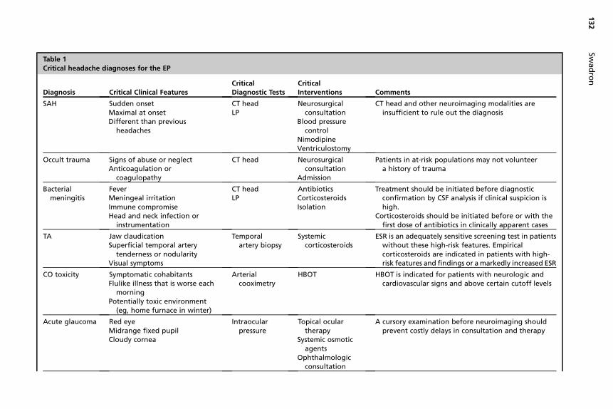
Table 1Critical headache diagnoses for the EP
Diagnosis Critical Clinical FeaturesCriticalDiagnostic Tests
CriticalInterventions Comments
SAH Sudden onsetMaximal at onsetDifferent than previous
headaches
CT headLP
Neurosurgicalconsultation
Blood pressurecontrol
NimodipineVentriculostomy
CT head and other neuroimaging modalities areinsufficient to rule out the diagnosis
Occult trauma Signs of abuse or neglectAnticoagulation or
coagulopathy
CT head Neurosurgicalconsultation
Admission
Patients in at-risk populations may not volunteera history of trauma
Bacterialmeningitis
FeverMeningeal irritationImmune compromiseHead and neck infection or
instrumentation
CT headLP
AntibioticsCorticosteroidsIsolation
Treatment should be initiated before diagnosticconfirmation by CSF analysis if clinical suspicion ishigh.
Corticosteroids should be initiated before or with thefirst dose of antibiotics in clinically apparent cases
TA Jaw claudicationSuperficial temporal artery
tenderness or nodularityVisual symptoms
Temporalartery biopsy
Systemiccorticosteroids
ESR is an adequately sensitive screening test in patientswithout these high-risk features. Empiricalcorticosteroids are indicated in patients with high-risk features and findings or a markedly increased ESR
CO toxicity Symptomatic cohabitantsFlulike illness that is worse each
morningPotentially toxic environment
(eg, home furnace in winter)
Arterialcooximetry
HBOT HBOT is indicated for patients with neurologic andcardiovascular signs and above certain cutoff levels
Acute glaucoma Red eyeMidrange fixed pupilCloudy cornea
Intraocularpressure
Topical oculartherapy
Systemic osmoticagents
Ophthalmologicconsultation
A cursory examination before neuroimaging shouldprevent costly delays in consultation and therapy
Swad
ron
132

Cervical arterydissection
SAH-like onsetFacial (carotid), neck (vertebral)
painCranial nerve abnormalities
Angiography Neurologic/neurosurgicalconsultation
Anticoagulation
In the absence of brain hemorrhage, anticoagulation isinitiated to reduce the risk of thrombus formationand embolization
Cerebral/duralVST
Hypercoagulable state (pregnancyand puerperum, oralcontraceptives, malignancy)
Head and neck infectionProptosis (cavernous sinus
thrombosis)
MR headVenography
Neurosurgicalconsultation
Systemicanticoagulation
A D-dimer may be falsely negative
Space-occupyinglesion
Progressively worse over timeNew onset in patient >50 years oldHistory of malignancyWorse in morningWorse in head-down position
CT head Neurosurgicalconsultation
ICP-loweringtherapies
Lesion-specifictherapies
Emergent ICP-lowering therapies may include elevatingthe head of the bed, restriction of intravenous fluids,mannitol, and hyperventilation
Lesion-specific therapies may include emergent surgery/neuroradiological procedures, corticosteroids, andantimicrobial agents
Cerebellarinfarction
Headache with dizzinessCerebellar signsCranial nerve abnormalities
CT head Neurologic/neurosurgicalconsultation
Although CT head is insensitive for infarction, it ishelpful initially to rule out hemorrhage and identifylife-threatening edema and mass effect
Idiopathicintracranialhypertension
Obese, young female patientCranial nerve 6 palsy (false
localizing sign)
LP CSF drainageNeurologic
referral
After negative neuroimaging, an LP will reveala markedly increased opening pressure and providetemporary headache relief
Pituitaryapoplexy
Thunderclap headacheVomitingVisual acuity, field deficitsOcular palsies
CT headMR head
Neurosurgicalconsultation
Many pituitary infarctions and hemorrhages will not beeasily visible on CT. MR is considered the diagnosticmodality of choice
Preeclampsia Postpartum(up to 4 weeks)
Complete bloodcount
Chemistry panelwith Liverfunction tests
Coagulationstudies
Intravenousmagnesium
Obstetricconsultation
Up to half of all patients present in the postpartumperiod, the majority with a chief complaint ofheadache
Abbreviations: CSF, cerebrospinal fluid; CT, computed tomography; ESR, erythrocyte sedimentation rate; HBOT, hyperbaric oxygen therapy; ICP, intracranial pres-sure; LP, lumbar puncture; MR, magnetic resonance; TA, temporal arteritis; VST, venous sinus thrombosis.
Pitfa
llsin
the
Man
ag
em
en
to
fH
ead
ach
e133
Swadron134
and lowering intraocular pressure with topical and systemic therapy. Patients withacute angle-closure glaucoma will have ocular findings such as a unilateral red eyewith decreased visual acuity, a fixed, midrange pupil, and corneal edema. A cursoryexamination before diagnostic studies that take the patient out of the ED areperformed will prevent delays to critical interventions.
Space-occupying lesions include primary and secondary neoplasms, infectiousprocesses such as brain abscesses and cysts, and vascular lesions such as unrup-tured giant cerebral aneurysms and arteriovenous malformations. Although this isa heterogeneous group of pathologies, headaches caused by space-occupyinglesions share common features in their presentation; the sine qua non being a progres-sive and unremitting course as the lesion expands in volume and increases intracranialpressure (ICP). Headaches also tend to be worse with the head-down position, and inthe morning after waking, when ICP is higher. The specific location of the lesion and itsadjacent structures within the calvarium will determine other features of the presenta-tion. Whether or not it is critical for the EP to uncover the presence of a space-occu-pying lesion differs from case to case. In the absence of neurologic findings onexamination, a patient with a slowly progressive headache of several weeks’ ormonths’ duration may be appropriately discharged from the ED for urgent neuroimag-ing and follow-up in a primary care setting. On the other hand, patients at high risk forpathology, such as those with a history of malignancy, recent head and neck infectionor surgery, immune compromise, or those more than 50 years of age with a new onsetof headache, often receive neuroimaging in the ED. The discovery of a large lesion, ora lesion in a critical location, will sometimes lead to the initiation of emergency thera-pies to prevent further expansion, brain injury, and herniation. If neuroimaging is per-formed in the ED, CT is usually sufficient; space-occupying lesions that are onlydetectable on MR are unlikely to alter ED management.
Cerebral and dural VST is another rare, but life-threatening and treatable, cause ofheadache. The characteristics of the headache are variable, but some patientspresent with a thunderclap. Patients at risk for VST include those with hypercoagu-lable states, including pregnancy and the postpartum period, oral contraceptiveuse, and the nephrotic syndrome, head and neck infections, malignancies, and vascu-litidies.43–45 Patients may present with signs of increased ICP, such as papilledema,and may continue to be symptomatic despite attempts at analgesia. If untreated,the thrombosis may progress to venous infarction and hemorrhage, which results inneurologic deficits that do not conform to an arterial territorial distribution. In somecases, VST eventually leads to brain herniation and death. Unless there are positivefindings on the neurologic examination, there may be few other clues to compel theEP to pursue this diagnosis, and many cases go undiagnosed on the initialpresentation.46,47
Idiopathic intracranial hypertension (IIH), previously known as pseudotumor cere-bri, is a poorly understood disease with a predilection for obese middle-agedwomen. In these patients, a sustained increase in ICP seems to be related to anobstruction of CSF drainage at the level of the arachnoid granulations. However,in at least some cases coexistent VST is visible on contrast imaging studies, and,in this subgroup, more emergent therapy is necessary to prevent rapid deteriora-tion.48,49 In the absence of macroscopic thrombosis, deterioration is more gradual,with progressive visual-field deterioration occurring if ICP is not reduced. The phys-ical examination most often reveals papilledema, and may reveal loss of peripheralvisual fields. In some cases, a unilateral or bilateral sixth cranial nerve palsy ispresent. This palsy is a result of increased ICP, not a localized process, and thusis considered a false localizing sign. The diagnosis of IIH should be suspected in
Pitfalls in the Management of Headache 135
patients with a headache pattern consistent with a space-occupying lesion whohave negative neuroimaging studies. Although the diagnosis is not as time criticalas with VST, it is strongly suggested by the finding of a grossly increased openingpressure during lumbar puncture (LP) that is accompanied by an immediateimprovement in the patient’s symptoms.
Pituitary apoplexy is an extremely rare cause of thunderclap headache. It is definedas hemorrhage or infarction of the pituitary gland, typically into a preexisting adenoma.Although headache may be the most prominent presenting complaint, pituitaryapoplexy is most often accompanied by visual symptoms and signs, such asdecreased acuity, reduction in visual fields, and ocular palsies.50
PITFALLS OF DIAGNOSTIC TESTINGRelying on Neuroimaging to Rule out SAH
Despite advances in CT technology, noncontrast CT imaging alone remains inade-quate to rule out nontraumatic SAH. In all but a single published case series,51 anLP was required after a negative CT scan to make the diagnosis of SAH in a substantialminority of cases.52–58 Although reported sensitivities in these case series typicallyexceed 90%, in practice, the sensitivity of CT for SAH is likely significantly lower, forseveral reasons. First and foremost is the critical issue of spectrum bias. Most studiesbegin their analysis with a group of patients ultimately diagnosed with SAH in hospital,potentially missing patients who are discharged from the ED and other outpatientsettings with less severe presentations. These are precisely the patients in whoma timely diagnosis of SAH is so important to prevent a second, more severe bleed.In addition, most studies are conducted at referral centers, at which the equipmentand the expertise of the radiologists are optimal. The ability of a general radiologistto detect small amounts of hemorrhage on CT is known to be inferior to that ofa subspecialist neuroradiologist.59 Lastly, small amounts of subarachnoid blood, afteran initial leak, are rapidly absorbed; following the initial 12-hour interval after symptomonset, the sensitivity of CT decreases over time.60
Other imaging modalities, including MR, MR angiography, CT angiography, andconventional angiography, have not eliminated the need for LP with CSF analysis incases of suspected SAH. MR is less sensitive than CT for the presence of blood inthe first several hours after the onset of bleeding.30 Even the addition of the fluid-atten-uated inversion-recovery (FLAIR) technique, with its enhanced ability to detect blood,is insufficient. In a recent study of 12 patients with a negative CT scan who subse-quently had SAH confirmed by CSF analysis, MR was only able to detect bleedingin 2.61
Although these technologies are continuing to evolve and improve, conventionalangiography, CT angiography, and MR angiography are not 100% sensitive for thedetection of cerebral aneurysms.62–65 More importantly, angiography by any methodis unable to distinguish between unruptured, asymptomatic aneurysms, which havean exceedingly low likelihood of rupture throughout a patient’s lifetime, and symptom-atic aneurysms that have already bled, and are likely to rebleed with serious conse-quences.66,67 Thus, an LP with CSF analysis to confirm SAH can be critical todetermine the need for surgical intervention when aneurysms are detected on angiog-raphy. Because the overall prevalence of cerebral aneurysms in the general populationranges from 2% to 6%,67 the indiscriminate application of angiography to an unse-lected population of patients with headaches could result in unnecessary and harmfulinvasive procedures.
Swadron136
Other Limitations of CT
CT is insensitive to detect VST. Although data are lacking to precisely estimate itssensitivity, in one consecutive series of 127 patients with VST, 17 patients whopresented with isolated headache had normal brain CT scan and CSF examination.46
The addition of contrast to CT will help identify some cases, but the diagnosis can onlybe ruled out using MR venography.68 However, because of the rarity of this diagnosis,it is an unreasonable expectation for MR venography to be performed emergently inpatients without neurologic findings unless risk factors and a strong clinical suspicionexist for VST.
Cerebellar infarction, like cerebral infarction, may not become apparent on CT scanfor several hours. However, cerebellar infarction deserves special consideration by theEP because it is more likely to present as a headache without weakness or other local-izing signs on neurologic examination.69 Moreover, because of its location in theposterior fossa, there is a greater risk for brain herniation as the lesion evolves andedema ensues. Although CT is less sensitive than MR to visualize the contents ofthe posterior fossa, edema from cerebellar infarction is often visible. Distortion or oblit-eration of the fourth ventricle from the resultant mass effect should prompt neurosur-gical consultation for possible decompression. Pituitary apoplexy, by contrast, isfrequently not visualized on CT, and MR is advised if clinical suspicion for this entityexists.50
Misinterpreting CSF Results
LP with CSF examination is critical in the evaluation of headaches suspicious for SAHor meningitis. In both instances, there are several common pitfalls that may interferewith a correct diagnosis. Within hours of the onset of subarachnoid bleeding, redblood cells (RBCs) are detectable in large numbers throughout the circulating CSF.In up to 15% of cases, LPs are traumatic, and RBCs from epidural vessels contami-nate the specimen, making it difficult to identify a true SAH.70 A common misunder-standing is that a progressive decrease in RBCs across serial collection tubeseliminates the possibility of SAH. Because SAH may coexist with RBCs that arisefrom a traumatic LP, the possibility of SAH can only safely be eliminated if the CSFcount in one of the tubes approaches zero. If blood is encountered at the beginningof the LP, wasting the first 2 or 3 mL of fluid as the CSF clears will increase the likeli-hood that the RBC count will approach zero. If it does not, it may be necessary torepeat the procedure at a different interspace.
In cases in which traumatic LP makes the interpretation of RBC counts difficult, thepresence of xanthochromia has been used to confirm the presence of true SAH. Xan-thochromia is the yellowish discoloration of CSF that occurs in the hours followingSAH as RBCs break down in vivo into bilirubin and oxyhemoglobin. However, xantho-chromia has also been demonstrated to occur in vitro in collected specimens, result-ing in falsely positive results.71 Moreover, the practice of waiting until 12 hours haveelapsed following the onset of headache, until the xanthochromia that develops invivo is more reliably present, is not advised. Any advantage of this technique regardingthe interpretation of CSF specimens is outweighed by the risk of a second aneurysmalbleed, which occurs more frequently in the hours immediately following the initialbleed than in any time period thereafter.72 Almost all laboratories in the United Statesmeasure xanthochromia by visual inspection after centrifugation of the CSF specimen.Using this technique, falsely negative results are also possible.73 Nonetheless, inpatients who present several days after the onset of headache pain, xanthochromia
Pitfalls in the Management of Headache 137
may be the only remaining sign of SAH on CSF analysis; it typically persists for2 weeks.74
Although a negative LP effectively rules out the diagnosis of SAH,75 it does not ruleout other vascular emergencies that may result in a thunderclap headache, such ascervical artery dissection, cerebral and dural VST, cerebellar infarction, and pituitaryapoplexy.
There are other important pitfalls of CSF interpretation in patients with a suspectedcentral nervous system infection. It may be difficult to distinguish between bacterialand viral meningitis using the initial results of CSF analysis available in the ED.Although bacterial meningitis is more likely to result in high cell counts with a prepon-derance of polymorphonuclear leukocytes, a low glucose, and a positive Gram stain,none of these features is reliably present, and there is a significant overlap in CSF find-ings with viral meningitis. If sufficient clinical suspicion exists for bacterial meningitis,the most prudent course is to proceed with broad-spectrum antibiotic coverage untilthe results of bacterial cultures are available.
Certain populations, such as infants, the elderly, and those with immune compro-mise, may have a limited cellular response on initial CSF analysis. Moreover, inpatients with suspected immune compromise, atypical pathogens, such as Crypto-coccus neoformans and mycobacteria should be considered. Specific tests for thesepathogens are necessary, and the collection of an additional tube of CSF for later useby the inpatient physician may be helpful.
In a traumatic LP, leukocytes should be present in small numbers in a fixed ratio withRBCs, typically in the range of 1 leukocyte for every 500 RBCs. This ratio may vary de-pending on the relative proportions of these cells in the peripheral blood. Althoughthere is some evidence to support using such ratios to rule out meningitis in a traumatictap, in the presence of large numbers of leukocytes and a high clinical suspicion formeningitis, it is advisable to repeat the LP at a different interspace.76 It should alsobe noted that the most important treatable cause of viral encephalitis, herpes simplexencephalitis (HSE), may present with red cells and leukocytes in the CSF.77,78
Complications of LP
The most common complication of LP is a postural headache that can be persistentand debilitating, often resulting in repeat visits to the ED. These headaches occur inup to one-third of patients in some series, and usually occur within 3 days of the proce-dure. The headache is typically described as worse in the upright position, forcingpatients to lie down; this is consistent with the theory that it is due to ongoing CSFleakage at the site of puncture and the resultant intracranial hypotension. Many ofthe traditional instructions given to patients to prevent post-LP headaches, includingpostprocedural bed rest, increased fluid intake, and caffeine administration, have littleor no basis in evidence.79,80 There is, however, sufficient evidence to support the prac-tice of using a small (22 gauge), atraumatic (noncutting tip) needle to prevent post-LPheadache.81,82 Epidural blood patch, the injection of 5 to 30 mL of the patient’s ownblood into the epidural space at the site of the prior procedure, has been found tobe an effective treatment of post-LP headache refractory to other therapies.83
The most feared complication of LP is brain herniation. Although it is rare, a strongtemporal correlation between the procedure and subsequent herniation lends strengthto a causal relationship.84 Most investigators recommend performing a CT scanbefore LP to minimize this risk. Although a normal CT scan does not completely elim-inate the risk of brain herniation with LP, in patients with mass lesions or other struc-tural changes, an LP is not advised. Although there may be a small increase in the riskof deterioration or herniation from an unsuspected lesion, it may be acceptable to omit

Swadron138
the CT scan in selected patients with headache, such as those less than 60 years ofage with no significant neurologic history and a normal neurologic examination.85,86
Other Laboratory Pitfalls
CO cooximetry values may be misleading. Low or undetectable levels may not rule outCO poisoning in the patient who presents many hours after exposure, as CO clearsfrom the blood over time. Even in toxic patients with high tissue levels that have accu-mulated over a long period of exposure, the administration of high-flow supplementaloxygen will accelerate clearance from the blood and may lead to levels in the normal ornear-normal range.34,37 If the diagnosis is strongly suspected, removal of the patientfrom the suspected toxic environment, and repeat testing with any recurrentsymptoms, is advised.
Although erythrocyte sedimentation rate (ESR) is an invaluable screening test for TAin older adult patients with headache, in the presence of an extremely high pretestprobability it is inadequately sensitive to rule out the diagnosis. Thus, in patientswith jaw claudication, nodularity or tenderness over the superficial temporal artery,or diploplia, the diagnostic evaluation should not end with a normal, or only mildlyincreased, ESR. In patients with such suggestive clinical features, or those with lessspecific features but a highly increased ESR, empirical corticosteroids should beadministered in the interval before a definite diagnostic determination is made withtemporal artery biopsy.39
The dimerized plasmin fragment D (D-dimer) test may help to identify some patientswith VST. However, its lack of specificity has been well documented, most notably inpatients with identified risk factors for VST, such as women in the peripartum period,and patients with malignancy, vasculitis, and other chronic inflammatory conditions.87
D-dimer is also falsely negative in a significant number of cases of VST diagnoseddefinitely with contrast neuroimaging studies, especially in patients who presentwith isolated headache.88–90
PITFALLS OF TREATMENTPoor Analgesic Agent Choices
Several investigators have been critical of the patterns of analgesic use for headachein the ED setting. One criticism is directed at the lack of specificity of the agents usedwith respect to the underlying diagnoses, especially for migraine. Another is in regardto the frequency of opioid use, which is said to be too high and inconsistent with pub-lished consensus guidelines.3,91,92 Many agents have been proven to be highly effec-tive for acute headache in the ED, including migraine, and, in any given patient, theremay be several acceptable choices. One of the key factors for the EP is the patient’sprevious response to the various agents available.
The dopamine antagonists, including droperidol, prochlorperazine, metochlopra-mide, and chlorpromazine, have a less well-delineated mechanism of action thanthe more migraine-specific DHE and triptans, which activate serotonin 1B/1D recep-tors. Nonetheless, these agents, when used alone93–99 or in combination with otherdrugs,100 have been shown to be equivalent or superior to drugs from all other classesin multiple controlled trials. Droperidol seems to be more efficacious than prochlorper-azine,101,102 which in turn seems to be more efficacious as a single agent than meto-chlopramide.103,104 All agents are more effective when they reach the central nervoussystem quickly, with intravenous administration superior to the intramuscular and oralroutes.104,105

Pitfalls in the Management of Headache 139
Although not as effective overall as the dopamine antagonists, opioids are alsosuccessful at abating headache. Although they may be used in some cases for whichother options are available, the treatment guidelines set forth by nonemergency medi-cine societies may not be an appropriate standard against which to evaluate ED prac-tices. The patient population in the ED is unique. The more severe spectrum of acuityin the ED, the lower likelihood of diagnostic precision, and the fact that many patientshave already tried 1 or more medications before being evaluated, all suggest thatdifferent practice patterns are to be expected. Nonetheless, wide regional differencesamong EPs, and the discrepancy between their responses to hypothetical scenariosand actual practices observed, indicate that other factors also play a role.106,107
Patients who visit the ED frequently with a chief complaint of headache are oftenstigmatized by EPs and ED staff. These patients, who make up a larger proportionof patients in the ED than in office-based and specialty clinic settings are often labeledas drug-seekers.18 They often request specific medications and are more frequentlyprescribed opioids. They also have a higher frequency of chronic headaches, anda large proportion use symptomatic headache therapies on a daily or almost dailybasis, especially combination preparations containing opioids and caffeine,a syndrome known as medication overuse headache. Higher rates of opioid use inthe ED may reflect, in part, the refractoriness of such patients to other therapies.18
With so many efficacious therapies available, pitfalls are more commonly related tomedication side effects. The EP must therefore be mindful of a few key contraindica-tions and complications of the commonly used agents. The dopamine antagonists arenotable for prolonging the QT interval on the electrocardiogram (ECG). Moreover, it isnot uncommon for 2 agents to be used that each cause some degree of QT prolonga-tion, such as ondansetron for nausea, combined with a dopamine antagonist.Although this is rarely of clinical consequence, in patients with preexisting conductionor electrolyte abnormalities, or those who are already receiving therapy with otheragents that prolong the QT interval, it can sometimes lead to torsades de pointes(TdP), a potentially fatal dysrhythmia. Women, who are disproportionately representedamong patients with migraine headache, are at higher risk than men.108 The relation-ship between QT-prolonging agents and TdP is unclear, and seems to be nonlinear.Agents that cause only minimal prolongation of the QT interval may cause TdP athigher rates than those that cause more marked prolongation.108 Because patientswith cardiac or electrolyte abnormalities are known to be at higher risk, it may beprudent to avoid dopamine antagonists in these patients, or to monitor them withtelemetry or serial ECGs during therapy.
The other, more common but less life-threatening, concern with the dopamineantagonists is akathisia, an uncomfortable sensation of not being able to sit still. Itoccurs with varying severity in a substantial minority of patients.109 In general, it hasnot limited the ability of this class of agents to be effective, and when severe, it is reli-ably abated by the use of anticholinergic agents such as diphenhydramine.109 Theincidence of akathisia may be decreased by the use of slower infusion rates,110 orby using an anticholinergic premedication.111
The triptans offer greater selectivity for serotonin receptors, and are available foradministration by oral and subcutaneous routes. They have largely replaced DHE inthe primary care and neurology specialty clinic settings. However, DHE is still in usein many EDs, possibly because of its low cost and availability as an intravenous prep-aration. Most investigators recommend avoiding DHE in patients with cardiovasculardisease, uncontrolled hypertension, and in pregnancy.112 Although triptans have beenshown to be more efficacious than DHE,113 many EPs have lingering concerns abouttheir cardiovascular risk, and are concerned about the frequent occurrence of chest
Swadron140
pain and pressure that occurs after their administration. Although there is evidencethat these symptoms are not cardiac related,114 coronary vasospasm has been docu-mented in the catheterization laboratory with the administration of triptans in patientswith diseased coronary arteries.115 Moreover, most subjects studied to establish thesafety of triptans have been those without evidence of cardiac disease. It is thus rec-ommended that these agents be avoided in patients with a history of coronary arterydisease.
Withholding Empirical Antibiotics and Steroids in Suspected Meningitis
In patients clinically suspected of having bacterial meningitis, such as those whoappear ill and are febrile with signs of meningeal irritation, broad-spectrum antibioticsshould not be withheld pending the results of diagnostic studies. Bacterial meningitisprogresses rapidly, and outcomes are most likely related to the time to antibioticadministration in patients with overt presentations.116–118 Moreover, a short intervalof antibiotics before LP is unlikely to obscure the diagnosis on CSF analysis.119
Corticosteroids have also been shown to reduce mortality and improve outcomes inpatients with bacterial meningitis when they are administered before, or concurrently,with antibiotics. Their empirical administration should similarly be considered inpatients in whom bacterial meningitis is strongly suspected.120,121 Because manypatients in the United States receive empirical antibiotics before CSF examinationbased on a low or moderate clinical suspicion, coadministering a large dose of corti-costeroids in all of these patients may expose the majority, who do not have menin-gitis, to unnecessary risks. There are limited data to address this question; the bestapproach may be to limit empirical steroid administration to those patients in whommeningitis is most strongly suspected.
After CSF results become available, additional antimicrobial therapy may be indi-cated to cover specific organisms and possible drug-resistant strains, especially inspecial at-risk populations.122,123 Acyclovir should be added to the empirical treat-ment regimen for patients with CSF pleocytosis and a negative Gram stain until theresults of more definitive tests are available, such as the polymerase chain reactiontest for the herpes simplex virus.77
PITFALLS OF DISCHARGE AND FOLLOW-UP MANAGEMENTPoor Discharge Planning
Although many patients leave the ED pain free, recurrence of headache in the dayfollowing the ED visit is a common problem occurring in most discharged patientswith a diagnosis of migraine in one study,124 and in almost one-third of patients withprimary headaches in another.125 It is important to consider the patient’s previoushistory of headache recurrence during discharge, as repeat ED visits can be avoidedin many cases. Risk factors associated with recurrence of moderate to severe painand functional impairment within 24 hours of discharge include incomplete resolutionat discharge,124,126 severe baseline pain, a longer duration of pain, baseline nausea,and a positive screen for depression.125 In patients who obtain headache relief withintramuscular sumatriptan, discharge with an oral dose, to be used in the event ofrecurrence, is appropriate.127 In other patients, the use of nonsteroidal antiinflamma-tory agents, acetaminophen, aspirin, or oral antiemetics at the first sign of recurrencemay be helpful. Several randomized, controlled studies have looked at the role of oralor intravenous dexamethasone to reduce the rate of headache recurrence. Althoughseveral of these studies were negative,126,128–130 a recent meta-analysis131 found anoverall benefit of parenteral dexamethasone at 72 hours posttreatment. Although it
Pitfalls in the Management of Headache 141
is difficult to recommend the routine use of corticosteroids, they seem to have a role,and may be especially useful in those patients with a headache of greater than 72hours duration before presentation.130
Patients should be given follow-up care with a primary care provider. In certaincases that present a diagnostic or therapeutic challenge, neurologist or headachespecialist follow-up may also be warranted. It is important for patients to be instructedto return to the ED if their symptoms worsen or if new symptoms evolve; many of thedangerous diagnoses discussed earlier may initially present as an isolated headachebut go on to develop additional symptoms and signs as they progress.
Patients who visit the ED frequently with exacerbations of primary headachepresent a special challenge. Although there are few data to describe successful strat-egies for their ED management, consistency in their care among EPs and other EDstaff, and regular communication among EPs, primary care providers, and specialists,may improve their outcomes.
REFERENCES
1. McCaig LF, Burt CW. National Hospital Ambulatory Medical Care Survey: 2002emergency department summary. Adv Data 2004;340:1.
2. Headache Classification Subcommittee of the International Headache Society.The International Classification of Headache Disorders: 2nd edition. Cephalal-gia 2004;24(Suppl 1):9.
3. Sahai-Srivastava S, Desai P, Zheng L. Analysis of headache management ina busy emergency room in the United States. Headache 2008;48:931.
4. Maizels M. Headache evaluation and treatment by primary care physicians in anemergency department in the era of triptans. Arch Intern Med 1969;161:2001.
5. Blumenthal HJ, Weisz MA, Kelly KM, et al. Treatment of primary headache in theemergency department. Headache 2003;43:1026.
6. Miner JR, Smith SW, Moore J, et al. Sumatriptan for the treatment of undifferenti-ated primary headaches in the ED. Am J Emerg Med 2007;25:60.
7. Trainor A, Miner J. Pain treatment and relief among patients with primary headachesubtypes in the ED. Am J Emerg Med 2008;26:1029.
8. Friedman BW, Hochberg ML, Esses D, et al. Applying the International Classifi-cation of Headache Disorders to the emergency department: an assessment ofreproducibility and the frequency with which a unique diagnosis can beassigned to every acute headache presentation. Ann Emerg Med 2007;49:409.
9. Fiesseler FW, Riggs RL, Holubek W, et al. Canadian Headache Society criteriafor the diagnosis of acute migraine headache in the ED – do our patients meetthese criteria? Am J Emerg Med 2005;23:149.
10. Pope JV, Edlow JA. Favorable response to analgesics does not predict a benignetiology of headache. Headache 2008;48:944.
11. Pfadenhauer K, Schonsteiner T, Keller H. The risks of sumatriptan administrationin patients with unrecognized subarachnoid haemorrhage (SAH). Cephalalgia2006;26:320.
12. Lipton RB, Mazer C, Newman LC, et al. Sumatriptan relieves migrainelike headachesassociated with carbon monoxide exposure. Headache 1997;37:392.
13. Abisaab J, Nevadunsky N, Flomenbaum N. Emergency department presenta-tion of bilateral carotid artery dissections in a postpartum patient. Ann EmergMed 2004;44:484.
14. Leira EC, Cruz-Flores S, Leacock RO, et al. Sumatriptan can alleviate head-aches due to carotid artery dissection. Headache 2001;41:590.
Swadron142
15. Prokhorov S, Khanna S, Alapati D, et al. Subcutaneous sumatriptan relievedmigraine-like headache in two adolescents with aseptic meningitis. Headache2008;48:1235.
16. Rosenberg JH, Silberstein SD. The headache of SAH responds to sumatriptan.Headache 2005;45:597.
17. Barclay CL, Shuaib A, Montoya D, et al. Response of non-migrainousheadaches to chlorpromazine. Headache 1990;30:85.
18. Maizels M. Health resource utilization of the emergency department headache‘‘repeater’’. Headache 2002;42:747.
19. Friedman BW, Serrano D, Reed M, et al. Use of the emergency department forsevere headache. A population-based study. Headache 2009;49:21.
20. Katsarava Z, Rabe K, Diener HC. From migraine to stroke. Intern Emerg Med2008;3(Suppl 1):S9.
21. Karras DJ, Ufberg JW, Harrigan RA, et al. Lack of relationship between hyper-tension-associated symptoms and blood pressure in hypertensive ED patients.Am J Emerg Med 2005;23:106.
22. Mitchell CS, Osborn RE, Grosskreutz SR. Computed tomography in theheadache patient: is routine evaluation really necessary? Headache 1993;33:82.
23. Reinus WR, Wippold FJ 2nd, Erickson KK. Practical selection criteria for non-contrast cranial computed tomography in patients with head trauma. Ann EmergMed 1993;22:1148.
24. Diaz M, Braude D, Skipper B. Concordance of historical questions used in risk-stratifying patients with headache. Am J Emerg Med 2007;25:907.
25. Harling DW, Peatfield RC, Van Hille PT, et al. Thunderclap headache: is itmigraine? Cephalalgia 1989;9:87.
26. Lledo A, Calandre L, Martinez-Menendez B, et al. Acute headache of recent onsetand subarachnoid hemorrhage: a prospective study. Headache 1994;34:172.
27. Edlow JA, Panagos PD, Godwin SA, et al. Clinical policy: critical issues in theevaluation and management of adult patients presenting to the emergencydepartment with acute headache. Ann Emerg Med 2008;52:407.
28. Vannemreddy P, Nanda A, Kelley R, et al. Delayed diagnosis of intracranialaneurysms: confounding factors in clinical presentation and the influence ofmisdiagnosis on outcome. South Med J 2001;94:1108.
29. Polmear A. Sentinel headaches in aneurysmal subarachnoid haemorrhage:what is the true incidence? A systematic review. Cephalalgia 2003;23:935.
30. Hesselink JR, Dowd CF, Healy ME, et al. MR imaging of brain contusions:a comparative study with CT. AJR Am J Roentgenol 1988;150:1133.
31. Thomas KE, Hasbun R, Jekel J, et al. The diagnostic accuracy of Kernig’s sign,Brudzinski’s sign, and nuchal rigidity in adults with suspected meningitis. ClinInfect Dis 2002;35:46.
32. Attia J, Hatala R, Cook DJ, et al. The rational clinical examination. Does this adultpatient have acute meningitis? JAMA 1999;282:175.
33. Uchihara T, Tsukagoshi H. Jolt accentuation of headache: the most sensitivesign of CSF pleocytosis. Headache 1991;31:167.
34. Kao LW, Nanagas KA. Carbon monoxide poisoning. Emerg Med Clin North Am2004;22:985.
35. Centers for Disease Control and Prevention (CDC). Unintentional non-fire-related carbon monoxide exposures – United States, 2001–2003. MMWRMorb Mortal Wkly Rep 2005;54:36.
36. Salameh S, Amitai Y, Antopolsky M, et al. Carbon monoxide poisoning inJerusalem: epidemiology and risk factors. Clin Toxicol (Phila) 2009;47:137.

Pitfalls in the Management of Headache 143
37. Keles A, Demircan A, Kurtoglu G. Carbon monoxide poisoning: how manypatients do we miss? Eur J Emerg Med 2008;15:154.
38. Hellmann DB. Temporal arteritis: a cough, toothache, and tongue infarction.JAMA 2002;287:2996.
39. Smetana GW, Shmerling RH. Does this patient have temporal arteritis? JAMA2002;287:92.
40. Schievink WI. Spontaneous dissection of the carotid and vertebral arteries.N Engl J Med 2001;344:898.
41. Saeed AB, Shuaib A, Al-Sulaiti G, et al. Vertebral artery dissection: warningsymptoms, clinical features and prognosis in 26 patients. Can J Neurol Sci2000;27:292.
42. Matthys LA, Coppage KH, Lambers DS, et al. Delayed postpartum preeclampsia:an experience of 151 cases. Am J Obstet Gynecol 2004;190:1464.
43. Cantu C, Barinagarrementeria F. Cerebral venous thrombosis associated withpregnancy and puerperium. Review of 67 cases. Stroke 1993;24:1880.
44. Akatsu H, Vaysburd M, Fervenza F, et al. Cerebral venous thrombosis innephrotic syndrome. Clin Nephrol 1997;48:317.
45. Ferro JM, Canhao P, Stam J, et al. Prognosis of cerebral vein and dural sinusthrombosis: results of the International Study on Cerebral Vein and Dural SinusThrombosis (ISCVT). Stroke 2004;35:664.
46. Cumurciuc R, Crassard I, Sarov M, et al. Headache as the only neurological signof cerebral venous thrombosis: a series of 17 cases. J Neurol NeurosurgPsychiatr 2005;76:1084.
47. Beeson MS, Vesco JA, Reilly BA, et al. Dural sinus thrombosis. Am J Emerg Med2002;20:568.
48. Lin A, Foroozan R, Danesh-Meyer HV, et al. Occurrence of cerebral venoussinus thrombosis in patients with presumed idiopathic intracranial hypertension.Ophthalmology 2006;113:2281.
49. Ooi LY, Walker BR, Bodkin PA, et al. Idiopathic intracranial hypertension: canstudies of obesity provide the key to understanding pathogenesis? Br J Neurosurg2008;22:187.
50. Randeva HS, Schoebel J, Byrne J, et al. Classical pituitary apoplexy: clinicalfeatures, management and outcome. Clin Endocrinol (Oxf) 1999;51:181.
51. Boesiger BM, Shiber JR. Subarachnoid hemorrhage diagnosis by computedtomography and lumbar puncture: are fifth generation CT scanners better atidentifying subarachnoid hemorrhage? J Emerg Med 2005;29:23.
52. Byyny RL, Mower WR, Shum N, et al. Sensitivity of noncontrast cranial computedtomography for the emergency department diagnosis of subarachnoid hemorrhage.Ann Emerg Med 2008;51:697.
53. van der Wee N, Rinkel GJ, Hasan D, et al. Detection of subarachnoid haemorrhageon early CT: is lumbar puncture still needed after a negative scan? J Neurol Neuro-surg Psychiatr 1995;58:357.
54. Sames TA, Storrow AB, Finkelstein JA, et al. Sensitivity of new-generationcomputed tomography in subarachnoid hemorrhage. Acad Emerg Med 1996;3:16.
55. Foot C, Staib A. How valuable is a lumbar puncture in the management ofpatients with suspected subarachnoid haemorrhage? Emerg Med (Fremantle)2001;13:326.
56. Morgenstern LB, Luna-Gonzales H, Huber JC Jr, et al. Worst headache andsubarachnoid hemorrhage: prospective, modern computed tomography andspinal fluid analysis. Ann Emerg Med 1998;32:297.
Swadron144
57. O’Neill J, McLaggan S, Gibson R. Acute headache and subarachnoid haemor-rhage: a retrospective review of CT and lumbar puncture findings. Scott Med J2005;50:151.
58. Sidman R, Connolly E, Lemke T. Subarachnoid hemorrhage diagnosis: lumbarpuncture is still needed when the computed tomography scan is normal.Acad Emerg Med 1996;3:827.
59. Schriger DL, Kalafut M, Starkman S, et al. Cranial computed tomographyinterpretation in acute stroke: physician accuracy in determining eligibility forthrombolytic therapy. JAMA 1998;279:1293.
60. van Gijn J, van Dongen K. The time course of aneurysmal haemorrhage oncomputed tomograms. Neuroradiology 1982;23:153–6.
61. Mohamed M, Heasly DC, Yagmurlu B, et al. Fluid-attenuated inversion recoveryMR imaging and subarachnoid hemorrhage: not a panacea. AJNR Am J Neuro-radiol 2004;25:545.
62. El Khaldi M, Pernter P, Ferro F, et al. Detection of cerebral aneurysms in nontrau-matic subarachnoid haemorrhage: role of multislice CT angiography in 130consecutive patients. Radiol Med 2007;112:123.
63. White PM, Wardlaw JM, Easton V. Can noninvasive imaging accurately depictintracranial aneurysms? A systematic review. Radiology 2000;217:361.
64. Chappell ET, Moure FC, Good MC. Comparison of computed tomographicangiography with digital subtraction angiography in the diagnosis of cerebralaneurysms: a meta-analysis. Neurosurgery 2003;52:624.
65. Johnson MR, Good CD, Penny WD, et al. Lesson of the week: playing the oddsin clinical decision making: lessons from berry aneurysms undetected bymagnetic resonance angiography. BMJ 2001;322:1347.
66. Wiebers DO. Unruptured intracranial aneurysms: natural history and clinicalmanagement. Update on the international study of unruptured intracranialaneurysms. Neuroimaging Clin N Am 2006;16:383.
67. Rinkel GJ, Djibuti M, Algra A, et al. Prevalence and risk of rupture of intracranialaneurysms: a systematic review. Stroke 1998;29:251.
68. Cortez O, Schaeffer CJ, Hatem SF, et al. Cases from the Cleveland Clinic: cere-bral venous sinus thrombosis presenting to the emergency department withworst headache of life. Emerg Radiol 2009;16:79.
69. Savitz SI, Caplan LR, Edlow JA. Pitfalls in the diagnosis of cerebellar infarction.Acad Emerg Med 2007;14:63.
70. Shah KH, Richard KM, Nicholas S, et al. Incidence of traumatic lumbar punc-ture. Acad Emerg Med 2003;10:151.
71. Graves P, Sidman R. Xanthochromia is not pathognomonic for subarachnoidhemorrhage. Acad Emerg Med 2004;11:131.
72. Naidech AM, Janjua N, Kreiter KT, et al. Predictors and impact of aneurysmrebleeding after subarachnoid hemorrhage. Arch Neurol 2005;62:410.
73. Sidman R, Spitalnic S, Demelis M, et al. Xanthrochromia? By what method? Acomparison of visual and spectrophotometric xanthrochromia. Ann EmergMed 2005;46:51.
74. EdlowJA,CaplanLR.Avoidingpitfalls in thediagnosisof subarachnoidhemorrhage.N Engl J Med 2000;342:29.
75. Perry JJ, Spacek A, Forbes M, et al. Is the combination of negative computedtomography result and negative lumbar puncture result sufficient to rule outsubarachnoid hemorrhage? Ann Emerg Med 2008;51:707.
76. Mazor SS, McNulty JE, Roosevelt GE. Interpretation of traumatic lumbar punctures:who can go home? Pediatrics 2003;111:525.

Pitfalls in the Management of Headache 145
77. Kennedy PG, Chaudhuri A. Herpes simplex encephalitis. J Neurol NeurosurgPsychiatr 2002;73:237.
78. Benson PC, Swadron SP. Empiric acyclovir is infrequently initiated in the emer-gency department to patients ultimately diagnosed with encephalitis. AnnEmerg Med 2006;47:100.
79. Lin W, Geiderman J. Myth: fluids, bed rest, and caffeine are effective in prevent-ing and treating patients with post-lumbar puncture headache. West J Med2002;176:69.
80. Ebinger F, Kosel C, Pietz J, et al. Strict bed rest following lumbar puncture inchildren and adolescents is of no benefit. Neurology 2004;62:1003.
81. Thomas SR, Jamieson DR, Muir KW. Randomised controlled trial of atraumaticversus standard needles for diagnostic lumbar puncture. BMJ 2000;321:986.
82. Halpern S, Preston R. Postdural puncture headache and spinal needle design.Metaanalyses. Anesthesiology 1994;81:1376.
83. Safa-Tisseront V, Thormann F, Malassine P, et al. Effectiveness of epidural bloodpatch in the management of post-dural puncture headache. Anesthesiology2001;95:334.
84. Rennick G, Shann F, de Campo J. Cerebral herniation during bacterial menin-gitis in children. BMJ 1993;306:953.
85. Hasbun R, Abrahams J, Jekel J, et al. Computed tomography of the head beforelumbarpuncture in adults with suspected meningitis.N Engl J Med2001;345:1727.
86. Schull MJ. Lumbar puncture first: an alternative model for the investigation oflone acute sudden headache. Acad Emerg Med 1999;6:131.
87. Haapaniemi E, Tatlisumak T. Is D-dimer helpful in evaluating stroke patients? Asystematic review. Acta Neurol Scand 2009;119:141.
88. Squizzato A, Ageno W. D-dimer testing in ischemic stroke and cerebral sinusand venous thrombosis. Semin Vasc Med 2005;5:379.
89. Misra UK, Kalita J, Bansal V. D-dimer is useful in the diagnosis of cortical venoussinus thrombosis. Neurol India 2009;57:50.
90. Bousser MG, Ferro JM. Cerebral venous thrombosis: an update. Lancet Neurol2007;6:162.
91. Colman I, Rothney A, Wright SC, et al. Use of narcotic analgesics in the emer-gency department treatment of migraine headache. Neurology 2004;62:1695.
92. Gupta MX, Silberstein SD, Young WB, et al. Less is not more: underutilization ofheadache medications in a university hospital emergency department. Headache2007;47:1125.
93. Friedman BW, Corbo J, Lipton RB, et al. A trial of metoclopramide vs sumatriptanfor the emergency department treatment of migraines. Neurology 2005;64:463.
94. Griffith JD, Mycyk MB, Kyriacou DN. Metoclopramide versus hydromorphone forthe emergency department treatment of migraine headache. J Pain 2008;9:88.
95. Ginder S, Oatman B, Pollack M. A prospective study of i.v. magnesium and i.v.prochlorperazine in the treatment of headaches. J Emerg Med 2000;18:311.
96. Tanen DA, Miller S, French T, et al. Intravenous sodium valproate versus prochlor-perazine for the emergency department treatment of acute migraine headaches:a prospective, randomized, double-blind trial. Ann Emerg Med 2003;41:847.
97. Callan JE, Kostic MA, Bachrach EA, et al. Prochlorperazine vs. promethazine forheadache treatment in the emergency department: a randomized controlledtrial. J Emerg Med 2008;35:247.
98. Lane PL, McLellan BA, Baggoley CJ. Comparative efficacy of chlorpromazineand meperidine with dimenhydrinate in migraine headache. Ann Emerg Med1989;18:360.
Swadron146
99. Bell R, Montoya D, Shuaib A, et al. A comparative trial of three agents in thetreatment of acute migraine headache. Ann Emerg Med 1990;19:1079.
100. Colman I, Brown MD, Innes GD, et al. Parenteral metoclopramide for acutemigraine: meta-analysis of randomised controlled trials. BMJ 2004;329:1369.
101. Weaver CS, Jones JB, Chisholm CD, et al. Droperidol vs prochlorperazine forthe treatment of acute headache. J Emerg Med 2004;26:145.
102. Miner JR, Fish SJ, Smith SW, et al. Droperidol vs. prochlorperazine for benignheadaches in the emergency department. Acad Emerg Med 2001;8:873.
103. Coppola M, Yealy DM, Leibold RA. Randomized, placebo-controlled evaluation ofprochlorperazine versus metoclopramide for emergency department treatment ofmigraine headache. Ann Emerg Med 1995;26:541.
104. Jones J, Pack S, Chun E. Intramuscular prochlorperazine versus metoclopra-mide as single-agent therapy for the treatment of acute migraine headache.Am J Emerg Med 1996;14:262.
105. Richman PB, Reischel U, Ostrow A, et al. Droperidol for acute migraine headache.Am J Emerg Med 1999;17:398.
106. Vinson DR. Treatment patterns of isolated benign headache in US emergencydepartments. Ann Emerg Med 2002;39:215.
107. Hurtado TR, Vinson DR, Vandenberg JT. ED treatment of migraine headache:factors influencing pharmacotherapeutic choices. Headache 2007;47:1134.
108. Gupta A, Lawrence AT, Krishnan K, et al. Current concepts in the mechanismsand management of drug-induced QT prolongation and torsade de pointes. AmHeart J 2007;153:891.
109. Vinson DR. Diphenhydramine in the treatment of akathisia induced by prochlor-perazine. J Emerg Med 2004;26:265.
110. Vinson DR, Migala AF, Quesenberry CP Jr. Slow infusion for the prevention ofakathisia induced by prochlorperazine: a randomized controlled trial. J EmergMed 2001;20:113.
111. Vinson DR, Drotts DL. Diphenhydramine for the prevention of akathisia induced byprochlorperazine: a randomized, controlled trial. Ann Emerg Med 2001;37:125.
112. Friedman BW, Grosberg BM. Diagnosis and management of the primary head-ache disorders in the emergency department setting. Emerg Med Clin North Am2009;27:71.
113. Colman I, Brown MD, Innes GD, et al. Parenteral dihydroergotamine for acutemigraine headache: a systematic review of the literature. Ann Emerg Med2005;45:393.
114. Tomita M, Suzuki N, Igarashi H, et al. Evidence against strong correlationbetween chest symptoms and ischemic coronary changes after subcutaneoussumatriptan injection. Intern Med 2002;41:622.
115. Dodick D, Lipton RB, Martin V, et al. Consensus statement: cardiovascularsafety profile of triptans (5-HT agonists) in the acute treatment of migraine.Headache 2004;44:414.
116. Proulx N, Frechette D, Toye B, et al. Delays in the administration of antibioticsare associated with mortality from adult acute bacterial meningitis. QJM 2005;98:291.
117. Miner JR, Heegaard W, Mapes A, et al. Presentation, time to antibiotics, andmortality of patients with bacterial meningitis at an urban county medical center.J Emerg Med 2001;21:387.
118. Radetsky M. Duration of symptoms and outcome in bacterial meningitis: ananalysis of causation and the implications of a delay in diagnosis. Pediatr InfectDis J 1992;11:694.

Pitfalls in the Management of Headache 147
119. Talan DA, Hoffman JR, Yoshikawa TT, et al. Role of empiric parenteral antibioticsprior to lumbar puncture in suspected bacterial meningitis: state of the art. RevInfect Dis 1988;10:365.
120. de Gans J, van de Beek D. Dexamethasone in adults with bacterial meningitis.N Engl J Med 2002;347:1549.
121. van de Beek D, de Gans J, McIntyre P, et-al: Corticosteroids for acute bacterialmeningitis. Cochrane Database Syst Rev, 2007;(1):CD004405.
122. Fitch MT, van de Beek D. Emergency diagnosis and treatment of adult meningitis.Lancet Infect Dis 2007;7:191.
123. Tunkel AR, Hartman BJ, Kaplan SL, et al. Practice guidelines for the managementof bacterial meningitis. Clin Infect Dis 2004;39:1267.
124. Ducharme J, Beveridge RC, Lee JS, et al. Emergency management of migraine:is the headache really over? Acad Emerg Med 1998;5:899.
125. Friedman BW, Hochberg ML, Esses D, et al. Recurrence of primary head-ache disorders after emergency department discharge: frequency andpredictors of poor pain and functional outcomes. Ann Emerg Med 2008;52:696.
126. Rowe BH, Colman I, Edmonds ML, et al. Randomized controlled trial of intrave-nous dexamethasone to prevent relapse in acute migraine headache. Headache2008;48:333.
127. Akpunonu BE, Mutgi AB, Federman DJ, et al. Subcutaneous sumatriptan fortreatment of acute migraine in patients admitted to the emergency department:a multicenter study. Ann Emerg Med 1995;25:464.
128. Donaldson D, Sundermann R, Jackson R, et al. Intravenous dexamethasone vsplacebo as adjunctive therapy to reduce the recurrence rate of acute migraineheadaches: a multicenter, double-blinded, placebo-controlled randomizedclinical trial. Am J Emerg Med 2008;26:124.
129. Kelly AM, Kerr D, Clooney M. Impact of oral dexamethasone versus placeboafter ED treatment of migraine with phenothiazines on the rate of recurrentheadache: a randomised controlled trial. Emerg Med J 2008;25:26.
130. Friedman BW, Greenwald P, Bania TC, et al. Randomized trial of IV dexameth-asone for acute migraine in the emergency department. Neurology 2007;69:2038.
131. Colman I, Friedman BW, Brown MD, et al. Parenteral dexamethasone for acutesevere migraine headache: meta-analysis of randomised controlled trials forpreventing recurrence. BMJ 2008;336:1359.