Pigmented villonodular synovitis of the … · 140 Pigmented villonodular synovitis of the...
7
140 Pigmented villonodular synovitis of the temporomandibular joint – computed tomography and magnetic resonance findings: a case report Il-Kyu Kim, Hyun-Young Cho, Hyun-Woo Cho, Ji-Hoon Seo, Dong-Hwan Lee, Wang Peng Department of Oral and Maxillofacial Surgery, Inha University College of Medicine, Incheon, Korea Abstract (J Korean Assoc Oral Maxillofac Surg 2014;40:140-146) Pigmented villonodular synovitis (PVNS) is a benign but locally aggressive and destructive disease originating in the synovial membranes. It is a pro- liferative disorder of unknown etiology. Involvement of the temporomandibular joint (TMJ) is very rare. Computed tomography clearly reveals areas of lytic bone erosion and sclerosis, and also clearly defines the extent of the tumor which is the focal areas of hyperdensity within the soft-tissue mass. Magnetic resonance images invariably show profound hypointensity on both T1- and T2-weighted sequences due to hemosiderin pigmentation. Ad- ditionally, high signal intensity on T2-weighted images may indicate cystic loculation of the joint fluid. This case study describes a rare case of PVNS of the TMJ with bone destruction of the mandibular condyle. Complete surgical excision of the lesion was performed through a preauricular approach with temporal extension. During the 10-year follow-up, two more operations were performed due to local recurrence and the fracture of the reconstruc- tion plate. Total joint reconstruction with Biomet was finally performed, and the absence of disease was confirmed with a biopsy report showing fibro- sis with hyalinization and mild inflammation of the excised soft tissue from the old lesion. Key words: Temporomandibular joint, Pigmented villonodular synovitis, Mandibular reconstruction [paper submitted 2014. 4. 13 / revised 2014. 5. 27 / accepted 2014. 5. 28] less frequently the disease involves the hip, shoulder, ankle, or wrist 1,2 . Although any joint can be affected, involvement of the temporomandibular joint (TMJ) is reportedly very rare 3,4 . Lapayowker et al. 5 first described PVNS of the TMJ in 1973. According to one literature search, since 1973, a total of 45 PVNS patients with TMJ involvement have been reported to date 6 . In this report, we describe a rare case with 10 years of fol- low-up of PVNS of the TMJ that presented as an aggressive tumor with local recurrence. We also discuss and review the previous literature on PVNS of the TMJ. II. Case Report In May 2004, a 38-year-old Korean man was referred to the Department of Oral and Maxillofacial Surgery at Inha University Hospital (Incheon, Korea) with a 2-year history of painless swelling in the right preauricular area. Review of his medical history revealed schizophrenia and diabetes mellitus, which were well controlled with medication. Physical exami- nation revealed an ill-defined, hard mass approximately 3× 4 cm in diameter that was mildly tender on palpation in the I. Introduction Pigmented villonodular synovitis (PVNS) is a benign, but locally aggressive, proliferative disorder affecting the synovium, often with infiltration and/or osteoinvasion 1-4 . It commonly arises in the tendon sheaths and long bone joints and rarely arises in the bursae. Jaffe et al. 1 introduced the term PVNS in 1941 to describe a yellow tumor-like synovial lesion. They described PVNS as both localized (affects only a portion of the joint lining or tendon sheath with a solitary pedunculated nodular lesion with no villi) and more com- monly diffuse (affects the entire synovial joint membrane or bursa). The most commonly affected joint is the knee, and CASE REPORT Il-Kyu Kim Department of Oral and Maxillofacial Surgery, Inha University College of Medicine, 27 Inhang-ro, Jung-gu, Incheon 400-711, Korea TEL: +82-32-890-2470 FAX: +82-32-890-2475 E-mail: [email protected] This is an open-access article distributed under the terms of the Creative Commons Attribution Non-Commercial License (http://creativecommons.org/licenses/by-nc/3.0/), which permits unrestricted non-commercial use, distribution, and reproduction in any medium, provided the original work is properly cited. CC Copyright Ⓒ 2014 The Korean Association of Oral and Maxillofacial Surgeons. All rights reserved. http://dx.doi.org/10.5125/jkaoms.2014.40.3.140 pISSN 2234-7550 · eISSN 2234-5930 This work was supported by the Inha University Research Grant .
Transcript of Pigmented villonodular synovitis of the … · 140 Pigmented villonodular synovitis of the...

140
Pigmented villonodular synovitis of the temporomandibular joint – computed tomography and magnetic resonance findings: a case report
Il-Kyu Kim, Hyun-Young Cho, Hyun-Woo Cho, Ji-Hoon Seo, Dong-Hwan Lee, Wang Peng
Department of Oral and Maxillofacial Surgery, Inha University College of Medicine, Incheon, Korea
Abstract (J Korean Assoc Oral Maxillofac Surg 2014;40:140-146)
Pigmented villonodular synovitis (PVNS) is a benign but locally aggressive and destructive disease originating in the synovial membranes. It is a pro-liferative disorder of unknown etiology. Involvement of the temporomandibular joint (TMJ) is very rare. Computed tomography clearly reveals areas of lytic bone erosion and sclerosis, and also clearly defines the extent of the tumor which is the focal areas of hyperdensity within the soft-tissue mass. Magnetic resonance images invariably show profound hypointensity on both T1- and T2-weighted sequences due to hemosiderin pigmentation. Ad-ditionally, high signal intensity on T2-weighted images may indicate cystic loculation of the joint fluid. This case study describes a rare case of PVNS of the TMJ with bone destruction of the mandibular condyle. Complete surgical excision of the lesion was performed through a preauricular approach with temporal extension. During the 10-year follow-up, two more operations were performed due to local recurrence and the fracture of the reconstruc-tion plate. Total joint reconstruction with Biomet was finally performed, and the absence of disease was confirmed with a biopsy report showing fibro-sis with hyalinization and mild inflammation of the excised soft tissue from the old lesion.
Key words: Temporomandibular joint, Pigmented villonodular synovitis, Mandibular reconstruction[paper submitted 2014. 4. 13 / revised 2014. 5. 27 / accepted 2014. 5. 28]
lessfrequentlythediseaseinvolvesthehip,shoulder,ankle,
orwrist1,2.Althoughanyjointcanbeaffected,involvementof
thetemporomandibularjoint(TMJ)isreportedlyveryrare3,4.
Lapayowkeretal.5firstdescribedPVNSoftheTMJin1973.
Accordingtooneliteraturesearch,since1973,atotalof45
PVNSpatientswithTMJinvolvementhavebeenreportedto
date6.
Inthisreport,wedescribeararecasewith10yearsoffol-
low-upofPVNSoftheTMJthatpresentedasanaggressive
tumorwithlocalrecurrence.Wealsodiscussandreviewthe
previousliteratureonPVNSoftheTMJ.
II. Case Report
InMay2004,a38-year-oldKoreanmanwasreferredto
theDepartmentofOralandMaxillofacialSurgeryatInha
UniversityHospital(Incheon,Korea)witha2-yearhistoryof
painlessswellingintherightpreauriculararea.Reviewofhis
medicalhistoryrevealedschizophreniaanddiabetesmellitus,
whichwerewellcontrolledwithmedication.Physicalexami-
nationrevealedanill-defined,hardmassapproximately3×
4cmindiameterthatwasmildlytenderonpalpationinthe
I. Introduction
Pigmentedvillonodularsynovitis (PVNS) isabenign,
but locallyaggressive,proliferativedisorderaffectingthe
synovium,oftenwithinfiltrationand/orosteoinvasion1-4.It
commonlyarisesinthetendonsheathsandlongbonejoints
andrarelyarisesinthebursae.Jaffeetal.1 introducedthe
termPVNSin1941todescribeayellowtumor-likesynovial
lesion.TheydescribedPVNSasbothlocalized(affectsonly
aportionofthejointliningortendonsheathwithasolitary
pedunculatednodular lesionwithnovilli)andmorecom-
monlydiffuse(affectstheentiresynovialjointmembraneor
bursa).Themostcommonlyaffectedjointistheknee,and
CASE REPORT
Il-Kyu KimDepartment of Oral and Maxillofacial Surgery, Inha University College of Medicine, 27 Inhang-ro, Jung-gu, Incheon 400-711, KoreaTEL: +82-32-890-2470 FAX: +82-32-890-2475E-mail: [email protected]
This is an open-access article distributed under the terms of the Creative Commons Attribution Non-Commercial License (http://creativecommons.org/licenses/by-nc/3.0/), which permits unrestricted non-commercial use, distribution, and reproduction in any medium, provided the original work is properly cited.
CC
Copyright Ⓒ 2014 The Korean Association of Oral and Maxillofacial Surgeons. All rights reserved.
http://dx.doi.org/10.5125/jkaoms.2014.40.3.140pISSN 2234-7550·eISSN 2234-5930
ThisworkwassupportedbytheInhaUniversityResearchGrant.

Pigmented villonodular synovitis of the temporomandibular joint
141
rightpreauriculararea.Maximummouthopeningwaslim-
itedto30mm,andmandibularopeningwasaccompaniedby
slightdeviationtotheright.Therewasnoevidenceoffacial
nerve-relatedweakness,andahistoryoffacialtraumawas
notnoted.
Fig. 1. The panoramic view shows the lobulated osteolytic lesion in the right condylar head.Il-Kyu Kim et al: Pigmented villonodular synovitis of the temporomandibular joint – computed tomography and magnetic resonance findings: a case report. J Korean Assoc Oral Maxillofac Surg 2014
Fig. 3. The axial (A) and coronal (B) T2-weighted images of magnetic reso-nance imaging show the multilobulated mass-like lesion with inner high signal intensity and outer low signal intensity. Il-Kyu Kim et al: Pigmented villonodular synovitis of the temporomandibular joint – computed tomogra-phy and magnetic resonance findings: a case report. J Korean Assoc Oral Maxillofac Surg 2014
Fig. 2. The axial (A) and coronal (B) views of computed tomography show a lobulated osteolytic lesion with the bony erosion (arrow) in the mandibular condyle.Il-Kyu Kim et al: Pigmented villonodular synovitis of the temporomandibular joint – computed tomogra-phy and magnetic resonance findings: a case report. J Korean Assoc Oral Maxillofac Surg 2014
Fig. 4. The axial T1-weighted image of magnetic resonance im-aging shows the intermediate signal intensity with peripheral low signal intensity rim. Il-Kyu Kim et al: Pigmented villonodular synovitis of the temporomandibular joint – computed tomography and magnetic resonance findings: a case report. J Korean Assoc Oral Maxillofac Surg 2014
J Korean Assoc Oral Maxillofac Surg 2014;40:140-146
142
suspicionofPVNS,orfibrousorinflammatorypseudo-tumor
oftheTMJ,surgicalexplorationoftherightTMJ,excisionof
themass,andreconstructionofthemandibularcondylewere
alsoplanned.
Thesurgerywasperformedundergeneralendotracheal
anesthesia.Thezygomaticarchandthejointcapsulewere
exposedbyapreauricularincisionwithatemporalextension.
Acompletecapsulectomywasperformedforexcisionof
themassinvolvingthecondylarheadandarticulardisk.The
surfacesof theglenoidfossaandarticulareminencewere
foundtobenormal.Afterreconstructionofthecondylewith
atitaniumcondylarheadandplate,thesurgicalwoundwas
sutured.
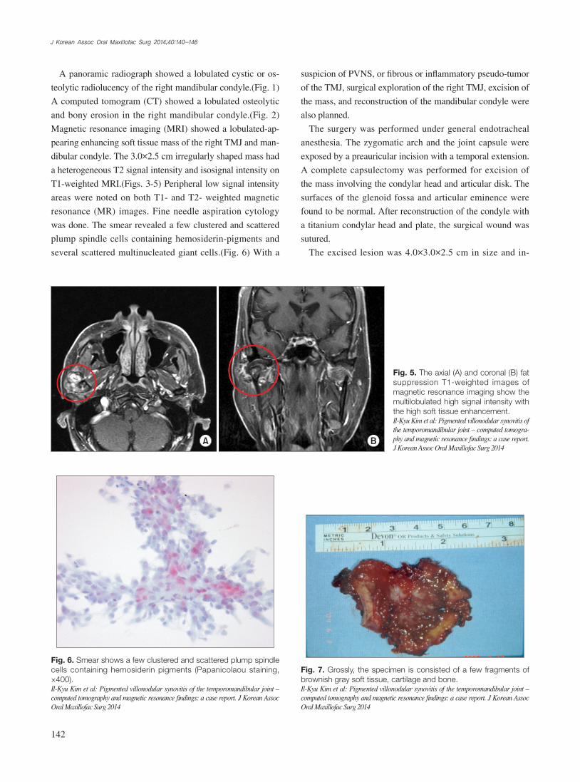
Theexcised lesionwas4.0×3.0×2.5cminsizeand in-
Apanoramicradiographshowedalobulatedcysticoros-
teolyticradiolucencyoftherightmandibularcondyle.(Fig.1)
Acomputedtomogram(CT)showedalobulatedosteolytic
andbonyerosionintherightmandibularcondyle.(Fig.2)
Magneticresonanceimaging(MRI)showedalobulated-ap-
pearingenhancingsofttissuemassoftherightTMJandman-
dibularcondyle.The3.0×2.5cmirregularlyshapedmasshad
aheterogeneousT2signalintensityandisosignalintensityon
T1-weightedMRI.(Figs.3-5)Peripherallowsignalintensity
areaswerenotedonbothT1-andT2-weightedmagnetic
resonance(MR) images.Fineneedleaspirationcytology
wasdone.Thesmearrevealedafewclusteredandscattered
plumpspindlecellscontaininghemosiderin-pigmentsand
severalscatteredmultinucleatedgiantcells.(Fig.6)Witha
Fig. 5. The axial (A) and coronal (B) fat suppression T1-weighted images of magnetic resonance imaging show the multilobulated high signal intensity with the high soft tissue enhancement. Il-Kyu Kim et al: Pigmented villonodular synovitis of the temporomandibular joint – computed tomogra-phy and magnetic resonance findings: a case report. J Korean Assoc Oral Maxillofac Surg 2014
Fig. 6. Smear shows a few clustered and scattered plump spindle cells containing hemosiderin pigments (Papanicolaou staining, ×400).Il-Kyu Kim et al: Pigmented villonodular synovitis of the temporomandibular joint – computed tomography and magnetic resonance findings: a case report. J Korean Assoc Oral Maxillofac Surg 2014
Fig. 7. Grossly, the specimen is consisted of a few fragments of brownish gray soft tissue, cartilage and bone. Il-Kyu Kim et al: Pigmented villonodular synovitis of the temporomandibular joint – computed tomography and magnetic resonance findings: a case report. J Korean Assoc Oral Maxillofac Surg 2014

Pigmented villonodular synovitis of the temporomandibular joint
143
Fig. 8. The mass is composed of many mononuclear histiocytic cells and irregularly interspersed multinucleated giant cells. Hemosiderin deposits are found (H&E staining, A: ×100, B: ×400).Il-Kyu Kim et al: Pigmented villonodular synovitis of the temporomandibular joint – computed tomography and magnetic resonance findings: a case report. J Korean Assoc Oral Maxil-lofac Surg 2014
Fig. 9. The axial (A) and coronal (B) views of computed tomography show the round soft tissue opacity on the condylar fossa of temporal bone (ar-row) and the skull base erosion with bony perforation (arrow head). Asterisk: metal condylar head.Il-Kyu Kim et al: Pigmented villonodular synovitis of the temporomandibular joint – computed tomogra-phy and magnetic resonance findings: a case report. J Korean Assoc Oral Maxillofac Surg 2014
Fig. 10. The axial (A) and coronal (B) fat suppression T1-weighted images of magnetic resonance imaging show the lobulated high signal intensity mixed up inner intermediate signal intensity with the moderate enhancement. The skull base perforation (arrow) is also seen.Il-Kyu Kim et al: Pigmented villonodular synovitis of the temporomandibular joint – computed tomogra-phy and magnetic resonance findings: a case report. J Korean Assoc Oral Maxillofac Surg 2014

J Korean Assoc Oral Maxillofac Surg 2014;40:140-146
144
sionoftheskullbaseofthetemporalbonewasnoticedto
accompanytheperforation.(Figs.9,10)ThefindingsonMRI
weresimilarwiththepreoperativesignalintensitiesofthe
T1W1,T2W1,andfatsuppressionT1W1images.(Figs.10-
12)SurgerywasperformedagainonJuly2008andtherecur-
renceofPVNSwasconfirmed.Duringthefollow-upwith
CTandMRIexamination,fractureofthereconstructionplate
wasnoticed,withthepatientcomplainingofsomediscomfort
andalimitationinmouthopening.InDecember2012,total
jointreconstructionwithBiometstockprosthesis(Biomet,
Jacksonvill,FL,USA)wasperformed,andbiopsyof the
excisedsofttissuefromtheoldlesionrevealedfibrosiswith
hyalinizationandmildinflammation.Therewerenoclinical
orimagingsignsofrecurrenceafter18monthsoffollow-up
followingthefinaloperation.(Fig.13)
III. Discussion
PVNSisan idiopathicproliferativedisorder involving
thesynoviumofthejoint,tendonsheaths,andbursae.More
volvedthemandibularcondyle,whichwaserodedandperfo-
ratedonitsposteriorsurface.(Fig.7)Grossly,thespecimen
consistedofafewfragmentsofbrownishgraysoft tissue,
cartilage,andbone.Microscopically, thetumorwascom-
posedofmanymononuclearhistiocyticcellsandirregularly
interspersedmultinucleatedgiantcells.Hemosiderinpig-
mentswerealsofound.Somefoamyhistiocyticcellswere
individuallyinterspersedorformedclusters.(Fig.8)Osseous,
cartilaginous,andsofttissueinvolvementwasalsoseen.The
lesionwasdiagnosedasaPVNSwithinvolvementof the
bone,cartilage,andadjacentsofttissue.
The4-yearfollow-upCTandMRIexaminationrevealed
arecurrenceofthePVNSwitha2.3×2.1×2.6cmmass.Ero-
Fig. 13. The panoramic view after 18 months of final operation later. Il-Kyu Kim et al: Pigmented villonodular synovitis of the temporomandibular joint – computed tomography and magnetic resonance findings: a case report. J Korean Assoc Oral Maxillofac Surg 2014
Fig. 11. The axial T1-weighted image of magnetic resonance imaging shows the isosignal intensity mixed up inner low signal intensity with peripheral low signal intensity rim.Il-Kyu Kim et al: Pigmented villonodular synovitis of the temporomandibular joint – computed tomography and magnetic resonance findings: a case report. J Korean Assoc Oral Maxillofac Surg 2014
Fig. 12. The axial (A) and coronal (B) T2-weighted images of magnetic reso-nance imaging show the heterogenous high signal intensity mixed up inner low signal intensity with peripheral low sig-nal intensity rim.Il-Kyu Kim et al: Pigmented villonodular synovitis of the temporomandibular joint – computed tomogra-phy and magnetic resonance findings: a case report. J Korean Assoc Oral Maxillofac Surg 2014

Pigmented villonodular synovitis of the temporomandibular joint
145
thetumorwellbecausethetumorhasallormostofitsfocal
areasofhyperdensitywithinthesoft-tissuemassthatfurther
enhancesafteradministrationofcontrastmaterial.CTen-
hancesthethickenedsynoviumandareasofhighattenuation
causedbyirondeposits11-13.Inthispatient,CTalsoshoweda
lobulatedosteolyticlesionaccompanyingthebonyerosion,
butdidnotfindbonysclerosisandareasoffocalhyperden-
sitywithinthesofttissue.(Figs.2,9)
TheappearanceofPVNSonMRimagecanbevariable,
dependingontherelativeproportionoflipids,hemosiderin,
fibrousstroma,pannus,fluids,andcellularelements2,13.For
example,dependingonthedegreeofhemosiderindeposition,
tumorsmaydemonstratelowsignal intensityonT1-,T2-,
andprotondensity-weightedsequences;thelowsignalinten-
sityistheresultofthepresenceofhemosiderindeposition.
Hemosiderin typicallydecreasedsignal intensityonlong-
repetitiontimeandlong-echotimespin-echo(SE)sequences
largelyduetotheferromagneticpropertiesofhemosiderin.
OnT2-weightedSEandgradient-echo(GRE)images, the
abnormal tissuemayappear largerbecauseof so-called
blooming.Ontheotherhand,theaccumulationoflipidsin
foamymacrophagesmayproduceareasofhighsignalinten-
sityonT1-weightedSEimagessimilartosubcutaneousfat.
Additionally,areasofhighsignalintensityonT2-weighted
imagescorrespondtoaloculatedcystofjointfluid.Another
well-recognizedcharacteristicsofPVNSonMRimagesare
hyperplasticsynoviumandboneerosioninthejointwithin-
sufficientintra-articularspaceforoutwardproliferationofthe
synovium.Inthispatient,the3.0×2.5cmirregularlyshaped
masshadheterogeneousT2signalintensityandisosignalin-
tensityonT1-weightedMRI.Peripherallowsignalintensity
areasarenotedonbothT1-andT2-weightedMRimages.
(Figs.3-5,10-12)Inthepresentcase, lowsignal intensity
onT1-weightedsequenceswasalsoobserved(Figs.4,11),
butthecausesofheterogeneoushighsignalintensityonT2-
weightedandfatsuppressionT1MRimagesonthemass
werethoughttobeduetotheprofoundfluidfromthecystic
lesionfromthecondyletothesynovium.(Figs.3,5,10,12)
TreatmentofPVNSisexclusivelysurgicalandconsistsof
completeexcisionoftheinvolvedboneandtotalsynovec-
tomybecausesynoviallesionshaveahighrateoflocalrecur-
rence.TherecurrenceofTMJPVNSoccursprimarilyinthe
diffusetypein8%to46%ofcasesandusuallyresultsfrom
incompleteresection,butcanalsobespontaneous14.Radia-
tiontherapymayproduceinvolutionofthelesionbutshould
bereservedforonlythoserecurrentcaseswithinvolvement
ofvitalstructures15.
than80%ofPVNScasesinvolvethekneesandtheupper
extremitiesareseldominvolved,suggestingapredilectionfor
weight-bearingjoints.Eighty-eightpercentageoflocalized
PVNSand59%ofdiffusePVNShaveapositivetraumatic
history.Bloodyfluidwasobtainedinmostofthecasesun-
dergoingsynovialfluidaspiration,andyellowfluidwasob-
tainedinafewcases7,8.
ReportsofPVNSintheTMJarerare,butPVNScanoccur
atanyage,withapreviousincidencepeakinthefourthand
fifthdecadesoflife.Nogenderdifferenceisseen9.Whenthe
diseasedoesoccurintheTMJ,itusuallyarisesasapreau-
ricularpalpablemassorswelling,andisfrequentlymistaken
foraparotidglandlesion.Othersymptomsmayincludeare-
strictedmandibularrangeofmotion,stiffness,numbness,and
pain.Inmostcases,thepainwasofminimalintensity.Are-
viewoftheliteratureandcarefulanalysisoftheclinicopatho-
logicfeaturesofPVNSoftheTMJrevealthat it typically
occursinthediffuseform,andthevastmajorityofreported
caseswereoftheextra-articularvariantofthisdisease,which
isassociatedwithmoreaggressivelocalinfiltrativebehavior,
suchasinvolvingtheparotidglandorskullbase,andahigher
rateoflocalrecurrencethanthelocalizedtype6,10,11.Thepa-
tientinthisstudywasthoughttohavethediffuseform,and
anaggressivelocalrecurrencewithskullbaseperforationby
bonyerosionsparingthedurawasfound4yearsaftertheop-
eration.
TheetiologyofPVNSstillremainscontroversial.While
Jaffeetal.1originally thought itwasaninflammatoryre-
sponsetoanunknownstimulus,othershaveattributedthe
process torepetitive intra-articularhemorrhagefollowing
trauma,disturbancesinlipidmetabolism,orabenignneo-
plasticproliferation7,9.Thepatientinthisstudydidnotrecall
ahistoryoffacialtrauma.
ThemacroscopicfeaturesofPVNSincludeathicksynovi-
umcomprisedofmattedmassesofvilliandsynovialfolds,
orpedunculatednodules.The lesionhasacharacteristic
rusty,red-browncolorresultingfromextensivehemosiderin
deposition.ThemicroscopicfeaturesofPVNSindicatea
nonspecificinflammatoryprocess,resultinginapapillary,
villous,andnodularappearanceofthestratifiedsynovium
withmultinucleatedgiantcells,foamcells,andhemosiderin
deposition3,9.
TheclassicplainradiographicfindingsofPVNSconsistof
cysticerosions,increaseddensityofthesynoviumsecond-
arytohemosiderindeposition,andperiarticularsoft-tissue
swellingwithnocalcification5,9.CTclearlyrevealsareasof
lyticboneerosionandsclerosis.CTalsodefinestheextentof

J Korean Assoc Oral Maxillofac Surg 2014;40:140-146
146
Conflict of Interest
Nopotentialconflictofinterestrelevanttothisarticlewas
reported.
References
1. JaffeHL,LichtensteinL,SutroCJ.Pigmentedvillonodularsynovi-tis,bursitisandtenosynovitis.ArchPathol1941;31:731-65.
2. KimKW,HanMH,ParkSW,KimSH,LeeHJ,JaeHJ,etal.Pig-mentedvillonodularsynovitisofthetemporomandibularjoint:MRfindingsinfourcases.EurJRadiol2004;49:229-34.
3. AoyamaS,IwakiH,AmagasaT,KinoK,OkadaN,KishimotoS.Pigmentedvillonodularsynovitisofthetemporomandibularjoint:differentialdiagnosisandcasereport.BrJOralMaxillofacSurg2004;42:51-4.
4. KişnişciRS,TüzHH,GünhanO,OnderE.Villonodularsynovitisof the temporomandibular joint:casereport.JOralMaxillofacSurg2001;59:1482-4.
5. LapayowkerMS,MillerWT,LevyWM,HarwickRD.Pigmentedvillonodularsynovitisofthetemperomandibularjoint.Radiology1973;108:313-6.
6. LiuYK,ChanJY,ChangCJ,HuangJS.Pigmentedvillonodularsynovitisofthetemporomandibularjointpresentingasamiddlecranialfossatumor.JOralMaxillofacSurg2012;70:367-72.
7. BemporadJA,ChaloupkaJC,PutmanCM,RothTC,TarroJ,Mi-
traS,etal.Pigmentedvillonodularsynovitisofthetemporoman-dibularjoint:diagnosticimagingandendovasculartherapeuticem-bolizationofarareheadandnecktumor.AJNRAmJNeuroradiol1999;20:159-62.
8. CaiJ,CaiZ,GaoY.Pigmentedvillonodularsynovitisofthetem-poromandibularjoint:acasereportandtheliteraturereview.IntJOralMaxillofacSurg2011;40:1314-22.
9. CasconeP,RinnaC,UngariC,PoladasG,FiliaciF.Pigmentedvillonodularsynovitisofthetemporomandibularjoint.JCraniofacSurg2005;16:712-6.
10. SafaeeM,OhT,SunMZ,ParsaAT,McDermottMW,El-SayedIH,etal.Pigmentedvillonodularsynovitisofthetemporomandibu-larjointwithintracranialextension:acaseseriesandsystematicreview.HeadNeck2014.doi:10.1002/hed.23717.
11. ChurchCA,RoweM,LlauradoR,LiwniczBH,MartinPA.Pig-mentedvillonodularsynovitisofthetemporomandibularjoint:areportoftwocases.EarNoseThroatJ2003;82:692-5.
12. LeWJ,LiMH,YuQ,ShiHM.Pigmentedvillonodularsynovitisofthetemporomandibularjoint:CTimagingfindings.ClinImag-ing2014;38:6-10.
13. StryjakowskaKK,MartelM,SasakiCT.Pigmentedvillonodularsynovitisofthetemporomandibularjoint:differentialdiagnosisoftheparotidmass.AurisNasusLarynx2005;32:309-14.
14. WongJJ,PhalPM,WiesenfeldD.Pigmentedvillonodularsynovi-tisofthetemporomandibularjoint:aradiologicdiagnosisandcasereport.JOralMaxillofacSurg2012;70:126-34.
15. O'SullivanTJ,AlportEC,WhistonHG.Pigmentedvillonodularsy-novitisofthetemporomandibularjoint.JOtolaryngol1984;13:123-6.