Pigment Epithelium-Derived Factor: Chemistry, Structure...
75
Pigment Epithelium-Derived Factor: Chemistry, Structure, Biology, and Applications Guest Editors: S. Patricia Becerra, Crispin R. Dass, Takeshi Yabe, and Susan E. Crawford Journal of Biomedicine and Biotechnology
Transcript of Pigment Epithelium-Derived Factor: Chemistry, Structure...
Pigment Epithelium-Derived Factor: Chemistry, Structure, Biology, and Applications
Guest Editors: S. Patricia Becerra, Crispin R. Dass, Takeshi Yabe, and Susan E. Crawford
Journal of Biomedicine and Biotechnology
Pigment Epithelium-Derived Factor: Chemistry,Structure, Biology, and Applications
Journal of Biomedicine and Biotechnology
Pigment Epithelium-Derived Factor: Chemistry,Structure, Biology, and Applications
Guest Editors: S. Patricia Becerra, Crispin R. Dass,Takeshi Yabe, and Susan E. Crawford

Copyright © 2012 Hindawi Publishing Corporation. All rights reserved.
This is a special issue published in “Journal of Biomedicine and Biotechnology.” All articles are open access articles distributed underthe Creative Commons Attribution License, which permits unrestricted use, distribution, and reproduction in any medium, providedthe original work is properly cited.

Editorial BoardThe editorial board of the journal is organized into sections that correspond to
the subject areas covered by the journal.
Agricultural Biotechnology
Ahmad Z. Abdullah, MalaysiaGuihua H. Bai, USAChristopher P. Chanway, CanadaRavindra N. Chibbar, CanadaAdriana S. Franca, Brazil
Ian Godwin, AustraliaHari B. Krishnan, USACarol A. Mallory-Smith, USADennis P. Murr, CanadaRodomiro Ortiz, Sweden
B. C. Saha, USAMariam B. Sticklen, USAChiu-Chung Young, Taiwan
Animal Biotechnology
E. S. Chang, USABhanu P. Chowdhary, USANoelle E. Cockett, USAPeter Dovc, SloveniaScott C. Fahrenkrug, USADorian J. Garrick, USAThomas A. Hoagland, USA
Tosso Leeb, SwitzerlandJames D. Murray, USAAnita M. Oberbauer, USAJorge A. Piedrahita, USADaniel Pomp, USAKent M. Reed, USALawrence Reynolds, USA
Lawrence B. Schook, USAMari A. Smits, The NetherlandsLeon Spicer, USAJ. Verstegen, USAMatthew B. Wheeler, USAKenneth L. White, USA
Biochemistry
David Ronald Brown, UKSaulius Butenas, USAVittorio Calabrese, ItalyMiguel Castanho, PortugalFrancis J. Castellino, USARoberta Chiaraluce, ItalyD. M. Clarke, CanadaFrancesca Cutruzzola, ItalyPaul W. Doetsch, USA
Hicham Fenniri, CanadaNick V. Grishin, USAJ. Guy Guillemette, CanadaPaul W. Huber, USAChen-Hsiung Hung, TaiwanMaria Jerzykiewicz, PolandMichael Kalafatis, USAB. E. Kemp, AustraliaPhillip E. Klebba, USA
Wen-Hwa Lee, USAGeorge Makhatadze, USALeonid Medved, USASusan A. Rotenberg, USAJason Shearer, USAAndrei Surguchov, USAJohn B. Vincent, USAY. George Zheng, USA
Bioinformatics
T. Akutsu, JapanMiguel A. Andrade, GermanyMark Y. Borodovsky, USARita Casadio, ItalyArtem Cherkasov, CanadaDavid Corne, UKSorin Draghici, USA
Stavros J. Hamodrakas, GreecePaul Harrison, USAGeorge Karypis, USAJack A. Leunissen, The NetherlandsGuohui Lin, CanadaSatoru Miyano, JapanZoran Obradovic, USA
Florencio Pazos, SpainZhirong Sun, ChinaYing Xu, USAAlexander Zelikovsky, USAAlbert Zomaya, Australia
Biophysics
Miguel Castanho, PortugalP. Bryant Chase, USAKuo-Chen Chou, USARizwan Khan, India
Ali A. Khraibi, Saudi ArabiaRumiana Koynova, USASerdar Kuyucak, AustraliaJianjie Ma, USA
S. B. Petersen, DenmarkPeter Schuck, USAClaudio M. Soares, Portugal
Cell Biology
Omar Benzakour, FranceSanford I. Bernstein, USAPhillip I. Bird, AustraliaEric Bouhassira, USAMohamed Boutjdir, USAChung-Liang Chien, TaiwanRichard Gomer, USAPaul J. Higgins, USAPavel Hozak, Czech Republic
Xudong Huang, USAAnton M. Jetten, USASeamus J. Martin, IrelandManuela Martins-Green, USAShoichiro Ono, USAGeorge Perry, USAM. Piacentini, ItalyGeorge E. Plopper, USALawrence Rothblum, USA
Michael Sheetz, USAJames L. Sherley, USAG. S. Stein, USARichard Tucker, USAThomas van Groen, USAAndre Van Wijnen, USASteve Winder, UKChuanyue Wu, USABin-Xian Zhang, USA
Genetics
Adewale Adeyinka, USAClaude Bagnis, FranceJ. Birchler, USASusan Blanton, USABarry J. Byrne, USAR. Chakraborty, USADomenico Coviello, ItalySarah H. Elsea, USACelina Janion, Poland
J. Spencer Johnston, USAM. Ilyas Kamboh, USAFeige Kaplan, CanadaManfred Kayser, The NetherlandsBrynn Levy, USAXiao Jiang Li, USAThomas Liehr, GermanyJames M. Mason, USAMohammed Rachidi, France
Raj S. Ramesar, South AfricaElliot D. Rosen, USADharambir K. Sanghera, USAMichael Schmid, GermanyMarkus Schuelke, GermanyWolfgang Arthur Schulz, GermanyJorge Sequeiros, PortugalMouldy Sioud, NorwayRongjia Zhou, China
Genomics
Vladimir Bajic, Saudi ArabiaMargit Burmeister, USASettara Chandrasekharappa, USAYataro Daigo, JapanJ. Spencer Johnston, USA
Vladimir Larionov, USAThomas Lufkin, SingaporeJohn L. McGregor, FranceJohn V. Moran, USAYasushi Okazaki, Japan
Gopi K. Podila, USAMomiao Xiong, USA
Immunology
Hassan Alizadeh, USAPeter Bretscher, CanadaRobert E. Cone, USATerry L. Delovitch, CanadaAnthony L. DeVico, USANick Di Girolamo, AustraliaDon Mark Estes, USASoldano Ferrone, USAJeffrey A. Frelinger, USAJohn Robert Gordon, Canada
James D. Gorham, USASilvia Gregori, ItalyThomas Griffith, USAYoung S. Hahn, USADorothy E. Lewis, USABradley W. McIntyre, USAR. Lee Mosley, USAMarija Mostarica-Stojkovic, SerbiaHans Konrad Muller, AustraliaAli Ouaissi, France
Kanury V. S. Rao, IndiaYair Reisner, IsraelHarry W. Schroeder, USAWilhelm Schwaeble, UKNilabh Shastri, USAYufang Shi, ChinaPiet Stinissen, BelgiumHannes Stockinger, AustriaJ. W. Tervaert, The NetherlandsGraham R. Wallace, UK
Microbial Biotechnology
Suraini Abd-Aziz, MalaysiaJozef Anne, BelgiumNuri Azbar, TurkeyYoav Bashan, MexicoMarco Bazzicalupo, ItalyHakan Bermek, TurkeyNico Boon, BelgiumJose Luis Campos, SpainYinguang Chen, ChinaLuca Simone Cocolin, Italy
Peter Coloe, AustraliaDaniele Daffonchio, ItalyHan de Winde, The NetherlandsRaf Dewil, BelgiumJose Domingos Fontana, BrazilPetros Gikas, GreeceTom Granstrom, FinlandIsmail Kiran, TurkeyHongjuan Liu, ChinaYanhe Ma, China
Paula Loureiro Paulo, BrazilBernd H A Rehm, New ZealandAlberto Reis, PortugalMuthuswamy Sathishkumar, SingaporeRamkrishna Sen, IndiaAngela Sessitsch, AustriaYa-Jie Tang, ChinaOrhan Yenigun, TurkeyEileen Hao Yu, United Kingdom
Microbiology
D. Beighton, UKSteven R. Blanke, USAStanley Brul, The NetherlandsIsaac K. O. Cann, USAStephen K. Farrand, USAAlain Filloux, UK
Gad Frankel, UKRoy Gross, GermanyHans-Peter Klenk, GermanyTanya Parish, UKGopi K. Podila, USAFrederick D. Quinn, USA
Didier A. Raoult, FranceIsabel Sa-Correia, PortugalP. L. C. Small, USAMichael Thomm, GermanyH. C. van der Mei, The NetherlandsSchwan William, USA
Molecular Biology
Rudi Beyaert, BelgiumMichael Bustin, USADouglas Cyr, USAK. Iatrou, GreeceLokesh Joshi, IrelandDavid W. Litchfield, Canada
Wuyuan Lu, USAPatrick Matthias, SwitzerlandJohn L. McGregor, FranceS. L. Mowbray, SwedenElena Orlova, UKYeon-Kyun Shin, USA
William S. Trimble, CanadaLisa Wiesmuller, GermanyMasamitsu Yamaguchi, Japan

Oncology
Colin Cooper, UKF. M. J. Debruyne, The NetherlandsNathan Ames Ellis, USADominic Fan, USAGary E. Gallick, USADaila S. Gridley, USAXin-yuan Guan, Hong KongAnne Hamburger, USAManoor Prakash Hande, SingaporeBeric Henderson, Australia
Steve B. Jiang, USADaehee Kang, Republic of KoreaAbdul R. Khokhar, USARakesh Kumar, USAMacus Tien Kuo, USAEric W. Lam, UKSue-Hwa Lin, USAKapil Mehta, USAOrhan Nalcioglu, USAP. J. Oefner, Germany
Allal Ouhtit, OmanFrank Pajonk, USAWaldemar Priebe, USAF. C. Schmitt, PortugalSonshin Takao, JapanAna Maria Tari, USAHenk G. Van Der Poel, The NetherlandsHaodong Xu, USADavid J. Yang, USA
Pharmacology
Abdel A. Abdel-Rahman, USAM. Badr, USAStelvio M. Bandiera, CanadaRonald E. Baynes, USAR. Keith Campbell, USAHak-Kim Chan, AustraliaMichael D. Coleman, UKJ. Descotes, FranceDobromir Dobrev, Germany
Ayman El-Kadi, CanadaJeffrey Hughes, USAKazim Husain, USAFarhad Kamali, UKMichael Kassiou, AustraliaJoseph J. McArdle, USAMark J. McKeage, New ZealandDaniel T. Monaghan, USAT. Narahashi, USA
Kennerly S. Patrick, USAVickram Ramkumar, USAMichael J. Spinella, USAQuadiri Timour, FranceTodd W. Vanderah, USAVal J. Watts, USADavid J. Waxman, USA
Plant Biotechnology
Prem L. Bhalla, AustraliaJ. R. Botella, AustraliaElvira Gonzalez De Mejia, USAShi-You Ding, USA
Metin Guru, TurkeyH. M. Haggman, FinlandLiwen Jiang, Hong KongPulugurtha Bharadwaja Kirti, India
Yong Pyo Lim, Republic of KoreaGopi K. Podila, USARalf Reski, GermanySudhir Kumar Sopory, India
Toxicology
Michael Aschner, USAJuergen Buenger, GermanyMichael L. Cunningham, USALaurence D. Fechter, USA
Hartmut Jaeschke, USAYoumin James Kang, USAM. Firoze Khan, USAPascal Kintz, France
Qaisar Mahmood, PakistanR. S. Tjeerdema, USAKenneth Turteltaub, USABrad Upham, USA

Virology
Nafees Ahmad, USAEdouard Cantin, USAEllen Collisson, USAKevin M. Coombs, CanadaNorbert K. Herzog, USATom Hobman, CanadaShahid Jameel, India
Fred Kibenge, CanadaFenyong Liu, USAEric Rassart, CanadaGerald G. Schumann, GermanyY.-C. Sung, Republic of KoreaGregory Tannock, Australia
Ralf Wagner, GermanyJianguo Wu, ChinaDecheng Yang, CanadaJiing-Kuan Yee, USAXueping Zhou, ChinaWen-Quan Zou, USA
Contents
Pigment Epithelium-Derived Factor: Chemistry, Structure, Biology, and Applications, S. Patricia Becerra,Crispin R. Dass, Takeshi Yabe, and Susan E. CrawfordVolume 2012, Article ID 830975, 2 pages
Identification of Pigment Epithelium-Derived Factor Protein Forms with Distinct Activities on TumorCell Lines, P. Subramanian, M. Deshpande, S. Locatelli-Hoops, S. Moghaddam-Taaheri, D. Gutierrez,D. P. Fitzgerald, S. Guerrier, M. Rapp, V. Notario, and S. P. BecerraVolume 2012, Article ID 425907, 12 pages
Efficacy of Continuously Administered PEDF-Derived Synthetic Peptides against Osteosarcoma Growthand Metastasis, Matthew L. Broadhead, Peter F. M. Choong, and Crispin R. DassVolume 2012, Article ID 230298, 10 pages
Cell and Molecular Biology Underpinning the Effects of PEDF on Cancers in General and Osteosarcomain Particular, Vijay Chandolu and Crispin R. DassVolume 2012, Article ID 740295, 9 pages
Role of Pigment Epithelium-Derived Factor in Stem/Progenitor Cell-Associated Neovascularization,Jung-Tung Liu, Yuh-Lien Chen, Wen-Chi Chen, Huey-Yi Chen, Yi-Wen Lin, Shu-Huei Wang,Kee-Ming Man, Hui-Min Wan, Wei-Hsian Yin, Po-Len Liu, and Yung-Hsiang ChenVolume 2012, Article ID 871272, 10 pages
The Emerging Role of PEDF in Stem Cell Biology, Mina Elahy, Swati Baindur-Hudson, and Crispin R. DassVolume 2012, Article ID 239091, 6 pages
PEDF in Diabetic Retinopathy: A Protective Effect of Oxidative Stress, Xiao-feng Zhu and Hai-dong ZouVolume 2012, Article ID 580687, 8 pages
PEDF and VEGF-A Output from Human Retinal Pigment Epithelial Cells Grown on NovelMicrocarriers, Torsten Falk, Nicole R. Congrove, Shiling Zhang, Alexander D. McCourt, Scott J. Sherman,and Brian S. McKayVolume 2012, Article ID 278932, 8 pages
Hindawi Publishing CorporationJournal of Biomedicine and BiotechnologyVolume 2012, Article ID 830975, 2 pagesdoi:10.1155/2012/830975
Editorial
Pigment Epithelium-Derived Factor: Chemistry, Structure,Biology, and Applications
S. Patricia Becerra,1 Crispin R. Dass,2 Takeshi Yabe,3 and Susan E. Crawford4
1 Section of Protein Structure and Function, Laboratory of Retinal Cell and Molecular Biology, National Eye Institute,Bethesda, MD 20892, USA
2 School of Biomedical Sciences, Victoria University, Melbourne, VIC, Australia3 Department of Medical Pharmacy, Setsunan University, Osaka 573-0101, Japan4 Department of Pathology, St. Louis University, St. Louis, MO 63104, USA
Correspondence should be addressed to S. Patricia Becerra, [email protected]
Received 27 August 2012; Accepted 27 August 2012
Copyright © 2012 S. Patricia Becerra et al. This is an open access article distributed under the Creative Commons AttributionLicense, which permits unrestricted use, distribution, and reproduction in any medium, provided the original work is properlycited.
Pigment epithelium-derived factor (PEDF) is a multi-functional serpin protein with demonstrable neurotro-phic, antiangiogenic, antitumorigenic, antimetastatic, anti-inflammatory, antioxidative properties among others. PEDFexists naturally in most organs of the human body, and itis released from most cell types as an extracellular diffusibleand circulating glycoprotein. It has been studied mostly inthe eye, where its levels are altered in diseases characterizedby retinopathies, such as age-related macular degenerationand diabetic retinopathy. The importance of PEDF in thedevelopment, maintenance, and function of the retina andCNS is evident in animal models for inherited and light-induced retinal degeneration, as well as for degenerationof spinal cord motor neurons. Pathological ocular neovas-cularization- and retinal degeneration-related animal modelshave prompted clinical development. Clinical trials to assessthe safety of a viral expression vector for PEDF in the contextof age-related macular degeneration have been performed.Interestingly, PEDF has multiple biological effects againsttumors, and its efficacy has been demonstrated in severalanimal models for tumor growth and progression. Themechanisms of PEDF action on tumors have been associatedto inhibition of tumor angiogenesis, and also negative effectsdirected on tumor cells. Recently, the involvement of PEDFin stem cell biology has been revealed. Moreover, PEDFis a potential diagnostic tool for several diseases triggeredby pathological neovascularization, retinal degenerations, ortumors.
The present issue includes three original research andfour review articles. In one article, V. Chandolu and C. R.Dass review the biological functions of PEDF against cancer,with a focus on a particular type of bone cancer calledosteosarcoma. They summarize the progress in understand-ing the function of PEDF in antiangiogenesis, tumor celldifferentiation, and direct tumor cell inhibition. In anotherreview article, J.-T. Liu et al. address the role of PEDF instem/progenitor cell-associated neovascularization, in parti-cular in cardiovascular and neurovascular biology. M. Elahyet al. review the promising significance of PEDF in stem cellbiology, specifically in human embryonic stem cells, mesen-chymal stem cells (MSCs), neural stem sells (NSCs), andstem cells overexpressing the PEDF gene. X.-F. Zhu et al.summarize the advances of PEDF in diabetic retinopathy, itsprotective effect on oxidative stress, which is the main triggerfor the pathology, and the potential application of PEDF indiabetic retinopathy. In a research article, P. Subramanian etal. report the identification of biochemical forms of PEDFwith distinct biological effects on tumor cells, which mayexplain the multifunctional modality of this protein. In asecond research article, M. L. Broadhead et al. report theeffects of two PEDF-derived peptides on a clinically relevantmurine model of osteosarcoma with spontaneous metastasis.Finally, T. Falk et al. report that human retinal pigmentepithelial cells grown on plastic-based microcarriers retainthe ability to produce both PEDF and vascular endothelialgrowth factor VEGF-A. This constitutes a novel candidate

2 Journal of Biomedicine and Biotechnology
delivery system for neurotrophic factors with potential appli-cation in neurodegenerative diseases, such as Parkinson’s.
In summary, given the multimodal nature of PEDF, thepresent issue aims to enhance our understanding of thechemistry, structure, biology, and application of PEDF inbiomedicine. It gives an overview of the current status ofresearch on PEDF and will prove useful as source of referencefor students and researchers.
S. Patricia BecerraCrispin R. Dass
Takeshi YabeSusan E. Crawford
Hindawi Publishing CorporationJournal of Biomedicine and BiotechnologyVolume 2012, Article ID 425907, 12 pagesdoi:10.1155/2012/425907
Research Article
Identification of Pigment Epithelium-Derived Factor ProteinForms with Distinct Activities on Tumor Cell Lines
P. Subramanian,1 M. Deshpande,1 S. Locatelli-Hoops,1
S. Moghaddam-Taaheri,1 D. Gutierrez,2 D. P. Fitzgerald,3, 4
S. Guerrier,1 M. Rapp,1 V. Notario,5 and S. P. Becerra1
1 Section of Protein Structure and Function, LRCMB, National Eye Institute, NEI, Bethesda, MD 20892-0608, USA2 Laboratory of Retinal Cell and Molecular Biology, National Eye Institute, NEI, Bethesda, MD 20892, USA3 Center for Cancer Research (CCR), National Cancer Institute, Bethesda, MD 20892, USA4 Otsuka Maryland Medicinal Laboratories, Rockville, MD 20850, USA5 Lombardi Comprehensive Cancer Center, Georgetown University Medical Center, Washington, DC 20057, USA
Correspondence should be addressed to S. P. Becerra, [email protected]
Received 16 December 2011; Accepted 7 March 2012
Academic Editor: Crispin Dass
Copyright © 2012 P. Subramanian et al. This is an open access article distributed under the Creative Commons AttributionLicense, which permits unrestricted use, distribution, and reproduction in any medium, provided the original work is properlycited.
Purpose. Pigment epithelium-derived factor (PEDF) is a multifunctional serpin. The purpose of this study is to identify PEDFprotein forms and investigate their biological activities on tumor cell lines. Methods. Recombinant human PEDF proteins werepurified by cation- and anion-exchange column chromatography. They were subjected to SDS-PAGE, IEF, deglycosylation, heparinaffinity chromatography, and limited proteolysis. Cell viability, real-time electrical impedance of cells, and wound healing assayswere performed using bladder and breast cancer cell lines, rat retinal R28, and human ARPE-19 cells. Results. Two PEDF proteinpeaks were identified after anion-exchange column chromatography: PEDF-1 eluting with lower ionic strength than PEDF-2.PEDF-1 had higher pI value and lower apparent molecular weight than PEDF-2. Both PEDF forms were glycosylated, boundto heparin, and had identical patterns by limited proteolysis. However, PEDF-2 emerged as being highly potent in lowering cellviability in all tumor cell lines tested, and in inhibiting tumor and ARPE-19 cell migration. In contrast, PEDF-1 minimally affectedtumor cell viability and cell migration but protected R28 cells against death caused by serum starvation. Conclusion. Two distinctbiochemical forms of PEDF varying in overall charge have distinct biological effects on tumor cell viability and migration. Theexistence of PEDF forms may explain the multifunctional modality of PEDF.
1. Introduction
Pigment epithelium-derived factor (PEDF) is a 50 kDa se-creted glycoprotein and a member of the serpin superfam-ily with no demonstrable protease inhibitory activity [1].PEDF is associated with several biological processes due toits antiangiogenic, anti-inflammatory, anti-oxidative, neu-rotrophic, and neuroprotective properties [2]. Moreover, ithas been implicated in another interesting role, as an antitu-mor and antimetastatic agent with applications in multiplemalignancies such as retinoblastoma, lung, breast, prostate,ovarian and pancreatic carcinomas, uveal melanoma, glioma,and osteosarcoma [3–8]. PEDF levels are decreased in tumorcells relative to normal cells, and PEDF addition inhibits
tumor formation and metastasis, blocks angiogenesis, andinduces apoptosis in tumor and endothelial cells. In contrast,PEDF promotes retinal cell survival and neuronal differentia-tion, protects retinal pigment epithelial cells against oxidativestress [2, 3, 9] and plays a role in expansion of neural stemcells [10]. The mechanisms that mediate the multimodalfunctionality to PEDF are not clear.
Previous structure-function studies revealed that twopeptide regions toward the amino end of the PEDF polypep-tide have distinct biological activities [2, 11, 12]. The peptideregion termed 34-mer (amino acid residues Asp44-Asn77 ofthe human PEDF sequence) forms alpha helix A of the 3Dstructure of human PEDF [13] and confers antiangiogenicand antitumorigenic properties to the PEDF polypeptide
2 Journal of Biomedicine and Biotechnology
[12]. The 44-mer peptide (Val78-Thr121) derived from theregion that forms alpha helices B, C, and part of D exhibitsneurite-outgrowth activity and protects spinal cord motorneurons against chronic toxicity [11, 14]. A smaller peptidederived from amino acid positions 82–121 exhibits effectiveneuroprotective properties in retinal ischemia [15].
Interestingly, the native PEDF has several isoformsdiffering in isoelectric point (pI) values [16, 17] implyingdifferences in posttranslational modifications of the polypep-tide backbone. Duh et al. reported that the secreted humanrecombinant PEDF from human embryonic kidney (HEK)cells has at least two species varying in their carbohydratecomposition of the N-glycosylation site and efficacy ofsuppressing vascular endothelial growth factor-induced pro-liferation and migration of retinal microvascular endothelialcells [18]. Maik-Rachline and Seger showed that the humanplasma PEDF is a phosphoprotein and that extracellularphosphorylation converts the recombinant protein froma neurotrophic to an antiangiogenic factor [19]. Konsonet al. demonstrated that a triple phosphomimetic-alteredPEDF is more efficient than wild-type PEDF in inhibitingneovascularization and tumor growth in vivo and suppressescultured endothelial cell proliferation and cell migrationmuch better than the wild-type PEDF [20].
Given the above, the aim of this study was to identifyand characterize PEDF isoforms that could contribute to thecomplexity of PEDF action and its multifunctional modality.
2. Methods
2.1. Cell Culture. Human bladder carcinoma T24 cells (CellLine collection, Lombardi Comprehensive Cancer Center,passage 33), human breast cancer 231-BR cells [21] (passage13), mouse breast cancer 4T1-BR5 cells [21] (passage 11),human breast tumor MDA-MB-231 cells (Cell Line Collec-tion, Lombardi Comprehensive Cancer Center, passage 6),mouse breast cancer 4T1 cells (Cell Line Collection, Lombar-di Comprehensive Cancer Center, passage 9), and rat retinalR28 (generous gift of Gail Seigel, passages 47–55), were cul-tured in DMEM medium. Human retinal pigment epithelialARPE-19 cells (American Type Culture Collection, passages27–32) were cultured in DMEM-12 medium. Media weresupplemented with 10% of fetal bovine serum (FBS) and 1%penicillin/streptomycin and cultures were incubated at 37◦Cwith 5% CO2.
2.2. Protein Purification. Recombinant human PEDF waspurified from the culture media of BHK cells harboring anexpression plasmid containing full-length PEDF cDNA [22].The culture media was concentrated by ammonium sulfateprecipitation and subjected sequentially to cation- andanion-exchange column chromatography as described beforewith the following modifications [13]. Cation-exchange col-umn chromatography was performed using a POROS S resinconnected to a BioCAD 700E perfusion chromatographysystem, with buffer S (20 mM Na phosphate, pH 6.5, 50 mMNaCl) and elutions were with a linear gradient of 50–500 mMNaCl in Buffer S. PEDF-containing fractions were pooled,
dialyzed against buffer Q (50 mM Tris-HCl, pH 8) and sub-jected to POROS Q column chromatography in buffer Qand elutions were done with a linear gradient of 100–300 mM NaCl in buffer Q. The PEDF-containing fractionswere pooled, concentrated, and the buffer exchanged to PBSusing ultrafiltration devices (Centricon-30 or Amicon-30,Millipore). Storage of the final samples was at −80◦C.
2.3. Protein Analyses. Proteins were analyzed by SDS-PAGEusing 10–20% polyacrylamide in SDS-Tricine gels (Invitro-gen) or NuPAGE 10% polyacrylamide gel in Bis-Tris bufferwith NuPAGE MOPS-SDS as running buffer (Invitrogen)under reducing conditions, and isoelectric focusing (IEF).Protein detection was accomplished with Coomassie Bluestain. PEDF protein identity was confirmed by WesternBlot. After separation by SDS-PAGE electrophoresis, proteinswere transferred to a nitrocellulose membrane, blocked for1 h at room temperature, and incubated with polyclonalantibody to PEDF (Bioproducts MD, Inc.) in blockingsolution at 1 : 5,000 or 1 : 10,000 dilution, followed bysecondary antibody anti-rabbit IgG (H + L) in a 1 : 1000dilution and the Vectastatin ABC Kit (Vector Laboratories)with colorimetric detection reagent 4-chloro-1-naphthol(BioRad Laboratories) as substrate. Alternatively, the sec-ondary antibody was affinity-purified peroxidase-labeledgoat anti-rabbit IgG (H + L) in a 1 : 200,000 dilution, andchemiluminescence detection with Super Signal West DuraExtended Duration Substrate (Thermo Scientific) on X-rayfilms.
Protein concentrations were determined using the Pro-tein Assay Kit (BioRad) and Beckman DU 640 Spectropho-tometer. Protein concentration was calculated from absorb-ance values using the formula:
[Absorbance595 nm × 3.5μg BSA
]
÷ [Sample volume(μL)× 0.2
]
= protein concentration(mg/mL
).
(1)
Isoelectric focusing was performed using pH 3–7 or pH3–10 IEF gels (Invitrogen), following manufacturer’s instruc-tions.
2.4. Enzymatic Deglycosylation. Two micrograms of proteinwas treated with N-glycosidase F (New England Biolabs,Ipswich, MA) following a previously described method [17].Briefly, the protein sample was denatured by boiling in asolution containing 0.5% SDS, 40 mM DTT. To avoid inacti-vation of the enzyme by SDS, a total of 1% NP-40 in 50 mMsodium phosphate, pH 7.5 was added to the denaturedsample before adding 1000 Units of N-glycosidase F in 20 μLfinal reaction volume. Enzymatic reactions were incubatedat 37◦C for 1 h followed by addition of SDS-PAGE samplebuffer and samples were boiled for 10 min.
2.5. Limited Proteolysis. PEDF was cleaved with limitingamounts of chymotrypsin. Reaction mixtures containedchymotrypsin (Worthington, Lakewood, NJ) and PEDF sub-strate: at a w/w ratio of 1 : 10 in 80 mM Tris-HCl, pH 7.5 and
Journal of Biomedicine and Biotechnology 3
100 mM CaCl2. Protein amounts were 1 μg and incubationtemperature was 25◦C. Incubation times were as indicated.The reactions were stopped by freezing in dry ice and theaddition of SDS-PAGE sample buffer. Reaction productswere analyzed by SDS-PAGE analysis of products.
2.6. Glycosaminoglycan-Binding Assays. Heparin-bindingassays were performed using heparin affinity column chro-matography as described previously [23]. Hyaluronan-bind-ing assays were performed using hyaluronan affinity columnchromatography as described before [24].
2.7. RT-CES Assay. The cells were plated on a microplate bi-osensor platform and real-time electrical impedance of cellswas followed with RT-CES system from ACEA Biosciences(San Diego, CA) (http://www.aceabio.com), a system de-scribed previously [25, 26]. T24, 231-BR, and 4T1-BR5 cellswere seeded at 6,000 cells per well (a 96-well format) andallowed to attach for 18 h. Then the cells were serum-starvedfor 8 h followed by addition of serum-free media containingPEDF. R28 cells were seeded at a density of 3000 cells/well on16-well strips of 96-well format with microelectrodes. Back-ground impedance was measured with media alone beforeadding the cells. The cells were allowed to attach for 8 h withmedia containing 5% FBS. This was followed by changing tomedia with or without PEDF at the desired concentration.Real-time electrical cell impendence was monitored everyhour in each well for several hours. Data from two replicateswas averaged.
2.8. Cell Viability Assays. At the end point of the RT-CESassay, cell viability was measured by determining the relativelevels of intracellular ATP as a biomarker for live cellsusing a CellTiter-Glo kit (Promega, Madison, WI) andfollowing instructions by manufacturer. After 30 min ofincubation at room temperature, the incubation solution ineach well was transferred into wells in a 96-well microtiterplate. The luminescence intensity was measured using anautomated plate reader (Envision, Perkin Elmer, MA). Inparallel experiments, cells cultured in 24-well plates intriplicates were imaged and at the end point, cell viability wasmeasured by determining the relative levels of mitochondrialdehydrogenase activity as live cell biomarker using theCell Counting Kit-8 (Dojindo) following instructions bymanufacturer. Cells in each well were incubated with 50 μLof CCK-8 solution diluted 1 : 25 and incubated for 4 h at37◦C. Absorbance of each well was measured at 450 nm usingan automated plate reader (Envision). In all the cases, theabsorbance reading for background was subtracted from thereadings of samples. Data from replicates were averaged andstatistical analysis was performed by a t-test. A P value of<0.05 was taken as significant.
2.9. In Vitro Migration Assay. Cell migration was assessedusing wound-healing assays. Confluent cultures of ARPE-19cells on 24-well culture plates and of MDA-MB-231 cells in6-well plates were scratched with 10-μL pipette tips to createfixed-width linear “wounds” in the cell monolayers, followed
0.05
0.04
0.03
0.02
0.01
0
Abs
orba
nce
(280
nm
)
100
50
00 5 10 15 20
INJ
(min)
Con
duct
ivit
y (m
S)-10
-7.5
-6
-3
pH
-PE
DF
-1 -2
PFDF-1-PFDF-2-
-1
-2
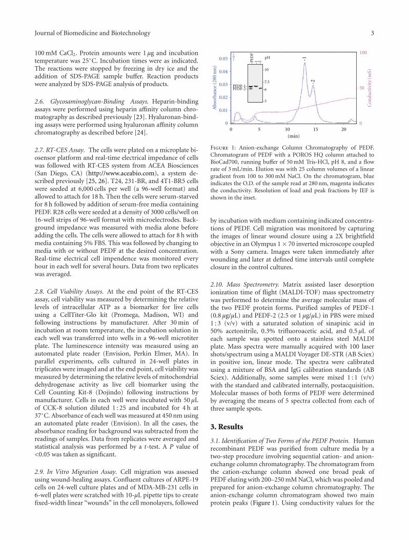
Figure 1: Anion-exchange Column Chromatography of PEDF.Chromatogram of PEDF with a POROS HQ column attached toBioCad700, running buffer of 50 mM Tris-HCl, pH 8, and a flowrate of 3 mL/min. Elution was with 25 column volumes of a lineargradient from 100 to 300 mM NaCl. On the chromatogram, blueindicates the O.D. of the sample read at 280 nm, magenta indicatesthe conductivity. Resolution of load and peak fractions by IEF isshown in the inset.
by incubation with medium containing indicated concentra-tions of PEDF. Cell migration was monitored by capturingthe images of linear wound closure using a 2X brightfieldobjective in an Olympus 1× 70 inverted microscope coupledwith a Sony camera. Images were taken immediately afterwounding and later at defined time intervals until completeclosure in the control cultures.
2.10. Mass Spectrometry. Matrix assisted laser desorptionionization time of flight (MALDI-TOF) mass spectrometrywas performed to determine the average molecular mass ofthe two PEDF protein forms. Purified samples of PEDF-1(0.8 μg/μL) and PEDF-2 (2.5 or 1 μg/μL) in PBS were mixed1 : 3 (v/v) with a saturated solution of sinapinic acid in50% acetonitrile, 0.3% trifluoroacetic acid, and 0.5 μL ofeach sample was spotted onto a stainless steel MALDIplate. Mass spectra were manually acquired with 100 lasershots/spectrum using a MALDI Voyager DE-STR (AB Sciex)in positive ion, linear mode. The spectra were calibratedusing a mixture of BSA and IgG calibration standards (ABSciex). Additionally, some samples were mixed 1 : 1 (v/v)with the standard and calibrated internally, postacquisition.Molecular masses of both forms of PEDF were determinedby averaging the means of 5 spectra collected from each ofthree sample spots.
3. Results
3.1. Identification of Two Forms of the PEDF Protein. Humanrecombinant PEDF was purified from culture media by atwo-step procedure involving sequential cation- and anion-exchange column chromatography. The chromatogram fromthe cation-exchange column showed one broad peak ofPEDF eluting with 200–250 mM NaCl, which was pooled andprepared for anion-exchange column chromatography. Theanion-exchange column chromatogram showed two mainprotein peaks (Figure 1). Using conductivity values for the
4 Journal of Biomedicine and Biotechnology
(a)
[DYKDDDDK]WT
PEDF form
1 2 1 2 1 2 1 20
5
10
15
20
25
30
Con
duct
ivit
y (m
S)
+2x2K, 1R 3A 3K, 1R 4A
(b)
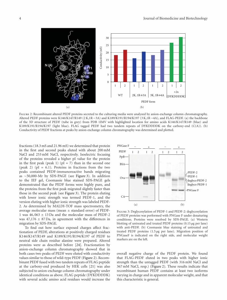
Figure 2: Recombinant altered PEDF proteins secreted to the culturing media were analyzed by anion-exchange column chromatography.Altered PEDF proteins were K146/K147/R149 (2 K,1R→3A) and K189/K191/R194/K197 (3 K,1R→4A), and FLAG-PEDF. (a) the backboneof the 3D structure of PEDF (tube in grey) from PDB 1IMV with highlighted location for amino acids K146/K147/R149 (blue) andK189/K191/R194/K197 (light blue). FLAG tagged PEDF had two tandem repeats of DYKDDDDK on the carboxy-end (C(A)). (b)Conductivity of PEDF fractions at peaks by anion-exchange column chromatography was determined and plotted.
fractions (18.3 mS and 21.96 mS) we determined that proteinin the first and second peaks eluted with about 200 mMNaCl and 253 mM NaCl, respectively. Isoelectric focusingof the proteins revealed a higher pI value for the proteinin the first peak (peak 1) (pI = 7) than in the second one(peak 2) (pI = 6.1). Proteins in fractions from the twopeaks contained PEDF-immunoreactive bands migratingas ∼50,000-Mr by SDS-PAGE (see Figure 3). In additionto the IEF gel, Coomassie blue stained SDS-PAGE gelsdemonstrated that the PEDF forms were highly pure, andthe proteins from the first peak migrated slightly faster thanthose in the second peak (see Figure 3). The protein elutingwith lower ionic strength was termed PEDF-1, and theversion eluting with higher ionic strength was labeled PEDF-2. As determined by MALDI-TOF mass spectrometry, theaverage molecular mass (mean ± standard error) of PEDF-1 was 46, 063 ± 13 Da and the molecular mass of PEDF-2was 47,176 ± 87 Da, in agreement with the differences inmigration by SDS-PAGE.
To find out how surface exposed charges affect frac-tionation of PEDF, alterations at positively charged residuesK146/K147/R149 and K189/K191/R194/K197 of PEDF toneutral side chain residue alanine were prepared. Alteredproteins were as described before [24]. Fractionation byanion-exchange column chromatography showed that inboth cases two peaks of PEDF were eluted with conductivityvalues similar to those of wild-type PEDF (Figure 2). Recom-binant PEDF fused with two tandem repeats of FLAG peptideat the carboxy-end produced by HEK cells [21] was alsosubjected to anion-exchange column chromatography underidentical conditions as above. FLAG peptide (DYKDDDDK)with several acidic amino acid residues would increase the
PpB
BSA
Ova
CA
PEDF
PNGase F
1 2 1 2
+−
(a)
deglycodeglyco
+
1 2 1 2
-PNGase F
PEDF-2PEDF-1
−
PEDF-2PEDF-1
(b)
Figure 3: Deglycosylation of PEDF-1 and PEDF-2: deglycosylationof PEDF proteins was performed with PNGase F under denaturingconditions. Proteins were resolved by SDS-PAGE. (a) Westernblotting of untreated and treated PEDF proteins (0.15 μg per lane)with anti-PEDF. (b) Coomassie blue staining of untreated andtreated PEDF proteins (1.5 μg per lane). Migration position ofPNGaseF is indicated on the right side, and molecular weightmarkers are on the left.
overall negative charge of the PEDF protein. We foundthat FLAG-PEDF eluted in two peaks with higher ionicstrength than the untagged PEDF (with 316 mM NaCl and367 mM NaCl, resp.) (Figure 2). These results indicate thatrecombinant human PEDF contains at least two isoformsvarying in charge and in apparent molecular weight, and thatthis characteristic is general.
Journal of Biomedicine and Biotechnology 5
0 30 60 0 30 60
uncleaved
cleaved
PEDF-1 PEDF-2
Incubation time(min)
(a)
U B U B
PEDF-1 PEDF-2
(b)
Figure 4: (a) Chymotrypsin Limited Proteolysis: reactions for the indicated times were performed with mixtures containing α-chymotrypsinand PEDF substrate at a ratio of 1 : 10 (w/w). Reactions were resolved by SDS-PAGE (0.1 μg per lane) followed by western blotting with anti-PEDF. Cleaved and the uncleaved forms are shown to the right. (b) Binding to Heparin. PEDF-1 and PEDF-2 were subjected to heparin-affinity column chromatography. Unbound (U) and bound (B) material was analyzed by SDS-PAGE and immunostained with anti-PEDF.
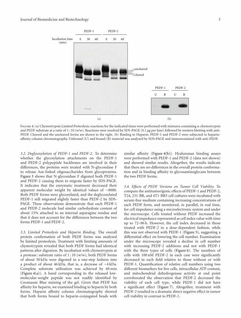
3.2. Deglycosylation of PEDF-1 and PEDF-2. To determinewhether the glycosylation attachments on the PEDF-1and PEDF-2 polypeptide backbones are involved in theirdifferences, the proteins were treated with N-glycosidase Fto release Asn-linked oligosaccharides from glycoproteins.Figure 3 shows that N-glycosidase F digested both PEDF-1and PEDF-2 causing them to migrate faster by SDS-PAGE.It indicates that the enzymatic treatment decreased theirapparent molecular weight by identical values of ∼8000.Both PEDF forms were glycosylated, and the deglycosylatedPEDF-1 still migrated slightly faster than PEDF-2 by SDS-PAGE. These observations demonstrate that each PEDF-1and PEDF-2 molecule had similar carbohydrate content ofabout 15% attached to an internal asparagine residue andthat it does not account for the differences between the twoforms PEDF-1 and PEDF-2.
3.3. Limited Proteolysis and Heparin Binding. The overallprotein confirmation of both PEDF forms was analyzedby limited proteolysis. Treatment with limiting amounts ofchymotrypsin revealed that both PEDF forms had identicalpatterns after digestion. By incubation with chymotrypsin ata protease : substrate ratio of 1 : 10 (w/w), both PEDF formsof about 50 kDa were digested in a one-step fashion intoa product of about 46 kDa, that is, a decrease of ∼4 kDa.Complete substrate utilization was achieved by 60 min(Figure 4(a)). A band corresponding to the released low-molecular-weight peptide was not readily identified byCoomassie Blue staining of the gel. Given that PEDF hasaffinity for heparin, we examined binding to heparin by bothforms. Heparin affinity column chromatography showedthat both forms bound to heparin-conjugated beads with
similar affinity (Figure 4(b)). Hyaluronan binding assayswere performed with PEDF-1 and PEDF-2 (data not shown)and showed similar results. Altogether, the results indicatethat there are no differences in the overall protein conforma-tion and in binding affinity to glycosaminoglycans betweenthe two PEDF forms.
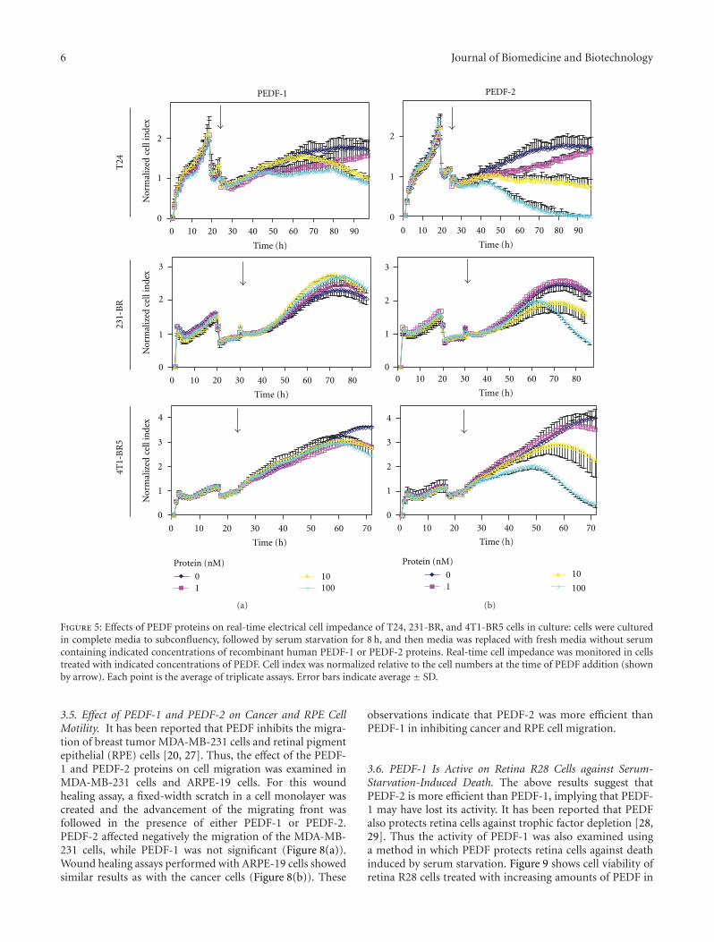
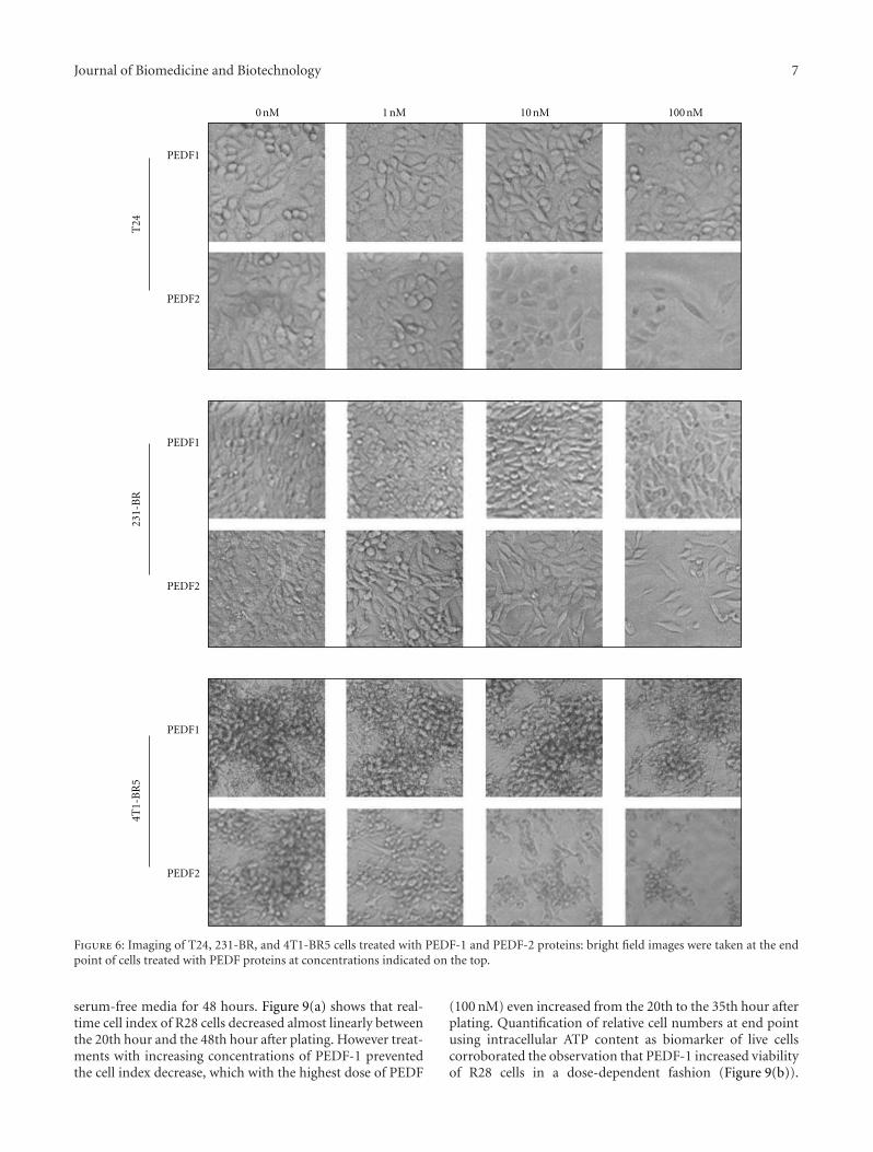
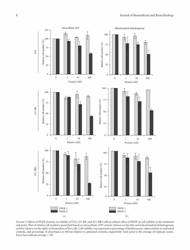
3.4. Effects of PEDF Versions on Tumor Cell Viability. Tocompare the antitumorigenic effects of PEDF-1 and PEDF-2,T24, 231-BR, and 4T1-BR5 cell cultures were incubated withserum-free medium containing increasing concentrations ofeach PEDF form, and monitored, in parallel, in real time,for cell impedance using a microelectronic system and underthe microscope. Cells treated without PEDF increased theelectrical impedance represented as cell index value with timeup to 72–96 h. However, the cell index decreased in thosetreated with PEDF-2 in a dose-dependent fashion, whilethis was not observed with PEDF-1 (Figure 5), suggesting adifferential effect on lowering the cell number. Examinationunder the microscope revealed a decline in cell numberwith increasing PEDF-2 additions and not with PEDF-1with the three types of cells (Figure 6). The numbers ofcells with 100 nM PEDF-2 in each case were significantlydecreased in each field relative to those without or withPEDF-1. Quantification of relative cell numbers using twodifferent biomarkers for live cells, intracellular ATP content,and mitochondrial dehydrogenase activity at end pointcorroborated the observation that PEDF-2 decreased theviability of each cell type, while PEDF-1 did not havea significant effect (Figure 7). Altogether, treatment withPEDF-2 resulted in a dramatic direct negative effect in tumorcell viability in contrast to PEDF-1.
6 Journal of Biomedicine and Biotechnology
0
1
2
0
1
2
3
0
1
2
3
4
0 10 20 30 40 50 60 70 80 90
0 10 20 30 40 50 60 70 80
0 10 20 30 40 50 60 70
Time (h)
Time (h)
Time (h)
Nor
mal
ized
cell
inde
xN
orm
aliz
edce
llin
dex
Nor
mal
ized
cell
inde
x
T24
PEDF-1
231-
BR
4T1-
BR
5
01
10100
Protein (nM)
(a)
0
1
2
0
1
2
3
0
1
2
3
4
0 10 20 30 40 50 60 70 80 90
0 10 20 30 40 50 60 70 80
0 10 20 30 40 50 60 70
Time (h)
Time (h)
Time (h)
PEDF-2
1
10
100
0
Protein (nM)
(b)
Figure 5: Effects of PEDF proteins on real-time electrical cell impedance of T24, 231-BR, and 4T1-BR5 cells in culture: cells were culturedin complete media to subconfluency, followed by serum starvation for 8 h, and then media was replaced with fresh media without serumcontaining indicated concentrations of recombinant human PEDF-1 or PEDF-2 proteins. Real-time cell impedance was monitored in cellstreated with indicated concentrations of PEDF. Cell index was normalized relative to the cell numbers at the time of PEDF addition (shownby arrow). Each point is the average of triplicate assays. Error bars indicate average ± SD.
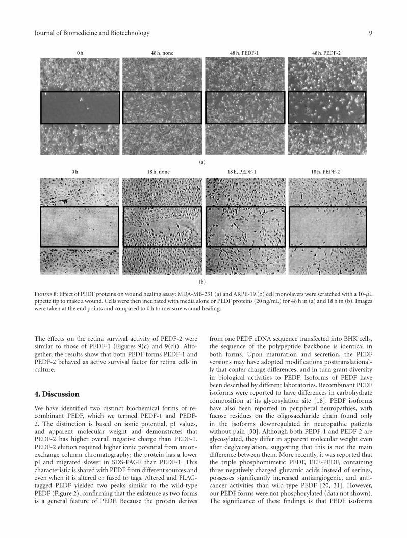
3.5. Effect of PEDF-1 and PEDF-2 on Cancer and RPE CellMotility. It has been reported that PEDF inhibits the migra-tion of breast tumor MDA-MB-231 cells and retinal pigmentepithelial (RPE) cells [20, 27]. Thus, the effect of the PEDF-1 and PEDF-2 proteins on cell migration was examined inMDA-MB-231 cells and ARPE-19 cells. For this woundhealing assay, a fixed-width scratch in a cell monolayer wascreated and the advancement of the migrating front wasfollowed in the presence of either PEDF-1 or PEDF-2.PEDF-2 affected negatively the migration of the MDA-MB-231 cells, while PEDF-1 was not significant (Figure 8(a)).Wound healing assays performed with ARPE-19 cells showedsimilar results as with the cancer cells (Figure 8(b)). These
observations indicate that PEDF-2 was more efficient thanPEDF-1 in inhibiting cancer and RPE cell migration.
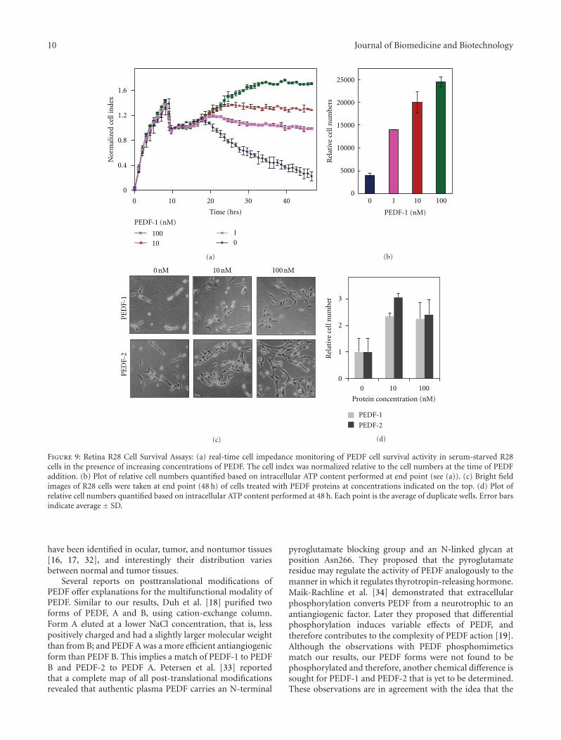
3.6. PEDF-1 Is Active on Retina R28 Cells against Serum-Starvation-Induced Death. The above results suggest thatPEDF-2 is more efficient than PEDF-1, implying that PEDF-1 may have lost its activity. It has been reported that PEDFalso protects retina cells against trophic factor depletion [28,29]. Thus the activity of PEDF-1 was also examined usinga method in which PEDF protects retina cells against deathinduced by serum starvation. Figure 9 shows cell viability ofretina R28 cells treated with increasing amounts of PEDF in
Journal of Biomedicine and Biotechnology 7
PEDF1
PEDF2
PEDF1
PEDF2
PEDF1
PEDF2
T24
231-
BR
4 T1-
BR
5
0 nM 1 nM 10 nM 100 nM
Figure 6: Imaging of T24, 231-BR, and 4T1-BR5 cells treated with PEDF-1 and PEDF-2 proteins: bright field images were taken at the endpoint of cells treated with PEDF proteins at concentrations indicated on the top.
serum-free media for 48 hours. Figure 9(a) shows that real-time cell index of R28 cells decreased almost linearly betweenthe 20th hour and the 48th hour after plating. However treat-ments with increasing concentrations of PEDF-1 preventedthe cell index decrease, which with the highest dose of PEDF
(100 nM) even increased from the 20th to the 35th hour afterplating. Quantification of relative cell numbers at end pointusing intracellular ATP content as biomarker of live cellscorroborated the observation that PEDF-1 increased viabilityof R28 cells in a dose-dependent fashion (Figure 9(b)).
8 Journal of Biomedicine and Biotechnology
4T1-
BR
5
0 1 10 1000
25
50
75
100
Protein (nM)
PEDF-1PEDF-2
Rel
ativ
e ce
ll n
um
ber
(%)
231-
BR
0 1 10 100
0
25
50
75
100
Protein (nM)
Rel
ativ
e ce
ll n
um
ber
(%)
T24
0 1 10 100
0
25
50
75
100
125Intracellular ATP
Protein (nM)
Rel
ativ
e ce
ll n
um
ber
(%)
(a)
0 1 10 100
0
25
50
75
100
Protein (nM)
PEDF-1PEDF-2
Rel
ativ
e ce
ll n
um
ber
(%)
0 1 10 100
0
25
50
75
100
Protein (nM)
Rel
ativ
e ce
ll n
um
ber
(%)
0 1 10 100
0
25
50
75
100
Mitochondrial dehydrogenase
Protein (nM)
Rel
ativ
e ce
ll n
um
ber
(%)
(b)
Figure 7: Effects of PEDF proteins on viability of T24, 231-BR, and 4T1-BR5 cells in culture: effect of PEDF on cell viability at the treatmentend point. Plot of relative cell numbers quantified based on intracellular ATP content (shown on the left) and mitochondrial dehydrogenaseactivity (shown on the right) as biomarkers of live cells. Cell viability was expressed as percentage of luminescence values relative to untreatedcontrols, and percentage of absorbance at 490 nm relative to untreated controls, respectively. Each point is the average of triplicate assays.Error bars indicate average ± SD.
Journal of Biomedicine and Biotechnology 9
48 h, PEDF-1 48 h, PEDF-248 h, none0 h
(a)
0 h 18 h, none 18 h, PEDF-1 18 h, PEDF-2
(b)
Figure 8: Effect of PEDF proteins on wound healing assay: MDA-MB-231 (a) and ARPE-19 (b) cell monolayers were scratched with a 10-μLpipette tip to make a wound. Cells were then incubated with media alone or PEDF proteins (20 ng/mL) for 48 h in (a) and 18 h in (b). Imageswere taken at the end points and compared to 0 h to measure wound healing.
The effects on the retina survival activity of PEDF-2 weresimilar to those of PEDF-1 (Figures 9(c) and 9(d)). Alto-gether, the results show that both PEDF forms PEDF-1 andPEDF-2 behaved as active survival factor for retina cells inculture.
4. Discussion
We have identified two distinct biochemical forms of re-combinant PEDF, which we termed PEDF-1 and PEDF-2. The distinction is based on ionic potential, pI values,and apparent molecular weight and demonstrates thatPEDF-2 has higher overall negative charge than PEDF-1.PEDF-2 elution required higher ionic potential from anion-exchange column chromatography; the protein has a lowerpI and migrated slower in SDS-PAGE than PEDF-1. Thischaracteristic is shared with PEDF from different sources andeven when it is altered or fused to tags. Altered and FLAG-tagged PEDF yielded two peaks similar to the wild-typePEDF (Figure 2), confirming that the existence as two formsis a general feature of PEDF. Because the protein derives
from one PEDF cDNA sequence transfected into BHK cells,the sequence of the polypeptide backbone is identical inboth forms. Upon maturation and secretion, the PEDFversions may have adopted modifications posttranslational-ly that confer charge differences, and in turn grant diversityin biological activities to PEDF. Isoforms of PEDF havebeen described by different laboratories. Recombinant PEDFisoforms were reported to have differences in carbohydratecomposition at its glycosylation site [18]. PEDF isoformshave also been reported in peripheral neuropathies, withfucose residues on the oligosaccharide chain found onlyin the isoforms downregulated in neuropathic patientswithout pain [30]. Although both PEDF-1 and PEDF-2 areglycosylated, they differ in apparent molecular weight evenafter deglycosylation, suggesting that this is not the maindifference between them. More recently, it was reported thatthe triple phosphomimetic PEDF, EEE-PEDF, containingthree negatively charged glutamic acids instead of serines,possesses significantly increased antiangiogenic, and anti-cancer activities than wild-type PEDF [20, 31]. However,our PEDF forms were not phosphorylated (data not shown).The significance of these findings is that PEDF isoforms
10 Journal of Biomedicine and Biotechnology
Time (hrs)
0
0.4
0.8
1.2
1.6
0 10 20 30 40
PEDF-1 (nM)
10010
1
0
Nor
mal
ized
cel
l in
dex
(a)
0
5000
10000
15000
20000
25000
0 1 10 100
PEDF-1 (nM)
Rel
ativ
ece
lln
um
b ers
(b)
PE
DF-
1P
ED
F-2
0 nM 10 nM 100 nM
(c)
0
1
2
3
Rel
ativ
ece
lln
um
ber
0 10 100
Protein concentration (nM)
PEDF-1
PEDF-2
(d)
Figure 9: Retina R28 Cell Survival Assays: (a) real-time cell impedance monitoring of PEDF cell survival activity in serum-starved R28cells in the presence of increasing concentrations of PEDF. The cell index was normalized relative to the cell numbers at the time of PEDFaddition. (b) Plot of relative cell numbers quantified based on intracellular ATP content performed at end point (see (a)). (c) Bright fieldimages of R28 cells were taken at end point (48 h) of cells treated with PEDF proteins at concentrations indicated on the top. (d) Plot ofrelative cell numbers quantified based on intracellular ATP content performed at 48 h. Each point is the average of duplicate wells. Error barsindicate average ± SD.
have been identified in ocular, tumor, and nontumor tissues[16, 17, 32], and interestingly their distribution variesbetween normal and tumor tissues.
Several reports on posttranslational modifications ofPEDF offer explanations for the multifunctional modality ofPEDF. Similar to our results, Duh et al. [18] purified twoforms of PEDF, A and B, using cation-exchange column.Form A eluted at a lower NaCl concentration, that is, lesspositively charged and had a slightly larger molecular weightthan from B; and PEDF A was a more efficient antiangiogenicform than PEDF B. This implies a match of PEDF-1 to PEDFB and PEDF-2 to PEDF A. Petersen et al. [33] reportedthat a complete map of all post-translational modificationsrevealed that authentic plasma PEDF carries an N-terminal
pyroglutamate blocking group and an N-linked glycan atposition Asn266. They proposed that the pyroglutamateresidue may regulate the activity of PEDF analogously to themanner in which it regulates thyrotropin-releasing hormone.Maik-Rachline et al. [34] demonstrated that extracellularphosphorylation converts PEDF from a neurotrophic to anantiangiogenic factor. Later they proposed that differentialphosphorylation induces variable effects of PEDF, andtherefore contributes to the complexity of PEDF action [19].Although the observations with PEDF phosphomimeticsmatch our results, our PEDF forms were not found to bephosphorylated and therefore, another chemical difference issought for PEDF-1 and PEDF-2 that is yet to be determined.These observations are in agreement with the idea that the
Journal of Biomedicine and Biotechnology 11
multifunctional modality of PEDF may be explained bydifferences in posttranslational modifications of the PEDFpolypeptide, which may regulate their biological activities.Given that synthetic peptides 34-mer and 44-mer derivedfrom PEDF are biologically active, these regions may requireto be properly exposed in the folded protein to interactwith cell surface receptors and trigger the necessary signalsfor activity. Posttranslational modifications may be tools toopen up or expose the active regions in the folded PEDFprotein. It is envisioned that in PEDF-2 the active region forantitumorigenic activity is more exposed than in PEDF-1.Other factors that enhance efficacy in PEDF-2 may includepost-translational modification(s) that confers an increase innegative charge to the protein. Further studies to identifythe chemical differences between the PEDF forms will beof great interest for the development of second generationPEDF molecules.
Authors’ Contribution
P. Subramanian and M. Deshpande share first coauthorshipand they contributed equally to this paper.
Acknowledgments
This work was supported, in part, by National Institutes ofHealth NEI Intramural Research Program and by NationalCancer Institute Grant CA134727 (to V. Notario). Theauthors thank Natalia Balko for assistance in purification ofPEDF protein and Gail Seigel for generously providing R28cells.
References
[1] S. P. Becerra, A. Sagasti, P. Spinella, and V. Notario, “Pigmentepithelium-derived factor behaves like a noninhibitory serpin.neurotrophic activity does not require the serpin reactiveloop,” Journal of Biological Chemistry, vol. 270, no. 43, pp.25992–25999, 1995.
[2] S. Patricia Becerra, “Focus on molecules: pigment epithelium-derived factor (PEDF),” Experimental Eye Research, vol. 82, no.5, pp. 739–740, 2006.
[3] C. J. Barnstable and J. Tombran-Tink, “Neuroprotective andantiangiogenic actions of pedf in the eye: molecular targetsand therapeutic potential,” Progress in Retinal and Eye Re-search, vol. 23, no. 5, pp. 561–577, 2004.
[4] M. L. Broadhead, C. R. Dass, and P. F. Choong, “In vitro and invivo biological activity of pedf against a range of tumors,” Ex-pert Opinion on Therapeutic Targets, vol. 13, no. 12, pp. 1429–1438, 2009.
[5] J. A. Doll, V. M. Stellmach, N. P. Bouck et al., “Pigment epithe-lium-derived factor regulates the vasculature and mass of theprostate and pancreas,” Nature Medicine, vol. 9, no. 6, pp. 774–780, 2003.
[6] E. T. H. Ek, C. R. Dass, K. G. Contreras, and P. F. M. Choong,“Pigment epithelium-derived factor overexpression inhibitsorthotopic osteosarcoma growth, angiogenesis and metasta-sis,” Cancer Gene Therapy, vol. 14, no. 7, pp. 616–626, 2007.
[7] D. Palmieri, D. Fitzgerald, S. M. Shreeve et al., “Analyses ofresected human brain metastases of breast cancer reveal the
association between up-regulation of hexokinase 2 and poorprognosis,” Molecular Cancer Research, vol. 7, no. 9, pp. 1438–1445, 2009.
[8] H. Yang and H. E. Grossniklaus, “Constitutive overexpressionof pigment epithelium-derived factor inhibition of ocularmelanoma growth and metastasis,” Investigative Ophthalmol-ogy and Visual Science, vol. 51, no. 1, pp. 28–34, 2010.
[9] P. K. Mukherjee, V. L. Marcheselli, S. Barreiro, J. Hu, D. Bok,and N. G. Bazan, “Neurotrophins enhance retinal pigmentepithelial cell survival through neuroprotectin d1 signaling,”Proceedings of the National Academy of Sciences of the UnitedStates of America, vol. 104, no. 32, pp. 13152–13157, 2007.
[10] C. Ramirez-Castillejo, F. Sanchez-Sanchez, C. Andreu-Agulloet al., “Pigment epithelium-derived factor is a niche signal forneural stem cell renewal,” Nature Neuroscience, vol. 9, no. 3,pp. 331–339, 2006.
[11] M. M. Bilak, S. Patricia Becerra, A. M. Vincent, B. H. Moss,M. S. Aymerich, and R. W. Kuncl, “Identification of theneuroprotective molecular region of pigment epithelium-derived factor and its binding sites on motor neurons,” Journalof Neuroscience, vol. 22, no. 21, pp. 9378–9386, 2002.
[12] S. Filleur, K. Volz, T. Nelius et al., “Two functional epitopes ofpigment epithelial-derived factor block angiogenesis and in-duce differentiation in prostate cancer,” Cancer Research, vol.65, no. 12, pp. 5144–5152, 2005.
[13] M. Simonovic, P. G. W. Gettins, and K. Volz, “Crystal structureof human pedf, a potent anti-angiogenic and neurite growth-promoting factor,” Proceedings of the National Academy ofSciences of the United States of America, vol. 98, no. 20, pp.11131–11135, 2001.
[14] E. Alberdi, M. S. Aymerich, and S. P. Becerra, “Binding of pig-ment epithelium-derived factor (pedf) to retinoblastoma cellsand cerebellar granule neurons. evidence for a pedf receptor,”Journal of Biological Chemistry, vol. 274, no. 44, pp. 31605–31612, 1999.
[15] H. Li, V. V. Tran, Y. Hu, W. Mark Saltzman, C. J. Barnstable,and J. Tombran-Tink, “A pedf n-terminal peptide protectsthe retina from ischemic injury when delivered in plga nano-spheres,” Experimental Eye Research, vol. 83, no. 4, pp. 824–833, 2006.
[16] J. Tombran-Tink, S. M. Shivaram, G. J. Chader, L. V. Johnson,and D. Bok, “Expression, secretion, and age-related downreg-ulation of pigment epithelium-derived factor, a serpin withneurotrophic activity,” Journal of Neuroscience, vol. 15, no. 7I, pp. 4992–5003, 1995.
[17] Y. Q. Wu, V. Notario, G. J. Chader, and S. P. Becerra, “Iden-tification of pigment epithelium-derived factor in the inter-photoreceptor matrix of bovine eyes,” Protein Expression andPurification, vol. 6, no. 4, pp. 447–456, 1995.
[18] E. J. Duh, H. S. Yang, I. Suzuma et al., “Pigment epithelium-derived factor suppresses ischemia-induced retinal neovascu-larization and vegf-induced migration and growth,” Investiga-tive Ophthalmology and Visual Science, vol. 43, no. 3, pp. 821–829, 2002.
[19] G. Maik-Rachline and R. Seger, “Variable phosphorylationstates of pigment-epithelium-derived factor differentially reg-ulate its function,” Blood, vol. 107, no. 7, pp. 2745–2752, 2006.
[20] A. Konson, S. Pradeep, C. W. D’Acunto, and R. Seger, “Pig-ment epithelium-derived factor and its phosphomimetic mu-tant induce jnk-dependent apoptosis and p38-mediated mi-gration arrest,” Journal of Biological Chemistry, vol. 286, no. 5,pp. 3540–3551, 2011.
[21] D. P. Fitzgerald, P. Subramanian, M. Deshpande et al., “Oppos-ing effects of pigment epithelium-derived factor on breast
12 Journal of Biomedicine and Biotechnology
cancer cell versus neuronal survival: implication for brainmetastasis and metastasis-induced brain damage,” CancerResearch, vol. 72, no. 1, pp. 144–153, 2012.
[22] E. Stratikos, E. Alberdi, P. G. W. Gettins, and S. P. Becerra,“Recombinant human pigment epithelium-derived factor(PEDF): characterization of pedf overexpressed and secretedby eukaryotic cells,” Protein Science, vol. 5, no. 12, pp. 2575–2582, 1996.
[23] E. Alberdi, C. C. Hyde, and S. P. Becerra, “Pigment epithelium-derived factor (PEDF) binds to glycosaminoglycans: Analysisof the binding site,” Biochemistry, vol. 37, no. 30, pp. 10643–10652, 1998.
[24] S. P. Becerra, L. A. Perez-Mediavilla, J. E. Weldon et al., “Pig-ment epithelium-derived factor binds to hyaluronan: mappingof a hyaluronan binding site,” Journal of Biological Chemistry,vol. 283, no. 48, pp. 33310–33320, 2008.
[25] Y. A. Abassi, J. A. Jackson, J. Zhu, J. Oconnell, X. Wang, andX. Xu, “Label-free, real-time monitoring of ige-mediated mastcell activation on microelectronic cell sensor arrays,” Journal ofImmunological Methods, vol. 292, no. 1-2, pp. 195–205, 2004.
[26] K. Solly, X. Wang, X. Xu, B. Strulovici, and W. Zheng, “Appli-cation of real-time cell electronic sensing (RT-CES) technol-ogy to cell-based assays,” Assay and Drug Development Tech-nologies, vol. 2, no. 4, pp. 363–372, 2004.
[27] X. Ma, L. Pan, X. Jin et al., “Microphthalmia-associated tran-scription factor acts through PEDF to regulate RPE cell migra-tion,” Experimental Cell Research, vol. 318, no. 3, pp. 251–261,2012.
[28] Y. Murakami, Y. Ikeda, Y. Yonemitsu et al., “Inhibition of nu-clear translocation of apoptosis-inducing factor is an essen-tial mechanism of the neuroprotective activity of pigment epi-thelium-derived factor in a rat model of retinal degeneration,”American Journal of Pathology, vol. 173, no. 5, pp. 1326–1338,2008.
[29] L. Notari, A. Miller, A. Martinez et al., “Pigment epithelium-derived factor is a substrate for matrix metalloproteinase type2 and type 9: implications for downregulation in hypoxia,” In-vestigative Ophthalmology & Visual Science, vol. 46, no. 8, pp.2736–2747, 2005.
[30] A. Conti, P. Ricchiuto, S. Iannaccone et al., “Pigment epitheli-um-derived factor is differentially expressed in peripheral neu-ropathies,” Proteomics, vol. 5, no. 17, pp. 4558–4567, 2005.
[31] A. Konson, S. Pradeep, and R. Seger, “Phosphomimetic mu-tants of pigment epithelium-derived factor with enhanced an-tiangiogenic activity as potent anticancer agents,” Cancer Re-search, vol. 70, no. 15, pp. 6247–6257, 2010.
[32] A. M. Rodrıguez-Pineiro, S. Blanco-Prieto, N. Sanchez-Otero,F. J. Rodrıguez-Berrocal, and M. Paez de la Cadena, “On theidentification of biomarkers for non-small cell lung cancer inserum and pleural effusion,” Journal of Proteomics, vol. 73, no.8, pp. 1511–1522, 2010.
[33] S. V. Petersen, Z. Valnickova, and J. J. Enghild, “Pigment-epithelium-derived factor (pedf) occurs at a physiologicallyrelevant concentration in human blood: purification andcharacterization,” Biochemical Journal, vol. 374, no. 1, pp. 199–206, 2003.
[34] G. Maik-Rachline, S. Shaltiel, and R. Seger, “Extracellularphosphorylation converts pigment epithelium-derived factorfrom a neurotrophic to an antiangiogenic factor,” Blood, vol.105, no. 2, pp. 670–678, 2005.
Hindawi Publishing CorporationJournal of Biomedicine and BiotechnologyVolume 2012, Article ID 230298, 10 pagesdoi:10.1155/2012/230298
Research Article
Efficacy of Continuously Administered PEDF-Derived SyntheticPeptides against Osteosarcoma Growth and Metastasis
Matthew L. Broadhead,1 Peter F. M. Choong,1, 2 and Crispin R. Dass3
1 Department of Orthopaedics and Department of Surgery, St. Vincent’s Hospital, University of Melbourne,Fitzroy VIC 3065, Australia
2 Sarcoma Service, Peter MacCallum Cancer Centre, East Melbourne, VIC 3002, Australia3 School of Biomedical and Health Sciences, Victoria University, St. Albans, VIC 3021, Australia
Correspondence should be addressed to Matthew L. Broadhead, [email protected]
Received 5 December 2011; Revised 18 February 2012; Accepted 19 February 2012
Academic Editor: Susan E. Crawford
Copyright © 2012 Matthew L. Broadhead et al. This is an open access article distributed under the Creative Commons AttributionLicense, which permits unrestricted use, distribution, and reproduction in any medium, provided the original work is properlycited.
The potent antiangiogenic pigment epithelium-derived factor (PEDF) has shown promise against osteosarcoma, a tumour thatoriginates in the bone and metastasises to the lungs. Neurotrophic, antiangiogenic, antiproliferative, and antimetastatic propertiesof PEDF have been attributed to a number of functional epitopes on the PEDF glycoprotein. StVOrth-2 (residues 78–102) andStVOrth-3 (residues 90–114) are two PEDF-derived peptides based on these functional epitopes. StVOrth-2 has previously beenshown to inhibit osteosarcoma cell proliferation, while StVOrth-3 increased osteosarcoma cell adhesion to collagen I in vitro.In this paper, we have evaluated systemically and continuously delivered StVOrth-2 and StVOrth-3 using a clinically relevantmurine model of osteosarcoma with spontaneous metastasis. Treatment with StVOrth-2 or StVOrth-3 with microosmotic pumpswas initiated after primary osteosarcoma was established in the tibia. While treatment with StVOrth-2 and StVOrth-3 did notappear to affect local tumour invasion, tumour necrosis or apoptosis, StVOrth-2 predominantly restricted the growth of primarytumours, while StVOrth-3 restricted the burden of pulmonary metastatic disease. No peptide caused gross toxicity in mouse tissuesas assessed by measuring weight of animals, serum biochemistry, and gross tissue observation. The differential effects exhibitedby StVOrth-2 and StVOrth-3 in this orthotopic model of osteosarcoma may be related to the functional epitopes on the PEDFglycoprotein that they represent.
1. Introduction
Pigment epithelium-derived factor (PEDF) is a 50 kDa en-dogenous glycoprotein that was first discovered in 1991 asa factor secreted by the pigment epithelium of the humanfoetal eye [1]. PEDF was shown to promote differentiationof retinoblastoma cells [2] and was implicated in a rangeof eye pathologies including diabetic retinopathy, maculardegeneration, and retinitis pigmentosa and glaucoma [3].PEDF is a potent antiangiogenic agent, more potent thanangiostatin, endostatin and thrombospondin-1 by endothe-lial cell migration assay [4]. PEDF has also been shown to bean antitumorigenic agent for malignancies including osteo-sarcoma, melanoma, glioma, lung, breast, prostatic, ovarianand pancreatic carcinomas [5].
Biochemical studies have enabled the identification ofmultiple functional epitopes for PEDF. The interactions
between these epitopes and receptors are likely to initi-ate divergent signalling pathways for the different cellu-lar effects of PEDF. Filleur et al. [6] first characterised34-mer (residues 24–57) and 44-mer (residues 58–101)PEDF-derived peptides that conferred antiangiogenic andneurotrophic activity, respectively. Additionally, another se-quence, ERT (residues 79–94), showed both antiangiogenicand differentiation activity. The properties of these peptideswere demonstrated in vitro by endothelial cell apoptosisand chemotaxis assays and Y-79 retinoblastoma differenti-ation assay. Using a subcutaneous tumour model and PC-3 prostate cancer cells, expression of the 34 mer peptidereduced tumour microvascular density and induced tumourcell apoptosis, effects not demonstrated for the 44-merpeptide [6]. The 34 mer peptide restricted angiogenesisthrough a c-jun-NH2 kinase (JNK-) dependent pathwayleading to NFATc2 deactivation and c-Flip antagonism [6].
2 Journal of Biomedicine and Biotechnology
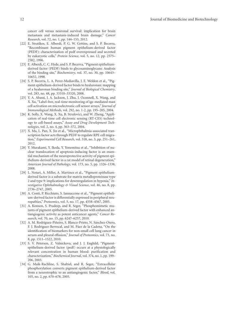
Inhibits OS cell proliferationStVOrth-278 102
StVOrth-3Promotes OS cell adhesion to collagen I
90 114
79 94ERT∗
Antiangiogenesis, prodifferentiation
44-mer∗
Prodifferentiation
4181
58 101
StVOrth-2 (residues 78–102) StVOrth-3 (residues 90–114)
Figure 1: StVOrth-2 and StVOrth-3 peptides. StVOrth-2 (highlighted in blue) consists of residues 78–102 of the parent PEDF sequence.StVOrth-3 (highlighted in red) consists of residues 90–114. StVOrth-2 and StVOrth-3 have previously been shown to inhibit osteosarcomacell proliferation and promote osteosarcoma cell adhesion to collagen I in vitro, respectively [7]. ∗ERT and 44-mer peptide sequences, asdescribed by Filleur et al. [6], overlap with the StVOrth-2 and StVOrth-3 sequences.
In another study, four different PEDF-derived peptides,termed StVOrth-1, StVOrth-2, StVOrth-3, and StVOrth-4, consisting of PEDF residues 40–64, 78–102, 90–114,and 387–411, respectively, were tested in vitro and in vivo[7]. In vitro, StVOrth-2 was the most potent inhibitorof SaOS-2 osteosarcoma cell proliferation, while StVOrth-3 dramatically promoted SaOS-2 adhesion to collagen I.StVOrth-4 inhibited SaOS-2 cell invasion through Matrigel.StVOrth-1, -2, and -3 all induced osteoblastic differentiation.StVOrth-3 and StVOrth-4 reduced VEGF expression inSaOS-2 osteosarcoma cells. StVOrth-2 and StVOrth-3 werethen evaluated in vivo using an orthotopic murine modelof osteosarcoma. Notably, StVOrth-2 (residues 78–102) andStVOrth-3 (residues 90–114) possessed sequences that over-lapped with the 44-mer (residues 58–101) and ERT (residues79–94) sequences described by Filleur et al. [6] (Figure 1).Both StVOrth-2 and StVOrth-3 restricted osteosarcomatumour growth and inhibited the development of pulmonarymetastases when SaOS-2 cells were treated prior to intratibialinjection.
The findings of both Filleur et al. [6] and Ek et al. [7]provide some insight into how PEDF structure relates toits multidimensional ability to restrict tumour progression.However, the study design and methods of peptide deliveryused in these models make it difficult to extrapolate thefindings for human use. Filleur et al. [6] used a subcutaneoustumour model with transfected PC-3 cells to demonstrate
the differential effects of the 34-mer and 44-mer PEDF-derived peptides. Gene therapies have yet to be proven safefor human application, making it unlikely that they will beused for osteosarcoma therapy in the near future [8]. Eket al. [7] used the SaOS-2 osteosarcoma cell line to achievea spontaneously metastasizing murine model of orthotopicosteosarcoma. SaOS-2 cells were treated with StVOrth-2and StVOrth-3 peptides prior to intraosseous injection,thus facilitating early uptake of peptides and change inphenotype. In order to evaluate the true efficacy of PEDF-derived peptides against established osteosarcoma, treatmentshould be delayed until after the establishment of primarytumours. This would better simulate the clinical presentationand treatment of osteosarcoma in humans.
2. Materials and Methods
2.1. Cells and Culture Conditions. The SaOS-2 human os-teosarcoma cell line (American Tissue Culture Collection,Manassas, VA, USA) was cultured in complete medium,CM, at 37◦C and in humidified 5% CO2. CM consisted ofMEM-Alpha+GlutaMAX (Invitrogen, Carlsbad, CA, USA)supplemented with 10% foetal bovine serum (Invitrogen,Carlsbad, CA, USA) and 1% antibiotic-antimycotic (Invitro-gen, Carlsbad, CA, USA). Exponentially growing cells, withpassage number always less than 20, were used for the studies.
Journal of Biomedicine and Biotechnology 3
2.2. Establishment of the Orthotopic Model of Osteosarcoma.5-week-old Balb/c nude mice were purchased from theAnimal Resource Centre, Perth, Australia and were housedat the St Vincent’s Hospital BioResources Centre underPC2 pathogen-free conditions. Animal ethics approval wasobtained from the St Vincent’s Hospital Melbourne AnimalEthics Committee. A 50% concentration of Matrigel wasused to dilute SaOS-2 osteosarcoma cells to a concentrationof 2 × 10⁶ cells/mL. Following anaesthesia with intraperi-toneal ketamine (100 mg/kg) and xylazine (10 mg/kg), a 27-gauge needle was introduced into the left tibia of each mouseusing a gentle drilling motion in order to avoid iatrogenicfracture [9], and a volume of 10 μL of SaOS-2/Matrigelsolution was injected. Postinjection, the needle was retractedslowly to prevent backflow of injectate.
Tumour growth and animal weights were monitoredtwice weekly until the endpoint of the study. Anteroposterior(AP) and lateral (L) dimensions of limbs were recordedusing digital callipers. Volumes were calculated from thesedimensions using the formula 4/3π[1/4(AP + L)]2 [7]. Thecontralateral nontumour-bearing limb was used as a controlto calculate actual tumour volume.
Primary orthotopic tumours were apparent at day 20after SaOS-2 inoculation, when average tumour volumewas 22.5 mm3, at which point mice were randomised intotreatment groups each consisting of four mice. As outlinedbelow, these groups received either: (1) sterile water ascontrol, (2) StVOrth-2 at 50 μg/kg/day, (3) StVOrth-2at 500 μg/kg/day, (4) StVOrth-3 at 50 μg/kg/day, or (5)StVOrth-3 at 500 μg/kg/day.
2.3. Delivery of PEDF-Derived Synthetic Peptides. PEDF-derived peptides, StVOrth-2 (residues 78–102) and StVOrth-3 (90–114), were designed and sourced previously accordingto the procedure outlined by Ek et al. [7]. This paper refersto the full-length human PEDF sequence, and amino acidnumbering is based on those sequences listed in GenBank(National Institutes of Health). StVOrth-2 is the aminoacidsequence 78VLLSP LSVAT ALSAL SLGAE QRTES102.StVOrth-3 is the sequence 90SALSL GAEQR TESII HRALYYDLIS114. High-performance liquid chromatography(HPLC) and mass spectrometry was used to confirm thepurity of these peptides. Ek et al. [7] showed that StVOrth-2inhibited SaOS-2 osteosarcoma cell proliferation, whileStVOrth-3 inhibited SaOS-2 osteosarcoma cell adhesion tocollagen I in vitro. StVOrth-2 and StVOrth-3 sequences over-lap with the 44-mer (residues 58–101), and ERT (residues79–94) sequences described by Filleur et al. [6] (Figure 1).
Sustained delivery of StVOrth-2, StVOrth-3, or sterilewater (negative control, placebo) was achieved by intraperi-toneally implanted Alzet microosmotic pump (Durect Corp.,Cupertino, CA, USA). Pumps were aseptically filled withthe different treatments and surgically implanted withinthe peritoneal cavity of animals for systemic delivery. Thissurgery was performed at day 20 after SaOS-2 injection.The mean pumping rate for the Alzet microosmotic pump(model 1002) is 0.25 μL/hr over 14 days, as determined by themanufacturer. StVOrth-2 and StVOrth-3 were administered
at 50 μg/kg and 500 μg/kg daily doses. Sterile water was usedas diluent for StVOrth-2 and StVOrth-3.
The human physiological serum concentration of PEDFranges between 4 ng/mL (80 pM) to 15 μg/mL (300 nM) [10–15]. In one study, inhibition of vessel formation in ischemia-induced retinopathy was achieved at a 50 nM concentration[16]. The 50 μg/kg and 500 μg/kg doses used in the presentstudy are equivalent to 1 μg/mL (20 nM) and 10 μg/mL(200 nM) concentrations of PEDF, respectively, which lieswithin the range mentioned above. This assumes an averagemouse weight of 20 grams and an average blood volumeof 1 mL [17]. It is known that the pumps are capableof delivering a steady state quantity of peptides into theabdominal cavity [18, 19], which will eventually be taken intothe microvasculature supplying the abdominal region.
2.4. Study Endpoint and Tissue Analysis. Tumours had grownto a disabling size for control animals at day 34 after SaOS-2 inoculation as expected. This was the humane endpoint ofthe study, and all animals were euthanized under anaesthesiaby cervical dislocation at this time. Following this, tumour-bearing limbs, lungs, hearts, small intestines, and skinwere harvested for examination. All specimens were fixedin 4% paraformaldehyde, followed by paraffin embedding.Blood samples were obtained after cervical dislocation anddissection through the thoracic cage and were immediatelytreated with anticoagulants. Affected limbs were X rayedat 35 kV for 30 s using a cabinet system (Faxitron Corp.,Wheeling, IL, USA).
Blood collected after euthanasia was analysed for renaland hepatic biochemical parameters (serum creatinine, ala-nine transaminase (ALT), and aspartate transferase (AST))using a Sysmex XE2100 instrument [20]. Tissues wereembedded in paraffin prior to histological preparationand analysis. For preparation of paraffin sections, 4 μmsections of tumours and viscera were cut by microtome.Tumours were sectioned to provide an en face surface for thequantification of per cent tumour necrosis and apoptosis.
Primary tumours, lungs, heart, small intestine, and skinsections were dehydrated through an ethanol series followedby xylene, then stained with haematoxylin and eosin.A terminal dUTP nick end labelling (TUNEL) assay kit(Promega, Madison, WI, USA) was used to detect apoptoticcells in primary tumours [21]. Pertex mounting agent wasused to seal coverslips to slide sections, and all tissues wereobserved using a Nikon Eclipse TE2000-U microscope(Nikon, Lidcombe, NSW, Australia) and photographed withSPOT Advanced software (SciTech, Aurora, IL, USA).
The effect of intraperitoneal StVOrth-2 and StVOrth-3on the development of pulmonary metastatic disease wasexamined histologically. Lungs were sectioned to achieve thegreatest cross-sectional area and stained with haematoxylinand eosin. Micrometastases were identified by systematicallyscanning lung sections under 20x magnification. Discernibleclusters of metastatic cells at this magnification were countedas micrometastases. Ten metastatic lesions were then ran-domly selected from each treatment group for measurementof cross-sectional areas.
4 Journal of Biomedicine and Biotechnology
WaterμStVOrth-2 50 g/kg/day
StVOrth-2 500 μg/kg/day
StVOrth-3 50 μg/kg/dayStVOrth-3 500 μg/kg/day
30
20
10
0
Wei
ght
(g)
20 23 27 30 34
Day
(a)
Wat
er
Wat
er
Wat
er
40
30
20
10
0
1500
1000
500
0
150
100
50
0
(mm
ol/L
)
(U/L
)
(U/L
)
Creatinine AST ALT
StV
Ort
h-2
50μ
g/kg
/day
StV
Ort
h-2
500μ
g/kg
/day
StV
Ort
h-3
50μ
g/kg
/day
StV
Ort
h-3
50oμ
g/kg
/day
StV
Ort
h-2
50μ
g/kg
/day
StV
Ort
h-2
500μ
g/kg
/day
StV
Ort
h-3
50μ
g/kg
/day
StV
Ort
h-3
50oμ
g/kg
/day
StV
Ort
h-2
50μ
g/kg
/day
StV
Ort
h-2
500μ
g/kg
/day
StV
Ort
h-3
50μ
g/kg
/day
StV
Ort
h-3
50oμ
g/kg
/day
(b)
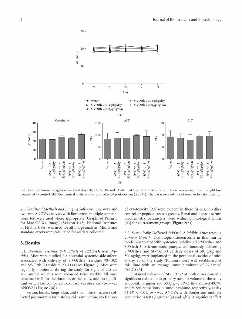
Figure 2: (a) Animal weights recorded at days 20, 23, 27, 30, and 34 after SaOS-2 intratibial injection. There was no significant weight losscompared to control. (b) Biochemical analysis of serum collected postmortem (±SEM). There was no evidence of renal or hepatic toxicity.
2.5. Statistical Methods and Imaging Software. One-way andtwo-way ANOVA analyses with Bonferroni multiple compar-isons test were used where appropriate (GraphPad Prism 5for Mac OS X). ImageJ (Version 1.45j, National Institutesof Health, USA) was used for all image analysis. Means andstandard errors were calculated for all data collected.
3. Results
3.1. Potential Systemic Side Effects of PEDF-Derived Pep-tides. Mice were studied for potential systemic side affectsassociated with delivery of StVOrth-2 (residues 78–102)and StVOrth-3 (residues 90–114) (see Figure 1). Mice wereregularly monitored during the study for signs of distressand animal weights were recorded twice weekly. All miceremained well for the duration of the study, and no signifi-cant weight loss compared to control was observed (two-wayANOVA) (Figure 2(a)).
Serum, hearts, lungs, skin, and small intestines were col-lected postmortem for histological examination. No features
of cytotoxicity [22] were evident in these tissues, in eithercontrol or peptide-treated groups. Renal and hepatic serumbiochemistry parameters were within physiological limits[23] for all treatment groups (Figure 2(b)).
3.2. Systemically Delivered StVOrth-2 Inhibits OsteosarcomaTumour Growth. Orthotopic osteosarcoma in this murinemodel was treated with systemically delivered StVOrth-2 andStVOrth-3. Microosmotic pumps, continuously deliveringStVOrth-2 and StVOrth-3 at daily doses of 50 μg/kg and500 μg/kg, were implanted in the peritoneal cavities of miceat day 20 of the study. Tumours were well established atthis time with an average tumour volume of 22.5 mm3
(±1.7 SEM).Sustained delivery of StVOrth-2 at both doses caused a
significant reduction in primary tumour volume at the studyendpoint. 50 μg/kg and 500 μg/kg StVOrth-2 caused 49.7%and 50.9% reductions in tumour volume, respectively, at day34 (P < 0.01, two-way ANOVA with Bonferroni multiplecomparisons test) (Figures 3(a) and 3(b)). A significant effect
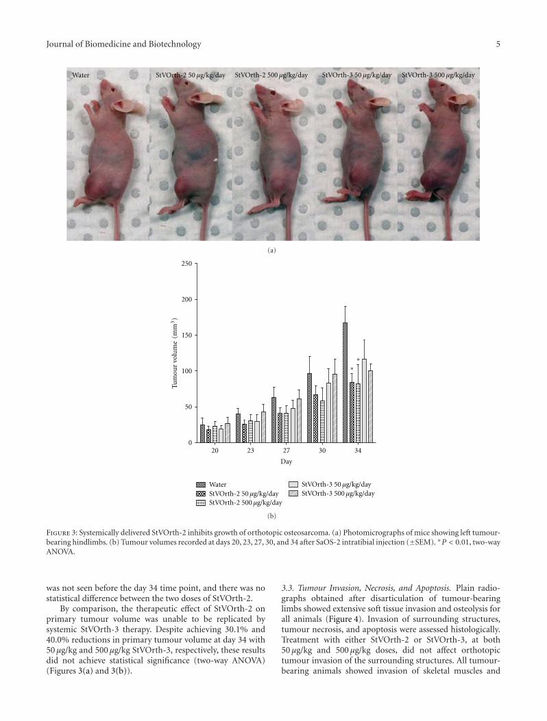
Journal of Biomedicine and Biotechnology 5
Water μStVOrth-2 50 g/kg/day StVOrth-2 500 μg/kg/day StVOrth-3 50 μg/kg/day StVOrth-3 500 μg/kg/day
(a)
250
200
150
100
50
0
Tum
our
volu
me
(mm
3)
20 23 27 30 34
Day
WaterμStVOrth-2 50 g/kg/day
StVOrth-2 500 μg/kg/day
StVOrth-3 50 μg/kg/dayStVOrth-3 500 μg/kg/day
∗∗
(b)
Figure 3: Systemically delivered StVOrth-2 inhibits growth of orthotopic osteosarcoma. (a) Photomicrographs of mice showing left tumour-bearing hindlimbs. (b) Tumour volumes recorded at days 20, 23, 27, 30, and 34 after SaOS-2 intratibial injection (±SEM). ∗P < 0.01, two-wayANOVA.
was not seen before the day 34 time point, and there was nostatistical difference between the two doses of StVOrth-2.
By comparison, the therapeutic effect of StVOrth-2 onprimary tumour volume was unable to be replicated bysystemic StVOrth-3 therapy. Despite achieving 30.1% and40.0% reductions in primary tumour volume at day 34 with50 μg/kg and 500 μg/kg StVOrth-3, respectively, these resultsdid not achieve statistical significance (two-way ANOVA)(Figures 3(a) and 3(b)).
3.3. Tumour Invasion, Necrosis, and Apoptosis. Plain radio-graphs obtained after disarticulation of tumour-bearinglimbs showed extensive soft tissue invasion and osteolysis forall animals (Figure 4). Invasion of surrounding structures,tumour necrosis, and apoptosis were assessed histologically.Treatment with either StVOrth-2 or StVOrth-3, at both50 μg/kg and 500 μg/kg doses, did not affect orthotopictumour invasion of the surrounding structures. All tumour-bearing animals showed invasion of skeletal muscles and
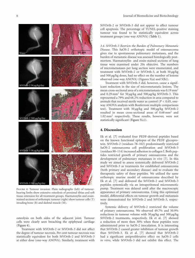
6 Journal of Biomedicine and Biotechnology
Water
μStVOrth-2 50 g/kg/day
StVOrth-2 500 μg/kg/day
StVOrth-3 50 μg/kg/day
StVOrth-3 500 μg/kg/day
T
T
T
T
T
B
B
B
B
M
Figure 4: Tumour invasion. Plain radiographs (left) of tumour-bearing limbs show extensive osteolysis of proximal tibiae and softtissue extension for all treatment groups. Haematoxylin and eosin-stained sections of orthotopic tumour (right) show tumour cells (T)invading bone (B) and skeletal muscle (M).
osteolysis on both sides of the adjacent joint. Tumourcells were clearly seen breaching the epiphyseal cartilage(Figure 4).
Treatment with StVOrth-2 or StVOrth-3 did not affectthe degree of tumour necrosis. Per cent tumour necrosis wasstatistically equivalent for both StVOrth-2 and StVOrth-3at either dose (one-way ANOVA). Similarly, treatment with
StVOrth-2 or StVOrth-3 did not appear to affect tumourcell apoptosis. The percentage of TUNEL-positive stainingtumour was found to be statistically equivalent acrosstreatment groups (one-way ANOVA) (Table 1).
3.4. StVOrth-3 Restricts the Burden of Pulmonary MetastaticDisease. This SaOS-2 orthotopic model of osteosarcomagives rise to spontaneous pulmonary metastases, and theburden of metastatic disease was assessed histologically post-mortem. Haematoxylin- and eosin-stained sections of lungtissue were examined under 20x objective. The numbersof micrometastases per lung section were enumerated, andtreatment with StVOrth-2 or StVOrth-3, at both 50 μg/kgand 500 μg/kg doses, had no effect on the number of lesionsobserved (one-way ANOVA) (Figures 5(a) and 5(b)).
Treatment with StVOrth-3 did, however, cause a signif-icant reduction in the size of micrometastatic lesions. Themean cross-sectional area of a micrometastasis was 0.19 mm2
and 0.29 mm2 for 50 μg/kg and 500 μg/kg StVOrth-3. Thisrepresented a 79% and 68.2% reduction in area compared toanimals that received sterile water as control (P < 0.05, one-way ANOVA analysis with Bonferroni multiple comparisonstest). Treatment with 50 μg/kg and 500 μg/kg StVOrth-2resulted in mean cross-sectional areas of 0.68 mm2 and1.02 mm2 respectively. These results, however, were notstatistically significant (Figure 5(c)).
4. Discussion
Ek et al. [7] evaluated four PEDF-derived peptides basedon the known functional epitopes of the PEDF glycopro-tein. StVOrth-2 (residues 78–102) predominantly restrictedSaOS-2 osteosarcoma cell proliferation and StVOrth-3(residues 90–114) increased adhesion to collagen I. Both pep-tides restricted growth of primary osteosarcoma and thedevelopment of pulmonary metastases in vivo [7]. In thisstudy we aimed to assess systemically delivered StVOrth-2and StVOrth-3 as treatments for established osteosarcoma(both primary and secondary disease) and to evaluate thetherapeutic safety of these peptides. We utilised the sameorthotopic murine model of osteosarcoma described byEk et al. [7] and delivered the StVOrth-2 and StVOrth-3peptides systemically via an intraperitoneal microosmoticpump. Treatment was delayed until after the macroscopicappearance of primary osteosarcoma. Using this optimisedmodel, differential effects on tumour growth and metastasiswere demonstrated for StVOrth-2 and StVOrth-3, respec-tively.
Systemic delivery of StVOrth-2 restricted the volumeof primary osteosarcoma. We observed 49.7% and 50.9%reductions in tumour volume with 50 μg/kg and 500 μg/kgStVOrth-2 treatments, respectively. Ek et al. [7] showeda reduction of more then 30% when these peptides wereadministered prior to SaOS-2 inoculation. It is interestingthat StVOrth-2 caused greater inhibition of tumour growththan StVOrth-3. Ek et al. [7] showed that StVOrth-2had a significant antiproliferative effect on SaOS-2 cellsin vitro, while StVOrth-3 did not exhibit this effect. The
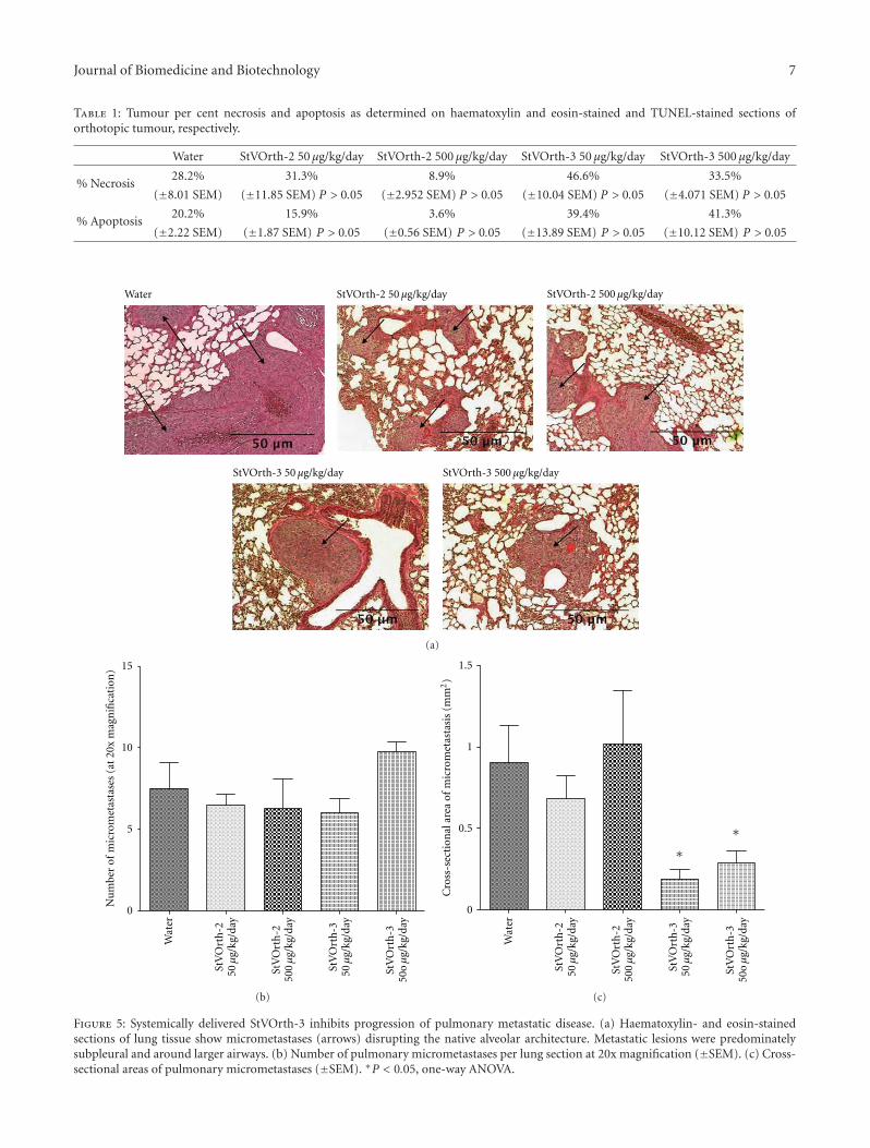
Journal of Biomedicine and Biotechnology 7
Table 1: Tumour per cent necrosis and apoptosis as determined on haematoxylin and eosin-stained and TUNEL-stained sections oforthotopic tumour, respectively.
Water StVOrth-2 50 μg/kg/day StVOrth-2 500 μg/kg/day StVOrth-3 50 μg/kg/day StVOrth-3 500 μg/kg/day
% Necrosis28.2% 31.3% 8.9% 46.6% 33.5%
(±8.01 SEM) (±11.85 SEM) P > 0.05 (±2.952 SEM) P > 0.05 (±10.04 SEM) P > 0.05 (±4.071 SEM) P > 0.05
% Apoptosis20.2% 15.9% 3.6% 39.4% 41.3%
(±2.22 SEM) (±1.87 SEM) P > 0.05 (±0.56 SEM) P > 0.05 (±13.89 SEM) P > 0.05 (±10.12 SEM) P > 0.05
μStVOrth-2 50 g/kg/day StVOrth-2 500 μg/kg/day
StVOrth-3 50 μg/kg/day StVOrth-3 500 μg/kg/day
Water
(a)
Wat
er
Nu
mbe
r of
mic
rom
etas
tase
s (a
t 20
x m
agn
ifica
tion
)
10
5
0
15
StV
Ort
h-2
50μ
g/kg
/day
StV
Ort
h-2
500μ
g/kg
/day
StV
Ort
h-3
50μ
g/kg
/day
StV
Ort
h-3
50oμ
g/kg
/day
(b)
Wat
er
1.5
1
0.5
0
Cro
ss-s
ecti
onal
are
a of
mic
rom
etas
tasi
s (m
m2)
∗∗
StV
Ort
h-2
50μ
g/kg
/day
StV
Ort
h-2
500μ
g/kg
/day
StV
Ort
h-3
50μ
g/kg
/day
StV
Ort
h-3
50oμ
g/kg
/day
(c)
Figure 5: Systemically delivered StVOrth-3 inhibits progression of pulmonary metastatic disease. (a) Haematoxylin- and eosin-stainedsections of lung tissue show micrometastases (arrows) disrupting the native alveolar architecture. Metastatic lesions were predominatelysubpleural and around larger airways. (b) Number of pulmonary micrometastases per lung section at 20x magnification (±SEM). (c) Cross-sectional areas of pulmonary micrometastases (±SEM). ∗P < 0.05, one-way ANOVA.
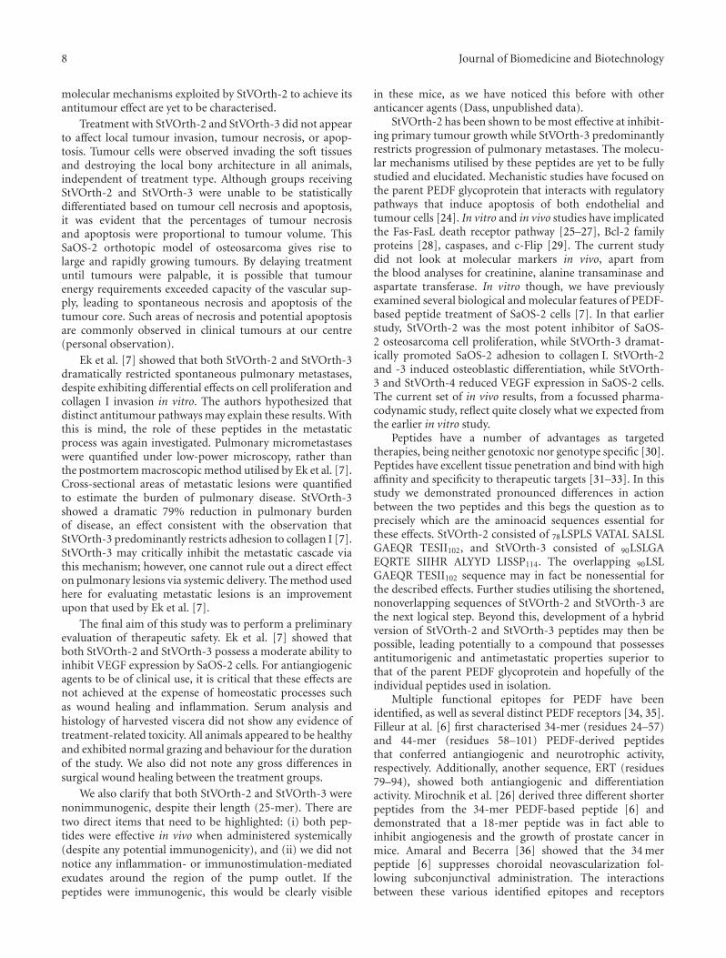
8 Journal of Biomedicine and Biotechnology
molecular mechanisms exploited by StVOrth-2 to achieve itsantitumour effect are yet to be characterised.
Treatment with StVOrth-2 and StVOrth-3 did not appearto affect local tumour invasion, tumour necrosis, or apop-tosis. Tumour cells were observed invading the soft tissuesand destroying the local bony architecture in all animals,independent of treatment type. Although groups receivingStVOrth-2 and StVOrth-3 were unable to be statisticallydifferentiated based on tumour cell necrosis and apoptosis,it was evident that the percentages of tumour necrosisand apoptosis were proportional to tumour volume. ThisSaOS-2 orthotopic model of osteosarcoma gives rise tolarge and rapidly growing tumours. By delaying treatmentuntil tumours were palpable, it is possible that tumourenergy requirements exceeded capacity of the vascular sup-ply, leading to spontaneous necrosis and apoptosis of thetumour core. Such areas of necrosis and potential apoptosisare commonly observed in clinical tumours at our centre(personal observation).
Ek et al. [7] showed that both StVOrth-2 and StVOrth-3dramatically restricted spontaneous pulmonary metastases,despite exhibiting differential effects on cell proliferation andcollagen I invasion in vitro. The authors hypothesized thatdistinct antitumour pathways may explain these results. Withthis is mind, the role of these peptides in the metastaticprocess was again investigated. Pulmonary micrometastaseswere quantified under low-power microscopy, rather thanthe postmortem macroscopic method utilised by Ek et al. [7].Cross-sectional areas of metastatic lesions were quantifiedto estimate the burden of pulmonary disease. StVOrth-3showed a dramatic 79% reduction in pulmonary burdenof disease, an effect consistent with the observation thatStVOrth-3 predominantly restricts adhesion to collagen I [7].StVOrth-3 may critically inhibit the metastatic cascade viathis mechanism; however, one cannot rule out a direct effecton pulmonary lesions via systemic delivery. The method usedhere for evaluating metastatic lesions is an improvementupon that used by Ek et al. [7].
The final aim of this study was to perform a preliminaryevaluation of therapeutic safety. Ek et al. [7] showed thatboth StVOrth-2 and StVOrth-3 possess a moderate ability toinhibit VEGF expression by SaOS-2 cells. For antiangiogenicagents to be of clinical use, it is critical that these effects arenot achieved at the expense of homeostatic processes suchas wound healing and inflammation. Serum analysis andhistology of harvested viscera did not show any evidence oftreatment-related toxicity. All animals appeared to be healthyand exhibited normal grazing and behaviour for the durationof the study. We also did not note any gross differences insurgical wound healing between the treatment groups.
We also clarify that both StVOrth-2 and StVOrth-3 werenonimmunogenic, despite their length (25-mer). There aretwo direct items that need to be highlighted: (i) both pep-tides were effective in vivo when administered systemically(despite any potential immunogenicity), and (ii) we did notnotice any inflammation- or immunostimulation-mediatedexudates around the region of the pump outlet. If thepeptides were immunogenic, this would be clearly visible
in these mice, as we have noticed this before with otheranticancer agents (Dass, unpublished data).
StVOrth-2 has been shown to be most effective at inhibit-ing primary tumour growth while StVOrth-3 predominantlyrestricts progression of pulmonary metastases. The molecu-lar mechanisms utilised by these peptides are yet to be fullystudied and elucidated. Mechanistic studies have focused onthe parent PEDF glycoprotein that interacts with regulatorypathways that induce apoptosis of both endothelial andtumour cells [24]. In vitro and in vivo studies have implicatedthe Fas-FasL death receptor pathway [25–27], Bcl-2 familyproteins [28], caspases, and c-Flip [29]. The current studydid not look at molecular markers in vivo, apart fromthe blood analyses for creatinine, alanine transaminase andaspartate transferase. In vitro though, we have previouslyexamined several biological and molecular features of PEDF-based peptide treatment of SaOS-2 cells [7]. In that earlierstudy, StVOrth-2 was the most potent inhibitor of SaOS-2 osteosarcoma cell proliferation, while StVOrth-3 dramat-ically promoted SaOS-2 adhesion to collagen I. StVOrth-2and -3 induced osteoblastic differentiation, while StVOrth-3 and StVOrth-4 reduced VEGF expression in SaOS-2 cells.The current set of in vivo results, from a focussed pharma-codynamic study, reflect quite closely what we expected fromthe earlier in vitro study.
Peptides have a number of advantages as targetedtherapies, being neither genotoxic nor genotype specific [30].Peptides have excellent tissue penetration and bind with highaffinity and specificity to therapeutic targets [31–33]. In thisstudy we demonstrated pronounced differences in actionbetween the two peptides and this begs the question as toprecisely which are the aminoacid sequences essential forthese effects. StVOrth-2 consisted of 78LSPLS VATAL SALSLGAEQR TESII102, and StVOrth-3 consisted of 90LSLGAEQRTE SIIHR ALYYD LISSP114. The overlapping 90LSLGAEQR TESII102 sequence may in fact be nonessential forthe described effects. Further studies utilising the shortened,nonoverlapping sequences of StVOrth-2 and StVOrth-3 arethe next logical step. Beyond this, development of a hybridversion of StVOrth-2 and StVOrth-3 peptides may then bepossible, leading potentially to a compound that possessesantitumorigenic and antimetastatic properties superior tothat of the parent PEDF glycoprotein and hopefully of theindividual peptides used in isolation.
Multiple functional epitopes for PEDF have beenidentified, as well as several distinct PEDF receptors [34, 35].Filleur at al. [6] first characterised 34-mer (residues 24–57)and 44-mer (residues 58–101) PEDF-derived peptidesthat conferred antiangiogenic and neurotrophic activity,respectively. Additionally, another sequence, ERT (residues79–94), showed both antiangiogenic and differentiationactivity. Mirochnik et al. [26] derived three different shorterpeptides from the 34-mer PEDF-based peptide [6] anddemonstrated that a 18-mer peptide was in fact able toinhibit angiogenesis and the growth of prostate cancer inmice. Amaral and Becerra [36] showed that the 34 merpeptide [6] suppresses choroidal neovascularization fol-lowing subconjunctival administration. The interactionsbetween these various identified epitopes and receptors
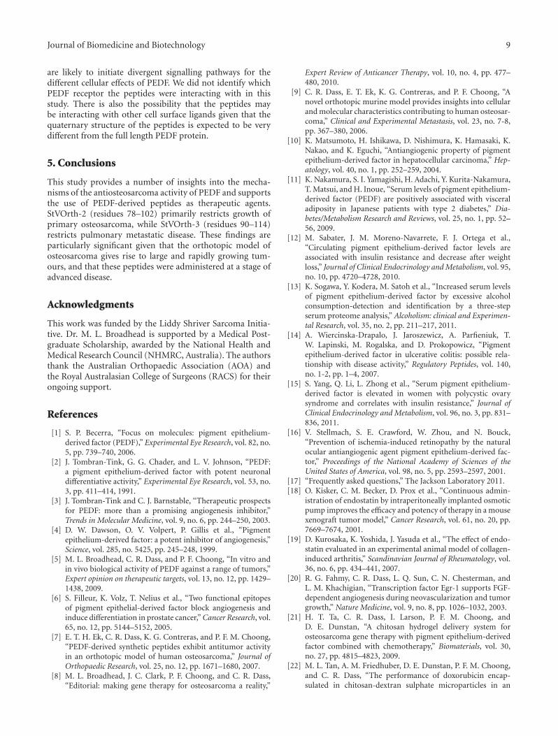
Journal of Biomedicine and Biotechnology 9
are likely to initiate divergent signalling pathways for thedifferent cellular effects of PEDF. We did not identify whichPEDF receptor the peptides were interacting with in thisstudy. There is also the possibility that the peptides maybe interacting with other cell surface ligands given that thequaternary structure of the peptides is expected to be verydifferent from the full length PEDF protein.
5. Conclusions
This study provides a number of insights into the mecha-nisms of the antiosteosarcoma activity of PEDF and supportsthe use of PEDF-derived peptides as therapeutic agents.StVOrth-2 (residues 78–102) primarily restricts growth ofprimary osteosarcoma, while StVOrth-3 (residues 90–114)restricts pulmonary metastatic disease. These findings areparticularly significant given that the orthotopic model ofosteosarcoma gives rise to large and rapidly growing tum-ours, and that these peptides were administered at a stage ofadvanced disease.
Acknowledgments
This work was funded by the Liddy Shriver Sarcoma Initia-tive. Dr. M. L. Broadhead is supported by a Medical Post-graduate Scholarship, awarded by the National Health andMedical Research Council (NHMRC, Australia). The authorsthank the Australian Orthopaedic Association (AOA) andthe Royal Australasian College of Surgeons (RACS) for theirongoing support.
References
[1] S. P. Becerra, “Focus on molecules: pigment epithelium-derived factor (PEDF),” Experimental Eye Research, vol. 82, no.5, pp. 739–740, 2006.
[2] J. Tombran-Tink, G. G. Chader, and L. V. Johnson, “PEDF:a pigment epithelium-derived factor with potent neuronaldifferentiative activity,” Experimental Eye Research, vol. 53, no.3, pp. 411–414, 1991.
[3] J. Tombran-Tink and C. J. Barnstable, “Therapeutic prospectsfor PEDF: more than a promising angiogenesis inhibitor,”Trends in Molecular Medicine, vol. 9, no. 6, pp. 244–250, 2003.
[4] D. W. Dawson, O. V. Volpert, P. Gillis et al., “Pigmentepithelium-derived factor: a potent inhibitor of angiogenesis,”Science, vol. 285, no. 5425, pp. 245–248, 1999.
[5] M. L. Broadhead, C. R. Dass, and P. F. Choong, “In vitro andin vivo biological activity of PEDF against a range of tumors,”Expert opinion on therapeutic targets, vol. 13, no. 12, pp. 1429–1438, 2009.
[6] S. Filleur, K. Volz, T. Nelius et al., “Two functional epitopesof pigment epithelial-derived factor block angiogenesis andinduce differentiation in prostate cancer,” Cancer Research, vol.65, no. 12, pp. 5144–5152, 2005.
[7] E. T. H. Ek, C. R. Dass, K. G. Contreras, and P. F. M. Choong,“PEDF-derived synthetic peptides exhibit antitumor activityin an orthotopic model of human osteosarcoma,” Journal ofOrthopaedic Research, vol. 25, no. 12, pp. 1671–1680, 2007.
[8] M. L. Broadhead, J. C. Clark, P. F. Choong, and C. R. Dass,“Editorial: making gene therapy for osteosarcoma a reality,”
Expert Review of Anticancer Therapy, vol. 10, no. 4, pp. 477–480, 2010.
[9] C. R. Dass, E. T. Ek, K. G. Contreras, and P. F. Choong, “Anovel orthotopic murine model provides insights into cellularand molecular characteristics contributing to human osteosar-coma,” Clinical and Experimental Metastasis, vol. 23, no. 7-8,pp. 367–380, 2006.
[10] K. Matsumoto, H. Ishikawa, D. Nishimura, K. Hamasaki, K.Nakao, and K. Eguchi, “Antiangiogenic property of pigmentepithelium-derived factor in hepatocellular carcinoma,” Hep-atology, vol. 40, no. 1, pp. 252–259, 2004.
[11] K. Nakamura, S. I. Yamagishi, H. Adachi, Y. Kurita-Nakamura,T. Matsui, and H. Inoue, “Serum levels of pigment epithelium-derived factor (PEDF) are positively associated with visceraladiposity in Japanese patients with type 2 diabetes,” Dia-betes/Metabolism Research and Reviews, vol. 25, no. 1, pp. 52–56, 2009.
[12] M. Sabater, J. M. Moreno-Navarrete, F. J. Ortega et al.,“Circulating pigment epithelium-derived factor levels areassociated with insulin resistance and decrease after weightloss,” Journal of Clinical Endocrinology and Metabolism, vol. 95,no. 10, pp. 4720–4728, 2010.
[13] K. Sogawa, Y. Kodera, M. Satoh et al., “Increased serum levelsof pigment epithelium-derived factor by excessive alcoholconsumption-detection and identification by a three-stepserum proteome analysis,” Alcoholism: clinical and Experimen-tal Research, vol. 35, no. 2, pp. 211–217, 2011.
[14] A. Wiercinska-Drapalo, J. Jaroszewicz, A. Parfieniuk, T.W. Lapinski, M. Rogalska, and D. Prokopowicz, “Pigmentepithelium-derived factor in ulcerative colitis: possible rela-tionship with disease activity,” Regulatory Peptides, vol. 140,no. 1-2, pp. 1–4, 2007.
[15] S. Yang, Q. Li, L. Zhong et al., “Serum pigment epithelium-derived factor is elevated in women with polycystic ovarysyndrome and correlates with insulin resistance,” Journal ofClinical Endocrinology and Metabolism, vol. 96, no. 3, pp. 831–836, 2011.
[16] V. Stellmach, S. E. Crawford, W. Zhou, and N. Bouck,“Prevention of ischemia-induced retinopathy by the naturalocular antiangiogenic agent pigment epithelium-derived fac-tor,” Proceedings of the National Academy of Sciences of theUnited States of America, vol. 98, no. 5, pp. 2593–2597, 2001.
[17] “Frequently asked questions,” The Jackson Laboratory 2011.[18] O. Kisker, C. M. Becker, D. Prox et al., “Continuous admin-
istration of endostatin by intraperitoneally implanted osmoticpump improves the efficacy and potency of therapy in a mousexenograft tumor model,” Cancer Research, vol. 61, no. 20, pp.7669–7674, 2001.
[19] D. Kurosaka, K. Yoshida, J. Yasuda et al., “The effect of endo-statin evaluated in an experimental animal model of collagen-induced arthritis,” Scandinavian Journal of Rheumatology, vol.36, no. 6, pp. 434–441, 2007.
[20] R. G. Fahmy, C. R. Dass, L. Q. Sun, C. N. Chesterman, andL. M. Khachigian, “Transcription factor Egr-1 supports FGF-dependent angiogenesis during neovascularization and tumorgrowth,” Nature Medicine, vol. 9, no. 8, pp. 1026–1032, 2003.
[21] H. T. Ta, C. R. Dass, I. Larson, P. F. M. Choong, andD. E. Dunstan, “A chitosan hydrogel delivery system forosteosarcoma gene therapy with pigment epithelium-derivedfactor combined with chemotherapy,” Biomaterials, vol. 30,no. 27, pp. 4815–4823, 2009.
[22] M. L. Tan, A. M. Friedhuber, D. E. Dunstan, P. F. M. Choong,and C. R. Dass, “The performance of doxorubicin encap-sulated in chitosan-dextran sulphate microparticles in an
10 Journal of Biomedicine and Biotechnology
osteosarcoma model,” Biomaterials, vol. 31, no. 3, pp. 541–551,2010.
[23] “Reference values for laboratory animals,” Research AnimalResources, University of Minnesota, 2009.
[24] M. L. Broadhead, C. R. Dass, and P. F. M. Choong, “Cancer cellapoptotic pathways mediated by PEDF: prospects for therapy,”Trends in Molecular Medicine, vol. 15, no. 10, pp. 461–467,2009.
[25] R. Abe, T. Shimizu, S. I. Yamagishi et al., “Overexpression ofpigment epithelium-derived factor decreases angiogenesis andinhibits the growth of human malignant melanoma cells invivo,” American Journal of Pathology, vol. 164, no. 4, pp. 1225–1232, 2004.
[26] Y. Mirochnik, A. Aurora, F. T. Schulze-Hoepfner et al., “Shortpigment epithelial-derived factor-derived peptide inhibitsangiogenesis and tumor growth,” Clinical Cancer Research, vol.15, no. 5, pp. 1655–1663, 2009.
[27] O. V. Volpert, T. Zaichuk, W. Zhou et al., “Inducer-stimulatedFas targets activated endothelium for destruction by anti-angiogenic thrombospondin-1 and pigment epithelium-derived factor,” Nature Medicine, vol. 8, no. 4, pp. 349–357,2002.
[28] T. Zhang, M. Guan, C. Xu, Y. Chen, and Y. Lu, “Pigmentepithelium-derived factor inhibits glioma cell growth in vitroand in vivo,” Life Sciences, vol. 81, no. 16, pp. 1256–1263, 2007.
[29] T. A. Zaichuk, E. H. Shroff, R. Emmanuel, S. Filleur, T.Nelius, and O. V. Volpert, “Nuclear factor of activated T cellsbalances angiogenesis activation and inhibition,” Journal ofExperimental Medicine, vol. 199, no. 11, pp. 1513–1522, 2004.
[30] S. K. Bhutia and T. K. Maiti, “Targeting tumors with peptidesfrom natural sources,” Trends in Biotechnology, vol. 26, no. 4,pp. 210–217, 2008.
[31] J. S. Mader and D. W. Hoskin, “Cationic antimicrobialpeptides as novel cytotoxic agents for cancer treatment,”Expert Opinion on Investigational Drugs, vol. 15, no. 8, pp.933–946, 2006.
[32] C. Ruegg, M. Hasmim, F. J. Lejeune, and G. C. Alghisi,“Antiangiogenic peptides and proteins: from experimentaltools to clinical drugs,” Biochimica et Biophysica Acta, vol.1765, no. 2, pp. 155–177, 2006.
[33] Y. L. Janin, “Peptides with anticancer use or potential,” AminoAcids, vol. 25, no. 1, pp. 1–40, 2003.
[34] L. Notari, V. Baladron, J. D. Aroca-Aguilar et al., “Identifica-tion of a lipase-linked cell membrane receptor for pigmentepithelium-derived factor,” Journal of Biological Chemistry,vol. 281, no. 49, pp. 38022–38037, 2006.
[35] A. Bernard, J. Gao-Li, C. A. Franco, T. Bouceba, A. Huet, andZ. Li, “Laminin receptor involvement in the anti-angiogenicactivity of pigment epithelium-derived factor,” Journal ofBiological Chemistry, vol. 284, no. 16, pp. 10480–10490, 2009.
[36] J. Amaral and S. P. Becerra, “Effects of human recombinantPEDF protein and PEDF-derived peptide 34-mer on choroidalneovascularization,” Investigative ophthalmology & visual sci-ence, vol. 51, no. 3, pp. 1318–1326, 2010.
Hindawi Publishing CorporationJournal of Biomedicine and BiotechnologyVolume 2012, Article ID 740295, 9 pagesdoi:10.1155/2012/740295
Review Article
Cell and Molecular Biology Underpinning the Effects of PEDF onCancers in General and Osteosarcoma in Particular
Vijay Chandolu and Crispin R. Dass
School of Biomedical and Health Sciences, Victoria University, Building 6, St Albans, VIC 3021, Australia
Correspondence should be addressed to Crispin R. Dass, [email protected]
Received 24 October 2011; Revised 9 March 2012; Accepted 23 March 2012
Academic Editor: Susan E. Crawford
Copyright © 2012 V. Chandolu and C. R. Dass. This is an open access article distributed under the Creative Commons AttributionLicense, which permits unrestricted use, distribution, and reproduction in any medium, provided the original work is properlycited.
Cancer is becoming an increasingly common disease in which abnormal cells aggressively grow, invade, and metastasize. In thispaper, we review the biological functions of PEDF (pigmented epithelium-derived factor) against cancer, with a focus on a partic-ular type of bone cancer called osteosarcoma. PEDF is a 50 kDa glycoprotein and is a potent inhibitor of angiogenesis, via its abilityto decrease proliferation and migration of endothelial cells. This paper critically examines the anticancer activities of PEDF via itsrole in antiangiogenesis, apoptosis-mediated tumor suppression, and increased tumor cell differentiation. Recently, an orthotopicmodel of osteosarcoma was used to show that treatment with PEDF had the greatest impact on metastases, warranting anevaluation of PEDF efficacy in other types of cancers.
1. Introduction
Cancer is characterized by uncontrolled growth and spreadof cells harboring some form of genetic aberration. Forma-tion of tumours requires sequential acquisition of defectsthat endow tumour cells with the ability to grow, invade, andeventually metastasize. Even after significant improvementsin surgery, chemotherapy, and radiotherapy, there are stillnumerous tumours that are unaffected by therapy. Bythe time a tumour is diagnosed, it has almost certainlybegun metastasizing. In such cases radiation therapy andchemotherapy are often used in combination with surgeryin the hope that the tumour and its metastases are effectivelycontrolled [1]. Targeted biological therapies, which are safer,are being studied and clinical trials are conducted to gaugeefficacy. The main objective of targeted therapy is to attackspecific pathways and tumour growth mechanisms withoutthe side effects attached to small molecule drugs andradiotherapy [2, 3].
PEDF is a 50-kDa glycoprotein, which is a member of theserine protease inhibitor (serpin) family, and it has multi-functional properties [4]. It is found to be a potent inhibitorof angiogenesis, proliferation and migration of endothelialcells, retinal vascular permeability, and tumour activity [5].
PEDF is responsible for the antiangiogenesis in various ocu-lar compartments [6]. These significant antiangiogenic prop-erties led the scientists to shift focus on to studies examiningthe potential antitumour activities of PEDF. PEDF expressionchanges in the course of progression of different tumourtypes [5]. Researchers did a number of studies and showedthat there is opposite relation between PEDF levels, gradeand metastatic potential of prostate tumours [7], pancreaticadenocarcinoma [8], prostate, melanoma, ovarian, osteosar-coma, glioma [4], hepatocellular carcinoma [9], and Wilm’stumours [10].
The human PEDF gene has been mapped, and it encodesa 418 amino acid protein [11]. It has an asymmetrical chargedistribution, with a high density of basic residues concen-trated on one side and of acidic residues on the opposite side.Negatively charged acidic PEDF binds to collagen, lacks neu-rotrophic activity, and may confer antiangiogenic properties(Figure 1). Mutational studies have shown that positivelycharged amino acids and negatively-charged amino acids areresponsible for heparin and collagen binding, respectively[12, 13]. There are three phosphorylation sites identifiedwhich collectively induce antiangiogenic and neurotrophicactivities. A 34-mer peptide and 44-mer peptide are the twomajor functional epitopes identified (as shown in Figure 2)
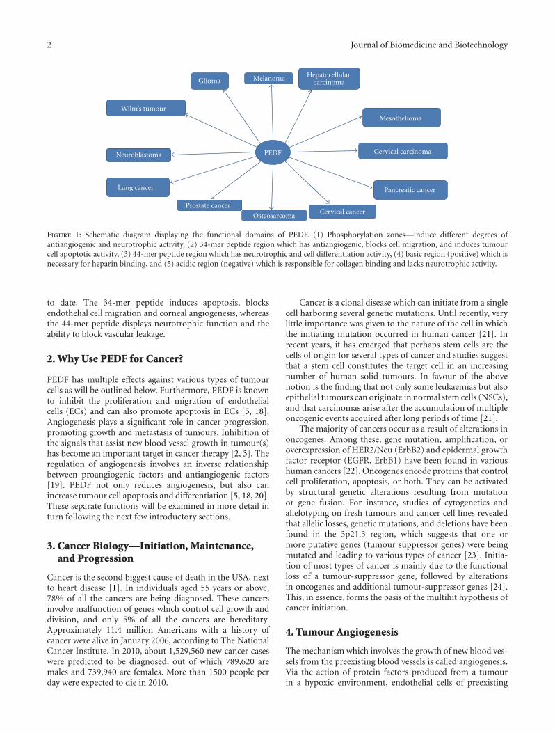
2 Journal of Biomedicine and Biotechnology
PEDF
Melanoma Hepatocellular carcinomaGlioma
Cervical carcinomaNeuroblastoma
OsteosarcomaCervical cancer
Prostate cancer
MesotheliomaWilm’s tumour
Lung cancer Pancreatic cancer
Figure 1: Schematic diagram displaying the functional domains of PEDF. (1) Phosphorylation zones—induce different degrees ofantiangiogenic and neurotrophic activity, (2) 34-mer peptide region which has antiangiogenic, blocks cell migration, and induces tumourcell apoptotic activity, (3) 44-mer peptide region which has neurotrophic and cell differentiation activity, (4) basic region (positive) which isnecessary for heparin binding, and (5) acidic region (negative) which is responsible for collagen binding and lacks neurotrophic activity.
to date. The 34-mer peptide induces apoptosis, blocksendothelial cell migration and corneal angiogenesis, whereasthe 44-mer peptide displays neurotrophic function and theability to block vascular leakage.
2. Why Use PEDF for Cancer?
PEDF has multiple effects against various types of tumourcells as will be outlined below. Furthermore, PEDF is knownto inhibit the proliferation and migration of endothelialcells (ECs) and can also promote apoptosis in ECs [5, 18].Angiogenesis plays a significant role in cancer progression,promoting growth and metastasis of tumours. Inhibition ofthe signals that assist new blood vessel growth in tumour(s)has become an important target in cancer therapy [2, 3]. Theregulation of angiogenesis involves an inverse relationshipbetween proangiogenic factors and antiangiogenic factors[19]. PEDF not only reduces angiogenesis, but also canincrease tumour cell apoptosis and differentiation [5, 18, 20].These separate functions will be examined in more detail inturn following the next few introductory sections.
3. Cancer Biology—Initiation, Maintenance,and Progression
Cancer is the second biggest cause of death in the USA, nextto heart disease [1]. In individuals aged 55 years or above,78% of all the cancers are being diagnosed. These cancersinvolve malfunction of genes which control cell growth anddivision, and only 5% of all the cancers are hereditary.Approximately 11.4 million Americans with a history ofcancer were alive in January 2006, according to The NationalCancer Institute. In 2010, about 1,529,560 new cancer caseswere predicted to be diagnosed, out of which 789,620 aremales and 739,940 are females. More than 1500 people perday were expected to die in 2010.
Cancer is a clonal disease which can initiate from a singlecell harboring several genetic mutations. Until recently, verylittle importance was given to the nature of the cell in whichthe initiating mutation occurred in human cancer [21]. Inrecent years, it has emerged that perhaps stem cells are thecells of origin for several types of cancer and studies suggestthat a stem cell constitutes the target cell in an increasingnumber of human solid tumours. In favour of the abovenotion is the finding that not only some leukaemias but alsoepithelial tumours can originate in normal stem cells (NSCs),and that carcinomas arise after the accumulation of multipleoncogenic events acquired after long periods of time [21].
The majority of cancers occur as a result of alterations inoncogenes. Among these, gene mutation, amplification, oroverexpression of HER2/Neu (ErbB2) and epidermal growthfactor receptor (EGFR, ErbB1) have been found in varioushuman cancers [22]. Oncogenes encode proteins that controlcell proliferation, apoptosis, or both. They can be activatedby structural genetic alterations resulting from mutationor gene fusion. For instance, studies of cytogenetics andallelotyping on fresh tumours and cancer cell lines revealedthat allelic losses, genetic mutations, and deletions have beenfound in the 3p21.3 region, which suggests that one ormore putative genes (tumour suppressor genes) were beingmutated and leading to various types of cancer [23]. Initia-tion of most types of cancer is mainly due to the functionalloss of a tumour-suppressor gene, followed by alterationsin oncogenes and additional tumour-suppressor genes [24].This, in essence, forms the basis of the multihit hypothesis ofcancer initiation.
4. Tumour Angiogenesis
The mechanism which involves the growth of new blood ves-sels from the preexisting blood vessels is called angiogenesis.Via the action of protein factors produced from a tumourin a hypoxic environment, endothelial cells of preexisting
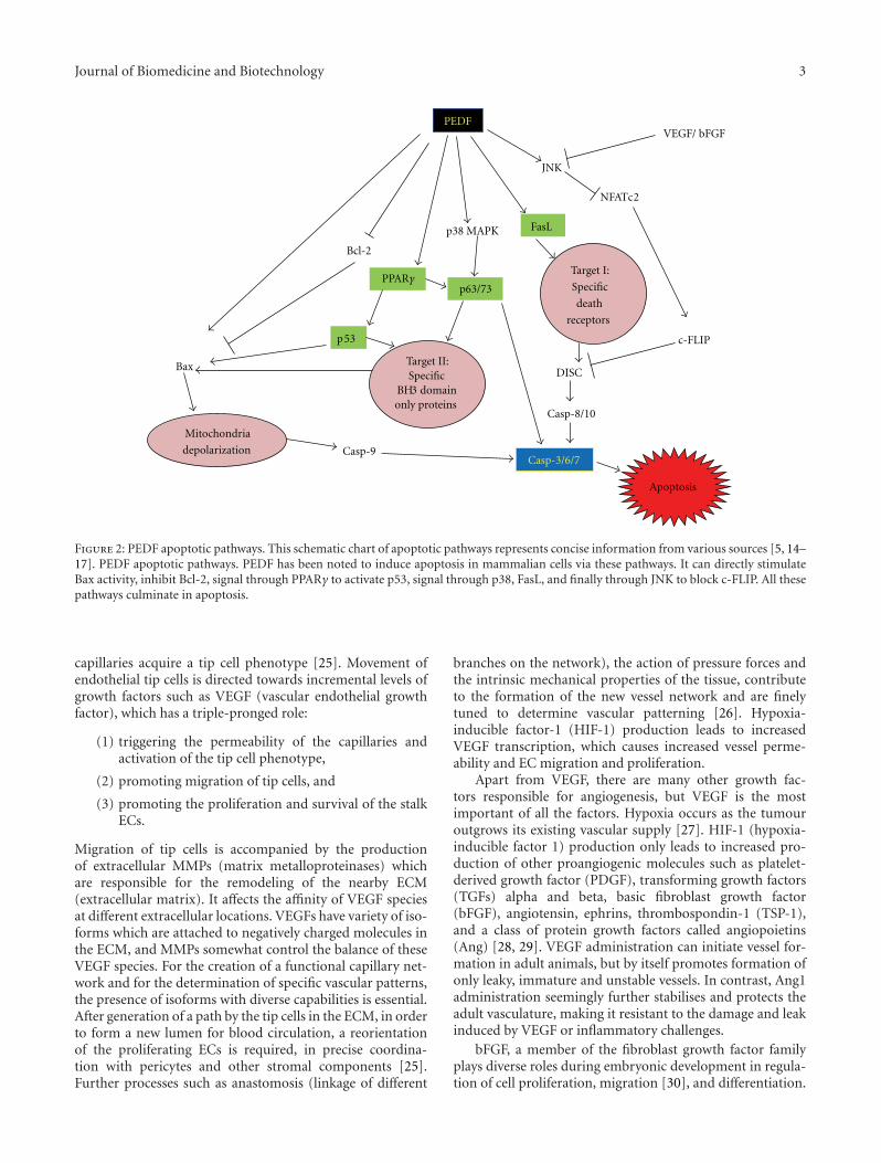
Journal of Biomedicine and Biotechnology 3
PEDF
Bax
Bcl-2
p53
Mitochondria
depolarization Casp-9
Target II: Specific
BH3 domain only proteins
Casp-3/6/7
Apoptosis
FasL
Target I:
Specific
death
receptors
DISC
Casp-8/10
VEGF/ bFGF
JNK
NFATc2
PPARγp63/73
p38 MAPK
c-FLIP
Figure 2: PEDF apoptotic pathways. This schematic chart of apoptotic pathways represents concise information from various sources [5, 14–17]. PEDF apoptotic pathways. PEDF has been noted to induce apoptosis in mammalian cells via these pathways. It can directly stimulateBax activity, inhibit Bcl-2, signal through PPARγ to activate p53, signal through p38, FasL, and finally through JNK to block c-FLIP. All thesepathways culminate in apoptosis.
capillaries acquire a tip cell phenotype [25]. Movement ofendothelial tip cells is directed towards incremental levels ofgrowth factors such as VEGF (vascular endothelial growthfactor), which has a triple-pronged role:
(1) triggering the permeability of the capillaries andactivation of the tip cell phenotype,
(2) promoting migration of tip cells, and
(3) promoting the proliferation and survival of the stalkECs.
Migration of tip cells is accompanied by the productionof extracellular MMPs (matrix metalloproteinases) whichare responsible for the remodeling of the nearby ECM(extracellular matrix). It affects the affinity of VEGF speciesat different extracellular locations. VEGFs have variety of iso-forms which are attached to negatively charged molecules inthe ECM, and MMPs somewhat control the balance of theseVEGF species. For the creation of a functional capillary net-work and for the determination of specific vascular patterns,the presence of isoforms with diverse capabilities is essential.After generation of a path by the tip cells in the ECM, in orderto form a new lumen for blood circulation, a reorientationof the proliferating ECs is required, in precise coordina-tion with pericytes and other stromal components [25].Further processes such as anastomosis (linkage of different
branches on the network), the action of pressure forces andthe intrinsic mechanical properties of the tissue, contributeto the formation of the new vessel network and are finelytuned to determine vascular patterning [26]. Hypoxia-inducible factor-1 (HIF-1) production leads to increasedVEGF transcription, which causes increased vessel perme-ability and EC migration and proliferation.
Apart from VEGF, there are many other growth fac-tors responsible for angiogenesis, but VEGF is the mostimportant of all the factors. Hypoxia occurs as the tumouroutgrows its existing vascular supply [27]. HIF-1 (hypoxia-inducible factor 1) production only leads to increased pro-duction of other proangiogenic molecules such as platelet-derived growth factor (PDGF), transforming growth factors(TGFs) alpha and beta, basic fibroblast growth factor(bFGF), angiotensin, ephrins, thrombospondin-1 (TSP-1),and a class of protein growth factors called angiopoietins(Ang) [28, 29]. VEGF administration can initiate vessel for-mation in adult animals, but by itself promotes formation ofonly leaky, immature and unstable vessels. In contrast, Ang1administration seemingly further stabilises and protects theadult vasculature, making it resistant to the damage and leakinduced by VEGF or inflammatory challenges.
bFGF, a member of the fibroblast growth factor familyplays diverse roles during embryonic development in regula-tion of cell proliferation, migration [30], and differentiation.
4 Journal of Biomedicine and Biotechnology
Angiopoietin-1(Ang-1) [31] plays a significant role at a laterstage of blood vessel formation. PDGF plays an importantrole in normal tissue growth and the pathophysiological pro-cesses of vascular diseases like atherosclerosis and restenosis[32]. During the initiation and progression of atheroscle-rosis, VSMCs are activated by growth factors like PDGF orcytokines, then proliferate and migrate from the media intothe intimal surface of the vessel, thus facilitating neointimalformation.
TSP-1 is the first naturally occurring angiogenicinhibitor, which is a multifunctional ECM protein. Down-regulation of TSP-1 causes tumour growth alteration bymodulating angiogenesis in various types of tumours. Itplays a critical role in inhibiting angiogenesis, resulting in thesuppression of tumour growth and experimental metastasis[33]. The TGF-β pathway occupies a central position in thesignaling networks that control growth and differentiation.TGF-β is known to have pleiotropic effects which differaccording to cell state and differentiation. This includesregulation of proliferation and apoptosis, and stimulationof epithelial-mesenchymal transition (EMT) which togetheris critical for the development of invasive and metastasispotential [34].
Angiostatin (38 KDa), an internal fragment of plasmino-gen, is a specific inhibitor of EC proliferation. Akin toangiostatin, researchers isolated an angiogenesis inhibitornamed endostatin, a 20 kDa carboxyl-terminal fragment ofcollagen XVIII. It is a specific inhibitor for endothelial cellproliferation and with systemic therapy, causes suppressionof tumour-induced angiogenesis and inhibits tumour growth[35]. It has been shown that when a combination of angio-statin and endostatin gene-encoded proteins are delivered totumour cells by gene transfer, they can exhibit synergisticantiangiogenic and antitumour effects [36].
5. Cell Cycling and Apoptosis in Cancer
The cell cycle plays a significant role in managing to maintainmulticellular organisms healthy and alive, in which theparent cell genome undergoes replication and the cellphysically divides into daughter cell via cytokinesis. However,genomic stability and cancer may occur due to unwantedmutations caused by some environmental and physiologicalevents. Cells have evolved certain checkpoints to preventthese types of mutations to daughter cells and allow DNArepair mechanism. These checkpoints are controlled byATM (ataxia telangiectasia mutated) and ATR (ataxia telang-iectasia and Rad3-related protein) kinases which activatedownstream effector kinases Chk1 (checkpoint kinase 1) andChk2, respectively. Checkpoints will repair damages in mostcases, however if the repair mechanisms cannot repair theDNA damage, cells are either permanently withdrawn fromthe cell cycle or are eliminated by programmed cell death(apoptosis). Cells may reenter the cell cycle in a process calledcheckpoint recovery [37]. It is known that programmed celldeath, that is, apoptosis, is required for normal developmentof multicellular organisms, whereby unwanted cells are
eliminated during physiological and certain pathologicalconditions.
Apoptosis is a programmed cell death event whichoccurs during embryogenesis, metamorphosis, endocrine-dependent tissue atrophy, and normal tissue turnover. Inmulticellular organisms, it is responsible for development,tissue homeostasis, and the immune response via two path-ways [38]. They are (i) an intrinsic pathway, which is initiatedwhen the cell is severely damaged, and (ii) the extrinsicpathway, activated when extracellular death ligands arebound by their cognate membrane-associated death recep-tors.
However, if there is a fault in apoptosis it leads to numberof serious diseases including cancer, autoimmunity, andneurodegeneration [39]. When genetically unstable cancercells tend to acclimatise to a tissue microenvironment that isdistant from the primary tumour, a process called metastasisoccurs [40]. Metastasis requires the cancer cells to detachfrom the primary tumour mass, move to and invade bloodvessels, survive within the circulation, attach to the endothe-lium of distant organs, penetrate the endothelial barrier, andestablish new tumour colonies. It is this multistage processwhich is the primary cause of cancer-related deaths, withthe primary tumour often resectable. PEDF is responsiblefor apoptosis of ECs either through the p38/MAPK pathway[14] or through the Fas/FasL pathway [41]. (Detailed PEDF-mediated apoptotic pathways are provided in Figure 2).
6. PEDF and Antiangiogenesis
Angiogenesis is the process by which new blood vessels areformed from a preexisting microvascular network, and itplays a major role in tumour growth and metastasis. Forevaluating antitumour efficacy, Yang et al. [42] constructedPEDF expressing-adenovirus (Ad-PEDF), and with success-ful transfer of the PEDF gene, antitumour efficacy in a mousetumour model was tested. The tumour volume was inhibited(2195.1 ± 462.9 mm3 approximately 50%) with Ad-PEDFwhen compared to the controls. The mouse which was inoc-ulated with Ad-PEDF showed a 50% survival rate at day 38,than controls (50% on day 13). By using TUNEL (terminaldeoxynucleotidyl transferase dUTP nick end labeling) assay,it was determined that the apoptosis index in tumour tissuewas higher (26.3%± 3.3%) in the Ad-PEDF group thancontrol groups (6.3%± 4.7% and 5.6%± 1.9%), confirmingthat increased apoptosis caused the accompanying decreasedtumour volume. For elucidating whether the increase inapoptosis of Ad-PEDF-treated tumour tissue was associatedwith the antiangiogenic effect of PEDF, scientists analysedmicrovessel density (MVD) of tumour tissues and found thatAd-PEDF demonstrated significant decrease in MVD whencompared to control groups.
In [43] determined the growth characteristics of a humanmalignant melanoma cell line overexpressing human PEDFin vitro and in vivo. In contrast to the empty vector-transfected group, PEDF overexpression completely inhib-ited the growth of the tumour. TUNEL assay was performedto determine the extent of apoptosis in the PEDF-transfected
Journal of Biomedicine and Biotechnology 5
and control vector-transfected tumours and it was found thatapoptotic cells increased 30% in the tumours derived fromthe PEDF-overexpressing G361 cells, when compared withcontrol vector-transfected tumour cells.
In this study, Garcia et al. [44] determined the resultsof overexpression of PEDF on both melanoma primarytumour growth and metastasis development. Overexpressionby melanoma cells inhibited subcutaneous tumour forma-tion. After tail vein injection, PEDF-overexpressing humanmelanoma cells were unable to grow and metastasise to thelung and liver. The authors also determined the effect ofPEDF on proliferation and apoptosis in the subcutaneoustumours and found tumour growth collapse, likely dueto impaired vascularisation. PEDF-positive melanoma cellsshowed significantly lower chemotaxis in a modified Boydenchamber assay. Quantitative data analysis showed a 3- to5-fold reduction in migration and invasiveness of PEDFpositive cells.
A metabolite isolated from ginseng protopanaxadiolsaponins called compound K (CK; 20-O-beta-D-gluco-pyranosyl-20(S)-protopanaxadiol) has an ability to induceapoptosis in various types of cancers. Based on this, Jeonget al. [45] evaluated the antiangiogenic effects of CK in vitroand in vivo. bFGF induces angiogenesis in human umbilicalvein ECs (HUVECs). In vitro tests determined that CKinhibited bFGF-induced proliferation, migration and tubeformation. In vivo antiangiogenic activity of CK was eval-uated by a Matrigel plug assay and it was found that CKinhibited bFGF-induced angiogenesis. By measuring thelevels of VEGF and PEDF, Jeong et al. evaluated the effectof CK on the phosphorylation of p38/MAPK, and AKT inbFGF-treated HUVECs and found decreased levels of VEGFand increased levels of PEDF following CK treatment. Thesefindings suggest that there is a relationship between PEDF,VEGF, and bFGF with respect to angiogenesis.
7. PEDF and Tumour Cell Differentiation
Angiogenesis underlies the processes of bone growth, repair,and remodelling and may account at least in part for theaggressive nature of osteosarcoma [46]. PEDF regulatesangiogenesis to underlie the physiological processes of boneformation, growth, and remodelling [47]. An elegant balanceexists between proangiogenic and antiangiogenic factors inbone so that the degree of vascularisation is appropriatefor the biology required. VEGF is widely regarded as themost important proangiogenic factor, while PEDF has beenidentified as the most-potent antiangiogenic factor [18]. Thebalance between these two factors is as important in bonephysiology as it is in other tissues.
PEDF was found to be expressed in the zones of activebone formation and its role in cell differentiation as well asthe maintenance of high growth rates in the mesenchymalcell layer was determined by Lord et al. [48] using a deerantler model. Tombran-Tink and Barnstable hypothesizedabout the significance of PEDF in bone based on RT-PCRand western blot analysis [14], and more recent studies [49]demonstrated that PEDF inhibits osteoclast formation,
survival and bone resorption function, and effects wereabrogated by VEGF.
PEDF expression by osteoblasts and osteoclasts in regionsof active bone formation regulates neovascularisation atthese sites, a process that underlies bone formation, growthand remodelling. Without a supporting vasculature, boneprecursors are unable to localise to those sites intended forbone development [50]. Angiogenesis in the ossification andlower hypertrophic zones is necessary for the continuedgrowth and functioning of osteoblasts and osteoclasts,enabling the replacement of cartilaginous tissue by bone,a process ultimately regulated by a balance between PEDFand VEGF [47]. Using immunohistochemistry and in situhybridisation, PEDF expression was shown to be largelyrestricted to the chondrocytes of the avascular resting,proliferative and upper hypertrophic layers of the epiphysis,and to regions of active bone remodelling.
In vitro and in vivo studies have revealed that in the caseof osteosarcoma, PEDF can induce both indirect and directsuppression of tumour growth and progression by potentantiangiogenic capability of PEDF targeting tumour vascu-lature and induction of osteosarcoma cell apoptosis, differ-entiation, and inhibition of cell cycling, respectively [2, 3].Osteosarcoma usually arises in the metaphysis of long bones.The epiphyseal plate is found between the epiphysis andmetaphysis of long bones, which is a natural barrier totumour invasion. The resistance of epiphyseal cartilage toosteosarcoma invasion is likely to be due to the differentialexpression of PEDF and VEGF in the zones of the epiphysis[51].
Metastasis is the foremost problem in the treatment ofosteosarcoma. In vitro studies with rPEDF revealed dose-dependent reduction in cellular invasion by matrigel assay aswell as an increase in cell adhesion to collagen type-1 usingthe human SaOS-2 cell line [52]. In vivo studies done onan orthotopic model [53] showed that treatment with PEDFhad the greatest impact on metastases. There was a 70%reduction in the development of pulmonary metastases and a40% reduction in primary tumour size in those mice treatedwith PEDF [52]. More recently, sustained administration ofPEDF via osmotic pumps implanted in mice bearing SaOS-2tumours demonstrated reduced metastasis to the lungs [54].Notably, treatment with PEDF was delayed until after themacroscopic appearance of primary tumours. Thus, in thisstudy, both the model used and the delivery regimen wereclose to the appropriate clinical treatment foreseeable in thefuture if PEDF is to be used as an osteosarcoma therapeutic.
The critical role of PEDF in regulating MMP activitymakes the area of metastasis an intriguing one. Initially,it was found that PEDF can significantly downregulate thelevels of MMP-14 but not MMP-2 and -9 in a humanchondrosarcoma cell line [55]. This was corroborated by adecreased invasion of cells through collagen-1 matrix in thepresence of PEDF. Human melanoma cells were partiallyinhibited in their invasion, with reduced trafficking ofmembrane-bound MMP-14 to the cell surface [56].
In [57] demonstrated the ability of PEDF as a multi-functional antitumour agent in neuroblastomas by inhibitingtumour angiogenesis and differentiating tumour cells to
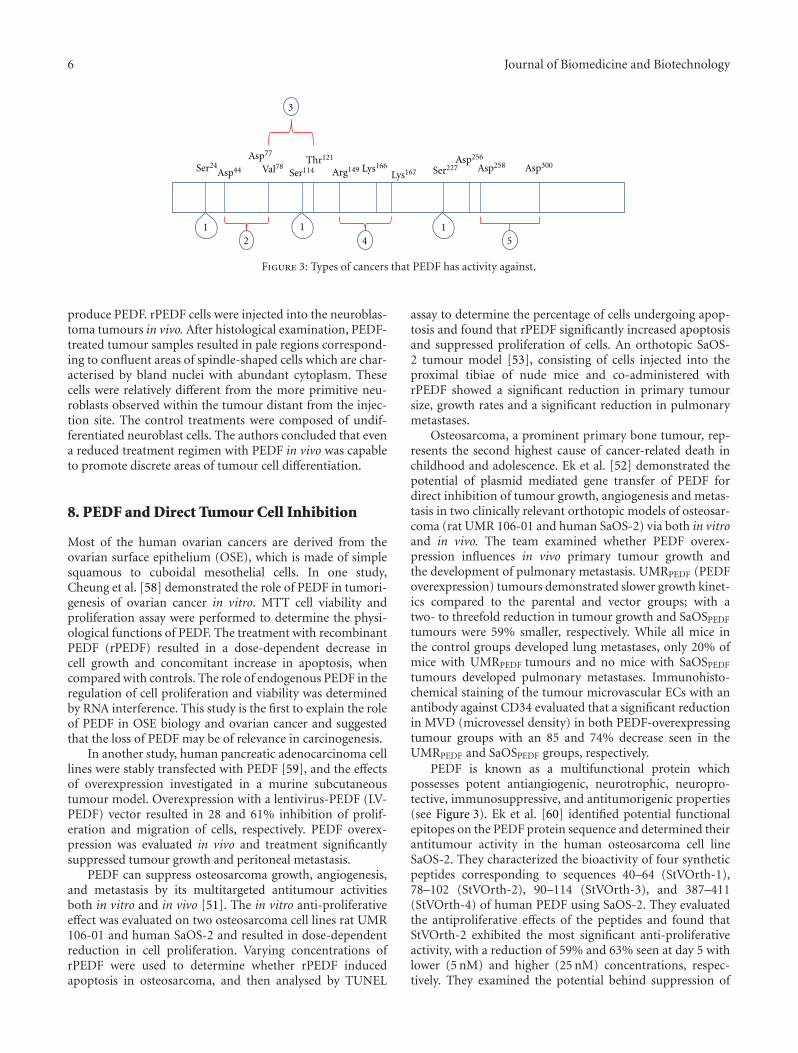
6 Journal of Biomedicine and Biotechnology
2 54
3
Ser24Asp44
Asp77
Ser114Val78Thr121
Arg149 Lys166Lys167
Asp256
Asp258Ser227 Asp300
1 1 1
Figure 3: Types of cancers that PEDF has activity against.
produce PEDF. rPEDF cells were injected into the neuroblas-toma tumours in vivo. After histological examination, PEDF-treated tumour samples resulted in pale regions correspond-ing to confluent areas of spindle-shaped cells which are char-acterised by bland nuclei with abundant cytoplasm. Thesecells were relatively different from the more primitive neu-roblasts observed within the tumour distant from the injec-tion site. The control treatments were composed of undif-ferentiated neuroblast cells. The authors concluded that evena reduced treatment regimen with PEDF in vivo was capableto promote discrete areas of tumour cell differentiation.
8. PEDF and Direct Tumour Cell Inhibition
Most of the human ovarian cancers are derived from theovarian surface epithelium (OSE), which is made of simplesquamous to cuboidal mesothelial cells. In one study,Cheung et al. [58] demonstrated the role of PEDF in tumori-genesis of ovarian cancer in vitro. MTT cell viability andproliferation assay were performed to determine the physi-ological functions of PEDF. The treatment with recombinantPEDF (rPEDF) resulted in a dose-dependent decrease incell growth and concomitant increase in apoptosis, whencompared with controls. The role of endogenous PEDF in theregulation of cell proliferation and viability was determinedby RNA interference. This study is the first to explain the roleof PEDF in OSE biology and ovarian cancer and suggestedthat the loss of PEDF may be of relevance in carcinogenesis.
In another study, human pancreatic adenocarcinoma celllines were stably transfected with PEDF [59], and the effectsof overexpression investigated in a murine subcutaneoustumour model. Overexpression with a lentivirus-PEDF (LV-PEDF) vector resulted in 28 and 61% inhibition of prolif-eration and migration of cells, respectively. PEDF overex-pression was evaluated in vivo and treatment significantlysuppressed tumour growth and peritoneal metastasis.
PEDF can suppress osteosarcoma growth, angiogenesis,and metastasis by its multitargeted antitumour activitiesboth in vitro and in vivo [51]. The in vitro anti-proliferativeeffect was evaluated on two osteosarcoma cell lines rat UMR106-01 and human SaOS-2 and resulted in dose-dependentreduction in cell proliferation. Varying concentrations ofrPEDF were used to determine whether rPEDF inducedapoptosis in osteosarcoma, and then analysed by TUNEL
assay to determine the percentage of cells undergoing apop-tosis and found that rPEDF significantly increased apoptosisand suppressed proliferation of cells. An orthotopic SaOS-2 tumour model [53], consisting of cells injected into theproximal tibiae of nude mice and co-administered withrPEDF showed a significant reduction in primary tumoursize, growth rates and a significant reduction in pulmonarymetastases.
Osteosarcoma, a prominent primary bone tumour, rep-resents the second highest cause of cancer-related death inchildhood and adolescence. Ek et al. [52] demonstrated thepotential of plasmid mediated gene transfer of PEDF fordirect inhibition of tumour growth, angiogenesis and metas-tasis in two clinically relevant orthotopic models of osteosar-coma (rat UMR 106-01 and human SaOS-2) via both in vitroand in vivo. The team examined whether PEDF overex-pression influences in vivo primary tumour growth andthe development of pulmonary metastasis. UMRPEDF (PEDFoverexpression) tumours demonstrated slower growth kinet-ics compared to the parental and vector groups; with atwo- to threefold reduction in tumour growth and SaOSPEDF
tumours were 59% smaller, respectively. While all mice inthe control groups developed lung metastases, only 20% ofmice with UMRPEDF tumours and no mice with SaOSPEDF
tumours developed pulmonary metastases. Immunohisto-chemical staining of the tumour microvascular ECs with anantibody against CD34 evaluated that a significant reductionin MVD (microvessel density) in both PEDF-overexpressingtumour groups with an 85 and 74% decrease seen in theUMRPEDF and SaOSPEDF groups, respectively.
PEDF is known as a multifunctional protein whichpossesses potent antiangiogenic, neurotrophic, neuropro-tective, immunosuppressive, and antitumorigenic properties(see Figure 3). Ek et al. [60] identified potential functionalepitopes on the PEDF protein sequence and determined theirantitumour activity in the human osteosarcoma cell lineSaOS-2. They characterized the bioactivity of four syntheticpeptides corresponding to sequences 40–64 (StVOrth-1),78–102 (StVOrth-2), 90–114 (StVOrth-3), and 387–411(StVOrth-4) of human PEDF using SaOS-2. They evaluatedthe antiproliferative effects of the peptides and found thatStVOrth-2 exhibited the most significant anti-proliferativeactivity, with a reduction of 59% and 63% seen at day 5 withlower (5 nM) and higher (25 nM) concentrations, respec-tively. They examined the potential behind suppression of
Journal of Biomedicine and Biotechnology 7
tumour cell invasion through Matrigel and found that allfour peptides significantly decreased Matrigel invasion bygreater that 50%. However StVOrth-4 provided that mostconsistent inhibition, with greater than 70% reductionobserved at all concentrations. Ek et al. examined treatmentwith the PEDF-derived peptides for understanding the alter-ation of osteosarcoma cell adhesion to collagen (a potentialmechanism in antimetastasis) and found that all four pep-tides considerably increased cell adhesion to collagen type 1,with StVOrth-3 demonstrating 53% greater ability than theother peptides. Prodifferentiation potential studies of thePEDF fragments in vitro revealed that administration ofStVOrth-1, -2, and -3 significantly increased nodule forma-tion in SaOS-2 cells. They further tested the in vivo activity ofStVOrth-2 and StVOrth-3 in an orthotopic model of humanosteosarcoma, using a model employing tumour cells pre-mixed with peptides, then injected orthotopically. StVOrth-2exhibited predominantly antiproliferation activity, whileStVOrth-3 caused mainly increased collagen adhesion andVEGF suppression. The results of an efficacy study usingsustained delivery of peptides in the orthotopic model ofosteosarcoma is included in this issue [5, 54].
In a more recent study, Mirochnik et al. [61] have demon-strated improved antiangiogenic activity by the small PEDFpeptide, only 18mer in length, called P18. P18 inhibited thegrowth of prostate tumour growth and the more aggressiveRenca cells in vivo in the subcutaneous xenograft model at10 mg/kg. At the same dose, the 34-mer had no significanteffect on tumour growth. The authors stated that P18 wasmore potent at blocking tumor angiogenesis and causinghigher levels of intratumoral apoptosis.
9. Future Directions
PEDF is a promising therapeutic agent for various typesof cancers. Until now it is known that it is capable ofreducing cancer via inhibiting angiogenesis, tumour growth,cell migration and inducing apoptosis and tumour cell dif-ferentiation. Only a little is known about the proangiogenicfactors like VEGF, bFGF, PDGF, and there is lot to be knownabout other proangiogenic factors that interact with PEDF.The fact that regions of PEDF have different functionalactivity may lead to synthesis of small and cheap peptideswhich can be tested as anticancer drugs in the near future.Recent biochemical studies have provided some insight intoPEDF structure and function. However, very few articles havelooked at PEDF receptor-mediated signalling in cancer. Somereceptors (G-protein-coupled receptor GPR39) can protectcells from death by increasing secretion of PEDF [62]. Morework has to be done in this area given that more than tworeceptors for this protein have been found to date.
10. Summary
Cancer is characterized by uncontrolled growth and spreadof abnormal cells. PEDF is known to be a multifunctionalprotein which possesses potent antiangiogenic,neurotrophic, neuroprotective, immunosuppressive, and
antitumorigenic properties. Scientists designed Ad-PEDFfor evaluating the antitumour efficacy and demonstratedrole of PEDF in apoptosis and angiogenesis. Other groupof researchers demonstrated that overexpression of PEDFsuppressed tumour growth and angiogenesis and enhancedthe rate of apoptosis. PEDF regulation of angiogenesisunderlies the physiological processes of bone formation,growth and remodelling. PEDF can induce both indirect anddirect suppression of tumour growth and progression bypotent antiangiogenic capability. An orthotopic model ofosteosarcoma studies showed that treatment with PEDF hadthe greatest impact on metastases. There is now justificationfor evaluation of PEDF efficacy in other types of cancers.
Conflict of Interests
The authors declare that there is not conflict of interests inwriting this paper.
Acknowledgments
The authors acknowledge funding from the Victoria Uni-versity Research Development Grant Scheme (VU-RDGS)and an ANZ Trustees/Medical Research and Technology inVictoria Grant to C. Dass.
References
[1] American Cancer Society, 2010, http://www.cancer.org/Research/CancerFactsFigures/CancerFactsFigures/index.
[2] E. T. H. Ek, C. R. Dass, and P. F. M. Choong, “Pigment epi-thelium-derived factor: a multimodal tumor inhibitor,” Molecular Cancer Therapeutics, vol. 5, no. 7, pp. 1641–1646, 2006.
[3] E. T. H. Ek, C. R. Dass, and P. F. M. Choong, “PEDF: apotential molecular therapeutic target with multiple anti-cancer activities,” Trends in Molecular Medicine, vol. 12, no. 10,pp. 497–502, 2006.
[4] A. R. Murray and J. X. Ma, “PEDF as a treatment for cervicalcancer,” Cancer Biology and Therapy, vol. 9, no. 12, pp. 975–977, 2010.
[5] M. L. Broadhead, C. R. Dass, and P. F. Choong, “In vitro andin vivo biological activity of PEDF against a range of tumors,”Expert Opinion on Therapeutic Targets, vol. 13, no. 12, pp.1429–1438, 2009.
[6] J. Yang, E. J. Duh, R. B. Caldwell, and M. Ali Behzadian,“Antipermeability function of PEDF involves blockade of theMAP kinase/GSK/β-catenin signaling pathway and uPAR ex-pression,” Investigative Ophthalmology and Visual Science, vol.51, no. 6, pp. 3273–3280, 2010.
[7] S. Halin, P. Wikstrom, S. H. Rudolfsson et al., “Decreased pig-ment epithelium-derived factor is associated with metastaticphenotype in human and rat prostate tumors,” Cancer Re-search, vol. 64, no. 16, pp. 5664–5671, 2004.
[8] H. Uehara, M. Miyamoto, K. Kato et al., “Expression ofpigment epithelium-derived factor decreases liver metastasisand correlates with favorable prognosis for patients withductal pancreatic adenocarcinoma,” Cancer Research, vol. 64,no. 10, pp. 3533–3537, 2004.
[9] K. Matsumoto, H. Ishikawa, D. Nishimura, K. Hamasaki, K.Nakao, and K. Eguchi, “Antiangiogenic property of pigment
8 Journal of Biomedicine and Biotechnology
epithelium-derived factor in hepatocellular carcinoma,” Hep-atology, vol. 40, no. 1, pp. 252–259, 2004.
[10] L. P. Abramson, V. Stellmach, J. A. Doll et al., “Wilms’ tumorgrowth is suppressed by antiangiogenic pigment epithelium-derived factor in a xenograft model,” Journal of PediatricSurgery, vol. 38, no. 3, pp. 336–342, 2003.
[11] J. Tombran-Tink, K. Mazuruk, I. R. Rodriguez et al., “Organi-zation, evolutionary conservation, expression and unusual Aludensity of the human gene for pigment epithelium-derivedfactor, a unique neurotrophic serpin.,” Molecular Vision, vol.2, p. 11, 1996.
[12] S. Patricia Becerra, “Focus on molecules: pigment epithelium-derived factor (PEDF),” Experimental Eye Research, vol. 82, no.5, pp. 739–740, 2006.
[13] N. Yasui, T. Mori, D. Morito et al., “Dual-site recogni-tion of different extracellular matrix components by anti-angiogenic/neurotrophic serpin, PEDF,” Biochemistry, vol. 42,no. 11, pp. 3160–3167, 2003.
[14] O. V. Volpert, T. Zaichuk, W. Zhou et al., “Inducer-stimulatedFas targets activated endothelium for destruction by anti-angiogenic thrombospondin-1 and pigment epithelium-derived factor,” Nature Medicine, vol. 8, no. 4, pp. 349–357,2002.
[15] T. A. Zaichuk, E. H. Shroff, R. Emmanuel, S. Filleur, T.Nelius, and O. V. Volpert, “Nuclear factor of activated T cellsbalances angiogenesis activation and inhibition,” Journal ofExperimental Medicine, vol. 199, no. 11, pp. 1513–1522, 2004.
[16] T. C. Ho, S. L. Chen, Y. C. Yang, C. L. Liao, H. C. Cheng, andY. P. Tsao, “PEDF induces p53-mediated apoptosis throughPPAR gamma signaling in human umbilical vein endothelialcells,” Cardiovascular Research, vol. 76, no. 2, pp. 213–223,2007.
[17] T. Zhang, M. Guan, C. Xu, Y. Chen, and Y. Lu, “Pigmentepithelium-derived factor inhibits glioma cell growth in vitroand in vivo,” Life Sciences, vol. 81, no. 16, pp. 1256–1263, 2007.
[18] D. W. Dawson, O. V. Volpert, P. Gillis et al., “Pigmentepithelium-derived factor: a potent inhibitor of angiogenesis,”Science, vol. 285, no. 5425, pp. 245–248, 1999.
[19] D. Hanahan and J. Folkman, “Patterns and emerging mech-anisms of the angiogenic switch during tumorigenesis,” Cell,vol. 86, no. 3, pp. 353–364, 1996.
[20] G. Maik-Rachline, S. Shaltiel, and R. Seger, “Extracellularphosphorylation converts pigment epithelium-derived factorfrom a neurotrophic to an antiangiogenic factor,” Blood, vol.105, no. 2, pp. 670–678, 2005.
[21] A. Castellanos, C. Vicente-Duenas, E. Campos-Sanchez etal., “Cancer as a reprogramming-like disease: Implicationsin tumor development and treatment,” Seminars in CancerBiology, vol. 20, no. 2, pp. 93–97, 2010.
[22] S. V. Sharma, D. W. Bell, J. Settleman, and D. A. Haber,“Epidermal growth factor receptor mutations in lung cancer,”Nature Reviews Cancer, vol. 7, no. 3, pp. 169–181, 2007.
[23] L. Ji and J. A. Roth, “Tumor suppressor FUS1 signaling path-way,” Journal of Thoracic Oncology, vol. 3, no. 4, pp. 327–330,2008.
[24] C. M. Croce, “Oncogenes and cancer,” New England Journal ofMedicine, vol. 358, no. 5, pp. 502–511, 2008.
[25] R. D. M. Travasso, E. C. Poire, M. Castro, J. C. Rodrguez-Manzaneque, and A. Hernandez-Machado, “Tumor angiogen-esis and vascular patterning: a mathematical model,” PLoSOne, vol. 6, no. 5, Article ID e19989, 2011.
[26] E. A. V. Jones, F. Le Noble, and A. Eichmann, “Whatdetermines blood vessel structure? Genetic prespecification vs.hemodynamics,” Physiology, vol. 21, no. 6, pp. 388–395, 2006.
[27] J. A. Forsythe, B. H. Jiang, N. V. Iyer et al., “Activationof vascular endothelial growth factor gene transcription byhypoxia-inducible factor 1,” Molecular and Cellular Biology,vol. 16, no. 9, pp. 4604–4613, 1996.
[28] G. D. Yancopoulos, S. Davis, N. W. Gale, J. S. Rudge, S. J.Wiegand, and J. Holash, “Vascular-specific growth factors andblood vessel formation,” Nature, vol. 407, no. 6801, pp. 242–248, 2000.
[29] R. K. Jain, “Molecular regulation of vessel maturation,” NatureMedicine, vol. 9, no. 6, pp. 685–693, 2003.
[30] J. W. Choi, S. Kim, T. M. Kim et al., “Basic fibroblastgrowth factor activates MEK/ERK cell signaling pathway andstimulates the proliferation of chicken primordial germ cells,”PLoS One, vol. 5, no. 9, Article ID e12968, 2010.
[31] G. Thurston, “Complementary actions of VEGF and Angio-poietin-1 on blood vessel growth and leakage,” Journal ofAnatomy, vol. 200, no. 6, pp. 575–580, 2002.
[32] C. H. Heldin and B. Westermark, “Mechanism of action andin vivo role of platelet-derived growth factor,” Physiological Re-views, vol. 79, no. 4, pp. 1283–1316, 1999.
[33] B. Ren, K. O. Yee, J. Lawler, and R. Khosravi-Far, “Regulationof tumor angiogenesis by thrombospondin-1,” Biochimica etBiophysica Acta, vol. 1765, no. 2, pp. 178–188, 2006.
[34] S. Sheahan, C. O. Bellamy, S. N. Harland, D. J. Harrison, andS. Prost, “TGFbeta induces apoptosis and EMT in primarymouse hepatocytes independently of p53, p21Cip1 or Rbstatus,” BMC Cancer, vol. 8, article no. 191, 2008.
[35] M. S. O’Reilly, L. Holmgren, Y. Shing et al., “Angiostatin: anovel angiogenesis inhibitor that mediates the suppression ofmetastases by a Lewis lung carcinoma,” Cell, vol. 79, no. 2, pp.315–328, 1994.
[36] S. Y. Paek, Y. S. Kim, and S. G. Choi, “The orientation-dependent expression of angiostatin-endostatin hybrid pro-teins and their characterization for the synergistic effects ofantiangiogenesis,” Journal of Microbiology and Biotechnology,vol. 20, no. 10, pp. 1430–1435, 2010.
[37] R. H. Medema and L. Macurek, “Checkpoint recovery in cells:how a molecular understanding can help in the fight againstcancer,” F1000 Biology Reports, vol. 3, no. 1, article no. 10,2011.
[38] M. O. Hengartner, “The biochemistry of apoptosis,” Nature,vol. 407, no. 6805, pp. 770–776, 2000.
[39] C. Gu, J. Zhang, Y. Chen, and J. Lei, “A trigger model ofapoptosis induced by tumor necrosis factor signaling,” BMCSystems Biology, vol. 5, no. 1, article no. 13, 2011.
[40] G. P. Gupta and J. Massague, “Cancer metastasis: building aframework,” Cell, vol. 127, no. 4, pp. 679–695, 2006.
[41] S. X. Zhang, J. J. Wang, G. Gao, K. Parke, and J. X. Ma,“Pigment epithelium-derived factor downregulates vascularendothelial growth factor (VEGF) expression and inhibitsVEGF-VEGF receptor 2 binding in diabetic retinopathy,”Journal of Molecular Endocrinology, vol. 37, no. 1, pp. 1–12,2006.
[42] L. P. Yang, P. Cheng, X. C. Peng et al., “Anti-tumor effectof adenovirus-mediated gene transfer of pigment epithelium-derived factor on mouse B16-F10 melanoma,” Journal of Ex-perimental and Clinical Cancer Research, vol. 28, no. 1, articleno. 75, 2009.
[43] R. Abe, T. Shimizu, S. I. Yamagishi et al., “Overexpression ofpigment epithelium-derived factor decreases angiogenesis andinhibits the growth of human malignant melanoma cells invivo,” American Journal of Pathology, vol. 164, no. 4, pp. 1225–1232, 2004.
Journal of Biomedicine and Biotechnology 9
[44] M. Garcia, N. I. Fernandez-Garcia, V. Rivas et al., “Inhibitionof xenografted human melanoma growth and preventionof metastasis development by dual antiangiogenic/antitumoractivities of pigment epithelium-derived factor,” CancerResearch, vol. 64, no. 16, pp. 5632–5642, 2004.
[45] A. Jeong, H. J. Lee, S. J. Jeong et al., “Compound K inhibitsbasic fibroblast growth factor-induced angiogenesis viaregulation of p38 mitogen activated protein kinase and AKTin human umbilical vein endothelial cells,” Biological andPharmaceutical Bulletin, vol. 33, no. 6, pp. 945–950, 2010.
[46] G. M. Quan, J. Ojaimi, and P. F. Choong, “Cartilage-derivedanti-angiogenic therapy for osteosarcoma: a novel approach,”International Journal of Cancer Prevention, vol. 1, pp. 411–419,2004.
[47] G. M. Y. Quan, J. Ojaimi, Y. Li, V. Kartsogiannis, H. Zhou, andP. F. M. Choong, “Localization of pigment epithelium-derivedfactor in growing mouse bone,” Calcified Tissue International,vol. 76, no. 2, pp. 146–153, 2005.
[48] E. A. Lord, S. K. Martin, J. P. Gray, C. Li, and D. E. Clark, “Cellcycle genes PEDF and CDKN1C in growing Deer antlers,”Anatomical Record, vol. 290, no. 8, pp. 994–1004, 2007.
[49] T. Akiyama, C. R. Dass, Y. Shinoda, H. Kawano, S. Tanaka,and P. F. M. Choong, “PEDF regulates osteoclasts via osteo-protegerin and RANKL,” Biochemical and Biophysical ResearchCommunications, vol. 391, no. 1, pp. 789–794, 2010.
[50] G. M. Y. Quan, J. Ojaimi, A. P. W. Nadesapillai, H. Zhou,and P. F. M. Choong, “Resistance of epiphyseal cartilage toinvasion by osteosarcoma is likely to be due to expression ofantiangiogenic factors,” Pathobiology, vol. 70, no. 6, pp. 361–367, 2002.
[51] E. T. H. Ek, C. R. Dass, K. G. Contreras, and P. F. M.Choong, “Pigment epithelium-derived factor overexpressioninhibits orthotopic osteosarcoma growth, angiogenesis andmetastasis,” Cancer Gene Therapy, vol. 14, no. 7, pp. 616–626,2007.
[52] E. T. H. Ek, C. R. Dass, K. G. Contreras, and P. F. M. Choong,“Inhibition of orthotopic osteosarcoma growth and metastasisby multitargeted antitumor activities of pigment epithelium-derived factor,” Clinical and Experimental Metastasis, vol. 24,no. 2, pp. 93–106, 2007.
[53] C. R. Dass, E. T. Ek, K. G. Contreras, and P. F. Choong,“A novel orthotopic murine model provides insights intocellular and molecular characteristics contributing to humanosteosarcoma,” Clinical and Experimental Metastasis, vol. 23,no. 7-8, pp. 367–380, 2006.
[54] M. L. Broadhead, C. R. Dass, and P. F. M. Choong, “Sys-temically administered PEDF against primary and secondarytumours in a clinically relevant osteosarcoma model,” BritishJournal of Cancer, vol. 105, no. 10, pp. 1503–1511, 2011.
[55] M. L. Tan, P. F. M. Choong, and C. R. Dass, “Anti-chondrosarcoma effects of PEDF mediated via moleculesimportant to apoptosis, cell cycling, adhesion and invasion,”Biochemical and Biophysical Research Communications, vol.398, no. 4, pp. 613–618, 2010.
[56] O. Ladhani, C. Sanchez-Martinez, J. L. Orgaz, B. Jimenez,and O. V. Volpert, “Pigment epithelium-derived factor blockstumor extravasation by suppressing amoeboid morphologyand mesenchymal proteolysis1,2,” Neoplasia, vol. 13, no. 7, pp.633–642, 2011.
[57] S. E. Crawford, V. Stellmach, M. Ranalli et al., “Pigmentepithelium-derived factor (PEDF) in neuroblastoma: a mul-tifunctional mediator of Schwann cell antitumor activity,”Journal of Cell Science, vol. 114, no. 24, pp. 4421–4428, 2001.
[58] L. W. T. Cheung, S. C. L. Au, A. N. Y. Cheung et al., “Pigmentepithelium-derived factor is estrogen sensitive and inhibits thegrowth of human ovarian cancer and ovarian surface epithelialcells,” Endocrinology, vol. 147, no. 9, pp. 4179–4191, 2006.
[59] R. Hase, M. Miyamoto, H. Uehara et al., “Pigment epithelium-derived factor gene therapy inhibits human pancreatic cancerin mice,” Clinical Cancer Research, vol. 11, no. 24, pp. 8737–8744, 2005.
[60] E. T. H. Ek, C. R. Dass, K. G. Contreras, and P. F. M. Choong,“PEDF-derived synthetic peptides exhibit antitumor activityin an orthotopic model of human osteosarcoma,” Journal ofOrthopaedic Research, vol. 25, no. 12, pp. 1671–1680, 2007.
[61] Y. Mirochnik, A. Aurora, F. T. Schulze-Hoepfner et al., “Shortpigment epithelial-derived factor-derived peptide inhibitsangiogenesis and tumor growth,” Clinical Cancer Research, vol.15, no. 5, pp. 1655–1663, 2009.
[62] S. Dittmer, M. Sahin, A. Pantlen et al., “The constitutivelyactive orphan G-protein-coupled receptor GPR39 protectsfrom cell death by increasing secretion of pigment epithelium-derived growth factor,” Journal of Biological Chemistry, vol.283, no. 11, pp. 7074–7081, 2008.
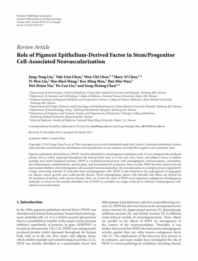
Hindawi Publishing CorporationJournal of Biomedicine and BiotechnologyVolume 2012, Article ID 871272, 10 pagesdoi:10.1155/2012/871272
Review Article
Role of Pigment Epithelium-Derived Factor in Stem/ProgenitorCell-Associated Neovascularization
Jung-Tung Liu,1 Yuh-Lien Chen,2 Wen-Chi Chen,3, 4 Huey-Yi Chen,3, 4
Yi-Wen Lin,3 Shu-Huei Wang,2 Kee-Ming Man,5 Hui-Min Wan,6
Wei-Hsian Yin,7 Po-Len Liu,6 and Yung-Hsiang Chen3, 4
1 Department of Neurosurgery, School of Medicine, Chung-Shan Medical University and Hospital, Taichung 402, Taiwan2 Department of Anatomy and Cell Biology, College of Medicine, National Taiwan University, Taipei 100, Taiwan3 Graduate Institutes of Integrated Medicine and Acupuncture Science, College of Chinese Medicine, China Medical University,Taichung 404, Taiwan
4 Departments of Urology, Obstetrics and Gynecology, and Medical Research, China Medical University Hospital, Taichung 404, Taiwan5 Department of Anesthesiology, Tungs’ Taichung Metroharbor Hospital, Taichung 433, Taiwan6 Department of Fragrance and Cosmetic Science, and Department of Respiratory Therapy, College of Medicine,Kaohsiung Medical University, Kaohsiung 807, Taiwan
7 School of Medicine, Faculty of Medicine, National Yang-Ming University, Taipei 112, Taiwan
Correspondence should be addressed to Po-Len Liu, [email protected] and Yung-Hsiang Chen, [email protected]
Received 12 December 2011; Accepted 26 March 2012
Academic Editor: Crispin Dass
Copyright © 2012 Jung-Tung Liu et al. This is an open access article distributed under the Creative Commons Attribution License,which permits unrestricted use, distribution, and reproduction in any medium, provided the original work is properly cited.
Pigment epithelium-derived factor (PEDF) was first identified in retinal pigment epithelium cells. It is an endogenously producedprotein that is widely expressed throughout the human body such as in the eyes, liver, heart, and adipose tissue; it exhibitsmultiple and varied biological activities. PEDF is a multifunctional protein with antiangiogenic, antitumorigenic, antioxidant,anti-inflammatory, antithrombotic, neurotrophic, and neuroprotective properties. More recently, PEDF has been shown to be themost potent inhibitor of stem/progenitor cell-associated neovascularization. Neovascularization is a complex process regulated bya large, interacting network of molecules from stem/progenitor cells. PEDF is also involved in the pathogenesis of angiogeniceye disease, tumor growth, and cardiovascular disease. Novel antiangiogenic agents with tolerable side effects are desired forthe treatment of patients with various diseases. Here, we review the value of PEDF as an important endogenous antiangiogenicmolecule; we focus on the recently identified role of PEDF as a possible new target molecule to influence stem/progenitor cell-related neovascularization.
1. Introduction
In the 1980s, pigment epithelium-derived factor (PEDF) wasidentified and isolated from primary human fetal retinal pig-ment epithelial cells [1]. It is a 50 kDa secreted glycoproteinthat is a noninhibitory member of the serpin (serine proteaseinhibitor) superfamily of proteins; its gene (SERPINF1) islocated on chromosome 17p13 [2]. PEDF is an endogenouslyproduced protein widely expressed throughout the humanbody such as in the eye, liver, heart, and adipose tissue,which exhibits multiple and varied biological activities [3, 4].PEDF was initially identified as a neurotrophic factor that
differentiates retinoblastoma cells into nonproliferating neu-rons [5]. PEDF has also been shown to be neuroprotective formotor neurons [6], hippocampal neurons [7], dopaminergicmidbrain neurons [8], and striatal neurons [9] in differenttoxin-induced models of neurodegeneration. These effectsare parallel to the effects of PEDF on neurogenesis inthe context of the neurovasculature. Thereafter, it wasfurther discovered that PEDF also had potent antiangiogenicactivity, greater than any other known endogenous factor[10, 11]. The implications of this discovery have proven tobe extensive, and many studies have investigated the role ofPEDF in various pathological conditions, including chronic
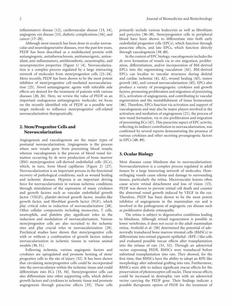
2 Journal of Biomedicine and Biotechnology
inflammatory disease [12], cardiovascular disease [13, 14],angiogenic eye disease [15], diabetic complications [16], andcancer [17–20].
Although most research has been done in ocular neovas-cular and neurodegenerative diseases, over the past few years,PEDF has been described as a multifaceted protein withantiangiogenic, antiatherosclerosis, antitumorigenic, antiox-idant, anti-inflammatory, antithrombotic, neurotrophic, andneuroprotective properties (Figure 1) [4]. Neovasculariza-tion is a complex process regulated by a large interactingnetwork of molecules from stem/progenitor cells [21–24].More recently, PEDF has been shown to be the most potentinhibitor of stem/progenitor cell-mediated neovasculariza-tion [25]. Novel antiangiogenic agents with tolerable sideeffects are desired for the treatment of patients with variousdiseases [20, 26]. Here, we review the value of PEDF as animportant endogenous antiangiogenic molecule; we focuson the recently identified role of PEDF as a possible newtarget molecule to influence stem/progenitor cell-relatedneovascularization therapeutically.
2. Stem/Progenitor Cells andNeovascularization
Angiogenesis and vasculogenesis are the major types ofpostnatal neovascularization. Angiogenesis is the processwhere new vessels grow from preexisting blood vessels;whereas vasculogenesis is the process of blood vessel for-mation occurring by de novo production of bone marrow(BM) stem/progenitor cell-derived endothelial cells (ECs),which, in turn, form blood capillaries (Figure 2) [27].Neovascularization is an important process in the functionalrecovery of pathological conditions, such as wound healingand ischemic diseases. Hypoxia is an important drivingforce for neovascularization in various ischemic conditionsthrough stimulation of the expression of many cytokinesand growth factors such as vascular endothelial growthfactor (VEGF), platelet-derived growth factor, insulin-likegrowth factor, and fibroblast growth factor (FGF), whichplay critical roles in induction of neovascularization [28].Other cellular components including monocytes, T cells,neutrophils, and platelets play significant roles in theinduction and modulation of neovascularization. Variousstem/progenitor cells are also recruited to the ischemicsites and play crucial roles in neovascularization [29].Preclinical studies have shown that stem/progenitor cellswith or without a combination of growth factors induceneovascularization in ischemic tissues in various animalmodels [30, 31].
Following ischemia, various angiogenic factors andcytokines are upregulated and promote homing of stem/progenitor cells to the site of injury [32]. It has been shownthat circulating stem/progenitor cells could be incorporatedinto the neovasculature within the ischemic tissue and coulddifferentiate into ECs [33, 34]. Stem/progenitor cells canalso differentiate into other supporting cells, which delivergrowth factors and cytokines to ischemic tissue and promoteangiogenesis through paracrine effects [35]. These cells
primarily include various leukocytes as well as fibroblastsand pericytes [36–38]. Stem/progenitor cells in peripheralblood have been shown to differentiate into both earlyendothelial progenitor cells (EPCs), which function throughparacrine effects, and late EPCs, which function directlythrough vasculogenesis [39, 40].
In the context of EPC biology, vasculogenesis includes thede novo formation of vessels via in situ migration, prolifer-ation, differentiation, and/or incorporation of BM-derivedEPCs into the regenerating vasculature [41]. BM-derivedEPCs can localize to vascular structures during skeletaland cardiac ischemia [41, 42], wound healing [43], tumorgrowth [44], and corneal neovascularization [45]. EPCs alsoproduce a variety of proangiogenic cytokines and growthfactors, promoting proliferation and migration of preexistingECs, activation of angiogenesis, and contributing to vascularregeneration and the reestablishment of tissue homeostasis[46]. Therefore, EPCs function via activation and support ofvasculogenesis and may also be major players involved in theactivation and mediation of angiogenesis [21], the process ofnew vessel formation, via in situ proliferation and migrationof preexisting ECs [47]. This paracrine aspect of EPC activity,reflecting its indirect contribution to neovascularization, wasconfirmed by several reports demonstrating the presence ofvarious cytokines and other secreting proangiogenic factorsin EPCs [48, 49].
3. Ocular Biology
Most diseases cause blindness due to neovascularization.Neovascularization is a complex process regulated in adulttissues by a large interacting network of molecules. Hem-orrhaging vessels cause edema and damage to surroundingtissues, particularly the retina. Microvascular lesions oftencause severe retinal detachment and loss of vision [15].PEDF was shown to prevent retinal cell death and counterthe abnormal vessel growth induced by VEGF in the eye.Therefore, PEDF has been shown to be the most potentinhibitor of angiogenesis in the mammalian eye and isinvolved in the pathogenesis of angiogenic eye disease suchas proliferative diabetic retinopathy.
The retina is subject to degenerative conditions leadingto blindness. Although retinal regeneration is possible inlower vertebrates, it does not occur in the adult mammalianretina. Arnhold et al. [50] determined the potential of ade-novirally transduced bone marrow stromal cells (BMSCs) todifferentiate into retinal-pigment-epithelial- (RPE-) like cellsand evaluated possible rescue effects after transplantationinto the retinas of rats [51, 52]. Through an adenoviralvector expressing PEDF, BMSCs were transduced beforesubretinal transplantation into rats. They showed, for thefirst time, that BMSCs have the ability to adopt an RPE-likemorphology after subretinal grafting into rats. Furthermore,BMSCs were able to induce significant rescue effects for thepreservation of photoreceptor cell nuclei. These rescue effectscould be increased in dystrophic rats with an adenoviralvector carrying the PEDF gene. Their findings indicate apossible therapeutic option of PEDF for the treatment of
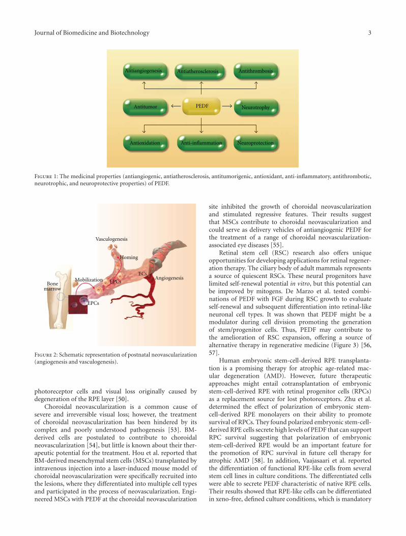
Journal of Biomedicine and Biotechnology 3
PEDF Neurotrophy
Antioxidation Anti-inflammation Neuroprotection
Antiangiogenesis Antiatherosclerosis Antithrombosis
Antitumor
Figure 1: The medicinal properties (antiangiogenic, antiatherosclerosis, antitumorigenic, antioxidant, anti-inflammatory, antithrombotic,neurotrophic, and neuroprotective properties) of PEDF.
marrow
EPCs
EPCsMobilization
Vasculogenesis
Homing
AngiogenesisECs
Bone
Figure 2: Schematic representation of postnatal neovascularization(angiogenesis and vasculogenesis).
photoreceptor cells and visual loss originally caused bydegeneration of the RPE layer [50].
Choroidal neovascularization is a common cause ofsevere and irreversible visual loss; however, the treatmentof choroidal neovascularization has been hindered by itscomplex and poorly understood pathogenesis [53]. BM-derived cells are postulated to contribute to choroidalneovascularization [54], but little is known about their ther-apeutic potential for the treatment. Hou et al. reported thatBM-derived mesenchymal stem cells (MSCs) transplanted byintravenous injection into a laser-induced mouse model ofchoroidal neovascularization were specifically recruited intothe lesions, where they differentiated into multiple cell typesand participated in the process of neovascularization. Engi-neered MSCs with PEDF at the choroidal neovascularization
site inhibited the growth of choroidal neovascularizationand stimulated regressive features. Their results suggestthat MSCs contribute to choroidal neovascularization andcould serve as delivery vehicles of antiangiogenic PEDF forthe treatment of a range of choroidal neovascularization-associated eye diseases [55].
Retinal stem cell (RSC) research also offers uniqueopportunities for developing applications for retinal regener-ation therapy. The ciliary body of adult mammals representsa source of quiescent RSCs. These neural progenitors havelimited self-renewal potential in vitro, but this potential canbe improved by mitogens. De Marzo et al. tested combi-nations of PEDF with FGF during RSC growth to evaluateself-renewal and subsequent differentiation into retinal-likeneuronal cell types. It was shown that PEDF might be amodulator during cell division promoting the generationof stem/progenitor cells. Thus, PEDF may contribute tothe amelioration of RSC expansion, offering a source ofalternative therapy in regenerative medicine (Figure 3) [56,57].
Human embryonic stem-cell-derived RPE transplanta-tion is a promising therapy for atrophic age-related mac-ular degeneration (AMD). However, future therapeuticapproaches might entail cotransplantation of embryonicstem-cell-derived RPE with retinal progenitor cells (RPCs)as a replacement source for lost photoreceptors. Zhu et al.determined the effect of polarization of embryonic stem-cell-derived RPE monolayers on their ability to promotesurvival of RPCs. They found polarized embryonic stem-cell-derived RPE cells secrete high levels of PEDF that can supportRPC survival suggesting that polarization of embryonicstem-cell-derived RPE would be an important feature forthe promotion of RPC survival in future cell therapy foratrophic AMD [58]. In addition, Vaajasaari et al. reportedthe differentiation of functional RPE-like cells from severalstem cell lines in culture conditions. The differentiated cellswere able to secrete PEDF characteristic of native RPE cells.Their results showed that RPE-like cells can be differentiatedin xeno-free, defined culture conditions, which is mandatory
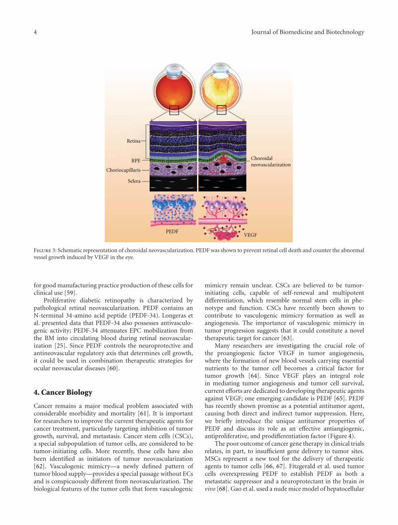
4 Journal of Biomedicine and Biotechnology
PEDFVEGF
Choroidalneovascularization
Retina
RPE
Choriocapillaris
Selera
Figure 3: Schematic representation of choroidal neovascularization. PEDF was shown to prevent retinal cell death and counter the abnormalvessel growth induced by VEGF in the eye.
for good manufacturing practice production of these cells forclinical use [59].
Proliferative diabetic retinopathy is characterized bypathological retinal neovascularization. PEDF contains anN-terminal 34-amino acid peptide (PEDF-34). Longeras etal. presented data that PEDF-34 also possesses antivasculo-genic activity; PEDF-34 attenuates EPC mobilization fromthe BM into circulating blood during retinal neovascular-ization [25]. Since PEDF controls the neuroprotective andantineovascular regulatory axis that determines cell growth,it could be used in combination therapeutic strategies forocular neovascular diseases [60].
4. Cancer Biology
Cancer remains a major medical problem associated withconsiderable morbidity and mortality [61]. It is importantfor researchers to improve the current therapeutic agents forcancer treatment, particularly targeting inhibition of tumorgrowth, survival, and metastasis. Cancer stem cells (CSCs),a special subpopulation of tumor cells, are considered to betumor-initiating cells. More recently, these cells have alsobeen identified as initiators of tumor neovascularization[62]. Vasculogenic mimicry—a newly defined pattern oftumor blood supply—provides a special passage without ECsand is conspicuously different from neovascularization. Thebiological features of the tumor cells that form vasculogenic
mimicry remain unclear. CSCs are believed to be tumor-initiating cells, capable of self-renewal and multipotentdifferentiation, which resemble normal stem cells in phe-notype and function. CSCs have recently been shown tocontribute to vasculogenic mimicry formation as well asangiogenesis. The importance of vasculogenic mimicry intumor progression suggests that it could constitute a noveltherapeutic target for cancer [63].
Many researchers are investigating the crucial role ofthe proangiogenic factor VEGF in tumor angiogenesis,where the formation of new blood vessels carrying essentialnutrients to the tumor cell becomes a critical factor fortumor growth [64]. Since VEGF plays an integral rolein mediating tumor angiogenesis and tumor cell survival,current efforts are dedicated to developing therapeutic agentsagainst VEGF; one emerging candidate is PEDF [65]. PEDFhas recently shown promise as a potential antitumor agent,causing both direct and indirect tumor suppression. Here,we briefly introduce the unique antitumor properties ofPEDF and discuss its role as an effective antiangiogenic,antiproliferative, and prodifferentiation factor (Figure 4).
The poor outcome of cancer gene therapy in clinical trialsrelates, in part, to insufficient gene delivery to tumor sites.MSCs represent a new tool for the delivery of therapeuticagents to tumor cells [66, 67]. Fitzgerald et al. used tumorcells overexpressing PEDF to establish PEDF as both ametastatic suppressor and a neuroprotectant in the brain invivo [68]. Gao et al. used a nude mice model of hepatocellular
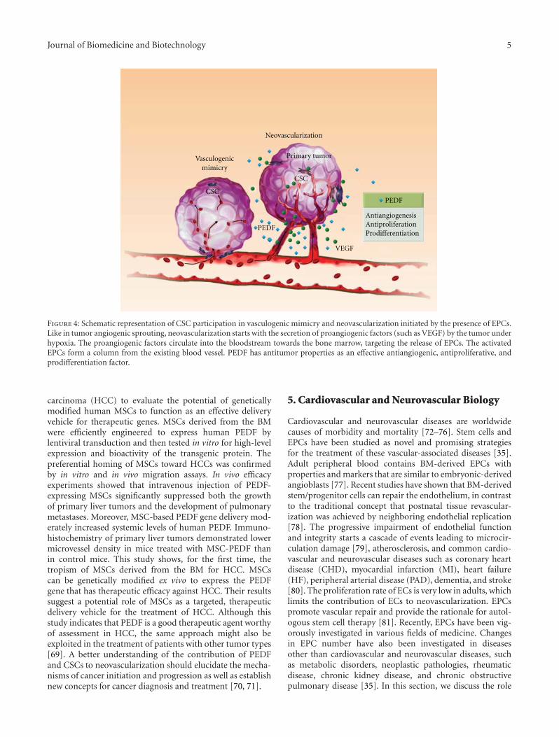
Journal of Biomedicine and Biotechnology 5
PEDF
AntiangiogenesisAntiproliferationProdifferentiation
Neovascularization
Vasculogenic
PEDF
VEGF
CSC
CSC
Primary tumor
mimicry
Figure 4: Schematic representation of CSC participation in vasculogenic mimicry and neovascularization initiated by the presence of EPCs.Like in tumor angiogenic sprouting, neovascularization starts with the secretion of proangiogenic factors (such as VEGF) by the tumor underhypoxia. The proangiogenic factors circulate into the bloodstream towards the bone marrow, targeting the release of EPCs. The activatedEPCs form a column from the existing blood vessel. PEDF has antitumor properties as an effective antiangiogenic, antiproliferative, andprodifferentiation factor.
carcinoma (HCC) to evaluate the potential of geneticallymodified human MSCs to function as an effective deliveryvehicle for therapeutic genes. MSCs derived from the BMwere efficiently engineered to express human PEDF bylentiviral transduction and then tested in vitro for high-levelexpression and bioactivity of the transgenic protein. Thepreferential homing of MSCs toward HCCs was confirmedby in vitro and in vivo migration assays. In vivo efficacyexperiments showed that intravenous injection of PEDF-expressing MSCs significantly suppressed both the growthof primary liver tumors and the development of pulmonarymetastases. Moreover, MSC-based PEDF gene delivery mod-erately increased systemic levels of human PEDF. Immuno-histochemistry of primary liver tumors demonstrated lowermicrovessel density in mice treated with MSC-PEDF thanin control mice. This study shows, for the first time, thetropism of MSCs derived from the BM for HCC. MSCscan be genetically modified ex vivo to express the PEDFgene that has therapeutic efficacy against HCC. Their resultssuggest a potential role of MSCs as a targeted, therapeuticdelivery vehicle for the treatment of HCC. Although thisstudy indicates that PEDF is a good therapeutic agent worthyof assessment in HCC, the same approach might also beexploited in the treatment of patients with other tumor types[69]. A better understanding of the contribution of PEDFand CSCs to neovascularization should elucidate the mecha-nisms of cancer initiation and progression as well as establishnew concepts for cancer diagnosis and treatment [70, 71].
5. Cardiovascular and Neurovascular Biology
Cardiovascular and neurovascular diseases are worldwidecauses of morbidity and mortality [72–76]. Stem cells andEPCs have been studied as novel and promising strategiesfor the treatment of these vascular-associated diseases [35].Adult peripheral blood contains BM-derived EPCs withproperties and markers that are similar to embryonic-derivedangioblasts [77]. Recent studies have shown that BM-derivedstem/progenitor cells can repair the endothelium, in contrastto the traditional concept that postnatal tissue revascular-ization was achieved by neighboring endothelial replication[78]. The progressive impairment of endothelial functionand integrity starts a cascade of events leading to microcir-culation damage [79], atherosclerosis, and common cardio-vascular and neurovascular diseases such as coronary heartdisease (CHD), myocardial infarction (MI), heart failure(HF), peripheral arterial disease (PAD), dementia, and stroke[80]. The proliferation rate of ECs is very low in adults, whichlimits the contribution of ECs to neovascularization. EPCspromote vascular repair and provide the rationale for autol-ogous stem cell therapy [81]. Recently, EPCs have been vig-orously investigated in various fields of medicine. Changesin EPC number have also been investigated in diseasesother than cardiovascular and neurovascular diseases, suchas metabolic disorders, neoplastic pathologies, rheumaticdisease, chronic kidney disease, and chronic obstructivepulmonary disease [35]. In this section, we discuss the role
6 Journal of Biomedicine and Biotechnology
of PEDF as a possible new target molecule to therapeuticallyinfluence cardiovascular and neurovascular diseases.
PEDF has been characterized in cardiovascular systems.It has a protective role in atherosclerosis, the main cause ofCHD, MI, and HF, due to its anti-inflammatory, antioxidant,and antithrombotic effects in the vessel wall and platelets.Expression of PEDF by ECs is essential for the inhibition ofproliferation and migration of smooth muscle cells after bal-loon injury [82]. The antioxidative properties of PEDF havebeen shown to block TNF-α-induced EC activation [83].These observations suggest that PEDF might have beneficialeffects on atherosclerosis by suppressing inflammatory pro-liferative responses to injury. Additionally, PEDF has strongantiangiogenic effects by inducing apoptosis [84] in ECsand by regulating the expression of other angiogenic factors.Local blocking of PEDF (e.g., in ischemic tissue in the heart)might favor angiogenesis, induce neovascularization, andlead to increased perfusion of the injured tissue. In contrast,local overexpression of PEDF restricted to atheroscleroticlesions might block angiogenesis, inflammation, and throm-bosis at these sites and thus counteract destabilization andrupturing of the lesion otherwise caused by inflammatoryactivation [85] and excessive angiogenesis, thereby inhibitingsubsequent thrombus formation [4].
In our previous study, we described a streamlinedmethod for the rapid isolation, growth, and ex vivo expan-sion of late outgrowth ECs from Wharton jelly of a humanumbilical cord; we evaluated the ability of these cellsto reendothelialize and inhibit neointimal hyperplasia ininjured femoral arteries of mice. We also examined the directeffects of EPC-conditioned medium on the migration andproliferation of human aortic smooth muscle cells and therole of PEDF in these effects. Our results showed that EPCtransplantation led to rapid reendothelialization of denudedarteries, which resulted in significant inhibition of neoin-timal thickening. This was the first report demonstratingthat EPCs derived from an umbilical cord aid in acceleratingreendothelialization and attenuating vascular remodeling atsites of arterial injury; these effects were closely associatedwith PEDF [14]. These findings have implications for a novelPEDF-related and cell-based therapy for cardiovascular andneurovascular diseases.
MSCs can ameliorate MI injury; however, MSCs fromolder donors are less efficacious than those from youngerdonors. More recently, Liang et al. determined how age-related expression of PEDF affects MSC therapeutic efficacyfor MI. Their data showed that PEDF expression wasincreased in MSCs from old mice compared to young miceresulting in significantly impaired therapeutic efficacy in oldMSCs, compared with that in young MSCs, for treatment ofmice subjected to MI. PEDF overexpression in young MSCsimpaired the beneficial effects against MI injury and inducedcellular profile changes in the infarct region that was similarto administration of old MSCs. Knocking down PEDFexpression in old MSCs improved MSC therapeutic efficacyand induced a cellular profile similar to administrationof young MSCs. PEDF secreted by MSCs regulated theproliferation and migration of cardiac fibroblasts. These dataprovide the first evidence that the paracrine factor PEDF
plays a critical role in the regulatory effects of MSCs againstMI injury. Furthermore, the impaired therapeutic abilityof aged MSCs is predominantly caused by increased PEDFsecretion [86]. These findings indicate PEDF as a promisingnovel genetic modification target for improving aged MSCtherapeutic efficacy.
Vascular and neural tissues are delicately intertwined infunctional neurovascular units. This codependence emergesearly in development with the coordinated growth and tissuemodeling of both cellular elements [87]. Stem/progenitorcells in the developing central nervous system and inneurogenic regions of the adult brain are stimulated to self-renew and generate more neurons by factors released fromvarious vascular cells [88]. In the mammalian brain, neuro-genesis persists in 2 germinal areas, the subventricular zone(SVZ), and the hippocampus, where continuous postnatalneuronal production seems to be supported by neural stemcells (NSCs) [89]. Ramırez-Castillejo et al. identified PEDFas critical for the communication between vascular andneural cells in an adult NSC niche, the SVZ [90]. A singlefactor that can stimulate brain tumor cells to differentiateand, at the same time, cut off their blood supply hasunlimited therapeutic value. PEDF levels decline with aging,possibly contributing to cell senescence and to age-relatedsusceptibility to cancer [91]. These data demonstrate thatPEDF is a niche-derived regulator of adult NSCs and provideevidence for a role for PEDF in NSC maintenance.
The potential of NSCs for brain repair depends ontheir capacity for self-renewal. Recent evidence for the closeapposition of adult periventricular NSCs and blood vesselshas confirmed the findings [88, 90] that factors derivedfrom the vasculature contribute to regulation of the adultNSC pool [92, 93]. A new study by Andreu-Agullo et al.revealed that the vasculature-derived PEDF promotes theNotch signaling-dependent renewal of adult periventricularNSCs through an unconventional mechanism [94]. Theyfound that Notch was active in astroglia-like NSCs but not intransit-amplifying progenitors of the murine subependymalzone and that the level of Notch transcriptional activity cor-related with self-renewal and multipotency. Moreover, divid-ing NSCs appeared to balance renewal with commitmentvia controlled segregation of Notch activity. PEDF enhancedNotch-dependent transcription in cells with low Notchsignaling, thereby subverting the output of an asymmetricaldivision to the production of 2 highly self-renewing cells.Mechanistically, PEDF induced a noncanonical activation ofthe nuclear factor (NF)-κB pathway. These data provide abasis for stemness regulation in vascular niches and indicatethat Notch and PEDF cooperate to regulate self-renewal [94].
6. Conclusions
PEDF has been described as a natural angiogenesis inhibitorwith neurotrophic and immune-modulation properties [95].It balances stem/progenitor cell-associated angiogenesis inthe eye, as well as cardiovascular and neurovascular systems,and blocks tumor progression. The mechanisms underlyingmost of these events are not completely clear; however, it
Journal of Biomedicine and Biotechnology 7
appears that PEDF acts via multiple high affinity ligandsand cell receptors. In this paper, we summarized the currentknowledge on the important endogenous antiangiogenicmolecule PEDF; we focused on the recently identified roleof PEDF as a possible new target molecule to influencestem/progenitor cell-related neovascularization. We dis-cussed the multimodal activities of PEDF and addressedthe therapeutic potential of PEDF in treating angiogenesis-,neurodegeneration-, and inflammation-related diseases.However, many questions remain to be resolved; thesespecific points must be addressed prior to initiation of anyhuman clinical trials using PEDF peptides.
Conflict of Interests
The authors declare that there is no conflict of interests.
Acknowledgments
This study is supported in part by Taiwan Departmentof Health Clinical Trial and Research Center of Excellence(DOH101-TD-B-111-004), National Science Council (NSC100-2320-B-039-008-MY2 and NSC 100-2320-B-037-009),and China Medical University (CMU100-NSC-04). Wethank Miss Jin-Mei Wang and Chi-Hsiang Wei for the paperpreparation.
References
[1] J. Tombran-Tink and L. V. Johnson, “Neuronal differentiationof retinoblastoma cells induced by medium conditioned byhuman RPE cells,” Investigative Ophthalmology and VisualScience, vol. 30, no. 8, pp. 1700–1707, 1989.
[2] F. R. Steele, G. J. Chader, L. V. Johnson, and J. Tombran-Tink,“Pigment epithelium-derived factor: neurotrophic activityand identification as a member of the serine protease inhibitorgene family,” Proceedings of the National Academy of Sciencesof the United States of America, vol. 90, no. 4, pp. 1526–1530,1993.
[3] E. T. H. Ek, C. R. Dass, and P. F. M. Choong, “PEDF: apotential molecular therapeutic target with multiple anti-cancer activities,” Trends in Molecular Medicine, vol. 12, no. 10,pp. 497–502, 2006.
[4] K. Rychli, K. Huber, and J. Wojta, “Pigment epithelium-derived factor (PEDF) as a therapeutic target in cardiovasculardisease,” Expert Opinion on Therapeutic Targets, vol. 13, no. 11,pp. 1295–1302, 2009.
[5] J. Tombran-Tink, G. G. Chader, and L. V. Johnson, “PEDF:a pigment epithelium-derived factor with potent neuronaldifferentiative activity[2],” Experimental Eye Research, vol. 53,no. 3, pp. 411–414, 1991.
[6] M. M. Bilak, A. M. Corse, S. R. Bilak, M. Lehar, J. Tombran-Tink, and R. W. Kuncl, “Pigment epithelium-derived factor(PEDF) protects motor neurons from chronic glutamate-mediated neurodegeneration,” Journal of Neuropathology andExperimental Neurology, vol. 58, no. 7, pp. 719–728, 1999.
[7] M. A. DeCoster, E. Schabelman, J. Tombran-Tink et al.,“Neuroprotection by pigment epithelial-derived factor againstglutamate toxicity in developing primary hippocampal neu-rons,” Journal of Neuroscience Research, vol. 56, no. 6, pp. 604–610, 1999.
[8] T. Falk, S. Zhang, and S. J. Sherman, “Pigment epitheliumderived factor (PEDF) is neuroprotective in two in vitromodels of Parkinson’s disease,” Neuroscience Letters, vol. 458,no. 2, pp. 49–52, 2009.
[9] T. Sanagi, T. Yabe, and H. Yamada, “Adenoviral gene deliveryof pigment epithelium-derived factor protects striatal neuronsfrom quinolinic acid-induced excitotoxicity,” Journal of Neu-ropathology and Experimental Neurology, vol. 69, no. 3, pp.224–233, 2010.
[10] D. W. Dawson, O. V. Volpert, P. Gillis et al., “Pigmentepithelium-derived factor: a potent inhibitor of angiogenesis,”Science, vol. 285, no. 5425, pp. 245–248, 1999.
[11] E. M. Levy, M. P. Roberti, and J. Mordoh, “Natural killercells in human cancer: from biological functions to clinicalapplications,” Journal of Biomedicine and Biotechnology, vol.2011, Article ID 676198, 11 pages, 2011.
[12] S. X. Zhang, J. J. Wang, G. Gao, C. Shao, R. Mott, and J. X. Ma,“Pigment epithelium-derived factor (PEDF) is an endogenousantiinflammatory factor,” FASEB Journal, vol. 20, no. 2, pp.323–325, 2006.
[13] S. I. Yamagishi, K. Nakamura, S. Ueda, S. Kato, and T.Imaizumi, “Pigment epithelium-derived factor (PEDF) blocksangiotensin II signaling in endothelial cells via suppression ofNADPH oxidase: a novel anti-oxidative mechanism of PEDF,”Cell and Tissue Research, vol. 320, no. 3, pp. 437–445, 2005.
[14] S. H. Wang, S. J. Lin, Y. H. Chen et al., “Late outgrowthendothelial cells derived from wharton jelly in humanumbilical cord reduce neointimal formation after vascularinjury: involvement of pigment epithelium-derived factor,”Arteriosclerosis, Thrombosis, and Vascular Biology, vol. 29, no.6, pp. 816–822, 2009.
[15] J. Tombran-Tink, “PEDF in angiogenic eye diseases,” CurrentMolecular Medicine, vol. 10, no. 3, pp. 267–278, 2010.
[16] H. Funatsu, H. Yamashita, S. Nakamura et al., “Vitreouslevels of pigment epithelium-derived factor and vascularendothelial growth factor are related to diabetic macularedema,” Ophthalmology, vol. 113, no. 2, pp. 294–301, 2006.
[17] J. A. Doll, V. M. Stellmach, N. P. Bouck et al., “Pigmentepithelium-derived factor regulates the vasculature and massof the prostate and pancreas,” Nature Medicine, vol. 9, no. 6,pp. 774–780, 2003.
[18] R. Hase, M. Miyamoto, H. Uehara et al., “Pigment epithelium-derived factor gene therapy inhibits human pancreatic cancerin mice,” Clinical Cancer Research, vol. 11, no. 24, pp. 8737–8744, 2005.
[19] S. Filleur, K. Volz, T. Nelius et al., “Two functional epitopesof pigment epithelial-derived factor block angiogenesis andinduce differentiation in prostate cancer,” Cancer Research, vol.65, no. 12, pp. 5144–5152, 2005.
[20] R. Abe, Y. Fujita, S. I. Yamagishi, and H. Shimizu, “Pigmentepithelium-derived factor prevents melanoma growth viaangiogenesis inhibition,” Current Pharmaceutical Design, vol.14, no. 36, pp. 3802–3809, 2008.
[21] A. L. George, P. Bangalore-Prakash, S. Rajoria et al., “Endothe-lial progenitor cell biology in disease and tissue regeneration,”Journal of Hematology and Oncology, vol. 4, article 24, 2011.
[22] M. Matthias, N. David, and N. Josef, “From bench to bedside:what physicians need to know about endothelial progenitorcells,” American Journal of Medicine, vol. 124, no. 6, pp. 489–497, 2011.
[23] A. J. Roks, K. Rodgers, and T. Walther, “Effects of therenin angiotensin system on vasculogenesis-related progenitorcells,” Current Opinion in Pharmacology, vol. 11, no. 2, pp.162–174, 2011.
8 Journal of Biomedicine and Biotechnology
[24] S. Fang and P. Salven, “Stem cells in tumor angiogenesis,”Journal of Molecular and Cellular Cardiology, vol. 50, no. 2, pp.290–295, 2011.
[25] R. Longeras, K. Farjo, M. Ihnat et al., “A PEDF-derived peptideinhibits retinal neovascularization and blocks mobilization ofbone marrow-derived endothelial progenitor cells,” Experi-mental Diabetes Research, vol. 2012, Article ID 518426, 11pages, 2012.
[26] W. D. Thompson, W. W. Li, and M. Maragoudakis, “Theclinical manipulation of angiogenesis: pathology, side-effects,surprises, and opportunities with novel human therapies,” TheJournal of Pathology, vol. 190, no. 3, pp. 330–337, 2000.
[27] J. M. Isner and T. Asahara, “Angiogenesis and vasculogenesisas therapeutic strategies for postnatal neovascularization,”Journal of Clinical Investigation, vol. 103, no. 9, pp. 1231–1236,1999.
[28] S. Patel-Hett and P. A. D’Amore, “Signal transduction in vas-culogenesis and developmental angiogenesis,” InternationalJournal of Developmental Biology, vol. 55, no. 4-5, pp. 353–369,2011.
[29] P. H. Huang, Y. H. Chen, H. Y. Tsai et al., “Intake of red wineincreases the number and functional capacity of circulatingendothelial progenitor cells by enhancing nitric oxide bioavail-ability,” Arteriosclerosis, Thrombosis, and Vascular Biology, vol.30, no. 4, pp. 869–877, 2010.
[30] J. Lu, V. J. Pompili, and H. Das, “Neovascularization andhematopoietic stem cells,” Cell Biochemistry and Biophysics. Inpress.
[31] M. B. Grant, W. S. May, S. Caballero et al., “Adult hematopoi-etic stem cells provide functional hemangioblast activityduring retinal neovascularization,” Nature Medicine, vol. 8, no.6, pp. 607–612, 2002.
[32] S. Rafii and D. Lyden, “Therapeutic stem and progenitor celltransplantation for organ vascularization and regeneration,”Nature Medicine, vol. 9, no. 6, pp. 702–712, 2003.
[33] I. Rajantie, M. Ilmonen, A. Alminaite, U. Ozerdem, K. Alitalo,and P. Salven, “Adult bone marrow-derived cells recruitedduring angiogenesis comprise precursors for periendothelialvascular mural cells,” Blood, vol. 104, no. 7, pp. 2084–2086,2004.
[34] B. A. Peters, L. A. Diaz, K. Polyak et al., “Contributionof bone marrow-derived endothelial cells to human tumorvasculature,” Nature Medicine, vol. 11, no. 3, pp. 261–262,2005.
[35] C. Napoli, T. Hayashi, F. Cacciatore et al., “Endothelialprogenitor cells as therapeutic agents in the microcirculation:an update,” Atherosclerosis, vol. 215, no. 1, pp. 9–22, 2011.
[36] J. Rehman, J. Li, C. M. Orschell, and K. L. March, “Peripheralblood “endothelial progenitor cells” are derived from mono-cyte/macrophages and secrete angiogenic growth factors,”Circulation, vol. 107, no. 8, pp. 1164–1169, 2003.
[37] T. Ziegelhoeffer, B. Fernandez, S. Kostin et al., “Bone marrow-derived cells do not incorporate into the adult growingvasculature,” Circulation Research, vol. 94, no. 2, pp. 230–238,2004.
[38] C. Urbich, A. Aicher, C. Heeschen et al., “Soluble factorsreleased by endothelial progenitor cells promote migration ofendothelial cells and cardiac resident progenitor cells,” Journalof Molecular and Cellular Cardiology, vol. 39, no. 5, pp. 733–742, 2005.
[39] J. Hur, C. H. Yoon, H. S. Kim et al., “Characterization of twotypes of endothelial progenitor cells and their different con-tributions to neovasculogenesis,” Arteriosclerosis, Thrombosis,and Vascular Biology, vol. 24, no. 2, pp. 288–293, 2004.
[40] Y. H. Chen, S. J. Lin, F. Y. Lin et al., “High glucose impairsearly and late endothelial progenitor cells by modifying nitricoxide-related but not oxidative stress-mediated mechanisms,”Diabetes, vol. 56, no. 6, pp. 1559–1568, 2007.
[41] T. Asahara, H. Masuda, T. Takahashi et al., “Bone marroworigin of endothelial progenitor cells responsible for postnatalvasculogenesis in physiological and pathological neovascular-ization,” Circulation Research, vol. 85, no. 3, pp. 221–228, 1999.
[42] M. Ii, H. Nishimura, A. Iwakura et al., “Endothelial progenitorcells are rapidly recruited to myocardium and mediate protec-tive effect of ischemic preconditioning via “imported” nitricoxide synthase activity,” Circulation, vol. 111, no. 9, pp. 1114–1120, 2005.
[43] S. M. Bauer, L. J. Goldstein, R. J. Bauer, H. Chen, M. Putt,and O. C. Velazquez, “The bone marrow-derived endothelialprogenitor cell response is impaired in delayed wound healingfrom ischemia,” Journal of Vascular Surgery, vol. 43, no. 1, pp.134–141, 2006.
[44] H. Masuda, C. Kalka, T. Takahashi et al., “Estrogen-mediatedendothelial progenitor cell biology and kinetics for physiolog-ical postnatal vasculogenesis,” Circulation Research, vol. 101,no. 6, pp. 598–606, 2007.
[45] T. Murayama, O. M. Tepper, M. Silver et al., “Determination ofbone marrow-derived endothelial progenitor cell significancein angiogenic growth factor-induced neovascularization invivo,” Experimental Hematology, vol. 30, no. 8, pp. 967–972,2002.
[46] N. I. Moldovan and T. Asahara, “Role of blood mononuclearcells in recanalization and vascularization of thrombi: past,present, and future,” Trends in Cardiovascular Medicine, vol.13, no. 7, pp. 265–269, 2003.
[47] J. Folkman and Y. Shing, “Angiogenesis,” Journal of BiologicalChemistry, vol. 267, no. 16, pp. 10931–10934, 1992.
[48] K. Jujo, M. Ii, and D. W. Losordo, “Endothelial progenitorcells in neovascularization of infarcted myocardium,” Journalof Molecular and Cellular Cardiology, vol. 45, no. 4, pp. 530–544, 2008.
[49] T. Asahara, A. Kawamoto, and H. Masuda, “Concise review:circulating endothelial progenitor cells for vascular medicine,”Stem Cells, vol. 29, no. 11, pp. 1650–1655, 2011.
[50] S. Arnhold, P. Heiduschka, H. Klein et al., “Adenovirallytransduced bone marrow stromal cells differentiate intopigment epithelial cells and induce rescue effects in RCS rats,”Investigative Ophthalmology and Visual Science, vol. 47, no. 9,pp. 4121–4129, 2006.
[51] M. O. Karl and T. A. Reh, “Regenerative medicine for retinaldiseases: activating endogenous repair mechanisms,” Trends inMolecular Medicine, vol. 16, no. 4, pp. 193–202, 2010.
[52] F. Gaillard and Y. Sauve, “Cell-based therapy for retinadegeneration: the promise of a cure,” Vision Research, vol. 47,no. 22, pp. 2815–2824, 2007.
[53] M. Caputo, H. Zirpoli, R. Di Benedetto, K. De Nadai, and M. F.Tecce, “Perspectives of choroidal neovascularization therapy,”Current Drug Targets, vol. 12, no. 2, pp. 234–242, 2011.
[54] T. Chan-Ling, J. E. Dahlstrom, M. E. Koina et al., “Evidenceof hematopoietic differentiation, vasculogenesis and angio-genesis in the formation of human choroidal blood vessels,”Experimental Eye Research, vol. 92, no. 5, pp. 361–376, 2011.
[55] H. Y. Hou, H. L. Liang, Y. S. Wang et al., “A therapeutic strategyfor choroidal neovascularization based on recruitment ofmesenchymal stem cells to the sites of lesions,” MolecularTherapy, vol. 18, no. 10, pp. 1837–1845, 2010.
[56] A. De Marzo, C. Aruta, and V. Marigo, “PEDF promotes reti-nal neurosphere formation and expansion in vitro,” Advances
Journal of Biomedicine and Biotechnology 9
in Experimental Medicine and Biology, vol. 664, pp. 621–630,2010.
[57] C. Aruta, F. Giordano, A. De Marzo et al., “In vitro differen-tiation of retinal pigment epithelium from adult retinal stemcells,” Pigment Cell and Melanoma Research, vol. 24, no. 1, pp.233–240, 2011.
[58] D. Zhu, X. Deng, C. Spee et al., “Polarized secretion of PEDFfrom human embryonic stem cell-derived RPE promotesretinal progenitor cell survival,” Investigative Ophthalmologyand Visual Science, vol. 52, no. 3, pp. 1573–1585, 2011.
[59] H. Vaajasaari, T. Ilmarinen, K. Juuti-Uusitalo et al., “Towardthe defined and xeno-free differentiation of functional humanpluripotent stem cell-derived retinal pigment epithelial cells,”Molecular Vision, vol. 17, pp. 558–575, 2011.
[60] S. I. Yamagishi, T. Matsui, K. Nakamura, S. Ueda, Y. Noda,and T. Imaizumi, “Pigment epithelium-derived factor (PEDF):its potential therapeutic implication in diabetic vascularcomplications,” Current drug targets, vol. 9, no. 11, pp. 1025–1029, 2008.
[61] C. H. Chang, C. S. Tsai, Y. F. Jim, H. C. Wu, C. C. Lin, and A.Kao, “Lumbar bone mineral density in prostate cancer patientswith bone metastases,” Endocrine Research, vol. 29, no. 2, pp.177–182, 2003.
[62] Y. Zhao, Q. Bao, A. Renner et al., “Cancer stem cells andangiogenesis,” International Journal of Developmental Biology,vol. 55, no. 4-5, pp. 477–482, 2011.
[63] X. H. Yao, Y. F. Ping, and X. W. Bian, “Contribution of cancerstem cells to tumor vasculogenic mimicry,” Protein and Cell,vol. 2, no. 4, pp. 266–272, 2011.
[64] W. Fan, R. Crawford, and Y. Xiao, “The ratio of VEGF/PEDFexpression in bone marrow mesenchymal stem cells regulatesneovascularization,” Differentiation, vol. 81, no. 3, pp. 181–191, 2011.
[65] K. B. Manalo, P. F. Choong, and C. R. Dass, “Pig-ment epithelium-derived factor as an impending therapeuticagent against vascular epithelial growth factor-driven tumor-angiogenesis,” Molecular Carcinogenesis, vol. 50, no. 2, pp. 67–72, 2011.
[66] L. Elzaouk, K. Moelling, and J. Pavlovic, “Anti-tumor activityof mesenchymal stem cells producing IL-12 in a mousemelanoma model,” Experimental Dermatology, vol. 15, no. 11,pp. 865–874, 2006.
[67] P. Gao, Q. Ding, Z. Wu, H. Jiang, and Z. Fang, “Therapeuticpotential of human mesenchymal stem cells producing IL-12in a mouse xenograft model of renal cell carcinoma,” CancerLetters, vol. 290, no. 2, pp. 157–166, 2010.
[68] D. P. Fitzgerald, P. Subramanian, M. Deshpande et al., “Oppos-ing effects of pigment epithelium-derived factor on breastcancer cell versus neuronal survival: implication for brainmetastasis and metastasis-induced brain damage,” CancerResearch, vol. 72, no. 1, pp. 144–153, 2012.
[69] Y. Gao, A. Yao, W. Zhang et al., “Human mesenchymal stemcells overexpressing pigment epithelium-derived factor inhibithepatocellular carcinoma in nude mice,” Oncogene, vol. 29, no.19, pp. 2784–2794, 2010.
[70] Y. F. Ping and X. W. Bian, “Consice review: contribution ofcancer stem cells to neovascularization,” Stem Cells, vol. 29,no. 6, pp. 888–894, 2011.
[71] L. Z. Zhang, C. Q. Zhang, Z. Y. Yan, Q. C. Yang, Y. Jiang, andB. F. Zeng, “Tumor-initiating cells and tumor vascularization,”Pediatric Blood and Cancer, vol. 56, no. 3, pp. 335–340, 2011.
[72] J. W. Chen, Y. H. Chen, and S. J. Lin, “Long-term exposureto oxidized low-density lipoprotein enhances tumor necrosisfactor-α-stimulated endothelial adhesiveness of monocytes by
activating superoxide generation and redox-sensitive path-ways,” Free Radical Biology and Medicine, vol. 40, no. 5, pp.817–826, 2006.
[73] C. S. Tsai, F. Y. Lin, Y. H. Chen et al., “Cilostazol attenuatesMCP-1 and MMP-9 expression in vivo in LPS-administratedballoon-injured rabbit aorta and in vitro in LPS-treatedmonocytic THP-1 cells,” Journal of Cellular Biochemistry, vol.103, no. 1, pp. 54–66, 2008.
[74] Y. H. Chen, S. J. Lin, Y. L. Chen, P. L. Liu, and J. W. Chen,“Anti-inflammatory effects of different drugs/agents withantioxidant property on endothelial expression of adhesionmolecules,” Cardiovascular and Hematological Disorders, vol.6, no. 4, pp. 279–304, 2006.
[75] C. H. Wu, B. R. Tsai, W. T. Hsieh, G. Y. Chang, S. J. T. Mao,and W. C. Chang, “The preventive effects of G115 on ballooninjury-induced neointima formation in rats,” Life Sciences, vol.70, no. 6, pp. 669–679, 2001.
[76] H. C. Huang, G. Y. Shi, S. J. Jiang et al., “Thrombomodulin-mediated cell adhesion: involvement of its lectin-like domain,”Journal of Biological Chemistry, vol. 278, no. 47, pp. 46750–46759, 2003.
[77] M. Peichev, A. J. Naiyer, D. Pereira et al., “Expression ofVEGFR-2 and AC133 by circulating human CD34+ cellsidentifies a population of functional endothelial precursors,”Blood, vol. 95, no. 3, pp. 952–958, 2000.
[78] R. K. Burt, Y. Loh, W. Pearce et al., “Clinical applications ofblood-derived and marrow-derived stem cells for nonmalig-nant diseases,” Journal of the American Medical Association,vol. 299, no. 8, pp. 925–936, 2008.
[79] E. Crimi, L. J. Ignarro, and C. Napoli, “Microcirculation andoxidative stress,” Free Radical Research, vol. 41, no. 12, pp.1364–1375, 2007.
[80] R. Karra, S. Vemullapalli, C. Dong et al., “Molecular evidencefor arterial repair in atherosclerosis,” Proceedings of theNational Academy of Sciences of the United States of America,vol. 102, no. 46, pp. 16789–16794, 2005.
[81] C. Napoli, C. Maione, C. Schiano, C. Fiorito, and L. J. Ignarro,“Bone marrow cell-mediated cardiovascular repair: potentialof combined therapies,” Trends in Molecular Medicine, vol. 13,no. 7, pp. 278–286, 2007.
[82] K. Nakamura, S. I. Yamagishi, T. Matsui et al., “Pigmentepithelium-derived factor inhibits neointimal hyperplasiaafter vascular injury by blocking NADPH oxidase-mediatedreactive oxygen species generation,” American Journal ofPathology, vol. 170, no. 6, pp. 2159–2170, 2007.
[83] S. I. Yamagishi, Y. Inagaki, K. Nakamura et al., “Pig-ment epithelium-derived factor inhibits TNF-α-inducedinterleukin-6 expression in endothelial cells by suppressingNADPH oxidase-mediated reactive oxygen species genera-tion,” Journal of Molecular and Cellular Cardiology, vol. 37, no.2, pp. 497–506, 2004.
[84] S. T. Kao, C. C. Yeh, C. C. Hsieh et al., “The Chinese medicineBu-Zhong-Yi-Qi-Tang inhibited proliferation of hepatomacell lines by inducing apoptosis via G0/G1 arrest,” Life Sciences,vol. 69, no. 13, pp. 1485–1496, 2001.
[85] S. H. Fang, Y. C. Hou, W. C. Chang, S. L. Hsiu, P. D. L.Chao, and B. L. Chiang, “Morin sulfates/glucuronides exertanti-inflammatory activity on activated macrophages anddecreased the incidence of septic shock,” Life Sciences, vol. 74,no. 6, pp. 743–756, 2003.
[86] H. Liang, H. Hou, W. Yi et al., “Increased expression of pig-ment epithelium-derived factor in aged mesenchymal stemcells impairs their therapeutic efficacy for attenuating myocar-dial infarction injury,” European Heart Journal. In press.
10 Journal of Biomedicine and Biotechnology
[87] N. L. Ward and J. C. LaManna, “The neurovascular unit andits growth factors: coordinated response in the vascular andnervous systems,” Neurological Research, vol. 26, no. 8, pp.870–883, 2004.
[88] Q. Shen, S. K. Goderie, L. Jin et al., “Endothelial cells stimulateself-renewal and expand neurogenesis of neural stem cells,”Science, vol. 304, no. 5675, pp. 1338–1340, 2004.
[89] K. Pumiglia and S. Temple, “PEDF: bridging neurovascularinteractions in the stem cell niche,” Nature Neuroscience, vol.9, no. 3, pp. 299–300, 2006.
[90] C. Ramırez-Castillejo, F. Sanchez-Sanchez, C. Andreu-Agulloet al., “Pigment epithelium-derived factor is a niche signal forneural stem cell renewal,” Nature Neuroscience, vol. 9, no. 3,pp. 331–339, 2006.
[91] D. Palmieri, J. M. Watson, and C. A. Rinehart, “Age-relatedexpression of PEDF/EPC-1 in human endometrial stromalfibroblasts: implications for interactive senescence,” Experi-mental Cell Research, vol. 247, no. 1, pp. 142–147, 1999.
[92] M. Tavazoie, L. Van der Veken, V. Silva-Vargas et al., “Aspecialized vascular niche for adult neural stem cells,” CellStem Cell, vol. 3, no. 3, pp. 279–288, 2008.
[93] Q. Shen, Y. Wang, E. Kokovay et al., “Adult SVZ stem cells liein a vascular Niche: a quantitative analysis of Niche cell-cellinteractions,” Cell Stem Cell, vol. 3, no. 3, pp. 289–300, 2008.
[94] C. Andreu-Agullo, J. M. Morante-Redolat, A. C. Delgado etal., “Vascular niche factor PEDF modulates notch-dependentstemness in the adult subependymal zone,” Nature Neuro-science, vol. 12, no. 12, pp. 1514–1523, 2009.
[95] S. Filleur, T. Nelius, W. De Riese, and R. C. Kennedy,“Characterization of pedf: a multi-functional serpin familyprotein,” Journal of Cellular Biochemistry, vol. 106, no. 5, pp.769–775, 2009.
Hindawi Publishing CorporationJournal of Biomedicine and BiotechnologyVolume 2012, Article ID 239091, 6 pagesdoi:10.1155/2012/239091
Review Article
The Emerging Role of PEDF in Stem Cell Biology
Mina Elahy, Swati Baindur-Hudson, and Crispin R. Dass
Department of Biomedical and Health Sciences, Victoria University, St Albans, VIC 3021, Australia
Correspondence should be addressed to Crispin R. Dass, [email protected]
Received 19 December 2011; Accepted 15 March 2012
Academic Editor: Susan E. Crawford
Copyright © 2012 Mina Elahy et al. This is an open access article distributed under the Creative Commons Attribution License,which permits unrestricted use, distribution, and reproduction in any medium, provided the original work is properly cited.
Encoded by a single gene, PEDF is a 50 kDa glycoprotein that is highly conserved and is widely expressed among many tissues.Most secreted PEDF deposits within the extracellular matrix, with cell-type-specific functions. While traditionally PEDF is knownas a strong antiangiogenic factor, more recently, as this paper highlights, PEDF has been linked with stem cell biology, and thereis now accumulating evidence demonstrating the effects of PEDF in a variety of stem cells, mainly in supporting stem cell survivaland maintaining multipotency.
1. Introduction
1.1. Introduction to PEDF. Pigment-epithelium-derived fac-tor (PEDF) is a glycoprotein that belongs to the superfamilyof serpin protease inhibitor proteins without inhibitoryfunction, encoded by the gene SERPINF1 located on chro-mosome 17p13 which is well conserved in evolution [1].It is a protein of 418 amino acids, with a size of 50 KDaand widely expressed in most body tissues [2]. It is anextracellular protein which shows the secondary and tertiarystructure of serpin and binds to collagen-1 and heparin. Theαβ-sheet is the dominant feature of the secondary structureand comprises the core structural domain of the protein,being closely involved in dynamic movements that are part ofserpin function [3]. Existence of a reactive centre loop (RCL)is another feature of serpins, and it is a proteinase recognitionsite and a critical component of the function of serpins [4].PEDF contains an RCL structure but the function of this isstill unknown [4].
PEDF was originally isolated from the conditionedmedium of cultured human fetal retinal pigment epitheliumcells [4]. As mentioned before, it can be found in almostall tissues but the highest amount of expression has beenobserved in the eye, fetal and adult liver tissue, adult testis,ovaries, placenta, and the pancreas. A significant reduction inthe expression of PEDF is found in senescent (aging) cells [5].
PEDF is a pluripotent molecule with neurotrophic qual-ities, and several biological activities have been ascribed
to it, including antiangiogenic, antiproliferative, prodiffer-entiation, neuroprotective, anti-inflammatory, and antitu-mour properties [6–8]. The antiangiogenic properties andneurotrophic activities of the protein are due to peptidesderived from the N-terminal region of this protein, whilethe C-terminal peptides interact with its membrane receptor[7]. How PEDF controls all these biological processesand how it intercepts growth-promoting signals, acceleratescell death cascades, and prolongs cellular lifespan remainswidely unknown but it presumably binds to cell surfacereceptors to trigger various signalling cascades. Evaluatingthe expression of different PEDF receptors (PEDF-Rs) couldhelp to determine the specific biological responses of PEDF.Studies show that there are at least two different PEDF-Rs, specific to neural or endothelial cells, respectively[9]. The signalling pathways activated by PEDF regulatea number of key transcription factors including nuclearfactor kappa-light-chain-enhancer of activated B cells (NF-κB) [10], nuclear factor of activated T cells (NFATs) [11],peroxisome proliferator-activated receptor (PPAR-γ) [12],and the potent promigratory urokinase-type plasminogenactivator (uPA)/receptor (uPAR) system [8, 13].
1.2. Introduction to Stem Cell Biology. Stem cells are recog-nised by two important features, firstly the self-renewal prop-erty and secondly the multilineage differentiation potential[14]. These characteristics make stem cells a preferred
2 Journal of Biomedicine and Biotechnology
candidate for cell-based therapy for diseases such as neuronaldamage [15].
Generally, stem cells are divided into two groups basedon their origins: embryonic stem cells (ESCs) which areharvested from the inner cell mass of blastocysts and have theability to produce all three embryonic germ layers-ectoderm,endoderm, and mesoderm and adult stem cells which arecritical for tissue homeostasis. Adult stem cells support tissueregeneration and replacement of ongoing cell loss due tonatural cell death and injury. These cells can be found inmost tissues such as the brain, bone marrow, liver, and retina[16, 17].
Stem cells divide via mitosis and produce one daughtercell to replace the divided parent cell (and thereby maintainthe stem cell pool) and one other cell which will go throughthe differentiation process. The cell which undergoes dif-ferentiation can do so to different types of cells. Recently,a phenomenon known as trans-differentiation has beenidentified which is when tissue-specific stem cells switchtheir lineage to that of some other tissue under suitableconditions. For example, bone marrow stem cells are able totransdifferentiate into skeletal muscle, smooth muscle, andneuronal cells [18].
In spite of the unique properties of stem cells (self-renewal and differentiation), adult stem cells stay dormantthrough most of their lifetime and are activated under certaincircumstances by specific environmental factors [19]. Thespecific surrounding environment includes stroma whichcontains fibroblasts, macrophages, neutrophils, endothelialcells and other differentiated cells, and the microenviron-ment around the stem cell [20].
The surrounding microenvironment of a stem cell isknown as the stem cell niche and it includes differentsignalling areas which helps the daughter cell commit todifferent fates. Stem cells need to be held within the niche andthis happens via adhesion between stem cells and underlyingbasement membrane or support cells. Upon division, if acell is placed outside the niche, it commits to differentiationdepending on the different microenvironmental stimuli andsignalling it encounters in its new niche [19].
Stem cells have the capacity to react to a broad rangeof growth factors and signalling molecules and expressmany of the important downstream signal transductioncomponents such as those signal transduction pathwaysthat are present and may be active in stem cells. Theseinclude Notch, transforming growth factor beta (TGFβ),Wnt, bone morphogenetic proteins (BMPs), Hedgehog, andJanus kinase/signal transducer and activator of transcription(Jak/Stat) family members [21].
2. PEDF and Stem Cells
2.1. PEDF and Human Embryonic Stem Cells. Humanembryonic stem cells (hESCs) grown in vitro require feedercells such as fibroblasts to maintain proliferation andpluripotency. In a study using microarray analysis to studythe gene expression profile of human foreskin fibroblasts,a number of molecular targets were identified that are
potentially involved in the ability of these cells to act as feedercells for hESCs. The most likely target genes they found werePEDF, c-Kit, and leptin. These findings were supported byreal-time polymerase chain reaction (RT-PCR) and virtualserial analysis of gene expression (SAGE) analysis. It has beenhypothesized that the protein products of these genes mayplay an important role in the support of hESC growth byhuman foreskin fibroblasts [22].
Age-related macular degeneration (AMD) is one ofthe common causes of blindness in the elderly and ischaracterised by degeneration and loss of retinal pigmentedepithelial (RPE) cells and photoreceptor cells in the macularregion. Local expression of PEDF is significantly decreasedin the eyes of patients with AMD [23]. There are two lateforms of the disease: in geographic atrophy (also knownas dry) there are confluent areas of RPE degeneration andloss in the macular region, while in neovascular AMD (alsoknown as wet), there is growth of blood vessels from thechoroid through Bruch’s membrane to the subretinal space—a process known as choroidal neovascularization (CNV)[24, 25].
hESC-derived retinal pigment epithelium (hES-RPE)seems to be a promising strategy for cell replacement inthis condition. Studies have shown that polarised hES-RPE cells secrete high levels of PEDF and that culturemedium containing this PEDF increased proliferation andsupported the survival of retinal progenitor cells (RPCs)isolated from human fetal eyes [23]. This role of PEDF wasconfirmed by the addition of neutralising PEDF antibody tothe culture media, which diminished or completely abolishedthe increase in RPC proliferation and survival.
Apart from the potential effects of PEDF on RPCs, PEDFalso has at least three important effects on the health ofRPEs. The first effect is the neuroprotective activity towardsphotoreceptors and other retinal neural cells against damageof cytotoxic injury. Second, its antiangiogenic effect preventspathologic neovascularisation through inhibiting endothelialcell migration and promoting endothelial apoptosis. Third,its antiaging function inhibits premature senescence of bothRPE cells and their neighbouring retinal cells [26] (Table 1).
2.2. PEDF and Mesenchymal Stem Cells (MSCs). Bonemarrow-derived stem cells (BMSCs) are also called mes-enchymal stem cells (MSCs) because these cells are able todifferentiate into a variety of mesodermal tissues includ-ing bone, cartilage, and adipose [14]. One of the mostabundant proteins identified in murine mesenchymal MSC-conditioned medium is PEDF [27]. Immunofluorescentstaining shows a high level of expression of PEDF in therough endoplasmic reticulum/Golgi areas [27]. PEDF is alsofound to be located near the plasma membrane and in theextracellular space (considering the ability of PEDF to bindto collagen and proteoglycans in the extracellular matrix).
The role of bone-marrow-derived SCs (BMSCs) or MSCsin angiogenesis is not clear but it has been suggested that theymay support and stabilise newly formed blood vessels. Theprocess of angiogenesis is regulated by various factors thatstimulate or inhibit angiogenesis. One of the key factors that
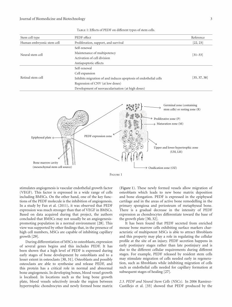
Journal of Biomedicine and Biotechnology 3
Table 1: Effects of PEDF on different types of stem cells.
Stem cell type PEDF effect Reference
Human embryonic stem cell Proliferation, support, and survival [22, 23]
Neural stem cell
Self-renewal
[31–33]Maintenance of multipotency
Activation of cell division
Antiapoptotic effects
Retinal stem cell
Self-renewal
[35, 37, 38]Cell expansion
Inhibits migration of and induces apoptosis of endothelial cells
Regression of CNV (at low doses)
Development of neovascularisation (at high doses)
Epiphyseal plate
Bone marrow cavity
(mesenchymal stem cell source)
Germinal zone (containing
stem cells) or resting zone (R)
Maturation zone (M)
Upper and lower hypertrophic zone
(UH, LH)
PEDF expression zone
Proliferative zone (P)
Ossification zone (OZ)
Figure 1
stimulates angiogenesis is vascular endothelial growth factor(VEGF). This factor is expressed in a wide range of cellsincluding BMSCs. On the other hand, one of the key func-tions of the PEDF molecule is the inhibition of angiogenesis.In a study by Fan et al. (2011), it was observed that PEDFexpression was much stronger than that of VEGF in BMSCs.Based on data acquired during that project, the authorsconcluded that BMSCs may not usually be an angiogenesis-promoting population in a normal environment [28]. Thisview was supported by other findings that, in the presence ofhigh cell numbers, MSCs are capable of inhibiting capillarygrowth [29].
During differentiation of MSCs to osteoblasts, expressionof several genes begins and this includes PEDF. It hasbeen shown that a high level of PEDF is expressed duringearly stages of bone development by osteoblasts and to alesser extent in osteoclasts [30, 31]. Osteoblasts and possiblyosteoclasts are able to synthesise and release PEDF, andthis protein has a critical role in normal and abnormalbone angiogenesis. In developing bones, blood vessel growthis localised. In locations such as the long bone growthplate, blood vessels selectively invade the region betweenhypertrophic chondrocytes and newly formed bone matrix
(Figure 1). These newly formed vessels allow migration ofosteoblasts which leads to new bone matrix depositionand bone elongation. PEDF is expressed in the epiphysealcartilage and in the areas of active bone remodelling in theprimary spongiosa and periosteum of metaphyseal bone.There is a gradual decrease in the intensity of PEDFexpression as chondrocytes differentiate toward the base ofthe growth plate [30, 32].
It has been found that PEDF secreted from enrichedmouse bone marrow cells exhibiting surface markers char-acteristic of multipotent MSCs is able to attract fibroblastsand this property may play a role in regulating the cellularprofile at the site of an injury. PEDF secretion happens inearly postinjury stages rather than late postinjury and isdue to the different cellular requirements during differentstages. For example, PEDF released by resident stem cellsmay stimulate migration of cells needed early in regenera-tion, such as fibroblasts while inhibiting migration of cellssuch as endothelial cells needed for capillary formation atsubsequent stages of healing [27].
2.3. PEDF and Neural Stem Cells (NSCs). In 2006 Ramırez-Castillejo et al. [33] showed that PEDF produced by the
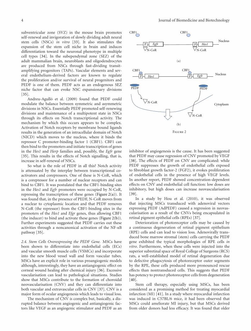
4 Journal of Biomedicine and Biotechnology
subventricular zone (SVZ) in the mouse brain promotesself-renewal and invigoration of slowly dividing adult neuralstem cells (NSCs) in vitro [33]. It also stimulates theexpansion of the stem cell niche in brain and inducesdifferentiation toward the neuronal phenotype in multiplecell types [34]. In the subependymal zone (SEZ) of theadult mammalian brain, neuroblasts and oligodendrocytesare produced from NSCs through fast-dividing transit-amplifying progenitors (TAPs). Vascular elements and sev-eral endothelium-derived factors are known to regulatethe proliferation and/or survival of neural progenitors andPEDF is one of them. PEDF acts as an endogenous SEZniche factor that can evoke NSC expansionary divisions[35].
Andreu-Agullo et al. (2009) found that PEDF couldmodulate the balance between symmetric and asymmetricdivisions in NSCs. Essentially PEDF promoted self-renewingdivisions and maintenance of a multipotent state in NSCsthrough its effects on Notch transcriptional activity. Themechanism by which this occurs appears to be complex.Activation of Notch receptors by membrane bound ligandsresults in the generation of an intracellular domain of Notch(NICD) which moves to the nucleus, where it binds therepressor C promoter-binding factor 1 (CBF1). CBF1 canthen bind to the promoters and initiate transcription of genesin the Hes1 and Herp families and, possibly, the Egfr gene[35]. This results in the effects of Notch signalling, that is,increase in self-renewal of NSCs.
So what is the role of PEDF in all this? Notch activityis attenuated by the interplay between transcriptional co-activators and corepressors. One of these is N-CoR, whichis a corepressor for a number of nuclear receptors and canbind to CBF1. It was postulated that the CBF1-binding sitesin the Hes1 and Egfr promoters were occupied by N-CoR,repressing the transcription of these genes (Figure 2(a)). Itwas found that, in the presence of PEDF, N-CoR moves froma nuclear to cytoplasmic location and that PEDF removesN-CoR (the repressor) from the CBF1-binding sites in thepromoters of the Hes1 and Efgr genes, thus allowing CBF1(the inducer) to bind and activate these genes (Figure 2(b)).Further experiments suggested that PEDF carries out theseactivities through a noncanonical activation of the NF-κBpathway [35].
2.4. Stem Cells Overexpressing the PEDF Gene. MSCs havebeen shown to differentiate into endothelial cells (ECs)and vascular smooth muscle cells (VSMCs) and incorporateinto the new blood vessel wall and form vascular tubes.MSCs have an explicit role in various proangiogenic modelsalthough, interestingly, they have an antiangiogenic effect oncorneal wound healing after chemical injury [36]. Excessivevascularisation can lead to pathological situations. Studiesshow that MSCs contribute to the formation of choroidalneovascularisation (CNV) and they can differentiate intoboth vascular and extravascular cells in CNV [37]. CNV is amajor form of ocular angiogenesis which leads to visual loss.
The mechanism of CNV is complex but, basically, a dis-rupted balance between angiogenic and antiangiogenic fac-tors like VEGF as an angiogenic stimulator and PEDF as an
CBF1
PNucleus
P
CBF1
Egfr Hes1
N-CoR N-CoR
(a)
Cytoplasm
PEDFPEDF
NucleusP
CBF1
P
CBF1
EgfrHes1
N-CoR
N-CoR
(b)
Figure 2
inhibitor of angiogenesis is the cause. It has been suggestedthat PEDF may cause regression of CNV promoted by VEGF[38]. The effects of PEDF on CNV are complicated: whilePEDF suppresses the growth of endothelial cells exposedto fibroblast growth factor-2 (FGF2), it evokes proliferationof endothelial cells in the presence of high VEGF levels.In another report, PEDF showed concentration-dependenteffects on CNV and endothelial cell function: low doses areinhibitory, but high doses can increase neovascularisation[39].
In a study by Hou et al. (2010), it was observedthat injecting MSCs transduced with adenoviral vectorsexpressing PEDF (AdPEDF) caused a regression in neovas-cularisation as a result of the CNVs being encapsulated inretinal pigment epithelial cells (RPEs) [37].
Deterioration of photoreceptor cells may be caused bya continuous degeneration of retinal pigment epithelium(RPE) cells and can lead to vision loss. Adenovirally trans-duced bone marrow stromal (stem) cells carrying the PEDFgene exhibited the typical morphologies of RPE cells invitro. Furthermore, when these cells were injected into thesuperior subretinal space of Royal College of Surgeons (RCS)rats, a well-established model of retinal degeneration dueto defective phagocytosis of photoreceptor outer segmentsby the RPE, these cells produced more pronounced rescueeffects than nontransduced cells. This suggests that PEDFhas potency to protect photoreceptor cells from degeneration[40].
Stem cell therapy, especially using MSCs, has beenconsidered as a promising method for treating myocardialinfarction (MI) [41]. In a study where myocardial infarctionwas induced in C57BL/6 mice, it had been observed thatMSCs could ameliorate MI injury, but that MSCs derivedfrom older donors had less efficacy. It was found that older
Journal of Biomedicine and Biotechnology 5
MSCs secreted higher levels of PEDF than younger ones. Fur-thermore, infarcts treated with (AdPEDF-) transduced MSCsthat over-expressed PEDF contained fewer ECs, VSMCs, andmacrophages but had increased number of fibroblasts [42].They concluded that it was the increased expression of PEDFin aged MSCs that impaired their therapeutic efficacy.
3. Conclusion
PEDF has a range of functions in different tissues and cells;however, the impact of this interesting protein on stemcells is not yet clear. PEDF expressed in feeder cells appearsto have a supportive effect on stem cells such as hESCsand RPCs. PEDF affects NSCs via the Notch transcriptionalpathway, promoting self-renewing divisions, maintaining amultipotent state of these cells and supporting expansion ofthe stem cell niche. Stem cells transduced with adenoviralvectors carrying PEDF exhibit potential for enhanced stemcell therapy in diseases such as macular degeneration.However, the effects of PEDF on stem cells in other diseasestates are clear. Whilst PEDF generally causes regression ofCNV established by VEGF, the effects may be concentrationdependent—both on the concentration of VEGF and PEDF.As for myocardial infarction, the presence of excessive PEDFis actually reported to reduce the efficacy of stem cell therapy.To conclude, the role of PEDF in stem cells in variouspathologies does differ, and while counterproductive in somecases such as myocardial infarction, it may have a positiverole to play in some instances. Further studies on PEDF andstem cells will help illuminate such instances and whetherthey can be used therapeutically.
References
[1] Z. Xu, S. Fang, Y. Zuo et al., “Combination of pigmentepithelium-derived factor with radiotherapy enhances theantitumor effects on nasopharyngeal carcinoma by down-regulating vascular endothelial growth factor expression andangiogenesis,” Cancer Science, vol. 102, no. 10, pp. 1789–1798,2011.
[2] Q. Gong, X. Yang, W. Cai, G. Gao, and Z. Yang, “Expressionand purification of functional epitope of pigment epithelium-derived factor in E. coli with inhibiting effect on endothelialcells,” Protein Journal, vol. 29, no. 3, pp. 167–173, 2010.
[3] S. T. Olson and P. G. Gettins, “Regulation of proteasesby protein inhibitors of the serpin superfamily,” Progress inMolecular Biology and Translational Science C, vol. 99, pp. 185–240, 2011.
[4] J. Tombran-Tink, S. Aparicio, X. Xu et al., “PEDF and theserpins: phylogeny, sequence conservation, and functionaldomains,” Journal of Structural Biology, vol. 151, no. 2, pp.130–150, 2005.
[5] T. Samkharadze, M. Erkan, C. Reiser-Erkan et al., “Pigmentepithelium-derived factor associates with neuropathy andfibrosis in pancreatic cancer,” American Journal of Gastroen-terology, vol. 106, no. 5, pp. 968–980, 2011.
[6] T. Akiyama, C. R. Dass, Y. Shinoda, H. Kawano, S. Tanaka,and P. F. M. Choong, “PEDF regulates osteoclasts via osteo-protegerin and RANKL,” Biochemical and Biophysical ResearchCommunications, vol. 391, no. 1, pp. 789–794, 2010.
[7] F. Sanchez-Sanchez, J. D. Aroca-Aguilar, I. Segura et al.,“Expression and purification of functional recombinanthuman pigment epithelium-derived factor (PEDF) secreted bythe yeast Pichia pastoris,” Journal of Biotechnology, vol. 134, no.1-2, pp. 193–201, 2008.
[8] M. L. Broadhead, C. R. Dass, and P. F. Choong, “In vitro andin vivo biological activity of PEDF against a range of tumors,”Expert Opinion on Therapeutic Targets, vol. 13, no. 12, pp.1429–1438, 2009.
[9] J. L. Orgaz, O. Ladhani, K. S. Hoek et al., “Loss of pigmentepithelium-derived factor enables migration, invasion andmetastatic spread of human melanoma,” Oncogene, vol. 28, no.47, pp. 4147–4161, 2009.
[10] T. Yabe, D. Wilson, and J. P. Schwartz, “NFκB activa-tion is required for the neuroprotective effects of pigmentepithelium-derived Factor (PEDF) on cerebellar granule neu-rons,” The Journal of Biological Chemistry, vol. 276, no. 46, pp.43313–43319, 2001.
[11] T. A. Zaichuk, E. H. Shroff, R. Emmanuel, S. Filleur, T.Nelius, and O. V. Volpert, “Nuclear factor of activated T cellsbalances angiogenesis activation and inhibition,” Journal ofExperimental Medicine, vol. 199, no. 11, pp. 1513–1522, 2004.
[12] T. C. Ho, S. L. Chen, Y. C. Yang, C. L. Liao, H. C. Cheng, andY. P. Tsao, “PEDF induces p53-mediated apoptosis throughPPAR gamma signaling in human umbilical vein endothelialcells,” Cardiovascular Research, vol. 76, no. 2, pp. 213–223,2007.
[13] C. R. Dass and P. F. M. Choong, “uPAR mediates anticanceractivity of PEDF,” Cancer Biology and Therapy, vol. 7, no. 8,pp. 1262–1270, 2008.
[14] Y. L. Si, Y. L. Zhao, H. J. Hao, X. B. Fu, and W. D. Han,“MSCs: biological characteristics, clinical applications andtheir outstanding concerns,” Ageing Research Reviews, vol. 10,no. 1, pp. 93–103, 2011.
[15] C. Trento and F. Dazzi, “Mesenchymal stem cells and innatetolerance: biology and clinical applications.,” Swiss MedicalWeekly, vol. 140, article w13121, 2010.
[16] A. G. Smith, “Embryo-derived stem cells: of mice and men,”Annual Review of Cell and Developmental Biology, vol. 17, pp.435–462, 2001.
[17] L. Li and T. Xie, “Stem cell niche: structure and function,”Annual Review of Cell and Developmental Biology, vol. 21, pp.605–631, 2005.
[18] R. M. Backly and R. Cancedda, “Bone marrow stemcells in clinical application: harnessing paracrine rolesand niche mechanisms,” Advances in Biochemical Engineer-ing/Biotechnology, vol. 123, pp. 265–292, 2010.
[19] A. Wilson, E. Laurenti, G. Oser et al., “Hematopoietic stemcells reversibly switch from dormancy to self-renewal duringhomeostasis and repair,” Cell, vol. 135, no. 6, pp. 1118–1129,2008.
[20] T. Yin and L. Li, “The stem cell niches in bone,” The Journal ofClinical Investigation, vol. 116, no. 5, pp. 1195–1201, 2006.
[21] T. Reya, S. J. Morrison, M. F. Clarke, and I. L. Weissman, “Stemcells, cancer, and cancer stem cells,” Nature, vol. 414, no. 6859,pp. 105–111, 2001.
[22] S. V. Anisimov, N. S. Christophersen, A. S. Correia et al.,“Identification of molecules derived from human fibroblastfeeder cells that support the proliferation of human embryonicstem cells,” Cellular and Molecular Biology Letters, vol. 16, no.1, pp. 79–88, 2011.
6 Journal of Biomedicine and Biotechnology
[23] D. Zhu, X. Deng, C. Spee et al., “Polarized secretion of PEDFfrom human embryonic stem cell-derived RPE promotesretinal progenitor cell survival,” Investigative Ophthalmologyand Visual Science, vol. 52, no. 3, pp. 1573–1585, 2011.
[24] J. D. Weiland, A. K. Cho, and M. S. Humayun, “Retinalprostheses: current clinical results and future needs,” Ophthal-mology, vol. 118, no. 11, pp. 2227–2237, 2010.
[25] P. van Wijngaarden and S. H. Qureshi, “Inhibitors of vascularendothelial growth factor (VEGF) in the management ofneovascular age-related macular degeneration: a review ofcurrent practice,” Clinical & Experimental Optometry, vol. 91,no. 5, pp. 427–437, 2008.
[26] I. H. Pang, H. Zeng, D. L. Fleenor, and A. F. Clark, “Pigmentepithelium-derived factor protects retinal ganglion cells,”BMC Neuroscience, vol. 8, article 11, 2007.
[27] H. Sarojini, R. Estrada, H. Lu et al., “PEDF from mousemesenchymal stem cell secretome attracts fibroblasts,” Journalof Cellular Biochemistry, vol. 104, no. 5, pp. 1793–1802, 2008.
[28] W. Fan, R. Crawford, and Y. Xiao, “The ratio of VEGF/PEDFexpression in bone marrow mesenchymal stem cells regulatesneovascularization,” Differentiation, vol. 81, no. 3, pp. 181–191, 2011.
[29] K. Otsu, S. Das, S. D. Houser, S. K. Quadri, S. Bhattacharya,and J. Bhattacharya, “Concentration-dependent inhibition ofangiogenesis by mesenchymal stem cells,” Blood, vol. 113, no.18, pp. 4197–4205, 2009.
[30] J. Tombran-Tink and C. J. Barnstable, “Osteoblasts andosteoclasts express PEDF, VEGF-A isoforms, and VEGFreceptors: Possible mediators of angiogenesis and matrixremodeling in the bone,” Biochemical and Biophysical ResearchCommunications, vol. 316, no. 2, pp. 573–579, 2004.
[31] C. Chiellini, O. Cochet, L. Negroni et al., “Characterizationof human mesenchymal stem cell secretome at early stepsof adipocyte and osteoblast differentiation,” BMC MolecularBiology, vol. 9, p. 26, 2008.
[32] G. M. Y. Quan, J. Ojaimi, Y. Li, V. Kartsogiannis, H. Zhou, andP. F. M. Choong, “Localization of pigment epithelium-derivedfactor in growing mouse bone,” Calcified Tissue International,vol. 76, no. 2, pp. 146–153, 2005.
[33] C. Ramırez-Castillejo, F. Sanchez-Sanchez, C. Andreu-Agulloet al., “Pigment epithelium-derived factor is a niche signal forneural stem cell renewal,” Nature Neuroscience, vol. 9, no. 3,pp. 331–339, 2006.
[34] Y. Mirochnik, A. Aurora, F. T. Schulze-Hoepfner et al., “Shortpigment epithelial-derived factor-derived peptide inhibitsangiogenesis and tumor growth,” Clinical Cancer Research, vol.15, no. 5, pp. 1655–1663, 2009.
[35] C. Andreu-Agullo, J. M. Morante-Redolat, A. C. Delgado,and I. Farinas, “Vascular niche factor PEDF modulates notch-dependent stemness in the adult subependymal zone,” NatureNeuroscience, vol. 12, no. 12, pp. 1514–1523, 2009.
[36] Y. O. Joo, K. K. Mee, S. S. Mi et al., “The anti-inflammatoryand anti-angiogenic role of mesenchymal stem cells in cornealwound healing following chemical injury,” Stem Cells, vol. 26,no. 4, pp. 1047–1055, 2008.
[37] H. Y. Hou, H. L. Liang, Y. S. Wang et al., “A therapeutic strategyfor choroidal neovascularization based on recruitment ofmesenchymal stem cells to the sites of lesions,” MolecularTherapy, vol. 18, no. 10, pp. 1837–1845, 2010.
[38] J. P. Tong and Y. F. Yao, “Contribution of VEGF and PEDFto choroidal angiogenesis: a need for balanced expressions,”Clinical Biochemistry, vol. 39, no. 3, pp. 267–276, 2006.
[39] R. S. Apte, R. A. Barreiro, E. Duh, O. Volpert, and T. A.Ferguson, “Stimulation of neovascularization by the anti-angiogenic factor PEDF,” Investigative Ophthalmology andVisual Science, vol. 45, no. 12, pp. 4491–4497, 2004.
[40] S. Arnhold, P. Heiduschka, H. Klein et al., “Adenovirallytransduced bone marrow stromal cells differentiate intopigment epithelial cells and induce rescue effects in RCS rats,”Investigative Ophthalmology and Visual Science, vol. 47, no. 9,pp. 4121–4129, 2006.
[41] R. Passier, L. W. van Laake, and C. L. Mummery, “Stem-cell-based therapy and lessons from the heart,” Nature, vol. 453,no. 7193, pp. 322–329, 2008.
[42] H. Liang, H. Hou, and W. Yi, “Increased expression of pigmentepithelium-derivedfactor in aged mesenchymal stem cellsimpairs their therapeutic efficacy for attenuating myocardialinfarction injury,” European Heart Journal. In press.
Hindawi Publishing CorporationJournal of Biomedicine and BiotechnologyVolume 2012, Article ID 580687, 8 pagesdoi:10.1155/2012/580687
Review Article
PEDF in Diabetic Retinopathy: A Protective Effect ofOxidative Stress
Xiao-feng Zhu and Hai-dong Zou
Department of Ophthalmology, Shanghai First People’s Hospital, Shanghai Jiao Tong University, Shanghai 200080, China
Correspondence should be addressed to Hai-dong Zou, [email protected]
Received 8 December 2011; Accepted 8 February 2012
Academic Editor: Takeshi Yabe
Copyright © 2012 X.-f. Zhu and H.-d. Zou. This is an open access article distributed under the Creative Commons AttributionLicense, which permits unrestricted use, distribution, and reproduction in any medium, provided the original work is properlycited.
Diabetic retinopathy (DR) is a major cause of blindness in working age adults, and oxidative stress plays a vital role in thepathogenesis of DR. Pigment-epithelium-derived factor (PEDF), a multifunctional protein, has shown to inhibit the developmentof DR by accumulating evidence. This paper highlights the current understanding of probable mechanism about how PEDF blocksthe deterioration of DR through its antioxidative properties and application prospects of PEDF as a novel therapeutic target in DR.Gene therapy of PEDF is becoming more and more acceptable and will widely be applied to the actual treatment in the near future.
1. Introduction
Diabetic retinopathy (DR) is the leading cause of acquiredvision loss among adults of working age in developedcountries worldwide and has been perceived as the mostcommon microvascular complications of diabetes [1–3].Therefore, within three to five years after the onset of type1 diabetes and shortly after the diagnosis of type 2 diabetes,the diabetic patients are recommended to be screened forretinopathy with an initial dilated and comprehensive eyeexamination by an ophthalmologist regularly [4]. Chronichyperglycemia which is well documented is a primaryinitiator of DR [5]. In principle, intensive glycemic controlcan delay the development of DR [5]. However, it isnoteworthy that some patients may still develop DR evenwith good glycemic control [6, 7]. This remaining effect,prior to glycemic control, suggests a “metabolic memory”phenomenon. Furthermore, a growing number of studieshave shown that the retina experience increased oxidativedamage continuously, even in tight glycemic control, andoxidative stress plays a vital role in the pathogenesis of DR[6, 8].
The molecular mechanisms of hyperglycemia-inducedDR are not fully clear, and the majority of publications focuson multiple biochemical pathways, including the augmenta-tion of polyol pathway [9], protein kinase C (PKC) activation
[10], increased advanced glycation endproducts (AGEs)formation, the receptor for AGEs and its activating ligands[11], and overactivity of the hexosamine pathway [12].However, all the mechanisms are activated by a single event:aberrant production of the mitochondria-derived reactiveoxygen species (ROS) to increase the level of oxidative stress[13]. Therefore, antioxidant therapy is being studied toprevent induction of the various pathogenic mechanismsof DR. With the failure of demonstrating that dietarysupplementations of multiantioxidants have beneficial effectsclinically [14], an antioxidant which could specifically targetpathogenesis of DR is no time to delay. Pigment-epithelium-derived factor (PEDF), a 50-kDa secreted glycoprotein,recently is shown to inhibit the development of DR throughits antioxidative properties. This paper summarizes theprobable protective mechanism of PEDF in high-glucose-induced oxidative stress and application prospects of PEDFas a novel therapeutic target in DR.
2. PEDF and Its Potential Protective Roleof Oxidative Stress in Diabetic Retinopathy
2.1. PEDF and Its Biological Function. PEDF is a 418-aminoacid, 50-KDa protein which was first purified from
2 Journal of Biomedicine and Biotechnology
conditioned medium from both fetal and adult retinal pig-ment epithelial (RPE) cells [15–18] and it is a noninhibitorymember of the serine protease inhibitor (serpin) family.PEDF is widely expressing throughout the body, especiallyin the nervous system and the retina. According to currentresearch, PEDF has shown that it is a multifunctional proteinwith demonstrable neurotrophic [19, 20], antiangiogenic[16], antivasopermeability [21], antiinflammatory [22],antifibrosis [23], and antitumorigenic [24] properties andinhibited the development of DR through its antioxidativeproperties by accumulating evidence [25–28].
Earlier clinical studies demonstrated an inverse correla-tion of the levels of intraocular PEDF and the developmentof abnormal angiogenesis in some ocular diseases, suchas proliferative diabetic retinopathy (PDR) [16, 29–32].Similarly, in mouse model of type 2 diabetes mellitus, lowervitreous or aqueous humour levels of PEDF are associatedwith early phase of experimental DR [16, 31, 33], anddecreased protein levels of PEDF in the retina are associatedwith ischemia-induced retinal neovascularization in theoxygen-induced retinopathy (OIR) model as well [34].
Therefore, existing research data demonstrated thatupregulation or substitution of PEDF may be a promisingtherapeutic target for DR [16, 25, 35–37], especially oxidativestress-involved retinal tissue damage [38]. Then, experimen-tal interventions to increase locally PEDF concentrations,either by an adenovirus expressing human PEDF or apurified recombinant PEDF protein, have shown to attenuateretinal tissue damage in different animal models [22, 39].Thus, finding a good way for controlling PEDF expressionand action in the retina has become a research focus.
In addition, PEDF suppresses vascular endothelialgrowth-factor-induced (VEGF-induced) retinal microvascu-lar endothelial cell proliferation and migration [40] andinhibits VEGF activation in human retinal endothelial cellsin vitro condition [41]. In contrast, PEDF has a synergisticaction with VEGF on cell proliferation in endothelial cellscultured in the presence of VEGF [42]. Moreover, the VEGFand PEDF expression in Muller cells is unbalanced underhigh-glucose concentration, which contributes to retinalneovascularization in DR [43]. Since VEGF plays a pivotalrole in the formation of ROS, to defining the specific effectsof PEDF, high-glucose-induced oxidative stress will also beimportant.
2.2. Antioxidative Properties of PEDF in Diabetic Retinopathy
2.2.1. Inhibit AGEs-Induced Injury. AGEs, senescent macro-proteins formed at an accelerated rate in diabetes, causeapoptotic cell death in retinal pericytes [44, 45]. Studieshave found AGEs significantly decreased endothelial mRNAlevels of PEDF in endothelial cells [46], and PEDF proteinsprotect cultured retinal pericytes from AGEs-induced injuryprobably via oxidative stress generation [25]. Thus, we willintroduce the different possible mechanisms of PEDF toinhibit AGEs-induced injury in DR.
Retinal Pericytes. Earlier studies have found that loss ofpericytes and increased vascular permeability, followed by
microvascular occlusion in the retinas, ultimately leadedto the development of DR [47, 48]. Experimental analysisrevealed that pericytes possessed of a membrane proteinwith binding affinity for PEDF which significantly inhib-ited AGEs-induced ROS generation and the subsequentdecrease in DNA synthesis and apoptotic cell death inpericytes. Furthermore, PEDF proteins completely restoredthe downregulation of bcl-2 (an antiapoptotic molecule)gene expressing in AGEs-exposed pericytes [25]. Similarly,the studies have demonstrated that PEDF completely blockedhigh-glucose- or H2O2-induced intracellular ROS generationand an increased ratio of bax to bcl-2 mRNA level withsubsequent activation of caspase-3 in pericytes [49]. Inaddition, PEDF protected high glucose- (30 mM) or H2O2-induced pericyte apoptosis and dysfunction through itsantioxidative properties via glutathione peroxidase (GPx)induction. Simultaneously, the study also found that PEDF’smRNA levels themselves were downregulated in high-glucose(HG-) or H2O2-exposed pericytes.
These results all demonstrated that PEDF proteins pro-tected cultured pericytes from AGEs-induced cytotoxicitythrough its antioxidative properties, and substitution ofPEDF proteins may be a promising strategy in treatment ofpatients with DR.
Monocyte Chemoattractant Protein-1 (MCP-1). Furtherwork have shown that PEDF prevented the AGEs-inducedupregulation of monocyte chemoattractant protein-1(MCP-1) mRNA contents as well as protein production inmicrovascular endothelial cells (ECs) [50]. Moreover, levelsof MCP-1 in vitreous fluids have been correlated with theseverity of PDR [51].
Rage. There is a growing body of evidence that RAGE is asignal-transducing receptor for AGEs, and that engagementof RAGE by AGEs elicits vascular inflammation and altersgene expression in retinal vascular wall cells, thereby it isinvolved in the development and progression of DR [45, 50,52–54]. Recent studies have shown that PEDF could inhibitdiabetes- (in the eye of diabetic rats) or AGEs-induced (invitro) RAGE gene expression by blocking the superoxide-mediated NF-kappaB activation [36].
Endothelial NO Synthase (eNOS). Studies have shown thatPEDF prevented the AGEs-elicited endothelial NO synthase(eNOS) reduction through its antioxidative properties inAGEs-exposed human umbilical vein ECs (HUVECs) [55].And endothelial dysfunction due to reduced synthesis and/orbioavailability of nitric oxide (NO) is an initial step ofatherosclerotic vascular disease in diabetes [56–58].
Platelet Activation and Aggregation. There is accumulatingevidence that the oxidative stress generation is involved inplatelet activation and aggregation [59, 60]. These observa-tions suggest that the inhibition of platelet activation andaggregation may be a novel therapeutic target for preventingthe development and progression of vascular complicationsin patients with diabetes. Further, the researchers have found
Journal of Biomedicine and Biotechnology 3
that PEDF prevented platelet activation and aggregation indiabetic rats or AGEs-injected rats through its antioxidativeproperties by suppressing NADPH oxidase-driven superox-ide generation, deleterious effects of AGEs [61].
The Src Pathway. PEDF inhibited AGEs-induced ROS gener-ation by increasing levels of SOD and GSH and also blockedthe activation of caspase-3. Furthermore, PEDF induced cellsurvival via the Src pathway by Src phosphorylation at Y419,as evidenced by a pharmacological inhibitor and Src mutants[62].
Phosphatidylinositol 3-Kinase (PI3K)/Akt Pathway. Recentstudies demonstrated that PEDF could inhibit the AGEs-BSA-induced permeability via phosphatidylinositol 3-kinase(PI3K)/Akt pathway. AGEs-BSA increased the ECs perme-ability by stimulating ROS generation via NADPH oxidaseactivity and Akt phosphorylation at Ser473. PEDF decreasedROS generation in AGEs-BSA-exposed endothelial cells bysuppressing the NADPH oxidase activity via downregu-lating the phosphorylation of p22PHOX at Thr147. Thisled to blockade of AGEs induction of PI3K/Akt activationin permeability. Furthermore, PEDF inhibited the AGEs-BSA-induced permeability by increased expression of tightjunction protein zona occludens-1 (ZO-1), coincident withan increase in barrier properties of endothelial monolayer[63].
2.2.2. Inhibit Leptin-Induced Injury. PEDF was found toinhibit the leptin-induced ROS generation and upregulationof VEGF mRNA levels including any increase in DNAsynthesis in microvascular ECs [64]. Indeed, leptin levels invitreous were correlated with PDR [65].
2.2.3. Inhibit Tumor Necrosis Factor-α (TNF-α-) InducedInjury. The studies demonstrated that PEDF inhibitedtumor necrosis factor-α (TNF-α-) induced redox-sensitivetranscriptional factor NF-kappaB activation and subsequentinterleukin IL-6 overexpression at both mRNA and proteinlevels in human umbilical vein endothelial cell (HUVEC)by suppressing NADPH oxidase-mediated ROS generation[66]. In addition, TNF-alpha which is initially involved inthe pathogenesis of atherosclerosis [67, 68] and the classicproinflammatory cytokines, downregulated PEDF mRNAlevels [66].
2.2.4. Inhibit Angiopoietin-II (Ang II-) Induced Injury. Theresearchers have found that Angiopoietin-II (Ang II) signifi-cantly induced NF-kappaB activation and subsequent MCP-1 expression in HUVEC, both of which were completelyinhibited by PEDF. Subsequently, PEDF inhibited Ang-II-induced upregulation of mRNA levels of p22PHOX, Nox4,and gp91PHOX/Nox2, which are membrane components ofNADPH oxidase and its enzymatic activity in HUVEC [69].Another study found that Ang II also significantly decreasedPEDF mRNA levels in ECs, which was completely reversed
by an Ang II type 1 receptor blocker, telmisartan [70]. Fur-thermore, anti-PEDF Ab significantly inhibited the growth-stimulating effects of cocultured ECs on pericytes. Theseresults demonstrated that PEDF, an EC-derived mitogen orsurvival factor for retinal pericytes, inhibited Ang-II-inducedECs activation by suppressing NADPH-oxidase-mediatedROS generation, and suppression by Ang II of the EC-derivedPEDF may be involved in exacerbation of DR in patients withhypertension. In addition, PEDF was found to completelyinhibit high-glucose- or H2O2-induced increase in a mRNAratio of Ang II to Angiopoietin-I (Ang I) and upregulationof VEGF mRNA levels in pericytes. VEGF and angiopoietin(Ang) have been known that they were the major regulatorsof vascular integrity and involved in DR as well [71].
2.2.5. Inhibit HOL-LDL-Induced Injury. PEDF amelioratedHOL-LDL-induced MCP-1 and the subsequent NF-kappaBactivation effectively. Moreover, PEDF significantly amelio-rated HOG-LDL-induced ROS generation through upregu-lation of superoxide dismutase 1 expression [72]. This studyrepresented a new mechanism for the salutary effect of PEDFin DR.
2.2.6. Inhibit High-Glucose (HG)-Induced JAK2/STAT3 Acti-vation. A recent report suggested that ACEI exerted aprotective effect on DR, and this protective effect couldbe reflected by a decreased VEGF-to-PEDF ratio, which isa result of reduced mitochondrial ROS production itselfscaused by ACEI-induced increase of proliferator-activatedreceptor gamma (PPARγ) and subsequent upregulation ofuncoupling protein-2 (UCP-2) expression [73]. Furtherwork in vitro has demonstrated that PEDF could decreasemitochondria-derived ROS generation and subsequentlydownregulate VEGF expression, possibly through inhibitingHG-induced JAK2/STAT3 activation [74]. These studies pavea new way for future in treatment of DR.
2.2.7. Regulation of PEDF Expression In Vivo. PEDF levelsin aqueous humor or vitreous were associated with totalantioxidant capacity in humans [38, 75] and suggested thatPEDF may act as an endogenous antioxidant in the eye andupregulation or substitution of PEDF may be a therapeutictarget for oxidative stress-involved eye diseases, especiallyPDR.
Retinal PEDF levels were reduced in diabetic rat, whichwere restored by PEDF injections. Decreased amplitudes ofa- and b-wave in the ERG in diabetic rats, which were inparallel with GFAP overexpression in the Muller cells, alsocould be blocked by PEDF injections. Further, retinal 8-OHdG, p22PHOX, VEGF levels, and NADPH oxidase activitywere increased, and BRB was broken in diabetic rats, both ofwhich were ameliorated by the treatment of PEDF [26].
In addition, studies with rats, intravenous administra-tion of AGEs, and simultaneous treatments with PEDFdemonstrated that PEDF decreased ROS generation in AGEs-exposed endothelial cells by suppressing NADPH oxidaseactivity via downregulation of mRNA levels of p22PHOX
and gp91PHOX and inhibited the AGEs-induced vascular
4 Journal of Biomedicine and Biotechnology
hyperpermeability, the characteristic feature of early DR, bysuppressing VEGF expression [76].
Furthermore, the action of PEDF not only varied withthe cell type but also depended on its concentration andenvironmental conditions [77].
3. Application Prospects of PEDF inDiabetic Retinopathy
Large prospective clinical studies have shown that intensiveblood glucose control reduced the incidence and progressionof DR [5, 48]. However, strict control of hyperglycemiais often difficult to maintain and may increase the riskof severe hypoglycemia in diabetic patients. In addition,photocoagulation and vitrectomy, current conventional ther-apeutic options for the treatment of PDR, are limitedby considerable side effects. Therefore, developing noveltherapeutic strategies that specifically target pathogenesis ofDR is desired for patients with diabetes. Based on the aboverole and regulation mechanism of PEDF in DR, the researchon the treatment of PDR has turned to the regulation ofangiogenesis inhibitors and growth factors.
3.1. Current and Potential Molecular Therapie of PEDF
3.1.1. PEDF and PEDF-Derived Peptide. The multifunctionalPEDF, more effective than other antiangiogenic factors, isa good candidate for treatment of DR. Because a largepart of endogenous PEDF-binding affinity to extracellularmatrix components and cell-surface receptors, it would notachieve a protective effect. However, injection of exogenousPEDF, competitive binding of PEDF in the extracellularbinding sites, will release PEDF from extracellular matrixand subsequently achieve therapeutic concentrations. Theproblems of current stage are as follow: (1) PEDF is a 50-kDaprotein, and its structural features limit practical applicationas pharmaceuticals; (2) underestimated the carrier itselfcould induce the inflammatory response; (3) currently thereis no good control methods for PEDF which has secretedinto the extracellular. However, many biologically activefragment of PEDF has been known, and if we can findthe active fragment, equivalent or near equivalent withPEDF, which is replaced with small peptides, we will avoidor reduce the inflammatory reaction by virus or othercarriers through slow release of directly permeating sclera orintravitreal injection [78]. A recent study demonstrated thatan antiangiogenic peptide, PEDF-34, reduced circulatingendothelial cells during ischemia-induced neovasculariza-tion [79]. Thus, the PEDF-34 peptide could be a superiorbiological therapeutic for the treatment of PDR and hasgreat potential for large-scale pharmaceutical development.A disadvantage, however, of using small peptide derivativesis that they tend to be cleared rapidly from tissues and thusmay be less effective therapeutically unless they are protectedfrom rapid enzymatic degradation and tissue clearance.
3.1.2. Biodegradable Nanospheres. Nanospheres, biocompat-ible, biodegradable, and producer of fewer side effects,
is one way to avoid disadvantage of peptide. Moreover,there is abundant experimental evidence that intravitrealinjections of drugs encapsulated in PLGA-poly (lactide-co-glycolide) nanospheres are both useful and effective [80, 81].A recent study has found that delivering PEDF82−121, a freepeptide, in PLGA nanospheres is an effective way of achievingcontrolled release of therapeutically active levels of thepeptide [82]. Such delivery systems can be manipulated toprovide controlled release of physiological levels of bioactiveproducts for both short- and long-term needs [83].
3.1.3. Gene Therapy. Gene therapy involves the replacementof a faulty gene or the insertion of a new gene. Treatmentof angiogenic disease can be achieved by the insertion ofgenes that encode antiangiogenic proteins, including thevitreous body and subretinal injection. Adenovirus andadeno-associated virus (AAV) has shown particular promisein the delivery of antiangiogenic DNA. The first human trialof recombinant PEDF introduced via the adenoviral vectorAdPEDF.11 suggested that intravitreal delivery is relativelywell tolerated with the antiangiogenic effect of PEDF per-sisting for several months [84]. PEDF transgenic (PEDF-Tg)mice that ubiquitously express human PEDF driven by theβ-actin promoter inhibited neovascular disorders such as DR[85]. These results have provided valuable information withregards to gene therapy for the treatment of DR.
4. Conclusion
PEDF, a multifunctional factor, has shown to inhibit thedevelopment of DR by accumulating evidence. This paperhighlights the current understanding of probable mechanismabout how PEDF blocks the deterioration of DR throughits antioxidative properties and application prospects ofPEDF as a novel therapeutic target in DR. Gene therapy isbecoming more and more acceptable and widely applied tothe actual treatment. PEDF and its fragments, transferredinto virus vector or made of biodegradable implant forlocal injection, have broad application prospects in treatmentof DR. However, the drug screening and selection of thebest way to enter the human body need large number ofresearches.
Acknowledgments
This paper was in part supported by Grants from theNatural Science Foundation of Shanghai Jiaotong UniversitySchool of Medicine, China (11XJ21056), Magnolia Sci-ence and Technology Talent Fund (MTF) of Science andTechnology Commission of Shanghai Municipality, China(11BA1409700), and medical-engineering cross fund ofShanghai Jiaotong University, China (YG2011MS11).
References
[1] S. Resnikoff, D. Pascolini, D. Etya’ale et al., “Global data onvisual impairment in the year 2002,” Bulletin of the WorldHealth Organization, vol. 82, no. 11, pp. 844–851, 2004.
Journal of Biomedicine and Biotechnology 5
[2] J. H. Kempen, B. J. O’Colmain, M. C. Leske et al., “Eyediseases prevalence research group. The prevalence of diabeticretinopathy among adults in the United States,” Archives ofOphthalmology, vol. 122, no. 4, pp. 552–563, 2004.
[3] J. B. Saaddine, A. A. Honeycutt, K. M. V. Narayan, X. Zhang, R.Klein, and J. P. Boyle, “Projection of diabetic retinopathy andother major eye diseases among people with diabetes mellitus:United States, 2005–2050,” Archives of Ophthalmology, vol.126, no. 12, pp. 1740–1747, 2008.
[4] American Diabetes Association, “Standards of medical care indiabetes—2007,” Diabetes Care, vol. 30, supplement 1, pp. S4–S41, 2007.
[5] H. Shamoon, H. Duffy, N. Fleischer et al., “The effect ofintensive treatment of diabetes on the development andprogression of long-term complications in insulin-dependentdiabetes mellitus,” New England Journal of Medicine, vol. 329,no. 14, pp. 977–986, 1993.
[6] R. A. Kowluru, M. Kanwar, and A. Kennedy, “Metabolicmemory phenomenon and accumulation of peroxynitrite inretinal capillaries,” Experimental Diabesity Research, vol. 2007,Article ID 21976, 2007.
[7] P. S. Chan, M. Kanwar, and R. A. Kowluru, “Resistance ofretinal inflammatory mediators to suppress after reinstitutionof good glycemic control: novel mechanism for metabolicmemory,” Journal of Diabetes and its Complications, vol. 24, no.1, pp. 55–63, 2010.
[8] R. A. Kowluru, “Effect of reinstitution of good glycemiccontrol on retinal oxidative stress and nitrative stress indiabetic rats,” Diabetes, vol. 52, no. 3, pp. 818–823, 2003.
[9] K. Naruse, J. Nakamura, Y. Hamada et al., “Aldose reductaseinhibition prevents glucose-induced apoptosis in culturedbovine retinal microvascular pericytes,” Experimental EyeResearch, vol. 71, no. 3, pp. 309–315, 2000.
[10] P. Geraldes, J. Hiraoka-Yamamoto, M. Matsumoto et al., “Acti-vation of PKC-and SHP-1 by hyperglycemia causes vascularcell apoptosis and diabetic retinopathy,” Nature Medicine, vol.15, no. 11, pp. 1298–1306, 2009.
[11] T. C. B. Moore, J. E. Moore, Y. Kaji et al., “The role of advancedglycation end products in retinal microvascular leukostasis,”Investigative Ophthalmology and Visual Science, vol. 44, no. 10,pp. 4457–4464, 2003.
[12] C. L. B. Kline, T. L. Schrufer, L. S. Jefferson, and S. R.Kimball, “Glucosamine-induced phosphorylation of the α-subunit of eukaryotic initiation factor 2 is mediated bythe protein kinase R-like endoplasmic-reticulum associatedkinase,” International Journal of Biochemistry and Cell Biology,vol. 38, no. 5-6, pp. 1004–1014, 2006.
[13] M. Brownlee, “The pathobiology of diabetic complications: aunifying mechanism,” Diabetes, vol. 54, no. 6, pp. 1615–1625,2005.
[14] R. Marchioli, C. Schweiger, G. Levantesi, L. Tavazzi, and F.Valagussa, “Antioxidant vitamins and prevention of cardiovas-cular disease: epidemiological and clinical trial data,” Lipids,vol. 36, pp. S53–S63, 2001.
[15] D. W. Dawson, O. V. Volpert, P. Gillis et al., “Pigmentepithelium-derived factor: a potent inhibitor of angiogenesis,”Science, vol. 285, no. 5425, pp. 245–248, 1999.
[16] J. Spranger, M. Osterhoff, M. Reimann et al., “Loss of theantiangiogenic pigment epithelium-derived factor in patientswith angiogenic eye disease,” Diabetes, vol. 50, no. 12, pp.2641–2645, 2001.
[17] J. Tombran-Tink and L. V. Johnson, “Neuronal differentiationof retinoblastoma cells induced by medium conditioned by
human RPE cells,” Investigative Ophthalmology and VisualScience, vol. 30, no. 8, pp. 1700–1707, 1989.
[18] J. Ortego, J. Escribano, S. P. Becerra, and M. Coca-Prados,“Gene expression of the neurotrophic pigment epithelium-derived factor in the human ciliary epithelium: synthesis andsecretion into the aqueous humor,” Investigative Ophthalmol-ogy and Visual Science, vol. 37, no. 13, pp. 2759–2767, 1996.
[19] S. P. Becerra, A. Sagasti, P. Spinella, and V. Notario, “Pigmentepithelium-derived factor behaves like a noninhibitory serpin.Neurotrophic activity does not require the serpin reactiveloop,” Journal of Biological Chemistry, vol. 270, no. 43, pp.25992–25999, 1995.
[20] T. Yabe, D. Wilson, and J. P. Schwartz, “NFκB ActivationIs Required for the Neuroprotective Effects of PigmentEpithelium-derived Factor (PEDF) on Cerebellar GranuleNeurons,” Journal of Biological Chemistry, vol. 276, no. 46, pp.43313–43319, 2001.
[21] H. Liu, J. G. Ren, W. L. Cooper, C. E. Hawkins, M. R. Cowan,and P. Y. Tong, “Identification of the antivasopermeabilityeffect of pigment epithelium-derived factor and its active site,”Proceedings of the National Academy of Sciences of the UnitedStates of America, vol. 101, no. 17, pp. 6605–6610, 2004.
[22] S. X. Zhang, J. J. Wang, G. Gao, C. Shao, R. Mott, and J. X. Ma,“Pigment epithelium-derived factor (PEDF) is an endogenousantiinflammatory factor,” FASEB Journal, vol. 20, no. 2, pp.323–325, 2006.
[23] M. Matsuoka, N. Ogata, T. Otsuji, T. Nishimura, K. Takahashi,and M. Matsumura, “Expression of pigment epitheliumderived factor and vascular endothelial growth factor inchoroidal neovascular neovascular membranes and poly-poidal choroidal vasculopathy,” British Journal of Ophthalmol-ogy, vol. 88, no. 6, pp. 809–815, 2004.
[24] H. Yang and H. E. Grossniklaus, “Constitutive overexpressionof pigment epithelium-derived factor inhibition of ocularmelanoma growth and metastasis,” Investigative Ophthalmol-ogy & Visual Science, vol. 51, no. 1, pp. 28–34, 2010.
[25] S. I. Yamagishi, Y. Inagaki, S. Amano, T. Okamoto, M.Takeuchi, and Z. Makita, “Pigment epithelium-derived factorprotects cultured retinal pericytes from advanced glycationend product-induced injury through its antioxidative prop-erties,” Biochemical and Biophysical Research Communications,vol. 296, no. 4, pp. 877–882, 2002.
[26] Y. Yoshida, S. I. Yamagishi, T. Matsui et al., “Protective roleof pigment epithelium-derived factor (PEDF) in early phaseof experimental diabetic retinopathy,” Diabetes/MetabolismResearch and Reviews, vol. 25, no. 7, pp. 678–686, 2009.
[27] S. I. Yamagishi, Y. Inagaki, K. Nakamura et al., “Pig-ment epithelium-derived factor inhibits TNF-α-inducedinterleukin-6 expression in endothelial cells by suppressingNADPH oxidase-mediated reactive oxygen species genera-tion,” Journal of Molecular and Cellular Cardiology, vol. 37, no.2, pp. 497–506, 2004.
[28] K. Nakamura, S. I. Yamagishi, T. Matsui et al., “Pigmentepithelium-derived factor inhibits neointimal hyperplasiaafter vascular injury by blocking NADPH oxidase-mediatedreactive oxygen species generation,” American Journal ofPathology, vol. 170, no. 6, pp. 2159–2170, 2007.
[29] N. Ogata, J. Tombran-Tink, M. Nishikawa et al., “Pigmentepithelium-derived factor in the vitreous is low in diabeticretinopathy and high in rhegmatogenous retinal detachment,”American Journal of Ophthalmology, vol. 132, no. 3, pp. 378–382, 2001.
[30] B. O. Boehm, G. Lang, B. Feldmann et al., “Proliferativediabetic retinopathy is associated with a low level of the
6 Journal of Biomedicine and Biotechnology
natural ocular anti-angiogenic agent pigment epithelium-derived factor (PEDF) in aqueous humor. A pilot study,”Hormone and Metabolic Research, vol. 35, no. 6, pp. 382–386,2003.
[31] B. O. Boehm, G. Lang, O. Volpert et al., “Low content ofthe natural ocular anti-angiogenic agent pigment epithelium-derived factor (PEDF) in aqueous humor predicts progressionof diabetic retinopathy,” Diabetologia, vol. 46, no. 3, pp. 394–400, 2003.
[32] N. Ogata, M. Matsuoka, K. Matsuyama et al., “Plasma con-centration of pigment epithelium-derived factor in patientswith diabetic retinopathy,” Journal of Clinical Endocrinologyand Metabolism, vol. 92, no. 3, pp. 1176–1179, 2007.
[33] M. P. Cohen, E. Hud, E. Shea, and C. W. Shearman, “Vitreousfluid of db/db mice exhibits alterations in angiogenic andmetabolic factors consistent with early diabetic retinopathy,”Ophthalmic Research, vol. 40, no. 1, pp. 5–9, 2007.
[34] L. Notari, A. Miller, A. Martınez et al., “Pigment epithelium-derived factor is a substrate for matrix metalloproteinase type2 and type 9: implications for downregulation in hypoxia,”Investigative Ophthalmology and Visual Science, vol. 46, no. 8,pp. 2736–2747, 2005.
[35] V. Stellmach, S. E. Crawford, W. Zhou, and N. Bouck,“Prevention of ischemia-induced retinopathy by the naturalocular antiangiogenic agent pigment epithelium-derived fac-tor,” Proceedings of the National Academy of Sciences of theUnited States of America, vol. 98, no. 5, pp. 2593–2597, 2001.
[36] S. I. Yamagishi, T. Matsui, K. Nakamura et al., “Pigment-epithelium-derived factor suppresses expression of receptorfor advanced glycation end products in the eye of diabeticrats,” Ophthalmic Research, vol. 39, no. 2, pp. 92–97, 2007.
[37] N. Katakami, H. Kaneto, Y. Yamasaki, and M. Matsuhisa,“Increased serum pigment epithelium-derived factor levels intype 1 diabetic patients with diabetic retinopathy,” DiabetesResearch and Clinical Practice, vol. 81, no. 1, pp. e4–e7, 2008.
[38] Y. Yoshida, S. I. Yamagishi, T. Matsui et al., “Positivecorrelation of pigment epithelium-derived factor and totalantioxidant capacity in aqueous humour of patients withuveitis and proliferative diabetic retinopathy,” British Journalof Ophthalmology, vol. 91, no. 9, pp. 1133–1134, 2007.
[39] J. J. Wang, S. X. Zhang, R. Mott et al., “Salutary effect ofpigment epithelium-derived factor in diabetic nephropathy:evidence for antifibrogenic activities,” Diabetes, vol. 55, no. 6,pp. 1678–1685, 2006.
[40] E. J. Duh, H. S. Yang, I. Suzuma et al., “Pigment epithelium-derived factor suppresses ischemia-induced retinal neovascu-larization and VEGF-induced migration and growth,” Inves-tigative Ophthalmology and Visual Science, vol. 43, no. 3, pp.821–829, 2002.
[41] S. Gurunathan and E. Duh, “PEDF inhibits VEGF activationof PI3 Kinase/ Akt and Src in human retinal endothelial cells,”Investigative Ophthalmology and Visual Science, vol. 46, 2005.
[42] H. Hutchings, M. Maitre-Boube, J. Tombran-Tink, and J.Ploueta, “Pigment epithelium-derived factor exerts oppositeeffects on endothelial cells of different phenotypes,” Biochem-ical and Biophysical Research Communications, vol. 294, no. 4,pp. 764–769, 2002.
[43] H. Mu, X. M. Zhang, J. J. Liu, L. Dong, and Z. L. Feng, “Effectof high glucose concentration on VEGF and PEDF expressionin cultured retinal Muller cells,” Molecular Biology Reports, vol.36, no. 8, pp. 2147–2151, 2009.
[44] S. I. Yamagishi, C. C. Hsu, M. Taniguchi et al., “Receptor-mediated toxicity to pericytes of advanced glycosylation endproducts: a possible mechanism of pericyte loss in diabetic
microangiopathy,” Biochemical and Biophysical Research Com-munications, vol. 213, no. 2, pp. 681–687, 1995.
[45] S. I. Yamagishi, S. Amano, Y. Inagaki et al., “Advanced gly-cation end products-induced apoptosis and overexpression ofvascular endothelial growth factor in bovine retinal pericytes,”Biochemical and Biophysical Research Communications, vol.290, no. 3, pp. 973–978, 2002.
[46] S. Yamagishi, T. Matsui, and H. Inoue, “Inhibition byadvanced glycation end products (AGEs) of pigmentepithelium-derived factor (PEDF) gene expression inmicrovascular endothelial cells,” Drugs under Experimentaland Clinical Research, vol. 31, no. 5-6, pp. 227–232, 2005.
[47] L. J. Mandarino, “Current hypotheses for the biochemicalbasis of diabetic retinopathy,” Diabetes Care, vol. 15, no. 12,pp. 1892–1901, 1992.
[48] R. N. Frank, “On the pathogenesis of diabetic retinopathy:a 1990 update,” Ophthalmology, vol. 98, no. 5, pp. 586–593,1991.
[49] S. Amano, S. I. Yamagishi, Y. Inagaki et al., “Pigmentepithelium-derived factor inhibits oxidative stress-inducedapoptosis and dysfunction of cultured retinal pericytes,”Microvascular Research, vol. 69, no. 1-2, pp. 45–55, 2005.
[50] Y. Inagaki, S. Yamagishi, T. Okamoto, M. Takeuchi, andS. Amano, “Pigment epithelium-derived factor preventsadvanced glycation end products-induced monocytechemoattractant protein-1 production in microvascularendothelial cells by suppressing intracellular reactive oxygenspecies generation,” Diabetologia, vol. 46, no. 2, pp. 284–287,2003.
[51] Y. Mitamura, S. Takeuchi, A. Matsuda, Y. Tagawa, Y. Mizue,and J. Nishihira, “Monocyte chemotactic protein-1 in thevitreous of patients with proliferative diabetic retinopathy,”Ophthalmologica, vol. 215, no. 6, pp. 415–418, 2001.
[52] A. W. Stitt, “The role of advanced glycation in the pathogenesisof diabetic retinopathy,” Experimental and Molecular Pathol-ogy, vol. 75, no. 1, pp. 95–108, 2003.
[53] S. I. Yamagishi, Y. Inagaki, S. Amano, T. Okamoto, M.Takeuchi, and Z. Makita, “Pigment epithelium-derived factorprotects cultured retinal pericytes from advanced glycationend product-induced injury through its antioxidative prop-erties,” Biochemical and Biophysical Research Communications,vol. 296, no. 4, pp. 877–882, 2002.
[54] T. Okamoto, S. I. Yamagishi, Y. Inagaki et al., “Angiogenesisinduced by advanced glycation end products and its preven-tion by cerivastatin,” The FASEB Journal, vol. 16, no. 14, pp.1928–1930, 2002.
[55] S. I. Yamagishi, S. Ueda, T. Matsui et al., “Pigment Epithelium-Derived Factor (PEDF) prevents Advanced Glycation Endproducts (AGEs)-elicited endothelial Nitric Oxide Synthase(eNOS) reduction through its anti-oxidative properties,”Protein and Peptide Letters, vol. 14, no. 8, pp. 832–835, 2007.
[56] V. M. Victor and M. Rocha, “Targeting antioxidants tomitochondria: a potential new therapeutic strategy for cardio-vascular diseases,” Current Pharmaceutical Design, vol. 13, no.8, pp. 845–863, 2007.
[57] I. Sudano, L. E. Spieker, F. Hermann et al., “Protection ofendothelial function: targets for nutritional and pharmaco-logical interventions,” Journal of Cardiovascular Pharmacology,vol. 47, no. 2, pp. S136–S150, 2006.
[58] S. I. Yamagishi, K. Nakamura, T. Matsui, S. I. Ueda,and T. Imaizumi, “Role of postprandial hyperglycaemia incardiovascular disease in diabetes,” International Journal ofClinical Practice, vol. 61, no. 1, pp. 83–87, 2007.
Journal of Biomedicine and Biotechnology 7
[59] K. Takenaka, S. I. Yamagishi, T. Matsui, K. Nakamura, andT. Imaizumi, “Role of advanced glycation end products(AGEs) in thrombogenic abnormalities in diabetes,” CurrentNeurovascular Research, vol. 3, no. 1, pp. 73–77, 2006.
[60] S. I. Yamagishi, D. Edelstein, X. L. Du, and M. Brownlee,“Hyperglycemia potentiates collagen-induced platelet acti-vation through mitochondrial superoxide overproduction,”Diabetes, vol. 50, no. 6, pp. 1491–1494, 2001.
[61] S. I. Yamagishi, T. Matsui, K. Takenaka, K. Nakamura, M.Takeuchi, and H. Inoue, “Pigment epithelium-derived factor(PEDF) prevents platelet activation and aggregation in dia-betic rats by blocking deleterious effects of advanced glycationend products (AGEs),” Diabetes/Metabolism Research andReviews, vol. 25, no. 3, pp. 266–271, 2009.
[62] S. Sheikpranbabu, R. Haribalaganesh, and S. Gurunathan,“Pigment epithelium-derived factor inhibits advanced glyca-tion end-products-induced cytotoxicity in retinal pericytes,”Diabetes and Metabolism, vol. 37, no. 6, pp. 505–511, 2011.
[63] S. Sheikpranbabu, R. Haribalaganesh, E. Banumathi, N.Sirishkumar, K. J. Lee, and S. Gurunathan, “Pigmentepithelium-derived factor inhibits advanced glycation end-product-induced angiogenesis and stimulates apoptosis inretinal endothelial cells,” Life Sciences, vol. 85, no. 21-22, pp.719–731, 2009.
[64] S. I. Yamagishi, S. Amano, Y. Inagaki, T. Okamoto, M.Takeuchi, and H. Inoue, “Pigment epithelium-derived factorinhibits leptin-induced angiogenesis by suppressing vascularendothelial growth factor gene expression through anti-oxidative properties,” Microvascular Research, vol. 65, no. 3,pp. 186–190, 2003.
[65] R. F. Gariano, A. K. Nath, D. J. D’Amico, T. Lee, andM. R. Sierra-Honigmann, “Elevation of vitreous leptin indiabetic retinopathy and retinal detachment,” InvestigativeOphthalmology and Visual Science, vol. 41, no. 11, pp. 3576–3581, 2000.
[66] S. I. Yamagishi, Y. Inagaki, K. Nakamura et al., “Pig-ment epithelium-derived factor inhibits TNF-α-inducedinterleukin-6 expression in endothelial cells by suppressingNADPH oxidase-mediated reactive oxygen species genera-tion,” Journal of Molecular and Cellular Cardiology, vol. 37, no.2, pp. 497–506, 2004.
[67] W. V. Berghe, L. Vermeulen, G. De Wilde, K. De Bosscher,E. Boone, and G. Haegeman, “Signal transduction by tumornecrosis factor and gene regulation of the inflammatorycytokine interleukin-6,” Biochemical Pharmacology, vol. 60,no. 8, pp. 1185–1195, 2000.
[68] J. M. Li, A. M. Mullen, S. Yun et al., “Essential role ofthe NADPH oxidase subunit p47phox in endothelial cellsuperoxide production in response to phorbol ester and tumornecrosis factor-α,” Circulation Research, vol. 90, no. 2, pp. 143–150, 2002.
[69] S. I. Yamagishi, K. Nakamura, S. Ueda, S. Kato, and T.Imaizumi, “Pigment epithelium-derived factor (PEDF) blocksangiotensin II signaling in endothelial cells via suppression ofNADPH oxidase: a novel anti-oxidative mechanism of PEDF,”Cell and Tissue Research, vol. 320, no. 3, pp. 437–445, 2005.
[70] S. I. Yamagishi, T. Matsui, K. Nakamura, and H. Inoue,“Pigment epithelium-derived factor is a pericyte mitogensecreted by microvascular endothelial cells: possible partic-ipation of angiotensin II-elicited PEDF downregulation indiabetic retinopathy,” International Journal of Tissue Reactions,vol. 27, no. 4, pp. 197–202, 2005.
[71] M. Lu and A. P. Adamis, “Vascular endothelial growthfactor gene regulation and action in diabetic retinopathy,”Ophthalmology Clinics of North America, vol. 15, no. 1, pp. 69–79, 2002.
[72] S. X. Zhang, J. J. Wang, A. Dashti et al., “Pigment epithelium-derived factor mitigates inflammation and oxidative stress inretinal pericytes exposed to oxidized low-density lipoprotein,”Journal of Molecular Endocrinology, vol. 41, no. 3-4, pp. 135–143, 2008.
[73] Z. Zheng, H. Chen, G. Ke et al., “Protective effect of perindo-pril on diabetic retinopathy is associated with decreasedvascular endothelial growth factor-to-pigment epithelium-derived factor ratio: involvement of a mitochondria-reactiveoxygen species pathway,” Diabetes, vol. 58, no. 4, pp. 954–964,2009.
[74] Z. Zheng, H. Chen, H. Zhao et al., “Inhibition of JAK2/STAT3-mediated VEGF upregulation under high glucose conditionsby PEDF through a mitochondrial ROS pathway in vitro,”Investigative Ophthalmology & Visual Science, vol. 51, no. 1,pp. 64–71, 2010.
[75] M. Yokoi, S. I. Yamagishi, A. Saito et al., “Positive associationof pigment epithelium-derived factor with total antioxidantcapacity in the vitreous fluid of patients with proliferativediabetic retinopathy,” British Journal of Ophthalmology, vol.91, no. 7, pp. 885–887, 2007.
[76] S. I. Yamagishi, K. Nakamura, T. Matsui et al., “Pigmentepithelium-derived factor inhibits advanced glycation endproduct-induced retinal vascular hyperpermeability by block-ing reactive oxygen species-mediated vascular endothelialgrowth factor expression,” Journal of Biological Chemistry, vol.281, no. 29, pp. 20213–20220, 2006.
[77] E. Banumathi, S. Sheikpranbabu, R. Haribalaganesh, and S.Gurunathan, “PEDF prevents reactive oxygen species genera-tion and retinal endothelial cell damage at high glucose levels,”Experimental Eye Research, vol. 90, no. 1, pp. 89–96, 2010.
[78] O. A. Anderson, J. W. B. Bainbridge, and D. T. Shima,“Delivery of anti-angiogenic molecular therapies for retinaldisease,” Drug Discovery Today, vol. 15, no. 7-8, pp. 272–282,2010.
[79] R. Longeras, J.-X. Ma, K. Farjo, and M. Ihnat, “A PEDF-derived peptide inhibits retinal neovascularization and blocksmobilization of bone marrow-derived endothelial progenitorcells,” Experimental Diabetes Research, vol. 2012, Article ID518426, 11 pages, 2012.
[80] R. Herrero-Vanrell and M. F. Refojo, “Biodegradable micro-spheres for vitreoretinal drug delivery,” Advanced Drug Deliv-ery Reviews, vol. 52, no. 1, pp. 5–16, 2001.
[81] M. K. Yeh, S. M. Tung, D. W. Lu, J. L. Chen, and C. H. Chiang,“Formulation factors for preparing ocular biodegradabledelivery system of 5-fluorouracil microparticles,” Journal ofMicroencapsulation, vol. 18, no. 4, pp. 507–519, 2001.
[82] H. Li, V. V. Tran, Y. Hu, W. Mark Saltzman, C. J. Barnstable,and J. Tombran-Tink, “A PEDF N-terminal peptide protectsthe retina from ischemic injury when delivered in PLGAnanospheres,” Experimental Eye Research, vol. 83, no. 4, pp.824–833, 2006.
[83] G. G. Giordano, P. Chevez-Barrios, M. F. Refojo, and C. A.Garcia, “Biodegradation and tissue reaction to intravitreousbiodegradable poly(D,L-lactic-co-glycolic)acid microspheres,”Current Eye Research, vol. 14, no. 9, pp. 761–768, 1995.
[84] P. A. Campochiaro, Q. D. Nguyen, S. M. Shah et al., “Aden-oviral vector-delivered pigment epithelium-derived factor forneovascular age-related macular degeneration: results of a

8 Journal of Biomedicine and Biotechnology
phase I clinical trial,” Human Gene Therapy, vol. 17, no. 2, pp.167–176, 2006.
[85] K. Park, J. Jin, Y. Hu, K. Zhou, and J.-X. Ma, “Overex-pression of pigment epithelium-derived factor inhibits retinalinflammation and neovascularization,” American Journal ofPathology, vol. 178, no. 2, pp. 688–698, 2011.
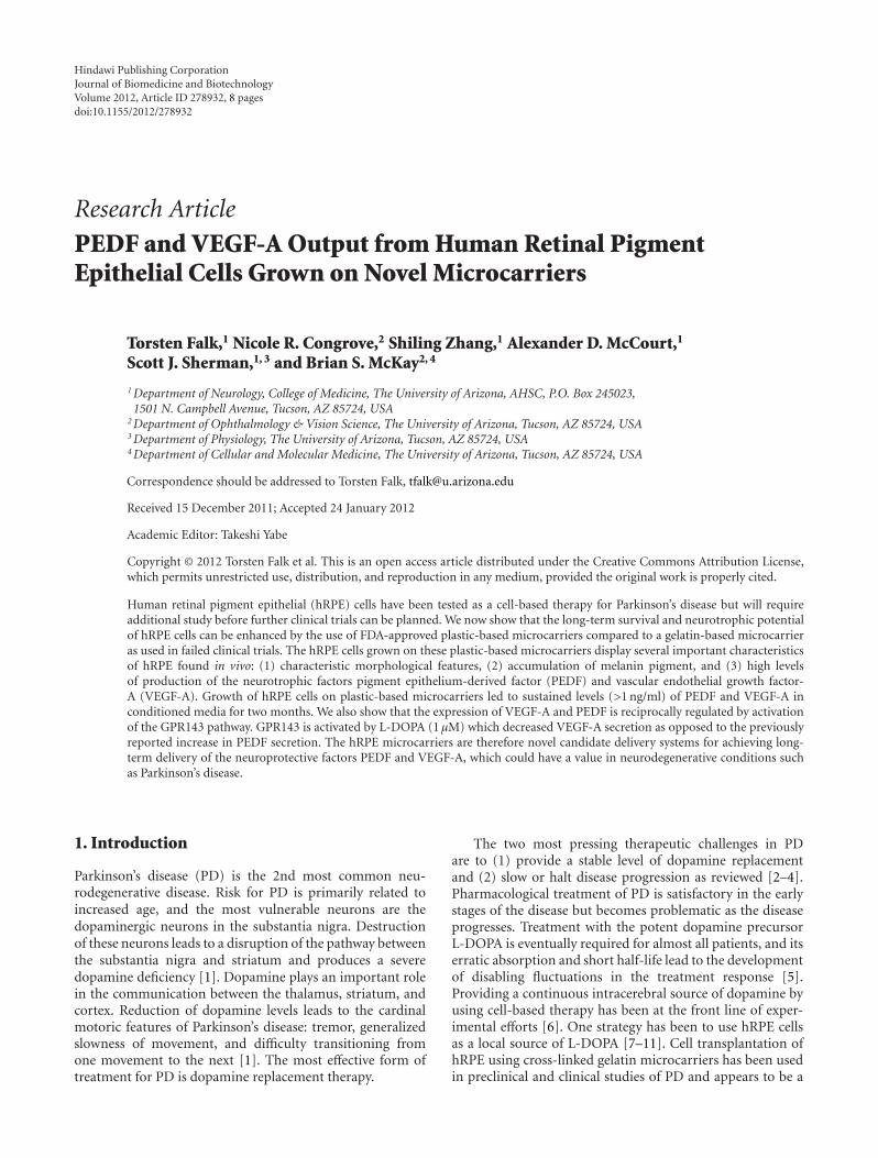
Hindawi Publishing CorporationJournal of Biomedicine and BiotechnologyVolume 2012, Article ID 278932, 8 pagesdoi:10.1155/2012/278932
Research Article
PEDF and VEGF-A Output from Human Retinal PigmentEpithelial Cells Grown on Novel Microcarriers
Torsten Falk,1 Nicole R. Congrove,2 Shiling Zhang,1 Alexander D. McCourt,1
Scott J. Sherman,1, 3 and Brian S. McKay2, 4
1 Department of Neurology, College of Medicine, The University of Arizona, AHSC, P.O. Box 245023,1501 N. Campbell Avenue, Tucson, AZ 85724, USA
2 Department of Ophthalmology & Vision Science, The University of Arizona, Tucson, AZ 85724, USA3 Department of Physiology, The University of Arizona, Tucson, AZ 85724, USA4 Department of Cellular and Molecular Medicine, The University of Arizona, Tucson, AZ 85724, USA
Correspondence should be addressed to Torsten Falk, [email protected]
Received 15 December 2011; Accepted 24 January 2012
Academic Editor: Takeshi Yabe
Copyright © 2012 Torsten Falk et al. This is an open access article distributed under the Creative Commons Attribution License,which permits unrestricted use, distribution, and reproduction in any medium, provided the original work is properly cited.
Human retinal pigment epithelial (hRPE) cells have been tested as a cell-based therapy for Parkinson’s disease but will requireadditional study before further clinical trials can be planned. We now show that the long-term survival and neurotrophic potentialof hRPE cells can be enhanced by the use of FDA-approved plastic-based microcarriers compared to a gelatin-based microcarrieras used in failed clinical trials. The hRPE cells grown on these plastic-based microcarriers display several important characteristicsof hRPE found in vivo: (1) characteristic morphological features, (2) accumulation of melanin pigment, and (3) high levelsof production of the neurotrophic factors pigment epithelium-derived factor (PEDF) and vascular endothelial growth factor-A (VEGF-A). Growth of hRPE cells on plastic-based microcarriers led to sustained levels (>1 ng/ml) of PEDF and VEGF-A inconditioned media for two months. We also show that the expression of VEGF-A and PEDF is reciprocally regulated by activationof the GPR143 pathway. GPR143 is activated by L-DOPA (1 μM) which decreased VEGF-A secretion as opposed to the previouslyreported increase in PEDF secretion. The hRPE microcarriers are therefore novel candidate delivery systems for achieving long-term delivery of the neuroprotective factors PEDF and VEGF-A, which could have a value in neurodegenerative conditions suchas Parkinson’s disease.
1. Introduction
Parkinson’s disease (PD) is the 2nd most common neu-rodegenerative disease. Risk for PD is primarily related toincreased age, and the most vulnerable neurons are thedopaminergic neurons in the substantia nigra. Destructionof these neurons leads to a disruption of the pathway betweenthe substantia nigra and striatum and produces a severedopamine deficiency [1]. Dopamine plays an important rolein the communication between the thalamus, striatum, andcortex. Reduction of dopamine levels leads to the cardinalmotoric features of Parkinson’s disease: tremor, generalizedslowness of movement, and difficulty transitioning fromone movement to the next [1]. The most effective form oftreatment for PD is dopamine replacement therapy.
The two most pressing therapeutic challenges in PDare to (1) provide a stable level of dopamine replacementand (2) slow or halt disease progression as reviewed [2–4].Pharmacological treatment of PD is satisfactory in the earlystages of the disease but becomes problematic as the diseaseprogresses. Treatment with the potent dopamine precursorL-DOPA is eventually required for almost all patients, and itserratic absorption and short half-life lead to the developmentof disabling fluctuations in the treatment response [5].Providing a continuous intracerebral source of dopamine byusing cell-based therapy has been at the front line of exper-imental efforts [6]. One strategy has been to use hRPE cellsas a local source of L-DOPA [7–11]. Cell transplantation ofhRPE using cross-linked gelatin microcarriers has been usedin preclinical and clinical studies of PD and appears to be a
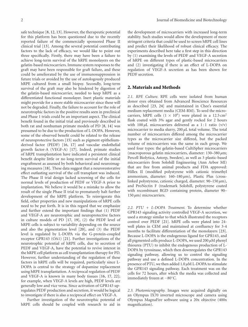
2 Journal of Biomedicine and Biotechnology
safe technique [8, 12, 13]. However, the therapeutic potentialfor this platform has been questioned due to the recentlyreported failure of the commercially sponsored Phase IIclinical trial [13]. Among the several potential contributingfactors to the lack of efficacy, we would like to point outthree specifically. Firstly, there may have been a failure toachieve long-term survival of the hRPE monolayers on thegelatin-based microcarriers. Immune system responses to thegraft may have been responsible for graft failure, and thesecould be ameliorated by the use of immunosuppression infuture trials or avoided by the use of autologously producedhRPE cultured from a small biopsy. Secondly, long-termsurvival of the graft may also be hindered by digestion ofthe gelatin-based microcarrier, needed to keep hRPE as adifferentiated functional monolayer. Inert plastic materialsmight provide for a more stable microcarrier since these willnot be degraded. Finally, the failure to account for the role ofneurotrophic factors in the positive results seen in preclinicaland Phase 1 trials could be an important aspect. The clinicalbenefit found in the initial trial and previously described inboth rat and nonhuman primate models of PD [8, 14] waspresumed to be due to the production of L-DOPA. However,some of the observed benefit could be related to the releaseof neuroprotective factors [15] such as pigment epithelium-derived factor (PEDF) [16, 17] and vascular endothelialgrowth factor-A (VEGF-A) [17]. Indeed, primate studiesof hRPE transplantation have indicated a persistent clinicalbenefit despite little or no long-term survival of the initialengraftment as assessed by both behavioral and neuroimag-ing measures [18]. These data suggest that a neurorestorativeeffect outlasting survival of the cell transplant was induced.The Phase II trial design lacked screening of the cells fornormal levels of production of PEDF or VEGF-A prior toimplantation. We believe it would be a mistake to allow theresult of the single Phase II trial to prematurely halt furtherdevelopment of the hRPE platform. To reinvigorate thisfield, other properties and new manipulations of hRPE cellsneed to be put forth. It is in this regard that we emphasizeand further extend the important findings that (1) PEDFand VEGF-A are neurotrophic and neuroprotective factorsin culture models of PD [17, 19], (2) the PEDF level ofhRPE cells is subject to variability depending on the sourceand also the pigmentation level [20], and (3) the PEDFlevel is regulated by L-DOPA via the G-protein-coupledreceptor GPR143 (OA1) [21]. Further investigations of theneurotrophic potential of hRPE cells, due to secretion ofPEDF and VEGF-A, have the potential to revive interest inthe hRPE cell platform as cell transplantation therapy for PD.However, further understanding of the regulation of thesefactors in hRPE cells will be required, particularly since L-DOPA is central to the strategy of dopamine replacementusing hRPE transplantation. A reciprocal regulation of PEDFand VEGF-A is known in many body tissues [16, 17, 22];for example, when VEGF-A levels are high, PEDF levels aregenerally low and vice versa. Since activation of GPR143 up-regulates PEDF production and secretion, it would be logicalto investigate if there is also a reciprocal effect on VEGF-A.
Further investigation of the neurotrophic potential ofhRPE cells should be coupled with research to aid in
the development of microcarriers with increased long-termstability. Such studies would allow the development of morestringent criteria that could be used to screen hRPE cell linesand predict their likelihood of robust clinical efficacy. Theexperiments described here take a first step in this directionby (1) examining the levels of PEDF and VEGF-A secretionof hRPE on different types of plastic-based microcarriersand (2) investigating if there is an effect of L-DOPA onregulation of VEGF-A secretion as has been shown forPEDF secretion.
2. Materials and Methods
2.1. RPE Culture. RPE cells were isolated from humandonor eyes obtained from Advanced Bioscience Resourcesas described [23, 24] and maintained in Chee’s essentialmedium replacement media (CEM-RM). To seed the micro-carriers, hRPE cells (1 × 10⁶) were plated in a 12.5 cm2
flask coated with 3% agar and gently rocked for 2 hourswith 100 μL microcarriers delivered as a 1 : 1 ratio (v/v)microcarrier to media slurry, 200 μL total volume. The totalnumber of microcarriers differed among the microcarriertypes as the microcarrier size differed, but the packedvolume of microcarriers was the same in each group. Weused four types: the gelatin-based CultiSpher microcarriers(macroporous gelatin microcarriers, diameter: 130–380 μm;Percell Biolytica, Astorp, Sweden), as well as 3 plastic-basedmicrocarriers from Solohill Engineering (Ann Arbor MI)that are free from animal products and FDA approved:Hillex II (modified polystyrene with cationic trimethylammonium, diameter: 160–180 μm), Plastic Plus (cross-linked polystyrene, cationic charged, diameter: 90–150 μm),and ProNectin F (trademark Solohill, polystyrene coatedwith recombinant RGD containing protein, diameter 90–150 μm) microcarriers.
2.2. PTU + L-DOPA Treatment. To determine whetherGPR143 signaling activity controlled VEGF-A secretion, weused a strategy similar to that which illustrated the receptorscontrol over PEDF [21]. hRPE cells were plated into 24-well plates in CEM and maintained at confluency for 3-4months to facilitate differentiation of the monolayers [23].Because L-DOPA is the endogenous ligand for GPR143, andall pigmented cells produce L-DOPA, we used 200 μM phenylthiourea (PTU) to inhibit the endogenous production of L-DOPA by tyrosinase, which then downregulates the GPR143signaling pathway, allowing us to control the signalingpathway and use a defined L-DOPA concentration. In thepresence of PTU, we then added 1.0 μM L-DOPA to stimulatethe GPR143 signaling pathway. Each treatment was on thecells for 72 hours, after which the media was collected andimmediately frozen at −80◦C.
2.3. Photomicrography. Images were acquired digitally onan Olympus IX70 inverted microscope and camera usingOlympus MagnaFire software using a 20x objective (400xmagnification).
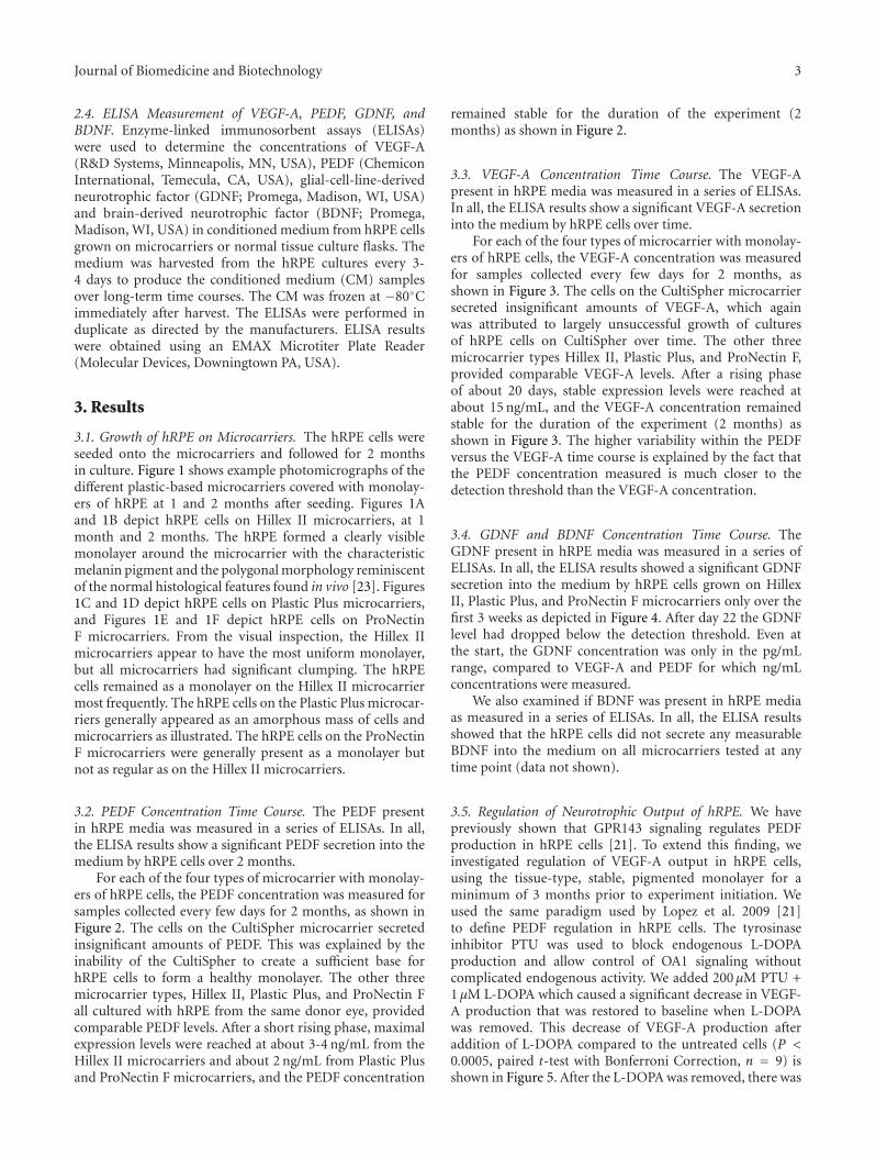
Journal of Biomedicine and Biotechnology 3
2.4. ELISA Measurement of VEGF-A, PEDF, GDNF, andBDNF. Enzyme-linked immunosorbent assays (ELISAs)were used to determine the concentrations of VEGF-A(R&D Systems, Minneapolis, MN, USA), PEDF (ChemiconInternational, Temecula, CA, USA), glial-cell-line-derivedneurotrophic factor (GDNF; Promega, Madison, WI, USA)and brain-derived neurotrophic factor (BDNF; Promega,Madison, WI, USA) in conditioned medium from hRPE cellsgrown on microcarriers or normal tissue culture flasks. Themedium was harvested from the hRPE cultures every 3-4 days to produce the conditioned medium (CM) samplesover long-term time courses. The CM was frozen at −80◦Cimmediately after harvest. The ELISAs were performed induplicate as directed by the manufacturers. ELISA resultswere obtained using an EMAX Microtiter Plate Reader(Molecular Devices, Downingtown PA, USA).
3. Results
3.1. Growth of hRPE on Microcarriers. The hRPE cells wereseeded onto the microcarriers and followed for 2 monthsin culture. Figure 1 shows example photomicrographs of thedifferent plastic-based microcarriers covered with monolay-ers of hRPE at 1 and 2 months after seeding. Figures 1Aand 1B depict hRPE cells on Hillex II microcarriers, at 1month and 2 months. The hRPE formed a clearly visiblemonolayer around the microcarrier with the characteristicmelanin pigment and the polygonal morphology reminiscentof the normal histological features found in vivo [23]. Figures1C and 1D depict hRPE cells on Plastic Plus microcarriers,and Figures 1E and 1F depict hRPE cells on ProNectinF microcarriers. From the visual inspection, the Hillex IImicrocarriers appear to have the most uniform monolayer,but all microcarriers had significant clumping. The hRPEcells remained as a monolayer on the Hillex II microcarriermost frequently. The hRPE cells on the Plastic Plus microcar-riers generally appeared as an amorphous mass of cells andmicrocarriers as illustrated. The hRPE cells on the ProNectinF microcarriers were generally present as a monolayer butnot as regular as on the Hillex II microcarriers.
3.2. PEDF Concentration Time Course. The PEDF presentin hRPE media was measured in a series of ELISAs. In all,the ELISA results show a significant PEDF secretion into themedium by hRPE cells over 2 months.
For each of the four types of microcarrier with monolay-ers of hRPE cells, the PEDF concentration was measured forsamples collected every few days for 2 months, as shown inFigure 2. The cells on the CultiSpher microcarrier secretedinsignificant amounts of PEDF. This was explained by theinability of the CultiSpher to create a sufficient base forhRPE cells to form a healthy monolayer. The other threemicrocarrier types, Hillex II, Plastic Plus, and ProNectin Fall cultured with hRPE from the same donor eye, providedcomparable PEDF levels. After a short rising phase, maximalexpression levels were reached at about 3-4 ng/mL from theHillex II microcarriers and about 2 ng/mL from Plastic Plusand ProNectin F microcarriers, and the PEDF concentration
remained stable for the duration of the experiment (2months) as shown in Figure 2.
3.3. VEGF-A Concentration Time Course. The VEGF-Apresent in hRPE media was measured in a series of ELISAs.In all, the ELISA results show a significant VEGF-A secretioninto the medium by hRPE cells over time.
For each of the four types of microcarrier with monolay-ers of hRPE cells, the VEGF-A concentration was measuredfor samples collected every few days for 2 months, asshown in Figure 3. The cells on the CultiSpher microcarriersecreted insignificant amounts of VEGF-A, which againwas attributed to largely unsuccessful growth of culturesof hRPE cells on CultiSpher over time. The other threemicrocarrier types Hillex II, Plastic Plus, and ProNectin F,provided comparable VEGF-A levels. After a rising phaseof about 20 days, stable expression levels were reached atabout 15 ng/mL, and the VEGF-A concentration remainedstable for the duration of the experiment (2 months) asshown in Figure 3. The higher variability within the PEDFversus the VEGF-A time course is explained by the fact thatthe PEDF concentration measured is much closer to thedetection threshold than the VEGF-A concentration.
3.4. GDNF and BDNF Concentration Time Course. TheGDNF present in hRPE media was measured in a series ofELISAs. In all, the ELISA results showed a significant GDNFsecretion into the medium by hRPE cells grown on HillexII, Plastic Plus, and ProNectin F microcarriers only over thefirst 3 weeks as depicted in Figure 4. After day 22 the GDNFlevel had dropped below the detection threshold. Even atthe start, the GDNF concentration was only in the pg/mLrange, compared to VEGF-A and PEDF for which ng/mLconcentrations were measured.
We also examined if BDNF was present in hRPE mediaas measured in a series of ELISAs. In all, the ELISA resultsshowed that the hRPE cells did not secrete any measurableBDNF into the medium on all microcarriers tested at anytime point (data not shown).
3.5. Regulation of Neurotrophic Output of hRPE. We havepreviously shown that GPR143 signaling regulates PEDFproduction in hRPE cells [21]. To extend this finding, weinvestigated regulation of VEGF-A output in hRPE cells,using the tissue-type, stable, pigmented monolayer for aminimum of 3 months prior to experiment initiation. Weused the same paradigm used by Lopez et al. 2009 [21]to define PEDF regulation in hRPE cells. The tyrosinaseinhibitor PTU was used to block endogenous L-DOPAproduction and allow control of OA1 signaling withoutcomplicated endogenous activity. We added 200 μM PTU +1 μM L-DOPA which caused a significant decrease in VEGF-A production that was restored to baseline when L-DOPAwas removed. This decrease of VEGF-A production afteraddition of L-DOPA compared to the untreated cells (P <0.0005, paired t-test with Bonferroni Correction, n = 9) isshown in Figure 5. After the L-DOPA was removed, there was
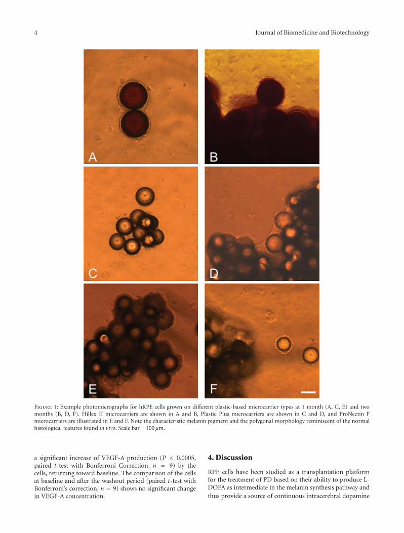
4 Journal of Biomedicine and Biotechnology
Figure 1: Example photomicrographs for hRPE cells grown on different plastic-based microcarrier types at 1 month (A, C, E) and twomonths (B, D, F). Hillex II microcarriers are shown in A and B, Plastic Plus microcarriers are shown in C and D, and ProNectin Fmicrocarriers are illustrated in E and F. Note the characteristic melanin pigment and the polygonal morphology reminiscent of the normalhistological features found in vivo. Scale bar = 100 μm.
a significant increase of VEGF-A production (P < 0.0005,paired t-test with Bonferroni Correction, n = 9) by thecells, returning toward baseline. The comparison of the cellsat baseline and after the washout period (paired t-test withBonferroni’s correction, n = 9) shows no significant changein VEGF-A concentration.
4. Discussion
RPE cells have been studied as a transplantation platformfor the treatment of PD based on their ability to produce L-DOPA as intermediate in the melanin synthesis pathway andthus provide a source of continuous intracerebral dopamine
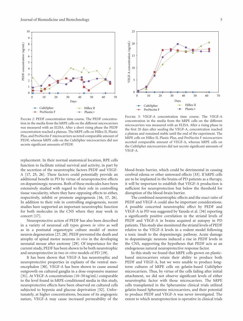
Journal of Biomedicine and Biotechnology 5
0
1
2
3
4
5
6
7
8
9
10
0 10 20 30 40 50 60 70
PE
DF
con
cen
trat
ion
(n
g/m
L)
Days
CultiSpherProNectin F
Hillex II Plastic+
Figure 2: PEDF concentration time course. The PEDF concentra-tion in the media from the hRPE cells on the different microcarrierswas measured with an ELISA. After a short rising phase the PEDFconcentration reached a plateau. The hRPE cells on Hillex II, PlasticPlus, and ProNectin F microcarriers secreted comparable amount ofPEDF, whereas hRPE cells on the CultiSpher microcarriers did notsecrete significant amounts of PEDF.
replacement. In their normal anatomical location, RPE cellsfunction to facilitate retinal survival and activity, in part bythe secretion of the neurotrophic factors PEDF and VEGF-A [17, 25, 26]. These factors could potentially provide anadditional benefit in PD by virtue of neuroprotective effectson dopaminergic neurons. Both of these molecules have beenextensively studied with regard to their role in controllingtissue vascularity, where they have opposing effects to either,respectively, inhibit or promote angiogenesis [16, 17, 26].In addition to their role in controlling angiogenesis, recentstudies have supported an important neurotrophic functionfor both molecules in the CNS where they may work inconcert [17].
Neuroprotective action of PEDF has also been describedin a variety of neuronal cell types grown in vitro as wellas in a postnatal organotypic culture model of motorneuron degeneration [27, 28]. PEDF prevented the death andatrophy of spinal motor neurons in vivo in the developingneonatal mouse after axotomy [29]. Of importance for thecurrent study, PEDF has been shown to be both neurotrophicand neuroprotective in two in vitro models of PD [19].
It has been shown that VEGF-A has neurotrophic andneuroprotective properties in explants of the ventral mes-encephalon [30]. VEGF-A has been shown to cause axonaloutgrowth on cultured ganglia in a dose-responsive manner[31]. At VEGF-A concentrations (10–50 ng/mL) comparableto the level found in hRPE-conditioned media in this study,neuroprotective effects have been observed on cultured cellssubjected to hypoxia and glucose deprivation [32]. Unfor-tunately, at higher concentrations, because of its angiogenicnature, VEGF-A may cause increased permeability of the
0
5
10
15
20
25
0 10 20 30 40 50 60 70
VE
GF-
A c
once
ntr
atio
n (
ng/
mL)
Days
CultiSpherProNectin F
Hillex II Plastic+
Figure 3: VEGF-A concentration time course. The VEGF-Aconcentration in the media from the hRPE cells on the differentmicrocarriers was measured with an ELISA. After a rising phase inthe first 20 days after seeding the VEGF-A, concentration reacheda plateau and remained stable until the end of the experiment. ThehRPE cells on Hillex II, Plastic Plus, and ProNectin F microcarrierssecreted comparable amount of VEGF-A, whereas hRPE cells onthe CultiSpher microcarriers did not secrete significant amounts ofVEGF-A.
blood-brain barrier, which could be detrimental in causingcerebral edema or other untoward effects [33]. If hRPE cellsare to be implanted in the brains of PD patients as a therapy,it will be important to establish that VEGF-A production issufficient for neuroprotection but below the threshold fordisruption of the blood-brain barrier.
The combined neurotrophic effects and the exact ratio ofPEDF and VEGF-A could also be important considerations.A possible concerted neurotrophic effect by PEDF andVEGF-A in PD was suggested by Yasuda et al. [34] reportinga significantly positive correlation in the striatal levels ofPEDF and VEGF-A in brains acquired at autopsy in PDpatients. This study also monitored the striatal levels of PEDFrelative to the VEGF-A levels in a rodent model followinga toxic insult to the dopaminergic pathway. Acute damageto dopaminergic neurons induced a rise in PEDF levels inthe CNS, supporting the hypotheses that PEDF acts as anendogenous natural neuroprotective response factor.
In this study we found that hRPE cells grown on plastic-based microcarriers retain their ability to produce bothPEDF and VEGF-A, but we were unable to produce long-term cultures of hRPE cells on gelatin-based CultiSphermicrocarriers. Thus, by virtue of the cells failing after initialattachment, we did not observe significant levels of eitherneurotrophic factor with those microcarriers. The hRPEcells transplanted in the Spheramine clinical trials utilizedgelatin-based Spheramine microcarriers, and their potentialto produce PEDF and VEGF-A was never investigated. Theextent to which neuroprotection is operative in clinical trials
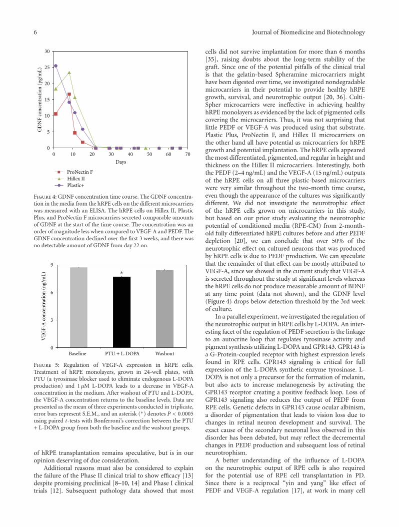
6 Journal of Biomedicine and Biotechnology
0
5
10
15
20
25
30
0 10 20 30 40 50 60 70
GD
NF
con
cen
trat
ion
(pg
/mL
)
Days
ProNectin FHillex IIPlastic+
Figure 4: GDNF concentration time course. The GDNF concentra-tion in the media from the hRPE cells on the different microcarrierswas measured with an ELISA. The hRPE cells on Hillex II, PlasticPlus, and ProNectin F microcarriers secreted comparable amountsof GDNF at the start of the time course. The concentration was anorder of magnitude less when compared to VEGF-A and PEDF. TheGDNF concentration declined over the first 3 weeks, and there wasno detectable amount of GDNF from day 22 on.
0
3
6
9
Baseline PTU + L-DOPA Washout
VE
GF-
A c
once
ntr
atio
n (
ng/
mL) ∗
Figure 5: Regulation of VEGF-A expression in hRPE cells.Treatment of hRPE monolayers, grown in 24-well plates, withPTU (a tyrosinase blocker used to eliminate endogenous L-DOPAproduction) and 1 μM L-DOPA leads to a decrease in VEGF-Aconcentration in the medium. After washout of PTU and L-DOPA,the VEGF-A concentration returns to the baseline levels. Data arepresented as the mean of three experiments conducted in triplicate,error bars represent S.E.M., and an asterisk (∗) denotes P < 0.0005using paired t-tests with Bonferroni’s correction between the PTU+ L-DOPA group from both the baseline and the washout groups.
of hRPE transplantation remains speculative, but is in ouropinion deserving of due consideration.
Additional reasons must also be considered to explainthe failure of the Phase II clinical trial to show efficacy [13]despite promising preclinical [8–10, 14] and Phase I clinicaltrials [12]. Subsequent pathology data showed that most
cells did not survive implantation for more than 6 months[35], raising doubts about the long-term stability of thegraft. Since one of the potential pitfalls of the clinical trialis that the gelatin-based Spheramine microcarriers mighthave been digested over time, we investigated nondegradablemicrocarriers in their potential to provide healthy hRPEgrowth, survival, and neurotrophic output [20, 36]. Culti-Spher microcarriers were ineffective in achieving healthyhRPE monolayers as evidenced by the lack of pigmented cellscovering the microcarriers. Thus, it was not surprising thatlittle PEDF or VEGF-A was produced using that substrate.Plastic Plus, ProNectin F, and Hillex II microcarriers onthe other hand all have potential as microcarriers for hRPEgrowth and potential implantation. The hRPE cells appearedthe most differentiated, pigmented, and regular in height andthickness on the Hillex II microcarriers. Interestingly, boththe PEDF (2–4 ng/mL) and the VEGF-A (15 ng/mL) outputsof the hRPE cells on all three plastic-based microcarrierswere very similar throughout the two-month time course,even though the appearance of the cultures was significantlydifferent. We did not investigate the neurotrophic effectof the hRPE cells grown on microcarriers in this study,but based on our prior study evaluating the neurotrophicpotential of conditioned media (RPE-CM) from 2-month-old fully differentiated hRPE cultures before and after PEDFdepletion [20], we can conclude that over 50% of theneurotrophic effect on cultured neurons that was producedby hRPE cells is due to PEDF production. We can speculatethat the remainder of that effect can be mostly attributed toVEGF-A, since we showed in the current study that VEGF-Ais secreted throughout the study at significant levels whereasthe hRPE cells do not produce measurable amount of BDNFat any time point (data not shown), and the GDNF level(Figure 4) drops below detection threshold by the 3rd weekof culture.
In a parallel experiment, we investigated the regulation ofthe neurotrophic output in hRPE cells by L-DOPA. An inter-esting facet of the regulation of PEDF secretion is the linkageto an autocrine loop that regulates tyrosinase activity andpigment synthesis utilizing L-DOPA and GPR143. GPR143 isa G-Protein-coupled receptor with highest expression levelsfound in RPE cells. GPR143 signaling is critical for fullexpression of the L-DOPA synthetic enzyme tyrosinase. L-DOPA is not only a precursor for the formation of melanin,but also acts to increase melanogenesis by activating theGPR143 receptor creating a positive feedback loop. Loss ofGPR143 signaling also reduces the output of PEDF fromRPE cells. Genetic defects in GPR143 cause ocular albinism,a disorder of pigmentation that leads to vision loss due tochanges in retinal neuron development and survival. Theexact cause of the secondary neuronal loss observed in thisdisorder has been debated, but may reflect the decrementalchanges in PEDF production and subsequent loss of retinalneurotrophism.
A better understanding of the influence of L-DOPAon the neurotrophic output of RPE cells is also requiredfor the potential use of RPE cell transplantation in PD.Since there is a reciprocal “yin and yang” like effect ofPEDF and VEGF-A regulation [17], at work in many cell
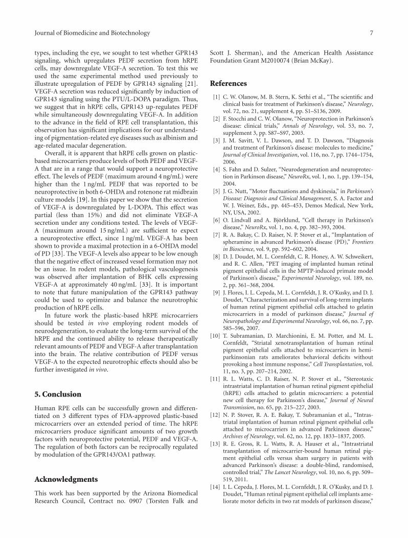
Journal of Biomedicine and Biotechnology 7
types, including the eye, we sought to test whether GPR143signaling, which upregulates PEDF secretion from hRPEcells, may downregulate VEGF-A secretion. To test this weused the same experimental method used previously toillustrate upregulation of PEDF by GPR143 signaling [21].VEGF-A secretion was reduced significantly by induction ofGPR143 signaling using the PTU/L-DOPA paradigm. Thus,we suggest that in hRPE cells, GPR143 up-regulates PEDFwhile simultaneously downregulating VEGF-A. In additionto the advance in the field of RPE cell transplantation, thisobservation has significant implications for our understand-ing of pigmentation-related eye diseases such as albinism andage-related macular degeneration.
Overall, it is apparent that hRPE cells grown on plastic-based microcarriers produce levels of both PEDF and VEGF-A that are in a range that would support a neuroprotectiveeffect. The levels of PEDF (maximum around 4 ng/mL) werehigher than the 1 ng/mL PEDF that was reported to beneuroprotective in both 6-OHDA and rotenone rat midbrainculture models [19]. In this paper we show that the secretionof VEGF-A is downregulated by L-DOPA. This effect waspartial (less than 15%) and did not eliminate VEGF-Asecretion under any conditions tested. The levels of VEGF-A (maximum around 15 ng/mL) are sufficient to expecta neuroprotective effect, since 1 ng/mL VEGF-A has beenshown to provide a maximal protection in a 6-OHDA modelof PD [33]. The VEGF-A levels also appear to be low enoughthat the negative effect of increased vessel formation may notbe an issue. In rodent models, pathological vasculogenesiswas observed after implantation of BHK cells expressingVEGF-A at approximately 40 ng/mL [33]. It is importantto note that future manipulation of the GPR143 pathwaycould be used to optimize and balance the neurotrophicproduction of hRPE cells.
In future work the plastic-based hRPE microcarriersshould be tested in vivo employing rodent models ofneurodegeneration, to evaluate the long-term survival of thehRPE and the continued ability to release therapeuticallyrelevant amounts of PEDF and VEGF-A after transplantationinto the brain. The relative contribution of PEDF versusVEGF-A to the expected neurotrophic effects should also befurther investigated in vivo.
5. Conclusion
Human RPE cells can be successfully grown and differen-tiated on 3 different types of FDA-approved plastic-basedmicrocarriers over an extended period of time. The hRPEmicrocarriers produce significant amounts of two growthfactors with neuroprotective potential, PEDF and VEGF-A.The regulation of both factors can be reciprocally regulatedby modulation of the GPR143/OA1 pathway.
Acknowledgments
This work has been supported by the Arizona BiomedicalResearch Council, Contract no. 0907 (Torsten Falk and
Scott J. Sherman), and the American Health AssistanceFoundation Grant M2010074 (Brian McKay).
References
[1] C. W. Olanow, M. B. Stern, K. Sethi et al., “The scientific andclinical basis for treatment of Parkinson’s disease,” Neurology,vol. 72, no. 21, supplement 4, pp. S1–S136, 2009.
[2] F. Stocchi and C. W. Olanow, “Neuroprotection in Parkinson’sdisease: clinical trials,” Annals of Neurology, vol. 53, no. 7,supplement 3, pp. S87–S97, 2003.
[3] J. M. Savitt, V. L. Dawson, and T. D. Dawson, “Diagnosisand treatment of Parkinson’s disease: molecules to medicine,”Journal of Clinical Investigation, vol. 116, no. 7, pp. 1744–1754,2006.
[4] S. Fahn and D. Sulzer, “Neurodegeneration and neuroprotec-tion in Parkinson disease,” NeuroRx, vol. 1, no. 1, pp. 139–154,2004.
[5] J. G. Nutt, “Motor fluctuations and dyskinesia,” in Parkinson’sDisease: Diagnosis and Clinical Management, S. A. Factor andW. J. Weiner, Eds., pp. 445–453, Demos Medical, New York,NY, USA, 2002.
[6] O. Lindvall and A. Bjorklund, “Cell therapy in Parkinson’sdisease,” NeuroRx, vol. 1, no. 4, pp. 382–393, 2004.
[7] R. A. Bakay, C. D. Raiser, N. P. Stover et al., “Implantation ofspheramine in advanced Parkinson’s disease (PD),” Frontiersin Bioscience, vol. 9, pp. 592–602, 2004.
[8] D. J. Doudet, M. L. Cornfeldt, C. R. Honey, A. W. Schweikert,and R. C. Allen, “PET imaging of implanted human retinalpigment epithelial cells in the MPTP-induced primate modelof Parkinson’s disease,” Experimental Neurology, vol. 189, no.2, pp. 361–368, 2004.
[9] J. Flores, I. L. Cepeda, M. L. Cornfeldt, J. R. O’Kusky, and D. J.Doudet, “Characterization and survival of long-term implantsof human retinal pigment epithelial cells attached to gelatinmicrocarriers in a model of parkinson disease,” Journal ofNeuropathology and Experimental Neurology, vol. 66, no. 7, pp.585–596, 2007.
[10] T. Subramanian, D. Marchionini, E. M. Potter, and M. L.Cornfeldt, “Striatal xenotransplantation of human retinalpigment epithelial cells attached to microcarriers in hemi-parkinsonian rats ameliorates behavioral deficits withoutprovoking a host immune response,” Cell Transplantation, vol.11, no. 3, pp. 207–214, 2002.
[11] R. L. Watts, C. D. Raiser, N. P. Stover et al., “Stereotaxicintrastriatal implantation of human retinal pigment epithelial(hRPE) cells attached to gelatin microcarriers: a potentialnew cell therapy for Parkinson’s disease,” Journal of NeuralTransmission, no. 65, pp. 215–227, 2003.
[12] N. P. Stover, R. A. E. Bakay, T. Subramanian et al., “Intras-triatal implantation of human retinal pigment epithelial cellsattached to microcarriers in advanced Parkinson disease,”Archives of Neurology, vol. 62, no. 12, pp. 1833–1837, 2005.
[13] R. E. Gross, R. L. Watts, R. A. Hauser et al., “Intrastriataltransplantation of microcarrier-bound human retinal pig-ment epithelial cells versus sham surgery in patients withadvanced Parkinson’s disease: a double-blind, randomised,controlled trial,” The Lancet Neurology, vol. 10, no. 6, pp. 509–519, 2011.
[14] I. L. Cepeda, J. Flores, M. L. Cornfeldt, J. R. O’Kusky, and D. J.Doudet, “Human retinal pigment epithelial cell implants ame-liorate motor deficits in two rat models of parkinson disease,”
8 Journal of Biomedicine and Biotechnology
Journal of Neuropathology and Experimental Neurology, vol. 66,no. 7, pp. 576–584, 2007.
[15] C. Jomary, R. M. Darrow, P. Wong, D. T. Organisciak, andS. E. Jones, “Expression of neurturin, glial cell line-derivedneurotrophic factor, and their receptor components in light-induced retinal degeneration,” Investigative Ophthalmologyand Visual Science, vol. 45, no. 4, pp. 1240–1246, 2004.
[16] J. Tombran-Tink and C. J. Barnstable, “PEDF: a multifacetedneurotrophic factor,” Nature Reviews Neuroscience, vol. 4, no.8, pp. 628–636, 2003.
[17] T. Falk, R. T. Gonzalez, and S. J. Sherman, “The Yin andYang of VEGF and PEDF: multifaceted neurotrophic factorsand their potential in the treatment of Parkinson’s disease,”International Journal of Molecular Sciences, vol. 11, no. 8, pp.2875–2900, 2010.
[18] Y. Ma, S. Peng, J. Flores et al., “Abnormal metabolic brainnetwork in parkinsonian macaques: modulation by retinalpigment epithelial (RPE) cell implantation,” Neurology, vol.71, pp. 154–155, 2008.
[19] T. Falk, S. L. Zhang, and S. J. Sherman, “PEDF is neurotrophicand neuroprotective in two in vitro models of Parkinson’sdisease,” Neuroscience Letters, vol. 458, no. 2, pp. 49–52, 2009.
[20] B. S. McKay, B. Goodman, T. Falk, and S. J. Sherman, “Retinalpigment epithelial cell transplantation could provide trophicsupport in Parkinson’s disease: results from an in vitro modelsystem,” Experimental Neurology, vol. 201, no. 1, pp. 234–243,2006.
[21] V. M. Lopez, C. L. Decatur, W. D. Stamer, R. M. Lynch, andB. S. McKay, “L-DOPA is an endogenous ligand for OA1,” PlosBiology, vol. 6, no. 9, article e236, 2008.
[22] J. I. Patel, J. Tombran-Tink, P. G. Hykin, Z. J. Gregor, and I.A. Cree, “Vitreous and aqueous concentrations of proangio-genic, antiangiogenic factors and other cytokines in diabeticretinopathy patients with macular edema: implications forstructural differences in macular profiles,” Experimental EyeResearch, vol. 82, no. 5, pp. 798–806, 2006.
[23] J. Hu and D. Bok, “A cell culture medium that supportsthe differentiation of human retinal pigment epithelium intofunctionally polarized monolayers,” Molecular Vision, vol. 7,pp. 14–19, 2001.
[24] D. J. Rak, K. M. Hardy, G. J. Jaffe, and B. S. McKay, “Ca++-switch induction of RPE differentiation,” Experimental EyeResearch, vol. 82, no. 4, pp. 648–656, 2006.
[25] D. Bok, “The retinal pigment epithelium: a versatile partner invision,” Journal of Cell Science, vol. 106, no. 17, pp. 189–195,1993.
[26] A. K. Olsson, A. Dimberg, J. Kreuger, and L. Claesson-Welsh,“VEGF receptor signalling—in control of vascular function,”Nature Reviews Molecular Cell Biology, vol. 7, no. 5, pp. 359–371, 2006.
[27] M. M. Bilak, A. M. Corse, S. R. Bilak, M. Lehar, J. Tombran-Tink, and R. W. Kuncl, “Pigment epithelium-derived factor(PEDF) protects motor neurons from chronic glutamate-mediated neurodegeneration,” Journal of Neuropathology andExperimental Neurology, vol. 58, no. 7, pp. 719–728, 1999.
[28] M. M. Bilak, S. P. Becerra, A. M. Vincent, B. H. Moss,M. S. Aymerich, and R. W. Kuncl, “Identification of theneuroprotective molecular region of pigment epithelium-derived factor and its binding sites on motor neurons,” Journalof Neuroscience, vol. 22, no. 21, pp. 9378–9386, 2002.
[29] L. J. Houenou, A. P. D’Costa, L. Li et al., “Pigment epithelium-derived factor promotes the survival and differentiation ofdeveloping spinal motor neurons,” Journal of ComparativeNeurology, vol. 412, no. 3, pp. 506–514, 1999.
[30] W. F. Silverman, J. M. Krum, N. Mani, and J. M. Rosenstein,“Vascular, glial and neuronal effects of vascular endothelialgrowth factor in mesencephalic explant cultures,” Neuro-science, vol. 90, no. 4, pp. 1529–1541, 1999.
[31] M. Sondell, G. Lundborg, and M. Kanje, “Vascular endothelialgrowth factor has neurotrophic activity and stimulates axonaloutgrowth, enhancing cell survival and Schwann cell prolifera-tion in the peripheral nervous system,” Journal of Neuroscience,vol. 19, no. 14, pp. 5731–5740, 1999.
[32] K. L. Jin, X. O. Mao, and D. A. Greenberg, “Vascularendothelial growth factor: direct neuroprotective effect in invitro ischemia,” Proceedings of the National Academy of Sciencesof the United States of America, vol. 97, no. 18, pp. 10242–10247, 2000.
[33] T. Yasuhara, T. Shingo, K. Muraoka et al., “The differencesbetween high and low-dose administration of VEGF todopaminergic neurons of in vitro and in vivo Parkinson’sdisease model,” Brain Research, vol. 1038, no. 1, pp. 1–10,2005.
[34] T. Yasuda, M. Fukuda-Tani, T. Nihira et al., “Correlationbetween levels of pigment epithelium-derived factor andvascular endothelial growth factor in the striatum of patientswith Parkinson’s disease,” Experimental Neurology, vol. 206,no. 2, pp. 308–317, 2007.
[35] E. S. Farag, H. V. Vinters, and J. Bronstein, “Pathologicfindings in retinal pigment epithelial cell implantation forParkinson disease,” Neurology, vol. 73, no. 14, pp. 1095–1102,2009.
[36] M. Ming, X. Li, X. Fan et al., “Retinal pigment epithelialcells secrete neurotrophic factors and synthesize dopamine:possible contribution to therapeutic effects of RPE cell trans-plantation in Parkinson’s disease,” Journal of TranslationalMedicine, vol. 7, article 53, 2009.