PHYTON - Zobodat - Welcome©Verlag Ferdinand Berger & Söhne Ges.m.b.H., Horn, Austria, download...
46
PHYTON ANNALES REI BOTANICAE VOL. 47, FASC. Phyton (Horn, Austria) 1-2 Vol. 47 PAG. 1-337 Fasc. 1-2 1-46 19. 19 12. . 12. 2007 2007 Dedicated to Univ.-Prof. Dr. Friedrich EHRENDORFER (Vienna) on the Occasion of his 80th Birthday Polyad Development and Karyology in Inga and Calliandra (Mimosaceae-Ingeae): A Reply to a Recent Paper in Flora. Herwig TEPPNER*) With 102 Figures Received August 28, 2007 Keywords: Acacia, Acacieae, Calliandra angustifolia, Inga feuillei, Ingeae, Leguminosae, Mimosaceae, Mimosoideae. - Embryology, karyology, chromosome numbers, meiosis, microsporogenesis, pollen adhesive, pollenkitt, polyads, pre- polyad, tapetal membrane. Summary TEPPNER H. 2007. Polyad development and karyology in Inga and Calliandra (Mimosaceae-Ingeae): A reply to a recent paper in Flora. - Phyton (Horn, Austria) 47 (1-2): 1-46, with 102 figures. - English with German summary. Inga and Calliandra anthers have transverse septa of parenchymatous tissue; in each locule-half one archesporial cell is present at the beginning. In Inga mitotic di- *) Pens. Univ.-Prof. Dr. Herwig TEPPNER, Institute of Plant Sciences, Division of Systematics and Geobotany, Karl-Franzens University Graz, Holteigasse 6, 8010 Graz, Austria, Europe; e-mail: [email protected] ©Verlag Ferdinand Berger & Söhne Ges.m.b.H., Horn, Austria, download unter www.biologiezentrum.at
Transcript of PHYTON - Zobodat - Welcome©Verlag Ferdinand Berger & Söhne Ges.m.b.H., Horn, Austria, download...

PHYTONANNALES REI BOTANICAE
VOL. 47, FASC.
Phyton (Horn, Austria)
1-2
Vol. 47
PAG. 1-337
Fasc. 1-2 1-46
19.
19
12.
. 12.
2007
2007
Dedicated to Univ.-Prof. Dr. Friedrich EHRENDORFER (Vienna)on the Occasion of his 80th Birthday
Polyad Development and Karyology in Inga andCalliandra (Mimosaceae-Ingeae):
A Reply to a Recent Paper in Flora.
Herwig TEPPNER*)
With 102 Figures
Received August 28, 2007
Keywords : Acacia, Acacieae, Calliandra angustifolia, Inga feuillei, Ingeae,Leguminosae, Mimosaceae, Mimosoideae. - Embryology, karyology, chromosomenumbers, meiosis, microsporogenesis, pollen adhesive, pollenkitt, polyads, pre-polyad, tapetal membrane.
S u m m a r y
TEPPNER H. 2007. Polyad development and karyology in Inga and Calliandra(Mimosaceae-Ingeae): A reply to a recent paper in Flora. - Phyton (Horn, Austria)47 (1-2): 1-46, with 102 figures. - English with German summary.
Inga and Calliandra anthers have transverse septa of parenchymatous tissue; ineach locule-half one archesporial cell is present at the beginning. In Inga mitotic di-
*) Pens. Univ.-Prof. Dr. Herwig TEPPNER, Institute of Plant Sciences, Division ofSystematics and Geobotany, Karl-Franzens University Graz, Holteigasse 6, 8010Graz, Austria, Europe; e-mail: [email protected]
©Verlag Ferdinand Berger & Söhne Ges.m.b.H., Horn, Austria, download unter www.biologiezentrum.at

visions and cytokinesis produce prepolyads of 8-12 pollen mother cells (PMCs),whereas in Calliandra one division takes place and only 2 PMCs are formed. In thePMCs meiosis occurs in the usual way with simultaneous cytokinesis and wall for-mation. In Inga the position of metaphase II-spindles and thus also the order of thepollen grains in the tetrads is variable (most frequent is the tetrahedral orientationwith two grains peripheral in the plane of the polyad). In Calliandra all spindles lieparallel to the plane of the polyad and thus all grains lie in the same plane. Thepostmeiotic secreted tapetal membrane with sporopollenin persists till maturity andin the open theca it is strongly appressed to the valves in both taxa. In Calliandra theapical grain of the polyad finally bears a drop of pollen adhesive which originates bypostmeiotic lysis of descendents of middle layer cells within the narrowed proximalend of the cavity of the locule (mucilage chamber).
Small amounts of pollenkitt can be verified by the contact zones of polyads inair hanging on the under side of a cover slip. The adherence for the polyad pre-sentation is reached by pollenkitt in Inga and Calliandra. Acacia clearly possessespollenkitt as well.
The chromosome number is 2n = 26 and n = 13 in Inga feuillei and 2n = 16 and n= 8 in Calliandra angustifolia. C. houstoniana var. calothyrsus shows 2n = 20 chro-mosomes.
It was stated in a recent paper, that in Calliandra the primary cell per locule-half is a PMC, that the first wall is oblique, the meiotic wall formation is successiveand that the drop of pollen adhesive is solid at the beginning. These statements areincorrect and will be corrected in the present paper.
Zusammenfassung
TEPPNER H. 2007. Polyaden-Entwicklung und Karyologie bei Inga und Callian-dra (Mimosaceae-Ingeae): Eine Entgegnung auf eine kürzlich in Flora erschienenePublikation. - Phyton (Horn, Austria) 47 (1-2): 1-46, mit 102 Abbildungen. - Eng-lisch mit deutscher Zusammenfassung.
Bei Inga und Calliandra sind die Antheren durch Parenchym quer septiert; injeder Lokulament-Hälfte ist zunächst eine Archesporzelle vorhanden. Durch Mitosenund nachfolgende Zellteilungen enthalten die Präpolyaden bei Inga schließlich 8-12,bei Calliandra stets nur zwei Pollenmutterzellen (PMZ). In den PMZ läuft die Meiosein der üblichen Weise ab, gefolgt von simultaner Wandbildung. Bei Inga ist die Stel-lung der Metaphase II-Spindeln variabel, so daß verschiedene Anordnung der Pol-lenkörner (PK) in den Tetraden möglich ist (am häufigsten tetrahedral mit 2 PKperipher in der Ebene der Polyade). Bei Calliandra liegen alle Spindeln parallel zurFläche der Polyade und daher auch alle 8 PK in einer Ebene. Die postmeiotisch ab-geschiedene Tapetum-Membran aus Sporopollenin persistiert bei beiden Taxa bis zurReife und ist in der geöffneten Theka völlig den Valven angepreßt. Bei Calliandraträgt das apikale Korn der Polyade schließlich einen Pollenklebstoff-Tropfen, derdurch postmeiotische Lysis von Zellen, die von der Mittelschicht abstammen, imverschmälerten, proximalen Ende des Raumes im Lokulament (Schleimkammer)entsteht.
Geringe Mengen an Pollenkitt können durch die Kontakthöfe trockener Poly-aden in Luft, die an der Unterseite eines Deckglases hängen, nachgewiesen werden.
©Verlag Ferdinand Berger & Söhne Ges.m.b.H., Horn, Austria, download unter www.biologiezentrum.at

Das Haften für die Präsentation der Polyaden wird sowohl bei Inga als auch beiCalliandra durch Pollenkitt erreicht. Acacia besitzt ebenfalls Pollenkitt.
Die Chromosomenzahlen sind 2n = 26 bzw. n = 13 bei Inga feuillei und 2n = 16bzw. n = 8 bei Calliandra angustifolia. C. houstoniana var. calothyrsus weist 2n = 20Chromosomen auf.
Die in einer kürzlich erschienen Publikation aufgestellten Behauptungen, daßdie primäre Zelle in der Lokulament-Hälfte eine PMZ sei, daß die erste Querwandschief angelegt werde, die Meiose mit sukzedaner Wandbildung verbunden sei unddaß der Pollenklebstoff-Tropfen am Anfang fest sei, sind unrichtig.
1. In t roduct ion
In a recent issue of Flora (201: 570-587, 2006) appeared a paper on'Ontogeny of the CaWiandra-massulae (Mimosaceae: Ingeae), and the as-sociated viscin body' by R. GREISSL. The main results of this paper are:
1) A single polyad should originate from one PMC (pollen mother-cell),the primary cell in each locule-half.
2) Wall formation in this assumed PMC is supposed to be successive,which is very unusual for dicotyledons (e. g., some Magnoliids). The firstwall is supposed to be oblique. In the gones (= meiotic products) successivedivisions should give the eight cells of each polyad.
3) The drop on the apical grain is - even if this is not explicitly said -supposed to be produced by this grain and this 'drop' is supposed to besolid when the anther opens.
These results are in contrast to what is known from other Mimosaceaesince the 19th century and also to observations of other scientists in Cal-liandra. Therefore they seem remarkable and interesting. But are these re-sults correct ? The aim of this paper is to answer this question. The existingliterature would have been sufficient as basis for a critical statement to theabove points. However, before a discussion it seems to be useful to de-monstrate some developmental facts for two examples. The misuse of theterms massula and viscin body in the case of Calliandra was discussed in aprevious paper (TEPPNER 2007), where the terminology used here is dis-cussed.
2. M a t e r i a l and Methods
The plants were grown in the greenhouse of the Institute for Plant Sciences ofthe University of Graz, Austria, Europe. The origin is as follows:
Inga feuillei DC: Seeds purchased in Lima, Peru, June 12, 1979, H. TEPPNER 79/428 & K. KEPLINGER (see TEPPNER 1998: 38-39 and TEPPNER & STABENTHEINER 2006:
142-143).Calliandra angustifolia BENTHAM: Peru, Dpt. Pasco, Pozuzo, ca. 820 m, August
30, 1981, H. TEPPNER 81/518 & K. KEPLINGER, live plant.Calliandra haematocephala HASSKARL var. haematocephala: Purchase from the
Emil KUR collection, Czech Republic, received July 1998, as C. emarginata and C.inaequilatera, respectively, three live plants.
©Verlag Ferdinand Berger & Söhne Ges.m.b.H., Horn, Austria, download unter www.biologiezentrum.at

Calliandra houstoniana (MILLER) STANDLEY var. calothyrsus (MEISNER) BARNEBY:Dominican Republic. - Chiltern Seeds, Cumbria, England, 2006: 234T (as C. ca-lothyrsus), sown March 22, 2006, start of germination April 1, 2006, c. 30-37 cm highseedlings only.
Acacia caven (MOLINA) MOLINA: Origin not known, old stock.
Acacia celastrifolia BENTH.: Western Australia, 'Collected in South-west Bota-nical Province'. - Kings Park & Bot. Garden, Perth, Western Australia 1995: 2358(seeds received as A. myrtifolia; det. G. PRENNER), sown August 2, 1995.
Acacia longifolia (ANDREWS) WILLD.: Bot. Garden Berlin-Dahlem 1978: 2230(seeds received as A. latifolia BENTH.; det. G. PRENNER), sown February 1979.
Since for defining and recognising PMCs meiosis is an important and essen-tial fact, it was decided to use a fixation for the flower buds adequate for chro-mosome studies - even though it is not well suited to discern cell borders and thinprimary cell walls. Inflorescence buds in different stages of development werefixed in ethanol : chloroform : acetic acid 5 : 3 : 1 and stained with aceto-carminesolution in the usual way (e. g., DARLINGTON & LA COUR 1963, SHARMA & SHARMA
1965); then the anthers were dissected on the slide in a drop of acetic acid; squashpreparations were also made. Observations and photos were made with a 'ZeissPhotomikroskop III'; Agfapan APX 100 professional was used. The negatives werescanned with Epson Perfection 2400 Photo and the images were edited with AdobePhotoshop 7.0.
The diameter of small inflorescences was measured without the surpassingbracts; the length of flower buds was measured along a straight line, even when ar-ched.
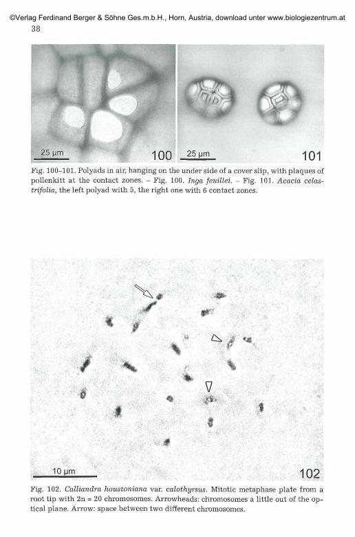
The detection of a thin film of pollenkitt surrounding the polyads (in other casessingle pollen grains) can be difficult as in the case of small polyads as in Acacia. Thefact, that the viscous liquid pollenkitt gets in contact with glass in a physicallycharacteristic way, offers a method for proving its presence. When grains in air comeclose to a glass plate, the pollenkitt leaps to the glass forming a contact zone betweenthe glass and the grain. In the LM this zone is brighter than the surroundings, ap-pears homogenous (without any structure of the exine visible) and, furthermore, isbordered by a dark line with a thin, bright halo (Fig. 100-101). Thus pollenkitt be-haves as an optical gel; the same effect is shown macroscopically by a drop of im-mersion oil between two slides. One possibility to proceed this attachment would beto shake the polyads from open anthers (by pushing flowers or filaments with a nee-dle) onto a slide, together with structures a little larger than the polyads (filaments,part of anthers, hairs or other material). A cover slip is fixed by paraffin or wax, thenthe slide is inverted and knocked against a table edge. Now, at least if pollenkitt ispresent, a part of the polyads will adhere to the cover slip and show distinct contactzones at all places of contact. So direct and easy comparison of polyads with contactzones to the cover slip on the upper side of the polyad, with others without contact onthe upper side, lying on the slide only, is possible (such a control may be helpful incritical cases with few pollenkitt).
A polyad is defined as an assembly of pollen grains; thus, for the developmentalstages from the archesporium to the PMCs the term p r e p o l y a d i s used here.
©Verlag Ferdinand Berger & Söhne Ges.m.b.H., Horn, Austria, download unter www.biologiezentrum.at

Abbrevia t ions used in the f igures:
c calloseco connectiveen endotheciumep epidermism middle layer (1-3 layered)s septumt tapetum or tapetum remainstm tapetal membrane
3. Inga feuillei DC.
3.1. Archesporium to PMCs
The four locules of Inga anthers have transverse septa of par-enchymatous tissue, thus each locule consists mostly of two locule-halves(8 locule-halves per anther). Because of the high number of stamens (75-120) in the complex androeceum, the anthers in a bud show different sizesand developmental stages. At a bud length (= calyx length) of c. 3.5 mm(corolla c. 1.8 mm), in the locule-halves the first sporogenous cell (primaryarchesporial cell) becomes visible (Fig. 1). At a bud length of c. 4.5 mm(corolla c. 2.4 mm) one to four cells form the archesporium, whereas at alength up to c. 6 mm (corolla c. 3-4 mm) the number of cells is near eight orthe final number of mostly eight cells (the PMCs) is reached. The primaryarchesporial cell in the locule-half is roundish to ellipsoid (Fig. 2) and be-comes flattened; the nucleus divides mitotically (Fig. 3), cytokinesis istransversal in respect of the cell or the cavity in the locule-half. Surpris-ingly, the next divisions in these prepolyads clearly do n o t occur synchro-nously; at a given time usually only one mitosis per prepolyad can be ob-served. Since the cell walls appear blurred due to the chosen method, timewas not wasted to clarify the sequence of divisions. It seems that at first aquadrant of cells is formed (Fig. 4) (+ round as seen from the flat side) andthen these cells divide for reaching the final number of cells (Fig. 5, 6, 9,10), the PMCs (usually eight, sometimes nine or ten, rarely twelve). Thedivisions are clearly mitotic as can be seen from the chromosome structure(doubled chromosomes, partite at the centromere; Fig. 3) and number (2n =26). For an isolated cell with the plate in side view see Fig. 3, for a polarview within the intact prepolyad Fig. 7, 8 and 20. The order of the PMCswithin a prepolyad in the case of eight PMCs is in four pairs (Fig. 15, 37,39) or one single cell is in a terminal position at one or both ends (Fig. 21,28).
Surrounded by the tapetum-sac, each prepolyad (now group of PMCs),increases up to c. 150 urn in length in premeiotic stage, which is approxi-mately the final size of the polyad. The premeiotic phase needs a lot oftime. Finally callose deposition begins (Fig. 9-11).
©Verlag Ferdinand Berger & Söhne Ges.m.b.H., Horn, Austria, download unter www.biologiezentrum.at
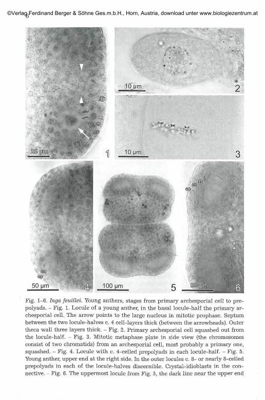
Fig. 1-6. Inga feuillei. Young anthers, stages from primary archesporial cell to pre-polyads. - Fig. 1. Locule of a young anther, in the basal locule-half the primary ar-chesporial cell. The arrow points to the large nucleus in mitotic prophase. Septumbetween the two locule-halves c. 4 cell-layers thick (between the arrowheads). Outertheca wall three layers thick. - Fig. 2. Primary archesporial cell squashed out fromthe locule-half. - Fig. 3. Mitotic metaphase plate in side view (the chromosomesconsist of two chromatids) from an archesporial cell, most probably a primary one,squashed. - Fig. 4. Locule with c. 4-celled prepolyads in each locule-half. - Fig. 5,Young anther, upper end at the right side. In the outer locules c. 8- or nearly 8-celledprepolyads in each of the locule-halves discernible. Crystal-idioblasts in the con-nective. - Fig. 6. The uppermost locule from Fig. 5, the dark line near the upper end
©Verlag Ferdinand Berger & Söhne Ges.m.b.H., Horn, Austria, download unter www.biologiezentrum.at

Fig. 7-10. Inga feuillei. Young anthers with prepolyads with the final number (ornearly so) of PMCs. - Fig. 7. Locule-half with the last mitotic divisions, a metaphaseplate in face view (parallel to the longitudinal axis of the prepolyad) at the right, aprophase nucleus in the next of the four segments. - Fig. 8. Drawing of the mitoticmetaphase plate with 2n = 26 chromosomes from Fig. 7. - Fig. 9. A locule with c. 8-celled prepolyads and the first signs of callose deposition. Septum c. 4-layered (be-tween the arrowheads). - Fig. 10. Prepolyad with PMCs surrounded by callose. Mid-dle layer already in part 2-layered (arrowhead). Slight distortion of epidermal cellsdue to pressure. Arrow: Limit between middle layer and septum.
of the upper prepolyad is a mitotic metaphase plate in side view, perpendicular to thelong axis of the prepolyad. The outer theca wall is 4-layered, i. e., the middle layerand the tapetum are already differentiated. The septum is c. 4-layered. - Abbrevia-tions at the end of chapter 2.
©Verlag Ferdinand Berger & Söhne Ges.m.b.H., Horn, Austria, download unter www.biologiezentrum.at

15Fig. 11-15. Inga feuülei. Anthers with fully developed prepolyads with the finalnumber of PMCs at premeiotic stage. - Fig. 11. Prepolyad in face view, in situ in thelocule-half, surrounded with callose. Middle layer in part 2-layered (arrowhead). -Fig. 12. Prepolyad in side (lateral) view, in situ in the locule-half, surrounded withcallose. The middle layer in part 3-layered (arrowhead). - Fig. 13. Centre of a loculewith a 4-layered septum (between the arrowheads). Prepolyads distinct due to thecallose layer, in face view. - Fig. 14. Centre of a locule with the whole septum and theadjacent connective. - Fig. 15. Prepolyad consisting of eight PMCs with callose(dark), surrounded by fragments of the tapetum-sac, squashed-out from the locule-half.
©Verlag Ferdinand Berger & Söhne Ges.m.b.H., Horn, Austria, download unter www.biologiezentrum.at

Fig. 16-20. Ingafeuülei. Locules with three polyads. - Fig. 16. One of the four loculesof an anther with exceptionally three similar cavities with premeiotic PMCs. - Fig. 17.Detail of Fig. 16, showing the central cavity (locule-third) and the adjacent par-enchymatous septa. The left septum in the thinnest part 3-4-layered, the other (theadditional one ?) 2-layered only. - Fig. 18. Central locule-third with premeiotic pre-polyad of another anther, also with parenchymatous septa. - Fig. 19. A locule-halfwith two premeiotic prepolyads with callose, partite by a 1-layered tapetal bridge. -Fig. 20. A locule half with two prepolyads with callose, one with the last mitosis, se-parated by a 2-layered tapetal bridge.
©Verlag Ferdinand Berger & Söhne Ges.m.b.H., Horn, Austria, download unter www.biologiezentrum.at

10
Sometimes in the one or another locule-half two prepolyads are de-veloped (Fig. 19-20). In such a case, a layer of tapetum lies between the twoand divides the lumen of the locule-half, i.e., each prepolyad is completelysurrounded by tapetum.
The parenchymatous transversal septum between the two locule-halves is c. four-layered (e. g., Fig. 1, 9, 13). Rarely two parenchymatoussepta divide one locule, so that it contains three cavities each with a poly ad(Fig. 16-18), evenly distributed along the locule; this was the case at timesin only one locule of an anther. A case of four polyads per locule in a ma-ture anther was seen only once, unfortunately the character of the septa-tion was not discernible.
When development of the archesporium starts, the theca wall is three-layered (epidermis, endothecial layer, third layer; Fig. 1) but soon the ta-petum is formed by divisions in the third layer and finally it is well de-veloped as a fourth layer (Fig. 6). Now the third layer is the middle layer,which, itself, becomes two- to three-layered (Fig. 11, 12). The outer wallsof the epidermis cells are flat or only slightly arched.
3.2. Meiosis
In flower buds of c. 6.0 - 7.0 mm in length (corolla c. 4.0 - 4.7 mm,length of anthers c. 0.3 mm) meiosis takes place.
The prepolyads are enclosed in the tapetum-sac and at this stage it isrelatively easy to separate the whole sac by dissection of the anther (e. g.,Fig. 24, 28, 29). Each PMC is surrounded by a callose layer. Meiosis occurss y n c h r o n o u s l y within all eight (or up to twelve) cells of a prepolyad(Fig. 24, 28, 29, 34). At metaphase I all spindles lie approximately parallel
Fig. 21-27. Inga feuillei. Anthers at Meiosis I. - Fig. 21. Prepolyad consisting of eightPMCs at pachytene, squashed-out from the locule-half; with thin callose and fewremains of the tapetum sac. - Fig. 22. Detail of Fig. 21, within the nuclei the threadsof the pachytene bivalents and the nucleoli. - Fig. 23. Prepolyad in lateral view atmetaphse I, with callose, in situ in the locule-half. - Fig. 24. Prepolyad with 9 PMCsat metaphase I, within the tapetum-sac, squashed-out from the locule-half. Varia-bility in the orientation of the metaphase I-spindles. - Fig. 25. Detail of a prepolyadwithin the tapetum-sac, squashed out from the locule-half. Variation in the orienta-tion of the spindles: nearly parallel to the plane of the polyad below and above leftand perpendicular to the plane of the polyad (arrowhead). - Fig. 26. Details of twoPMCs in late metaphase I, with spindles parallel to the plane of the polyad (thus bi-valents in lateral view) and more (left) or less radially. Above left a tapetal cell andcallose. - Fig. 27. The n = 13 bivalents of a late metaphase I plate in lateral view. Bi-valents laying superimposed, drawn side by side.
©Verlag Ferdinand Berger & Söhne Ges.m.b.H., Horn, Austria, download unter www.biologiezentrum.at

22
50 um 24
; V ' >
10 26 10 Mm 27
©Verlag Ferdinand Berger & Söhne Ges.m.b.H., Horn, Austria, download unter www.biologiezentrum.at
12
• £ " • ' .
28
25 um 29Fig. 28-29. Inga feuillei. Meiosis I. Prepolyads with 8 PMCs with callose in the tape-tum-sac, squashed out from the locule-halves. - Fig. 28. PMCs at metaphase I, allspindles more or less parallel to the plane of the prepolyad and radially oriented. -Fig. 29. PMCs at anaphase I, prepolyad a little damaged at the bottom due tosquashing. Orientation of the spindles similar to those in Fig. 28, a few a little moreoblique (above right).
©Verlag Ferdinand Berger & Söhne Ges.m.b.H., Horn, Austria, download unter www.biologiezentrum.at

IS
to the plane and are oriented more or less radially. The formation of biva-lents prove these divisions as meiotic and thus the cells as PMCs. Usuallyrod bivalents dominate; n = 13 can be easily counted at metaphase I (Fig.25-27) and in the later stages (Fig. 29, 32, 34-36). The outer walls of thePMCs are more distinct than the adjacent ones. Due to the orientation ofthe metaphase I and anaphase I spindles, one of the two interkinesis nucleiin each PMC lies near the outer side, the other near the inner side of thePMC (Fig. 33). In the metaphase II and anaphase II the two spindles withinone PMC usually lie approximately at right angles: the peripheral one liesin the plane of the polyad whereas the spindle near the centre is perpen-dicular (Fig. 34 arrows, Fig. 35 left). There is some variability, e. g., in asmaller number of cells both plates can also lie parallel to the plane of thepolyad (Fig. 34 arrowheads, Fig. 35 right) and intermediate positions alsooccur. Postmeiotic cytokinesis is simultaneous. The order of the pollengrains in the tetrad within the PMC, and thus in the polyad, depends onthe spindle orientation and as a consequence of it, the position of the tel-ophase II nuclei. Usually the two grains from the peripheral dyad of a PMClie in the plane of the polyad, whereas the other two are oriented perpen-dicular in two planes (as in six PMCs or tetrads, respectively, in Fig. 37).The next frequent possibility is, that the grains from both dyads lie paral-lel and perpendicular to the plane of the polyad, so that in two planes twograins are visible (as in four cases in Fig. 38).
The outer walls of the epidermal cells are flat to arched, endothecialcells show the start of the thickening of the baseplate. The middle layerconsists of three cell layers. The tapetum is well developed.
3.3. Postmeiotic Development
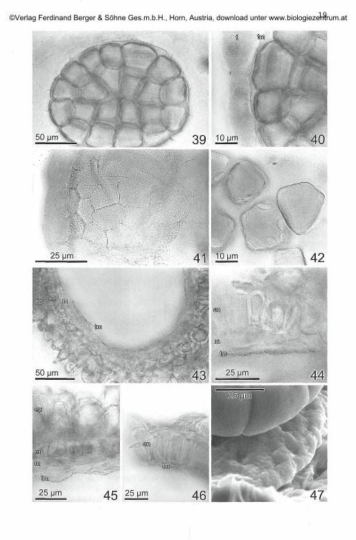
In buds of c. 8 mm length (corolla c. 5 mm) development has largelyprogressed. In the polyads pollen grain exine is well developed (Fig. 40,42), as well as the pores [(4?-)6-8 per grain (Fig. 42 )]. The callose layer isreabsorbed, the tapetum is still well developed and has secreted a thin,hyaline, distinctly granulated membrane (tapetal membrane with orbi-cules) on its inner side (Fig. 39-41); the orbicules sit on the inner (polyad)side of the membrane (Fig. 40, 41); secretion also takes place in the furrowsbetween the tapetum cells; thus the outlines of the tapetum cells areclearly figured on the membrane (Fig. 41). In part it is already loosenedfrom the tapetum (spontaneous or due to the preparation ?). Epidermalpapillae start to develop, the formation of the endothecial thickeningsprogresses.
At 14 mm bud length (corolla nearly of the same length as the calyx)anthers are fully developed, the tapetum has disappeared, only the tapetalmembrane is present and appressed to the middle layer or directly to the
©Verlag Ferdinand Berger & Söhne Ges.m.b.H., Horn, Austria, download unter www.biologiezentrum.at

14
endothecium or the transversal septum (Fig. 43-46). The middle layershows 2-3 layers of cells with more or less degenerated content or hasdisappeared.
Before anther opening approximately the half of the longitudinal sep-tum along the stomium and the central part of the transversal septum aredissolved. In the mature, opened anthers the tapetal membrane is sostrongly and completely appressed to the inner side of the remaining thecawall, that it was not observed by TEPPNER & STABENTHEINER 2006: 146.Higher magnification show the fine granulation on the inner side of thevalves, proving the persistence of the tapetal membrane (Fig. 47). Antheropening and polyad presentation is described in TEPPNER & STABENTHEINER
2006.
4. Calliandra angustifolia BENTH.
4.1. Archesporium to PMCs
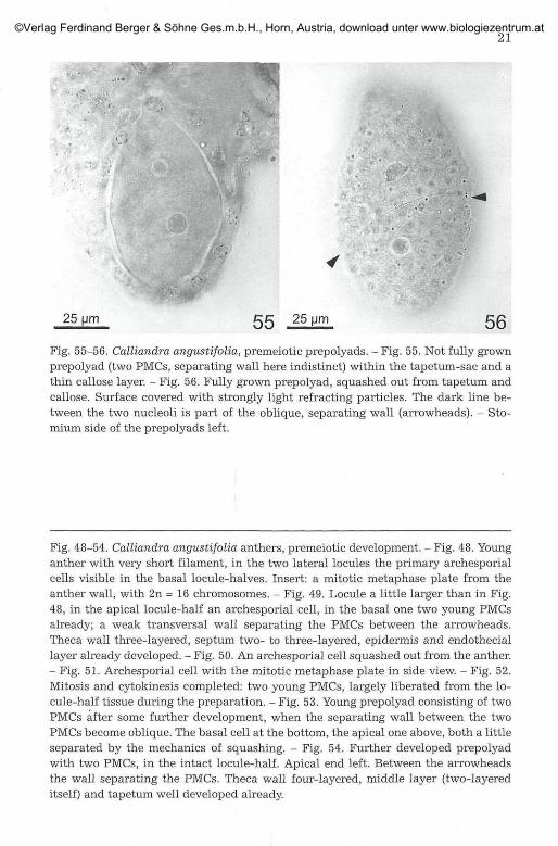
Just like Inga, Calliandra also shows transverse septate locules andtherefore also has two locules or four locule-halves per theca. At a budlength of c. 1.1-1.5 mm [inflorescence (head) diameter c. 2.5-3.5 mm], theanthers on very short filaments fill only the lower part within the corolla.In each locule-half the first cell, an archesporial cell, can be observed; it isroundish or rather elliptic in outline (Fig. 48-50). The first division of thenucleus is clearly mitotic (metaphase plate in side view: Fig. 51) and be-cause of the mitotic spindle lying in the longitudinal axis of the arche-sporial cell, the wall separating the two daughter cells is perpendicular atright angle to the longitudinal axis of the primary cell (Fig. 49, 52) and tothe theca wall. As shown later, the two resulting cells are PMCs already. Inthe next developmental phase the cells become flattened and the elliptic
Fig. 30-36. Inga feuillei. Meiosis I and II in the anthers. - Fig. 30. PMC with an ana-phase I nucleus in lateral view. On the right a fragment of callose layer and tapetalcell nuclei. - Fig. 31. Drawing of Fig. 30, the two plates drawn approached and allchromosomes (consisting of two chromatids each) drawn in one plane. - Fig. 32. Asingle anaphase I plate with n = 13 chromosomes in face (polar) view. - Fig. 33. Pre-polyad with 9 PMCs within the tapetum-sac, squashed out from the locule-half, atinterkinesis (two nuclei in each PMC). - Fig. 34. Prepolyad with 8 PMCs at metaphaseII, within the tapetum-sac, at the left end a little damaged by the preparation. Thetwo spindles perpendicular to each other [the outer one parallel, the central oneperpendicular to the plane of the polyad (arrows)] or both spindles more or less per-pendicular to the plane of the polyad (arrowheads). - Fig. 35. Detail of two PMCs atmetaphase II, the two extreme orientations of the spindles described for Fig. 34 sideby side (the outer spindle parallel to the plane of the polyad: left). - Fig. 36. Drawingof a PMC at metaphase II with the spindles of the two nuclei perpendicular to eachother.
©Verlag Ferdinand Berger & Söhne Ges.m.b.H., Horn, Austria, download unter www.biologiezentrum.at

16
20 um 30 31
10um
50 urn
3 2 50 Mm
34
33
36-
A*
10 Mm \ ^ x 36
©Verlag Ferdinand Berger & Söhne Ges.m.b.H., Horn, Austria, download unter www.biologiezentrum.at

16
shape of the two PMCs changes to heteropolar and asymmetric in faceview: one cell (at the distal end of the locule-half) remains rounded (laterthe basal end of the polyad) whereas the other at the proximal end of thelocule-half, becomes more or less acute (later the apical end of the polyad).The wall between the two cells of the prepolyad becomes oblique: it insertsat the outer margin (stomium side) nearer the base and passes to the innerside (connective-side) nearer to the apex. The final form and size isreached at premeiotic stage (Fig. 53-56) and thus, the final shape of thepolyad already is discernible at this early stage. Since the young PMCs fillthe lumen of the locule-half without any interstice, the change of the shapeof the PMCs is probably affected by changes of the shape of the cavity bygrowing of the locule walls (accompanied by shifts by the growth of thePMCs itself ?).
The theca wall during archespore and young PMC stage is three-layered (Fig. 49), during the early growth of the PMCs the fourth layer, thetapetum originates (Fig. 54). The growth of the anther causes many divi-sions in the theca walls, thus often the somatic chromosome number of 2n= 16 can be counted (Fig. 48, insert).
4.2. Meiosis
In buds of c. 1.0-1.6 mm in length (inflorescence diameter c. 3.5-3.8 mm), in which the anthers fill the space within the corolla, premeioticPMCs, meiosis and postmeiotic polyads can be observed.
Meiosis begins synchronously in the two PMCs of one locule-half (Fig.57-59), but may not be exactly synchronous in the whole locule, theca oranther. Metaphase I spindles lie parallel to the plane of the PMCs and ap-proximately parallel to each other. Because of the oblique position of theseparating wall, one spindle pole is nearer to this wall on the outer side inthe apical PMC and on the inner side in the basal PMC (Fig. 57, 59, 64-67).This causes also the position of the daughter-nuclei in the following stages,e. g., Fig. 64-66. The result of the following anaphase I is interkinesis withtwo nuclei in each PMC (Fig. 63). The spindles of metaphase II and conse-quently also anaphase II lie parallel to the face of the cells as well andmore or less oblique to the metaphase I spindles (Fig. 64-67). Thus allspindles lie in the same plane. At telophase II four nuclei are formed ineach of the PMCs (postmeiotic: Fig. 70), and as a consequence, all 8 nucleilie also in the same plane. After meiosis furrowing and formation of thewalls between the four gones (later pollen grains) are clearly simultaneousin both PMCs (Fig. 71-74). [For a review of the cytokinesis types in PMCssee, for e.g., BHANDARI 1984: 106-107]. Thus the final form and position ofthe pollen grains is formed. During meiosis (from premeiotic to postmeioticstage), relatively thin but distinct amounts of callose usually surround the
©Verlag Ferdinand Berger & Söhne Ges.m.b.H., Horn, Austria, download unter www.biologiezentrum.at
17
50 gm 37
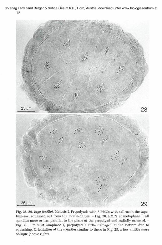
50 38Fig. 37-38. Inga feuülei. Postmeiotic polyads, still with callose around the pollengrains, within the tapetum-sac. The majority of the PMCs show the normal order ofgrains with the two peripheral grains side by side in the plane of the polyad whereasthe two grains on the centre side are superimposed. In the remaining PMCs (arrow-heads) the peripheral and the central grains are superimposed. - Fig. 37. A polyadwith 8 PMCs. - Fig. 38. A polyad with 10 PMCs.
©Verlag Ferdinand Berger & Söhne Ges.m.b.H., Horn, Austria, download unter www.biologiezentrum.at

18
PMCs (e. g., Fig. 55, 71), to a smaller extent callose can be found also in thecleft between the two neighbouring PMCs (e. g., Fig. 60, 63, 65, 73). Rarelythick layers of callose can be observed (Fig. 75). These break down post-meiotically. In all suited stages the meiotic chromosome number of n = 8 iseasy to count (e. g., Fig. 58-62, 64-69).
The epidermal cells are still smooth, the endothecium has no thicken-ings. The tapetum is well developed since premeiotic stages. Prepolyadsand polyads liberated during preparation frequently remain included inthe tapetum-sac (e. g, Fig. 55, 71, 74).
4.3. Postmeiotic Development
In the postmeiotic phase, in flower buds from c. 1.5 mm onwards (in-florescence diameter c. 3.5-5.0 mm) the rapid formation of the exine is veryprominent (Fig. 75-85). In the relevant epidermal cells the growth of thepapillae begins.
In the second (endothecial) layer in the arched parts of the theca wallsand in a bridge between them [narrower (5-6 cells high) on the outer sideof the theca (Fig. 86 ), wider (9-10 cells high) on the inner side (Fig. 87)] thethickenings of the endothecium are formed (with the baseplate character-istic for Mimosaceae) (Fig. 97). The middle layers, at least the outer one,remain intact. The originally intact tapetum secretes a thin, hyaline tapetalmembrane with orbicules (Fig. 82-91), which surrounds the polyads like aclose-fitting sac, which is attached to the polyad at the tip of the apical
Fig. 39-47. Inga feuillei. Postmeiotic polyad and anther development. - Fig. 39. Apolyad with 32 grains (from 8 PMCs) within tapetum-sac and tapetal membrane.Exine well developed, callose disintegrated. - Fig. 40. Detail of Fig. 39, tapetum cellsstill intact, tapetal membrane largely adhering to the inner side of the tapetum-sac,thick exine in the outer walls of the pollen grains only. - Fig. 41. Tapetal membrane,left optical section through tapetum cells and adhering tapetal membrane, otherwisesurface view of the membrane at different levels in relation to the optical plane. - Fig.42. Pollen grains, loosened from the polyad, in surface view and optical section. Inone grain at least 6 pores discernible. Others show the different thickness of the exinearound the grain. - Fig. 43. Fully developed anther, shortly before anthesis. Opticalsection through a locule-half, polyad removed. Tapetum completely disappeared, ta-petal membrane, spreading the cavity, appressed to the inner middle layer. - Fig. 44and 45. Tapetal membrane at the inner middle layer. - Fig. 46. Middle layer reduced,tapetal membrane nearly at the endothecium. - Fig. 47. ESEM image of the inner sideof the valve of an open anther after the night of anthesis. Tapetal membrane withorbicules strongly appressed to the valve. Above part of a polyad, below the marginof the valve with shrivelled epidermal papillae. Similar view as in Fig. 43 in TEPPNER& STABENTHEINER 2006: 155 (phot. STABENTHEINER).
©Verlag Ferdinand Berger & Söhne Ges.m.b.H., Horn, Austria, download unter www.biologiezentrum.at
19©Verlag Ferdinand Berger & Söhne Ges.m.b.H., Horn, Austria, download unter www.biologiezentrum.at

20
.-.>.. 25- 50 51 [_ 52
25 Mm 53 PI!
©Verlag Ferdinand Berger & Söhne Ges.m.b.H., Horn, Austria, download unter www.biologiezentrum.at
25 Mm 5 5 25 pm 5ß
Fig. 55-56. Calliandra angustifolia, premeiotic prepolyads. - Fig. 55. Not fully grownprepolyad (two PMCs, separating wall here indistinct) within the tapetum-sac and athin callose layer. - Fig. 56. Fully grown prepolyad, squashed out from tapetum andcallose. Surface covered with strongly light refracting particles. The dark line be-tween the two nucleoli is part of the oblique, separating wall (arrowheads). - Sto-mium side of the prepolyads left.
Fig. 48-54. Calliandra angustifolia anthers, premeiotic development. - Fig. 48. Younganther with very short filament, in the two lateral locules the primary archesporialcells visible in the basal locule-halves. Insert: a mitotic metaphase plate from theanther wall, with 2n = 16 chromosomes. - Fig. 49. Locule a little larger than in Fig.48, in the apical locule-half an archesporial cell, in the basal one two young PMCsalready; a weak transversal wall separating the PMCs between the arrowheads.Theca wall three-layered, septum two- to three-layered, epidermis and endotheciallayer already developed. - Fig. 50. An archesporial cell squashed out from the anther.- Fig. 51. Archesporial cell with the mitotic metaphase plate in side view. - Fig. 52.Mitosis and cytokinesis completed: two young PMCs, largely liberated from the lo-cule-half tissue during the preparation. - Fig. 53. Young prepolyad consisting of twoPMCs after some further development, when the separating wall between the twoPMCs become oblique. The basal cell at the bottom, the apical one above, both a littleseparated by the mechanics of squashing. - Fig. 54. Further developed prepolyadwith two PMCs, in the intact locule-half. Apical end left. Between the arrowheadsthe wall separating the PMCs. Theca wall four-layered, middle layer (two-layereditself) and tapetum well developed already.
©Verlag Ferdinand Berger & Söhne Ges.m.b.H., Horn, Austria, download unter www.biologiezentrum.at

22
grain (Fig. 89, 90, 98). Then the tapetum progressively reduces to a de-generated thin layer with flattened nuclei (Fig. 81-85). Soon after meiosisthe lysis of the potential mucilage cells begins and thus the border betweenthese cells and the transversal septum becomes clearer (Fig. 75). It becomesapparent that the small septum between two opposite mucilage chambershas 2-4 layers of small cells (Fig. 75-85) and only this part is homologousto the septum as in Inga. Inwards, the septum dilates abruptly to a widestructure of 7-8 layers of much larger cells (Fig. 75-85); this is an extension(protrusion) of the connective. If this interpretation is correct, than themucilage chamber is the narrowed, proximal end of the cavity of the lo-cule-half.
According to the investigations the mucilage cells are descendents ofcells of the middle layer. C. 10-20 small cells are involved. The lysis beginsalways soon after meiosis at the periphery, with the cells adjacent to theseptum (Fig. 76) and progresses towards the polyad tip (Fig. 77). Lysis oc-curs whereas the tapetum is still intact (Fig. 77, 78, 80). The tapetum dis-integrates considerably later and apparently in another way.
At a flower bud total length of c. 2.8-3.3 mm (inflorescence diameter c.6 mm) the buds are arched distinctly claw-like. The corolla surpass thecalyx tips for c. 0.5-1 mm, the filaments are long and coiled and the styleshows 1 1/2 - 2 1/2 screw-turns. In the anthers the epidermal papillae arealready well developed. The endothecial thickenings are fully developed,traversing the transversal septum as a smaller zone of cells, especially onthe outer side (Fig. 86), where the zone of cells with thickenings is c. half aswide only, compared to the inner side (Fig. 87). The middle layer is fullyintact. The tapetum has disappeared (Fig. 88), only the granulated tapetalmembrane is still present. The mucilage drops are fully developed, somesubdivisions as relics of the previous cellular structure may be still dis-cernible (Fig. 98). With the applied methods it is difficult to discern, if themucilage drop is attached to the polyad tip at this stage or later on. It is notimpossible, that this takes place during the early stages of anther opening.
Fig. 57-62. Calliandra angustifolia. PMCs at meiosis I: metaphase I. - Fig. 57. Surveyof a locule-half with the two PMCs, synchronously at metaphase I (arrows); separat-ing wall indistinct here. The two arrowheads point to the limit between septum andthe later mucilage cells. - Fig. 58. Detail of Fig. 57 with the two nuclei at metaphase I(8 bivalents each). - Fig. 59. Another locule-half with the two PMCs at late meta-phase I, separating wall distinct here (between the arrowheads). - Fig. 60. Detail ofFig. 59 with the upper left metaphase I plate. - Fig. 61. Drawing of the n = 8 bivalentsin the late metaphase I-nucleus of Fig. 60. - Fig. 62. Prepolyad with two PMCs atmetaphase I squashed-out from the anther. The arrowhead points to the position ofthe separating wall.
©Verlag Ferdinand Berger & Söhne Ges.m.b.H., Horn, Austria, download unter www.biologiezentrum.at

23
50 um 12.5 um 58
5 9 ! 25 um
1
\ '•'
60
61
©Verlag Ferdinand Berger & Söhne Ges.m.b.H., Horn, Austria, download unter www.biologiezentrum.at

24
At a bud length of c. 3.8-4 mm (corolla surpassing the calyx-tips for c.1.4-1.8 mm, inflorescence diameter c. 7 mm) no dramatical changes wereobserved except that from the polyads removed from the locule-halves thepercentage with mucilage drops is higher.
In buds of c. 4.5-5 mm in length (corolla surpassing the calyx-tips forc. 2 - 2.5 mm, inflorescence diameter c. 8 mm) the middle layers are in theprocess of degeneration; often the cells show hyaline, homogenous,strongly refracting drops as content (Fig. 95). They remain intact for longerperiod at the outer side near the polyad apex (Fig. 92). The tapetal mem-brane is appressed to the remains of the middle layer (Fig. 88, 92-95).
Fully developed buds of C. angustifolia were no longer available whenfinishing this paper. Thus buds of C. haematocephala at the day beforeanthesis were used. The cells of the middle layer are largely degenerated(Fig. 96-99), cells are usually discernible, sometimes the remains are only athin layer. The content of the cells mostly forms one to many drops whichare strongly light refracting. The tapetal membrane is still appressed to theremains of the middle layer and only by the mechanics of preparationsometimes loosened (Fig. 96, 97, 99). Epidermis and endothecial layer showseemingly normal nuclei, usually, especially in the region of the eye in thecentre of the stomium, otherwise the cell content is degenerated in thesame manner as described for the middle layer. The transversal septumappears ruptured (mechanically by the preparation or spontaneously bylysis ?).
5. Discussion
54.
The ontogeny of the Calliandra angustifolia flowers is presented inPRENNER 2004. For Inga no such studies seem to exist but the paper of VANHEEL 1993 on the related genus Archidendron gives an impression of thepossible flower development.
Fig. 63-66. Callianadra angustifolia, PMCs at meiosis: interkinesis and metaphase II.- Fig. 63. The two PMCs of one locule-half at interkinesis, squashed out, apical PMCabove. Nuclei a little distorted during preparation. - Fig. 64. The two PMCs (pre-polyad) at metaphase II, squashed out from the locule-half. All four plates in side-view (spindles parallel to the plane of the prepolyad), apical PMC above, separatingwall (with callose) distinct (arrowhead). Insert: Drawing of one metaphase II-plate(the one below right), the chromosomes consisting of two chromatids partite by thecentromere. - Fig. 65. Another prepolyad at metaphase II, squashed plates in sideview, apical PMC above, separating wall distinct. - Fig. 66. Ditto, photographed intwo optical planes, the arrowhead points to the position of the separating wall. - Inall four figures the stomiumside of the prepolyads left. Position of the nuclei and or-ientation of the spindles always basically the same.
©Verlag Ferdinand Berger & Söhne Ges.m.b.H., Horn, Austria, download unter www.biologiezentrum.at

25
25 um
25 um 66
©Verlag Ferdinand Berger & Söhne Ges.m.b.H., Horn, Austria, download unter www.biologiezentrum.at

26
Some fundamentals of the polyad development in Mimosaceae are al-ready known, at least since ROSANOFF 1866 who studied six species (Ingaand Calliandra included), and observed the development in Acacia. Hedescribed one 'Mutterzelle' per locule-half and a granulated membrane,which clearly can be assigned as the tapetal membrane with orbicules.More comprehensive and precise is the treatment of ENGLER 1876 (Inga andCalliandra mainly p. 282-284): In the cases concerning us here, the deve-lopment within one locule-half starts with one primary archesporial celleach, the 'Urmutterzelle' of the old German literature. By one to some di-visions groups of two to more PMCs ('Pollenmutterzellen') are formed.ENGLER clearly states, that a 'Pollengruppe' (now polyad) is composed oftwo to some pollen tetrads and postulates that the inclusion of the arche-sporial cells in its own tapetum is the prerequisite for the formation ofpolyads; a good summary is included in ENGLER 1887: 152-153. An exactand clear presentation of the first developmental stages from the primaryarchesporial cell per locule-half to the four PMCs in Albizia is included inYAMASAKI 1956. Obviously Acacia (Acacieae) has found greater interest (fore. g., NEWMAN 1933, FITZGERALD & al. 1993). A short review about micro-sporogenesis in the family is included in PRAKASH 1987: 242, 252-253(YAMASAKI 1956 not considered).
For the tapetum cells which surround the prepolyads, especially themeiotic ones, excised from the locule-halves, the term t a p e t u m - s a c isvery obvious and therefore this self-explanatory term is used here. How-ever it is necessary to avoid confusion with the 'culture sac' (= a peritapetalmembrane in the sense of BHANDARI 1984: 85) of HESLOP-HARRISON 1969:542 or the tapetal membrane; the latter is often named spore sac by Acaciaresearchers (e. g., KENRICK 2003: 121). The baseplate in the endothecialcells is a characteristic of Mimosaceae according to MANNING & STIRTON
1994: 145.Sometimes three (rarely four) po lyads per locu le are formed in
Inga feuillei. This is possible in two manners. One of the locule-halves can
Fig. 67-70. Calliandra angustifolia, PMCs at meiosis II: anaphase II and telophase II.- Fig. 67 a. Prepolyad at anaphase II squashed out from the locule-half, apical PMCabove. Surface of the PMCs convered with light refracting particles, arrows point tothe four anaphase II - nuclei. Fig. 67 b. Drawing of Fig. 67 a. - Fig. 68. Two anaphaseII nuclei with n = 8 undoubled chromosomes in each of the four plates. - Fig. 69.Prepolyad at anaphase II squashed out from the locule-half and photographed in twooptical planes. The arrowhead points to the position of the separating wall betweenthe two PMCs. - Fig. 70. The prepolyad at late telophase II, apical cell above, thenucleoli (four per PMC) reappearing, largely squashed out from the tapetum-sac. Thearrowhead show the position of the separating wall between the two PMCs, no wallswithin the PMCs. - In all four figures the stomium-side of the prepolyads right.
©Verlag Ferdinand Berger & Söhne Ges.m.b.H., Horn, Austria, download unter www.biologiezentrum.at

25 um 67a 25Mm 67b
25 urn 25 Mm 70
©Verlag Ferdinand Berger & Söhne Ges.m.b.H., Horn, Austria, download unter www.biologiezentrum.at

28
be parted into two compartments separated by a layer of tapetum (Fig. 19,20). Otherwise, a second parenchymatous septum can be developed, so thatthree similar compartments (locule-thirds) arise within a locule (Fig. 16-18). Two conclusions can be drawn. 1) According to ENGLER 1876: 282I. affinis and I. edulis possess 4-5 polyads per locule (and some other Mi-mosaceae much more; compare ENGLER 1876, DNYANSAGAR 1954, 1970: 93-95, SEIJO & SOLI'S NEFFA 2004); thus the condition of two locule-halves perlocule and of one archesporial cell and polyad, respectively, per locule-halfare clearly derived by reduction. 2) In studies of Mimosaceae, when thelocules are organized in compartments, it is very important to distinguishbetween septation of locules by parenchymatous tissue or by tapetal layers(tapetal bridges). In the latter case it can be discussed, if the term septumis applicable for these ephemeral cells. At least an adjective would be ob-ligatory for distinction such as parenchymatous septum and tapetal sep-tum, respectively.
Whereas anther development in Inga seems not to be treated sinceENGLER 1876, Calliandra has found more interest. Especially in DNYANSA-
GAR 1958: 4-6, 11-12 one primary sporogenous cell and two PMCs per lo-cule-half forming a polyad are clearly and correctly described and figured.
For summaries about different types of po l l en adhes ive see VOGEL1984 and 2002. In Calliandra the pollen adhesive is formed by lysis ofdescendents of the middle layer (chapter 4.3.), neither from the tapetum, assupposed by RICHTER 1929, nor from the septum as believed by PRENNER &TEPPNER 2005; NEVLING & ELIAS 1971: 79 noted only, that the mucilage isformed between the apices of the two opposed polyads in a locule. Thus,the pollen adhesive of Calliandra can be added as a forth type to the threeenumerated by VOGEL 2002.
As I am aware, many details of the polyad development in Inga andCalliandra are not described till now, this is especially true for meiosis andthe existence of a t a p e t a l m e m b r a n e with sporopollenin [cutinisationin the older literature (e.g., MAHESHWARI 1950: 36, 37, KOSMAT 1927); gen-eral: BHANDARI 1984: 83-85, PACINI 1997: 1453, HUYSMANS & al. 1998, 2000].
Fig. 71-74 (numbered in the ontogenetic order). Calliandra angustifolia, meiosis: si-multaneous cytokinesis. - Fig. 71. Prepolyad, covered with a thin callose layer, withinthe tapetum sac. In the apical PMC arrowheads are marking the four nucleoli. Thearrow points to the first sign of furrowing. - Fig. 72. The two PMCs with callose be-tween them and with furrowing and begin of wall formation within the PMCs. -Fig. 73. Detail of Fig. 72 with the two central cells (later pollen grains). - Fig. 74.Polyad within the tapetum sac with the primary walls of the four cells (the pollengrains) within each PMC. - Outer margin (stomium-side) of the polyad at the rightside in Fig. 71 and 74, left in Fig. 72 and 73.
©Verlag Ferdinand Berger & Söhne Ges.m.b.H., Horn, Austria, download unter www.biologiezentrum.at

29
25 25 um
25 um 7 0 12.5 um 73
©Verlag Ferdinand Berger & Söhne Ges.m.b.H., Horn, Austria, download unter www.biologiezentrum.at

30
12.5 . n . ' VürfSm^}
100 7 9 : 25 Mm
Fig. 75-80. Calliandra angustifoUa, forming of the mucilage cells originating from themiddle layer, immediately after meiosis. - Fig. 75. The difference between the cells ofthe septum (in the centre two layers) and the potential mucilage cells becomes dis-tinct (marked with arrowheads at the left side). - Fig. 76. Degeneration of cells andmucilage formation begins in the cells adjacent to the septum (arrowheads). - Fig. 77.Mucilage production progressed in the direction to the polyad, tapetum still intact,also over the tip of the polyad. Septum in the centre three-layered. - Fig. 78. Ditto,especially the intact tapetum distinct. - Fig. 79. A postmeiotic anther somewhat afterthe start of mucilage production (dark dots). - Fig. 80. Detail of Fig. 79 showing theintact tapetum and the septum which is in its centre, between the mucilage cellgroups, three-layered.
©Verlag Ferdinand Berger & Söhne Ges.m.b.H., Horn, Austria, download unter www.biologiezentrum.at

The tapetal membrane consists of two components, the orbicules and theorbicular membrane, connecting and bearing the orbicules (e. g., EL-GHA-
ZALY & NILSSON 1991). In contrast to Acacia (e. g., KENRICK & KNOX 1979),in Inga and Calliandra the thin tapetal membrane is so strongly unitedwith the theca-valve, that it is easy to overlook in the open anther. Thetapetal membrane which covers the degenerated remains of the middlelayer, is possibly important for the surface properties of the inner side ofthe open valves in connection with the adherence of the presented polyads(for Inga see TEPPNER & STABENTHEINER 2006, for Calliandra TEPPNER &STABENTHEINER 2007). Functions of the orbicules associated with the dis-persal of pollen grains [in cases with single grains (monads)], are discussedsometimes earlier, e. g., ECHLIN 1971: 53, KEIJZER 1987: 502, 503, PACINI &FRANCHI 1993: 5, PACINI 2000: 33 and HUYSMANS & al. 1998: 248-249.Especially non-wettability is discussed for angiosperms (e. g. KEIJZER
1987: 502, 503).In Inga feuillei there is some v a r i a b i l i t y in the n u m b e r of PMCs
per prepolyad (8-12) and consequently also in the number of pollen grains(32-48), but 32 is undoubtedly the most frequent one. Usually the o rde rof t he po l len g r a i n s in the polyad reflects the former PMCs clearly inboth genera and shows also high variability in Inga; here, within the PMC,the most abundant bauplan has its origin in a tetrahedral order of thetelophase II nuclei in the PMCs and corresponds to the model of grain or-ientation drawn by WODEHOUSE 1935: 433 for 16-grained polyads in Aca-cia (two grains from each PMC peripheral in the plane of the polyad, theother two, the central ones, perpendicular in two planes). Because of themany deviations which are possible, counting of the grains on both sidesof the polyad (in young or cleared polyads) is necessary for a correctnumber of pollen grains. In the scheme Fig. 20 h-1 in YAMASAKI 1956: 439the natural three-dimensional order of the grains in the 16-grained poly-ads of Albizia glabrior is masked, because all spindles and grains aredrawn in one and the same plane.
It is supposed that the positions of the meiotic spindles in the PMCs(in all its variability) and the resulting positions of the telophase II nucleiare sufficient conditions for the configuration of the grains within thetetrad. It does not seem to be necessary to assume postmeiotic movementsof the cells under the effects of surface tension (e. g., MELVILLE 1981).Furthermore, in Inga and Calliandra such movements would be hinderedby the lack of space.
The TEM study of Acacia paradoxa by FITZGERALD & al. 1993 showsthe details of polyad development very well. Surprising are the premeioticdegeneration of tapetum and middle layer (!), but compare the doubts onsuch an early degeneration of the tapetum in ROWLEY 1993: 36 as well asSANTOS & al. 2003: 196. Comparable TEM studies do not exist for Inga andCalliandra.
©Verlag Ferdinand Berger & Söhne Ges.m.b.H., Horn, Austria, download unter www.biologiezentrum.at

32
50 um 25 um 83 j
84
©Verlag Ferdinand Berger & Söhne Ges.m.b.H., Horn, Austria, download unter www.biologiezentrum.at

33
Fig. 86-87. Calliandra angustifolia. Distribution of endothecial thickenings. - Fig. 86.Between the locule-halves, endothecial cells with thickenings (dark dots in opticalsection) form a narrow band only on the outer side of the theca. - Fig. 87. A wide zoneof endothecial cells with thickenings is developed on the inner valve of the theca. Inthe figure, the marginal ones only lie in the optical plane (as a consequence of thearching of the valve).
Fig. 81-85. Callianadra angustifolia. Anthers with tapetum reduced to a thin layer,with tapetal membrane, with fully developed mucilage and the growing of the epi-dermal papillae. - Fig. 81. Longitudinal optical section through the whole anthershowing the two polyads of one locule, the massive connective and the filamentisthmus. - Fig. 82. Detail of Fig. 81 with the mucilage drops in the chambers belowthe 'eye', the septum between the drops and the connective protrusion below. - Fig.83. Details of 'eye', mucilage drop, polyad tip, septum and connective protrusion. Insitu the tapetal membrane is often difficult to discern (arrowheads). - Fig. 84. Basalend of a polyad with tapetal membrane distinct. Endothecial thickenings can be seenin this optical section. - Fig. 85. As Fig. 83. Septum between the mucilage dropsthree-layered, arrowheads point to the tapetal membrane.
©Verlag Ferdinand Berger & Söhne Ges.m.b.H., Horn, Austria, download unter www.biologiezentrum.at
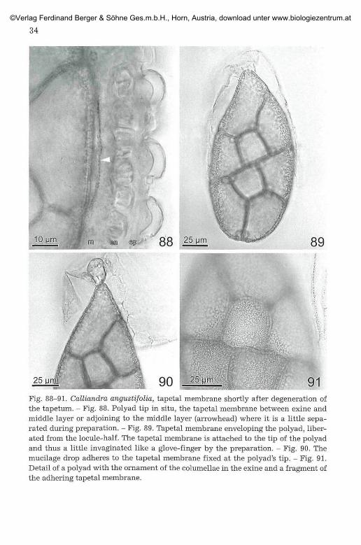
Fig. 88-91. Calliandra angustifolia, tapetal membrane shortly after degeneration ofthe tapetum. - Fig. 88. Polyad tip in situ, the tapetal membrane between exine andmiddle layer or adjoining to the middle layer (arrowhead) where it is a little sepa-rated during preparation. - Fig. 89. Tapetal membrane enveloping the polyad, liber-ated from the locule-half. The tapetal membrane is attached to the tip of the polyadand thus a little invaginated like a glove-finger by the preparation. - Fig. 90. Themucilage drop adheres to the tapetal membrane fixed at the polyad's tip. - Fig. 91.Detail of a polyad with the ornament of the columellae in the exine and a fragment ofthe adhering tapetal membrane.
©Verlag Ferdinand Berger & Söhne Ges.m.b.H., Horn, Austria, download unter www.biologiezentrum.at

35
Fig. 92-95. Calliandra angustifolia. Anthers with completely disappeared tapetumand content of the middle layer cells more or less in degeneration. Tapetal membraneadjoining or appressed to the middle layer. - Fig. 92-94. Middle layer cells with dis-tinct nuclei. - Fig. 95. Content of middle layer, endothecial and epidermis cells com-pletely degenerated to strongly light refracting drops.
In respect of the few investigated examples, the conclusion of PACINI
1997: 1453 that orbicules 'are absent in species with strictly entomophilous[should probably be replaced by zoophilous in contrast to anemophily ?]pollination like Cucurbita pepo in which pollenkitt is present' seems to beoverhasty. Inga as well as Calliandra possess o rb i cu l e s and p o l l e n -k i t t . Thus the older formulation in PACINI & FRANCHI 1993: 5 seems to bemore appropriate (' ... in most entomophilous taxa they coexist with pol-lenkitt. ...').• In Acacia the situation concerning pollenkitt seems to be notso clear. PACINI 1997: 1455 cites KENRICK & KNOX 1989 for the completelylack of pollen coating in polyads, but no relevant phrase was found in thispaper. It is BERNHARDT 1989: 266, 268 who writes that polyads appear tolack copious deposits of pollenkitt and about absence of pollenkitt and yetfigures small droplets of pollenkitt in Acacia pycnantha (in or on the
©Verlag Ferdinand Berger & Söhne Ges.m.b.H., Horn, Austria, download unter www.biologiezentrum.at

36
<m
25 (jm 96 2 5 97<m <§(p>
dOB
25 9S, .25 99Fig. 96-99. Calliandra haematocephala. Ripe anthers at the day before anthesis. Cellsof middle layer with degenerated content or nearly completely reduced. - Fig. 96.Polyad removed from the locule-halve, cavity coated with the tapetal membrane, onefragment of it broken away by the preparation. - Fig. 97. Middle layer distinctly twolayered (arrowhead) with degenerated cell content. In some endothecial cells thebaseplate discernible. - Fig. 98. The left mucilage chamber empty by the preparation,in the right one, the mucilage drop a little attached to the wall has stringed a thread(thus it is liquid). Tapetal membrane between mucilage drop and tip of the apical cellof the polyad. - Fig. 99. Middle layer largely lacking, tapetal membrane nearly at theendothecium.
©Verlag Ferdinand Berger & Söhne Ges.m.b.H., Horn, Austria, download unter www.biologiezentrum.at

pores ?). KNOX 1984: 218 himself writes, that the polyads of Acacia havesticky pollen-coat materials. According to the literature (e. g. KENRICK &KNOX 1979: 415) and own observations (ESEM, with E. STABENTHEINER,
mainly on Acacia celastrifolia, A. longifolia and A. caven), the tapetalmembrane is more robust and less strongly connected with the valve inmelittophilous Acacia. The tapetal membrane-envelope of each polyadopens separately with or shortly after the start of anther-valves opening;the membrane may be more or less connected with the valve (and is thenbent outwards) or stands free between valve and polyad. The polyads, withfaces parallel to each other, loosely adhere with the margins, usually, in thedepth between the inward bulges of the two valves of a theca. Because ofthe smallness of anthers and polyads I was not able to visualize pollenkittdirectly. Excellent TEM images, e. g. FITZGERALD & al. 1993: 57, e. g. Fig.17A, show no structure between tapetal membrane and pollen grain, butbecause of the dehydration with acetone this has no validity. From theobservation of the bright contact zones of dry polyads in air on a cover slip(see methods; Fig. 100-101), it can be seen, that the Acacia polyads arecovered throughout with a thin film of pollenkitt. Thus, also Acacia pos-sesses orbicules and pollenkitt as well and the latter is responsible for theadherence of the polyads with their margins between the inward foldedlongitudinal bulges of the valves (or, after disturbance, with a face on abulge) in the open anthers.
Because of the persistence of the tapetal membrane the pollenkittmust be secreted through the tapetal membrane (e. g., KEIJZER 1987: 501;compare also Fig. 15 and 16 in SANTOS & al. 2003). Beyond that, nothingcan be said about the fate of the pollenkitt in our material because ofethanol and chloroform in the fixative.
The chromosome n u m b e r in Inga feuillei with 2n = 26 and n = 13is the same as in earlier reports for other species (HANSON 1997). Chro-mosome morphology in I. feuillei is treated by TEPPNER 1998: 43 frommaterial of the same population. The number for Calliandra angustifolia(2n = 16, n = 8) is reported for the first time and is the same as indicatedfor the majority (6) of the counted species (9); one (C. pittieri) is tetra-ploid on this base: see the Index of Plant Chromosome Numbers at theMissouri Bot. Garden's web site www.mobot.org . The reports for sect.Calliandra ser. Calliandra only, seem to be problematic and need revisionbecause n = 8 is reported for C. houstoniana and 2n = 22 for C. hous-toniana var. calothyrsus (as 'C. confusa') and C. physocalyx. The result ofthe own counts on a number of good plates from root tip cells from oneindividual of C. houstoniana var. calothyrsus is 2n = 20 (Fig. 102)! Thusfurther investigations to clarify the appropriate chromosome number(s)in this group are needed urgently.
©Verlag Ferdinand Berger & Söhne Ges.m.b.H., Horn, Austria, download unter www.biologiezentrum.at
38
101Fig. 100-101. Polyads in air, hanging on the under side of a cover slip, with plaques ofpollenkitt at the contact zones. - Fig. 100. Inga feuillei. - Fig. 101. Acacia celas-trifolia, the left polyad with 5, the right one with 6 contact zones.
* • ' #
,"•*
10 Mm 102Fig. 102. Calliandra houstoniana var. calothyrsus. Mitotic metaphase plate from aroot tip with 2n = 20 chromosomes. Arrowheads: chromosomes a little out of the op-tical plane. Arrow: space between two different chromosomes.
©Verlag Ferdinand Berger & Söhne Ges.m.b.H., Horn, Austria, download unter www.biologiezentrum.at

39
5.2. Comparision of Inga and Calliandra
The anther development and microsporogenesis in Inga and Callian-dra are principally the same, but Calliandra shows remarkable specializa-tions.
It is evident that the c. eight PMCs in each locule-half in Inga is a moreprimitive condition and the two PMCs in Calliandra are derived. In bothgenera metaphase I spindles lie in one plane. Metaphase II spindles areoriented perpendicular (decussate) within one PMC (as usual in manyother dicots) in Inga, whereas all spindles are in the same plane in Cal-liandra, with the corresponding consequences for the order of the pollengrains. In Inga, because of the high number of grains, the polyad is com-plex with c. the half of the grains in two planes (e.g., the figures in TEPPNER
& STABENTHEINER 2006: 153-156). In Calliandra the order is stronger withall grains in one plane, two central ones and one grain forming the apex ofthe polyad; deviations are relatively rare. Whereas the Inga polyads areroundish to elliptic and distinctly asymmetric in side view only, the poly-ads of Calliandra are asymmetric in all directions. The apparent hetero-polar condition of the polyad in Calliandra is clearly a derived characteralso. The size of the 32-grained polyads in Inga feuillei and the 8-grainedones in Calliandra angustifolia is approximately the same (both c. 150 \imlong). The association of the grains is much stronger in Calliandra than inInga. The tapetal membrane with the sporopollenin orbicules has the sameappearance and fate in both genera, but in Calliandra it must breakaround the base of the drop before or during anther opening. The drop ofpollen adhesive in Calliandra is unique within the family. A possible pre-sence in the related Guinetia can not be seen clearly from Rico ARCE & al.2000. It may be permitted to speculate, that the mucilage drop could be aresult of specialization during evolution. If so, a primitive, unspecializedcondition could have been a middle layer (or more probably the inner layerof a two or three-layered middle layer) becoming mucilaginous at a largeextent which covers the polyad largely with a continuous film of slime.Over restriction of the mucilage to the corner of the cavity near the septumthe specialized condition of Calliandra could have been arisen. Thus, infurther research in Mimosaceae it should be paid attention to the possibi-lity of extratapetal mucilage occurrence. A thin layer of mucilage could bedifficult to be distinguished from pollenkitt.
The parenchymatous, transversal septum in Calliandra remains verysmall in comparison to Inga, thus the lumen of the locule-half appearsnarrowed at the proximal end and forms the mucilage chamber. Whereasthe transversal septum in Inga is dissolved as usual for the longitudinalseptum (see figures in TEPPNER & STABENTHEINER 2006), this septum is de-generated in Calliandra in part, so that cell structure is no longer dis-cernible and separates the two mucilage chambers of one valve. It could
©Verlag Ferdinand Berger & Söhne Ges.m.b.H., Horn, Austria, download unter www.biologiezentrum.at

40
not be decided if it separates from the connective protrusion before orduring anther opening. From the connective protrusion the valve breaksmost probably during anther opening.
An interesting candidate for a detailed comparison with Calliandrawould be Guinetia with similar polyads, which '... shows a poorly devel-oped basal cell and a very reduced sticky appendage, ...' (Rico ARCE & al.2000: 977) (= a lesser differentiated apical cell and very few pollen adhesivein our terminology).
5.3. Comments to the Calliandra Paper in Flora
This paper contains a lot of incorrect statements and wrong conclu-sions. The corrections and the reasons for it are presented in this chapter.
Back to introduction, point 1): The primary cell in the locule halfshould be a PMC. It has been described and documented with photos in theprevious pages, that the primary cell is always an archesporial cell whichshows seven mitotic divisions (followed by cytokinesis) or few more in Ingaand only one in Calliandra, thus, in the latter, two PMCs are formed inwhich then meiosis occurs. This is basically known since ENGLER 1876 and1887 and well described in YAMASAKI 1956 for Albizia and in DNYANSAGAR
1958 for Calliandra. It is paradox, that the correct statement of DNYANSA-
GAR is explicitly contradicted by GREISSL 2006: 580 [citing the wrong page(p. 12), correct would be p. 6, among others with the phrase '... in Callian-dra species, the number of microspore mother-cells formed per micro-sporangium is limited to 2' and the relevant figures.] Such apparent mis-takes are rare, I know only two further ones: KAPP 1969: 245 in a wrongdefinition of polyad ('a symmetrical group of pollen grains which developas a single unit, apparently from a single microspore mother cell') andCHEN 1973. The latter shows the basis of his conclusions by a series ofphotos of his cuts through anthers of Calliandra haematocephala, thus thepaper is helpful for an understanding of the origin of such mistakes. CHEN
describes primary archesporial cells (his Fig. 3-5) and the two young PMCsafter the mitotic division of the archesporial cell (Fig. 6-12). [Fig. 5 is anexcellent figure for a three-layered, Fig. 7 and 8 for a fully developed thecawall]. The archesporial cell is mistaken as PMC and consequently the two-celled stage is erroneously seen as meiotic product. Then, meiosis is over-looked. The following sequences (Fig. 13 onwards) are cuts through moreor less fully developed polyads. From cuts in which not all grains of thepolyads or wedges or degenerated grains are affected, he constructed acurious, non existing sequence of divisions. GREISSL has not demonstratedany basis for his conclusions, but the evidence suggest similarities, evenwhen GREISSL 2006: 579 writes '... the details given by CHEN (1973) couldnot be confirmed'. The postulated sequences of divisions (p. 572, 574-575,578-580) are unreal and not based on observations.
©Verlag Ferdinand Berger & Söhne Ges.m.b.H., Horn, Austria, download unter www.biologiezentrum.at

41
This first misinterpretation leads to a lot of further consequences.Among others, the first division and cytokinesis in the locule has nothingto do with meiosis. The furrowing or wall formation, respectively, in thetwo PMCs, which lead to the 2 x 4 pollen grains in the polyad is doubtlesssimultaneous, as nearly obvious in dicotyledons.
If, in Mimosaceae, in the primary cell in the locule-half meiosis shouldoccur and the four (as consequence of meiosis) daughter cells should dividemitotically (e. g., p. 580: 'the cells of the tetrad secondarily increase innumber') and result in other grains which produce the male gametophytes(other words for the theory in GREISSL 2006), then we would have a thirdgeneration between sporophyte and gamentophyte (a minute, haploid,second sporophyte): This would be very curious and interesting, thoughhighly improbable for an angiosperm!!
Point 2) of the introduction: In the alleged PMC (in reality an arche-sporial cell) the first wall should be oblique (p. 572, 577-578) with an angleof 120° of the wall between the two new cells originating from the firstdivision. What is the reference point for the angle ? Some papers, for ex-ample, SINNOT 1960 and WODEHOUSE 1935, are used for strengthening thereasons for an angle of 120°. At first the wall in the archesporial cell (inrespect to the cell itself) is originally transversal. Secondly in SINNOT 1960:46 the ominous 120° are mentioned for a completely other angle: in theexample, a round cell is divided transversally as the archesporial cells (fore. g., our Fig. 49 and 52); in the case of free cells (not true in Inga and Cal-liandra) and theoretically with a liquid film surface, an intention is formedwhere the new wall meets the mother cell surface; thus at the meetingpoint an angle of 120° is formed between the transversal new wall and thetangent to the circumference circles of each daughter cell. Nothing is saidabout an oblique position of the first wall. Thirdly, to WODEHOUSE 1935.The reference pages are also not cited, but I think that p. 177-200 aremeant, where he discusses the angle of 120° in describing the triradiatesymmetry, for e. g., the arrangement of colpi on the surface of a pollengrain and the mode of contact of three cells such as pollen grains in a tet-rad. To my opinion there is no connection of the mentioned results with theone wall separating the two PMCs after division of the archesporial cell.
Furthermore, on p. 572, 574 and 578 a 'tetrad stage' is stressed. If - ashere - the first cell is counted as PMC, a postulated tetrad stage can notexist. Or the two PMCs at interkinesis together should be regarded as tet-rad (four nuclei in the prepolyad) ? Also improbable, because there are nowalls between the two nuclei in each PMC and the size of the tetrad is in-dicated as more than 50 % of the polyad, whereas it is 100 % at the inter-kinesis stage. So from the text it is not to be concluded what is meant bythe 'tetrad stage'. 'The final, successive divisions [after tetrad stage] show aclear evidence of bipolarity'. The strong heteropolarity of the PMCs is fully
©Verlag Ferdinand Berger & Söhne Ges.m.b.H., Horn, Austria, download unter www.biologiezentrum.at

41
expressed at premeiotic stage. 'In Calliandra, polarity is induced by thetheca. (DAWE & FREELING 1992)'. The cited paper concerns not Calliandrabut exclusively Zea mays anthers; chimeric anthers were used to estimatethe number of initial cells, the origin of the symmetry of the mature antheretc. Every anther or theca shows polarity, which is nearly isopolar in Cal-liandra; crucial is the heteropolarity of the locule-halves and in relationwith the polyad in Calliandra, only these can be important for the induc-tion of heteropolarity.
The reproduction of the figures of the polyad of Calliandra hous-toniana var. anomala from MOHL 1834: Tab. V Fig. 11 (as Inga anomala) isinverted without notice. The two figures are praised as 'a beautiful draw-ing' (p. 574), but they are not fully correct because the side view is shownas exactly symmetrical. Matter of fact is that the Calliandra polyads areasymmetrical in side view, even when this can be lesser expressed in somespecies (for e. g., C. haematocephala).
On p. 575, 576, 580 stainability and small quantity of pollenkitt arediscussed but there are no detailed indications of localization, amount andappearance of it.
To point 3): The origin of the drop of pollen adhesive is not men-tioned explicitly. But phrases on p. 572 and p. 580 (for e. g., 'The viscinbody is formed on the modified octad tip cell as a final step.') suggest,that a secretion from the apical grain is supposed. According to our in-vestigations, the origin of the pollen adhesive has nothing to do with thepolyad, its origin is clearly extratapetal (PRENNER & TEPPNER 2005,TEPPNER 2007: 234 and this paper chapter 4.3.). More than once it isstressed, that the pollen adhesive should be a solid body (for e. g., p.572: 'When the theca opens, this drop-like body... seems to be solid, ...'),but even with a good stereomicroscope, during anther opening, thecharacter of a viscous fluid can be seen. The scheme of the behaviour ofthe drop of pollen adhesive during and after adherence on p. 582 is nice;what other a drop of similar viscosity should do according the physicallaws ? Same is true for the Fig. on p. 580; additionally, comments on thepotential function of pollenkitt are lacking here. Surprising is the visc-osity diagram on p. 582 with the degree of viscosity and the time-scaleas vectors. No methods for measurements of viscosity are indicated, al-though by the graph such measurements are suggested. The determina-tion of melting points can not be a scale for viscosity. And the diagramwith a solid initial phase is wrong in any case. Our impression (from theobservation of the drops, without measurements) is, that the viscosity islowest in the moment of anther opening and increases with desiccation.The diagram is imaginary according to my knowledge. A pity, that theauthor didn't have proper guidance.
©Verlag Ferdinand Berger & Söhne Ges.m.b.H., Horn, Austria, download unter www.biologiezentrum.at

6. A c k n o w l e d g e m e n t s
The figures are an essential part of this paper; therefore specific, sincere thanksto Mr. P. KOSNIK for the developing of the negatives and the scans of the images andespecially also to Ass.-Prof. Dr. W. OBERMAYER for the troublesome and time con-suming editing of the figures. Without this help the original intention for this paper,to avoid further misunderstandings by the presentation of a lot of figures, would nothave been possible to realize. My heartfelt thanks to Mag. Dr. U. BROSCH for procur-ing literature which was not available in our institute. Many thanks to Ass.-Prof. Dr.E. STABENTHEINER for an ESEM photo (Fig. 47) as well as for the possibility to observeAcacia anthers in the ESEM. I am grateful to Prof. Dr. V. RIBITSCH, Dr. A. BIZZARI andMag. Dr. R. TEPPNER for helpful discussions concerning the interpretation of pollen-kitt as an optical gel. Sincere thanks to Ass.-Prof. Dr. E. STABENTHEINER and Mag.P. HARVEY for the careful check of the language.
7. References
BERNHARDT P. 1989. The floral ecology of Australian Acacia. - In: STIRTON C. H. &ZARUCCHI J. L. (eds.), Advances in Legume Biology. - Monogr. syst. Bot. Mis-souri bot. Garden 29: 263-281.
BHANDARI N. N. 1984. 2. The Microsporangium. - In: JOHRI B. M. (ed.), Embryology ofAngiosperms, p. 53-121. - Springer-Verlag, Berlin, Heidelberg, New York, To-kyo.
CHEN Y.-Y. 1973. Studies on the development of polyad grains of Calliandra haema-tocephala HASSK. with electron microscopic technique. - Taiwania 17(1): 18-28.
DARLINGTON C. D. & LA COUR L. F. 1963. Methoden der Chromosomenuntersuchung. -Kosmos, Frankh'sche Verlagshandlung, Stuttgart.
DAWE R. K. & FREELING M. 1992. The role of initial cells in maize anther morpho-genesis. - Development 116: 1077-1085.
DNYANSAGAR V. R. 1954. Embryological studies in the Leguminosae. VI. Inflorescence,sporogenesis, and gametophytes of Dichrostachys cinerea W. & A. and Parkiabiglandulosa W. & A. - Lloydia 17(4): 263-274.
— 1958. Embryological studies in the Leguminosae. VIII. Acacia auriculaeformisA. CUNN., Adenanthera pavonina LINN., Calliandra hematocephala HASSK.,and Calliandra grandiflora BENTH. - Lloydia 21(1): 1-25.
— 1970. Leguminosae. - Proceedings of the symposium of comparative embryol-ogy of Angiosperms. - Bull, indian nation. Sei. Acad. 41: 93-103.
ECHLIN P. 1971. The role of the tapetum during microsporogenesis of Angiosperms. -In: HESLOP-HARRISON J. (ed.), Pollen: Development and physiology, p. 41-61. -Butterworths, London.
EL-GHAZALY G. A. & NILSSON S. 1991. Development of tapetum and orbicules ofCatharanthus roseus {Apocynaceae). - In: BLACKMORE S. & BARNES S. H.(eds.)., Pollen and Spores. - Systematics Association special Volume 44: 317-329. - Clarendon Press, Oxford.
ENGLER A. 1876. Beiträge zur Kenntniss der Antherenbildung der Metaspermen. -Jahrb. wiss. Bot. 10: 275-316, 5 plates.
— 1887. Angiospermae. - In: ENGLER A. & PRANTL K. (eds.), Die natürlichenPflanzenfamilien, 2(1/Lief. 13): 145-176. - Engelmann, Leipzig.
©Verlag Ferdinand Berger & Söhne Ges.m.b.H., Horn, Austria, download unter www.biologiezentrum.at
44
FITZGERALD M. A., CALDER D. M. & KNOX R. B. 1993. Character states of developmentand initiation of cohesion between compound pollen grains of Acacia para-doxa. - Annals Bot. 71: 51-59.
HANSON L. 1997. Chapter 3. Cytology. - In: PENNINGTON T. D., The genus Inga. Botany,p. 31-33. - The Royal Botanic Gardens, Kew.
HEEL W. A. VAN 1993. Floral ontogeny of Archidendron lucyi {Mimosaceae), with re-marks on Amherstia nobilis (Caesalpiniaceae). - Bot. Jahrb. Syst. 114(4): 551-560.
HESLOP-HARRISON J. 1969. An acetolysis-resistant membrane investing tapetum andsporogenous tissue in the anthers of certain Compositae. - Canad. J. Bot. 47:541-542, 2 plates.
HUYSMANS S., EL-GHAZALY G. & SMETS E. 1998. Orbicules in Angiosperms: morphol-ogy, function, distribution, and relation with tapetum types. - Bot. Review64(3): 240-272.
— , — & — 2000. Orbicules: still a well-hidden secret of the anther. - In:NORDENSTAM B., EL-GHAZALY G. & KASSAS M. (eds.), Plant systematics for the21st century. - Wenner-Gren international Series 77: 201-212. - Portland Press,London.
KAPP R. O. 1969. Pollen and spores. - The picutred-key nature series. - Wm. C. BrownComp. Publ., Dubuque, Iowa.
KEIJZER C. J. 1987. The processes of anther dehiscence and pollen dispersal. II. Theformation and the transfer mechanism of pollenkitt, cell-wall development ofthe loculus tissues and a function of orbicules in pollen dispersal. - NewPhytologist 105(3): 499-507, 4 plates.
KENRICK J. 2003. Review of pollen-pistil interactions and their relevance to the re-productive biology of Acacia. - Austral, syst. Bot. 16: 119-130.
— & KNOX R. B. 1979. Pollen development and cytochemistry in some Australianspecies of Acacia. - Austral. J. Bot. 27: 413-427.
— & — 1989. Pollen-pistil interactions in Leguminosae (Mimosoideae). - In:STIRTON C. H. & ZARUCCHI J. L. (eds.), Advances in Legume Biology. - Monogr.syst. Bot. Missouri bot. Garden 29: 127-156.
KNOX R. B. 1984. The pollen grain. - In: JOHRI B. M. (ed.), Embryology of Angios-perms, p. 197-271. - Springer-Verlag, Berlin, Heidelberg, New York.
KOSMAT L. 1927. Studien über das Antherentapetum. - Österr. bot. Z. 76(3): 235-241.MAHESHWARI P. 1950. An introduction to the embryology of Angiosperms. - McGraw-
Hill Book Company, New York, Toronto, London.MANNING J. C. & STIRTON C. H. 1994. Endothecial thickenings and phylogeny of the
Leguminosae. - In: FERGUSON I. K. & TUCKER S. (eds.), Adv. Leg. Syst. 6: 141-163.
MELVILLE R. 1981. Surface tension, diffusion and the evolution and morphogenesis ofpollen aperture patterns. - Pollen et Spores 23(2): 179-203.
MOHL H. 1834. Ueber den Bau und die Formen der Pollenkörner. Mit VI litho-graphirten Tafeln. - Beiträge zur Anatomie und Physiologie der Gewächse, 1,IV + 130 Seiten. - Fischer, Bern.
NEVLING L. I. & ELIAS T. S. 1971. Calliandra haematocephala: History, morphology,and taxonomy. - J. Arnold Arboretum 52: 69-85.
©Verlag Ferdinand Berger & Söhne Ges.m.b.H., Horn, Austria, download unter www.biologiezentrum.at
*§
NEWMAN I. V. 1933. Studies in the Australian Acacias. - II. The life-history of AcaciaBaileyana (F. v. M.). Part I. Some ecological and vegetative features, sporeproduction, and chromosome number. - J. linn. Soc. Bot. 49: 145-171, 4 plates.
PACINI E. 1997. Tapetum character states: analytical keys for tapetum types and ac-tivities. - Canad. J. Bot. 75: 1448-1459.
— 2000. From anther and pollen ripening to pollen presentation. - Plant Syst.Evol. 222: 19-43.
— & FRANCHI G. G. 1993. Role of the tapetum in pollen and spore dispersal. -Plant Syst. Evol., Suppl. 7:1-11.
— , — & HESSE M. 1985. The tapetum: Its form, function, and possible phy-logeny in Embryophyta. - Plant Syst. Evol. 149(3-4): 155-185.
PRAKASH N. 1987. Embryology of the Leguminosae. - In: STIRTON C. H. (ed.), Adv. Leg.Syst. 3: 241-278.
PRENNER G. 2004. Floral ontogeny in Calliandra angustifolia (Leguminosae: Mimo-soideae: Ingeae) and its systematic implications. - Int. J. Plant Sei. 165(3): 417-426.
— & TEPPNER H. 2005. Anther development, pollen presentation and pollen ad-hesive of parenchymatous origin in Calliandra angustifolia (Leguminosae-Mi-mosoideae-Ingeae). - Phyton (Horn, Austria) 45(2): 267-286.
RICHTER S. 1929. Über den Öffnungsmechanismus der Antheren bei einigen Ver-tretern der Angiospermen. - Planta (Berlin) 8(1-2): 154-184.
Rico ARCE M. DE L., SOUSA S. M. & FUENTES S. S. 2000 ('1999'). Guinetia: a new genusin the tribe Ingeae (Leguminosae: Mimosoideae) from Mexico. - Kew Bull.54(4): 975-981.
ROSANOFF S. 1866. Zur Kenntniss des Baues und der Entwickelungsgeschichte desPollens der Mimoseae. - Jahrb. wiss. Bot. 4: 441-450, 2 plates.
ROWLEY J. R. 1993. Cycles of hyperactivity in tapetal cells. - Plant Syst. Evol., Suppl.7: 23-37.
SANTOS R. P., MARIATH J. E. A. & HESSE M. 2003. Pollenkitt formation in Ilex para-guariensis A. ST.-HIL. (Aquifoliaceae). - Plant Syst. Evol. 237: 185-198.
SEIJO J. G. & SOLI'S NEFFAV. G. 2004. The cytological origin of the polyads and theirsignificance in the reproductive biology of Mimosa bimucronata. - Bot. J. linn.Soc. 144: 343-349.
SHARMA A. K. & SHARMA A. S. 1965. Chromosome techniques. Theory and practice. -Butterworths, London.
SINNOT E. W. 1960. Plant morphogenesis. - McGraw-Hill Book Comp., New York,Toronto, London.
TEPPNER H. 1998. Inga feuillei DC. (Mimosaceae-Ingeae). - Samentauschverzeichnis1998 (Bot. Garten Inst. Bot. Univ. Graz), p. 37-45.
— 2007. Notes on terminology for Mimosaceae polyads, especially in Calliandra.- Phyton (Horn, Austria) 46(2): 231-236.
— & STABENTHEINER E. 2006. Inga feuillei (Mimosaceae-Ingeae): Anther openingand polyad presentation. - Phyton (Horn, Austria) 46(1): 141-158.
— & — 2007. Anther opening, polyad presentation, pollenkitt and pollen ad-hesive in four Calliandra species (Mimosaceae-Ingeae). - Phyton (Horn, Aus-tria) 47(1): 291-320.
©Verlag Ferdinand Berger & Söhne Ges.m.b.H., Horn, Austria, download unter www.biologiezentrum.at
46
VOGEL S. 1984. Blütensekrete als akzessorischer Pollenkitt. - In: EHRENDORFER F. &al. (eds.), Mitteilungsband. Kurzfassungen der Beiträge. Botaniker-Tagung inWien. 9.-14. September 1984, p. 123.
— 2002. Extra-tapetal pollen adhesives: Where they occur and how they func-tion. - In: SCHÖNENBERGER J., BALTHAZAR M. V. & MATTHEWS M. (eds.), Flowers,diversity, development & evolution. - Zürich Meeting 5 - 7 July, p. 48.
WODEHOUSE R. P. 1935. Pollen grains. Their structure, identification and significancein science and medicine. - McGraw-Hill Book Company, New York and Lon-don.
YAMASAKI N. 1956. Reifeteilung, Pollenkornteilung und Polliniumbildung an Albizziaglabrior OHWI var. speciosa KOIDZUMI. - Cytologia (Tokyo) 21(4): 433-441.
©Verlag Ferdinand Berger & Söhne Ges.m.b.H., Horn, Austria, download unter www.biologiezentrum.at