Phytochemical Analysis, Antimicrobial and Antioxidant...
5
International Journal of Pharmacy and Chemistry 2016; 2(2): 39-43 http://www.sciencepublishinggroup.com/j/ijpc doi: 10.11648/j.ijpc.20160202.16 Phytochemical Analysis, Antimicrobial and Antioxidant Study of Nigella sativa L Sania Feroz * , Ghias Uddin Institute of Chemical Sciences, University of Peshawar, K. P. K Peshawar, Pakistan Email address: [email protected] (S. Feroz) * Corresponding author To cite this article: Sania Feroz, Ghias Uddin. Phytochemical Analysis, Antimicrobial and Antioxidant Study of Nigella sativa L. International Journal of Pharmacy and Chemistry. Vol. 2, No. 2, 2016, pp. 39-43. doi: 10.11648/j.ijpc.20160202.16 Received: August 30, 2016; Accepted: November 14, 2016; Published: December 17, 2016 Abstract: The current investigation was designed to assess the preliminary phytochemical analysis, antimicrobial and anti- radical effect of crude methanolic extract and isolated fractions of Nigella sativa seeds using established protocol. The phytochemical analysis revealed the presence of alkaloids, terpenoids, tannins, reducing sugars, saponins, steroids, proteins and amino acids, fatty acids and caumarine. The crude methanolic extract and isolated fraction were subjected to antibacterial, antifungal and antioxidant activities. The ethyl acetate and chloroform fractions demonstrated significant (80% and 78%) antioxidant activity. Regarding antibacterial activity of N. sativa, chloroform fraction showed good antibacterial effect as compared to the rest of solvent extracted fractions. Fractions were tested for antifungal activity as well. Amongst all, MeOH fraction showed good antifungal activity against A. N and A. S. n- Hexane showed no activity while other fractions showed moderate activity. The current finding suggests that N. sativa contains potential molecules which exhibited excellent antioxidant and antibacterial effect. Keywords: Nigella Sativa, Phytochemical Analysis, Antifungal Activity, Anti Bacterial Activity, Antioxidant Activity 1. Introduction Medicinal herbs are significant source of synthetic and herbal drugs. In the commercial market, medicinal herbs are used as raw drugs, extracts or tinctures. Isolated active constituents are used for applied research. For the last few decades, phytochemistry (study of plants) has been making rapid progress and herbal products are becoming popular [1]. Nigella sativa is considered to be one of the greatest healing herbs of all times. This herb has been used for millenniums to strengthen the immune system, cleanse the body, purify the blood, protect against irritants and diseases and support healthy long span of life. N. sativa is an annual herbaceous plant, which is believed to be aboriginal to the Mediterranean region but has been cultivated in other parts of the world including Saudi Arabia, northern Africa and parts of Asia [2]. Black seeds have been used as herbal medicine by various cultures and civilizations to treat and prevent a number of diseases including fever, common cold, headache, asthma, rheumatic diseases, and microbial infections and to expel worms from the intestines and as useful for treating “Sartan” (cancer). In addition, it is used for flavoring bread and pickles [3]. 2. Experimental 2.1. Plant Material N. sativa seeds were purchased from local market of Peshawar city. The plant material was identified by Prof. Dr. Abdur Rashid, Department of Botany, University of Peshawar. A voucher Specimen was deposited in the herbarium of the said department. 2.2. Extraction and Fractionation Shade dried seeds of N. sativa were grinded with local grinder machine. The crude extract was soaked in Methanol for 5 days. The crude extract was suspended in water and fractionated to various solvent such as n-hexane, chloroform, ethyl acetate and methanol.
Transcript of Phytochemical Analysis, Antimicrobial and Antioxidant...

International Journal of Pharmacy and Chemistry 2016; 2(2): 39-43
http://www.sciencepublishinggroup.com/j/ijpc
doi: 10.11648/j.ijpc.20160202.16
Phytochemical Analysis, Antimicrobial and Antioxidant Study of Nigella sativa L
Sania Feroz*, Ghias Uddin
Institute of Chemical Sciences, University of Peshawar, K. P. K Peshawar, Pakistan
Email address: [email protected] (S. Feroz) *Corresponding author
To cite this article: Sania Feroz, Ghias Uddin. Phytochemical Analysis, Antimicrobial and Antioxidant Study of Nigella sativa L. International Journal of
Pharmacy and Chemistry. Vol. 2, No. 2, 2016, pp. 39-43. doi: 10.11648/j.ijpc.20160202.16
Received: August 30, 2016; Accepted: November 14, 2016; Published: December 17, 2016
Abstract: The current investigation was designed to assess the preliminary phytochemical analysis, antimicrobial and anti-
radical effect of crude methanolic extract and isolated fractions of Nigella sativa seeds using established protocol. The
phytochemical analysis revealed the presence of alkaloids, terpenoids, tannins, reducing sugars, saponins, steroids, proteins
and amino acids, fatty acids and caumarine. The crude methanolic extract and isolated fraction were subjected to antibacterial,
antifungal and antioxidant activities. The ethyl acetate and chloroform fractions demonstrated significant (80% and 78%)
antioxidant activity. Regarding antibacterial activity of N. sativa, chloroform fraction showed good antibacterial effect as
compared to the rest of solvent extracted fractions. Fractions were tested for antifungal activity as well. Amongst all, MeOH
fraction showed good antifungal activity against A. N and A. S. n- Hexane showed no activity while other fractions showed
moderate activity. The current finding suggests that N. sativa contains potential molecules which exhibited excellent
antioxidant and antibacterial effect.
Keywords: Nigella Sativa, Phytochemical Analysis, Antifungal Activity, Anti Bacterial Activity, Antioxidant Activity
1. Introduction
Medicinal herbs are significant source of synthetic and
herbal drugs. In the commercial market, medicinal herbs are
used as raw drugs, extracts or tinctures. Isolated active
constituents are used for applied research. For the last few
decades, phytochemistry (study of plants) has been making
rapid progress and herbal products are becoming popular [1].
Nigella sativa is considered to be one of the greatest
healing herbs of all times. This herb has been used for
millenniums to strengthen the immune system, cleanse the
body, purify the blood, protect against irritants and diseases
and support healthy long span of life. N. sativa is an annual
herbaceous plant, which is believed to be aboriginal to the
Mediterranean region but has been cultivated in other parts of
the world including Saudi Arabia, northern Africa and parts
of Asia [2]. Black seeds have been used as herbal medicine
by various cultures and civilizations to treat and prevent a
number of diseases including fever, common cold, headache,
asthma, rheumatic diseases, and microbial infections and to
expel worms from the intestines and as useful for treating
“Sartan” (cancer). In addition, it is used for flavoring bread
and pickles [3].
2. Experimental
2.1. Plant Material
N. sativa seeds were purchased from local market of
Peshawar city. The plant material was identified by Prof. Dr.
Abdur Rashid, Department of Botany, University of
Peshawar. A voucher Specimen was deposited in the
herbarium of the said department.
2.2. Extraction and Fractionation
Shade dried seeds of N. sativa were grinded with local
grinder machine. The crude extract was soaked in Methanol
for 5 days. The crude extract was suspended in water and
fractionated to various solvent such as n-hexane, chloroform,
ethyl acetate and methanol.

International Journal of Pharmacy and Chemistry 2016; 2(2): 39-43 40
2.3. Phytochemical Profiling
The crude extract and its different fractions were
processed to phytochemical tests to recognize bioactive
secondary metabolites by using standard methods [4-10].
Alkaloids: 0.5ml of each of the fraction was warmed with
2% H2SO4 for two minutes. The reaction mixtures were
cooled and filtered and added a few drops of Dragendroff’s
reagent to each filterate. Orange red precipitate indicates the
presence of alkaloids moiety.
Tannins: 1ml of each extract was mixed with water and
heated on water bath and filtered. A few drops of ferric
chloride were added to each of the filterate. A dark green
solution indicates the presence of tannins.
Anthraquinones: 0.5 ml of each extract was boiled with
10% HCl for few minutes on water bath. The reaction
mixtures were cooled and filtered. Equal volume of CHCl3
was added to each filterate. Few drops of 10% ammonia were
added to each mixture and heated. Rose pink color formation
signifies the presence of anthraquinones.
Glycosides: Each extract was hydrolyzed with HCl and
neutralized with NaOH solution. A few drops of Fehling’s
solution A and B were added to each mixture. Formation of
red precipitate signifies the presence of glycosides.
Reducing Sugars: Each extract was shaken with distilled
water and filtered. The filterates were boiled with few drops
of Fehling’s solution A and B for few minutes. An orange red
precipitates signifies the presence of reducing sugars.
Saponins: 1 ml of each extract was shaken with 3 ml of
distilled water and heated to boiling. Frothing (appearance of
creamy miss of small bubbles) signifies the presence of
saponins.
Flavonoids: 0.2 ml of each extract was dissolved in diluted
NaOH and few drops of HCl were added. A yellow solution
that turns colorless implies the presence of flavonoids.
Phlobatanins: 0.5 ml of each extract was dissolved in
distilled water and filtered. The filterate was boiled with 2%
HCl solution. Red precipitate implies the presence of
phlobatanins.
Steroids: To 1 ml of each extract, few drops of acetic acid
were added. It was gently warmed and then cooled and then a
drop of H2SO4 was added. The color changes to green which
shows the presence of steroids.
Terpenoids: 0.2 ml of each extract was mixed with 2 ml of
chloroform and concentrated H2SO4 (3 ml) was carefully
added to form a layer. The formation of a reddish brown
coloration at the interface signifies positive results for the
presence of terpenoids.
Test for Caumarin: Exact 3 ml of 10% NaOH was added to
2ml of aqueous extract. Formation of yellow color indicates
the presence of caumarin.
Test for Emodins: Exact 2ml of NH4OH and 3ml of
benzene was added to extract. Appearance of red color
indicates the presence of emodins.
Test for Anthocyanin and Betacyanin: To 2ml of plant
extract, 1ml of 2N NaOH was added and heated for 5
minutes at 100°C. Formation of bluish green color indicates
the presence of anthocyanin and formation of yellow color
indicates the presence of betacyanin.
Test for Carbohydrates: Few drops of Molisch’s reagent
were added to each of the portion dissolved in distilled water;
this was then followed by addition of 1 ml of conc. H2SO4 by
the side of the test tube. The mixture was then allowed to
stand for two minutes and then diluted with 5 ml of distilled
water. Formation of a red or dull violet color at the interphase
of the two layers was a positive test.
2.4. Anti-bacterial Activity
One strain of Gram-negative bacteria (Klebsiella
pneumonia) and two strains of Gram-positive bacteria
(Staphylococcus aureus and Staphylococcus epidermidis)
were used for this activity. These organisms were kept in
Muller-Hinton agar in the refrigerator at 4°C.
The tests for responsiveness were done using modified
agar well diffusion method to tryout the antibacterial activity
of various fractions. To assess antibacterial activity, standard
protocol was used [11]. The Muller Hinton agar was the
medium. The cultures were taken in triplicates at cultivation
temperature of 37°C for 1 to 3 days. The broth culture of the
test organism was placed in a sterilized Petri-dish to which
20 ml of the sterilized molten MHA was added. Wells were
bored into the medium using 0.2 ml of the extracts. The
standard antimicrobial agent used was streptomycin
(2mg/ml). Inoculation was done for 1 h to make possible the
diffusion of the antimicrobial agent into the medium. The
inoculation plates were incubated at 37°C for 24 h and the
diameter of the zone of inhibition of bacterial growth were
measured in the plate in millimeters.
2.5. Antioxidant Activity
DPPH radical scavenging assay was used to perform
antioxidant activity by using standard protocol [12, 13]. The
oxidation abilities of the mentioned fractions were measured
from the bleaching of the 2, 2-diphenyl-1-picrylhydrazyl
(DPPH), purple-colored solution in methanol. Concisely, a
1mM solution in methanol was prepared by dissolving DPPH
in it and 1ml of this solution was mixed with 3ml of each
fraction solutions in methanol (containing 10-100µg) and
blank (without sample). Each solution was kept in dark for
30 min, and then absorbance was measured at 517 nm.
Decline in the DPPH solution absorbance confirms an
increase in the DPPH radical scavenging activity. As percent
radical scavenging activities (%RSA) by using DPPH, the
scavenging of free radicals was calculated as:
%DPPH= (OD blank/control s– OD sample) X 100 / OD
blank
Where OD blank/control indicates the absorbance of the
blank solution and OD sample shows the absorbance of
samples or standard sample.
2.6. Antifungal Activity
The most used method for screening antifungal agents is
the Agar tube dilution method [14]. 22 mg of each extract

41 Sania Feroz and Ghias Uddin: Phytochemical Analysis, Antimicrobial and Antioxidant Study of Nigella sativa L
was taken in clean and sterile vials. These weighed samples
were then dissolved in 1ml sterile DMSO. They were then
properly homogenized. The growth media used for fungus in
this bioassay was SDA (Sabourad dextrose agar). 9.75 g of
the powdered SDA was taken and was dissolved in 150 ml
distilled water. It was homogenized. The media and
micropipette tips were sterilized by Autoclave. The tubes
were allowed to cool at 50°C after autoclaving and test
samples were loaded in to the non-solidified SDA in
biological safety cabinet. Each sample was loaded in test
tubes for the given fungal cultures and for negative control.
Tubes were arranged with their corresponding samples in
slanting positions on the table at room temperature for 24
hours to form slants. Next day slants were checked for their
sterility and each sample tube was inoculated with 4mm
diameter of fungus removed from seven day old cultures of
fungus. The tubes were incubated in fungal incubators at
27°C-29°C for 3-7 days. The following formula is used to
calculate inhibition of fungal growth [15]
Percentage inhibition = 100 – Linear growth in test sample
(mm) / Linear growth in control (mm) X 100
3. Results
Phytochemical analysis of crude extract and subsequent
solvent fractions are given in Table 1, antimicrobial
efficiency are listed in Table 2, 3 while antioxidant activity is
given in Figure 1.
Table 1. Phytochemical analysis of Nigella sativa seeds extract/fractions.
Test for
constituent
Chlorofor
m extract
Ethyl acetate
extract
n-hexane
extract
Methanol
extract
Alkaloids + + + +
Tannins _ + + +
Anthraquinones _ _ _ _
Reducing sugars _ _ _ _
Saponins _ _ + +
Flavonoids _ _ _ _
Glycosides _ _ _ _
Phlobatanins _ _ _ _
Terpenoids + + + +
Steroids + + _ +
Protein &Amino
acid + + + +
Fatty acids + + + +
Caumarin + _ _ _
Emodine _ _ _ _
Anthocyanin and
Betacyanin _ _ _ _
Carbohydrates _ _ _ _
Table 2. Antibacterial sensitivity of different fractions of Nigella sativa.
% Zone of inhibition (mm)
S. A S. E K. P
Standard (Streptomycin) 28 26 28
n-hexane 0 0 0
CHCl3 14 12 12
EtOAc 12 0 0
MeOH 14 12 12
Key words: well size = 6mm, 0 = not active
Figure 1. Petri dishes showing Antibacterial activity.
International Journal of Pharmacy and Chemistry 2016; 2(2): 39-43 42
Table 3. Antifungal activity of N. sativa seeds extracts.
Name of Fungus % Zone of inhibition (mm)
EtOAc n- Hexane MeOH CHCl3 Standard MIC (µg/ml)
Aspergillus niger 20 0 20 10 Miconazole 110.8
Aspergillus flavus 20 0 0 15 Miconazole 110.8
Alternaria solani 0 0 30 0 Miconazole 110.8
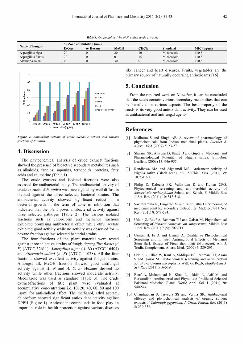
Figure 2. Antioxidant activity of crude alcoholic extract and various
fractions of N. sativa.
4. Discussion
The phytochemical analysis of crude extract/ fractions
showed the presence of bioactive secondary metabolites such
as alkaloids, tannins, saponins, terpenoids, proteins, fatty
acids and caumarine (Table 1).
The crude extracts and isolated fractions were also
assessed for antibacterial study. The antibacterial activity of
crude extracts of N. sativa was investigated by well diffusion
method against the three selected bacterial strains. The
antibacterial activity showed significant reduction in
bacterial growth in the term of zone of inhibition that
indicated that the plant poses antibacterial activity against
three selected pathogen (Table 2). The various isolated
fractions such as chloroform and methanol fractions
exhibited promising antibacterial effect while ethyl acetate
exhibited good activity while no activity was observed for n-
hexane fraction against selected bacterial strains.
The four fractions of the plant material were tested
against three selective strains of fungi; Aspergillus flavus (A.
F) (ATCC 32611), Aspergillus niger (A. N) (ATCC 16404)
and Alternaria solani (A. S) (ATCC 11078). All the four
fractions showed excellent activity against fungal strains.
Amongst all, MeOH fraction showed good antifungal
activity against A. N and A. S. n- Hexane showed no
activity while other fractions showed moderate activity.
Miconazole was used as standard (Table 3). The crude
extract/fractions of title plant were evaluated at
accumulative concentrations i.e. 10, 20, 40, 60, 80 and 100
µg/ml for anti-radical effect. The methanol, ethyl acetate,
chloroform showed significant antioxidant activity against
DPPH (Figure 1). Antioxidant compounds in food play an
important role in health protection against various diseases
like cancer and heart diseases. Fruits, vegetables are the
primary source of naturally occurring antioxidants [16].
5. Conclusion
From the reported work on N. sativa, it can be concluded
that the seeds contain various secondary metabolites that can
be beneficial in various aspects. The best property of the
seeds is its very good antioxidant activity. They can be used
as antibacterial and antifungal agents.
References
[1] Malhotra S and Singh AP. A review of pharmacology of phytochemicals from Indian medicinal plants. Internet J. Altern. Med. (2007) 5: 23-27.
[2] Sharma NK, Ahirwar D, Jhade D and Gupta S. Medicinal and Pharmacological Potential of Nigella sativa. Ethnobot. Leaflets. (2009) 13: 946-955.
[3] Randhawa MA and Alghamdi MS. Anticancer activity of Nigella sativa (Black seed). Am. J. Chin. Med. (2011) 39: 1075-1091.
[4] Philip D, Kaleena PK, Valtivittan K and Kumar CPG. Phytochemical screening and antimicrobial activity of Sansevieria roxburghiana Schult. and Schult. F. Middle-East J. Sci. Res. (2011) 10: 512-518.
[5] Savithramma N, Lingarao M and Suhrulatha D. Screening of medicinal plant for secondary metabolites. Middle-East J. Sci. Res. (2011) 8: 579-584.
[6] Uddin G, Rauf A, Rehman TU and Qaisar M. Phytochemical Screening of Pistacia chinensis var. integerrima. Middle-East J. Sci. Res. (2011) 7 (5): 707-711.
[7] Usman H, Fi A and Usman A. Qualitative Phytochemical Screening and in vitro Antimicrobial Effects of Methanol Stem Bark Extract of Ficus thonningii (Moraceae), Afr. J. Tradit. Complement. Altern. Med. (2009) 6: 289-295.
[8] Uddin G, Ullah W, Rauf A, Siddiqui BS, Rehman TU, Azam S and Qaisar M. Phytochemical screening and antimicrobial activity of Cornus microphylla Wall, ex Roxb, Middle-East J. Sci. Res. (2011) 516-519.
[9] Rauf A, Muhammad N, Khan N, Uddin N, Atif M, and Barkatullah. Antibacterial and Phytotoxic Profile of Selected Pakistani Medicinal Plants. World Appl. Sci. J. (2011) 20: 540-544.
[10] Chandrabhan S, Trivedia SS and Verma SK. Antibacterial efficacy and phytochemical analysis of organic solvent extracts of Calotropis gigantean. J. Chem. Pharm. Res. (2011) 3: 330-336.

43 Sania Feroz and Ghias Uddin: Phytochemical Analysis, Antimicrobial and Antioxidant Study of Nigella sativa L
[11] Merli M, Devi A, Priya I, Singh RJ, Jeyakumar P. Isolation, identification and antimicrobial activity of bioactive compound from Saraca asoca. Int J Curr Pharm Res 2016; 8 (1): 57-61.
[12] Uddin G, Rauf A, Siddiqui BS and Shah SQ. Preliminary Comparative phytochemical Screening of Diospyros Lotus Stewart, Middle-East J. Sci. Res. (2011) 1: 78-81.
[13] Uddin G and Rauf A. Phytochemical screening and biological activity of the aerial parts of Elaeagnus umbellate. Sci. Res. Essays (2012) 7 (43): 3690-3694.
[14] Martin G, Oswaldo C, José P, Cárdenas CA, Jovanny A, Gómez C. Antifungal activity of chloroform and acetone extracts of Solanum dolichosepalum against Fusarium oxysporum. Int J Pharm Pharm Sci 2016. 8 (8); 373-4.
[15] Rauf A, Khan A, Rasool S, Shah ZA, Saleem M. In-vitro Antifungal activity of three selected Pakistani medicinal plants. Middle-East J Med Plants Res 2012. 1 (2); 41-43.
[16] Velioglu YS, Mazza G, Gao L, Oomah BD. Antioxidant activity and total phenolics in selected fruits, vegetables and grain products. J. Agr. Food Chem. (1998) 46: 4113-4117.