Physiotherapy scoliosis-specific exercises – a comprehensive ...
52
REVIEW Open Access Physiotherapy scoliosis-specific exercises – a comprehensive review of seven major schools Hagit Berdishevsky 1*† , Victoria Ashley Lebel 2† , Josette Bettany-Saltikov 3 , Manuel Rigo 4 , Andrea Lebel 5 , Axel Hennes 6 , Michele Romano 7,8 , Marianna Białek 9 , Andrzej M’hango 9 , Tony Betts 10 , Jean Claude de Mauroy 11 and Jacek Durmala 12 Abstract In recent decades, there has been a call for change among all stakeholders involved in scoliosis management. Parents of children with scoliosis have complained about the so-called “wait and see” approach that far too many doctors use when evaluating children’ s scoliosis curves between 10° and 25°. Observation, Physiotherapy Scoliosis Specific Exercises (PSSE) and bracing for idiopathic scoliosis during growth are all therapeutic interventions accepted by the 2011 International Society on Scoliosis Orthopaedic and Rehabilitation Treatment (SOSORT). The standard features of these interventions are: 1) 3-dimension self-correction; 2) Training activities of daily living (ADL); and 3) Stabilization of the corrected posture. PSSE is part of a scoliosis care model that includes scoliosis specific education, scoliosis specific physical therapy exercises, observation or surveillance, psychological support and intervention, bracing and surgery. The model is oriented to the patient. Diagnosis and patient evaluation is essential in this model looking at a patient-oriented decision according to clinical experience, scientific evidence and patient’ s preference. Thus, specific exercises are not considered as an alternative to bracing or surgery but as a therapeutic intervention, which can be used alone or in combination with bracing or surgery according to individual indication. In the PSSE model it is recommended that the physical therapist work as part of a multidisciplinary team including the orthopeadic doctor, the orthotist, and the mental health care provider - all are according to the SOSORT guidelines and Scoliosis Research Society (SRS) philosophy. From clinical experiences, PSSE can temporarily stabilize progressive scoliosis curves during the secondary period of progression, more than a year after passing the peak of growth. In non-progressive scoliosis, the regular practice of PSSE could produce a temporary and significant reduction of the Cobb angle. PSSE can also produce benefits in subjects with scoliosis other than reducing the Cobb angle, like improving back asymmetry, based on 3D self-correction and stabilization of a stable 3D corrected posture, as well as the secondary muscle imbalance and related pain. In more severe cases of thoracic scoliosis, it can also improve breathing function. This paper will discuss in detail seven major scoliosis schools and their approaches to PSSE, including their bracing techniques and scientific evidence. The aim of this paper is to understand and learn about the different international treatment methods so that physical therapists can incorporate the best from each into their own practices, and in that way attempt to improve the conservative management of patients with idiopathic scoliosis. These schools are presented in the historical order in which they were developed. They include the Lyon approach from France, the Katharina Schroth Asklepios approach from Germany, the Scientific (Continued on next page) * Correspondence: [email protected] † Equal contributors 1 Conservative Care for Spine and Scoliosis, ColumbiaDoctors Midtown, Columbia University Medical Center, New York, NY, USA Full list of author information is available at the end of the article © 2016 The Author(s). Open Access This article is distributed under the terms of the Creative Commons Attribution 4.0 International License (http://creativecommons.org/licenses/by/4.0/), which permits unrestricted use, distribution, and reproduction in any medium, provided you give appropriate credit to the original author(s) and the source, provide a link to the Creative Commons license, and indicate if changes were made. The Creative Commons Public Domain Dedication waiver (http://creativecommons.org/publicdomain/zero/1.0/) applies to the data made available in this article, unless otherwise stated. Berdishevsky et al. Scoliosis and Spinal Disorders (2016) 11:20 DOI 10.1186/s13013-016-0076-9
Transcript of Physiotherapy scoliosis-specific exercises – a comprehensive ...

REVIEW Open Access
Physiotherapy scoliosis-specific exercises –a comprehensive review of seven majorschoolsHagit Berdishevsky1*† , Victoria Ashley Lebel2†, Josette Bettany-Saltikov3, Manuel Rigo4, Andrea Lebel5,Axel Hennes6, Michele Romano7,8, Marianna Białek9, Andrzej M’hango9, Tony Betts10, Jean Claude de Mauroy11
and Jacek Durmala12
Abstract
In recent decades, there has been a call for change among all stakeholders involved in scoliosis management. Parents ofchildren with scoliosis have complained about the so-called “wait and see” approach that far too many doctors use whenevaluating children’s scoliosis curves between 10° and 25°. Observation, Physiotherapy Scoliosis Specific Exercises (PSSE)and bracing for idiopathic scoliosis during growth are all therapeutic interventions accepted by the 2011 InternationalSociety on Scoliosis Orthopaedic and Rehabilitation Treatment (SOSORT). The standard features of these interventionsare: 1) 3-dimension self-correction; 2) Training activities of daily living (ADL); and 3) Stabilization of the corrected posture.PSSE is part of a scoliosis care model that includes scoliosis specific education, scoliosis specific physical therapy exercises,observation or surveillance, psychological support and intervention, bracing and surgery. The model is oriented to thepatient. Diagnosis and patient evaluation is essential in this model looking at a patient-oriented decision according toclinical experience, scientific evidence and patient’s preference. Thus, specific exercises are not considered as analternative to bracing or surgery but as a therapeutic intervention, which can be used alone or in combination withbracing or surgery according to individual indication. In the PSSE model it is recommended that the physical therapistwork as part of a multidisciplinary team including the orthopeadic doctor, the orthotist, and the mental health careprovider - all are according to the SOSORT guidelines and Scoliosis Research Society (SRS) philosophy. From clinicalexperiences, PSSE can temporarily stabilize progressive scoliosis curves during the secondary period of progression,more than a year after passing the peak of growth. In non-progressive scoliosis, the regular practice of PSSEcould produce a temporary and significant reduction of the Cobb angle. PSSE can also produce benefits insubjects with scoliosis other than reducing the Cobb angle, like improving back asymmetry, based on 3Dself-correction and stabilization of a stable 3D corrected posture, as well as the secondary muscle imbalanceand related pain. In more severe cases of thoracic scoliosis, it can also improve breathing function.This paper will discuss in detail seven major scoliosis schools and their approaches to PSSE, including theirbracing techniques and scientific evidence. The aim of this paper is to understand and learn about thedifferent international treatment methods so that physical therapists can incorporate the best from each intotheir own practices, and in that way attempt to improve the conservative management of patients withidiopathic scoliosis. These schools are presented in the historical order in which they were developed. Theyinclude the Lyon approach from France, the Katharina Schroth Asklepios approach from Germany, the Scientific(Continued on next page)
* Correspondence: [email protected]†Equal contributors1Conservative Care for Spine and Scoliosis, ColumbiaDoctors Midtown,Columbia University Medical Center, New York, NY, USAFull list of author information is available at the end of the article
© 2016 The Author(s). Open Access This article is distributed under the terms of the Creative Commons Attribution 4.0International License (http://creativecommons.org/licenses/by/4.0/), which permits unrestricted use, distribution, andreproduction in any medium, provided you give appropriate credit to the original author(s) and the source, provide a link tothe Creative Commons license, and indicate if changes were made. The Creative Commons Public Domain Dedication waiver(http://creativecommons.org/publicdomain/zero/1.0/) applies to the data made available in this article, unless otherwise stated.
Berdishevsky et al. Scoliosis and Spinal Disorders (2016) 11:20 DOI 10.1186/s13013-016-0076-9

(Continued from previous page)
Exercise Approach to Scoliosis (SEAS) from Italy, the Barcelona Scoliosis Physical Therapy School approach(BSPTS) from Spain, the Dobomed approach from Poland, the Side Shift approach from the United Kingdom,and the Functional Individual Therapy of Scoliosis approach (FITS) from Poland.
BackgroundIn recent decades, there has been a call for change amongall stakeholders involved in scoliosis management. Parentsof children with scoliosis have complained about the so-called “wait and see” approach that far too many doctorsuse when evaluating children’s scoliosis curves between 10°and 25° [1]. Numerous physical therapists have reportedthat children with scoliosis and their parents are reacting totheir lack of empowerment as they wonder how to helpthemselves beyond simply waiting and bracing. Physicaltherapists, most of whom are still inadequately educatedand equipped to provide quality scoliosis treatment, havesearched for new treatment methods. Orthotists haverecognized that traditional braces lack the ability to make3D corrections, producing flat back or other poor cosmeticchanges, and are looking for more effective options. Finally,doctors have sought out alternatives to help patients whoare not good candidates for surgery [2].The Society of Scoliosis Orthopedic Rehabilitation and
Treatment (SOSORT) was founded in 2004 in reactionto this growing awareness. SOSORT promotes and en-courages conservative, evidence-based medicine regard-ing scoliosis and provides education, guidelines andconsensus about treatment options to people with scoli-osis [3]. Every scoliosis approach or ‘school’ around theworld subscribes to SOSORT’s principles and shares acommon mission. The shared goal is not simply to lookat the spine in the coronal plane but to look at theaffected subject and family under a more holistic psy-chosocial model, where present and future quality of lifeis the main objective.SOSORT uses the term Physiotherapy Scoliosis Specific
Exercises (PSSE) in connection with all of the schoolsrepresented within the organization. The effectiveness ofPSSE in treating patients with Adolescent IdiopathicScoliosis (AIS) has been demonstrated by recent studies.While a Cochrane review published in 2012 [4] reported alow to very low quality of evidence for the proposition thatPSSE was effective in improving Cobb angle, angle of trunkrotation, pain and quality of life, since the time of thatreview, four randomized controlled trials (RCTs), which aregenerally recognized as the highest level of evidence forprimary studies, have provided strong proof that PSSE areindeed effective for treating AIS patients with mild andmoderate curves. The four RCTs were conducted in differ-ent parts of the world – in Italy by Monticone et al. [5](2013), in Canada by Schreiber et al. [1] (2015), inEngland by Williamson et al. 2015 [6], and in Turkey by
Kuru et al. [7] (2015) – and are summarized in the bodyof this paper.Seven major scoliosis schools and their approaches to
PSSE, including their bracing techniques, will be dis-cussed in detail in this paper. The differences betweenthe schools are related to PSSE used by each school. Thepurpose of this paper is not to determine which scoliosisschool and treatment approach is superior to the others.Rather, the aim is to understand and learn about thedifferent treatment methods around the world so thatphysical therapists can incorporate the best from eachinto their own practices, and in that way attempt toimprove the conservative management of patients withidiopathic scoliosis.The schools are presented in the historical order in
which they were developed. They include the Lyonapproach from France, the Katharina Schroth Asklepiosapproach from Germany, the Scientific Exercise Approachto Scoliosis (SEAS) from Italy, the Barcelona ScoliosisPhysical Therapy School approach (BSPTS) from Spain,the Dobomed approach from Poland, the Side Shiftapproach from the United Kingdom, and the FunctionalIndividual Therapy of Scoliosis approach (FITS) fromPoland.
The Lyon approach (France)IntroductionThe Lyon school of physiotherapy for scoliosis, managedby Dr. Jean Claude de Mauroy, the head of the ortho-pedic medicine department at Clinique du Parc, Lyon,France (Fig. 1), is one of the oldest physiotherapyschools in France and one of the first schools to be inte-grated into the Faculty of Medicine program in Lyon.Physiotherapy is an integral part of the Lyon approachto the management of scoliosis in conjunction with cast-ing and bracing.
HistoryDr. Gabriel Pravaz, an orthopedic surgeon, created thefirst orthopedic physiotherapy center in Lyon two cen-turies ago. In the middle of the 20th century, Dr. PierreStagnara established an organized nonsurgical approachto manage scoliosis with casts and braces, and in 1947he created the Lyon brace. More recently, the ARTbracehas been developed, which obviates the need for a plastercast [8]. Although the Lyon method is primarily focusedon the use of bracing – and in the recent past on both
Berdishevsky et al. Scoliosis and Spinal Disorders (2016) 11:20 Page 2 of 52

casting and bracing – it includes scoliosis specific exer-cises to support treatment.
Definition of treatmentThe Lyon method traditionally combined PSSE with theLyon brace and casting, and more recently has com-bined PSSE with bracing alone in the form of the newLyon ARTbrace (Asymmetrical Rigid Torsion brace).Physiotherapeutic treatment includes 3D mobilization ofthe spine, mobilization of the ilio-lumbar angle (lumbarscoliosis), patient education, and activities of daily living,including correction of the sitting position.
Treatment indications, goals and age specificsThe 2011 SOSORT Guidelines provide clear, scientificindications regarding what type of treatment (observation,physical therapy, bracing, surgery) is appropriate for patientswith scoliosis [9]. Under the Lyon approach, the treatmentis more specifically determined by the type of scoliosis;chaotic or linear [10]. Chaotic scoliosis is a true 3D struc-tural deformity of the spine, which occurs in approximately2.5 % of adolescents with scoliosis curves <20° Cobb angle.This is a dynamic scoliosis, which can be influenced bymany environmental factors. Because of the uncertainty ofits progression, chaotic scoliosis can best be described bydeterministic chaos. While, according to Newton’s Law ofGravity, we can predict where the apple that falls from a treewill land, we cannot similarly predict where a leaf that fallsfrom a tree will land. The tree leaf falls according to thesame laws, but the precise location of its landing remainsunpredictable because the leaf is more sensitive to the
wind. This type of unpredictability also defines the devel-opment and progression of scoliosis in an individual. Thespine is highly sensitive to the influence of the nervoussystem on its growth and development, and any change inthe nervous system leads to the deterministic chaos ofscoliosis below 20°. The progression of scoliosis is neithercertain nor predictable as the nervous system is constantlytrying to adapt and correct the asymmetric growth duringthe early development of the scoliosis [11].The other category of scoliosis is linear scoliosis,
which occurs in approximately 0.25 % of adolescentswith scoliosis curves >20°. Madame Duval Beaupère ini-tially described the linear progression of scoliosis inpolio patients. A triggering event brings scoliosis into a‘vicious cycle’ (Fig. 2) that was later described by IanStokes and R. Geoffrey Burwell in detail in their studyon the biomechanics of scoliosis progression [12]. Stokesand Burwell explain that the vicious cycle begins with atriggering event, which results in the formation ofwedged vertebrae. What follows is continuous asymmet-ric loading on the spine, which can potentially promoteasymmetric growth and advancement of the progression.The aim of PSSE is to intervene in this vicious cycle atthe level of patient education and promotion of spinaland trunk alignment by reducing or even stopping theasymmetric loading and by potentially helping to stopscoliosis progression.The goals of the Lyon method are improved motiv-
ation with bracing, patient education, including aware-ness of postural defects, and increased range ofmotion, neuromuscular control of the spine, coordination,trunk stabilization, muscular strength, respiration, andergonomics.The scoliosis treatment protocol of the Lyon
method depends on the patient’s age. Juvenile patients(younger than 15 to 17 years) do not do stretching.Adolescent patients complete the entire program. Withadult patients, the focus is on pain reduction and discprotection.
Classification systemThe classification system used for physiotherapy and forbracing are the Ponseti and the Lenke classifications,respectively.
Principles of the Lyon methodThe Lyon method of scoliosis treatment involves fivestages:
1. The Lyon approach to assessment.2. Awareness of trunk deformity.3. What to do: sample exercises.4. What not to do and why.5. Sport or only physiotherapy?
Fig. 1 Dr. Jean Claude de Mauroy, co-inventor of the new LyonARTbrace (Asymmetrical Rigid Torsion Brace)
Berdishevsky et al. Scoliosis and Spinal Disorders (2016) 11:20 Page 3 of 52

Stage I: Lyon approach to assessmentThe Lyon approach considers three factors in determin-ing the regimen of therapy to pursue: the patient’s age,postural imbalance, and the Cobb angle.
Stage II: Awareness of trunk deformityThe Lyon approach uses visualization with mirrors andvideo to help with curve correction (Fig. 3).
Stage III: What to do: sample exerciseThe basis of the Lyon method is to avoid spinal extensionduring exercise and enhance kyphosis of the thoracic regionwith lordosis of the lumbar spine as well as frontal planecorrection, segmental mobilization, core stabilization, pro-prioception, balance and stabilization (Figs. 4, 5, 6, 7, 8 and9). In the Lyon approach, a great emphasis is given to exer-cises done in the plaster cast prior to bracing (Fig. 10) andduring bracing (Fig. 11) to encourage equilibrium and mus-cular strength and endurance while in the cast or brace.
Stage IV: What not to do and whyThe Lyon method avoids sagittal plane extreme move-ments (flexion and extension) and exercises causingshortness of breath.
Stage V: Sport or only physiotherapy?The Lyon method teaches patients how to playsports and the best and worst sports for scoliosis(Fig. 12).
The use of breathing mechanics, muscle activation, andmobilizationThe Lyon method uses rotational angular breathingwith the diaphragm as well as a breathing machine toincrease lung capacity (Fig. 13). The Lyon method alsoimproves the endurance of the deep paraspinal and coremusculature and focuses on mobilization to improvecorrection (Figs. 14, 15 and 16).
Treatment tools: active and passiveThe Lyon method uses mirrors and video to assist incorrection and to help patients develop perception oftheir spine and their postural defects.
Description of the most relevant exercise mechanics(see Figures in section Classification system)
1. Lying: kyphotization with a cushion.2. Rolling: fetal position with a cushion and derotation
on a Swiss ball in kyphosis.
Fig. 2 Image on the left: The Vicious cycle. Dr. Stokes and Burwell hypothesized that the vicious cycle of scoliosis curve progression begins witha triggering event which leads to the formation of wedged vertebrae. Wedged vertebrae cause the spine to curve which results in continuousasymmetric loading on the spine. This in turn, can potentially promote asymmetric growth of the spine and progression of scoliosis curves asasymmetric growth increases the wedging of the vertebrae and perpetuates the cycle to continue. The image on the right shows a scoliosispatient sitting with increased asymmetric loading of the spine as described in Stokes’ ‘Vicious Cycle”. The large red “X” on the image indicatesthat this is not the desired posture. The image on the right shows the same scoliosis patient sitting with improved asymmetric loading of thespine as she performs scoliosis specific physiotherapy exercises in accordance with the Lyon approach
Berdishevsky et al. Scoliosis and Spinal Disorders (2016) 11:20 Page 4 of 52

3. Sitting: adjustments of the lumbar lordosis in thesitting position and mobilization on a Swiss ball.
4. Standing.
Activities of daily living and sportThe Lyon method helps patients develop the correctposture while sitting at a table to write or type on acomputer. Sports, such as basketball (Fig. 17), are anessential part of the Lyon method.
Scientific evidenceThe Lyon approach is not supported by scientific evi-dence for cases where the Cobb angle is less than 20°.For cases where the Cobb angle is 20° or greater, themethod depends primarily on casting and bracing forits effectiveness. Under this approach, the physicaltherapy exercises are properly viewed as supplemental
to the casting and bracing, and in each case areadapted to the individual’s particular needs. Asexpressed by Dr. Jean Claude de Mauroy, the physicaltherapy elements of the Lyon approach are better de-scribed as the “Lyon experience” than the “Lyonmethod” [10]. It must be noted that, while outside thescope of this paper, there does exist scientific supportfor the effectiveness of the casting and bracing es-poused by the Lyon approach.
The ARTbraceThe ARTbrace (Fig. 18) is a new brace: asymmetric,rigid, made from 4 mm polycarbonate [8]. The brace re-produces the shape of a twisted column opposite to thescoliosis. Both polycarbonate lateral hemi-shells are ar-ticulated on a posterior metal bar. Both anterior and in-ferior closures are rigid; the upper third is a velcro strap.The ARTbrace is the only asymmetrical brace with lat-eral hemi-shells.As with the old Lyon brace, the ARTbrace is adjustable,
but many new concepts were used to build the brace:
1. The mathematical basis of the detorsion is thecircled helicoid with horizontal circle generator.
2. The multiple 3 points system is replaced by a globaldetorsion.
3. Three regional 2D individual moldings are superposedto obtain a 3D helicoidal correction with coupledmovements.
4. The sagittal plane is fixed in a physiological postureto improve flat back if necessary.
5. The upper part of the brace supports the trunk likea baby lift.
Fig. 4 Active thoracic mobilization, promoting kyphosis, using theLyon method
Fig. 5 Active lumbar correction, promoting lordosis, using theLyon method
Fig. 3 Scoliosis patient developing self-awareness of postural defectswith the help of a video recorder and real-time video feedback
Berdishevsky et al. Scoliosis and Spinal Disorders (2016) 11:20 Page 5 of 52

6. In the middle, under the breast, the clamping of thetwo hemi-shells realizes the “tube mayonnaise” effectwith passive axial lengthening and geometricdetorsion.
7. The polycarbonate-skin interface is a soft contacttype with mechanical detorsion of a cylinder.
8. Global detorsion is like a wrench and bolt along thevertical axis.
The Lyon method of physiotherapy is adapted toprepare the child in order to facilitate the regional mold-ing and detorsion over a period of time.In Figs. 19 and 20 the case of ‘S’ demonstrates the results
of brace wearing over two years in a progressive case.
The Schroth method (Germany)IntroductionBased upon typical physiotherapeutic principles, theSchroth method was developed by Katharina Schroth in1920, and continuously refined through the treatment ofapproximately 3,000 scoliosis cases per year. The AsklepiosKatharina Schroth Spinal Deformities Rehabilitation Centrein Germany (Fig. 21) offers a scoliosis-specific intensiveinpatient rehabilitation program. In addition to the treat-ment offered at the Centre, 2,500 trained and certifiedSchroth therapists treat patients through the center’s resi-dential outpatient treatment program.The broad network of therapists enables the continu-
ation and actualization of the Schroth method through-out much of the world, including in Germany, Russiaand many other European countries, in Canada and theUnited States, in Australia, and several countries in Asia.The leading educator of Schroth therapists is AxelHennes (Fig. 22), who is the head physical therapist atthe Medical Spine Center in Bad Sobernheim, Germany.Another central figure in the school today is Dr. HansWeiss, the grandson of Katharina Schroth, who has pub-lished numerous studies regarding the Schroth method(see details in Scientific evidence).The main goals of the Schroth method are to provide
effective treatment for patients, and training and educationfor physiotherapists. The treatment approach includes bothintensive inpatient rehabilitation and residential outpatientphysiotherapy provided by certified Schroth therapists [13].
HistoryKatharina Schroth, born in Dresden, Germany, in 1894,was suffering from moderate scoliosis and underwenttreatment with a steel brace before she decided todevelop a more functional approach to treat her scoliosis
A B
Fig. 6 (a, b): Active thoracic shift exercise with a dowel (a) and a Swiss-ball (b) using the Lyon method
Fig. 7 Active thoracic shift and derotation exercise using the Lyonmethod. Arrows in the radiograph and the diagram show thedirection of the thoracic shift and the derotation of the ribcage asthe exercise is performed using the Lyon method
Berdishevsky et al. Scoliosis and Spinal Disorders (2016) 11:20 Page 6 of 52

and improve her quality of life. Inspired by the way inwhich a balloon is inflated, in 1910 she tried to correcther own deformity by breathing into the concavities ofher trunk in front of a mirror. She recognized that 3Dpostural correction could only be achieved with a seriesof corrective exercises designed to support a correctedposture and change the postural perception of the per-son suffering with scoliosis. The principles of active 3Dposture correction, corrective breathing, and correctionof postural perception form the foundation for whatcame to be known as the Schroth method of scoliosistreatment [14] (Fig. 23).By 1921, Katharina Schroth’s success with her own
scoliosis was attracting attention, and with the help ofher daughter, Christa Lehnert-Schroth, she begantreating others with scoliosis in her small institute in
Meissen, Germany. By the late 1930’s, the Schrothmethod was widely recognized as the best conservativescoliosis treatment method in all of Germany. AfterWorld War II, Katharina Schroth and her daughtermoved to West Germany and opened an institute inBad Sobernheim, which soon grew into a full-scalescoliosis treatment clinic that served more than 150inpatients at a time [14]. In the 1980’s, the institutewas renamed the Asklepios Katharina Schroth Klinik.Hans-Rudolf Weiss, an orthopedic surgeon, and the
grandson of Katharina Schroth, was the medical directorof the Asklepios Katharina Schroth Rehabilitation Centrefrom 1995 to 2008. In the summer of 2009 he openedhis own practice for orthopedics and rehabilitation andnow offers new concepts of bracing and physiotherapybased on the Schroth method. He has conducted andpublished a substantial amount of research supportingthe effectiveness of the Schroth method on improvingthe Cobb angle, the angle of trunk rotation, vital cap-acity, pain, quality of life, as well as the effectiveness ofbracing in reducing the need for surgery. (Details ofthese studies can be found in Scientific evidence).Today, the Asklepios Katharina Schroth Klinik accom-
modates 200 inpatients and has a waiting list for its re-nowned treatment courses. Christa Lehnert-Schroth(1924–2015) was involved in the treatment of more than10,000 scoliosis patients over the course of her 50-yearcareer at the Asklepios Schroth Klinik.
Classification systemThe Schroth system of classification [14] is derived fromthe Schroth principle of dividing the body into ‘BodyBlocks’. This symbolic description helps to explain the
A B
Fig. 8 (a, b): Balance and proprioception exercises on a Swiss-ball (a) and on a balance board (b) using the Lyon method
Fig. 9 Spinal stabilization exercises using the Lyon method
Berdishevsky et al. Scoliosis and Spinal Disorders (2016) 11:20 Page 7 of 52

Fig. 10 Several standard Lyon exercises in a Lyon plaster cast promoting core strength (top left), breathing and thoracic shift (bottom), and elongation
A B
Fig. 11 (a, b): Several standard Lyon exercises in a Lyon plaster cast promoting postural correction (a) and core strengthening (b)
Berdishevsky et al. Scoliosis and Spinal Disorders (2016) 11:20 Page 8 of 52

scoliotic alterations as compensatory adaptations. The BodyBlocks depict the trunk deformation as a change in theirgeometric form from a rectangle to a trapezium shape.Side-shift and rotation as well as compression on the con-cave side and expansion on the convex side are clearly vis-ible. In the standing static position the body blocks shouldbe aligned perpendicularly with their center of gravity inte-grated in the central sacral line (CSL) as seen in Fig. 24.The scoliotic trunk asymmetry is a loss of symmetry andshows the blocks skewed and off-center (Fig. 24).The Schroth classification system gives the direction
of the side deviation and rotation of the main importantbody blocks (major curves) and a clear orientation forthe standardized therapy plan which includes the ther-apy diagram, exercise-program with home-exercises, andnecessary mobilizing technique.According to the Schroth classification system, the dif-
ferent scoliosis types always start with the major curveand are followed by relevant secondary curves.
The uppercase letters represent the body blocks and thelowercase letters describe the direction of the lateral devi-ation and rotation: right = ri, left = le. Schroth body blocks:H – Hip-pelvic block including the lower limbs reach-
ing the lower end vertebra (LEV) of the lumbar curve.L – Lumbar block enclosed by upper end vertebra
(UEV) and LEV of the lumbar curve or thoracolumbarcurve respectively.T – Thoracic block between UEV and LEV of the
thoracic curve.S – Shoulder block represents the cervical thoracic
(proximal thoracic) curve located between UEV of thethoracic curve and UEV of the proximal thoracic curve.The following is an overview of the classifications:
1. Thoracic scoliosis (means that the major curve islocated in the thoracic spine, and the curve can beto the right or to the left).a. Thoracic only.
Fig. 12 Activity level recommendations by age per the Lyon Schoolin accordance with the Lyon treatment principles
Fig. 13 Lyon method breathing exercises using a breathingmachine, performed while wearing a Lyon plaster cast, increaseslung capacity
Fig. 14 Active thoracic mobilization using the Lyon method. Arrowsin the diagram on the right show the direction of thoracicmobilization of the ribcage
Fig. 15 Active lumbar mobilization using the Lyon method. Thediagram on the right shows lumbar scoliosis
Berdishevsky et al. Scoliosis and Spinal Disorders (2016) 11:20 Page 9 of 52

b. Thoracic with lumbar to opposite side with hipsin center.
c. Thoracic with lumbar and hips protruding to theopposite side of the thoracic curve (along withthe lumbar).
2. Lumbar scoliosis (means that the major curve islocated in the lumbar spine, and the curve can be tothe right or to the left).a. Lumbar only with hips protruding to the opposite
side of the curve.b. Lumbar curve with thoracic and hips protruding
to the opposite side of the lumbar curve.c. Lumbar and thoracic curves with hips in center.
3. Sagittal plane deformities including increasedthoracic kyphosis (round back), decreased thoracickyphosis (flat back) and increased lumbar kyphosisor loss of the normal anatomical lordosis (curve) ofthe lumbar spine.
Treatment indications and goalsTreatment indication for the Schroth method is basedon the SOSORT guidelines [9].Both individual and group treatments share these same
goals:
1. Proactive spinal corrections to avoid surgery.2. Postural training to avoid or decelerate
progression.3. Information to support a decision-making
process.4. Teaching a home-exercise program.5. Support help for self-help.6. Prevention and coping strategies for pain.
Age specificsThe Schroth method is primarily used for idiopathicscoliosis, including AIS and late juvenile idiopathic
Fig. 16 Mobilization of the costovertebral joints using the Lyon method
Fig. 17 The Lyon method encourages athletic activities whilewearing the Lyon brace. This group of scoliosis patients are playingbasketball, which aids vertical stretching and spinal flexibility
Fig. 18 Anterior (left) and posterior (right) views of the newasymmetric rigid torsion brace (ARTbrace) made from 4 mmpolycarbonate. The main biomechanical concepts are based onelongation along the vertical axis, lateral inflexion in the frontalplane, and derotation of the spine in order to obtain a correctionof the scoliotic curve
Berdishevsky et al. Scoliosis and Spinal Disorders (2016) 11:20 Page 10 of 52

scoliosis (JIS). People with early onset scoliosis (EOS), andadults, are treated with modified principles. Sagittal planedeformities such as hyper-kyphosis (Scheuermann’s ky-phosis) and lordosis (inverted back) can also be treatedwith Schroth exercises. Treatment of JIS involves a lessintense and modified Schroth method as well. Treatmentof AIS using strict Schroth principles is aimed at prevent-ing curve progression before the end of growth. Treat-ment of adult onset scoliosis implements a modifiedSchroth method based on the severity of pain and thedegree and rigidity of the spinal deformity.
3D principles of correctionIn the Schroth method there are five pelvic correctionsthat are assumed prior to the execution of the main prin-ciples of correction. These five pelvic corrections ensurethat the pelvis is best aligned with the trunk prior to themajor corrections.The five principles of the Schroth method are: 1) Auto-
elongation (detorsion); 2) Deflection; 3) Derotation; 4) Ro-tational breathing; and 5) Stabilization. During the appli-cation of these principals, as with the BSPTS method, thepatient is taught how to de-collapse the concaved areas ofthe trunk and how to reduce the prominences.
The use of breathing mechanics, muscle activation, andmobilizationThe use of specific and special Rotation Angular Breathing(RAB) (also called orthopaedic breathing) will be discussedin detail in The Use of Breathing Mechanics, Muscle Acti-vation, and Mobilization below.The method also includes mobilization and flexibility in
the spine and between ribs to enhance joint mobility priorto the exercises. Muscle activation is done via specificactivation of muscles that can improve the correction,such as the iliopsoas, the quadratus lumborum anderector spinae.In Figs. 25 and 26 the use of RAB and specific
mobilization and flexibility are demonstrated.
Description of Schroth method exercisesFour of the most commonly used exercises in theSchroth method are the “50 x Pezziball” exercise, Prone
Fig. 19 (a, b, c, d): Radiographic series of a female patient with progressive scoliosis (patient ‘S’). Initial PA (a) and lateral (c) radiographs at thetime of diagnosis at age 13 show a thoracic T5-T12 scoliosis of 39° Cobb angle (a) and a 25° hypokyphosis (c). Repeat radiographs taken in theARTbrace show a decreased Cobb angle on PA radiograph (b) and an increased kyphosis on lateral radiograph (d)
1 year 2 years
A B C
Fig. 20 (a, b, c): Radiographic series of patient ‘S’ at one year (b) andtwo years (c) after brace weaning. Compared to the initial radiograph(a), the final radiograph (c) shows a reduction in curve Cobb angle ofover 50 %
Berdishevsky et al. Scoliosis and Spinal Disorders (2016) 11:20 Page 11 of 52

exercise, Sail exercise, and the Muscle-cylinder exercise.All of these exercises can be used for all curve types.The “50 x Pezziball” exercise works on auto-self-elongation and activation of muscles in the trunk thatforce the convexities in the trunk “forward and inward”and the concavities “outward and backward” (Fig. 27).
The Prone exercise corrects the thoracic curve usingshoulder traction (ST) and shoulder counter-traction(SCT) and the lumbar curve via activation of the iliop-soas muscle (Fig. 28). The Sail exercise is a very effectivestretching exercise, which helps elongate the thoracicconcavity (Fig. 29). The Muscle-cylinder engages thequadratus lumborum muscle to correct the lumbar curveagainst gravity (Fig. 30). Other exercises related to theSchroth method involve postural correction during activ-ities of daily living. These exercises focus on correctingposture while resting, sitting, or standing.
Activities of daily livingThe Schroth method emphasizes teaching postural cor-rections throughout the day in order to change habitualdefault postures and improve alignment, pain and pro-gression (Fig. 31). The main advantage of this programlies in its application to ordinary daily activity for thepurpose of changing the asymmetrical loading on thebody in order to decrease progression and pain. Thisalso reduces the amount of time needed to practice thehighly demanding exercises and allows patients to spendmore time in leisure activities and to live a normal life.
Scientific evidenceAmong all PSSE approaches, the Schroth method [14] isamong the most studied and widely used specific exer-cise approaches for scoliosis. Numerous studies havebeen written by Dr. Hans Weiss, the medical director ofthe Asklepios Katharina Schroth Rehabilitation Centerfrom 1995 to 2008, and by Dr. Manuel Rigo, director ofthe Barcelona Scoliosis Physical Therapy School (BSPTS).
Fig. 21 Asklepios Katharina Schroth Spinal Deformities Rehabilitation Centre in Bad Sobernheim, Germany. Formerly called the KatharinaSchroth Klinik
Fig. 22 Axel Hennes, head of the physical therapy department atthe Asklepios Katharina Schroth Spinal Deformities RehabilitationCentre in Bad Sobernheim, Germany
Berdishevsky et al. Scoliosis and Spinal Disorders (2016) 11:20 Page 12 of 52

Fig. 23 History of the Schroth method. Katharina Schroth with her daughter, Christa Lehnert-Schroth (top right). Patients with scoliosis exercisingoutdoors at the Katharina Schroth Klinik (bottom right; left)
Shoulder block
SThoracic block
TLumbar block
L
Hip - pelvic
block H
+ A B C D
Fig. 24 (a, b, c, d): Schroth Body Blocks. The Schroth system of scoliosis curve classification is derived from the Schroth principle of dividing the bodyinto “body blocks” as pictured anatomically (a) and schematically (b). Scoliosis causes the body blocks to become deformed, changing their geometricshape from a rectangle (b) to a trapezium (c). A patient with a major lumbar scoliosis left convex curve has a lumbar block shifted to the left and ahip-pelvic block shifted to the right (d)
Berdishevsky et al. Scoliosis and Spinal Disorders (2016) 11:20 Page 13 of 52
Their studies [15–29] demonstrate positive outcomesfrom use of the Schroth method on back muscle strength,breathing function, pain, quality of life and self-image,slowing curve progression, improving Cobb angles anddecreasing the prevalence of surgery.A recent study by Kuru et al., suggests that Schroth
exercises performed in a clinic under supervision are su-perior to home exercise programs only, with results indi-cating significant improvement in Cobb angle, quality oflife and trunk rotation [7]. A study by Schriber et al.,confirms in a RCT improved self-image and quality oflife in patients that were assigned to a Schroth exercisegroup as compared to a control group [1]. Anotherstudy that followed the Schroth principles and theBSPTS protocol showed an improvement in back asym-metry and spinal imbalance both in the frontal planeand in the transversal plane [30]. The Schroth method
has been shown to positively influence the Cobb angle,vital capacity, strength and postural defects in AIS [31].Furthermore, in reducing the proportion of childrenwith AIS requiring surgery, conservative methods ofscoliosis treatment should never be ruled out from scoli-osis management as they afford patients a viable alterna-tive to surgical treatment [15].
Scientific exercise approach to scoliosis (Italy)IntroductionThe Scientific Exercise Approach to Scoliosis (SEAS) isan individualized exercise program scientifically adapted
A B C
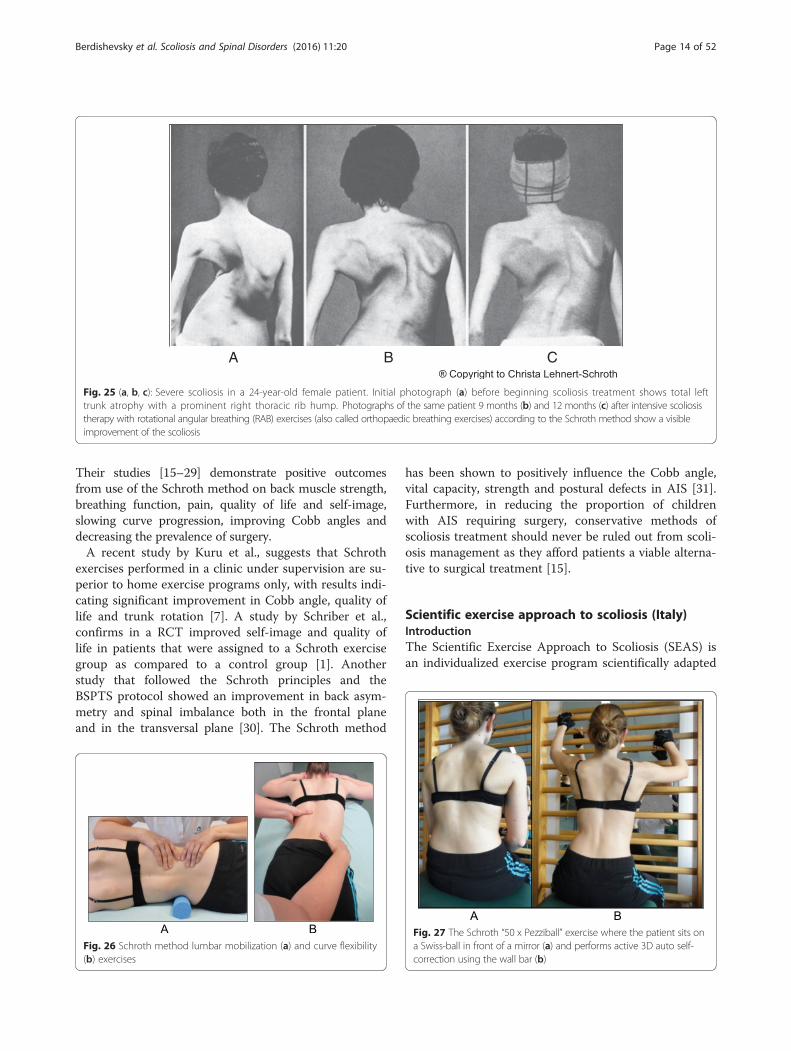
Fig. 25 (a, b, c): Severe scoliosis in a 24-year-old female patient. Initial photograph (a) before beginning scoliosis treatment shows total lefttrunk atrophy with a prominent right thoracic rib hump. Photographs of the same patient 9 months (b) and 12 months (c) after intensive scoliosistherapy with rotational angular breathing (RAB) exercises (also called orthopaedic breathing exercises) according to the Schroth method show a visibleimprovement of the scoliosis
Fig. 26 Schroth method lumbar mobilization (a) and curve flexibility(b) exercises
Fig. 27 The Schroth “50 x Pezziball” exercise where the patient sits ona Swiss-ball in front of a mirror (a) and performs active 3D auto self-correction using the wall bar (b)
Berdishevsky et al. Scoliosis and Spinal Disorders (2016) 11:20 Page 14 of 52

to all aspects of the conservative treatment of scoliosisbased on the most current research, and is continuouslyevolving with the introduction of new knowledge fromthe scientific literature. For mild-moderate curves duringactive growth, SEAS is used alone to reduce the need forbracing. In moderate-severe curves during active growth,SEAS is used in combination with bracing in order toslow down, halt, and possibly reverse curve progression,and in preparation to wean the patient off the brace. Inadult scoliosis patients, either with progressive scoliosiscurves or fused spines, SEAS helps stabilize the spineand reduce disability.
The SEAS method [32] is based on a scoliosis-specificactive self-correction technique performed without anyexternal aids and incorporated into functional exercises.Evaluation tests guide the choice of the exercises mostappropriate to the individual patient. Improvement ofthe stability of the spine in active self-correction is theprimary objective of SEAS. SEAS exercises train neuro-motor systems to activate a reflex of self-correction ofposture during activities of daily living. SEAS can be per-formed as an outpatient (2–3 times a week for 45 min)or as a home exercise program of 20 min daily inconjunction with expert physiotherapy sessions of 1.5 h
Concavities
Shoulder traction (ST)
Corrective pads
Convexities/curves shoulder counter-traction (SCT) (forward - inward)
Elongation
Fig. 28 The Schroth prone exercise with activation of the iliopsoasmuscle (right hip flexion). Blue arrows represent trunk elongationwith caudal and cranial forces. Red arrows represent areas of muscleactivation around the convexities towards the midline. Green half-moons represent areas of expansion of the concavities. Red circlesrepresent additional corrective forces: red circles around the rightlower extremity and the right upper extremity represent iliopsoasactivation and shoulder traction/counter-traction, respectively,resulting in correction of the lumbar and thoracic curves
Fig. 29 The Schroth “Sail” exercise where the patient stands on ahalf foam-roll with two poles and performs active stabilization. Thered circle represents the concavity (weak side according to Schroth).During active stabilization, the patient is consciously expanding theleft rib cage with right directional breathing, opening the collapsedleft lung, while maintaining 3D postural correction
Berdishevsky et al. Scoliosis and Spinal Disorders (2016) 11:20 Page 15 of 52

Fig. 30 The “Muscle-cylinder” exercise (also known as the “Side-lying” exercise), focusing mainly on the correction of the lumbar scoliosis curve.During this exercise, the patient lies on the lumbar convex side. The lumbar convexity is supported by a rice bag to help align the spine in thehorizontal plane. The patient’s right leg is supported by a stool (in case of 4C/major lumbar scoliosis) and the patient’s right arm is supported ona chair during the exercise. Light blue arrows represent trunk elongation with cranial and caudal forces. Green half-moons represent areas ofexpansion of the concavities. Red arrows represent areas of muscle activation, approximating the convexities towards midline, and the directionof the correction. The dark blue arrow pointing upwards from the right elbow represents the shoulder traction, which is an isometric tension from theshoulder in a lateral/outward direction with a fixed scapula as a continuation of the transversal expansion in the proximal thoracic region
Fig. 31 Patients performing Schroth 3D postural corrections in sitting and standing positions. These postural corrections are practiced duringactivities of daily living in order to change habitual default postures and improve alignment, pain, and curve progression
Berdishevsky et al. Scoliosis and Spinal Disorders (2016) 11:20 Page 16 of 52

every three months for continuous assessment and theadapted modification of the therapeutic program.
HistoryThe SEAS method [36] originated with the Lyon approachof conservative scoliosis treatment. In the early 1960’s,Antonio Negrini (Fig. 32a) and Nevia Verzini founded ascoliosis center that later became known as the “CentroScoliosi Negrini” (CSN) in Vigevano, Italy. In 2002, thename was changed to the Instituto Scientifico ItalianiColonnaVertebrale (ISICO), or the Italian Scientific SpinalInstitute, which taught the SEAS approach based on sci-entific principles. Today, Michele Romano and AlessandraNegrini (Fig. 32b-c), both physical therapists and the de-velopers and trainers of the approach, are the leaders ofthe school, treating and educating around the world.
The SEAS methodThe SEAS method is a scoliosis treatment method thatfocuses on regaining postural control and improvingspinal stability through exercises involving active 3Dself-correction of the scoliotic posture. Active 3D self-correction is accomplished first through patient educa-tion and increasing the patient’s awareness of theirdeformity. Once the patient is aware of their deformityand the changes required correcting it, the patient isable to consciously make adjustments to their posture(active self-correction) to find the best possible align-ment of the spine within 3D spatial planes. The SEASmethod then focuses on spinal stabilization and posturemaintenance through a variety of exercises according tothe physiotherapeutic literature to help achieve eventualsubconscious self-correction of posture through stimula-tion of neurosensory mechanisms of posture mainten-ance. Active 3D self-correction can be replicated in athousand different exercises with “distracting” situationsthat place demand on neuromuscular connections to in-crease stability while performing movements, daily ac-tions, and exercises such as sit-to-stand, ascending anddescending stairs, balancing on one leg or reaching withthe arm above the head, thereby “strengthening” the
neuromuscular connections involved in posture correc-tion and neuromotor rehabilitation (active exercises tolearn behavior).Another very important element of the SEAS method
is the “team approach” involving the physician, the phys-ical therapist, the orthotist, and the patient’s family. Thisapproach is based on the belief that teamwork producesgreater success in treating such patients than the workof a single professional. Teamwork improves patientcompliance with exercises, leading to an improved out-come. Family counseling, with strong involvement fromall family members throughout the treatment course, isalso an important aspect of the SEAS treatment plan.
Classification systemThe first attempt to develop a classification system foridiopathic scoliosis was made in 1950 by Ponseti andFriedman. Ponseti and Friedman developed a classificationsystem for idiopathic scoliosis in 1950 based on the num-ber of curves and the location of the curves. In their clas-sification, idiopathic scoliosis was classified as singlecurve, double curve, or triple curve. These curve patternswere then described based on the location of the curveapices – cervico-thoracic, thoracic (apex above T12-L1),thoracolumbar (apex at T12-L1), and lumbar (apex belowT12-L1), and combined double primary.Double curve scoliosis has a higher risk of progression
than single curve scoliosis, and thoracolumbar andlumbar curve patterns have higher risks of progressionthan thoracic curve patterns. Although fundamental toclassification, curve type and location alone do not ac-curately describe the complex 3D deformity. Moreover,this strict classification system does not account for thefact that these curves are dynamic, constantly changingin size and location as the patient with scoliosis grows.Later idiopathic scoliosis classification systems havebeen developed to address these shortcomings. Accuratescoliosis curve descriptions are important in decidingtreatment.
Treatment indications and treatment goalsAs with the other scoliosis treatment methods, the indica-tions for scoliosis treatment with the SEAS method arebased on the SOSORT guidelines. The primary therapeuticgoal of the SEAS method is to increase spinal stability.Other goals include developing postural balance, preserva-tion of the physiological sagittal orientation, halting andeven possibly reversing Stokes’ ‘vicious cycle’ of curve pro-gression, and improving vital capacity and quality of life.The SEAS method can also be used in AIS patients
wearing corrective braces. An generalized exercise pro-gram helps activate muscles to stabilize the spine and tostimulate ventilation in the lungs. Once the brace is re-moved, the patient is able to maintain a corrected posture
A B C
Fig. 32 Scientific Exercise Approach to Scoliosis (SEAS) school leadersAntonio Negrini (a), Michele Romano (b), and Alessandra Negrini (c)
Berdishevsky et al. Scoliosis and Spinal Disorders (2016) 11:20 Page 17 of 52

due to increased core muscle strength, improved vital cap-acity and maximized oxygen intake. Physical exercise,therefore, helps reduce impairments and disabilities dueto orthotic wear. Furthermore, because braces induce a“negative body image” in growing children and adoles-cents, which in turn leads to illness, low self-esteem andpsychological problems, exercises help reduce disabilityinduced by wearing the brace and the patient’s feeling ofinferiority compared to their friends. More specifically, anexercise program increases the corrective forces exertedby the brace. The idea is that exercises are “dynamic tools”and amplify the “static” forces applied by the orthosis.Exercises also help prevent muscular hypotrophy causedby immobilization of the ribs and spine by the brace byexercising these muscles during bracing.The SEAS method can be used in preparation for bracing
(Fig. 33), during the brace-wearing period (Fig. 34), andduring brace weaning. Prior to bracing, SEAS is rec-ommended to increase the range of motion of thespine along all planes so as to allow the brace to exertthe maximum possible correction; these mobilizationexercises are to be continued during the first phase ofbrace wearing. During the brace-wearing period,
modeling exercises to increase brace pressure on thehumps as well as muscular endurance strengtheningexercises, requiring lumbar lordosis and thoracic ky-phosis preservation, are recommended. Breathing acti-vation exercises are recommended when significantreductions in vital capacity are detected.
Age specificsRegardless of the age of the scoliosis patient, the treat-ment objective is the same: slow down and/or halt curveprogression. In children and adolescents, active 3D self-correction is the key to treatment in order to reduce theprogressive deformation of the vertebrae while the spineis still growing. Because bone plasticity ends at the endof skeletal growth and bone vertebral deformities arefixed, the primary treatment goal in adults is not to re-align the spine and reduce the magnitude of the curve,but rather, to stabilize the spine and prevent furthercurve progression. Although all adult patients still per-form active 3D self-correction, the purpose of these ex-ercises is not to reduce curve magnitudes as in childrenand adolescents, but rather to stabilize the spine andprevent curve progression.
3D principles of correctionActive 3D self-correction with SEAS requires that thepatient ask themselves four questions and respondaccordingly:
1. “Is my spine supported and not relaxed?”While performing SEAS exercises, patients arealways told to start from where the spine is in aposition of basic support. Once the patient is awarethat their spine is supported and not relaxed, thepatient then performs the self-correction first withthe assistance of a mirror and later without.
Fig. 33 Right thoracic curve mobilization in preparation for bracingis aimed at increasing the range of motion of the spine according tothe SEAS method
A B C DFig. 34 SEAS exercises in brace. The patient is in a relaxed position lying prone (a) and then lifts the trunk away from the sternal part of thebrace to increase the thoracic kyphosis (b). Similarly, the patient is in a relaxed standing position (c) and moves the abdomen posteriorly awayfrom the abdominal part of the brace to increase the force on the lumbar pressure pad (d)
Berdishevsky et al. Scoliosis and Spinal Disorders (2016) 11:20 Page 18 of 52

2. “Is my body more symmetrical than before?”To verify that they have successfully performed theself-correction, the patient must ask if their body ismore symmetrical than before. Because the patientinitially performs self-correction in front of a mirror,the first test is visual (I see that my body is nowmore symmetrical than before). But over time, asthe patient becomes more in-tune with theirsensory-motor perceptions, they are able to feelthat their body is more symmetrical than beforeand are able to perform exercises without the helpof a mirror.
3. “While doing the exercise, am I able to maintain thecorrection?”The answer to this question helps the therapistadjust the level of difficulty of the exercises. If thepatient is able to maintain the correction, thetherapist may decide to increase the difficulty of theexercise. If the patient is unable to maintain thecorrection, the therapist will know that the patientshould perform an exercise that is less difficult.
4. “Am I able to recognize that my body returns to theoriginal position that it was in before performing theself-correction?”The patient performs the exercise for about tenseconds, then slowly relaxes, returning from theself-corrected position to their normal position. Byanswering “yes” to this question, it means that thepatient was able to observe a change in positionfrom the self-corrected position to the usual relaxedposition. This question is very important to verifythat the exercise was carried out properly. If thepatient answers “no” to this question, that meansthat the self-correction was lost at some pointduring the execution of the exercise, and the exerciseperformed has lost its corrective specificity. If thepatient is unable to correctly perform the exercisebecause the patient finds the exercise too difficult tomaintain, then the self-corrective exercise should bechanged to a less demanding one until the patient isable to answer “yes” to all four questions pertaining toa specific exercise.
The use of breathing mechanics, muscle activation, andmobilizationControlled breathing mechanics help with the correctivemovements. Muscle activation helps to stabilize thetrunk and maintain the correct alignment. Stabilizationof the trunk is one of the primary objectives of SEAS.Exercising the muscles helps achieve self-correction dur-ing activities of daily living. Mobilization and flexibilityexercises of the spine and other parts of the body arealso important (Fig. 35).
Active and passive assistive devices during exerciseAssistive equipment such as balance boards (Fig. 36) isused only at the beginning of SEAS to help the patientachieve more effective self-correction; later, it is re-moved. The mirror is the only tool that helps the patientwith active self-correction during SEAS.
Description of the most relevant exercise mechanicsOne of the principle differences between the SEAS methodand other methods of scoliosis treatment is that there is nosingle exercise that is considered better than the others.The goal of SEAS treatment is postural rehabilitationthrough increasingly difficult exercises that challenge thepatient to achieve and maintain active self-correction.Through a sequence of corrective movements specific to a
Fig. 35 SEAS mobilization and flexibility exercises of the spine toimprove joint mobility for better posture correction
Fig. 36 Assistive devices like balance boards are used at the beginningof learning the SEAS method
Berdishevsky et al. Scoliosis and Spinal Disorders (2016) 11:20 Page 19 of 52

patient’s curve type, the patient is challenged to attain aspinal alignment that is as physiologic as possible. Activeself-correction along three spatial planes is the most im-portant component of SEAS. For the choice of the direc-tion of the self-correction, the SEAS method tries to adaptthe concept to the patient. This means that in the SEASapproach, there is not a defined sequence of self-correctionmovements but rather an individual choice of adapted self-correction that is based on the radiographic and posturalevaluation, as well as on observed asymmetries.The goal of treatment is to stimulate a reaction against the
deviation. This reaction cannot be properly invoked unlessthe patient has been able to train themselves properly. It isnot useful to set up a self-correction exercise that is theoret-ically “better” for a specific scoliosis case if the patient is notable to perform it properly and hold it for the required lengthof time. It is important to settle for a simpler movement thatthe patient performs correctly and then to focus gradually onan increase in exercise difficulty. Once the patient has suc-cessfully learned the correct movements, the active self-correction is performed by the patient independently andthen applied to every exercise the patient performs.SEAS also focus on muscular endurance and strength-
ening in the correct posture, development of balance re-actions (Fig. 37), and neuromotor integration. Muscleendurance strengthening aims at developing the paraver-tebral, abdominal, lower limbs and scapulo-humeral girdlemuscles through isometric contractions to increase themuscular support of the spine in order to stabilize the scoli-otic spine. The development of balance reactions is aimedat improving axial, static, and dynamic balance of the trunk.This is important in posture rehabilitation because of theimpairments in cortical centers of the brain that controlbalance in scoliosis. Neuromotor integration aims to inte-grate everyday behaviors with more correct and balancedpostures, progressively developing the ability to react with
active self-correction to the different requirements of sociallife, and challenging the patient to maintain the self-correction during activities of daily living (Fig. 38). Theseexercises associate active self-correction with global move-ments, e.g., walking with a simple gait and ocular-manualeducation exercises, even on unstable planes.
Activities of daily living and sportDuring complete brace weaning, SEAS teaches ergonomicelements aimed at avoiding spinal damage in adulthood.During the course of the brace treatment, it is of funda-mental importance to continually preserve aerobic func-tion and develop a positive body image. For this reason, itis recommended that AIS patients increase participationin athletic activities, professional and/or recreational, evenduring fulltime bracing (Figs. 39 and 40). The SEAS
Fig. 37 SEAS exercises aimed to improve balance while maintainingactive self-correction either by standing on one leg on a balanceboard (a) or by performing a knee-bending exercise on the balanceboard (b)
a b
e f
c d
Fig. 38 SEAS principles of maintaining self-correction duringactivities of daily living such as sitting (a), sitting leaning forward inpreparation for standing and sit-to-stand (b, c), standing (d), andlanding on a wall (e, f)
Fig. 39 SEAS encourages patients to participate in sports andathletic activities
Berdishevsky et al. Scoliosis and Spinal Disorders (2016) 11:20 Page 20 of 52

method holds that the brace should not impose anylimitations on a young patient’s personal and social life; itencourages an active lifestyle and promotes a positivebody image.
Scientific evidenceA study in 2008 [33], designed to confirm whether theindication for treatment with specific exercises for AIShas changed in recent years, found that with only a sin-gle exception, all studies confirmed the efficacy of exer-cises (Figs. 41 and 42) in reducing the progression rate(mainly in early puberty) and/or improving the Cobbangle (around the end of growth). One RCT (mentionedin the above 2008 review) showed improvement ofcurvature in all treated patients after six months; the
exercises were also shown to be effective in reducing theneed for brace prescription and surgery. Another paperfrom 2008 set out to compare the effect of SEAS exer-cises with “usual care” rehabilitation programs and con-firmed the effectiveness of exercises in patients withscoliosis who are at a high risk of progression and thatcompared with non-adapted exercises, a specific andpersonalized treatment (SEAS) appeared to be more ef-fective [33]. Other papers supported the SEAS approachto scoliosis exercise treatment concluding that SEASexercises can reduce bracing and in the case of patientswearing the brace, they assure the maintenance of thecorrection achieved [34].Moreover, SEAS has a strong modern neurophysio-
logical basis, to reduce requirements for patients andpossibly the costs for families linked to the frequencyand intensity of treatment and evaluations. Therefore,SEAS allows for treatment of a large number of pa-tients coming from far away [32]. Furthermore, exer-cises can help reduce the correction loss in braceweaning for AIS [32].
SEAS bracesSIBILLA BRACE (Fig. 43)In mild progressive AIS (up to 30° Cobb) that cannot
be controlled through SEAS exercises, the first aim is toavoid progression while allowing the maximum possiblefreedom in activities of daily living and reducing the dis-comfort caused by the brace. In such cases, the chosenbrace will be less rigid (Sibilla) and will have to be wornfor 18 to 20 h each day until the end of the progressiveperiod (up to Risser stage 3), at which point the patientwill be weaned off the brace.
Fig. 40 SEAS encourages patients to live a normal life
Fig. 41 Photograph and radiograph of a patient with scoliosis before SEAS (a) and 24 months later after 2 years of SEAS without bracing (b)
Berdishevsky et al. Scoliosis and Spinal Disorders (2016) 11:20 Page 21 of 52

SFORZESCO BRACE (Fig. 44)In severe adolescent scoliosis (up to 45°–50° Cobb,
and over if the patient does not want to be operated onor if surgery is not a viable option), the aim is, at mini-mum, to avoid progression (and surgery) [35], and pos-sibly even to reduce the magnitude of curvature;however, this does not guarantee stability in adulthood.In these cases, a brace is worn for the entire day for atleast one year, and the most rigid brace is chosen (Sfor-zesco). Afterwards, brace wearing is gradually reducedby one or two hours every six months, while maintainingthe results, even if the brace must be worn up to 18 h aday, until Risser stage 3.
Barcelona scoliosis physical therapy school(Spain)IntroductionThe Barcelona Scoliosis Physical Therapy School (BSPTS)is based on the principles developed by Katharina Schroth[14], and is used primarily to treat AIS, certain forms ofcongenital scoliosis, and sagittal deformities such asScheuermann’s disorder. The indications for PSSE are ori-ented to the particular patient. Treatment is based on anintegral scoliosis care model, which includes specific edu-cation, observation or surveillance, psychological supportand intervention, bracing in accordance with Rigo-Chêneau principles, and surgery. Diagnosis and patientevaluation are essential in this model aimed at patient-centered decision-making according to clinical experience,external evidence and the patient’s preference. Thus,specific exercises are not considered as an alternative tobracing or surgery but as a therapeutic intervention, whichcan be used alone or in combination with bracing orsurgery according to individual indication.
HistoryThe predecessor to BSPTS was founded in 1968 inBarcelona, Spain, by Spanish physiotherapist Elena Salvá(1926–2007) (Fig. 45). The school adopted the Schrothprinciples and the original intensive inpatient rehabilita-tion exercise program of the Katharina Schroth Clinic inBad Sobernheim, Germany. Elena Salvá met KatharinaSchroth and her daughter, Christa Lehnert-Schroth, cre-ators of the Schroth method, in Germany during the
Fig. 42 Photograph and radiograph of a patient with scoliosis before SEAS (a) and 43 months later after 3.5 years of SEAS without bracing (b)
Fig. 43 The Sibilla brace. Designed for mild progressive AIS, theSibilla brace is prescribed for scoliosis curves up to 30° Cobb angle
Berdishevsky et al. Scoliosis and Spinal Disorders (2016) 11:20 Page 22 of 52
1960’s. Salvá became close friends with Schroth andLehnert-Schroth, who taught Salvá about the Schrothmethod for the conservative treatment of scoliosis. Salváreturned to Spain with a new perspective on scoliosistreatment and founded the Elena Salvá Institute for theconservative treatment of spinal deformities in Barcelona.Salvá was dedicated to the treatment and rehabilitation ofpatients with scoliosis and other spinal deformities, suchas kyphosis. She used the Schroth method for more thanforty years before her passing in 2007.In 1989, Elena Salvá’s daughter, Gloria Quera-Salvá, and
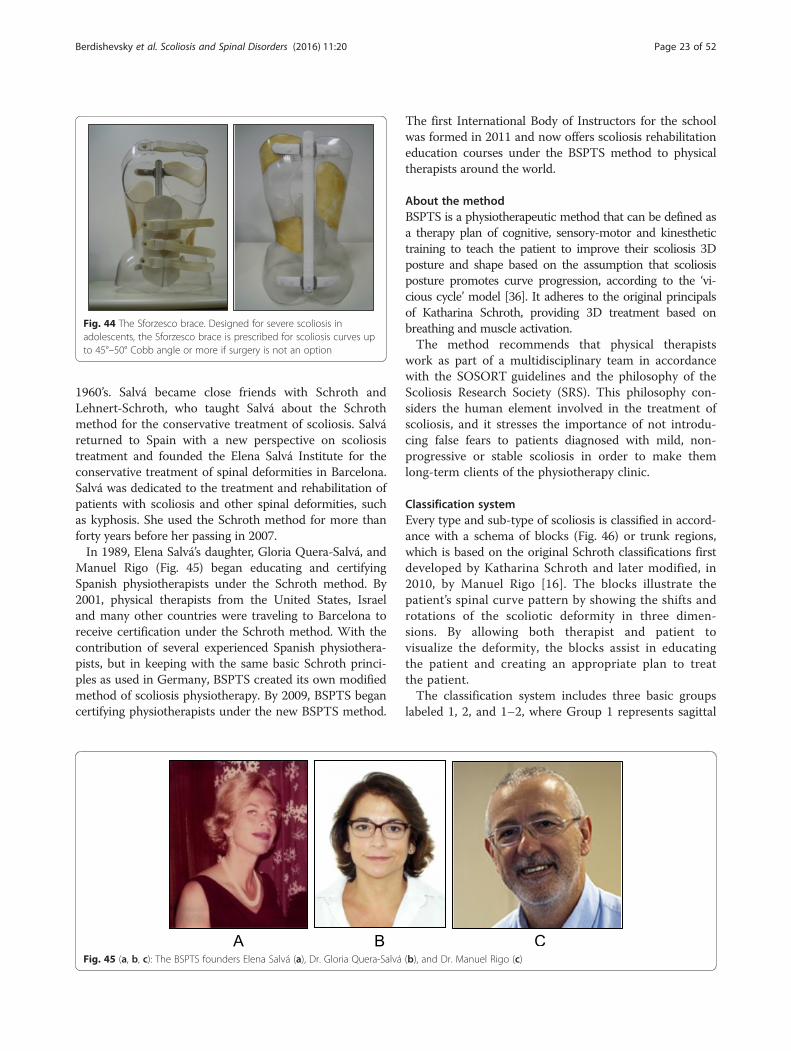
Manuel Rigo (Fig. 45) began educating and certifyingSpanish physiotherapists under the Schroth method. By2001, physical therapists from the United States, Israeland many other countries were traveling to Barcelona toreceive certification under the Schroth method. With thecontribution of several experienced Spanish physiothera-pists, but in keeping with the same basic Schroth princi-ples as used in Germany, BSPTS created its own modifiedmethod of scoliosis physiotherapy. By 2009, BSPTS begancertifying physiotherapists under the new BSPTS method.
The first International Body of Instructors for the schoolwas formed in 2011 and now offers scoliosis rehabilitationeducation courses under the BSPTS method to physicaltherapists around the world.
About the methodBSPTS is a physiotherapeutic method that can be defined asa therapy plan of cognitive, sensory-motor and kinesthetictraining to teach the patient to improve their scoliosis 3Dposture and shape based on the assumption that scoliosisposture promotes curve progression, according to the ‘vi-cious cycle’ model [36]. It adheres to the original principalsof Katharina Schroth, providing 3D treatment based onbreathing and muscle activation.The method recommends that physical therapists
work as part of a multidisciplinary team in accordancewith the SOSORT guidelines and the philosophy of theScoliosis Research Society (SRS). This philosophy con-siders the human element involved in the treatment ofscoliosis, and it stresses the importance of not introdu-cing false fears to patients diagnosed with mild, non-progressive or stable scoliosis in order to make themlong-term clients of the physiotherapy clinic.
Classification systemEvery type and sub-type of scoliosis is classified in accord-ance with a schema of blocks (Fig. 46) or trunk regions,which is based on the original Schroth classifications firstdeveloped by Katharina Schroth and later modified, in2010, by Manuel Rigo [16]. The blocks illustrate thepatient’s spinal curve pattern by showing the shifts androtations of the scoliotic deformity in three dimen-sions. By allowing both therapist and patient tovisualize the deformity, the blocks assist in educatingthe patient and creating an appropriate plan to treatthe patient.The classification system includes three basic groups
labeled 1, 2, and 1–2, where Group 1 represents sagittal
Fig. 44 The Sforzesco brace. Designed for severe scoliosis inadolescents, the Sforzesco brace is prescribed for scoliosis curves upto 45°–50° Cobb angle or more if surgery is not an option
Fig. 45 (a, b, c): The BSPTS founders Elena Salvá (a), Dr. Gloria Quera-Salvá (b), and Dr. Manuel Rigo (c)
Berdishevsky et al. Scoliosis and Spinal Disorders (2016) 11:20 Page 23 of 52

deformities and Group 2 and Group 1–2 represent scoli-osis, and here is their description:
1) Group 1 describes sagittal deformities such ashyperkyphosis (mainly due to Scheuermann’sKyphosis), inverted back (hypokyphosis), and flatback.
2) Group 2 defines a structural scoliosis in the mainthoracic region, with no lumbar curve or combinedwith a minor functional or minor structural ormajor structural lumbar or thoracolumbar curve.Group 2 can be subdivided into three differentpatterns: 3 Curves, 4 Curves and non 3–non 4.a. Three-curve scoliosis pattern (3C) means a
major thoracic curvature with a majorstructural lumbar curvature that is combinedwith the pelvis. The lumbar spine and the pelvisfunction as one unit in the schema of bodyblocks, and will shift and rotate to the oppositeside of the thoracic curvature.
b. Four-curve scoliosis pattern (4C) is a majorlumbar curvature with a compensatory thoraciccurvature and a pelvis that shifts and rotates tothe opposite side of the lumbar curvature.
c. Non 3-non 4 (N3N4) is defined by a majorthoracic curvature with or without a lumbar
curvature with a pelvis that is not shifted and notrotated, i.e. one that is balanced in the center.
3) Group 1-2 defines a lumbar or thoracolumbar curvewith a rectilinear thoracic spine.
Rigo’s Radiological Classification System [16] uses object-ive radiological criteria to confirm the functional curve type(Fig. 47). This current classification system was developedspecifically by Dr. Rigo in 2010 to correlate with brace de-sign and physiotherapy [16]. Patients must be classified as3C, 4C, N3N4 (Group 2) or single lumbar/TL (Group 1–2),as described above, based on clinical observation. Later theradiological criteria are used to confirm the initial clinicaldiagnosis. From a clinical perspective, Group 2 correlates toA, B and C types, respectively, in the radiological classifica-tion. A, B and C types can be at the same time subdividedas A1, A2, A3, B1, B2, C1 and C2. Group 1–2 correlates toE type (E1 and E2) in the radiological classification. Thepresence of a structural curve in the proximal thoracic re-gion is defined as ‘D modifier’.
Treatment indicationsIndications for treatment are outlined in the SOSORTguidelines [9] and focus primarily on the conservative treat-ment available to prevent curve progression. The BSPTSmethod is designed specifically for physiotherapists. The
A B C DA B C DFig. 46 (a, b, c, d): The BSPTS system of scoliosis curve classification illustrated with photographs and body block diagrams. The four scoliosiscurve types in this classification system are 3C (a), 4C (b), N3N4 (c), and single lumbar or thoracolumbar (d). The 3C curve is a major thoracicscoliosis curve with a compensatory lumbar and pelvic shift (a). The 4C curve is a major lumbar scoliosis curve with a thoracic and lumbar shift(b). The N3N4 curve is a major thoracic scoliosis with or without a lumbar curve but with the pelvis in a neutral position (c). The single lumbar orthoracolumbar curve is a single curve scoliosis with an uncoupled pelvic shift and no thoracic curvature (d)
Berdishevsky et al. Scoliosis and Spinal Disorders (2016) 11:20 Page 24 of 52

physiotherapist requires extensive training and many clin-ical years of experience in order to perfect the BSPTSmethod. There are some elements of the BSPTS methodthat may benefit patients with other spinal deformities, butthe BSPTS approach has been used primarily for idiopathicscoliosis (late JIS and AIS). Other types of scoliosis may betreated with modified principles. Sagittal plane deformitiessuch as hyper-kyphosis (Scheuermann’s kyphosis) and lor-dosis (inverted back) can also be treated with Schroth exer-cises. A modified Schroth program is used to treat painfuldegenerative adult scoliosis. BSPTS principles, but not thefull active plan of exercises used classically for adolescentsor adults, can be used in early onset scoliosis.
GoalsThe goals of the BSTPS method are to 1) correct the‘scoliotic posture’ (Fig. 48) and improve aesthetics, 2)stabilize the spine and arrest the curve progression, 3)educate patients and families about the condition andtreatment options, 4) improve breathing function, 5) in-crease activity, including activities of daily living andfunctional mobility, 6) improve overall self-image andself-esteem, and 7) decrease pain. The higher the risk ofcurve progression, the more intense the conservativetreatment plan should be in order to meet the goals oftherapy. However, this objective should not delay therecommendation for bracing or surgery when indicated.
BSPTS is not an alternative to or substitution for bracingor surgery and has its own indications.
3D Principles of correctionBSPTS principles of correction are based on the ori-ginal principles described by Katharina Schroth [14].The treatment is individualized depending on the curvetype (described in About the method above) and isdone only after the individual has achieved their bestglobal postural alignment by organizing the lower ex-tremities, the pelvis and the trunk in the best possibleposture. The principles of correction follow the globalpostural alignment and are applied with high intensityforces created inside the body (‘from inside’) involvingisometric tensions, expansions and specific breathing.The end result (Fig. 49) is a corrected posture wherethe collapsed areas of the trunk (the concavities) areopen and expanded and the prominences (the convex-ities) are contained.The following is a detailed description of the principles:
1. 3D postural correction is made through movementsof translation, rotation and mixed (sagittalexpansions). The correction follows a schema ofblocks, which is based on the classification offunctional types first developed by Schroth and latermodified by Rigo. The blocks are deformed,translated and rotated in accordance with the
Fig. 47 Rigo classification for BSPTS bracing and physical therapy
Berdishevsky et al. Scoliosis and Spinal Disorders (2016) 11:20 Page 25 of 52

spinal curve pattern, and the 3D posturalcorrection is referred not only to a combined butto a real synchronized correction of the position(translation and rotation) and shape (deformity) ofall the blocks. Thus, the applied principles ofcorrection can be described as deflection,derotation and sagittal normalization.
2. The expansion/contraction technique (Fig. 50) is usedto achieve the ‘best possible correction.’ It facilitates theso-called ‘corrective breathing.’ The best possiblecorrection is only possible, at the beginning of thetherapy, with the help of some external aids,including passive and passive-active manual aidsoffered by the physical therapist. Expansion/contraction technique is about expanding any partof the trunk in any direction ‘from inside,’ by usingonly the muscle force (independent of breathingmovements). The expansion can be side to side orone side against a fixed point. Only the collapsedareas of the trunk will be expanded, while theprominences will be contracted. This techniquefacilitates the later introduction of ‘rotatory correctivebreathing.’ The overall goal is not only to expand andbreathe into the collapses or concavities, but to do itin a corrective direction according to well-describedbiomechanical rules.
3. Stabilization by muscle tension. Once the bestpossible correction has been achieved in any specificstarting position (the starting positions can vary inaccordance with the above described functionaltypes), the subject will be asked to produce muscletension in order to maintain the correction. Thus,muscle tension can be defined as isometric tension.The maintenance of the correction during this partof the therapy, by creating muscle tension, willproduce an isometric eccentric contraction of thepreviously shortened muscles and concentriccontraction of the previously over-elongated muscles.Before creating the tension, the muscle balance has
Fig. 49 Patient with a major left lumbar-thoracolumbar scoliosis curvewith a right pelvic shift performs a two-pole standing exercise applyingthe BSPTS principles of correction 1–5. Light brown arrows representbilateral shoulder traction, which is required for stabilization duringactive self-correction. Light blue arrows represent bilateral shouldercounter-traction, which is required for midline spinal alignment.The light blue arrow pointing to the patient’s pelvis representspelvic correction from the right to the midline, which is required exercisewhen the patient has a major lumbar or thoracolumbar scoliosis curve
A B Fig. 48 (a, b): Active 3D self-correction exercises. During active 3D self-correction, patients expand the collapsed areas and open the concavitiesby performing rotational angular breathing (RAB) and specific arm positions (a). During the Schroth-derotation sitting exercise (b), the patient sitson a chair, with a pole in either hand planted on the ground, while performs corrections 1–5, while stabilizing her curve specific corrections. (48bprovided with the permission of Andrea Lebel, RPT, MCPA, Ottawa, Canada)
Berdishevsky et al. Scoliosis and Spinal Disorders (2016) 11:20 Page 26 of 52

been improved and the new achieved balance is notlost when creating this final muscle tension.
4. Integration. After the exercise, the subject is askedto relax while still maintaining the 3D postural self-correction. Eventually coming again into the bestpossible correction or going back into the badposture, the patient notes (proprioception) and sees(indirect by mirror or direct by camera-screen) thedifferences between scoliotic posture, 3D posturalself-correction and ‘best possible correction.’Repetition of the exercises and the integrativestrategies allow the subject to bring the correctioninto activities of daily living.
The use of breathing mechanics, muscle activation, andmobilizationThe success of the method is based on strengtheningexercises tailored to each individual scoliosis patient andtheir specific curve pattern. Unique rotational angularbreathing (RAB) exercises (Fig. 51) originally developedby Katharina Schroth [14] help in vertebral and rib cagederotation and in increasing vital capacity. This uniquebreathing technique helps expand the ribs from insidethe rib cage by pushing the ribs “sideways and back-wards” and helps return the vertebrae closer to theirnormal, untwisted position. Muscle activation of coremuscle groups, like the iliopsoas (Fig. 52), thoracic andlumbar fascicles of the erector spinae and quadratuslumborum, help stabilize and maintain the expanded ribsand derotated vertebral bodies. Encouraging mobilizationand flexibility (Fig. 53) helps to release tension and assists
in postural correction. Wall bars, pads, poles, belts, straps,mirrors, elastic-bands, dowels, balls, yoga blocks, stools,and foam rollers are equipment commonly used to assistSchroth exercises.The training period can vary and an individual can be
trained on a one-to-one basis or in a group setting. Thetherapy is always individualized and even in the groupsetting, every patient is treated individually by the phys-ical therapist. Only very experienced therapists can man-age individual treatment in a group session. There are
A B
Fig. 51 (a, b): Before (a) and during (b) rotation angular breathing(RAB). The arrows represent directional breathing used to fill thecollapsed lungs with air and reshape the thorax (b)
Fig. 52 Diagrammatic depiction of the activation of the iliopsoasmuscle in a lumbar scoliosis curve. The arrows show the direction ofactivation from the origin to the insertion points of the iliopsoas,promoting curve de-flexion and derotation towards the right
Fig. 50 Patient using postural correction and the correctiveexpansion/contention technique to achieve the best possiblecorrection. The blue and black arrows represent trunk expansion duringthe first principle of correction. Later, the blue arrows are convertedinto forces represented by the red shapes, which work around theprominences to move the prominences forward and inward
Berdishevsky et al. Scoliosis and Spinal Disorders (2016) 11:20 Page 27 of 52

two reasons to hold group sessions: first, the cost of thetherapy can be reduced, and second, and moreimportant, the environment created is one of grouptherapy and support. The number of hours necessary totrain the subject to do the exercises with efficacy andsafety varies, and depends also on the modality, privateor group. The groups are limited but this limitation de-pends also on the therapist’s experience and capability.Sixty hours in-group and 20 h in private is typicallyenough to achieve a high level technique, but after a fewhours of treatment (e.g. nine hours in group) the pa-tients can reproduce the correction in several startingpositions and can start practicing at home. The patientcan always improve the level of performance with thehelp of the therapist, but perfection is not required inorder to obtain positive results.
Description of the BSPTS exercisesOne of the important aspects of exercising in differentpositions is that the individual trunk areas are beingchallenged to work against or with gravity in differentpositions. The decision of which exercise position toutilize depends on the needs and goals of the patient,with a gravity-eliminated position to assist the patient inactivation of intended trunk musculature and an anti-gravity position to increase endurance and muscleactivation.Four of the most commonly used exercises in the
BSPTS method are Supine exercises (Fig. 54), Side-lyingexercises (Fig. 55), Prone on stool (Fig. 56) and theMuscle Cylinder (Fig. 57). The first three exercises canbe employed in all of the functional types of scoliosiscurve patterns. The ‘muscle cylinder’ exercise is forhighly trained patients and is used mainly in the majorlumbar (4C) pattern (although there is an old classicalversion for the major thoracic (3C) curve pattern).
Exercises in the supine position eliminate the force ofgravity on the spine so the patient can focus more onsmall corrections in posture with increased precision. Ex-ercises in the side-lying position are best for corrections inthe frontal plane. Additionally, the side-lying position isuseful to address a lumbar concavity by placing it upwardsand facilitating it against gravity. Exercises in the proneposition allow the collapsed areas in the back to workmore intensively as they are working against gravity whilethe prominences have the advantage of facing down andare in a gravity-eliminated position.The muscle cylinder exercise (Fig. 57) is an advanced
level exercise involving very high-level muscle activationagainst gravity. The entire program of exercises includesmany additional exercises, but their description falls out-side the scope of this paper. Other exercises related to theBSPTS method involve postural correction during activ-ities of daily living. These exercises focus on correctingthe scoliosis posture while sleeping, resting, sitting, orstanding, carrying a bag, bending, reaching, and exercisingin brace. During these activities, the spine is in a neutralposition but the patient focuses on consciously maintain-ing the correct posture.
Activities of daily livingThe BSPTS believes that it is an important part of its ap-proach to teach patients maintain good posture in allparts of their life. This includes learning how to sit,stand, sleep and move with better alignment and in aspecific way based on their unique curve patterns(Figs. 58 and 59). Regarding participation in sports, BSPTSfocuses on the entire child/patient and not only on thetreatment of scoliosis. BSPTS encourages patients to con-tinue living their lives and to pursue normal psychosocialgrowth and maturation. This may include a passion forsports, which should be allowed and even facilitated.
Fig. 53 Physical therapist, Dr. Hagit Berdishevsky, assisting a patient in mobilizing the collapsed ribs on the left concave side and expanding theribcage in an outwards and backwards direction
Berdishevsky et al. Scoliosis and Spinal Disorders (2016) 11:20 Page 28 of 52

Scientific evidenceBSPTS is based on the principles developed by KatharinaSchroth. The scientific evidence may be found in thesection describing the evidence for the Schroth method(Scientific evidence).
BSPTS bracingBSPTS principles are fully compatible with brace con-cepts such as the Rigo-Chêneau [37] brace type. The
Rigo-Chêneau brace (Fig. 60) is a rigid asymmetric cus-tom brace. The original technique was inspired by theplaster cast correction principles. The main objective isto bring the patient into the best possible 3D correctionsimilar to that utilized in a plaster cast, but in this casewithout any passive traction. A positive plaster moldfrom the patient is modified in order to define very se-lective contact areas and correspondent expansion areas.The pads for contact are designed to act at a properlevel and with a proper shape and orientation to movethe trunk into the best spinal correction. The correctiveprinciple is a combination of three-point systems, re-gional derotation and sagittal balance, and physiologicalprofile. The lack of compression/”sandwich” effect con-verts the passive rigid brace into a 4D dynamic brace.Breathing movements, and growth and development, arefacilitated in the corrective direction by the brace, sotime is essential as a corrective principle as well.This type of brace can potentially prevent the increase
in lordosis commonly observed in full-contact bracesworking through static pressures rather than correctivemovements. The principles of correction applied in theconstruction of this brace are based on the correctivemovements from the BSPTS principles. In Fig. 61 a cor-rection of scoliosis is observed on x-ray.
The Dobomed method (Poland)IntroductionDobosiewicz’s method for the treatment of scoliosis(Dobomed) is a conservative management approach foridiopathic scoliosis that addresses both trunk deformityand respiratory function impairment. The Dobomed ap-proach has incorporated both Klapp’s position for kypho-tization of the thoracic spine [38] and Lehnert-Schroth’sapproach for active asymmetrical breathing [14].
HistoryThe Dobomed method was developed in 1979 byPolish physiotherapist and physician Professor KrystynaDobosiewicz (1931–2007) (Fig. 62), and has been usedroutinely in Poland for the conservative treatment of scoli-osis since 1982. Prof. Dobosiewicz was very familiar withKlapp’s and Lehnert-Schroth’s methods. It was from thesebeginnings that Prof. Dobosiewicz started to create herown approach to the treatment of scoliosis. Initially, theDobomed method was tested on an outpatient group,continuously being improved and modified by Prof.Dobosiewicz before being adopted by the Departmentof Rehabilitation at the Medical University of Katowice,Poland, in 2000, as an intensive inpatient rehabilitationapproach for patients with scoliosis. Since the beginning,the Dobomed approach has been used either as a solephysiotherapy method or in combination with Chêneaubracing [37].
Concavities
Shoulder traction (ST)
Corrective pads
Convexities/curves shoulder counter-traction (SCT) (forward - inward)
Elongation
Fig. 54 Schroth supine exercise for patients with a major lumbarcurve. Turquoise arrows represent the cranial elongation and caudaltraction force. The Green half-moon represents the area of expansionof the concavity. Light blue arrows on the patient’s arms representbilateral shoulder traction, which is an isometric tension from theshoulder in a lateral/outward direction with a fixed scapula as acontinuation of the transversal expansion in the proximal thoracicregion. The arm position and muscle activation during bilateralshoulder traction can assist in active self-elongation and in preventingpostural collapse. Red arrows represent counter traction forces –contraction around the convexities/the curves in a forward andinward direction towards neutral spine
Berdishevsky et al. Scoliosis and Spinal Disorders (2016) 11:20 Page 29 of 52

Definition of treatmentThe Dobomed approach is a biodynamic method of 3Dauto-correction based on the pathomechanics of idio-pathic scoliosis. The basic technique of the Dobomedapproach is active 3D correction involving mobilizationof the primary curve towards curve correction, with spe-cial emphasis on “kyphotization” of the thoracic spineand/or “lordotization” of the lumbar spine [39]. Thismobilization of the primary curve is performed in closedkinetic chains and developed upon a symmetrically posi-tioned pelvis and shoulder girdle. The pelvis and shoul-ders are positioned first and kept stable for the durationof the exercise and during the inspiration and expirationphases of active asymmetrical breathing (Fig. 63). Frontalplane correction occurs automatically as the sagittal andaxial planes are corrected. Lateral flexion of the spine isnot required for thoracic curves. This symmetrical posi-tioning of the pelvis and the shoulder girdle is somethingthat is unique to the Dobomed method.
Treatment indications, goals, and age specificsTreatment indications for the Dobomed method followthe 2011 SOSORT guidelines [9]. Small, moderate, andlarge scoliosis curves can all be treated with the Dobomedmethod, either with or without Chêneau bracing.
The goals of the Dobomed method are to stabilizeand correct the spinal deformity, and to prevent pro-gression and/or decrease the curvature of the scoliosis.Another aim of this method is to improve the overallfunctional status of the patient, particularly the re-spiratory function.Cooperation is the basic requirement in using the
Dobomed method. Therefore, Dobomed is not recom-mended for small children who are unable to understandand perform the exercises. For older patients, the focus ison stabilization exercises rather than active 3D correction.
Classification systemThe Dobomed method uses its own classification systemfor scoliosis and scoliosis treatment. Each scoliosis patienthas his/her own unique curve pattern and is evaluatedseparately. A patient’s personalized exercise program de-pends on the number of primary and secondary curvesand the location of these curves.
3D Principles of correctionThe following are the principle distinctive features of theDobomed method:
1. Symmetrical positions for exercising.
Fig. 55 The Schroth “Side-lying” exercise for major lumbar curves (top) and major thoracic curves (bottom). During this exercise, the patient lieson the lumbar convex side. Light blue arrows represent trunk elongation with cranial and caudal forces. Green half-moons represent areas ofexpansion of the concavities. Red arrows represent areas of muscle activation, approximating the convexities towards midline, and the directionof the correction to correct the convexities. The dark blue arrow pointing upwards from the right elbow represents the shoulder traction
Berdishevsky et al. Scoliosis and Spinal Disorders (2016) 11:20 Page 30 of 52

2. Asymmetrical active movements to accomplish 3Dscoliosis correction.
3. Thoracic spine mobilization to increase thoracicflexion.
4. Transverse plane derotation, with specific treatmentemphasis focused on the area of the curve apex.
5. Concave rib mobilization to expand and derotatethe ribs.
6. External facilitation.7. Directed movements of the thorax and spine to
improve respiratory function.8. 3D displacement of vertebrae to obtain 3D scoliosis
correction.
The use of breathing mechanics, muscle activation, andmobilizationThe Dobomed method uses the ‘phased-lock’ respirationtechnique to assist in spinal correction and stabilization.
During ‘phased-lock’ respiration [40] (Figs. 64 and 65), astrong local pressure is applied on the concave side duringinspiration while a subtle facilitation is applied on the con-vex side during expiration. During expiration, isometriccontraction of the trunk muscles helps to stabilize the cor-rection or hypercorrection. Additionally, activation of theinternal intercostal muscles on the convex side duringboth inspiratory and expiratory phases of breathing allowsfor asymmetric breathing which results in the ribs comingcloser together on the convex side, and mobilization andderotation of the spine.
Treatment tools: active and passiveThe Dobomed method employs the use of mirrors, pho-tographs, and video to assist with scoliosis treatmentexercises.
Description of the most relevant exercise mechanicsThe Dobomed method consists of three parts:
1. Forward bending phase: Active 3D self-correction ofthe spine and ribcage during forward bending is anoriginal component and the main corrective techniqueof the Dobomed method. Forward bending exercisesare designed in closed kinetic chains in order toenhance their effectiveness. Forward bending isdone with strict fixation of the pelvis and theshoulder girdle with the upper and lower limbs.This bending position is thought to facilitate activeself-correction between two symmetrical andstable zones, and helps to consolidate the correctpostural habit beyond the therapeutic session.
Fig. 56 The Schroth prone exercise. The leg of the lumbar convexside (left leg) is abducted and the pelvis is supported and elevatedby a footstool. The lower abdomen is supported by a roll, as is theright shoulder, to facilitate trunk stabilization during the exercise.Blue arrows on the shoulders represent bilateral shoulder traction.Turquoise arrows represent trunk elongation with cranial elongationand caudal traction forces. Red arrows represent areas of muscleactivation around the convexities towards the midline. Greenhalf-moons represent areas of expansion of the concavities
Fig. 57 The “Muscle-Cylinder” exercise for major lumbar curves inpatients with moderate-severe scoliosis (as seen in the radiograph)helps patients achieve spinal alignment and a corrected posture
Berdishevsky et al. Scoliosis and Spinal Disorders (2016) 11:20 Page 31 of 52
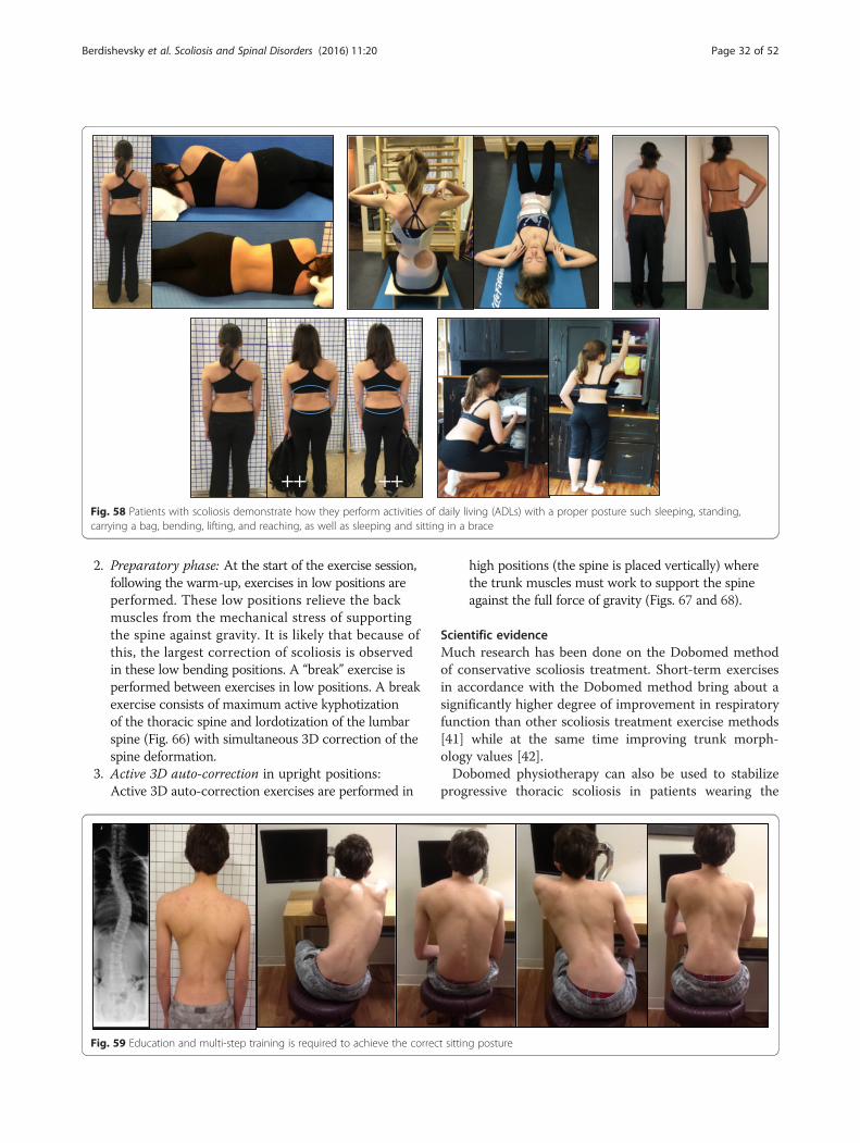
2. Preparatory phase: At the start of the exercise session,following the warm-up, exercises in low positions areperformed. These low positions relieve the backmuscles from the mechanical stress of supportingthe spine against gravity. It is likely that because ofthis, the largest correction of scoliosis is observedin these low bending positions. A “break” exercise isperformed between exercises in low positions. A breakexercise consists of maximum active kyphotizationof the thoracic spine and lordotization of the lumbarspine (Fig. 66) with simultaneous 3D correction of thespine deformation.
3. Active 3D auto-correction in upright positions:Active 3D auto-correction exercises are performed in
high positions (the spine is placed vertically) wherethe trunk muscles must work to support the spineagainst the full force of gravity (Figs. 67 and 68).
Scientific evidenceMuch research has been done on the Dobomed methodof conservative scoliosis treatment. Short-term exercisesin accordance with the Dobomed method bring about asignificantly higher degree of improvement in respiratoryfunction than other scoliosis treatment exercise methods[41] while at the same time improving trunk morph-ology values [42].Dobomed physiotherapy can also be used to stabilize
progressive thoracic scoliosis in patients wearing the
Fig. 58 Patients with scoliosis demonstrate how they perform activities of daily living (ADLs) with a proper posture such sleeping, standing,carrying a bag, bending, lifting, and reaching, as well as sleeping and sitting in a brace
Fig. 59 Education and multi-step training is required to achieve the correct sitting posture
Berdishevsky et al. Scoliosis and Spinal Disorders (2016) 11:20 Page 32 of 52

Chêneau brace [37, 43]. Stabilization of scoliosis curvesin children with the Dobomed method can also be seenradiographically [39, 44]. Asymmetric mobilization ofthe trunk in strictly symmetrical positions, as a part ofthe Dobomed method, has been shown to decreaseCobb angles and vertebral rotation [39], or, at minimum,
halt scoliosis progression [43, 45, 46]. Moreover, asym-metrical mobilization significantly rebuilds physiologicalthoracic kyphosis in cases of idiopathic scoliosis accompan-ied by a straight back [44]. Additionally, conservative treat-ment of idiopathic scoliosis using the Dobomed method ofintensive 3D respiratory exercises helps preserve normal
Fig. 60 Different views of the Rigo-Chêneau brace. From left to right: later view of the thoracic concave side, frontal, posterior, and lateral view ofthe thoracic convex side
Fig. 61 (a, b, c): Series of radiographs of a patient with progressive scoliosis treated with the Rigo-Chêneau brace. a The initial radiograph showsa thoracic scoliosis curve of 38° Cobb. b In-brace radiograph shows a greater than 50 % scoliosis curve correction. c Out-of-brace radiograph atRisser 4 (end of growth) shows that the thoracic curve has been decreased significantly to 24° Cobb angle, a curve reduction of >35 % comparedto the initial radiograph
Berdishevsky et al. Scoliosis and Spinal Disorders (2016) 11:20 Page 33 of 52

exercise efficiency of patients with idiopathic scoliosis asmeasured by the ventilatory anaerobic threshold [41].
Side shift (United Kingdom)IntroductionThe Side Shift approach to conservative idiopathic scoli-osis treatment, led by Tony Betts (Fig. 69), a physiother-apist who specializes in spinal deformities at the RoyalNational Orthopaedic Hospital in London, is built uponthe theory that a flexible curve can be stabilized withlateral movements. Excessive side movements of the
trunk correct the lateral deviation of the trunk alongthe coronal plane. These lateral movements promote areduction in postural forces, which aim to affect the de-velopment of the structural curve.
HistoryThe Side Shift approach to correct scoliosis curves wasdeveloped by Dr. Min Mehta in 1984 and has beenused by therapists at the Royal National OrthopaedicHospital since that time. Dr. Mehta initially used thisapproach to treat congenital scoliosis curves in chil-dren; she proposed that growth could be a correctiveforce for spinal deformity in children [47]. Through re-petitive actions the direction of growth opposes thecurve of the spine. While correcting the curve positionthrough trunk shifts, the body is using muscular forcesand connective tissue stretches to increase mobilityand re-align the soft tissue components of the scoli-osis. It is believed that frequent repetition of correctivemovements also helps to promote somatosensory inte-gration of the spinal position to a more upright andphysiological posture.
Classification systemThe consultants at the Royal National Orthopaedic Hos-pital use the King and Lenke systems for surgical classi-fication of scoliosis curves. The Side Shift treatmentmethod of scoliosis uses the King classification as astarting point for the descriptive classification of thecurve types and diagnosis of the area of the curve. Aclassification system for Side Shift has been developed[48] which examines the mobility of the spine and itsability to correct towards the midline (schematic dia-gram X classification). According to this classification
Fig. 62 Dr. Krystyna Dobosiewicz (1931–2007), founder of the DoboMedtreatment method of scoliosis
A BFig. 63 (a, b): Patient performing typical DoboMed method exercises before the application of thoracic kyphosis (a) and with thoracic kyphosis(b). Thoracic kyphosis is obtained by fixation of the pelvis and shoulder girdle using the upper and lower limbs
Berdishevsky et al. Scoliosis and Spinal Disorders (2016) 11:20 Page 34 of 52

system, there are three types of scoliosis curves withclassifications based on the flexibility of the curve andthe ability of the patient to auto-correct the deformedspine during a side shift movement. Type I is any curvepattern that can be corrected by shifting the trunk be-yond the coronal midline to the contralateral side of thescoliosis curve (very flexible curve). Type II is any curvepattern that can be corrected to the midline of the coronal
plane, aligning the spine with the pelvis, with partial dero-tation of the vertebrae (moderately flexible curves). TypeIII is any curve pattern that cannot correct to the midline,remains shifted during the side shift exercise, and wherethe vertebrae do not de-rotate (extremely rigid curves,which may represent a severe structural curve).
Treatment indications and goalsThe Side Shift approach includes principles of the Schrothmethod and treatment indications as outlined in the 2011SOSORT guidelines [9]. The goal of the Side Shift methodof scoliosis treatment is the active correction of the spinalcurve directed at the apex of the scoliosis, with Side Shiftmovements of the trunk towards the concavity, includ-ing active postural corrections in all planes. The start-ing point of analysis is the coronal plane (Cobb angle)curvature with emphasis towards the apex of the curvein this plane [49]. Core stabilization of the spine is alsoimportant through isometric lower abdominal, gluteal,and scapulae strengthening exercises that are all includedin the treatment program. The main aim of the exercisesfor AIS is the correction of postural deviation from themidline in pre- or post-operative patients. In adults, themain aim is the reduction of mechanical pain caused byexcessive postural deviations of the spine from the neutralmidline. In adults, the sagittal plane is sometimes consid-ered the important starting place for correction dependingupon the radiological findings. Breathing exercises are
Fig. 64 ‘Phased-lock’ respiration exercise showing in nine sequential photographs the complete filling of the collapsed lung on the left thoracicconcave side. Local subtle pressure applied with a finger facilitates lung expansion
Fig. 65 A diagrammatic depiction (right) of rotation angular breathing(RAB) exercises with the arrows showing the corrective direction of theribcage is demonstrated by the patient (left) as she expands the lungon the concave side with the goal of reshaping the thorax
Berdishevsky et al. Scoliosis and Spinal Disorders (2016) 11:20 Page 35 of 52
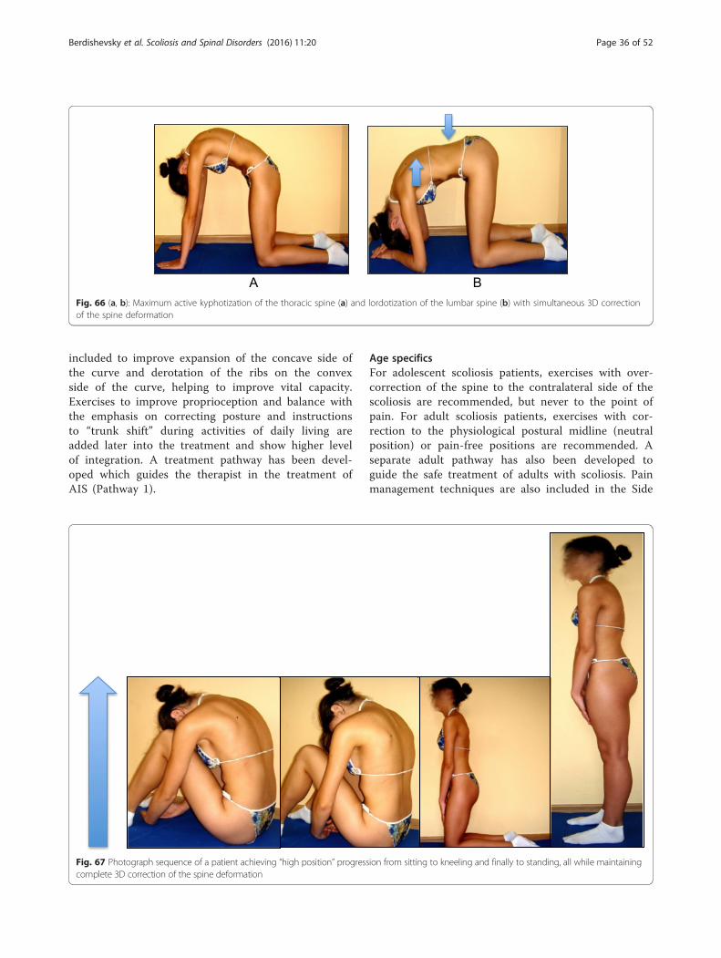
included to improve expansion of the concave side ofthe curve and derotation of the ribs on the convexside of the curve, helping to improve vital capacity.Exercises to improve proprioception and balance withthe emphasis on correcting posture and instructionsto “trunk shift” during activities of daily living areadded later into the treatment and show higher levelof integration. A treatment pathway has been devel-oped which guides the therapist in the treatment ofAIS (Pathway 1).
Age specificsFor adolescent scoliosis patients, exercises with over-correction of the spine to the contralateral side of thescoliosis are recommended, but never to the point ofpain. For adult scoliosis patients, exercises with cor-rection to the physiological postural midline (neutralposition) or pain-free positions are recommended. Aseparate adult pathway has also been developed toguide the safe treatment of adults with scoliosis. Painmanagement techniques are also included in the Side
A BFig. 66 (a, b): Maximum active kyphotization of the thoracic spine (a) and lordotization of the lumbar spine (b) with simultaneous 3D correctionof the spine deformation
Fig. 67 Photograph sequence of a patient achieving “high position” progression from sitting to kneeling and finally to standing, all while maintainingcomplete 3D correction of the spine deformation
Berdishevsky et al. Scoliosis and Spinal Disorders (2016) 11:20 Page 36 of 52

Shift method, including acupuncture, assisted bracingfor support, medication and transcutaneous electricalnerve stimulation (TENS).
3D principles of correctionAIS Treatment Pathway:In designing a treatment protocol the therapist has to
answer these questions:
1. What is the most important curve? (for example,Cobb angle and the amount of rotation)
2. Is shift/translation necessary? (Measure: Riboverhanging – trunk frontal plumb-line)
3. Is elongation necessary? (Measure: plumb-line sagit-tal – slumped posture)
4. Is derotation necessary? (Measure: rib hump/ATR,breathing/twisting)
5. Is lordosing/kyphosing necessary? (Measure: sagittalplane alignment)
6. What is the easiest movement to perform/remember?(Only do corrective movements at home that areaccurately/easily reproduced at clinic)
The Side Shift approach has been modified withpractice, experience, and clinical re-evaluation. It in-cludes several principles from the Schroth method,including active 3D auto-correction (along transverse,frontal, and sagittal planes), overcorrection move-ments beyond the midline, side shifting of the trunkin the direction opposite to the convexity of the pri-mary curve, and repetition of corrective movementsduring growth to influence the direction of spinalgrowth. Moreover, the patient must be old enough tounderstand instructions and perform the exercisesindependently.Two principle exercises of the Side Shift method in-
clude the hitch exercise and the hitch-shift exercise(Figs. 70 and 71). The hitch exercise is used for lumbar
Fig. 68 A summary of the different symmetrical positions of the DoboMed method
Berdishevsky et al. Scoliosis and Spinal Disorders (2016) 11:20 Page 37 of 52

or thoracolumbar curves, whereas the hitch-shift exer-cise is an option for double major curves. During thehitch exercise, in the standing position, the patient isinstructed to lift their heel on the convex side of thecurve while keeping their hip and knee straight and tohold the “hitch position” for ten seconds before return-ing to a neutral position. The patient is required to re-peat this exercise at least 30 times a day. In the hitchposition, the pelvis on the convex side of the curve islifted, resulting in a decreased lateral tilt at the inferioraspect of the vertebrae. This corrects the curve and re-duces the asymmetry of the indented waistline. Duringthe hitch-shift exercise, in the standing position, the pa-tient is instructed to lift their heel on the convex side ofthe lower curve, as done in the hitch exercise, while im-mobilizing the lower curve with their hand and shiftingtheir trunk to the concavity of the upper curve. The pa-tient is required to hold this position for ten seconds be-fore returning to a neutral position and to repeat thisexercise at least 30 times a day.
The use of breathing mechanics, muscle activation, andmobilizationThe Side Shift approach uses the same breathingmechanics as the Schroth (Section 2) and DoboMed
Fig. 69 Tony Betts, school leader and physical therapist teachingthe Side-Shift method (UK)
A B CFig. 70 (a, b, c): “Hitch” exercise. A patient with a left thoracolumbar scoliosis curve seen on radiograph (a) stands in a neutral position (b). She isinstructed to transition into the “hitch” position (c) by lifting her left heel on the same side as the convexity of the curve while keeping her hipand knee straight. The “hitch” position reduces the asymmetry of the patient’s waistline (c)
Berdishevsky et al. Scoliosis and Spinal Disorders (2016) 11:20 Page 38 of 52

(Section 5) methods, based on the principles of rota-tional angular breathing that targets breathing into theconcavity of the ribs. Muscle activation (Fig. 72) isachieved with isometric muscle bracing (via plank or‘bird-dog’) to provide dynamic correction to the SideShift corrective movement (incorporating Pilates andcore strengthening exercises). Active muscle controlhelps to prevent muscle atrophy and provides greaterforces to the corrective movements of the Side Shiftmethod. The Side Shift method also includes the princi-ples of Maitland and myofascial release techniques toincrease mobilization and flexibility of the joint tissuesand the soft tissues, respectively (Fig. 73).
Treatment tools: active and passiveThe Side Shift approach uses a combination of mirrors,photographs, and videos to assist patients with correc-tion during exercises.
Description of the most relevant exercise mechanicsSide Shift exercises can be performed either in thesitting or standing position and either with or with-out a brace (Figs. 74 and 75). The patient actively“shifts” their trunk away from the convexity of thecurve, while either sitting or standing, and holdsthe position for ten seconds. A more advanced ex-ercise involves maintaining the side shift positionaway from the convexity of the curve while goingfrom a seated to a standing position, and later onunsteady surfaces to challenge the balance and theproprioceptive system (Fig. 76). This exercise en-courages transitional postural control during every-day movements.
Activities of daily living and sportThe Side Shift approach encourages the mantra of “thinkshift” with activities of daily living and incorporates
Fig. 71 (a, b, c, d): The “Hitch-Shift” exercise is indicated for patients with double scoliosis curves. A patient with a double scoliosis curve seen onradiograph (a) stands in a neutral position (b). She is instructed to transition into the “hitch” position (c) by lifting her left heel on the same sideas the convexity of the lumbar curve while keeping her hip and knee straight. She then immobilizes the lumbar curve using her hand and “shifts”her trunk to the concavity of the thoracic curve (d)
Berdishevsky et al. Scoliosis and Spinal Disorders (2016) 11:20 Page 39 of 52

side shift exercises into everyday movements, such asgoing from a sitting position to a standing position(Fig. 77).
Scientific evidenceDr. Min Mehta [47] first described the Side Shift ex-ercise for the treatment of scoliosis in 1985. Dr.Mehta used single and multiple case reports of 35 pa-tients with idiopathic scoliosis to demonstrate positiveclinical and radiological corrections of scoliosis withSide Shift exercises. This study was part of an un-blinded retrospective study presenting observationaland radiological results of over 2,530 patients. In2002 and 2008 two studies from Japan [50, 51] thatinvestigated the Side Shift method in 39 females withAIS concluded that Side Shift exercises and the hitchexercises are useful options for idiopathic scoliosis.
Functional individual therapy of scoliosis (Poland)IntroductionFunctional Individual Therapy of Scoliosis (FITS) wascreated in 2004 by Marianna Białek PT, Ph.D., andAndrzej M'hango PT, M.Sc., D.O. (Fig. 78a-b). FITStreatment [52] is based upon the inclusion of many ele-ments selected from a variety of other therapeutic ap-proaches that have been adapted and modified to form adifferent scoliosis treatment concept. The FITS methodalso contains many techniques developed by the schoolleaders. FITS may be used as a separate system forscoliosis correction, as a supportive therapy for bracing,in preparation of children for surgery, or for the correc-tion of the shoulder and pelvic girdles after surgicalintervention [52]. The FITS method may also beapplied to other structural and non-structural spinaldeformities.
Fig. 72 Trunk stabilization exercises using the side-shift method. The “bird-dog” (left) and the plank (right) exercises are performed whilemaintaining the side shift position
Fig. 73 (a, b): Lateral (a) and sagittal (b) trunk mobilization exercises using the Side Shift method
Berdishevsky et al. Scoliosis and Spinal Disorders (2016) 11:20 Page 40 of 52

HistoryMarianna Białek and Andrzej M'hango collaboratedfor the first time in the late 1990’s during a scoliosismanual therapy training course. At that time, bothwere involved in various courses, training sessionsand conferences on the most recent physiotherapeuticmethods for the conservative treatment of scoliosis.Between 1999 and 2000, they prepared and conductedthe course “Proprioception Neuromuscular Facilitation(PNF) in Scoliosis” for physiotherapists. In 2002, they or-ganized the first two-week rehabilitation camp for chil-dren with scoliosis. Taking advantage of their experiencein treating young patients with scoliosis and the expert-ise of their colleagues, including Wieslaw Chwała of theDepartment of Biokinetics at AWF Kracow, who at thetime was carrying out EMG examination and a 3D gait
analysis in the Vicon system, they created their ownprogram of scoliosis therapy. Through 2006, FITS(Functional Individual Therapy of Scoliosis) continuedto evolve with the cooperation of Professor TomaszKotwicki from the Department of Pediatric Orthopedicsand Traumatology, University of Medical Sciences inPoznań, Poland.
Definition of treatmentFITS is a complex, asymmetrical and individual methodof treating patients with scoliosis. It is based on a num-ber of physiotherapeutic techniques from which thecreators of the method selected those treatment ap-proaches that they believed were most useful andthen adapted them as necessary. In addition, many ofthe techniques used in FITS were developed by the
Fig. 74 Patient with a right thoracic scoliosis curve demonstrates a sequence of Side Shift exercises with assistive correction in the standingposition. The arrows in the diagram on the right illustrate the corrective movement of the spine during the Side Shift exercise
A B C DFig. 75 (a, b, c, d): Patient with a left thoracolumbar scoliosis curve (a, b) performs a sequence of side shift exercises to the right while wearingher brace (c, d)
Berdishevsky et al. Scoliosis and Spinal Disorders (2016) 11:20 Page 41 of 52

creators of the method based on their own experienceas physiotherapists [53]. A description of the FITSmethod, its treatment indications and guidelines waspublished by Białek, M’hango [52, 54] in 2008 and2011.FITS can be used in a child of any age regardless of
the Cobb angle and has been proven to be significantlyeffective in a short period of time [55]. FITS requires achild to be active in the therapy process, which isguided by an experienced and specially trained therap-ist. FITS therapy may be conducted in an outpatientclinic or as a one to two-week in-patient treatmentcourse. FITS encourages physiotherapists to work
together with orthopedists and psychologists in treat-ing the patient.
Classification systemUnlike other methods of scoliosis treatment, the FITSmethod has no traditional classification system; rather,it relies on an individual approach. Each patient’sscoliosis is different with reference to the number ofcurves, the location of the curves, the degree ofcurvature and trunk rotation, the body’s compensation(structural or functional) for the deformity, the pos-ition of the scapulae and pelvis, the muscle tension,the setting of the sagittal plane, and the patient’sbreathing patterns, sense of stability, coordination,and psychological state. Based on the individual char-acteristics of the patient’s scoliosis, the scoliosis de-formity is described as low, moderate, or severe. Eachpatient is assigned an individualized treatment pro-gram based on their unique deformity.
Treatment indication and guidelinesIn line with other scoliosis treatment methods, theFITS indications for scoliosis treatment are based onthe 2011 SOSORT guidelines [9] with some modifica-tions. Observation is not recommended for JIS pa-tients. Rather, it is recommended that all JIS patientsreceive FITS therapy regardless of age or Cobb angle.If a JIS patient requires bracing, only rigid bracing isrecommended. Soft bracing is not part of the FITSindication for therapy in any age group. FITS recom-mends part-time rigid bracing for JIS curves 21°-25°degrees and full-time rigid bracing for JIS curvesgreater than 26°. For AIS patients, observation only
Fig. 76 (a, b, c, d, e): Patient with a right thoracic scoliosis curve demonstrates side shift balance stabilization exercises against a wall (a, b) and inthe standing position (c, d, e)
A B
Fig. 77 (a, b): Patient with a right thoracolumbar scoliosis curveperforms a side shift to the left in a sitting-to-standing position (a)and in the standing position (b) as part of side shift exercises whichcan be done as part of activities of daily living (ADLs)
Berdishevsky et al. Scoliosis and Spinal Disorders (2016) 11:20 Page 42 of 52

is recommended in curves up to 15°. Curves greaterthan 15° require FITS therapy regardless of the Cobbangle. For AIS curves greater than 30° in patients withRisser 0–2 (bone maturity), full-time rigid bracing isrecommended. Additionally, patients can participate inFITS’s Special Inpatient Rehabilitation program regardlessof Cobb angle, Risser sign, and age.
Treatment goals and age specificsFITS goals are divided into short-term goals and long-termgoals. The short-term goals of FITS include increased pa-tient awareness (psychological goal), improved shoulder
and pelvic girdle alignment (aesthetic goal), patient educa-tion of 3D breathing and improved function, myofascial re-lease, and teaching the correct shift. The long-term goals ofFITS include decreasing scoliosis, stabilizing the scoliosis(stop curve progression), and improving aesthetics andbody function in patients who do not undergo surgery orwho are post-operative patients.The nine main goals of the FITS concept:
1. Awareness of existing deformation of the spineand the trunk, and of the direction of the scoliosiscorrection.
2. Sensory-motor balance training (Fig. 79).3. Release of myofascial structures that limit three-plane
corrective movement (Fig. 80).4. Lumbo-pelvic stabilization (Fig. 81).
Fig. 78 The Functional Individual Therapy of Scoliosis (FITS) schoolleaders, Marianna Białek and Andrzej M'hango
Fig. 79 FITS method sensory-motor balance training. With a videocamera positioned behind her, the patient is able to see her postureon the screen in front of her in real-time while moving from thesitting to the standing position and make corrections according tothe instructions of the physical therapist
Fig. 80 Physical therapist demonstrating the release of myofascialstructures (relaxation of the hamstring muscles) that limit three-plane corrective movements
Fig. 81 Lumbo-pelvic stabilization. The physical therapist instructsthe patient to slowly extend her left hip while maintaining a stablelumbar spine and pelvis with the support of her left hand
Berdishevsky et al. Scoliosis and Spinal Disorders (2016) 11:20 Page 43 of 52
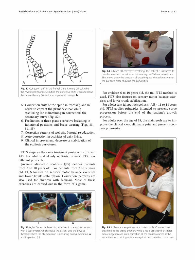
5. Correction shift of the spine in frontal plane inorder to correct the primary curve whilestabilizing (or maintaining in correction) thesecondary curve (Fig. 82).
6. Facilitation of three-plane corrective breathing infunctional positions and brace wearing (Figs. 83,84, 85).
7. Correction patterns of scoliosis. Postural re-education.8. Auto-correction in activities of daily living.9. Clinical improvement, decrease or stabilization of
the scoliosis curvatures.
FITS employs the same treatment protocol for JIS andAIS. For adult and elderly scoliosis patients FITS usesdifferent protocols.Juvenile idiopathic scoliosis (JIS) defines patients
from 3 to 10 years old. For patients from 3 to 5 yearsold, FITS focuses on sensory motor balance exercisesand lower trunk stabilization. Correction patterns arealso used for children with scoliosis. Most of theseexercises are carried out in the form of a game.
For children 6 to 10 years old, the full FITS method isused. FITS also focuses on sensory motor balance exer-cises and lower trunk stabilization.For adolescent idiopathic scoliosis (AIS), 11 to 18 years
old, FITS applies principles intended to prevent curveprogression before the end of the patient's growthprocess.For adults over the age of 18, the main goals are to im-
prove the clinical view, eliminate pain, and prevent scoli-osis progression.
Fig. 82 Correction shift in the frontal plane is more difficult whenthe myofascial structures limiting the corrective shift. Diagram showsthe before therapy (a), and after myofascial therapy (b)
Fig. 83 (a, b): Corrective breathing exercises in the supine positionwith a scoliometer, which shows the patient and the physicaltherapist where the rib expansion is occurring during expiration (a)and inspiration (b)
Fig. 84 In-brace 3D corrective breathing. The patient is instructed tobreathe into the concavities while wearing her Chêneau-style brace.The arrows show the direction of breathing and the red markings onthe patient’s brace showing the convexities
Fig. 85 A physical therapist assists a patient with 3D correctionalbreathing in the sitting position, while a red elastic band facilitatesauto-elongation and auto-correction of the scoliosis curves at thesame time as providing resistance against the corrective movements
Berdishevsky et al. Scoliosis and Spinal Disorders (2016) 11:20 Page 44 of 52

3D Principles of correction: The 3 StagesThe FITS concept consists of three main stages of cor-rection: patient examination and education, preparationfor correction, and three-plane correction.
Stage IPatient examination and education include physical exam-ination of the flexibility of the scoliotic spine in functional
A BFig. 86 (a, b): A physical therapist demonstrates how to test the flexibility of the scoliotic spine in the sitting (a) and standing (b) positions
Fig. 87 Physical therapist using radiographs and spine models to helpthe patient visualize and gain awareness of her trunk deformity causedby scoliosis
Fig. 88 FITS sensory-motor balance training. With a video camerapositioned behind her, the patient is able to see her posture on thescreen in front of her in real-time while making postural correctionson the balance board
Berdishevsky et al. Scoliosis and Spinal Disorders (2016) 11:20 Page 45 of 52
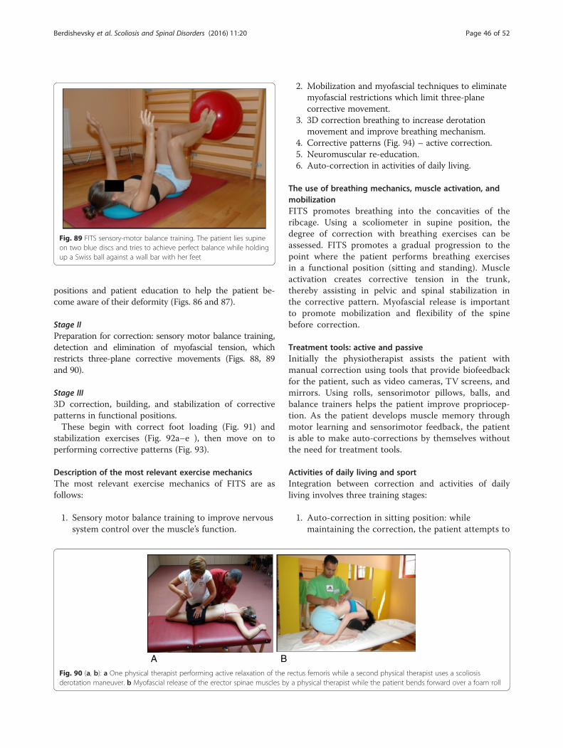
positions and patient education to help the patient be-come aware of their deformity (Figs. 86 and 87).
Stage IIPreparation for correction: sensory motor balance training,detection and elimination of myofascial tension, whichrestricts three-plane corrective movements (Figs. 88, 89and 90).
Stage III3D correction, building, and stabilization of correctivepatterns in functional positions.These begin with correct foot loading (Fig. 91) and
stabilization exercises (Fig. 92a–e ), then move on toperforming corrective patterns (Fig. 93).
Description of the most relevant exercise mechanicsThe most relevant exercise mechanics of FITS are asfollows:
1. Sensory motor balance training to improve nervoussystem control over the muscle’s function.
2. Mobilization and myofascial techniques to eliminatemyofascial restrictions which limit three-planecorrective movement.
3. 3D correction breathing to increase derotationmovement and improve breathing mechanism.
4. Corrective patterns (Fig. 94) – active correction.5. Neuromuscular re-education.6. Auto-correction in activities of daily living.
The use of breathing mechanics, muscle activation, andmobilizationFITS promotes breathing into the concavities of theribcage. Using a scoliometer in supine position, thedegree of correction with breathing exercises can beassessed. FITS promotes a gradual progression to thepoint where the patient performs breathing exercisesin a functional position (sitting and standing). Muscleactivation creates corrective tension in the trunk,thereby assisting in pelvic and spinal stabilization inthe corrective pattern. Myofascial release is importantto promote mobilization and flexibility of the spinebefore correction.
Treatment tools: active and passiveInitially the physiotherapist assists the patient withmanual correction using tools that provide biofeedbackfor the patient, such as video cameras, TV screens, andmirrors. Using rolls, sensorimotor pillows, balls, andbalance trainers helps the patient improve propriocep-tion. As the patient develops muscle memory throughmotor learning and sensorimotor feedback, the patientis able to make auto-corrections by themselves withoutthe need for treatment tools.
Activities of daily living and sportIntegration between correction and activities of dailyliving involves three training stages:
1. Auto-correction in sitting position: whilemaintaining the correction, the patient attempts to
Fig. 89 FITS sensory-motor balance training. The patient lies supineon two blue discs and tries to achieve perfect balance while holdingup a Swiss ball against a wall bar with her feet
A BFig. 90 (a, b): a One physical therapist performing active relaxation of the rectus femoris while a second physical therapist uses a scoliosisderotation maneuver. b Myofascial release of the erector spinae muscles by a physical therapist while the patient bends forward over a foam roll
Berdishevsky et al. Scoliosis and Spinal Disorders (2016) 11:20 Page 46 of 52
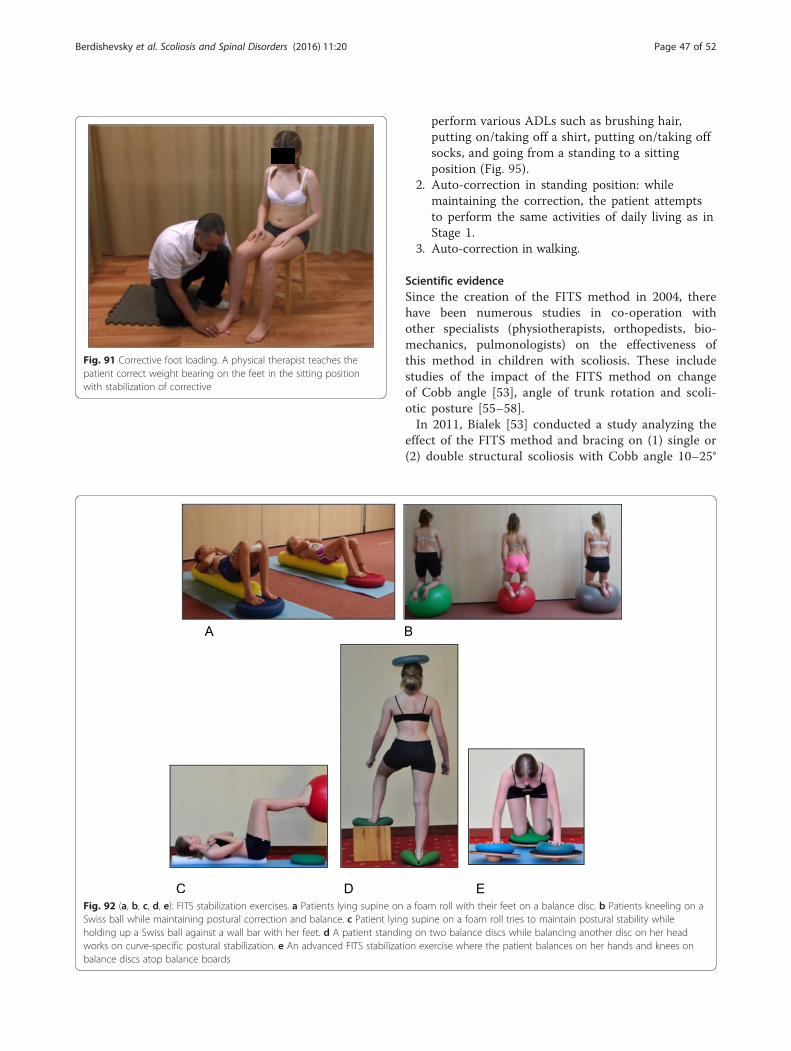
perform various ADLs such as brushing hair,putting on/taking off a shirt, putting on/taking offsocks, and going from a standing to a sittingposition (Fig. 95).
2. Auto-correction in standing position: whilemaintaining the correction, the patient attemptsto perform the same activities of daily living as inStage 1.
3. Auto-correction in walking.
Scientific evidenceSince the creation of the FITS method in 2004, therehave been numerous studies in co-operation withother specialists (physiotherapists, orthopedists, bio-mechanics, pulmonologists) on the effectiveness ofthis method in children with scoliosis. These includestudies of the impact of the FITS method on changeof Cobb angle [53], angle of trunk rotation and scoli-otic posture [55–58].In 2011, Bialek [53] conducted a study analyzing the
effect of the FITS method and bracing on (1) single or(2) double structural scoliosis with Cobb angle 10–25°
Fig. 91 Corrective foot loading. A physical therapist teaches thepatient correct weight bearing on the feet in the sitting positionwith stabilization of corrective
Fig. 92 (a, b, c, d, e): FITS stabilization exercises. a Patients lying supine on a foam roll with their feet on a balance disc. b Patients kneeling on aSwiss ball while maintaining postural correction and balance. c Patient lying supine on a foam roll tries to maintain postural stability whileholding up a Swiss ball against a wall bar with her feet. d A patient standing on two balance discs while balancing another disc on her headworks on curve-specific postural stabilization. e An advanced FITS stabilization exercise where the patient balances on her hands and knees onbalance discs atop balance boards
Berdishevsky et al. Scoliosis and Spinal Disorders (2016) 11:20 Page 47 of 52

Fig. 93 Patients demonstrating typical FITS exercises with elastic bands, which help facilitate the scoliosis specific correctional patterns. Initially,exercises are performed with the assistance of a physical therapist and then later are performed independently
Fig. 94 Patients demonstrating typical FITS exercises with elastic bands, which help facilitate the scoliosis specific correctional patterns
Berdishevsky et al. Scoliosis and Spinal Disorders (2016) 11:20 Page 48 of 52

(Group A) and on patients with Cobb between 20–45°(Group B). In Group A: (1) in single scoliosis, 50.0 %of patients improved, 46.2 % were stable and 3.8 %progressed, while (2) in double scoliosis, 50.0 % ofpatients improved, 30.8 % were stable and 19.2 %progressed. In group B: (1) in single scoliosis, 20.0 %of patients improved, 80.0 % were stable, and no pa-tient progressed, while (2) in double scoliosis, 28.1 %of patients improved, 46.9 % were stable and 25.0 %progressed. The study concluded that best resultswere obtained in 10–25° scoliosis, indicating that
therapy should begin where a patient shows a 10–25°curve in order to prevent greater structural changeswithin the spine.The study of the effectiveness of FITS Therapy in
Early Onset Idiopathic Scoliosis (range 4 to 9 yearsold) was published in Medicine [59]. In this study asignificant decrease of Cobb angle and angle oftrunk rotation was observed in close to 80 % of thesubjects.Regardless of the angle of curvature, studies demon-
strate that FITS therapy has a beneficial effect on theclinical appearance of each child (Fig. 96 and 97).
ConclusionSeven major schools operating under the SOSORT ban-ner focus on the treatment of adolescent idiopathicscoliosis. As can be seen by the therapeutic approach ofeach school, and the scientific evidence supporting eachschool’s method of treatment, each of the seven schoolspromotes a unique technique and unique exercises.However, the schools’ overall goals are the same, aseach method seeks to treat all aspects of the 3D scoliosisdeformity by realigning the spine, rib cage, shoulders andpelvis to ‘normal’ anatomical postures (see Table 1). Theevidence supporting the effectiveness of PSSE is growing,with more high quality research studies being published inrecent years. The research must continue in order to fur-ther study the effectiveness of the various schools and todetermine which methods and which exercises are mostbeneficial for patients.
A BFig. 95 (a, b): Posture correction in the sitting position, beforecorrection (a) and after active self-correction (b)
Fig. 96 A 12-year-old female with a left thoracolumbar scoliosis hasan improved physical appearance and aesthetic after being treatedwith bracing and the FITS method
Fig. 97 A 15-year-old female patient with a 32° Cobb angle left upperthoracic curve and a 36° Cobb angle right thoracolumbar curve as seenon the radiograph on the right, has an improved physical appearanceand aesthetic with FITS therapy
Berdishevsky et al. Scoliosis and Spinal Disorders (2016) 11:20 Page 49 of 52

Table 1 The similarities and the differences between the methods
School name Treatmentindication
Age treated Principles of correction Classification system Breathingtechnique
Mobilization andflexibility
Brace used Evidence
Lyon (France) SOSORT 2011guidelines
* JuvenileAdolescent** Adult
Focuses on physical therapyexercises in preparation forbrace wearing and in brace
PonsetiLenke
Rotational angularbreathing (RAB)
Encouraging thoracickyphosis and lumbarlordosis
3D ARTbrace(AsymmetricalRigid Torsion brace)
De Mauroy JC et al.Scoliosis. 2015;10:26
Schroth (Germany) SOSORT 2011guidelines
* Early onset* JuvenileAdolescent** Adult
Reshape the thorax throughisometric muscle activationaround the prominences(the convexities) and aspecific breathing technique(called rotational angularbreathing (RAB) or simplyorthopedic breathing) in thecollapsed areas (the concavities)
KatharinaSchroth’s BodyBlocks
Rotational angularbreathing (RAB)
Rib cage, spinemobilization andlower extremitiesflexibility
3D Chêneau brace RCT - Schreiber S et al.Scoliosis. 2015;10:24
Weiss H et al. Hard Tissue.2013; 2(3): 27RCT - Kuru T et al. ClinilRehabil. 2015Weiss HR et al. StudHealth Technol Inform.2002;91:342–7
Scientific ExerciseApproach toScoliosis (SES) (Italy)
SOSORT 2011guidelines
Same treatmentobjectives for allages
Educates and trains patientsto actively self-correct theirposture and to incorporatethat self-correction intofunctional exercises
Ponseti Rotational angularbreathing (RAB)
Pre-bracingmobilization
3D Sibilla brace(Cobb <30°)Sforzesco brace(Cobb 30°–50°)
RCT - Monticone M et al. EurSpine J. 2014;23(6):1204–14Negrini S et al. Disability andRehabilitation2008;30(10):772–785
Barcelona ScoliosisPhysical Therapy School(BSPTS) (Spain)
SOSORT 2011guidelines
* JuvenileAdolescent** Adult
Based on the original principlesof correction established byKatharina Schroth. Like theSchroth method, the school’saim to improve the scolioticposture via muscle activationand the RAB techniquementioned above.
KatharinaSchroth’s BodyBlocks andManuel Rigo’sradiologicalclassification
Rotational angularbreathing (RAB)
Rib cage, spineand lowerextremitiesflexibility
3D RigoChêneau brace
Schroth evidence above plus:Rigo M et al. Scoliosis 2010,5:1Otman SN et al. Saudi Med J.2005; 26(9): 1429–35
Dobomed (Poland) SOSORT 2011guidelines
Same treatmentobjectives for allages
Involves mobilization of theprimary curve toward curvecorrection with a specialemphasis on ‘kyphotization’of the thoracic spine and/or‘lordotization’ of the lumbarspine in closed kinetic chains
Dobomed Specific Rotationalangular breathingin a ‘phased-lock’respiration technique
Increase thoracicspine kyphosis andlumbar lordosis
3D Cheneaubrace
Durmala J et al. OrtopTraumatol Rehabil.2003;5(l):80–5Durmala J et al. Scoliosis.2009;4(Suppl 2):029
Side-Shift (UnitedKingdom)
SOSORT 2011guidelines
Adolescent** Adult
Built on the theory that aflexible curve can be stabilizedwith lateral movements andthat repetitive side movementsof the trunk will correct thelateral deviation of the trunkalong with the coronal plane
King Rotational angularbreathing (RAB)
Principles ofMaitland andmyofascial releasetechniques
Unspecified
Functional IndividualTherapy of Scoliosis(FITS) (Poland)
SOSORT 2011guidelines
JuvenileAdolescent** Adult
Based on a number ofphysiotherapeutic techniquesthat were selected and adaptedspecifically for the treatment ofscoliosis. Examples areProprioception NeuromuscularFacilitation (PNF) and myofascialrelease techniques
No traditionalclassificationsystem
3D correctivebreathing intothe concavities
Mobilization andmyofascialtechniques toeliminatemyofascialrestrictions
3D Cheneaubrace
Białek M. Scoliosis.2011;28;6(1):25Białek M. Medicine.2015;94(20):e863
* Exercises are modified to allow participation of young children** Exercises are modified and focus on reduction of pain
Berdishevskyet
al.Scoliosisand
SpinalDisorders
(2016) 11:20 Page
50of
52

AbbreviationsADL, activity of daily living; AIS, adolescent idiopathic scoliosis; ARTbrace,Asymmetrical Rigid Torsion brace; BSPTS, Barcelona Scoliosis Physical TherapySchool; CSL, central sacral line; CSN, Centro Scoliosi Negrini; EOS, early onset;FITS, Functional Individual Therapy of Scoliosis; ISICO, Instituto ScientificoItaliani Colonna Vertebrale; JIS, juvenile idiopathic scoliosis; LEV, lower endvertebra; N3N4, Non 3-Non 4 scoliosis pattern; PNF, proprioception neuromuscularfacilitation; PSSE, Physiotherapy Scoliosis Specific Exercises; QOL, quality of life; RAB,rotation angular breathing; RCT, randomized controlled trial; SCT, shouldercounter-traction; SEAS, Scientific Exercises Approach to Scoliosis; SOSORT,The Society of Scoliosis Orthopaedic Rehabilitation and Treatment; SRS,Scoliosis Research Society; SRS-22r, Scoliosis Research Society 22r questionnaire;ST, shoulder traction; TENS, transcutaneous electrical nerve stimulation; TL,thoracolumbar; UEV, upper end vertebra; 3D, three-dimensional; 3C, three-curvescoliosis pattern; 4C, four-curve scoliosis pattern
AcknowledgementsI thank Dr. Theodoros B. Grivas, co-Editor-in-Chief of the Scoliosis and SpinalDisorders journal, for encouraging me to turn a presentation into this manuscript,and for his constant support throughout the preparation of the manuscript. I alsothank Kelly Grimes - physical therapist and specialist in spine and Schroth therapy,and Patti Orthwein - Juvenile scoliosis, pediatric physical therapist and myco-teacher for the Schroth method at the BSPTS for reviewing the manuscriptand improving it with their wise and sensible suggestions. Finally, I want to thankthe patients whose photographs appear in the manuscript, and their parents.
Authors’ contributionsHB is the principal researcher and author of the manuscript. VL assisted in draftingthe manuscript. JBS assisted in drafting and editing the manuscript. MR assisted indrafting the section titled Barcelona Scoliosis Physical Therapy School and inediting the manuscript. AL assisted in drafting and editing the manuscript. AHassisted in drafting the section titled The Schroth Method. MR assisted in draftingthe section titled Scientific Exercise Approach to Scoliosis. MB and AM assisted indrafting the section titled Functional Individual Therapy of Scoliosis. TB assistedin drafting the section titled Side-Shift. JCM assisted in drafting the sectiontitled The Lyon Approach. JM assisted in drafting the section titledDobomed. All authors read and approved the final manuscript.
Competing interestsNone of the authors have any competing interests in the manuscript.
Consent for publicationWritten informed consent was obtained from the patients’ parents fortreatment, photos, and publication of this case study. Copies of the writtenconsent forms are available for review by the co-Editors of this journal.
Author details1Conservative Care for Spine and Scoliosis, ColumbiaDoctors Midtown,Columbia University Medical Center, New York, NY, USA. 2Saba UniversitySchool of Medicine, Saba, Dutch Caribbean, Netherlands. 3TeessideUniversity, Middleborough, UK. 4Institut Elena Salvá, Barcelona, Spain.5Scoliosis Physiotherapy Posture and Rehabilitation Centre, Ottawa, ON,Canada. 6Asklepsios Katharina Schroth Spinal Deformities RehabilitationCentre, Bad Sobernheim, Germany. 7ISICO (Italian Scientific Spine Institute),Milan, Italy. 8Italian Scoliosi Study Group (GSS), Vigevano, Italy. 9FITS Center,Jawor, Poland. 10Royal National Orthopaedic Hospital, London, UK. 11Cliniquedu Parc, Lyon, France. 12Department of Rehabilitation, Medical University ofSilesia, Katowice, Poland.
Received: 1 December 2015 Accepted: 21 June 2016
References1. Schreiber S, Parent EC, Hedden DM, Hill D, Moreau MJ, Lou E, Watkins
EM,Southon SC. The effect of Schroth exercises added to the standard ofcare on the quality of life and muscle endurance in adolescents withidiopathic scoliosis—an assessor and statistician blinded randomizedcontrolled trial: “SOSORT 2015 Award Winner”. Scoliosis. 2015;10:24.
2. Weiss H, Moramarco M, Moramarco K. Risks and long-term complications ofadolescent idiopathic scoliosis surgery vs. non-operative and natural historyoutcomes. Hard Tissue. 2013;2(3):27.
3. Negrini S, Hresko TM, O’Brien JP, Price N, SOSORT Boards and SRS Non-Operative Committee SOSORT Boards and SRS Non-Operative Committee.Recommendations for research studies on treatment of idiopathic scoliosis:Consensus 2014 between SOSORT and SRS non–operative managementcommittee. Scoliosis. 2015;10:8.
4. Romano M, Minozzi S, Bettany-Saltikov J, Zaina F, Chockalingam N, KotwickiT, Maier-Hennes A, Negrini S. Exercises for adolescent idiopathic scoliosis.Cochrane Database Syst Rev. 2012;8:CD007837. DOI: 10.1002/14651858.CD007837.pub2.
5. Monticone M, Ambrosini E, Cazzaniga D, Rocca B, ferrante S. Active self-correctionand task-orientated exercises reduce spinal deformity and improve quality of lifein subjects with mild adolescent idiopathic scoliosis. Results of a randomizedcontrolled trial. Eur Spine J. 2014;23(6):1204–14.
6. Williams MA, Heine JP, Williamson EM, Toye F, Dritsaki M, Petrou S, Crossman R,Lall R, Barker KL, Fairbank J, Harding I, Gardner A, Slowther AM, Coulson N, LambSE. Active Treatment for Idiopathic Adolescent Scoliosis (ACTIvATeS): a feasibilitystudy. Health Technol Assess. 2015;19(55).
7. Kuru T, Yeldan İ, Dereli EE, Özdinçler AR, Dikici F, Çolak İ. The efficacy ofthree-dimensional Schroth exercises in adolescent idiopathic scoliosis: Arandomised controlled clinical trial. Clinil Rehabil. 2016;30(2):181–90.
8. De Mauroy JC, Journe A, Gagaliano F, Lecante C, Barral F, Pourret S. Thenew lyon ART brace versus the historical Lyon brace: a prospective caseseries of 148 consecutive scoliosis with short time results after 1 yearcompared with a historical retrospective case series of 100 consecutivescoliosis; SOSORT award 2015 winner. Scoliosis. 2015;10:26.
9. Negrini S, Aulisa AG, Aulisa L, Circo AB, Claude de Mauroy J, Durmala J,Grivas, TB, Knott P, Kotwicki T, Maruyama T, Minozzi S, O’Brien JP,Papadopoulos D, Rigo M, Rivard CH, Romano M, Wynne JH, Villagrasa M,Weiss HR, Zaina F. 2011 SOSORT guidelines: Orthopaedic and Rehabilitationtreatment of idiopathic scoliosis during growth. Scoliosis. 2012;7:3.
10. De Mauroy JC. Email Interview. April 201511. Burwell RG, Dangerfield PH, Moulton A, Grivas TB, Cheng JCY. Whither the
etiopathogenesis (and scoliogeny) of adolescent idiopathic scoliosis?Incorporating presentations on scoliogeny at the 2012 IRSSD and SRSmeetings. Active self-correction and task-orientated exercises reduce spinaldeformity and improve quality of life in subjects with mild adolescentidiopathic scoliosis. Results of a randomized controlled trial. Scoliosis. 2013;8:4.
12. Stokes IAF, Burwell RG, Dangerfield PH. Biomechanical spinal growthmodulation and progressive adolescent scoliosis – a test of the ‘viciouscycle’ pathogenetic hypothesis: Summary of an electronic focus groupdebate of the IBSE. Scoliosis. 2003;1:16.
13. Weiss HR. The method of Katharina Schroth - history, principles and currentdevelopment. Scoliosis. 2011;6:17.
14. Lenhert-Schroth C. Three diamentional treatment of scoloisis. 200715. Rigo M, Reiter CH, Weiss HR. Effect of conservative management on the
prevalence of surgery in patients with adolescent idiopathic scoliosis.Pediatr Rehabil. 2003;6(3–4):209–14.
16. Rigo M, Villagrasa M, Gallo D. A specific scoliosis classification correlatingwith brace treatment: description and reliability. Scoliosis. 2010;5:1.
17. Rigo M et al. Scoliosis intensive out-patient rehabilitation based on Schrothmethod. Stud Health Technol Inform. 2008;135:208–27.
18. Weiss HR. Physical therapy intervention studies on idiopathic scoliosis:review with the focus on inclusion criteria. Scoliosis. 2012;7(4):1–11.
19. Weiss HR, Goodall D. The treatment of adolescent idiopathic scoliosis (AIS)according to present evidence. A systematic review. Eur J Phys Rehabil Med.2008;44(2):177–93.
20. Weiss HR, Maier-Hennes H. Specific exercises in the treatment of scoliosis–differential indication. Stud Health Technol Inform. 2008;135:173–90.
21. Weiss HR, Negrini S, Hawes MC, Rigo M, Kotwicki T, Grivas TB, Maruyama T,and members of the SOSORT. Physical exercises in the treatment ofidiopathic scoliosis at risk of brace treatment – SOSORT consensus paper2005. Scoliosis. 2006;1:6.
22. Weiss HR. Adolescent idiopathic scoliosis (AIS) - an indication for surgery?A systematic review of the literature. Disabil Rehabil. 2008;30(10):799–807.
23. Weiss HR. Is there a body of evidence for the treatment of patients withAdolescent Idiopathic Scoliosis (AIS)? Scoliosis. 2007;2:12.
24. Weiss HR et al. Preliminary results and worst-case analysis of in patientscoliosis rehabilitation. Pediatr Rehabil. 1997;1(1):35–40.
25. Weiss HR, Weiss G, Schaar HJ. Conservative management in patients withscoliosis—does it reduce the incidence of surgery? Stud Health TechnolInform. 2002;91:342–7.
Berdishevsky et al. Scoliosis and Spinal Disorders (2016) 11:20 Page 51 of 52

26. Weiss HR, Weiss G, Schaar HJ. Incidence of surgery in conservatively treatedpatients with scoliosis. Pediatr Rehabil. 2003;6(2):111–8.
27. Weiss HR. Influence of an in-patient exercise program on scoliotic curve.Ital J Orthop Traumatol. 1992;18(3):395–406.
28. Weiss HR. The effect of an exercise program on vital capacity and ribmobility in patients with idiopathic scoliosis. Spine. 1991;16(1):88–93.
29. Weiss HR. The Schroth scoliosis-specific back school—initial results of aprospective follow-up study. Z Orthop Ihre Grenzgeb. 1995;133(2):114–7.
30. Jelačić M, Villagrasa M, Pou E, Quera-Salvá G, Rigo M. Barcelona Scoliosis PhysicalTherapy School – BSPTS – based on classical Schroth principles: short termeffects on back asymmetry in idiopathic scoliosis. Scoliosis. 2012;7 Suppl 1:057.
31. Otman SN, Kose N, Yakut Y. The efficacy of Schroth s 3-dimensional exercisetherapy in the treatment of adolescent idiopathic scoliosis in Turkey. SaudiMed J. 2005;26(9):1429–35.
32. Romano M, Negrini A, Parzini S, Tavernaro M, Zaina F, Donzelli S, Negrini S.SEAS (Scientific Exercises Approach to Scoliosis): a modern and effectiveevidence based approach to physiotherapic specific scoliosis exercises.Scoliosis. 2015;10:3.
33. Negrini S, Fusco C, Minozzi S, Atanasio S, Zaina F, Romano M. Exercisesreduce the progression rate of adolescent idiopathic scoliosis: Results of acomprehensive systematic review of the literature. Disabil Rehabil. 2008;30(10):772–85.
34. Zaina F, Negrini S, Atanasio S, Fusco C, Romano M, Negrini A. Specificexercises performed in the period of brace weaning can avoid loss ofcorrection in Adolescent Idiopathic Scoliosis (AIS) patients: Winner ofSOSORT’s 2008 Award for Best Clinical Paper. Scoliosis. 2009;4:8.
35. Negrini S, Minozzi S, Romano M. Braces for idiopathic scoliosis in adolescents.Spine. 2010;35(13):1285–93.
36. Dobosiewicz K, Durmala J, Czernicki K, Jendrzejek H. Pathomechanic basicsof conservative treatment of progressive idiopathic scoliosis according toDobosiewicz method based upon radiologic evaluation. Stud HealthTechnol Inform. 2002;91:336–41.
37. Rigo M, Weiss HR. The Chêneau concept of bracing- Biomechanical aspects.Stud Health Technol Inform. 2008;135:303–19.
38. Lunes DH, Cecilio MB, Dozza MA, Almeida PA. Quantitative photogrammetricanalysis of the Klapp method for treating scoliosis. Rev Bras Fisioter, São Carlos.2010;14(2):133–4.0.
39. Durmala J, Dobosiewicz K, Kotwicki T, Jendrzejek H. Influence of asymmetricmobilization of the trunk on the Cobb angle and rotation in idiopathicscoliosis in children and adolescents. Ortop Traumatol Rehabil. 2003;5(1):80–5.
40. Durmala J, Dobosiewicz K, Jendrzejek H, Pius W. Exercise efficiency of girlswith idiopathic scoliosis based on the ventilator anaerobic threshold. StudHealth Technol Inform. 2002;91:357–60.
41. Fabian KM, Rożek-Piechura K. Exercise tolerance and selected motor skills inyoung females with idiopathic scoliosis treated with differentphysiotherapeutic methods. Ortop Traumatol Rehabil. 2014;16(5):507–22.
42. Wnuk J, Frackiewicz J, Durmala J, Czernicki K, Wadolowski K. Short-termeffects of combination of several physiotherapy methods on the respiratoryfunction – a case report of idiopathic adolescent scoliosis. Stud HealthTechnol Inform. 2012;176:402–6.
43. Durmala J, Kotwicki T, Piotrowski J. Stabilization of progressive thoracicadolescent idiopathic scoliosis using brace treatment and DoboMedphysiotherapy. Scoliosis. 2009;4 Suppl 2:O29.
44. Dobosiewicz K, Durmala J, Czernicki K, Piotrowski J. Radiological results ofDobosiewicz method of three-dimensional treatment of progressiveidiopathic scoliosis. Stud Health Technol Inform. 2006;123:267–72.
45. Durmala J, Dobosiewicz K, Czernicki K. A retrospective analysis (October1999 – September 2004) of the effects of conservative treatment of in-patients with progressive scoliosis in the Department of Rehabilitation inKatowice. Poland Ann Acad Med Siles. 2007;61(1):1–3.
46. Dobosiewicz K, Durmala J, Jendrzejek H, Czernicki K. Influence of method ofasymmetric trunk mobilization on shaping of a physiological thoracic kyphosisin children and youth suffering from progressive idiopathic scoliosis. StudHealth Technol Inform. 2002;91:348–51.
47. Mehta MH. Active Correction by Side-Shift: An alternative treatment for earlyidiopathic scoliosis, Scoliosis prevention. New York: Praeger; 1985. p. 126–40.
48. Betts T. The development of a classification system for the treatment ofscoliosis by the side shift. Scoliosis. 2014;9 Suppl 1:O66.
49. Betts T. The development of a decision making pathway for the physiotherapytreatment of adult scoliosis. Scoliosis. 2014;9 Suppl 1:O47.
50. Maruyama T, Takeshita K, Kitagawa T, Nakao Y. Side shift exercise and hitchexercise. Stud Health Technol Inform. 2002;91:361–4.
51. Maruyama T et al. Conservative treatment for adolescent idiopathicscoliosis: can it reduce the incidence of surgical treatment? Pediatr Rehabil.2003;6(3–4):215–9.
52. Białek M, M'hango A. FITS concept - functional individual therapy ofscoliosis. studies in health technology and informatics. the conservativescoliosis treatment. 1st SOSORT instructional course lectures book. IOS.2008;135:250–61.
53. Białek M. Conservative treatment of idiopathic scoliosis according to FITSconcept: presentation of the method and preliminary, short termradiological and clinical results based on SOSORT and SRS criteria. Scoliosis.2011;6(1):25.
54. Białek M., M'hango A. complex diagnostic and therapy of spinal curvaturesand scoliosis according to fits concept. Dysfunctions of the locomotorsystem. University of Medical Sciences 1/2010. Poznań 2011.
55. Czaprowski D, Stoliński Ł, Stolińska E, Białek M, Szczygieł A. Effect of intensivephysiotherapy carried out according to FITS concept to change the angle oftrunk rotation in children with idiopathic scoliosis. Advances in Rehabilitation IInternational Congress PTR I PTF T. XXII, Warszawa 2009: 123–124
56. Białek M, Chwała W, M'hango A. Evaluation symmetry of functionalbiopotentials in back muscle action on the basis of EMG (percutaneous) indouble scoliosis undergoing intensive, 2-weeks intensive individual FITStherapy. Fizjoterapia Polska, Medsportpress, Warszawa, Volume 5, Number 2,2005.
57. Białek M. Kotwicki T, M'hango A, Szulc A. The value of the angle of trunkrotation within the primary and compensatory scoliosis in children withidiopathic scoliosis undergoing intensive, individual physiotherapy accordingFITS Method. Annales Academiae Medicae Silesiensis, T.6, Nr 1/2007.
58. Białek M, Brzęk A, Białek E. FITS therapy impact on the value of the angle oftrunk rotation in girls diagnosed with double idiopathic scoliosis.Fizjoterapia: diagnosis and therapy in the management of people withdifferent needs. Red.: R.Plinta, M. Kosińska, L. Niebrój, Eukrasja, Vol.17,Katowice 2013.
59. Białek M. Mild Angle Early Onset Idiopathic Scoliosis Children AvoidProgression Under FITS Method (Functional Individual Therapy of Scoliosis).Medicine. 2015;94(20):e863.
• We accept pre-submission inquiries
• Our selector tool helps you to find the most relevant journal
• We provide round the clock customer support
• Convenient online submission
• Thorough peer review
• Inclusion in PubMed and all major indexing services
• Maximum visibility for your research
Submit your manuscript atwww.biomedcentral.com/submit
Submit your next manuscript to BioMed Central and we will help you at every step:
Berdishevsky et al. Scoliosis and Spinal Disorders (2016) 11:20 Page 52 of 52