Oral submucous fibrosis: etiology, pathogenesis, and future research
Upload
stephen-coxCategory
view
222download
7
Physiotherapeutic treatment improves oral opening inoral submucous fibrosis
Stephen Cox, Hans Zoellner
The Cellular and Molecular Pathology Research Unit, Oral Pathology and Oral Medicine, The Faculty of Dentistry, The Universityof Sydney, Westmead Centre for Oral Health, Westmead, NSW, Australia
BACKGROUND: In oral submucous fibrosis (OSF) fib-
rous bands and burning mucosal pain restrict oral open-
ing to limit speech and eating. The pathogenesis of OSF
remains unclear, while surgical and pharmacological
treatments have limited success, and are often inacces-
sible in communities using areca nut where OSF is pre-
valent. Improved outcomes are reported for surgical
treatment when followed by physiotherapy. We tested
the hypothesis that physiotherapy alone can modify tissue
remodelling in OSF to increase oral opening.
MATERIALS AND METHODS: Fifty-four Nepali OSF
patients were managed for 4 months in three randomly
assigned groups receiving either: five times daily physio-
therapy by inter-positioning tongue spatulas between
teeth and adding a new spatula every 5–10 days; local
injection of hyaluronidase with steroids; or no active
treatment.
RESULTS: More males presented with OSF than females
(p < 0.05). All patients reported reduced opening and
47% had mucosal pain. Progressive mucosal involvement
was always in the same order, starting with the soft pal-
ate, and then progressing to the fauces, unilateral buccal
mucosa, bilateral buccal mucosa, floor of mouth and
finally lip mucosa (p < 0.006). Physiotherapy improved
oral opening (p < 0.0005), but not oral pain, while no
clear improvement was seen in untreated patients as well
as patients managed by injection.
CONCLUSIONS: We conclude OSF in the Nepali popu-
lation progresses in a predictable pattern, and that
physiotherapy is effective for increasing the oral opening.
We further suggest physiotherapy can be readily used to
improve OSF in communities with otherwise limited
health resources.
J Oral Pathol Med (2009) 38: 220–226
Keywords: clinical trial; physiotherapy; submucous fibrosis
Introduction
Oral submucous fibrosis (OSF) is a debilitating oralmucosal condition with pre-malignant potential for oralsquamous cell carcinoma (OSCC), and is stronglyassociated with areca nut chewing in communities wherethis habit is common (1, 2).
OSF is characterized by progressive fibrosis of theoral mucosa, particularly the buccal mucosa (1–3), whilethe particular sites affected appears to reflect thepeculiarities of areca nut habits in different geographicregions (4). Fibrosis restricts oral opening, while manyOSF patients report burning mucosal pain so thatspeaking and eating become difficult (1–8).
The biological basis forOSF remains unclear, althoughcyto-toxic, apoptotic and proliferative effects have beenreported for areca nut derived agents, and suggested ascontributing to the disease (9–13). Cells release cytokineswhen stimulated with areca nut components, indirectlymediating apoptotic, proliferative and collagen syntheticeffects (9, 14–16), while immune activity may also play arole (17–19). Further, genetic polymorphisms in collagenor immune genes confer increased susceptibility to OSF(19–21). Interestingly, a small number of OSF patientsdeny areca nut use (22), raising the possibility that as yetunidentified agents initiate OSF with areca nut exacer-bating rather than causing the disease.
Improved oral opening is an important objective ofOSF treatment. Treatment based upon a presumedinflammatory basis supports use of steroids, interferon-gamma or anti-inflammatory placental extracts (23, 24),while modifications of these regimes include dietarysupplementation with iron, Vitamin A, or Vitamin B(24, 25), as well as injection of degradative enzymes tofacilitate fibrous tissue removal (14, 23, 24, 26). Excisionof fibrous tissue is effective at increasing oral openingbut this is often followed by relapse (27, 28), althoughthere are improved outcomes with post-surgical stents orphysiotherapy (24, 27). Unfortunately, outcomes are
Correspondence: Dr Stephen Cox, The Cellular and MolecularPathology Research Unit, Oral Pathology and Oral Medicine, TheFaculty of Dentistry, The University of Sydney, Westmead Centre forOral Health, Westmead, NSW 2145, Australia. Tel: +61 2 9845 7892,Fax: +61 2 9893 8671, E-mail: [email protected] for publication May 18, 2008
J Oral Pathol Med (2009) 38: 220–226
ª 2008 The Authors. Journal compilation ª 2008 Blackwell Munksgaard Æ All rights reserved
www.blackwellmunksgaard.com/jopm
doi: 10.1111/j.1600-0714.2008.00696.x

uncertain (25, 27), while there is often limited access tothese complex therapies in communities with high arecanut use and OSF.
Irrespective of its uncertain pathogenesis, OSFinvolves tissue remodelling, and we considered thatit might be possible to exploit the accelerated tissueremodelling of OSF to improve oral opening. Havingearlier determined the lower limit of normal oralopening in Nepal is 32 mm (29), we tested the hypo-thesis that simple jaw stretching exercises would beeffective for improving oral opening in OSF. Withregard to this, we were also mindful of the need fordevelopment of cost-effective treatments independent ofexpensive pharmacological agents or surgery, and suit-able for delivery in communities with limited healthresources.
Materials and methodsData collected from OSF patients included in the studyThe clinical end-point of specific interest in the currentstudy was inter-incisal distance, which was determinedin two recordings per visit between left maxillary andmandibular central incisors, or the nearest appropriateteeth if absent, using vernier callipers. Measurementswere always passive without force applied, whileoral mucosal pain associated with OSF was alsostudied.
Oral submucous fibrosis patients included in thisstudy presented over a 5-year period to Patan Hospitalin Kathmandu Nepal. OSF was confirmed by biopsy,while all patients had clinical evidence of mucosalfibrosis and subjective reduction in oral opening. Thirty-two millimetres was recognized as the lower limit ofnormal inter-incisal distance in the Nepali population(29), providing an objective measure of reduced oralopening. Patients excluded from the study either hadOSCC or alternatively, had oral opening so restrictedthat surgical treatment appeared the only reasonableapproach.
Ethical approval was from the Ethical Committee ofPatan Hospital. Trained investigators fluent in the locallanguage interviewed patients for: gender, age, areca nuthabit, and the occurrence of pain. Recognizing that theareca nut is available in many different forms andchewed or held in the mouth for various periods of time,the use of the nut was recorded either as daily, weekly,or less frequent. Also, patients identified specific prep-arations of areca nut as being either: supari, being theareca nut alone; non-proprietary betel quid comprisedof areca nut and spices wrapped in the leaf of the betelpiper vine and held in the buccal sulcus; or proprietaryforms of the areca nut mixed with other spices and soldunder a range of retail names including Pan Masala, PanParag, and Gutka. Mucosal pain was recorded as eitherabsent, stimulated by eating, or spontaneous. Theseinvestigators also examined patients for the measure-ment of inter-incisal distance, identified and biopsiedsites with OSF, and also recorded the presence of anymucosal lesions. Blood tests for anaemia were alsooffered to patients.
Although data was incomplete for some patients,meaningful statistical evaluation was possible. In par-ticular, changes in oral opening over time were assessedusing Wilcoxon’s ranked sign test, while the Mann–Whitney U-test was used to assess the statistical signif-icance of reducing oral opening with increasing mucosalinvolvement. The Chi-Square test was used to evaluatedifferences in proportion between male and femalepopulations.
Assignment of subjects to treatment groupsRandom numbers were used for assignation of 54subjects (38 males mean age 36.4 years; 16 females meanage 35.1 years) to physiotherapy, injection with hyal-uronidase and steroid, or control groups. However,patients unable to attend bi-weekly injection wereassigned for physiotherapy with the next subject assignedfor injection. Control and injection enrolment ceased forethical reasons when sufficient control patients returned,and injection was recognized as having poor outcomes.
Treatment common to all three study groupsAll patients were advised of the association betweenareca nut and OSF, and recommended to cease arecanut use, and assume a bland diet avoiding spicy foods.Where patients accepting blood tests were found to beanaemic, anti-helminthic and haematinic supplementswere prescribed, comprising Mebendazol, 100 mg twicedaily for 3 days, ferrous sulphate 200 mg daily, and folicacid 5 mg daily. Treatment of all groups was for4 months followed by evaluation, with opportunity formonthly recall for support.
Management of patients in each group testedThe group treated with hyaluronidase and steroidreceived bi-weekly submucosal injections over 4 weeksof hyaluronidase (1500 units) and hydrocortisone(100 mg) from Sigma-Aldrich Corp. (Bangalore, India)in sterile water, with a total volume of 4 ml, as publishedelsewhere (25, 30).
Patients in the physiotherapy group were asked toundertake jaw exercises five times a day, in which tonguespatulas were positioned passively between anteriorteeth, spatula number determined by comfortablemaximal oral opening. The jaws were opened five timesin each session, and held in position with the teethresting on the spatulas for 1 minute on each occasion.An additional spatula was added every fifth day unlessthis caused pain, in which case the additional spatulawas added on the tenth day. Physiotherapy was to takeplace without pain, considered a sign of inflammationand potential exacerbation. To reduce discomfort,patients were encouraged to take aspirin (200 mg) orparacetamol (250 mg), 30 minutes prior to the exercise.
ResultsMales predominated amongst OSF patients in this studyand some patients denied areca nut useOf the 61 OSF patients presented, seven were excluded:three (two females and one male) because of the need for
Physiotherapy for oral submucous fibrosis
Cox and Zoellner
221
J Oral Pathol Med

surgical intervention; and four (one female, three males)for concomitant OSCC. As seen in Table 1, of the 54subjects included in the study, there was a preponder-ance of males comprising 70% of patients (p < 0.05).While it is unfortunate that areca nut use was recordedfor only 48 of the OSF patients included in this study (32males, 16 females), of those patients for whom areca nuthabit was recorded, four patients (8.3%, one male, threefemales) denied areca nut use, while no mucosal stainingwas noted by the examining investigators (Table 1).Twenty-four males and 10 females accepted blood tests,and of these, six females (60%) and seven males (29%)were anaemic. Two female and two male anaemicpatients were in the control group; two female andtwo male anaemic patients in the hyaluronidase andsteroid injection group; and two female with three maleanaemic patients received physiotherapy. Only 28 (52%)of initially enrolled patients returned for 4-monthevaluation (Table 1).
The major clinical symptoms of oral submucous fibrosiswere restricted oral opening and painWhile all 54 OSF patients subjectively experiencedreducing oral opening, 44 (81%) presented with aninter-incisal distance £32 mm, the lower limit of normalopening in Nepal. Twenty-one of 34 (47%) patientsfor whom a pain history was available reported oralmucosal pain. There was no statistically significantdifference or relationship between the incidence of painand reduced opening, while there was similarly nostatistically significant difference between the sexes withregard to pain or opening (Fig. 1). Nonetheless, 13 ofthese 34 patients (38%) reported pain during eating,while three of 34 (9%) had almost constant pain. Nopre-malignant mucosal lesions other than OSF werefound.
Oral submucous fibrosis affected different mucosalsurfaces in a predictable order and this correlatedwith reduced oral openingOSF affected discrete mucosal surfaces in a predictableorder, with the soft palate being first affected, followedby the fauces, unilateral buccal mucosa, bilateral buccalmucosa, floor of mouth, and lips (p < 0.006) (Fig. 2).The disease always appeared to progress in this order inthe Nepali population studied.
Oral opening improved with physiotherapyAll subjects reported they had ceased their regularchewing habit. Sufficient patients in both the physio-therapy and control groups returned at 4 months topermit a meaningful statistical evaluation of physio-therapy. There was no significant improvement amongstcontrol patients, but there was substantial improvementin patients undergoing physiotherapy (p < 0.0005)(Fig. 3). Figure 4 shows the relationship between initialoral opening and the response to physiotherapy, and it isof note that the only two patients who failed to improvewith physiotherapy had started the study with inter-incisal distances in the normal range. While thereappeared to be a trend for patients with most restricted T
able
1Dem
ographic
detailsandareca
nuthabitsofstudyparticipants
ineach
groupstudiedincludinguntreatedcontrols,those
injected
withhyaluronidase
andsteroid,andphysiotherapy
Treatm
entgroup
Age(years)
Sex
(number
of
participants)
Reported
areca
nutuse
(number
ofparticipants
recorded
inonly
48patients)
Frequency
of
areca
nutuse
Completed
treatm
ent(no.of
participants
&relative
%)
Average
Range
MF
None
Areca
nut
alone
Proprietary
betel
quid
preparations
Non-proprietary
betel
quid
Daily
Weekly
Less
frequent
No.
Relative
%
Control
35.8
±14.9
19–77
13
32
13
48
93
28
50%
Hyaluronidase
and
steroid
injections
37.7
±16.1
12–62
96
211
34
10
21
427%
Physiotherapy
35±
12.5
17–60
16
71
19
310
16
51
16
70%
Total
36.4
+14.8
12–77
38
16
543
10
22
34
87
28
52%
Whilemalesexceeded
females(p
<0.05),therewerenostatisticallysignificantdifferencesbetweentreatm
entgroups.Also,althoughaminority
ofsubjects(4)deniedareca
nutuse,those
whodid
report
achew
inghabitoften
usedtheareca
nutin
multiple
ways,includingchew
ingtheareca
nutaloneandtheuse
ofboth
proprietary
andnon-proprietary
betel
quid
preparations.It
should
be
notedthatalthoughpatients
treatedbyinjectionwithhyaluonidase
andsteroidsappearedless
likelyto
complete
treatm
entcomparedwiththose
inthecontrolorphysiotherapygroups,
this
difference
wasnotstatisticallysignificant.
Physiotherapy for oral submucous fibrosis
Cox and Zoellner
222
J Oral Pathol Med

Figure 1 Scattergram showing the inter-incisal distance of individualoral submucous fibrosis patients grouped according to whether theyhave no pain, pain on eating or constant pain, and indicating to whichof the three treatment groups each patient was assigned. Althoughthere was substantial variation in oral opening, with many patientshaving an inter-incisal distance of <32 mm considered the lowestopening in the normal range (dashed line), there was no convincingcorrelation between limited oral opening and the severity of painsuffered by patients.
Figure 2 Scattergram showing oral opening according to the pro-gressively increasing oral mucosal surfaces affected with oral submu-cous fibrosis (OSF), and indicating to which of the three treatmentgroups each patient was assigned. OSF affected the oral mucosa in ahighly predictable way, such that fibrosis affected first the soft palate,and then the fauces, unilateral buccal mucosa, bilateral buccal mucosa,floor of mouth and lips in that order. This was accompanied byprogressive reduction in the inter-incisal distance (p < 0.006), withmost patients presenting with an oral opening <32 mm (dashed line)being the lower end of the normal range of opening in Nepal.
Figure 3 Scattergram showing the change in inter-incisal distanceafter 4 months of either the absence of any active treatment, oralternatively treatment with hyaluronidase and steroid injections,or physiotherapy. There was no statistically significant change inoral opening in the group without active treatment, and thiscontrasted strongly with an increase in oral opening for all buttwo patients who received physiotherapy (p < 0.0005). Insufficientpatients receiving injections of hyaluronidase and steroids returnedfor evaluation to permit meaningful statistical evaluation of this modeof treatment.
Figure 4 Scattergram showing the relationship between the inter-incisal distance before physiotherapy and the change in oral openingafter 4 months of physiotherapy. The two patients for whom physio-therapy did not improve oral opening commenced the study with inter-incisal distances in the normal range (over 32 mm), so that improve-ment was seen in all patients who had objectively verifiable restrictionin oral opening.
Physiotherapy for oral submucous fibrosis
Cox and Zoellner
223
J Oral Pathol Med
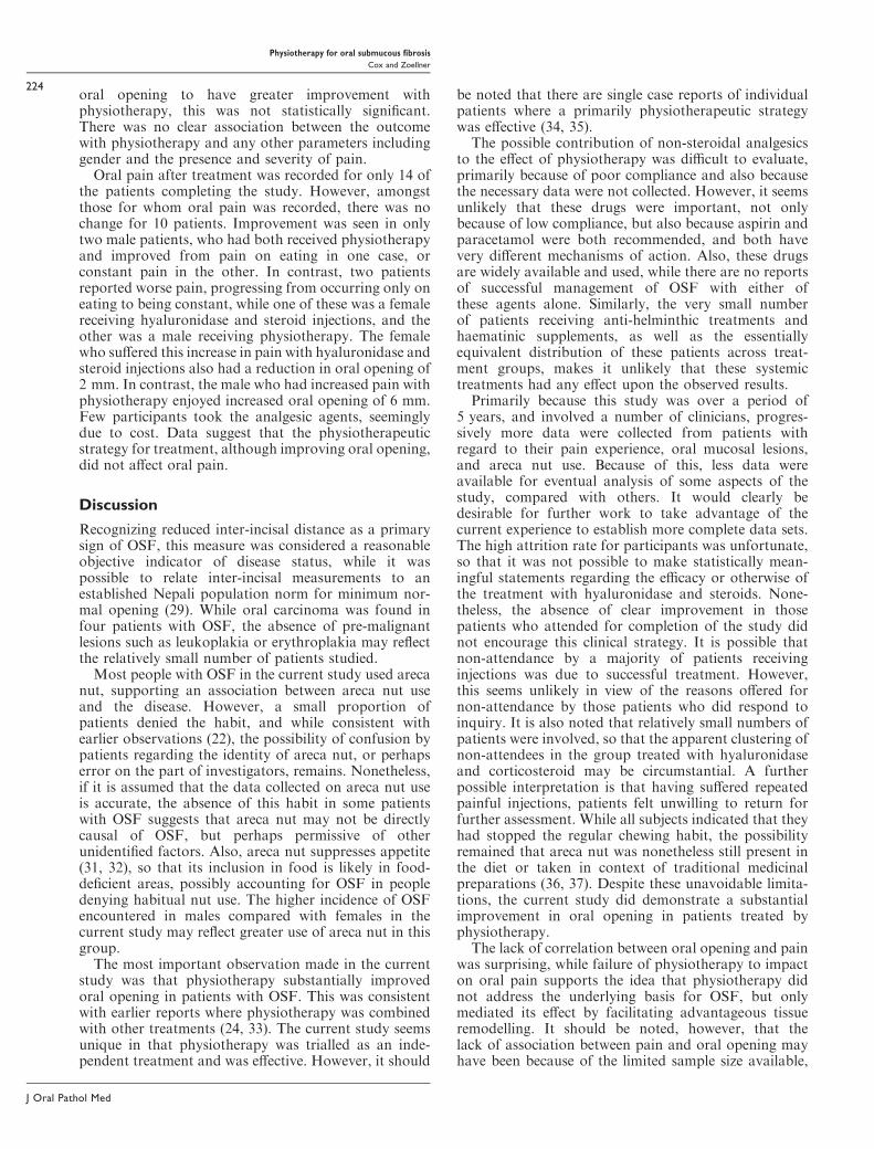
oral opening to have greater improvement withphysiotherapy, this was not statistically significant.There was no clear association between the outcomewith physiotherapy and any other parameters includinggender and the presence and severity of pain.Oral pain after treatment was recorded for only 14 of
the patients completing the study. However, amongstthose for whom oral pain was recorded, there was nochange for 10 patients. Improvement was seen in onlytwo male patients, who had both received physiotherapyand improved from pain on eating in one case, orconstant pain in the other. In contrast, two patientsreported worse pain, progressing from occurring only oneating to being constant, while one of these was a femalereceiving hyaluronidase and steroid injections, and theother was a male receiving physiotherapy. The femalewho suffered this increase in pain with hyaluronidase andsteroid injections also had a reduction in oral opening of2 mm. In contrast, the male who had increased pain withphysiotherapy enjoyed increased oral opening of 6 mm.Few participants took the analgesic agents, seeminglydue to cost. Data suggest that the physiotherapeuticstrategy for treatment, although improving oral opening,did not affect oral pain.
Discussion
Recognizing reduced inter-incisal distance as a primarysign of OSF, this measure was considered a reasonableobjective indicator of disease status, while it waspossible to relate inter-incisal measurements to anestablished Nepali population norm for minimum nor-mal opening (29). While oral carcinoma was found infour patients with OSF, the absence of pre-malignantlesions such as leukoplakia or erythroplakia may reflectthe relatively small number of patients studied.Most people with OSF in the current study used areca
nut, supporting an association between areca nut useand the disease. However, a small proportion ofpatients denied the habit, and while consistent withearlier observations (22), the possibility of confusion bypatients regarding the identity of areca nut, or perhapserror on the part of investigators, remains. Nonetheless,if it is assumed that the data collected on areca nut useis accurate, the absence of this habit in some patientswith OSF suggests that areca nut may not be directlycausal of OSF, but perhaps permissive of otherunidentified factors. Also, areca nut suppresses appetite(31, 32), so that its inclusion in food is likely in food-deficient areas, possibly accounting for OSF in peopledenying habitual nut use. The higher incidence of OSFencountered in males compared with females in thecurrent study may reflect greater use of areca nut in thisgroup.The most important observation made in the current
study was that physiotherapy substantially improvedoral opening in patients with OSF. This was consistentwith earlier reports where physiotherapy was combinedwith other treatments (24, 33). The current study seemsunique in that physiotherapy was trialled as an inde-pendent treatment and was effective. However, it should
be noted that there are single case reports of individualpatients where a primarily physiotherapeutic strategywas effective (34, 35).
The possible contribution of non-steroidal analgesicsto the effect of physiotherapy was difficult to evaluate,primarily because of poor compliance and also becausethe necessary data were not collected. However, it seemsunlikely that these drugs were important, not onlybecause of low compliance, but also because aspirin andparacetamol were both recommended, and both havevery different mechanisms of action. Also, these drugsare widely available and used, while there are no reportsof successful management of OSF with either ofthese agents alone. Similarly, the very small numberof patients receiving anti-helminthic treatments andhaematinic supplements, as well as the essentiallyequivalent distribution of these patients across treat-ment groups, makes it unlikely that these systemictreatments had any effect upon the observed results.
Primarily because this study was over a period of5 years, and involved a number of clinicians, progres-sively more data were collected from patients withregard to their pain experience, oral mucosal lesions,and areca nut use. Because of this, less data wereavailable for eventual analysis of some aspects of thestudy, compared with others. It would clearly bedesirable for further work to take advantage of thecurrent experience to establish more complete data sets.The high attrition rate for participants was unfortunate,so that it was not possible to make statistically mean-ingful statements regarding the efficacy or otherwise ofthe treatment with hyaluronidase and steroids. None-theless, the absence of clear improvement in thosepatients who attended for completion of the study didnot encourage this clinical strategy. It is possible thatnon-attendance by a majority of patients receivinginjections was due to successful treatment. However,this seems unlikely in view of the reasons offered fornon-attendance by those patients who did respond toinquiry. It is also noted that relatively small numbers ofpatients were involved, so that the apparent clustering ofnon-attendees in the group treated with hyaluronidaseand corticosteroid may be circumstantial. A furtherpossible interpretation is that having suffered repeatedpainful injections, patients felt unwilling to return forfurther assessment. While all subjects indicated that theyhad stopped the regular chewing habit, the possibilityremained that areca nut was nonetheless still present inthe diet or taken in context of traditional medicinalpreparations (36, 37). Despite these unavoidable limita-tions, the current study did demonstrate a substantialimprovement in oral opening in patients treated byphysiotherapy.
The lack of correlation between oral opening and painwas surprising, while failure of physiotherapy to impacton oral pain supports the idea that physiotherapy didnot address the underlying basis for OSF, but onlymediated its effect by facilitating advantageous tissueremodelling. It should be noted, however, that thelack of association between pain and oral opening mayhave been because of the limited sample size available,
Physiotherapy for oral submucous fibrosis
Cox and Zoellner
224
J Oral Pathol Med

suboptimal pain measures, cultural differences inexpressing pain, or even the continued use of areca nutproducts by some subjects.
Supporting the idea that physiotherapy improved oralopening by remodelling of the tissues was that improve-ment in oral opening was only unsuccessful in thosepatients who already had opening in the normal range, sothat only negligible remodelling and improvement mightbe expected. It was also interesting that OSF progressedin a highly predictable manner, with buccal mucosa neveraffected in isolation to the soft palate and the anteriorpillar of the fauces for example. This appears to be thefirst report illustrating this highly predictable pattern ofdisease in Nepal. Regional differences in OSF presenta-tion are reported, and have been postulated as due todifferences in chewing habits, including where the quid isheld and whether the juice is spat out or swallowed.Lesions in the anterior mouth seem associated withspitting, while the posterior oral cavity seems moreinvolved when the juice is swallowed (4). Similarly, aswallowing habit correlates with increased erythroplakia(38). In Nepal the use of areca nut is highly variable, inthat the nut can be chewed alone, or alternatively held inthe buccal sulcus over many hours as a quid wrappedin the leaf of the betel piper vine, with or without spices orchewing tobacco. Whether the resultant saliva is swal-lowed or spat also varies greatly, and future detailedstudy of specific chewing habits is needed.
Patients using physiotherapy were encouraged to useanalgesics to ensure that there was no pain associatedwith the jaw opening exercises. However, few patientsused analgesics in the ways advised, and it seemsunlikely that the analgesics made any significant contri-bution to the observations made.
The failure of physiotherapy to address the problemof oral mucosal pain suggests a need to develop andapply a suitable analgesic therapy. This in combinationwith the physiotherapeutic approach described here mayprovide a successful strategy for symptomatic manage-ment of OSF, despite continuing uncertainty of thebiological basis for the disease.
References
1. Gupta PC, Ray CS. Smokeless tobacco and health in Indiaand South Asia. Respirology 2003; 8: 419–31.
2. Pindborg JJ. Oral submucous fibrosis: a review. Ann AcadMed Singapore 1989; 18: 603–7.
3. Lal D. Diffuse oral submucous fibrosis. J All India DentAssoc 1953; 26: 1–3.
4. Bhonsle RB, Murti PR, Daftary DK, et al. Regionalvariations in oral submucous fibrosis in India. CommunityDent Oral Epidemiol 1987; 15: 225–9.
5. Schwartz J. Atrophica idiopathica (tropica) mucosa oris.Presented at the Eleventh International Dental Congress1952; London.
6. Chiu CJ, Lee WC, Chiang CP, Hahn LJ, Kuo YS, ChenCJ. A scoring system for the early detection of oralsubmucous fibrosis based on a self-administered question-naire. J Public Health Dent 2002; 62: 28–31.
7. Chang YC, Lii CK, Tai KW, Chou MY. Adverse effectsof arecoline and nicotine on human periodontal
ligament fibroblasts in vitro. J Clin Periodontol 2001; 28:277–82.
8. Huang L, Wang C, Kao L. Neuronal activity modulatingcomponents inbetelquid.SymposiumonBetelQuidChewingand Its Health Effect. Kaohsiung, Taiwan; 1993; p16.
9. Tilakaratne WM, Klinikowski MF, Saku T, Peters TJ,Warnakulasuriya S. Oral submucous fibrosis: review onaetiology and pathogenesis. Oral Oncol 2006; 42: 561–8.
10. Chang MC, Wu HL, Lee JJ, et al. The induction ofprostaglandin E2 production, interleukin-6 production,cell cycle arrest, and cytotoxicity in primary oral kerati-nocytes and KB cancer cells by areca nut ingredients isdifferentially regulated by MEK⁄ERK activation. J BiolChem 2004; 279: 50676–83.
11. Jeng JH, Wang YJ, Chang WH, et al. Reactive oxygenspecies are crucial for hydroxychavicol toxicity toward KBepithelial cells. Cell Mol Life Sci 2004; 61: 83–96.
12. Harvey W, Scutt A, Meghji S, Canniff JP. Stimulation ofhuman buccal mucosa fibroblasts in vitro by betel-nutalkaloids. Arch Oral Biol 1986; 31: 45–9.
13. Tsai CL, Kuo MY, Hahn LJ, Kuo YS, Yang PJ, Jeng JH.Cytotoxic and cytostatic effects of arecoline on oralmucosal fibroblasts. Proc Natl Sci Counc Repub China B1997; 21: 161–7.
14. Haque MF, Meghji S, Nazir R, Harris M. Interferongamma (IFN-gamma) may reverse oral submucous fibro-sis. J Oral Pathol Med 2001; 30: 12–21.
15. Feng Y, Ling T. Changes of cytokines secreted by humanoral mucosa keratinocytes from oral submucous fibrosis invitro. Hua Xi Kou Qiang Yi Xue Za Zhi 2000; 18: 23–5.
16. Rajalalitha P, Vali S. Molecular pathogenesis of oralsubmucous fibrosis – a collagen metabolic disorder. J OralPathol Med 2005; 34: 321–8.
17. Balaram P, Pillai MR, Abraham T. Immunology ofpremalignant and malignant conditions of the oral cavity.II. Circulating immune complexes. J Oral Pathol 1987; 16:389–91.
18. Shah N, Kumar R, Shah MK. Immunological studies inoral submucous fibrosis. Indian J Dent Res 1994; 5: 81–7.
19. Liu CJ, Lee YJ, Chang KW, Shih YN, Liu HF, Dang CW.Polymorphism of the MICA gene and risk for oralsubmucous fibrosis. J Oral Pathol Med 2004; 33: 1–6.
20. Chiu CJ, Chang ML, Chiang CP, Hahn LJ, Hsieh LL,Chen CJ. Interaction of collagen-related genes andsusceptibility to betel quid-induced oral submucous fibro-sis. Cancer Epidemiol Biomarkers Prev 2002; 11: 646–53.
21. Chiu CJ, Chiang CP, Chang ML, et al. Associationbetween genetic polymorphism of tumor necrosis factor-alpha and risk of oral submucous fibrosis, a pre-cancerouscondition of oral cancer. J Dent Res 2001; 80: 2055–9.
22. Seedat HA, van Wyk C. Submucous fibrosis in non-betelnut chewing subjects. J Biol Buccale 1988; 16: 3–6.
23. Kakar PK, Puri RK, Venkatachalam VP. Oral submucousfibrosis – treatment with hyalase. J Laryngol Otol 1985; 99:57–9.
24. Lai DR, Chen HR, Lin LM, Huang YL, Tsai CC. Clinicalevaluation of different treatment methods for oral sub-mucous fibrosis. A 10-year experience with 150 cases.J Oral Pathol Med 1995; 24: 402–6.
25. Borle RM, Borle SR. Management of oral submucousfibrosis: a conservative approach. J Oral Maxillofac Surg1991; 49: 788–91.
26. Gupta D, Sharma SC. Oral submucous fibrosis – a newtreatment regimen. J Oral Maxillofac Surg 1988; 46: 830–3.
27. Le PV, Gornitsky M, Domanowski G. Oral stent astreatment adjunct for oral submucous fibrosis. Oral
Physiotherapy for oral submucous fibrosis
Cox and Zoellner
225
J Oral Pathol Med

Surg Oral Med Oral Pathol Oral Radiol Endod 1996; 81:148–50.
28. Mokal NJ, Raje RS, Ranade SV, Prasad JS, Thatte RL.Release of oral submucous fibrosis and reconstructionusing superficial temporal fascia flap and split skin graft –a new technique. Br J Plast Surg 2005; 58: 1055–60.
29. Cox SC, Walker DM. Establishing a normal range formouth opening: its use in screening for oral submucousfibrosis. Br J Oral Maxillofac Surg 1997; 35: 40–2.
30. Chaturvedi VN. Oral submucous fibrosis. Natl Med JIndia 1989; 2: 11–7.
31. Strickland SS, Veena GV, Houghton PJ, Stanford SC,Kurpad AV. Areca nut, energy metabolism and hunger inAsian men. Ann Hum Biol 2003; 30: 26–52.
32. Strickland SS, Duffield AE. Anthropometric status andresting metabolic rate in users of the areca nut andsmokers of tobacco in rural Sarawak. Ann Hum Biol 1997;24: 453–74.
33. Sharma JK, Gupta AK, Mukhija RD, Nigam P. Clinicalexperience with the use of peripheral vasodilator in oraldisorders. Int J Oral Maxillofac Surg 1987; 16: 695–9.
34. Yusuf H, Yong SL. Oral submucous fibrosis in a 12-year-old Bangladeshi boy: a case report and review of literature.Int J Paediatr Dent 2002; 12: 271–6.
35. Reichart PA, Philipsen HP. Oral submucous fibrosis in a31-year-old Indian women: first case report from Ger-many. Mund Kiefer Gesichtschir 2006; 10: 192–6.
36. Murthy SKR. Astanga Samgraha of Vagbhata. Varanasi:Chaukhambha Orientalia, 1995; 1: 312–317.
37. Murthy SKR. Astanga Samgraha of Vagbhata. Varanasi:Chaukhambha Orientalia, 1996; II: 433.
38. Hashibe M, Sankaranarayanan R, Thomas G, et al.Alcohol drinking, body mass index and the risk of oralleukoplakia in an Indian population. Int J Cancer 2000;88: 129–34.
Acknowledgements
We thank Dr M Malla, Dr R Stringer and Mr U Rajkarnikar for their
assistance in collecting data and seeing patients, and the staff of the Dental
Department at Patan Hospital, Kathmandu, Nepal, for their assistance.
Physiotherapy for oral submucous fibrosis
Cox and Zoellner
226
J Oral Pathol Med