Physiology week 15 - EmergencyPedia · RENAL PHYSIOLOGY - ANATOMY • Glomerulus + renal tubule •...
22
RENAL PHYSIOLOGY WESTMEAD PRIMARY EXAM
Transcript of Physiology week 15 - EmergencyPedia · RENAL PHYSIOLOGY - ANATOMY • Glomerulus + renal tubule •...

RENAL PHYSIOLOGY WESTMEAD PRIMARY EXAM

RENAL PHYSIOLOGY - ANATOMY
• Glomerulus + renal tubule
• Each kidney has 1.3 million nephrons
• Cortical nephrons (85%) have shorter Loop of Henle than Juxtamedullary nephrons (15%)
• The ascending loop finishes with the specialised macula densa cells next to afferent more than efferent arterioles
• Macula densa + renin secreting juxtaglomerular cells = juxtaglomerular apparatus
• Primary convoluted tubule - brush border, apical tight junction
• Collecting duct - Principle cells (Na reabsorption and ADH related H2O rehabilitation) and intercalated cells (acid secretion and HCO3)


STRUCTURE OF THE GLOMERULUS • Blind end of the renal tubule is bowman’s capsule which is
invaginated by tufts of glomerular capillaries
• Afferent and efferent arterioles
• Endothelium is fenestrated 70 - 90 micrometers
• Podocytes have foot processes which wrap around capillaries making filtration slits which are 25 nano-m
• Glomerular basement membrane
• Mesangial cells -


RENAL BLOOD FLOW
• Total renal blood flow is: 1100 ml/min
• 20 - 25% of cardiac output
• 90% flows to cortex
• Total renal plasma flow is around 605ml.min
• The filtration fraction is around 20%
• The medullary flow is low because of high resistance in the vasa recta - this low flow is required to maintain the concentration gradient
• Renal plasma flow is the amount of a substance excreted per unit of time divided by the renal AV difference as long as the passage of blood through the kidney is un altered and is measured use p amino hipputric acid

AUTOREGULATION AND BLOOD PRESSURE
• Blood flow is virtually constant over BP range of 80 - 180 mmHg due to auto regulation - this is the case for RBF and GFR
• The afferent arterioles are the major site of regulation of resistance
• This effectively blunts significant changes in glomerular capillary pressure
• These effects are seen in both isolated and transplanted kidneys - therefore theses mechanisms are predominantly intra renal

AUTOREGULATION AND BLOOD PRESSURE • Myogenic mechanisms
• Tubuloglomerular feedback
• Sympathetic control
• Angiotensin 2
• Intrarenal baro-receptors
• Macula Densa
• Renal SNS
• Intrarenal prostaglandins

RENAL AUTOREGULATION
• Myogenic mechanism - smooth muscle contraction in response to stretch/tension
• Tubulo glomerular feedback
• Primarly regulates GFR, chanes in RBG being secondary
• Increased renal artery pressure -
• Increased glomerular capillary pressure
• Increase net filtration pressure and grr
• Increased flow to proximal tubule and loop and macula densa.
• This increased rate of flow is detected by the macula densa. by detecting an increased luminal Na,Cl,osmolality and these cells release vasoconstrictor mediators
• Aniotensin 2, Prostaglandins, Adenosine —> afferent arteriolar constriction

RENAL AUTOREGULATION
• Sympathetic control - there is a rich CNS supply to kidney, afferent > efferent
• tone is reflexively mediated by aortic and carotid sinus bark-receptors
• alpha receptors cause vasoconstriction
• beta receptors are present but these are relatively int, such that adrenaline causes only vasoconstriction
• because both afferent and efferent vessels are innervated SNS stimulation causes a greater reduction in RBF than GFR, efferent tone raises glomerular capillary pressure but adds series resistance to the total renal vascular resistance
• Increased SNS tone —> increases BP —> decreases Na + H2O excretion via decreases to GFR and increases to tubular reabsorption of Na and H20
• Intrarenal baro-receptors
• Renin secreting granular cells act as baro-receptors measure pressure in the late afferent arteriole and secretion of renin is inversely related to the pressure
RENAL AUTOREGULATION
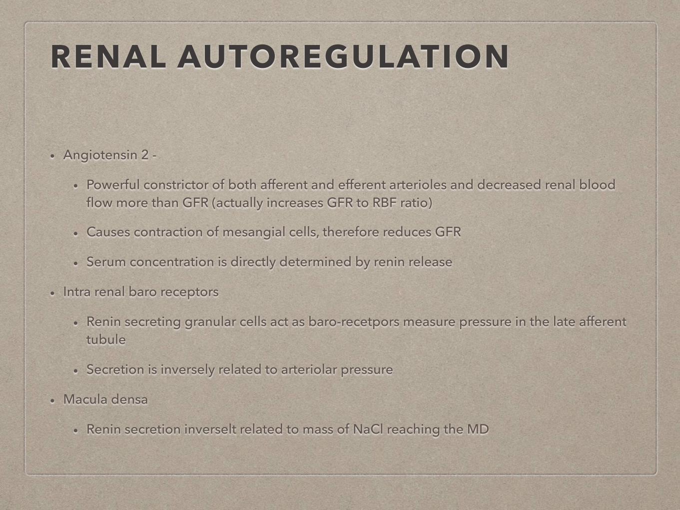
• Angiotensin 2 -
• Powerful constrictor of both afferent and efferent arterioles and decreased renal blood flow more than GFR (actually increases GFR to RBF ratio)
• Causes contraction of mesangial cells, therefore reduces GFR
• Serum concentration is directly determined by renin release
• Intra renal baro receptors
• Renin secreting granular cells act as baro-recetpors measure pressure in the late afferent tubule
• Secretion is inversely related to arteriolar pressure
• Macula densa
• Renin secretion inverselt related to mass of NaCl reaching the MD

RENAL AUTOREGULATION
• Renal sympathetic NS
• Produces both direct and in direct effects
• Indirect - afferent aa. constriction and decreased aa. pressure which decreases NaCl load leaving the loop and reaching the macular densa.
• Direct - stimulation of beta 1 receptors on the granular cells and stimulation of renin secretion
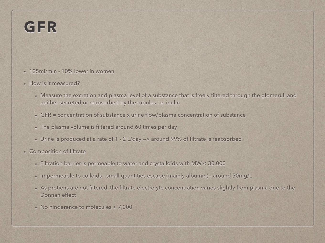
GFR
• 125ml/min - 10% lower in women
• How is it measured?
• Measure the excretion and plasma level of a substance that is freely filtered through the glomeruli and neither secreted or reabsorbed by the tubules i.e. inulin
• GFR = concentration of substance x urine flow/plasma concentration of substance
• The plasma volume is filtered around 60 times per day
• Urine is produced at a rate of 1 - 2 L/day —> around 99% of filtrate is reabsorbed
• Composition of filtrate
• Filtration barrier is permeable to water and crystalloids with MW < 30,000
• Impermeable to colloids - small quantities escape (mainly albumin) - around 50mg/L
• As protiens are not filtered, the filtrate electrolyte concentration varies slightly from plasma due to the Donnan effect
• No hinderence to molecules < 7,000
GFR - THE FILTRATION BARRIER
• Endothelial fenestra
• Basement membrane
• Slit diaphragms
• Slit
• This entire pathway is extracellular - the greatest restriction is the basement membrane through the hydrated spaces between glycoprotein chains
• Also important is that the cell coats of the endothelium basement membrane and cell coats of the podocytes are all poly-anions —> at any MW, anions are selectively restricted

DETERMINANTS OF GFR

TUBULAR FUNCTION
• Qualitatively a different process from GF, the later being bulk flow - relatively little bulk flow occurs across the tubular membrane due to small filtration pressures and low hydraulic permeability
• Transport mechanisms
• Simple diffusion - determined by electrochemical gradient and occurs through cell membranes and is determined to some degree by lipid solubility
• Simple facilitated diffusion - also down an EC gradient but dependent on reaction to specific membrane proteins
• Secondary active transport - two or more molecules + protein —> facilitate movement. The molecule under going SAT usually travels against its electrochemical gradient - the angry for this is derived from the movement of the other ion ‘down hill’
• Primary active transport - molecule + protein + ATP , displays characteristics of competition, specificity and saturatibility
• Endocytosis - a form of primary active transport because ATP is required
• Amount excreted = urine flow x urine concentration of substance
• Amount filtered = GFR x plasma concentration of substance

THE MANAGEMENT OF NA
• Reabsorption of Na and Cl is the key in water homeostasis
• Na is paired to glucose, amino acid, organic acid, phosphate, electrolytes
• Moves down electrical and concentration gradient into tubular epithelial cells then actively transported into the interstitium by Na/K ATPase
• 60% of the filtered Na+ is reabsorbed in the proximal tubule, primarily by Na/H
• 30% is absorbed by the Na-2Cl-K co transporter in the distal convoluted tubule
• 3% is absorbed in the ENAC channels in the collecting ducts mediated by aldosterone

THE MANAGEMENT OF GLUCOSE AND OTHER ORGANIC NUTRIENTS
• Glucose, amino acids, water soluble vitamins, lactate
• Main site of absorption from the tubule is proximal tubule
• They share the following characteristics:
• reabsorption is an active process
• this uphill transport is SAT coupled with Na across the luminal membrane
• they cross the basolateral membrane by simply diffusion

RENAL HANDLING OF K
• Freely filtered at the glomerulus - although final urinary K reflect 10 - 15% of the filtered fraction
• Tubular reabsorption dominates but the tubules can secrete K
• PT - 50% reabsorbed primarily by passive diffusion
• Distal PT and DLH - K secretion dominates this occurs by diffusion due to the high interstitial K concentration in the renal medulla
• In the ALH - passive reabsorption commences again
• The late DT and CT play a role in both K absorption and secretion

TUBULOGLOMERULAR FEEDBACK
• Increasing rate of flow through sending LOH —> GFR at that nephron - this maintains a constant load on the DCT
• Sensor is the macular densa which triggers adenosine release which then triggers Ca entrance in to the cells of the afferent arteriole causing vasoconstriction which then lowers EGFR

GLOMERULAR TUBULAR BALANCE
• The more that is filtered there more that is absorbed in the proximal tubule