PHYSIOLOGY OF THE CARDIOVASCULAR SYSTEM The Heart.
25
PHYSIOLOGY OF THE CARDIOVASCULAR SYSTEM The Heart
-
Upload
laurence-thornton -
Category
Documents
-
view
224 -
download
1
Transcript of PHYSIOLOGY OF THE CARDIOVASCULAR SYSTEM The Heart.

PHYSIOLOGY OF THE CARDIOVASCULAR SYSTEM
The Heart
Introduction
Cardiovascular system is comprised of the Heart and Blood vessels
Heart is the central pump and the blood vessels are series of distributing and collecting tubes.
CVS forms one of the major coordinating system of the human body

The Heart – Anatomy and Physiology The heart is located in the thoracic cavity
between the lungs. This area is called the mediastinum.
The base of the cone-shaped heart is uppermost, behind the sternum, and the great vessels enter or leave here.
The apex (tip) of the heart points downward and is just above the diaphragm to the left of the midline.


Chambers
It is divided into four chambers: a right and a left atrium both lying superiorly, and a right and left ventricle both lying inferiorly and larger.
The atria are separated by a thin interatrial partition called interatrial septum, while the ventricles are separated by thick muscular partition called interventricular septum.
The thickness of the right ventricular wall is 0.3-0.5cm and while that of the left ventricular wall is 1.3-1.5 cm
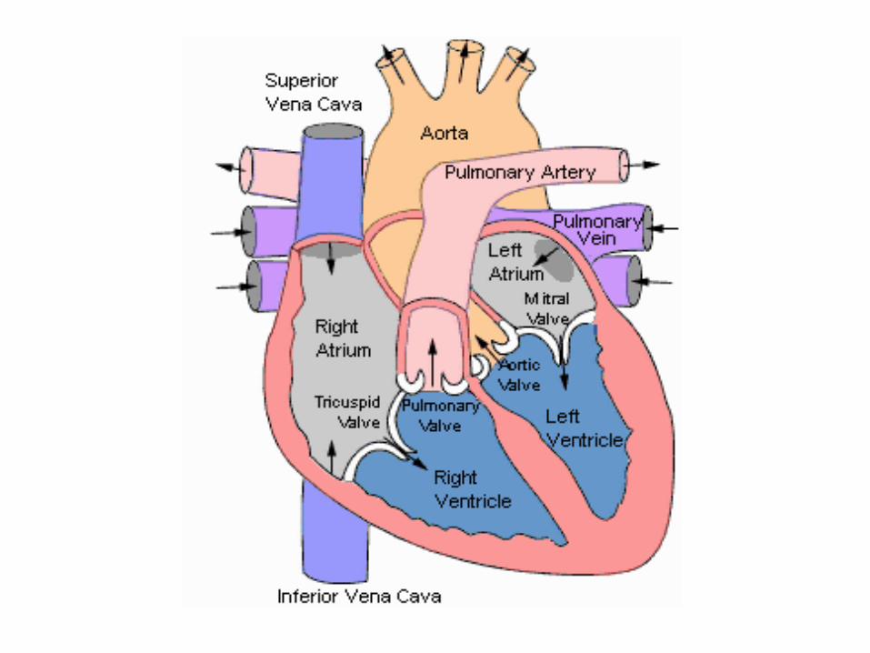

Movement of the Blood
Valves
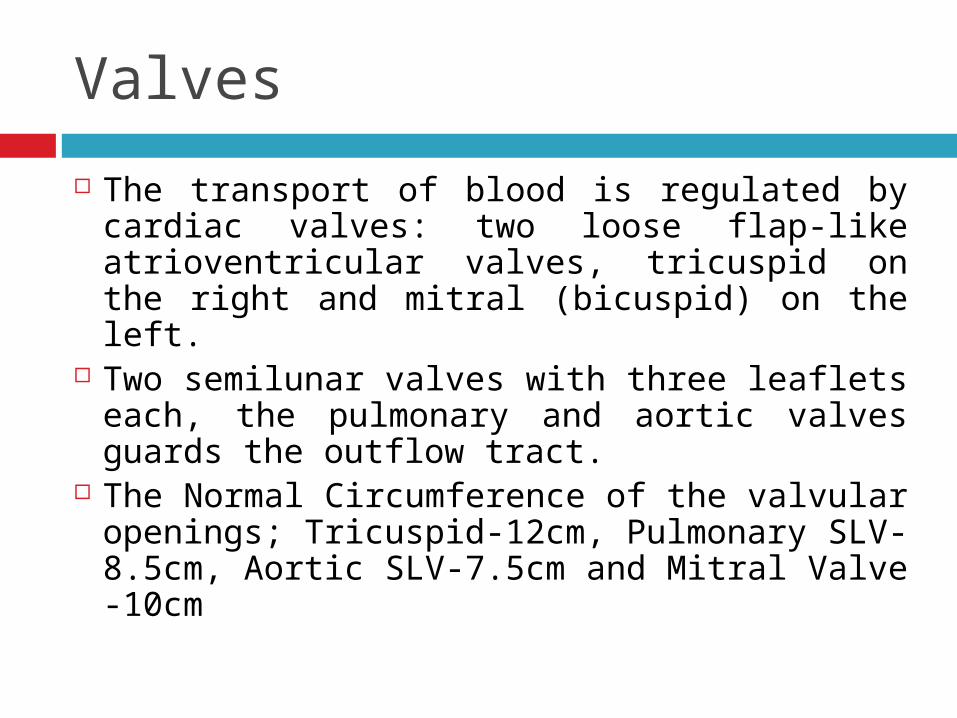
The transport of blood is regulated by cardiac valves: two loose flap-like atrioventricular valves, tricuspid on the right and mitral (bicuspid) on the left.
Two semilunar valves with three leaflets each, the pulmonary and aortic valves guards the outflow tract.
The Normal Circumference of the valvular openings; Tricuspid-12cm, Pulmonary SLV-8.5cm, Aortic SLV-7.5cm and Mitral Valve -10cm
Layers of the Heart
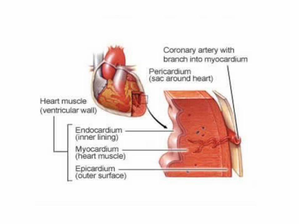
The wall of the heart consists mainly of the myocardium which is covered externally by thin membrane, the epicardium or visceral pericardium, and lined internally by another thin layer, the endocardium.

Myocardium
Muscle tissue of the heart Composed of syncytium of branching
and anatomizing, transversely striated muscle fibers arranged in parallel fashion.
Contains a rich capillary network and loose connective tissue.
Myocardial fibers are connected to each other by intercalated discs.
Myocardium
They form tight junctions for free transport of ions and action potential.
Cardiac myocytes are rich in Mitochondria.
It also contains more sarcoplasmic reticulum
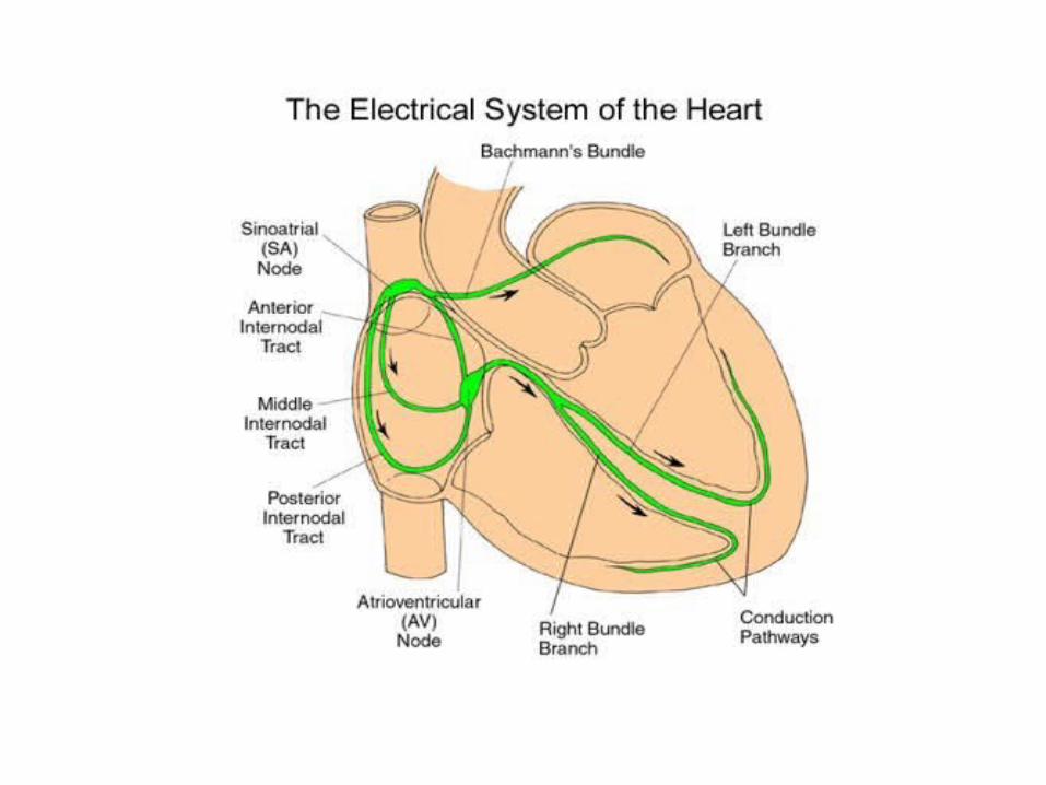
The conduction system of the heart in the myocardium is responsible for regulating rate and rhythm of the heart.
Pericardium
The heart is enclosed in the pericardial membranes, of which there are three layers:
The outermost is the fibrous pericardium, a loose fitting sac of strong fibrous connective tissue that extends inferiorly over the diaphragm and superiorly over the bases of the large vessels that enter and leave the heart.

Pericardium
The serous pericardium is a folded membrane; the fold gives it two layers, parietal and visceral. Lining the fibrous pericardium is the parietal pericardium.
On the surface of the heart muscle is the visceral pericardium, often called the epicardium.
Between the parietal and visceral pericardial membranes is serous fluid, which prevents friction as the heart beats.


Endocardium
Smooth shiny inner lining of the myocardium Covers all cardiac chambers, cardiac valves,
the chordae tendineae and the papillary muscles.
It is lined by endothelium with connective tissue and elastic fibres in its deeper part.
The valve cusps and semilunar leaflets are delicate and translucent structures.
The valves are strengthened by collage and elastic tissue and covered by a layer of endothelium (valvular).

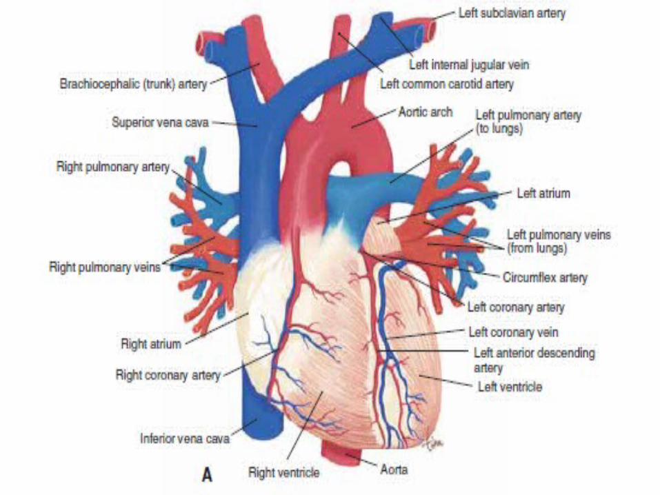
Blood Supply
Blood is transported to the myocardium by Coronary arteries.
Most of the blood supply occurs during diastole.
There are three major coronary trunks The Anterior Descending Branch of the Left
Coronary Artery The Circumflex Branch of the Left Coronary
Artery The Right Coronary Artery

Blood Supply
The Anterior Descending Branch of the Left Coronary Artery Supplies most of the apex of the heart Anterior surface of the left ventricle Adjacent third of the anterior wall of the right
ventricle Anterior two third of the interventricular septum
The Circumflex Branch of the Left Coronary Artery Supplies left atrium Small portion of the lateral aspect of the left
ventricle

Blood Supply
The Right Coronary Artery Supplies Right Atrium Anterior surface of the right ventricle The adjacent half of the posterior wall of
the left ventricle and The posterior third of the interventricular
septum

Anatomic Patterns of Blood Supply Three anatomic patterns of distribution
of the blood supply, depending upon the blood vessel that crosses the Crux.
Crux is the region on the posterior surface of the heart where all the four chambers and interatrial and interventricular septa meet.
Anatomic Patterns of Blood Supply Balanced Coronary Artery Distribution
Right and Left ventricles receives blood from Right and Left Coronary artery.
Posterior part of the interventricular septum is supplied by branch of right coronary artery
Anterior part is supplied by the branch of Left Coronary artery

Anatomic Patterns of Blood Supply Left Coronary Artery Preponderance
Entire left ventricle receives blood from LCA, whole of interventricular septum and
Posterior wall of the Right ventricle Right Coronary Artery Preponderance
Entire Right ventricle Posterior half of the interventricular septum Posterior wall of the left ventricle by
crossing the Crux.

A. Balanced Coronary Circulation
B. LCA Preponderance
C. RCA Preponderance