Physiological or Biological Approach to Psychology€¦ · Web viewMcGuire Taxi Drivers Brains....
33
Physiological or Biological Approach to Psychology McGuire Taxi Drivers Brains Sperry – Split Brain Dement and Kleitman – Dreaming 1
Transcript of Physiological or Biological Approach to Psychology€¦ · Web viewMcGuire Taxi Drivers Brains....

Physiological or Biological Approach to Psychology
McGuire Taxi Drivers Brains
Sperry – Split Brain
Dement and Kleitman – Dreaming
1

McGuire Taxi Drivers Brains – how plastic are our brains? Do they structurally change with new experience? Is the Hippocampus the centre for spatial awareness and map learning?
Sperry – Split Brain – how independently does each side of the brain work? What would happen to our behaviour if our two hemispheres are split? What information and skills does each hemisphere contain?
Dement and Kleitman – Dreaming – what electrical activity is going on in our brains when we are asleep? When do we dream? How often do we dream? Why do our eyes move when we dream?
2
The Physiological Approach to Psychology
The assumptions of the physiological approach are that our biology has a significant impact on our behaviour. Some physiological psychologists would say that our brain and nervous system determine most of our behaviour. They study the brain, the nervous system, the endocrine system (hormones such as adrenaline, testosterone and oestrogen etc), heredity and disease, to determine their effects. In terms of the nature v nurture debate they consider nature to be most important. In the past studies have often been carried out on animals or on identical twins. However the onset of EEG machines to measure brainwaves and MRI scanning together with genetic engineering has recently extended our understanding of the human brain and how it works. The physiological approach also includes the work of evolutionary psychologists who study instinctive behaviours which are common to all animals and determine the reproduction of their species.
There are considerable strengths to the physiological approach as many tests which can be carried out such as EEG’s, MRI’s and DNA testing are highly scientific and therefore reliable. We all have very similar biological brains and nervous systems so data is also highly generalisable. However brain imaging techniques are relatively new and we cannot always be sure we are interpreting them correctly. We may see changes in brain functions but these techniques do not tell us what is causing those changes. Otherwise we often have to rely on the self report method when studying the brain and so demand characteristics may be a problem. Studies which are carried out in laboratories or hospital settings could also be said to lack ecological validity.
McGuire Taxi Drivers Brains and the Hippocampus
The Hippocampus helps us to navigate and find our way around both familiar and new environments. This part of the brain plays an important part in spatial memory and tests in laboratory animals have shown a tendency for the male species to have better spatial memories than females. This is thought to be an evolutionary difference.
3

London Taxi Drivers were used in the study as they undergo an intensive two year training of route and map learning of the London area before they are allowed to obtain a taxi licence. This training is known as ‘The Knowledge’.
Method: an experiment using independent groups comparing brains of taxi drivers with non taxi drivers. In addition correlational analysis was done to see if the hippocampus changes increased with time of being a taxi driver.
Subjects: 16 right handed male taxi drivers with a mean age of 44yrs. They were all healthy and had a minimum of two years experience ‘on the knowledge’. There was a control group of 50 right handed males with the same mean age range who volunteered to have an MRI scan of their brains.
Only right handed males were used as females are known to have slightly different functions in their hippocampus and whether you are left or right handed also affects how your brain is used and developed.
Structural MRI scans were used (which measure the structure rather than the function of the brains).
The Dependent variables were measured by: VBM (voxel-based morphometry) to measure the changes in shape of the grey matter. Pixel counting was also carried out on the scans of both the taxi drivers and non taxi drivers’ hippocampus to measure the volume. They sliced the scans into 26 slices looking at different sections of the hippocampus, posterior (back), anterior (front) and main body.
Control: a pixel counter was used who was highly experienced but did not know which group any of the pictures belonged so as to avoid bias.
Results:
1. The VBM analysis showed there was in increase in grey matter on both sides at the back or posterior of the hippocampus and a decrease in grey matter at the front (anterior) of the taxi drivers. This showed that the brain is ‘plastic’ (can change shape).
2. Pixel counting showed there was no difference in the volume of the two groups hippocampus so proving the grey matter had shifted rather than expanded.
3. The correlation analysis showed change with length of time as a taxi driver in the volume of the right anterior hippocampus only.
Conclusions:
The work and learning involved in being a taxi driver in London caused structural changes to the brain. This shows that people who choose to do this kind of work do not have specifically different brains to start with but their brains develop and change with experience. It also shows that brains are plastic (can change shape with new experiences). The findings also support work carried out on animals such as monkeys and rodents as well as with patients with brain damage.
The study is important as it indicates that different parts of our hippocampus have different functions. The posterior is the part that is used when we are using previously learned information to find our way about whilst the front part is used
4

when we are learning new layouts. This has implications for brain damaged patients and their recovery as it shows that given the right environmental stimulation some brain functions can be increased. The study also suggests that the right and left side of the hippocampus have different functions. The right side may hold more mental maps whilst the left side more memories of events.
Strengths of the study are its usefulness to the understanding of brain recovery in brain damaged patients and the ability of the brain to adapt to environmental stimulation. This supports theories of evolution which show how animals adapt over time and change their body structure and function to adapt to their changing environments.
Validity: this study does appear to be highly valid (does measure what is says it is measuring). Firstly reliable scientific techniques were used to measure the differences in brain function and secondly not only were taxi drivers compared to the control group but also correlation analysis supported the fact that as time went by the changes became more significant. The validity is also supported by the fact that the same changes have been seen in the study of rodents and other mammals.
Problems: of course learning such detailed mapping is a skill that many of us do not do and do not need. We also cannot generalise to women or even people who are left handed as there may be significant differences between these two groups. But the strengths do seem to outweigh the disadvantages on this study.
Two changes to the study and how it would affect the data:
One change I would make to the McGuire study is to use women. Other studies on mammals have shown that women do have different spatial abilities than men. However this maybe due to the fact that they have different roles in society and so have different experiences. If the same study was done one women taxi drivers it would show if the study can be generalised to both sexes or if the hippocampus changes would be different in women than men. This would be useful for the nature nurture debate as it would show if some sex differences in the brain are down to nature or simply to the experiences of the society in which women are brought up.
Another change I would make is to include left handed people. Many people are left handed. It would be interesting to see if left handed people have the same function in their hippocampus and if the same side of the hippocampus enlarges in a left handed person or if the left side of the hippocampus would enlarge rather than the right. This would enable us to know if handedness does affect brain plasticity or not and if handedness has any other effects on the brain such as spatial awareness.
5

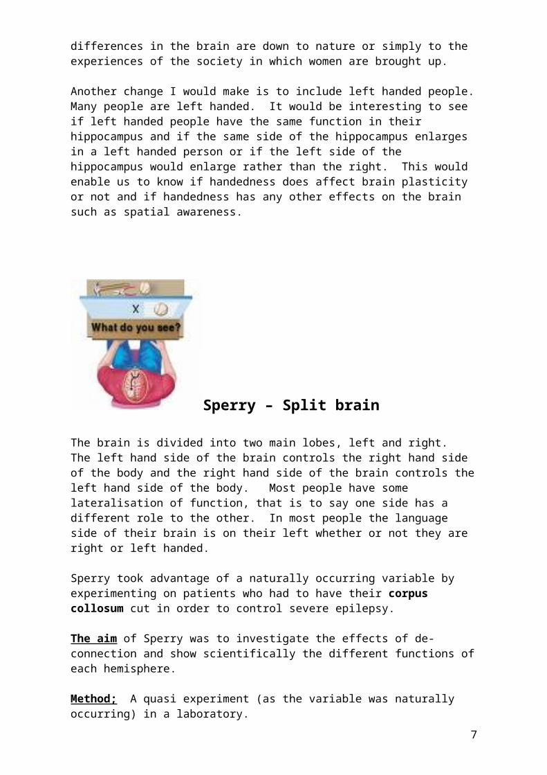
Sperry – Split brain
The brain is divided into two main lobes, left and right. The left hand side of the brain controls the right hand side of the body and the right hand side of the brain controls the left hand side of the body. Most people have some lateralisation of function, that is to say one side has a different role to the other. In most people the language side of their brain is on their left whether or not they are right or left handed.
Sperry took advantage of a naturally occurring variable by experimenting on patients who had to have their corpus collosum cut in order to control severe epilepsy.
The aim of Sperry was to investigate the effects of de-connection and show scientifically the different functions of each hemisphere.
Method; A quasi experiment (as the variable was naturally occurring) in a laboratory.
Subjects: 11 patients who had already experienced a commissurotomy for their epilepsy. 9 were recent patient 2 had their surgery some years before.
Procedure: A table with a screen. The screen had a space beneath it so the patient could put their hand below the screen and touch objects on the table. The experimenter sat one side of the table and the participant the other side and objects were projected through the screen into one or other of their visual fields (either left or right).
6
Results:
1. Objects shown to the left visual field could not be named but could be picked up by the right hand or drawn by the right hand. Objects shown to the right visual field could be named and picked up by the left hand.
2. When shown an object in one visual field it could only be recognized again if shown to the same visual field.
3. If objects were put in the right hand they could name it but would say they would not have any awareness of it if it was put in the left hand. However they could find it by touch with the left hand if put in a bag along with other objects.
4. When subjects saw embarrassing pictures of a nude appear in their left visual hemisphere they giggled even though they could not say what they had seen. This suggests the hemisphere has its own awareness just not the speech.
5. Right hemispheres were better at carrying out mathematical problems.
Advantages and conclusions:
The study is useful in that at the time it was groundbreaking in demonstrating how each side of the hemispheres work. It also revealed the purpose of the corpus collosum in being the communication pathway between the two hemispheres. It also showed how independently the two sides of the brain can work and how each has its own purpose and function.
Validity
The problem with this study is that the patients all had severe epilepsy so it could be argued that the results are not valid to ‘normal’ brains.
Reliability:
The sample was small and we cannot repeat the experiment on people with a normal brain due to ethics
Key words:
Left and right hemisphere
Corpus Collosum
Epilepsy
Quasi experiment
Commissurotomy
7

Two changes to this study and how it would change the data:
This question is almost impossible to answer as we cannot objectively experiment on ‘normal’ patients as this would be unethical. When surgeons carry out brain surgery they often have the patient awake so they can talk and answer questions. By probing different parts of the brain the surgeon can then tell which part is responsible for which activity and so avoid damaging certain parts responsible for speech and movement. Brain scans which can show a brain working can now be carried out. A patient is given an injection of glucose which has a radio active tracer. They are then given tasks to do whilst their brain is scanned. The part of the brain which is being used the most uses more glucose and so the tracer shows up highlighted on the scan. This is now the best way to study brain function and would make the data more valid as we could do this on healthy patients. The only other way we could improve the study would be to make it more reliable by repeating the same experiments with more subjects to be sure that the sane results are found. I do not think this question is likely to come up.
8
Dement and Kleitman – Sleep and Dreams
Our bodies have different patterns or rhythms which are built into our biology. Circadian rhythms are those that happen each day based on a 24 hour cycle and all animals and plants have some kind of built in pattern of life. Plants, animals, birds respond to changes in light and temperature stimulating their sleep, migration or breeding patterns. Humans have a body clock or rhythm which determines their sleep wake cycle and also their body temperature. Our body temperature falls at night reaching its lowest point in the early hours of the morning. This is one reason why night shift workers find it so hard to sleep in the day time.
No one knows the precise reason for sleep although sleep deprivation studies have shown how brain function reduces without sleep and long term sleep deprivation can even result in paranoia and mental illness. The assumption is that sleep is required for psychological and physiological restoration. Accidents in the work place often occur at night when workers are functioning at their worst part of the circadian cycle.
Evolutionary theory suggests that we sleep to preserve resources and keep us safe in times of darkness and extreme cold however if this was true one might think that humans would have evolved to need less sleep than they do.
As well as a 24 hour cycle we also have a cycle to our sleep pattern. You need to understand this cycle and occasionally you will be asked about this cycle in an examination question:
9

The Sleep Cycle: the whole sleep cycle lasts for about 90 minutes
1. Stage One: we start to feel drowsy often as the light fades and the pineal gland starts to produce melatonin in the brain.
2. Stage Two: deeper stage but still can be woken easily and heart rate and breathing start to slow.
3. Stage Three deeper sleep with more long slow delta waves on a EEG machine. Difficult to wake up, heart rate and blood pressure still dropping.
4. Stage Four delta or quiet sleep and hardest to wake up from and last in bursts of about 30 minutes.
5. Stage Five – we return to where stage one would be but instead go in REM sleep (rapid eye movement sleep). Our eyes move but our bodies cannot. We stay in this stage for 5 – 15 minutes and then go through stages two to four again and again all night.
Aim of the Dement study was to see if the REM sleep part of the cycle does correspond to dreaming and to see if the eye movements match the content of the dream. Dement also wanted to find our if subjects could tell how long they had been dreaming for.
Method: laboratory experiment in sleep lab.
Apparatus :
1. EEG machine to measure sleep brain waves2. Electrodes attached to eyes to measure eye movements3. A tape recorder so subjects could self report and a bell to wake the subjects
Subjects: 7 adults makes and 2 adult females. Only 5 were studied intensively the other 4 were used to confirm the findings.
Controls:
Subjects told not to have any caffeine or alcohol on days of trialsWere woken by bell so experimenter did not effect dream recall
Procedures:
1. Subjects woken at random and asked to report if dreaming2. Subjects woken between 5 and 15 minutes when in REM sleep and asked if
they could estimate length of time they had been dreaming3. Subjects asked to explain dram and this was matched to eye movements.
10

Results and conclusions:
1. Most dreaming occurred in REM sleep2. Dreams matched eye movement (when eyes went up subject said he had
been climbing a ladder.3. Subjects were very good at estimating time they had been dreaming.
4. Dement and Kleitman concluded that the measurement of REM during sleep can be used as an objective measure of dreaming and that dreaming appears to occur in real time.
The study was useful in that it was a first in scientifically measuring when we dream but it fails to tell us why we dream or indeed why we go through specific cycles of sleep. It also could be said to lack ecological validity.
Two changes to this study and how it would affect the data:
One change I would make to the study is to carry out the experiment in peoples homes. This would have an effect of making the data higher in ecological validity as we cannot be sure that sleeping in a laboratory did not affect their sleep patterns or their dreaming. However it would mean that the study was less controlled as people would not be as closely monitored in their own homes as they would be in a laboratory. This would therefore make the data less scientific. Another change that I would make to this study would be to make sure that their were an equal amount of subjects that were male and female studied as well as a range of different ages as this would make the data more generalisable. At present we cannot be sure that women are exactly the same in their dream and sleep patterns as men and the study does not tell us anything about the effect of age on sleep patterns and dreaming.
11

Examination Questions to Practice
Some of the core studies take a physiological approach to human behaviour andexperience. This approach considers how our hormones, nervous system, and functions of the brain interact to determine our behaviour.
Using the core studies listed below, answer the questions that follow.
McGuire (taxi drivers brains)Sperry (split-brain)Dement and Kleitman (sleep and dreaming) (a) What physiological processes were involved in each of the studies? [12]
(b) Using examples, give two strengths and two weaknesses of the physiologicalapproach. [12]
McGuire – Taxi Drivers Brains
1. Describe the sample and say why they were chosen for this study (4)2. What is meant by the term plasticity in the McGuire study (2)3. What is the name given to the part of the brain that stores spatial
awareness (2)4. What part of the brain did McGuire find changed the most in taxi drivers
who had spent a long time on the job (2)5. What is meant by the knowledge (2)6. Name the different parts of the brain which were changed by learning the
knowledge (2)7. Why did McGuire only use right handed participants (2)8. Why did McGuire only use men (2)9. Give one control used by McGuire (2)10. Describe the usefulness of the McGuire study (4)11. How were the dependent variables measured in the McGuire study (6) 12. Give two reasons why the McGuire study is considered to be valid (4)13. Describe what is meant by Pixel counting (2)14. Describe what changes the Pixel counting showed (2)15. Describe what VBM (voxel-based morphometry) was measuring (2)
Dement & Kleitman – Sleep and Dreams
1. From the study by Dement & Kleitman, give two measures that the
12
researchers made while their subjects were sleeping. (2
2. What did their measurements tell them about the pattern of normalsleep? (2
3. From the study by Dement & Kleitman on sleep, what does an electroencephalogram (EEG) record? (2
4. Outline one problem with using an EEG to investigate dreaming. (2
5. From the study by Dement & Kleitman on sleep, give four characteristics ofREM sleep. (4
6. The data table shown below contains the results from the dream-durationEstimates made in the study by Dement & Kleitman. Identify two conclusionsThat we can draw from the data (4
7. Results of dream-duration estimates after 5 or 15 minutes of rapid eye Movements
Estimates (in minutes) Estimates (in minutes)After 5 minutes REM after 15 minutes REM
Subject Right Wrong Right Wrong
DN 8 2 5 5IR 11 1 7 3KC 7 0 12 1WD 13 1 15 1PM 6 2 8 3
Total 45 6 47 13
8. Identify one of the aims of the study on sleep and dreaming carried out by Dement and Kleitman. (2
9. Outline the results of the study in relation to this aim. (2
10. The study by Dement and Kleitman (sleep and dreaming) involved Participants self reports of dreams and the use of equipment to measureREM and NREM
11. Outline one finding of the relationship between sleep and dreaming (2
12. Give one reason why the conclusions of this study might not be valid (2
13. In the study on sleep and dreaming, Dement and Kleitman point out that
13

dream recall from NREM was higher if the awakening occurred within eight minutes of the end of REM than if the awakening occurred later. How did they explain this difference (2
14. Outline another possible explanation for this difference. (2
15. In their study on sleep and reaming, Dement and Kleitman found some participants recalled dreams following NREM sleep. How did they explain this? (2
16. What evidence is there in the study to support this explanation? (2
17. In the study on sleep and dreaming by Dement and Kleitman, it is suggested that Rapid Eye Movements (REM) only occur during dreaming. Give one piece of evidence which supports this suggestion, and one piece of evidence that challenges it. (4
18. From the study on sleep and dreaming by Dement and Kleitman, outline one way in which the study is low in ecological validity. (2
19. Identify two controls used in the study on sleep and dreaming by Dement and Kleitman.(2
20. Explain why one of these controls was used. 2 )
Sperry – Split Brains
1 From the study by Sperry on split brainsGive one piece of evidence that illustrates the language limitations of the right hemisphere of the brain. (2
Give one piece of evidence that illustrates that this same hemisphere is not completely “word blind”. (2
2. a. In the study by Sperry, what is meant by the term “split brain”? (2
b Explain one problem with making generalisations about normal brain activity from a study if people with split brains. (2)
3. a. What technique did Sperry use to present information to only one side of the brain?(2
b Why does this technique not present a problem to people with “normal” brains?(2)
4. From the paper by Sperry on split brain patients, outline the evidence which indicates that language is processed in the left hemisphere of the brain. (4
5 In the paper by Sperry on split brain patients, he writes, “the second hemisphere does not know what the first hemisphere has been doing.”
a. give one piece of evidence to support this statement. (2
b Explain why this problem does not matter in the everyday activity of the patients in this study. (2
14

6 In the study by Sperry (split brains) patients had problems with material presented to their left visual field.
a. Give one example of these problems. (2
b. Suggest one way in which patients could overcome these problems in everyday life. (2
7. a. From the split brain study by Sperry, if a word such as “key” was presented only to the right hemisphere, participants were not able to mane it. Identifyhow they were able to respond to show that they had seen the word. (2
b. What does this tell us about how “normal” brains function? (2
8. a. From Sperry’s split-brain study, outline one difference between the ability of split brain patients and “normal” people to identify objects by touch. (2b. Give one explanation for this difference. (2
9. Outline two of the tests used by Sperry on split brain patients. (4
10. From the study by Sperry on split-brains:
a. give one piece of evidence that shows the language limitations of the right hemisphere of the brain. (2
b give one piece of evidence that shows that the right hemisphere is not completely ‘word-blind’.
Appendix
15

Feature: Taxi Drivers Brains
We seem to hold an internal map of the world in our heads. Understanding how the brain stores such maps could help people whose memory has been damaged by a stroke or other trauma.
In a study that generated considerable media interest (and won her an Ig Nobel Prize), in 2000 Dr Eleanor Maguire scanned the brains of 16 London black-cab drivers, who had spent an average of two years learning 'the Knowledge' – street names and routes in London. The taxi drivers had a larger right hippocampus than control subjects, and the longer they had been on the job, the larger their hippocampus was. These findings seem to indicate that the right hippocampus plays an important role in storing spatial memories.
Dr Maguire, a Wellcome Trust Senior Research Fellow at the Wellcome Department of Imaging Neuroscience at University College London, is continuing her investigations into what happens in our brains when we navigate large-scale space. "When we travel down a route we are familiar with, we often can't see our destination. Instead, we have an image of it in our mind, and a mental map of how to get there. But this mental map is very different from a street map. I'm trying to understand how we create internal three-dimensional representation of space and our position within it."
16

She is also interested in another form of memory, linked to spatial memory, which appears to be mediated by the left, rather than the right hippocampus. "Having created an internal representation of large-scale space, how do we structure episodic memories – particular events and personal experiences that occurred at a specific time and place – within that environment?"
Dr Maguire will be using magnetic resonance imaging (MRI) techniques * to establish which neural elements, or brain circuits, are involved in storing and recollecting these two types of memory: spatial and episodic. Results from the MRI scans will be combined with findings from psychological tests to shed light on the cognitive processes that support neural activity in the formation of memory.
Together, she hopes these neural and cognitive findings will provide an integrated, holistic picture of how we understand who and where we are, by locating ourselves in the world and recalling the personal experiences that constitute our recent and distant past.
Mimicking reality Measuring someone's brain activity while they navigate large-scale space poses a challenge, since MRI techniques require the subject to lie in a brain scanner. Dr Maguire has therefore developed a series of virtual-reality tasks that the subject can carry out, without physically moving, on a screen inside a scanner. † The complex, naturalistic nature of these tasks closely mimics 'real-life' situations.
"We ask subjects to find their way around a virtual city using a joystick or keypad, and scan their brains while they're doing it, to find out which brain regions are activated," explains Dr Maguire. "We vary factors like familiarity by allowing some subjects to practise first; or we get people to take detours or short-cuts or we put a road block in their normal route." Developing these virtual-reality environments can be done by adapting commercially available video games. "A lot of video games have good editors and are therefore adaptable. We take out all the shooting and monsters so we're left with the basic environment, which is ideal for our purposes."
She has adapted the same method of 'in-scanner' testing for tasks involving episodic memory. Unlike recall of facts or object recognition, memories of personal experiences are coloured with emotions and played out in a rich spatial, temporal and social context. To understand how the brain stores and recalls this form of memory, it is important to evoke the 'whole' memory during MRI scanning. One way of doing this is to project a photo of a party or wedding from a family album onto the screen, prompting the subject to recall and re-experience this particular event in their past.
Plastic brains As well as understanding how these memories are structured in 'normal' healthy subjects, Dr Maguire also hopes to find out more about what happens in the brains of people who lose these abilities.
"The ability to find our way around an environment and to remember the events that occur within it – both thought to be mediated by the hippocampus – are fundamental to normal functioning in daily life," she says. "Unfortunately, the hippocampus is vulnerable to brain damage by epilepsy, dementia, and anoxia (when the brain is deprived of oxygen), which impacts on both these capacities, leaving patients severely debilitated and dependent on others for day-to-day living."
17

The symptoms can be very problematic. "If spatial cognition is affected, people literally don't know where they are, and if episodic memory is damaged, it can lead to amnesia," says Dr Maguire. People with amnesia live permanently in the present. Their speech and general intellect tends to remain intact, because remembering facts and general knowledge is not dependent on the hippocampus, but everything is frozen in time: they cannot remember anything that occurs after the damage took place. "If they do a couple of hours of tests with me, for example, and I leave the room for ten minutes and come back, they can't remember anything about me or what they had been doing. They can't live alone because they can't remember if they turned the gas off or paid their bills. Sometimes, which is very sad, if a spouse dies, they can't remember their loved one is now gone."
For people suffering from hippocampal damage and associated difficulties with spatial and episodic memory, the question of whether the brain can mend itself and memory be recovered is a pressing one. "It has long been thought that the adult brain only has a limited amount of plasticity," says Dr Maguire. "But findings like those from our study of London cab drivers show that structural changes can occur in healthy human brains. Perhaps in the future we could use that kind of understanding to help people with hippocampal damage."
Dr Maguire is therefore attempting to characterise the brain's plasticity in more detail by taking a unique lifespan approach to her study of memory. "We'll be measuring the effects of disease or injury on the brain and memory at all stages of life – from developmental disorders in children, to dementia in the elderly – and comparing these with healthy subjects." ‡ As well as examining memories at single points in time, she will also be tracking how people's brains change over short and longer timescales, again comparing diseased or injured brains with healthy brains of the same age. "If you see a measurable difference in the brain of a particular individual over time, such as growth of their hippocampus, then you know that structural changes have definitely occurred in that particular person's brain. This is different from simply comparing groups of people at one point in time."
Alongside these projects, Dr Maguire will be conducting further work on black-cab drivers. This time she will be testing retired cabbies to see if the plasticity goes both ways – whether the hippocampus shrinks again when they stop full-time navigation around London. She also wants to establish whether this plasticity is limited to navigation, or whether it is generalisable to other areas of the brain.
Findings from her research will be used to provide benchmarks for assessing the effects of disease or injury on memory, for people of different ages, and could aid development of clinical memory tests for early diagnosis of pathology. "In the long term, we hope we'll be able to use this information to develop new kinds rehabilitation programmes for people with hippocampal damage – but we still have a very long way to go."
The World Memory Championships Dr Maguire's study on London black-cab drivers showed distinct structural changes to the brain linked to the memorising of large interconnected spatial environments. She was interested to find out if similar changes accompanied another feat of exceptional memory, those on show at the World Memory Championships, which take place every year in London.
"People entering the World Memory Championships can do amazing things," she says. "They can memorise the order of cards in deck after deck of cards, for example. One
18

memory champion passed time waiting in reception prior to his scan by memorising pages from the phone book – pretty well too; I tested him on it."
Despite their high performance on memory tasks, however, Dr Maguire could find no structural changes. "I then asked them what strategies they used. Nine out of ten of them used the same strategy: an ancient Greek method, called the method of loci. It's based on navigation: they imagine going down a street they know well, place items at certain positions along the street, then mentally re-trace their route to find the items."
Although this strategy uses spatial memory to boost performance, the amount of large-scale space memorised is small, possibly accounting for the lack of structural changes in the right hippocampus. "Their brain doesn't have to change to accommodate a large map of London in their heads as it does for the cab drivers; the memory champions just need to memorise a couple of routes in detail."
Split Brain Behaviour
Maria VasiliadisOf all the syndromes in neurology and of all the discoveries in brain research, none is more wondrous than the behavior of a split-brain human patient. Every time split-brain patients are examined, they reveal truths about how brain enables mind. Split-brain effects have to be exposed in a laboratory, where special techniques separately test each half-brain. >From such tests we can discover the amazing effects of disconnection. Over the years hundreds of experiments have been carried out, and they mainly reveal that the thoughts and perceptions of one hemisphere go on outside the realm of awareness of the other. The left brain is crammed with devices that give humans an edge in the animal kingdom. This is the hemisphere that is adept at problem solving and thinking. While the right brain is better at things like facial recognition, the left brain is crucial for our intelligence agency. The split-brain patient seems to have two minds. What the left brain learns and thinks is unknown to the right brain, and vice versa . This paper will discuss the implications of a split-brain patient, the behavioral observations of the effect of a split-brain and the conclusions drawn to identify the differences between the left and right hemispheres of the brain.
We are aware that different parts of the brain are specialized to perform different functions. Over the years, scientists have devised increasingly sophisticated ways of exploring the geography of the brain, and they have succeeded in mapping it in considerable detail. One of the sources of information used is split-brain research, in which scientists study the effect of severing the corpus callosum, a bundle of millions of nerve fibers that connects the two halves of the brain and allows for communication between the hemispheres.(The operation is a rare one, done to treat intractable epilepsy.) By observing the performance of split-brain patients researchers have learned about the distinctive characteristics of the right and left hemispheres of the brain.
Surgeons treat some cases of epilepsy, when no other options are available, by cutting the corpus callosum, which connects the two cerebral hemispheres. Robert Sperry is a psychobiologist who conducted the landmark split-brain experiments. He discovered that human beings are of two minds. He found that the human brain has specialized functions on
19

the right and left, and that the two sides can operate practically independently. In a normal brain, the stimulus entering one hemisphere is quickly transferred through the corpus callosum to the other hemisphere in order for the brain to function as a whole. In the split-brain patient the two hemispheres can not communicate. This procedure known as commissurotomy is done by opening the skull and exposing the corpus callosum between the two hemispheres. The surgeon then cuts through the corpus callosum, cutting the communication between the two hemispheres. This experiment demonstrated significant differences in the mental capabilities of the brain's two hemispheres. The left hemisphere was shown to be logical, analytic, quantitative, rational and verbal, whereas the right hemisphere was revealed to be conceptual, holistic, intuitive, imaginative and non-verbal.
The unusual behavior of the split brain patient has revealed many differences between the two brain hemispheres. Roger Sperry along with Michael Gazzaniga were the first to study the split brain in humans. Research showed that split brain patients present superiority on the right hemisphere when it comes to spatial tasks, such as arranging blocks. Researchers also showed drawings to the left and right hemispheres and the patient was asked to draw what he saw from both hemispheres. The conclusions were that the left-handed drawings were better drawn. Sperry and Gazzaniga also found that split-brain patients are less likely to talk about their feelings and emotions. In one of their patients, "Paul S." his right hemisphere was more developed in language ability before the operation. This is uncommon but does sometimes happen. The fact that Paul's right hemisphere was developed in verbal response enabled Sperry and Gazzaniga to interview both sides of the split brain. When the researchers asked the right side what he wanted to be, he answered an automobile racer while his left side stated he wanted to be a draftsman. Paul was asked other similar questions, which gave the researchers insight on the hidden differences between the hemispheres. Another patient also exhibited strange behaviors with his right and left hands. His right hand was trying to pull up his pants while the left hand was trying to pull then down. A similar incident occurred when a split-brain patient was having an argument with his wife. The patient was attacking his wife with his left hand while his right hand was defending her. These studies of split-brain abnormalities in patients as opposed to normal people are offering researchers insight on hemispheric differences and specialization. We can look at an example of callosal apraxia in both an intact brain and a split brain patient. Callosal apraxia, a type of limb apraxia is due to damage to the anterior corpus callosum. If a patient with limb apraxia is asked to raise both hands, the left side of the brain analyzes the verbal message and triggers the prefrontal cortex, which contains the memory of the movement. The information is then passed to the motor cortex, which controls the movement. In order for the right motor cortex to be signaled so the left hand can be raised the signals of the verbal command must be passed form the left to the right hemisphere through the corpus callosum. Therefore the right arm will be raised but the left arm will not. In a split-brain patient we observe something different. The patient shows a marked aparxia of the left hand to the verbal command. This is due to the fact that the right side, which controls the left hand, has less language understanding (5). Another interesting example of how the split brain affects the patient's perception of the outside world is seen in an experiment done by Roger Sperry and Ronald Meyers. An experiment was done by flashing a word so the right hemisphere of the brain would interpret the information. When the patient wrote down the word his left hand wrote down the correct word flashed. But when asked what he wrote done the patient did not know. Since the brain was split the information that was given to the right half could not relay the message to the left side.
Research was also done at the McLean Hospital by Fredric Schiffer MD, who studied two split brain patients. The McLean researchers studied the fact that each side of the brain controls the movement of the hand on the opposite side of the body. They devised a way for the left hand to answer for the right brain and for the right hand to answer for the left brain. Their experiment consisted of placing a row of five pegs in front of both the left and right
20

hands of the split brain patients. These pegs represented a response, ranging from no response to an extreme response. The response was made by pointing simultaneously and independently with each hand immediately after the questions were asked. Dr. Schiffer observed that the left brain reported little or no anger toward a question asked about past bullies who had attacked him when he was young. But, his right brain was still upset about his early experiences. Another patient's two brains were asked questions about perceptions of himself. His right side perceived him as good while his left hemisphere thought he was insufficient. Dr. Schiffer and his staff found that each patient's right and left hemisphere seems to have its own opinions and emotions.
The end result of all the crossing over from one side of the body to the other is a bewildering network of interlacing nerve fibers that extends the total length of the human spinal cord. The key question is, of course, what purpose is served by such an arrangement. At this point neuroscientists have yet to formulate a really convincing answer. Experiments such as the split-brain enable scientists to grasp a better understanding of how the brain functions. To put the matter differently: The human brain has yet to discover this aspect of the mystery of its own functioning.
Clinical & Research News
’53 REM Discovery Launched Study Of Sleep Disorders, Treatment Lynne Lamberg
Pioneers in the study of sleep and sleep disorders recount the early days of the field and its role in psychiatry’s future.
Laboratory study of dreaming helped expand understanding of consciousness. Fifty years ago University of Chicago researchers reported finding that rapid eye movements (REMs) occur periodically in sleep. Sleepers awakened while having REMs usually recounted elaborate, visual dreams. If awakened while eyes were motionless, the same individuals rarely recalled dreaming.
Most scientists of that era thought the brain merely idled in sleep. The discovery of REMs and their link to dreaming, however, showed the sleeping brain is active. This finding, described in a 1.5-page report published in the September 4, 1953, Science sparked the modern field of sleep medicine and led to the recognition and treatment of 84 discrete disorders of sleep and the sleep/wake cycle.
The Chicago researchers, Eugene Aserinsky, a graduate student in physiology, and Nathaniel Kleitman, Ph.D., chair of physiology, roused sleepers after seeing that the heart beat faster and breathing quickened—signs of emotional arousal—when REMs occurred. But they were more interested in the physiological concomitants of REMs than in their psychiatric implications.
"Aserinsky and Kleitman sneered at Freud. I thought Freud was God," William Dement, M.D., Ph.D., recalled in a recent interview. Dement, in 1952 a second-year medical student
21

planning a career in psychiatry, helped Aserinsky record subjects and went on to write or co-write more than 500 papers on sleep.
Aserinsky and Kleitman reported observing "rapid, jerky, and binocularly symmetrical eye movements" via electrooculograms in 20 sleeping adults. By today’s standards, their number of subjects was small: they interrogated 10 subjects in 27 REM episodes, hearing 20 dream reports. Of 23 interrogations of the same subjects while eyes were still, 19 elicited no dream recall.
Aserinsky and Kleitman thought dreaming occurred in a light stage of sleep. It was not until 1959 that Michel Jouvet, M.D., of Claude Bernard University in Lyon, France, recognized that REM sleep is distinct from both non-REM (NREM) sleep and waking; that is, it is a third state of existence. Because REM sleep involves brain activity much like that of wakefulness, but near paralysis of the body, Jouvet called it "paradoxical sleep." NREM sleep, a time of reduced brain activation, is termed "quiet sleep." NREM sleep does not involve motor inhibition.
While much has been learned about REM sleep since the initial research, Jouvet said in a talk at a session marking the 50th anniversary of the REM discovery at the annual meeting of the Associated Professional Sleep Societies (APSS) last summer, "the function of dreams remains an unsolved mystery."
Aserinsky received his Ph.D. in 1953 and left Chicago, miffed by having to include Kleitman as a co-author on the paper reporting the doctoral research for which Kleitman had served as adviser. Although he and Kleitman published a 1955 paper expanding their
original report, Aserinsky eschewed sleep studies for roughly the next decade. He later studied ocular activity and respiration in sleep at Jefferson Medical College in Philadelphia, where he eventually became a professor of physiology. He published 20 papers and a few book chapters. He died in 1998 at age 77 after his car veered off the road and hit a tree; Kleitman died in 1999 at age 104.
More About REM Uncovered
Dement meanwhile immersed himself in sleep research. In the summer of 1953, he studied 17 people with chronic schizophrenia and 18 healthy medical students at Manteno (Ill.) State Hospital. Knowing that Freud viewed dreams as a mental "safety valve," Dement hypothesized that people with schizophrenia would not have REMs in sleep. To his surprise, the sleeping patients and the medical students experienced REMs equally often. About half the subjects with schizophrenia, he reported in 1955, recounted dreams of isolated, inanimate objects apparently hanging in space. None of the medical students had such dreams.
By 1955, when he graduated from medical school, Dement had divided sleep into non-REM (NREM) stages 1 to 4, or lightest to deepest sleep, plus REM, categories still used today. In 1957 he and Kleitman defined the normal human sleep cycle, showing REM periods range from 3 minutes early in sleep to 50 minutes at the end of sleep and recur roughly every 90 minutes. It was clear that everyone dreams several times every night, not just in response to a stressful situation, as had been hypothesized prior to the REM discovery.
Dement received his Ph.D. in 1957 and went to Mt. Sinai School of Medicine in New York City to work with Charles Fisher, M.D., a psychoanalyst and dream researcher. One of Dement’s studies there showed people deprived of "dreaming," that is, sleep in which REMs occurred, preferentially recovered it when next permitted to sleep. Dement today is the Lowell W. and Josephine Q. Berry Professor of Psychiatry and Behavioral Sciences at
22

Stanford University School of Medicine in Palo Alto, Calif., and director of its Center of Excellence in Sleep Disorders.
Research Reveals Complexity
In 1959 Howard Roffwarg, M.D., a resident in psychiatry at Mt. Sinai, heard Dement lecture and started to work with him. Roffwarg and colleagues published a lengthy review article in Science in 1966 that summarized key developments in sleep research up to that time. They reported finding that newborns spend half of their sleep time in REM sleep and hypothesized that this state plays a vital role in development, perhaps by periodically
stimulating the central nervous system. In a memorable phrase, they suggested that dreams "appear to be born in the brainstem but clothed in the cortex."
In other early work, Roffwarg explored whether eye movements and muscle activity in REM sleep were correlated with dream content. In one instance, Roffwarg recalled in a recent interview, "We picked up rhythmic left-wrist and right-ankle movement. The sleeper, who was left-handed, reported he was dreaming about playing his guitar.
"This is a powerful story," Roffwarg added. "It’s not just one system that’s involved."
REM sleep behavior disorder offers an experiment in nature, noted Roffwarg, now a professor of psychiatry and director of sleep medicine at the University of Mississippi Medical Center in Jackson. Sleepers with this disorder, who usually have a neurological illness or injury, lack normal muscle atonia in REM sleep. Their eye movements and behavior, such as thrashing or hitting, often show a relationship to dreams. They might
report, for example, "I was fighting a knife-wielding robber."
Speculation that dreaming also occurs outside of REM periods arose soon after the discovery of REMs. While visual, storylike reports may emerge in all sleep stages, they come more often from REM sleep than from NREM sleep.
There’s a difference in formed visual imagery across the states, Allan Hobson, M.D., a professor of psychiatry at Harvard Medical School, agreed. "Visual imagery reaches a high zenith in REM sleep," he said in a session on dreaming at this year’s APSS meeting. "This is reciprocal to thought. As internal perception increases, thought declines."
Two-Pronged Approach Makes Sense
Dream research today is torn between two paradigms, said Hobson: the old focus on dreams as interpretable, á la Sigmund Freud, and the new focus on dreaming as a state of consciousness that helps reveal how the brain is organized.
Dement sees neurocognitive research as complementary to psychological exploration, not a replacement for it. In REM sleep, he asserted, the brain creates a world all by itself. "You can see, hear, smell; you perceive the world around you to be real.
"Although dreams mirror concerns from the day, they are never exactly like the day," he said. "But that they are meaningful is obvious."
Just before the APSS meeting, Dement had some trouble with his computer and worried about being able to finish his talk. "For three nights in a row," he recalled, "I dreamed about this. In one dream, I was desperately trying to find the manuscript for my talk. In another,
23
the audience sank out of view." Once things came together, he said, he no longer had such dreams.
Roffwarg finds scrutiny of dreams useful in psychotherapy, even in brief treatment. "I tell patients early on to write down a dream they remember and bring it in to discuss," he said.
"I ask for their associations and what’s going on in their life and mind. Occasionally, we hit gold."
24