Physiological Mini Reviews 8 9 - pmr.safisiol.org.ar · Volume 8 Physiological Mini Reviews Volume...
83
Physiological Mini Reviews Volume 9 Vol. 9 #7 , October, 2016 ISSN 1669-5410 (Online) pmr.safisiol.org.ar Special Issue Chilean Society of Physiological Sciences XXXI Annual Meeting
Transcript of Physiological Mini Reviews 8 9 - pmr.safisiol.org.ar · Volume 8 Physiological Mini Reviews Volume...

Volume8
Physiological Mini Reviews
Volume9
Vol. 9 #7, October, 2016ISSN 1669-5410 (Online)pmr.safisiol.org.ar
Special IssueChilean Society of Physiological SciencesXXXI Annual Meeting
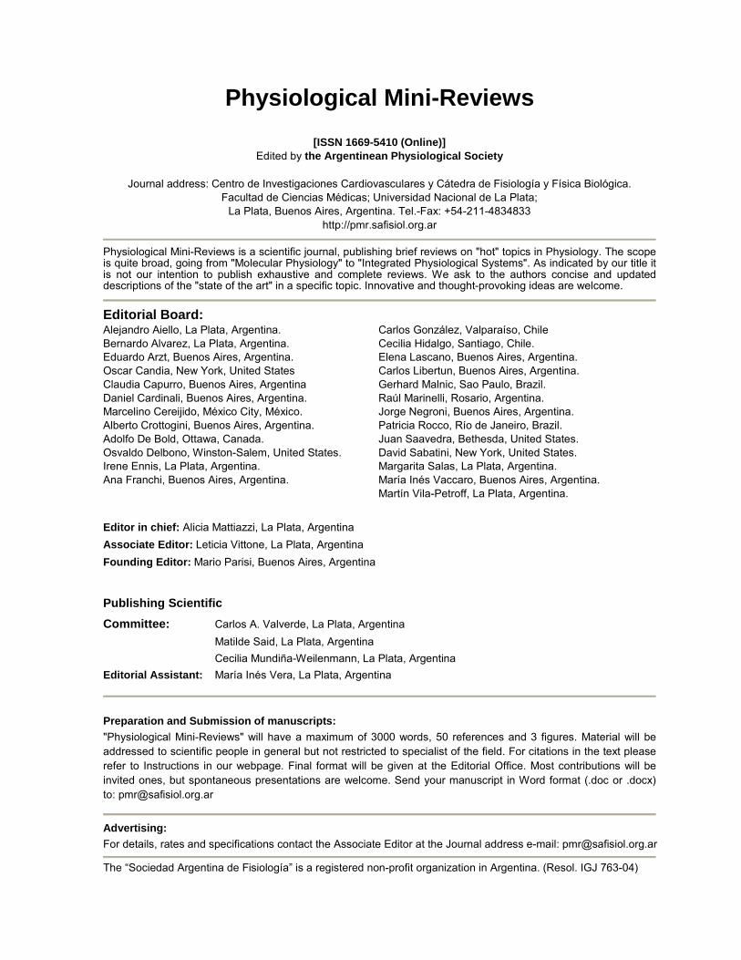
Physiological Mini-Reviews
[ISSN 1669-5410 (Online)] Edited by the Argentinean Physiological Society
Journal address: Centro de Investigaciones Cardiovasculares y Cátedra de Fisiología y Física Biológica.
Facultad de Ciencias Médicas; Universidad Nacional de La Plata; La Plata, Buenos Aires, Argentina. Tel.-Fax: +54-211-4834833
http://pmr.safisiol.org.ar
Physiological Mini-Reviews is a scientific journal, publishing brief reviews on "hot" topics in Physiology. The scope is quite broad, going from "Molecular Physiology" to "Integrated Physiological Systems". As indicated by our title it is not our intention to publish exhaustive and complete reviews. We ask to the authors concise and updated descriptions of the "state of the art" in a specific topic. Innovative and thought-provoking ideas are welcome.
Editorial Board: Alejandro Aiello, La Plata, Argentina. Bernardo Alvarez, La Plata, Argentina. Eduardo Arzt, Buenos Aires, Argentina. Oscar Candia, New York, United States Claudia Capurro, Buenos Aires, Argentina Daniel Cardinali, Buenos Aires, Argentina. Marcelino Cereijido, México City, México. Alberto Crottogini, Buenos Aires, Argentina. Adolfo De Bold, Ottawa, Canada. Osvaldo Delbono, Winston-Salem, United States. Irene Ennis, La Plata, Argentina. Ana Franchi, Buenos Aires, Argentina.
Carlos González, Valparaíso, Chile Cecilia Hidalgo, Santiago, Chile. Elena Lascano, Buenos Aires, Argentina. Carlos Libertun, Buenos Aires, Argentina. Gerhard Malnic, Sao Paulo, Brazil. Raúl Marinelli, Rosario, Argentina. Jorge Negroni, Buenos Aires, Argentina. Patricia Rocco, Río de Janeiro, Brazil. Juan Saavedra, Bethesda, United States. David Sabatini, New York, United States. Margarita Salas, La Plata, Argentina. María Inés Vaccaro, Buenos Aires, Argentina. Martín Vila-Petroff, La Plata, Argentina.
Editor in chief: Alicia Mattiazzi, La Plata, Argentina Associate Editor: Leticia Vittone, La Plata, Argentina Founding Editor: Mario Parisi, Buenos Aires, Argentina
Publishing Scientific Committee: Carlos A. Valverde, La Plata, Argentina Matilde Said, La Plata, Argentina Cecilia Mundiña-Weilenmann, La Plata, Argentina Editorial Assistant: María Inés Vera, La Plata, Argentina
Preparation and Submission of manuscripts: "Physiological Mini-Reviews" will have a maximum of 3000 words, 50 references and 3 figures. Material will be addressed to scientific people in general but not restricted to specialist of the field. For citations in the text please refer to Instructions in our webpage. Final format will be given at the Editorial Office. Most contributions will be invited ones, but spontaneous presentations are welcome. Send your manuscript in Word format (.doc or .docx) to: [email protected]
Advertising: For details, rates and specifications contact the Associate Editor at the Journal address e-mail: [email protected]
The “Sociedad Argentina de Fisiología” is a registered non-profit organization in Argentina. (Resol. IGJ 763-04)

Physiological Mini Reviews Vol 9 # 7, 2016
PhysiologicalMini
Reviews
SAFISSociedad Argentina de Fisiología
It is the intention of the Editorial Committee of PMR to add to our publications, special issues with the abstracts of the Congresses of the physiological societies of the Latin American countries who agree to collaborate with our journal, with the main goal of increasing the scientific and academic interaction among our countries.

1
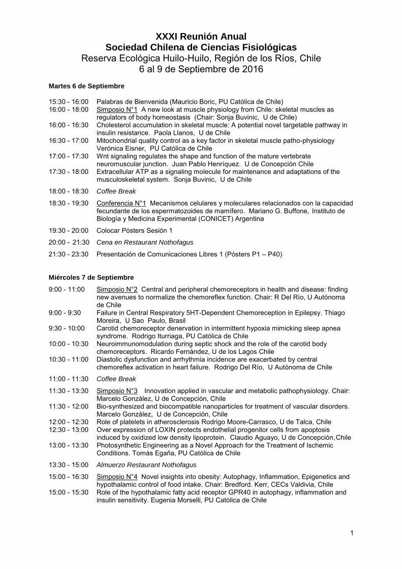
XXXI Reunión Anual Sociedad Chilena de Ciencias Fisiológicas
Reserva Ecológica Huilo-Huilo, Región de los Ríos, Chile 6 al 9 de Septiembre de 2016
Martes 6 de Septiembre 15:30 - 16:00 Palabras de Bienvenida (Mauricio Boric, PU Católica de Chile) 16:00 - 18:00 Simposio N°1 A new look at muscle physiology from Chile: skeletal muscles as
regulators of body homeostasis (Chair: Sonja Buvinic, U de Chile) 16:00 - 16:30 Cholesterol accumulation in skeletal muscle: A potential novel targetable pathway in
insulin resistance. Paola Llanos, U de Chile 16:30 - 17:00 Mitochondrial quality control as a key factor in skeletal muscle patho-physiology
Verónica Eisner, PU Católica de Chile 17:00 - 17:30 Wnt signaling regulates the shape and function of the mature vertebrate
neuromuscular junction. Juan Pablo Henríquez. U de Concepción Chile 17:30 - 18:00 Extracellular ATP as a signaling molecule for maintenance and adaptations of the
musculoskeletal system. Sonja Buvinic, U de Chile
18:00 - 18:30 Coffee Break
18:30 - 19:30 Conferencia N°1 Mecanismos celulares y moleculares relacionados con la capacidad fecundante de los espermatozoides de mamífero. Mariano G. Buffone, Instituto de Biología y Medicina Experimental (CONICET) Argentina
19:30 - 20:00 Colocar Pósters Sesión 1
20:00 - 21:30 Cena en Restaurant Nothofagus
21:30 - 23:30 Presentación de Comunicaciones Libres 1 (Pósters P1 – P40) Miércoles 7 de Septiembre
9:00 - 11:00 Simposio N°2 Central and peripheral chemoreceptors in health and disease: finding new avenues to normalize the chemoreflex function. Chair: R Del Río, U Autónoma de Chile
9:00 - 9:30 Failure in Central Respiratory 5HT-Dependent Chemoreception in Epilepsy. Thiago Moreira, U Sao Paulo, Brasil
9:30 - 10:00 Carotid chemoreceptor denervation in intermittent hypoxia mimicking sleep apnea syndrome. Rodrigo Iturriaga, PU Católica de Chile
10:00 - 10:30 Neuroimmunomodulation during septic shock and the role of the carotid body chemoreceptors. Ricardo Fernández, U de los Lagos Chile
10:30 - 11:00 Diastolic dysfunction and arrhythmia incidence are exacerbated by central chemoreflex activation in heart failure. Rodrigo Del Río, U Autónoma de Chile
11:00 - 11:30 Coffee Break
11:30 - 13:30 Simposio N°3 Innovation applied in vascular and metabolic pathophysiology. Chair: Marcelo González, U de Concepción, Chile
11:30 - 12:00 Bio-synthesized and biocompatible nanoparticles for treatment of vascular disorders. Marcelo González, U de Concepción, Chile
12:00 - 12:30 Role of platelets in atherosclerosis Rodrigo Moore-Carrasco, U de Talca, Chile 12:30 - 13:00 Over expression of LOXIN protects endothelial progenitor cells from apoptosis
induced by oxidized low density lipoprotein. Claudio Aguayo, U de Concepción, Chile 13:00 - 13:30 Photosynthetic Engineering as a Novel Approach for the Treatment of Ischemic
Conditions. Tomás Egaña, PU Católica de Chile
13:30 - 15:00 Almuerzo Restaurant Nothofagus
15:00 - 16:30 Simposio N°4 Novel insights into obesity: Autophagy, Inflammation, Epigenetics and hypothalamic control of food intake. Chair: Bredford. Kerr, CECs Valdivia, Chile
15:00 - 15:30 Role of the hypothalamic fatty acid receptor GPR40 in autophagy, inflammation and insulin sensitivity. Eugenia Morselli, PU Católica de Chile

2
15:30 - 16:00 Role of non-opioid dynorphin peptides in control of food intake and physical activity through the paraventricular hypothalamic nucleus. Claudio Pérez-Leighton, U Andrés Bello, Chile
16:00 - 16:30 Hypothalamus-adipose tissue interplay for the proper control of body weight, let´s talk from the epigenetic point of view. Bredford Kerr, CECs Valdivia, Chile
16:30 - 17:00 Coffee Break
17:00 - 18:30 Simposio N°5 Nuevas propuestas para un aprendizaje activo de la biología y la fisiología. Chair: Victoria Velarde, PU Católica de Chile
17:00 - 17:30 Utilización de plataformas computacionales en el aprendizaje activo del laboratorio de Fisiología. Loreto Véliz, PU Católica de Chile
17:30 - 18:00 El video juego como una herramienta para el aprendizaje de la fisiología. Victoria Velarde, PU Católica de Chile
18:00 - 18:30 ¿Cuánto contribuye la asistencia a clases en el aprendizaje de los estudiantes universitarios? Beatriz Ramirez, U de Santiago, Chile
18:30 - 19:30 Reconocimiento-Homenaje a la Dra. Beatriz Ramirez U. Nombramiento como Socia Honoraria de la de la Sociedad Chilena de Ciencias Fisiológicas.
19:00 – 19:30 Asamblea de Socios
19:30 - 20:00 Colocar Pósters Sesión 2
20:00 -21:30 Cena en Restaurant Nothofagus
21:30 - 23:30 Presentación de Comunicaciones Libres 2 (Pósters P41 – P80) Jueves 8 de Septiembre
9:00 - 11:00 Simposio N°6 Conexinns and Pannexins in health and disease, from regulation to cell physiology. Chair: Mauricio Retamal, U del Desarrollo, Chile
9:00 - 9:30 Oligodendrocytes form gap junctions constituted of endogenously expressed Pannexin1. Juan Carlos Sáez, PU Católica de Chile
9:30 - 10:00 Rol de hemicanales en ansiedad y memoria. Jimmy Stehberg, U Andrés Bello Chile 10:00 - 10:30 Hyperactive Hemichannels in Syndromic Deafness. Agustín Martínez, U de
Valparaíso, Chile 10:30 - 11:00 Transmisores gaseosos como moduladores de hemicanales formados por Cxs
Mauricio Retamal, U del Desarrollo, Chile
11:00 - 11:30 Coffee Break
11:30 - 13:30 Simposio N°7 Avances en Cardiología Molecular (Chair: Paulina Donoso, U de Chile) 11:30 - 12:00 Modificaciones postraduccionales del RyR2 y arritmias de reperfusión. Matilde Said,
U Nacional de la Plata, Argentina 12:00 - 12:30 Rol de policystina 1 en cardiomiocitos. Zully Pedrozo, U de Chile 12:30 - 13:00 Equilibrio nitroso-redox en el miocito cardiaco. Daniel González, U de Talca, Chile 13:00 - 13:30 Fibroblastos cardiacos: células centinela en el tejido cardiaco. Guillermo Díaz, U de
Chile
13:30 - 15:00 Almuerzo Restaurant Nothofagus
15:00 - 18:30 Tarde Libre: Visitas a senderos auto-guiados, etc.
18:30 - 20:00 Simposio N°8 Molecular targets for the treatment of pulmonary vascular disease. Chair: Mauricio Henríquez, U de Chile
18:30 - 19:00 Current and expected pathways in the therapy against pulmonary arterial hypertension. Mónica Zagolin, Instituto Nacional del. Tórax, Chile
19:00 - 19:30 Heme oxygenase induction, an evolution's old recipe in a new therapeutical strategy German Ebensperger, U de Chile
19:30 - 20:00 Purinergic signaling as alternative pathway to treat adult Pulmonary Arterial Hypertension. Mauricio Henríquez, U de Chile
20:00 - 21:00 Conferencia N°2 Anti-apoptotic Bcl-2 and intracellular Ca2+-signaling modulation in health and disease Geert Bultink, U Leuven, Belgium
21:00 - 22:30 Cena en Cervecería Petermann
22:30 - 2:00 Fiesta

3
Viernes 9 de Septiembre
9:30 - 11:30 Simposio N°9 Canales de iones: Más allá de las neuronas. Chair: Carlos Flores, CECs, Valdivia, Chile
9:30 - 10:00 Vascular Control by voltage-dependent ion channels in the endothelial cells. Xavier Figueroa, PU Católica de Chile
10:00 - 10:30 Nuevos mecanismos de regulación de canales TRPs. Oscar Cerda U de Chile 10:30 - 11:00 Functional effect of the interaction of the angiotensin receptor type 1 with the L-type
calcium channel. Diego Varela, U de Chile 11:00 - 11:30 Rol del canal KCa3.1 en la fisiología de las vías aéreas. Carlos Flores, CECs,
Valdivia, Chile
11:30 - 12:00 Coffee Break
12:00 - 13:00 Conferencia N°3 Ecología para el desarrollo de la industria del vino: acortando el abismo entre la producción y la conservación de la biodiversidad. Olga Barbosa, U Austral de Chile, Valdivia, Chile.
13:00 - 13:30 Palabras de Cierre
XXXI Reunión Anual Sociedad Chilena de Ciencias Fisiológicas
4
Libro de Resúmenes XXXI Reunión Anual
Sociedad Chilena de Ciencias Fisiológicas Reserva Ecológica Huilo-Huilo, Región de los Ríos, Chile
6 al 9 de Septiembre de 2016 CONFERENCES (CONFERENCIAS) “Mecanismos celulares y moleculares relacionados con la capacidad fecundante de los espermatozoides de mamífero” [C.1.] Los espermatozoides de ratón realizan la exocitosis acrosomal en los segmentos altos del oviducto. La Spina FA1, Romarowski A1, Puga Molina L1, Krapf D2, Falzone T1
, Luque GM. Hirohashi N3, Buffone MG1. 1IBYME-CONICET, Buenos Aires, Argentina; 2IBR-CONICET, Rosario Argentina, 3Oki Marine Biological Station, Shimane University, Japón.
Introducción: Los espermatozoides de mamífero no son capaces de fertilizar inmediatamente después de la eyaculación. Primero deben atravesar un proceso denominado capacitación dentro del tracto reproductor femenino. Esto les permite desarrollar dos características fundamentales para el proceso de fertilización: la mobilidad hiperactivada y la exocitosis acrosomal (EA). Durante mucho tiempo, diversas evidencias experimentales postulaban que la EA ocurre por la interacción del espermatozoide con la zona pelúcida (ZP) del ovocito. Sin embargo, estudios recientes realizados in vitro demostraron que el espermatozoide que penetra al ovocito y fertiliza es aquel que realizó la EA previo contacto con la ZP.
El objetivo de este trabajo fue estudiar, utilizando un modelo transgénico murino, la migración de los espermatozoides a través del complejo cúmulus-ovocitos (COCs) in vitro, y la migración de los mismos a través del tracto reproductor femenino en tiempo real y dilucidar el sitio fisiológico donde ocurre la EA.
Metodología: Se utilizaron ratones transgénicos que expresan las proteínas EGFP en el acrosoma y RFP en mitocondrias (Acr3-EGFP/su9-DsRed2), y por lo tanto, podemos abalizar el estado acrosomal en tiempo real. Se evaluó el estado acrosomal de espermatozoides capacitados que atraviesan los COCs in vitro mediante microscopía de fluorescencia, y de espermatozoides dentro del tracto reproductor feme-nino post apareo con ratones macho transgénicos por microscopía de epifluorescencia con sistema live imaging, z-stacking por microscopía confocal y por criocortes del oviducto.
Resultados: 1) La frecuencia de EA de espermatozoides penetrando los COCs in vitro fue del ~5%/hora luego de la inseminación. 2) La migración de los espermatozoides a través del oviducto ocurre gradualmente, presentando en su gran mayoría acrosoma intacto en las partes bajas del oviducto. Sin embargo, identificamos una proporción de espermatozoides que realizaron la EA en los segmentos altos (~%38) mientras que el 95% se encontraba reaccionado en la ámpula.
Conclusión: En este trabajo demostramos por primera vez en el modelo murino, que los espermatozoides realizan la EA fisiológicamente en los segmentos altos del oviducto previa interacción con los COC.
Financiamiento: NIH-R01TW008662.
XXXI Reunión Anual Sociedad Chilena de Ciencias Fisiológicas
5
[C.2.] Anti-apoptotic Bcl-2 and intracellular Ca2+-signaling modulation in health and disease. Bultynck G1
1 KU Leuven & Leuven Kanker Instituut, Lab. Molecular & Cellular Signaling, Dep. Cellular & Molecular Medicine, Leuven, Belgium
Introduction: Anti-apoptotic Bcl-2 proteins not only prevents apoptosis by neutralizing pro-apoptotic Bcl-2-family members at the mitochondrial level but also by controlling Ca2+ fluxes that originate at the endoplasmic reticulum.
Conclusions: Bcl-2 directly targets different Ca2+ transport systems, including IP3 receptors, ryanodine receptors and voltage-dependent anion channels, thereby impacting their functional properties. These “Ca2+-signaling” functions of Bcl-2 proteins contribute to their anti-apoptotic effects in cells. Moreover, different anti-apoptotic Bcl-2-family members, like the much related Bcl-2 and Bcl-XL, appear to target the same Ca2+-transport systems, though often via different molecular mechanisms, molecular domains and affinities resulting in different functional outcomes. Indeed, while Bcl-2 inhibits excessive, pro-apoptotic IP3 receptor activity via its BH4 domain, Bcl-XL enhances spontaneous/low-level, pro-survival IP3 receptor activity via its hydrophobic cleft. These mechanisms are an integral part of the cell survival functions of these proteins. Moreover, cancer cells exploit these “Ca2+-signaling” functions of Bcl-2 proteins as a survival strategy. Tools, like BIRD-2 (Bcl-2 / IP3 receptor Disrupter-2) that target the BH4 domain of Bcl-2 and thus disrupt IP3 receptor/Bcl-2-complex formation elicit pro-apoptotic intracellular Ca2+ overload in B-cell cancers, including diffuse large B-cell lymphoma and chronic lymphocytic leukemia. Thus, cancer cells are addicted to Bcl-2 at the ER to suppress Ca2+ signaling. This addiction to Bcl-2 is due to a combination of the high expression levels of the type 2 IP3 receptor, the most sensitive IP3 receptor isoform, and elevated basal IP3 signaling in certain type of B-cell cancers. Hence, anti-apoptotic Bcl-2 proteins critically control intracellular Ca2+ dynamics in cells, a property exploited by cancer cells as a survival strategy.
Funding: Research Foundation – Flanders, Research Council Leuven, Stichting Tegen Kanker

XXXI Reunión Anual Sociedad Chilena de Ciencias Fisiológicas
6
SYMPOSIA (SIMPOSIOS) Symposium 1: “A new look at muscle physiology from Chile: skeletal muscles as regulators of body homeostasis”. (Coordinator: Sonja Buvinic) [S.1.] Cholesterol accumulation in skeletal muscle: A potential novel targetable pathway in insulin resistance. Llanos P.1,2 1Institute for Research in Dental Sciences, Facultad de Odontología, Universidad de Chile. 2CEMC, Facultad de Medicina, Universidad de Chile.
Skeletal muscle plays an important role in glucose homeostasis, and defects in glucose uptake in muscle are involved in states of insulin resistance (IR). An alarming increase in the prevalence of IR during the past 20 years has received worldwide attention. Alterations in glucose homeostasis, due to deficient GLUT4 translocation and reduced insulin sensitivity in skeletal muscle characterize to the IR. Skeletal muscle is the main source of GLUT4-mediated glucose transport in animals; in fact, this tissue removes >80% of circulating glucose after a meal. Most GLUT4-mediated glucose transport occurs in the transverse tubules (TT), a specialized plasma membrane system of skeletal muscle highly enriched in cholesterol. In IR, GLUT4 translocation to the TT membrane is defective. However, the role of TT cholesterol content on glucose transport is unknown. Recently, we have focused our efforts in understanding the role of cholesterol on TT and its relation to IR. New insights into GLUT4 trafficking reveal that compounds that partially reduce membrane cholesterol content modulate insulin-independent GLUT4 translocation and glucose uptake in adipocytes and muscle cell lines. We recently reported similar effects in adult muscle fibers from high fat diet (HFD)-fed obese mice. Insulin-stimulated glucose uptake in fibers from HFD-fed mice was lower than in controls whereas cholesterol levels in triads from HFD-fed mice were 30% higher compared to NCD-fed mice. Pre-incubation with Methyl-β-cyclodextrin (MβCD) to decrease membrane cholesterol, reduced Akt phosphorylation and increased both basal and insulin-induced glucose uptake in muscle fibers from controls or HFD-fed mice. Indinavir, a GLUT4 antagonist, inhibited the effects of MβCD. MβCD increased membrane GLUT4 content and elicited intracellular calcium signals that was inhibited by Dantrolene, which prevents the interaction Cav1.1/RyR1. Dantrolene also reduced MβCD-mediated glucose uptake. In HFD-fed mice, four subcutaneous injections of MβCD improved their defective glucose tolerance test and normalized their high fasting glucose levels. In conclusion, cholesterol accumulation on TT has a role in regulation of glucose homeostasis during the IR condition. These emerging results may provide a rationale for the clinical evaluation of cyclodextrins, as an approach to the treatment of IR and the control of type 2 diabetes. Supported by: FONDECYT 11150243 & 1151293; FIOUCh-Enlace 001/2015. [S.2.] Mitochondrial quality control as a key factor in skeletal muscle patho-physiology. Castro M, Vial J, Brandan E, Eisner V. Departamento Biología Celular y Molecular, Facultad de Ciencias Biológicas, Pontificia Universidad Católica de Chile; [email protected]
All cells require balanced mitochondrial bioenergetics system to support its metabolism and function, in particular, skeletal muscle relay on mitochondria a key source of ATP. Healthy mitochondria relay on dynamic processes such as fusion, fission and motility along the cytoskeleton. We have previously demonstrated that mitochondrial fusion is active in highly structured adult skeletal muscle and that it is key for excitation-contraction coupling calcium regulation. Moreover, mitochondria fusion is targeted by pathological conditions such as alcoholic myopathy. Yet, despite the highly organized architecture of the muscle fiber, little is known about the relevance of mitochondrial-to-microtubules interaction on mitochondrial bioenergetics. We are currently studying mitochondrial fusion in Duchenne Muscle Dystrophy skeletal muscle; a fibrosis disease characterized the absence of dystrophin, an extracellular matrix-to-cytoskeleton anchor protein. Interestingly, mdx muscle fibers display microtubule-aberrant
XXXI Reunión Anual Sociedad Chilena de Ciencias Fisiológicas
7
organization, and thus, allow us studying the role of mitochondria-to-cytoskeleton regulatory alliance, by means of in vivo electroporation of exogenous markers and imaging of mitochondrial dynamics. Our results show that the mdx dystrophic adult skeletal muscle displays aberrant mitochondrial topology and suffers both inhibition and increased mitochondrial fusion. Fibrosis is triggered by Connective Tissue Growth Factor (CTGF) signaling. Our data shows that muscle fibers from mdx-CTGF (+/-) mice, that displays inhibition of fibrosis and muscle function restoration, rescue the mitochondrial fusion pattern to the level of wild type fibers. We sought to understand the relevance of mitochondrial-to-cytoskeleton interaction in skeletal muscle mitochondrial bioenergetics. Funding: FONDECYT 11550677 to VE and 1150106 and CARE-PFB-12/2007/Conicyt to EB. [S.3.] Signaling pathways regulating the shape and function of the mature neuromuscular junction. Ojeda J1#, Pérez V1#, Tabares L2, Bronfman F3, Henríquez JP1* (*[email protected]) (#equal contribution). 1University of Concepcion, Concepcion, Chile; 2University of Seville, Seville, Spain; 3P. Universidad Católica de Chile, Santiago, Chile; 1,3Millenium Nucleus for Regenerative Biology (MINREB).
The neuromuscular junction (NMJ) is an archetypical model to analyze synapse formation, maturation and maintenance. Clustering of nicotinic acetylcholine receptors (AChR) is a hallmark of postsynaptic differentiation at the NMJ. Early NMJ assembly relies on both, pre- and postsynaptic signals. However, the molecular mechanisms involved in post-natal NMJ maturation and maintenance are still to be fully elucidated. In the last years, our laboratory has focused on studying the effect that pathways activated by Wnts and neurotrophins could exert at the mature NMJ. To analyze Wnt signaling, we have focused on Frizzled-9 (Fzd9), a muscle Wnt receptor which expression depends on innervation. Fzd9 inhibits agrin-induced acetylcholine receptor (AChR) clustering in cultured myotubes. In vivo, co-localization studies showed that Fzd9 distributes in postsynaptic AChR rich-areas from P0 to P28. In turn, the Wnt effector beta-catenin distributes in regions devoid of AChRs at early stages and in AChR rich-areas as NMJ maturation proceeds. Gain- and loss-of-function experiments through muscle electroporation showed that NMJs with altered expression of Fzd9 significantly modified the proportion and size of complex mature postsynaptic shapes. Accordingly, electrophysiological postsynaptic recording revealed altered synaptic transmission and input resistance in fibers where Fzd9 expression has been modified. The role of neurotrophin-mediated signaling has been analyzed in mice null for p75, a co-receptor for all Trk receptors. p75 mutants display a marked walking phenotype and impaired motor coordination, in spite of exhibiting similar sensory responses. p75 disruption also resulted in muscle weakness, decreased muscle fiber diameter and increased slow fibers. In addition, contractile activity recording showed impaired muscle resistance in mutant muscles upon high-frequency presynaptic stimulation. p75 null animals displayed delayed NMJ maturation with smaller postsynaptic apparatuses. In vitro primary myotube cultures reveal that muscle-derived p75 is not involved in the observed postsynaptic defects. In turn, ultrastructural analyses by EM showed a significant reduction in the number of presynaptic vesicles at the motor terminal of p75 null NMJs. Remarkably, chronic administration of acetylcholinesterase inhibitors significantly rescued the motor coordination performance of p75 null mice. Together, our findings show that Wnt signaling and neurotrophins are required to maintain functional postsynaptic apparatuses by acting on different cell types of the mature NMJ. Funding FONDECYT 1110321 and MINREB RC120003, Chile. [S.4.] Extracellular ATP as a signaling molecule for maintenance and adaptations of the musculoskeletal system Arias-Calderón M 1, Rojas C 1, Hernández N 1, Bohle P 1, Morales C 1,2, Balanta J1,3, Gómez F 1, Vicencio N
1, Verdejo C 1, Casas M 4, Llanos P 1, and Buvinic S 1 1 Lab. Biología Celular y Molecular, Instituto de Investigación en Cs. Odontológicas, Facultad de Odontología, Universidad de Chile, Santiago, Chile. 2 Facultad de Salud, Pontificia Universidad Javeriana, Cali, Colombia. 3 Escuela de Odontología, Universidad del Valle, Cali, Colombia. 4 Centro de Estudios Moleculares de la Célula, ICBM, Facultad de Medicina, Universidad de Chile, Santiago, Chile.

XXXI Reunión Anual Sociedad Chilena de Ciencias Fisiológicas
8
Molecular basis of muscle-bone crosstalk is an unsolved issue in the whole musculoskeletal system, but specially in head and neck tissues, that are embryological and biochemically different than the trunk and limbs ones. In hind limb muscles we have demonstrated that extracellular ATP is a relevant mediator between membrane depolarization and gene expression. Depolarization of muscle membrane promotes ATP release through Pannexin 1 (Panx1) channels, which activates P2Y receptors (P2YR) and promotes changes in gene expression. Molecules involved in this process (voltage sensor, Panx1, P2YR, signaling molecules) assemble a multiprotein complex to finely regulate its activity. We are currently studying the molecular mechanisms for muscle plasticity and muscle-bone crosstalk at the mouse masticatory system. Historically, it has been thought that muscles and bones are just related through mechanical processes. However, it has now emerged the idea of a biochemical communication, where they interact through soluble molecules – “myokines” and “osteokines” - to maintain the homeostasis of the whole system. Considering that both muscle and bone releases and responds to extracellular ATP, we postulate it would be a relevant molecule for muscle-bone crosstalk. We have detected mRNA for purinergic receptors (P2YR, P2XR) in masseter and digastric muscles, mandible and maxilla from BalbC mouse. The components of the multiprotein complex previously described in limb muscles, are also expressed in masseter muscle (dihydropyridine receptor, Panx1 and P2Y2R). Tetanic electrical stimulation (20Hz, 270 pulses, 0.3 msec each) evokes ATP release from masseter muscle. Exogenous 100 µM ATP regulated the expression pattern of interleukin 1beta (IL1β), interleukin6 (IL6), troponinI fast/slow, PGC1alpha and citrate synthase in mouse masseter muscle. Tetanic electrical stimulation also increases mRNA levels of IL1β and IL6 up to 15 and 150 folds, respectively. Increase in gene expression is abolished when the purinergic pathway is blocked with apyrase (ATP metabolizing enzyme) or suramin (P2Y/P2X receptors blocker), or when the voltage sensor DHPR is inhibited with nifedipine. Conditioned medium derived from masseter muscle resembles the effect of 1 µM ATP in osteoclastogenesis of the pre-osteoclast RAW264.7 cell line. Differentiation to a giant multinucleated phenotype with increased expression of osteoclastogenic markers is observed. In sum, we propose that masticatory system have a functional purinergic signaling pathway relevant for muscle plasticity and muscle-bone crosstalk. Funded by Fondecyt-1151353(SB)-11150243(PLl), Conicyt 21151035-63140009-21150059.

XXXI Reunión Anual Sociedad Chilena de Ciencias Fisiológicas
9
Symposium 2: “Central and peripheral chemoreceptors in health and disease: finding new avenues to normalize the chemoreflex function” (coordinator Rodrigo Del Río) [S.5.] Failure in Central Respiratory 5HT-Dependent Chemoreception in a Genetic Model of Epilepsy. Moreira TS1, Totola, LT1, de Oliveira JA2, Garcia-Cairasco N2, Takakura AC3. 1Dept. of Physiology and Biophysics, Institute of Biomedical Sciences, Univ. of São Paulo, São Paulo, SP, 05508, Brazil; 2Dept. of Physiology, School of Medicine of Ribeirão Preto, University of São Paulo, Ribeirão Preto, SP, Brazil; 3Dept. of Pharmacology, Institute of Biomedical Sciences, Univ. of São Paulo, São Paulo, SP, 05508, Brazil
Introduction: Sudden unexpected death in epilepsy (SUDEP) is the leading cause of death in patients with refractory epilepsy. There is still a debate in the literature if the respiratory arrest is the primary cause of death. Respiratory chemoreceptors neurons located in retrotrapezoid nucleus (RTN) constitute one of the main groups responsible for controlling breathing automaticity and receive a dense serotonergic innervation. Objective: Here, we ask whether alterations of a type of chemoreceptor neurons that expresses the transcription factor Phox2b and is non-catecholaminergic (Phox2b+/TH-) could affect breathing in a rat model of tonic-clonic seizures. Methods: We used a rat model of tonic-clonic seizures, with susceptibility to audiogenic seizures (the Wistar Audiogenic Rat (WAR) strain (WAR: 340-496 g, n = 6-7). Results: The number of Phox2b+/TH- neurons in the RTN was reduced (79±11%) in WAR. The WARs have reduced resting ventilation (VE) by 35 ± 10% and reduced increase in VE elicited by hypercapnia (7% CO2) by 51±7%. Furthermore, we also showed that the VE response to serotonin (1 mM - 50 nl) into the RTN region was significantly hampered in WARs due to the reduction in the number of 5-HT varicosities within the marginal layer of the RTN region. Conclusion: Our results suggest a respiratory disorder, as well as a reduction of the serotonergic neurotransmission within the RTN region, which justify to further use WAR as a suitable model to study increased risk factors and the mechanisms associated with SUDEP. Financial Support: FAPESP, CNPq and CAPES/PROEX [S.6.] Cardiorespiratory acclimatization induced by intermittent hypoxia: A new role for the carotid body chemoreceptor. Iturriaga R Laboratorio Neurobiología, Facultad de Ciencias Biológicas, Pontificia Universidad Católica de Chile.
The carotid body is considered the main oxygen chemoreceptor in mammals, which mediates reflex respiratory and cardiovascular adjustments to acute hypoxia and the ventilatory acclimation to sustained chronic hypoxia in high altitude. However, the most usual form of chronic hypoxia in humans is the intermittent hypoxia resulting from obstructive sleep apnea (OSA). This sleep breathing disorder is considered an independent risk factor for hypertension and stroke. Endothelial dysfunction, oxidative stress, inflammation and sympathetic activation have been proposed as potential mechanisms involved in the hypertension induced by OSA. However, evidence for a unique pathogenic mechanism has been difficult to establish in OSA patients because of concomitant comorbidities. Thus, animal models have been developed to study the pathological consequences of exposure to chronic intermittent hypoxia. Since OSA patients and animals exposed to chronic intermittent hypoxia (CIH) show enhanced ventilatory, sympathetic and cardiovascular responses to hypoxia, it has been proposed that and enhanced carotid body responsiveness to hypoxia is involved in the autonomic changes induced by OSA and in the development of the hypertension. This proposal received further support from recordings of carotid body chemosensory discharges showing that intermittent hypoxia increases basal carotid chemosensory discharges and the responses to hypoxia. In this symposium, I will discuss new experimental evidence supporting an important role for the carotid body in the progression of cardiorespiratory acclimatization induced by CIH and the contribution of oxidative stress, endothelin-1 and pro-inflammatory molecules in the potentiation of the carotid body chemosensory function induced by CIH.
XXXI Reunión Anual Sociedad Chilena de Ciencias Fisiológicas
10
In addition, I will show new evidence that carotid body ablation normalized the elevated arterial blood pressure in conscious rats exposed to CIH, and restored the cardiac autonomic and baroreflex function. These results suggest that autonomic alterations induced by CIH depend on the enhanced carotid responsiveness and support a main role for the CB in the CIH-induced hypertension in OSA patients. Funding: Supported by FONDECYT 1150040 [S.7.] Carotid/sinus nerve stimulation modifies plasma cytokines profile and prevents multiple organ dysfunction in lipopolysaccharide-induced septic rats. Fernández R1, Cortés PP1, Reyes EP2. 1Departamento de Salud, Universidad de Los Lagos, Osorno, Chile. 2Facultad de Medicina, Clínica Alemana-Universidad del Desarrollo, Santiago, Chile.
Introduction: We recently demonstrated that bilateral carotid chemo/baro-denervation modifies the neural, endocrine and inflammatory responses to sepsis, anticipating multiple organ dysfunction (MOD) onset and death. Since carotid body senses inflammatory status and its stimulation provokes a wide array of cardiopulmonary and autonomic reflexes as well as endocrine responses (e.g., plasma release of catecholamines and cortisol), we propose that carotid/sinus nerve stimulation could play a protective role during sepsis. Objective: To determine the effect of carotid/sinus nerve electrical stimulation (STIM) in sepsis progression to MOD in septic rats. We suggest that restoring carotid chemo/baro-sensory function could delay MOD onset and increase survival time. Methods: In anesthetized male rats, we measured cardiorespiratory variables and plasma cytokines/chemokines profile, glucocorticoids, epinephrine, and MOD markers levels 90 min after I.P. administration of lipopolysaccharide (LPS) under three experimental conditions, control (SHAM surgery); bilateral carotid chemo/baro-denervated (BCN); and, BCN + STIM rats; the latter condition, by using two-episodes of 2 min stimulation (20 Hz, 0.2 ms, 100 µA), 10 min before LPS and immediately after LPS administration. Results: LPS administration to rats with both carotid/sinus nerves intact (SHAM-LPS) increases plasma IL-1β, IL-10 and TNF-α. LPS administration to BCN rats (BCN-LPS) increases both IL-2 and TNFα, and decreases IL-1β and IL-10. Electrical stimulation of septic denervated rats (BCN-STIM-LPS) evokes a similar pattern of inflammatory profile compared with saline-treated SHAM rats. Plasma markers of MOD were reduced compared with BCN-LPS. Finally, BCN-STIM-LPS rats show both hypotensive and tachycardic response to LPS, but these responses were lower than those observed in BCN-LPS. Furthermore, mortality rate was higher in BCN-LPS rats than BCN-STIM-LPS rats. Conclusion: The complete absence of carotid chemo/baro-sensory function modifies the neural, endocrine and inflammatory responses to sepsis. Carotid/sinus nerve stimulation restores, at least in part, the tonic control of systemic inflammation exerted by peripheral chemoreceptors. Carotid chemo- and baroreflexes are important modulators of sympathetic activity and their peripheral activation elicits respiratory and cardiovascular effects and a sympatho-excitatory response that could reverse sepsis dysautonomia. Suppported by Fondecyt 1120976; Dirección de Investigación, Universidad de Los Lagos. [S.8.] Central Chemoreflex Activation Exacerbates Diastolic Dysfunction and Arrhythmia Incidence in HFpEF. Del Rio R 1, Toledo C 1, Andrade DC 1, Lucero C 1, Díaz H 1, Aliaga V 1, Arce-Alvarez A 1, Manríquez M 1, Faundez M 1. 1Lab. Cardiorespiratory Control, Universidad Autónoma de Chile, Santiago, Chile.
Background: Heart failure with preserved ejection fraction (HFpEF) is characterized by increased sympathetic drive and breathing disorders. Tonic and/or episodic chemoreflex activation contributes to autonomic imbalance in heart failure and is associated with poor prognosis. Objective: We sought to determine whether acute activation of the central chemoreflex (CC) exacerbates autonomic imbalance, cardiac arrhythmogenesis and cardiac dysfunction in HFpEF rats.
SHAM-saline

XXXI Reunión Anual Sociedad Chilena de Ciencias Fisiológicas
11
Methods: Volume overload (a-v anastomosis) was used to induce HFpEF in adult male Sprague-Dawley rats. Ventilatory responses to acute hypercapnia (FiCO2-7% in O2) were measured by plethysmography to assess CC sensitivity, and a conductance catheter was used to measure pressure-volume relationships as a measure of cardiac function. Autonomic balance was assessed by indirect heart rate variability (HRV) method. Neuronal activation was assessed by measuring FosB expression in brainstem micropunches from chemosensitive regions of the retrotrapezoid nucleus (RTN) and pre-sympathetic regions of the rostral ventrolateral medulla (RVLM). Results: Ejection fraction did not differ between HFpEF and sham rats (51±3 vs. 50±7 %, HFpEF vs. sham). HFpEF rats compared to sham rats exhibited cardiac hypertrophy (heart/body weight, 6.1±0.3 vs. 4.0±0.5 mg/g, HFpEF vs. sham), pulmonary congestion (lung wet/dry weight, 4.4±0.1 vs. 3.8±0.1 g/g, HFpEF vs. sham), increased arrhythmia incidence (104±34 vs. 11±2 events/h, HFpEF vs. sham), and greater ventricular stiffness (β, 7.4±1.4 vs. 4.3±0.7 mmHg/ml, HFpEF vs. sham). In HFpEF rats, CC sensitivity was increased (165.3±9.1 vs. 127.3±10.3 ml/min/100g, HFpEF vs. sham), and CC stimulation (normocapnia vs. hypercapnia) significantly increased (P<.05) arrhythmia incidence (104±34 vs. 1164±204 events/h,) and β (7.4±1.4 vs. 17.5±7.5 1/ml). Furthermore, CC activation leads to further sympathoexcitation to the heart. Indeed, HRV showed a shift towards sympathetic modulation of heart rate. Accordingly, the low to high frequency ratio of the HRV (LF/HF) change from 0.5±0.1 in normoxia to 1.3±0.4 during CC activation with hypercapnia HFpEF rats. RVLM activation (FosB expression) was increased in HFpEF rats compared to sham animals. Conclusion: Our results indicate that the CC is enhanced in in HFpEF, neuronal activation is increased in pre-sympathetic regions of the brainstem, and acute CC activation further exacerbates diastolic dysfunction and arrhythmia incidence in HFpEF. Funding: Supported by Fondecyt 1140275.

XXXI Reunión Anual Sociedad Chilena de Ciencias Fisiológicas
12
Symposium 3: “Innovation applied in vascular and metabolic pathophysiology” (Coordinator M González) [S.9.] Bio-synthesized and biocompatible nanoparticles for treatment of vascular disorders Haensgen A1, Parada MS2, RojasS1, Rodríguez-Llamazares3, Mondaca MA4, Fernández K2, González M1,5 1Laboratorio de Fisiología Vascular, Departamento de Fisiología, Facultad de Ciencias Biológicas, Universidad de Concepción. 2Departamento de Ingeniería Química, Facultad de Ingeniería, Universidad de Concepción. 3Centro de Investigación en Polímeros Avanzados (CIPA), Concepción. 4Departamento de Microbiología, Facultad de Ciencias Biológicas, Universidad de Concepción. 5Grupo de Investigación e Innovación en Salud Vascular (GRIVAS).
The incidence of cardiovascular diseases in Chilean population drives the search for innovative therapies and prevention in early pathophysiological stages. The association of cardiovascular dysfunction with oxidative stress in early steps of the pathology and with the endothelial dysfunction open the possibility for the use of different antioxidant tools as an approach that need the combination of multidiscipline knowledge. In this filed, the main problem with the consumption of natural antioxidants is related with the lower in vivo bioavailability after oral intake. A novel biotechnology strategy to avoid this problem is the use of nanoparticles for improves the bioavailability in circulation, protecting the molecules against degradation and improving sustained release. The aim of this work was to establish a multidisciplinary collaboration to find significant application using the combination of nanoparticles engineering and endothelial cell biology. Our results shows that the selenium nanoparticles biosynthesized by the bacteria Pantoea agglomerans or the nanoparticles with extracts of grapes of leaves of nalca or murta have high concentration of antioxidants and induces protection and nitric oxide synthesis in human endothelial cells, open the possibility to uses these particles as nutraceutics of functional additives for the prevention of cardiovascular diseases. These results derive from innovative collaboration with other disciplines like microbiology; polymers biotechnology and chemical engineer for apply physiology, showing the potentiality of the combination of endothelial cell biology with biotechnology. Funding: FONDEF–IDeA CA12I10374, FONDECYT 1120148, FONDECYT 11100192 and VRID-Asociativo 213.A84.014-1.0 [S.10.] Role of platelets in atherosclerosis Moore-Carrasco R1, Huilcaman R1, Brown N2. 1Departamento de Bioquímica Clínica e Inmunohematología, Facultad de Ciencias de la Salud, Universidad de Talca. 2Centro de Investigaciones Médicas, Escuela de Medicina, Universidad de Talca.
Introduction: The role of platelets in atherogenesis is of high interest in biomedical research. Recently multiple functions have been described, both in physiological and pathological processes. Platelets cells exhibit a pleiotropic and inflammatory behavior that could help understand in more detail atherogenesis and find new therapeutic targets. Aims: Determine the ability of platelets to migrate through a monolayer of endothelium in pro-atherogenic conditions and assess the role of Peroxisome proliferator-activated receptor (PPARs). Methods: Transendothelial migration assays were done using Transwell plates. HMEC-1 endothelial cells were grown in monolayers and above them we added: platelets, THP-1 cells (monocytes) ora combination of both cell types. Cell migration was induced by fMLP peptide application, oxidized LDL (oxLDL) and cholesterol alone or in combination. We identified cells that passed through to the lower well using specific antibodies against platelets (anti-CD61) and SYTOX Green (monocytes) followed by confocal microscopy imaging. We also performed assays in the presence of Quercetin (a PPARγ agonist) and GW6471 (a PPARαantagonist) to elucidate the role of PPAR in atherogenesis. Results: The number of platelets that migrated through the endothelial monolayer in presence of fMLP peptide was increased when they were co-cultured with monocytes compared to platelets alone (p <0.05). Therefore, leukocytes in the co-culture favored both platelet migration alone and in association with monocytes. Endothelial monolayers in pro-atherogenic conditions were more permissive to platelet migration (p <0.05), effect was augmented by monocytes. Quercetin blocked the migratory ability of
XXXI Reunión Anual Sociedad Chilena de Ciencias Fisiológicas
13
platelets alone and in association with monocytes in pro-atherogenic conditions. In the other hand, GW6471 exacerbated platelet phagocytosis during transmigration and cell migration. Conclusions: Platelets were able to migrate through endothelial monolayer. Interestingly our data indicates a paracrine effect of monocytes on platelet migration. Furthermore, PPAR agonists were able to block migration of both platelet and monocytes. In the other hand, PPAR antagonists favored platelet phagocytosis by monocytes. In sum, the results open a new mechanism in the prevention and treatment of atherosclerosis. [S.11.] Overexpression of LOXIN protects endothelial progenitor cells from apoptosis induced by oxidized low density lipoprotein Reyes C1 Ormazabal V1, Willis ND2, Toledo J3, Radojkovic C1, Zuñiga FA1, Escudero C4,5, Aguayo C1,4,5 1Department of Clinical Biochemistry and Immunology, Faculty of Pharmacy, University of Concepción, Concepción, Chile. 2Human Nutrition Research Centre, Institute of Cellular Medicine, Newcastle University, Newcastle upon Tyne, United Kingdom. 3Departament of Physiopathology, Faculty of Biological Sciences, University of Concepción, Concepción, Chile. 4Vascular Physiology Laboratory, Group of Investigation in Tumor Angiogenesis (GIANT), Department of Basic Sciences, University of Bio Bío, Chillán, Chile. 5Group of Research and Innovation in Vascular Health (GRIVAS Health).
Introduction: Human endothelial progenitor cells (hEPC) are adult stem cells located in the bone marrow and peripheral blood. hEPCs play an important role in the recovery and repair of injured endothelium, however their quantity and functional capacity is reduced in several diseases including hypercholesterolemia. Recently it has been demonstrated that hEPC express lectin-like oxidized low-density lipoprotein receptor-1 (LOX-1) and its activation by oxidized low-density lipoproteins (oxLDL) induces cellular dysfunction and apoptosis. Aim: to investigate whether overexpression of LOXIN, acts as a dominant negative plays a protective role against oxLDL-induced apoptosis in hEPC. Methods: hEPC were isolated from 50 mL peripheral venous blood by lymphocyte separation medium. Ethidium homodimer-1 incorporation was used to confirm the apoptotic effect of ox-LDL in hEPC. The plasmid pCMV6 was used to obtain the plasmid pMK-Loxin and inserted by enzymatic ligation to the shuttle-vector pAdTrack-CMV. The insertion of the gene of interest into the adenoviral genome was achieved by homolog recombination in E. coli BJ5183. Results: Human endothelial progenitor cells exposed to oxLDL showed a significant increase in LOX-1 expression, and apoptosis began at oxLDL concentrations above 50 µg/mL. All hEPC apoptosis at 200 µg/mL oxLDL. High LOXIN expression was generated using adenoviral systems in hEPC and SiHa cells transduced with 100 colony-forming units/cell. Transduced LOXIN localized to the plasma membrane and blocked oxLDL uptake mediated by LOX-1. Overexpression of LOXIN protected hEPC from oxLDL-induced apoptosis and therefore maybe a novel way of improving hEPC function and quantity. Conclusion: These results suggest that adenoviral vectors of LOXIN may provide a possible treatment for diseases related to oxLDL and vascular endothelium dysfunction, including atherosclerosis. Supported by INNOVA CORFO Chile (12IDL2-13351) and CONICYT-PII2015 0053 [S.12.] Photosynthetic engineering as a novel approach for the treatment of ischemic conditions Chávez MN1,2, Holmes C3, Miranda M2, Egaña T3. 1 Advanced Center for Chronic Diseases (ACCDiS) & Center for Molecular Studies of the Cell (CEMC), Facultad de Ciencias Químicas y Farmacéuticas & Facultad de Medicina, Universidad de Chile, Santiago, Chile. 2 FONDAP Center for Genome Regulation, Facultad de Ciencias, Universidad de Chile, Santiago, Chile. 3 Instituto de Ingeniería Biológica y Médica, Facultades de Ingeniería, Ciencias Biológicas y Medicina. Pontificia Universidad de Chile, Santiago, Chile.
Introduction: The extreme dependency to external oxygen supply observed in humans, represents a serious clinical issue as several pathological conditions are related to low oxygen tension in tissues. Objectives: In order to circumvent the limitation of oxygen self-production in animals, and improved tissue oxygenation, the main focus of our research is to engineer symbiotic approaches to generate

XXXI Reunión Anual Sociedad Chilena de Ciencias Fisiológicas
14
photosynthetic plant-vertebrate chimeric organisms (plantebrates), which could be further use for medical purposes. Methods and Results: In this context, we have created the first generation of photosynthetic materials that produce and release oxygen upon light stimulation, thus improving tissue regeneration. Further, in order to provide other therapeutic molecules in addition to oxygen, we have genetically engineered microalgae to generate materials capable to release human recombinant proteins in vitro and in vivo. Conclusions: Altogether the results represent a step forward in the development of autotrophic tissues, and suggest the use of engineered microalgae to treat a broad spectrum of hypoxic conditions. Funding: CIRM-BMBF Early Translational II Award, ICGEB CRP/CHI11-01, FONDAP 15090007.
XXXI Reunión Anual Sociedad Chilena de Ciencias Fisiológicas
15
Symposium 4: “Novel insights into obesity: Autophagy, Inflammation, Epigenetics and hypothalamic control of food intake”. (Coordinator: B. Kerr) [S.13.] “Role of the hypothalamic fatty acid receptor GPR40 in autophagy and inflammation” Hernández MP, C., Toledo L, Ávalos Y, Morselli E. Departmento de Fisiología, Facultad de Ciencias Biologicas, Pontificia Universidad Católica de Chile, Santiago, Chile.
Introduction: Chronic consumption of high fat diets, rich in saturated fatty acids, such as palmitic acid (PA), induces obesity. PA inhibits autophagy, a catabolic process that maintains cellular homeostasis, promoting hypothalamic inflammation and metabolic disorders. PA-mediated activation of the fatty acid receptor G protein–coupled receptor 40 (GPR40) has been shown to modulate inflammation; however whether PA-mediated activation of GPR40 affects autophagy and production of pro-inflammatory cytokines in the hypothalamus, specifically in neurons, is unknown. Objective: To determine if PA stimulates GPR40 inhibiting autophagy and promoting inflammation in hypothalamic neurons. Methods: In vivo, we evaluated if GPR40 localizes in the mouse hypothalamus by immunofluorescence (IF). In vitro experiments were performed on the hypothalamic neuronal cell line N43/5. Cells were exposed to pro-obesigenic concentration of PA (100µM) for 3 hours, in presence or absence of the GPR40 antagonist GW1100 (1µM). To assess PA-mediated GPR40 activation we evaluated changes in intracellular Ca2+ in cultures loaded with Fura 2-AM. Autophagy was quantified by IF and western blot while production of pro-inflammatory cytokines by qPCR. Results: In vivo, GPR40-positive cells co-localize with a neuronal marker. In vitro, PA increases intracellular Ca2+ levels, inhibits autophagy and stimulates the production of pro-inflammatory cytokines by GPR40, as indicated by the significant reduction in these responses in presence of the GPR40 antagonist GW1100. Conclusions: These results suggest PA-mediated GPR40 activation leads to defective hypothalamic autophagy and enhanced inflammation. Both of these conditions characterize hypothalamic neurons in condition of obesity and obesity-associated diseases. Funding: Fondecyt Regular #1160820 [S.14.] Role of non-opioid dynorphin peptides in control of food intake and physical activity through the paraventricular hypothalamic nucleus. Perez-Leighton, CE1,2 1CIMIS, Facultad de Medicina, Universidad Andres Bello, Santiago, Chile. 2Department of Food Science And Nutrition, University of Minnesota, USA.
Background: Understanding the neuronal mechanisms that regulate food intake and energy expenditure is central for the physiopathology of obesity. The dynorphin (DYN) neuropeptides regulate different behaviors, including food intake and selection. DYN peptides can be opioid or non-opioids, depending on whether they act through different receptors. Opioid DYN peptides promote food intake through its actions at the hypothalamic paraventricular nucleus (PVN), an important site in the regulation of energy balance. Aims: Our recent work has focused in the role of non-opioid DYN-A2-17 on food intake and physical activity through PVN in mice. Recently we published that injection of DYN-A2-17 in PVN simultaneously increases locomotor activity and food intake. Methods and Results: Here, we discuss unpublished data that further explores the cellular mechanisms and behavioral effects of DYN-A2-17. First, we show that DYN-A2-17 increases intracellular calcium in hypothalamic mice cell line, suggesting it acts as an excitatory neuropeptide in the hypothalamus. Next, we show that when injected into PVN, DYN-A2-17 increases spontaneous physical activity, energy expenditure and running wheel activity. Repeated injections of DYN-A2-17 increases short-term increases in food intake without altering body composition. Finally, we compared the effects of DYN-A2-17 in hedonic food intake with opioid DYN-A1-13 and orexin-A in PVN as the orexin/dynorphin (ox/dyn) neurons located in the lateral hypothalamus release both orexin and dynorphin (DYN) peptides. Our data suggest differential roles of these three peptides in food selection, such that at low doses DYN-A1-13 increasing intake of
XXXI Reunión Anual Sociedad Chilena de Ciencias Fisiológicas
16
preffered snacks and the high dose increasing intake of all foods while DYN-A2-17 increased intake of preferred foods and decreased intake of non-preferred foods and orexin-A increased chow and decreased snack intake without significant effects on intake of preferred and non-preferred foods. This experiment suggest that our food choice is modulated by the balance between multiple neuropeptides. Conclusion: Together, our data demonstrate that the non-opioid peptide DYN-A2-17 modulates hedonic food intake and energy expenditure through its actions in PVN. Funding: Fondecyt Regular 1150274. [S.15.] “Hypothalamus-adipose tissue interplay for the proper control of body weight, let´s talk from the epigenetic point of view” Valdivia S, Ojeda P, Guzmán L, Hernández S, Stolzenbach F, Pérez G, Kerr B. Centro de Estudios Científicos, CECs. Valdivia. Chile.
Background: The hypothalamus-adipose tissue interplay is essential for the proper control of feeding behavior and body weight. Hypothalamic neurons respond to the adipose tissue-derived hormone leptin, to command a proper feeding behavior and energy expenditure to maintain an adequate control of body weight. At the same time, hypothalamic neurons control lipid metabolism through maintaining a proper sympathetic tone on adipose tissue to regulate the expression of lipogenic and lipolytic genes. Hypothalamic neuronal circuits involved in this interplay maintain high levels of plasticity even during adulthood. One of the mechanisms underlying its plasticity is the permanent control of gene expression by chromatin remodeling, a process in which cytosine methylation plays a pivotal role. The methylation reader MECP2 is a transcription factor with a dual role on gene expression that is able to activate or repress its target genes by binding to its methylated promoter. Some patients carrying Mecp2 mutations exhibit alterations in body weight. Moreover, evidence from our lab have shown that mice lacking a fully functional Mecp2 allele exhibit a defective body weight regulation, which is associated with an altered hypothalamic response to signals connecting energy homeostasis and feeding behavior. Aims: The goal of our lab during the last few years has been elucidating the mechanisms through which environmental factors impact on epigenetic control of gene expression and its consequence on Hypothalamus-adipose tissue interplay. Results: Our results have demonstrated that hypothalamic neurons express the methylation reader Mecp2 and its absence disrupts body weight balance by increasing adiposity. In addition, Mecp2 absence alters post-translational modifications in leptin-signaling components that regulate the expression of genes commanding the anorexigenic and orexigenic tone. Moreover, the increased adiposity observed in absence of Mecp2 is associated to a decrease expression of adrenergic receptors coding genes and an unbalanced expression of lipogenic and lipolytic genes in the white adipose tissue. Environmental factors related to changes in energy demands, alter the expression of proteins commanding chromatin remodeling modifying the hypothalamic expression of neuronal plasticity genes, which impacts on hypothalamus-adipose tissue interplay and feeding behavior. Conclusions: Our results highlight the role of chromatin remodeling for the proper interplay between hypothalamus and adipose tissue required for an adequate control of feeding behavior and body weight Funding: Fondecyt 1140162, PFB01/2007.

XXXI Reunión Anual Sociedad Chilena de Ciencias Fisiológicas
17
Simposio 5: “Nuevas propuestas para un aprendizaje activo de la biología y la fisiología” (Coordinador V Velarde) [S.16.] Evolución de tecnologías para un aprendizaje activo. Véliz LP. Departmento de Fisiología, Facultad de Ciencias Biológicas, Pontificia Universidad Católica de Chile, Santiago, Chile.
Introducción: El aprendizaje activo se define como una estrategia de enseñanza-aprendizaje que se centra en el estudiante, promoviendo su participación a través de actividades que les permitan adquirir habilidades y actitudes que los comprometen con su propia educación. Los trabajos prácticos o actividades experimentales son actividades complejas que promueven un aprendizaje activo. La adaptación de los trabajos prácticos clásicos de Fisiología con modelos de estudio en animales, se reemplazó por el uso de nuevas tecnologías que permiten el registro de parámetros fisiológicos en sus propios compañeros, el análisis crítico de los datos obtenidos y una mejor comprensión del contenido disciplinar. Objetivos: Incorporar nuevas tecnologías en los trabajos prácticos, para lograr aprendizaje activo en estudiantes de cursos masivos de Fisiología. Métodos: Se trabajó con estudiantes de distintas carreras de Ciencias de la Salud incorporando laboratorios de Fisiología utilizando equipos análogos-digitales PowerLab (ADInstruments). Este equipo permitía la adquisición de datos de variables fisiológicas, registrarlas y analizarlas a través del software LabTutor. En una segunda etapa se incorporó la utilización de LabTutor On line (LTO), para que los estudiantes tuviesen acceso desde su casa a los laboratorios realizados. Resultados: Los docentes adaptaron los prácticos a sus necesidades utilizando el software Lab Author, lo que permitió que cada carrera pudiese tener distintos laboratorios acordes con los perfiles de egreso de los estudiantes. Se logró promover el aprendizaje activo de los estudiantes, mejorando la autonomía debido a que el sistema es de fácil manejo y podían acceder al laboratorio desde su casa. Además, se pudo aumentar el número de trabajos prácticos realizados por curso y la eficiencia de estos en términos de tiempo mejoró con el uso de LTO. Los estudiantes declaran una mejor comprensión de los contenidos luego de realizadas las actividades prácticas de laboratorio. Conclusión: Concluimos que LabTutor y LTO son programas que favorecen el aprendizaje activo de los estudiantes, favoreciendo la realización de actividades prácticas en cursos masivos y permitiéndole al docente adaptar las actividades de acuerdo a los requerimientos de cada carrera. Financiamiento: 25º Fondedoc, 2012. [S.17.] El video juego como una herramienta para el aprendizaje de la fisiología. Velarde V1, Opliger L1, Decap V2, Rosemblatt I2, Ulisses C2, Rosenblatt P2. 1Facultad de Ciencias Biológicas, P. Universidad Católica de Chile. 2Fundación Ciencias Para la Vida.
Introducción: La fisiología es una ciencia que frecuentemente resulta dificil de aprender para los estudiantes del sistema escolar, pues habitualmente se enseña de forma memorística sin muchas actividades experimentales. Por otra parte recientemente las leyes chilenas hacen dificil realizar experimentos en animales en la educación primaria. Proponemos entonces una metodología alternativa para enseñar fisiología utilizando un video juego que incorpore un simulador anatómico del cuerpo humano. Objetivos: evaluar el aprendizaje de la fisiología en niños de educación primaria que han utilizado el videojuego como complemento a la enseñanza tradicional. Métodos: se evaluó el uso del videojuego en 12 cursos de 5°básico (306 estudiantes) de la red de colegios SIP, adaptando la metodología para cubrir los contenidos de los planes y programas del Gobierno de Chile. Resultados: Se observó un leve aumento que no fue significativo en los puntajes promedios de las pruebas de los diferentes tópicos en los diferentes colegios en que se utilizó el juego. Los profesores observaron una mayor motivación en estudiantes que utilizaron el juego en clases. Fallas en la gráfica del

XXXI Reunión Anual Sociedad Chilena de Ciencias Fisiológicas
18
videojuego y dificultades técnicas impidieron una implementación más eficiente del videojuego en el aula de clases. Conclusión: Pese a presentar dificultades, el videojuego aumenta la motivación de los estudiantes por aprender fisiología. Financiamiento: FONDEF IDEAS CA12i10263. [S.18.] ¿Cuánto contribuye la asistencia a clases en el aprendizaje de los estudiantes universitarios? Ramírez BU. Escuela de Medicina, Universidad de Santiago de Chile Es una creencia bastante extendida que los estudiantes que tienen una mayor asistencia a clases obtendrán notas superiores a las de aquellos estudiantes que suelen faltar. Sin embargo, varios docentes universitarios que han evaluado el rendimiento académico de sus estudiantes en función de la asistencia no han encontrado una correlación entre ambas variables. Por otra parte, en la literatura se ha descrito tanto ausencia de correlación entre rendimiento y asistencia a clases como la existencia de una correlación positiva entre ambos elementos. En este último caso, la correlación ha tenido siempre un valor muy bajo aunque haya sido estadísticamente significativa. Las clases expositivas todavía constituyen una parte importante de las actividades docentes en muchas carreras del área de la salud. Sin embargo, éstas están siendo reemplazadas progresivamente por otras actividades que incentivan principalmente el aprendizaje activo y el desarrollo de habilidades para el pensamiento crítico, como el trabajo colaborativo en grupos pequeños, que se emplea en seminarios, talleres y trabajos prácticos. En esta presentación mostraré evidencias de que los estudiantes que tuvieron una mayor asistencia a actividades que incentivaban el aprendizaje activo percibieron que habían aprendido más que aquellos que asistieron a un menor porcentaje de las actividades. Además, dicha percepción coincidió en general con las notas obtenidas. Sin embargo, la asociación entre asistencia y aprendizaje no es universal, ya que no ocurrió en todos los estudiantes. Esto puede deberse a diversos factores que afectan el aprendizaje, los que podremos discutir.

XXXI Reunión Anual Sociedad Chilena de Ciencias Fisiológicas
19
Symposium 6: “Conexinns and Pannexins in health and disease, from regulation to cell physiology” (Coordinator: M. Retamal) [S.19.] Endogenous pannexin1 forms gap junctions in oligodendrocytes Sáez JC1,2, Soto PA1, Vielma A2, Cisternas BA1, Schmachtenberg O2, Jara O2, Martínez AD2 1Departamento de Fisiología, Pontificia Universidad Católica de Chile, Santiago, Chile, 2Centro Interdisciplinario de Neurociencia de Valparaíso, Valparaíso, Chile.
Introduction: Oligodendrocytes of the corpus callosum are coupled via Cx32/Cx47 gap junctions (GJs). However, oligodendrocytes of spinal white matter are not dye coupled with Lucifer yellow (LY) or Neurobiotin, although they have many GJs. Oligodendrocytes express pannexin1 (Panx1) that form hemichannels. Of mammalian cells, C6 glioma and HeLa cells transfected with Panx1 are dye coupled. However, there is no evidence of Panx1 GJ channels (GJCs) between oligodendrocytes or any other cells endogenously expressing Panx1. Goals: To provide evidence of functional Panx1 GJCs between primary oligodendrocytes in culture, TC620 cells (rat oligodendrocyte-derived cell line) and HeLa cells transfected with Panx1. Methods: TC620 cells (ATCC), rat oligodendrocytes from non-callosal, subcortical white matter, and HeLa cells transfected with Panx1, Cx29, Cx32 or Cx43 were used. Expression of Cx29, Cx32 and Panx1 was studied by immunofluorescence and immunoblotting. Dye coupling was assessed by testing for intercellular transfer of different permeability tracers. Electrical coupling was evaluated using paired patch clamp in whole-cell mode. Results: Panx1, but not Panx2 or Panx3, were detected in TC620 cells and cultured oligodendrocytes. TC620 cells expressed Cxs 32 and 47, but not Cx29. However, niether Panx1 nor Cx junctional plaques were detected by immunofluorescent labeling in in TC620 and cultures oligodendrocyte. However, intercellular transfer of DAPI was evident in confluent cultures of oligodendrocytes, TC620 cells and HeLa-Panx1 cells. This coupling was resistant to octanol (Oct, 1mM) but blocked by 0.1 mM probenecid (Pbc) or 5 µM carbenoxolone (Cbx), which preferentially block Panx1 hemichannels over Cx- based channels, suggesting an involvement of Panx1. Electrical coupling between TC620 cells was frequently observed and sensitive to Oct or Cbx, but completely blocked by Oct+Cbx, supporting a contribution of both Cxs and Panx1. On the other hand, dye coupling between HeLa-Cx32 and -Cx43 cells was completely blocked by Oct, but unaffected by Pbc or Cbx. Furthermore, in re-aggregating TC620 or HeLa-Panx1 cells, dye coupling was prevented by the Panx1 mimetic peptide 10Panx1, but not by the Cx mimetic peptide Gap26. In TC620 cells, dye coupling was also prevented by Panx1 siRNA, which markedly reduced Panx1 levels. Conclusions: Panx1 GJCs couple oligodendrocytes in culture and TC620 cells and can co-exist with Cx-based GJs. To our knowledge, this is the first evidence that endogenously expressed Panx1 can form GJCs between cells. Funding: P09-022-F from ICM-ECONOMIA (JCS, OSch, ADM) and FONDECYT 1150291 (JCS). [S.20.] Role of hemichannels in anxiety and memory Stehberg J. Centro de Investigaciones Biomedicas. Universidad Andres Bello.. Abstract not available [S.21.] Hyperactive Hemichannels in Syndromic Deafness 1García IE, 1Pupo A, 1Pahisa A, 1Jara O, 1Maripillán J, 2Villanelo F, 1,2Perez-Acle T, 1González C, 1Martínez AD 1Centro Interdisciplinario de Neurociencia de Valparaíso, Instituto de Neurociencia, Facultad de Ciencias, Universidad de Valparaíso, Valparaíso, Chile. 2Laboratorio de Biología Computacional. Fundación Ciencia para la Vida. Santiago. Chile
Introduction: Mutations in Cx26 can produce non-syndromic or syndromic deafness, like KID syndrome, in which deafness is associated to severe skin disease. Our previous studies support the idea that syndromic deafness is caused by formation of aberrant hyperactive Hemichannels (HCs) that can be
XXXI Reunión Anual Sociedad Chilena de Ciencias Fisiológicas
20
homomeric or heteromeric. Hyperactive HCs produce, large HCs currents, intracellular Ca2+ overload and release of ATP. Remarkably, some Cx26 syndromic mutations only produce hyperactive HCs after formation of heteromeric channels with co-expressed wild type Cx26 or Cx43. Objective: To determine the molecular and cellular mechanisms of mutations in the Cx26 causing homomeric and heteromeric hyperactive HCs linked to syndromic deafness. Methods: To achieve this aims, human mutants Cx26 and wild type Cx26 or Cx43, were expressed in HeLa cells and Xenopus Oocytes. The functional state of HCs was determined by dye uptake, calcium imaging and ATP release, and by two-electrode voltage clamp in HeLa cells and Oocytes, respectively. The effect of mutation on HCs transport to plasma membrane was studied by confocal and TIRF microscopy, and also by molecular modeling and molecular simulations techniques. Results: i.- All mutation studied (G12R, S17F) presents trafficking defects. However, co-expression of mutant Cx26 with WT Cx43, rescues the localization of HCs in plasma membrane, which is consequence of hetero-oligomerization. ii.- Mutation G12R produces loss of the fast voltage gating mechanism without changing the extracellular Ca2+ Kd. Single channels present a conductance similar to WTCx26, suggesting that channel size do not change significantly by mutation, however, the mean open time of single channels is longer. iii.-Mutation S17F does not produce functional channels in oocytes, as well as human Cx43, however, co-expression of Cx26S17F and Cx43 produces large HCs currents with slow deactivation rates. Recordings of single heteromeric Cx43/Cx26S17S HCs present large open time events, even at low positive or at resting membrane potentials. iv.- Expression of hyperactive HCs increases intracellular Ca2+ concentration, release of ATP, and decrease cellular viability by making the cultures more susceptible to apoptosis under resting or staurosporine treatment. Conclusion: Hyperactive homomeric or heteromeric HCs are easier to open and/or remaining open for longer times, even at resting voltage and extracellular Ca+2 conditions. Funding: FONDECYT 1130855 (A.D.M), NN (I.G.) and 1160574 (T.P.A.). CINV (P09-022-F) and PFB16 (FCV). [S.22.] Transmisores gaseosos como moduladores de hemicanales formados por Cxs. Retamal MA. Centro de Fisiología Celular e Integrativa, Facultad de Medicina, Clínica Alemana Universidad del Desarrollo.
Introducción: Los hemicanales están formados por seis proteínas de transmembrana llamadas conexinas (Cxs). Cuando los hemicanales son insertados en la membrana plasmática presentan una muy baja probabilidad de apertura, pero suficiente como para permitir la liberación de ATP y Glutamato al medio extracelular. Así, en condiciones fisiológicas los hemicanales participan en la comunicación paracrina entre células. En cambio en condiciones patológicas, la actividad de los hemicanales aumenta y esto se ha asociado a mal funcionamiento celular y en condiciones extremas a la muerte celular. Por lo que determinar los mecanismos que controlan a estos hemicanales se hace muy importante. Se han descubierto algunos mecanismos que controlan la apertura y cierre de estos canales, entre los que se cuentan la fosforilación, clivaje e interacciones proteína-proteína. En el laboratorio nos hemos interesado en la acción de los transmisores gaseosos como el NO, CO y H2S. Resultados: Se ha observado que el NO, CO así como el H2S tienen efectos distintos en los hemicanales formados por las Cx43 y 46. Así, por ejemplo, mientras que el NO induce la apertura de los hemicanales formados por la Cx43, en el caso de la Cx46, modifica su permeabilidad y algunas propiedades electrofisiológicas. Conclusiones: Cambios en el potencial redox modifican la actividad de los hemicanales formados por Cxs, algunas de estas modificaciones pueden estar mediadas por la acción de transmisores gaseosos. Financiamiento: Fondecyt 1160227.
XXXI Reunión Anual Sociedad Chilena de Ciencias Fisiológicas
21
Symposium 7: “Avances en Cardiología Molecular” (Coordinador P. Donoso) [S.23.] Modificaciones postraduccionales del RyR2 y arritmias de reperfusión Becerra R. 1, Román B. 1, Di Carlo MN. 1, Mariangelo JIE. 1, Salas M. 1, Sanchez G.2, Donoso P.3, Vittone L.1, Wehrens Xander HT 4, Mundiña-Weilenmann C. 1, Said M.1 1Centro de Investigaciones Cardiovasculares, CCT-CONICET La Plata, Facultad de Medicina, Universidad Nacional de La Plata, Argentina. 2Programa de Fisiopatología, Instituto de Ciencias Biomédicas, Facultad de Medicina, Universidad de Chile, Chile. 3Programa de Fisiología y Biofísica, Instituto de Ciencias Biomédicas, Facultad de Medicina, Universidad de Chile, Chile. 4Cardiovascular Research Institute, Department of Molecular Physiology and Biophysics, Department of Medicine (in Cardiology), Pediatrics, Baylor College of Medicine, Houston, TX 77030, USA
Introducción: resultados previos de nuestro laboratorio muestran que la fosforilación del receptor de rianodina (RyR2) por la quinasa dependiente de Ca2+ y calmodulina (CaMKII) es crucial pero no la única responsable de la producción de arritmias en reperfusión, sugiriendo la existencia de otros mecanismos que cooperan en forma aditiva para producir alteraciones del ritmo eléctrico cardíaco. El estrés oxidativo es una caracterítica de la injuria por isquemia y reperfusión. Tanto CaMKII como RyR2 son suceptibles de ser modificadas por cambios redox. Objetivos:este trabajo fue diseñado para dilucidar si ocurren cambios redox del RyR2 o de CaMKII durante la reperfusión y si esas modificaciones están involucradas en la génesis de las arritmias. Métodos: se emplearon corazones perfundidos (Langendorff) de ratas o ratones transgénicos con ablación genética del sitio Ser2814 de RyR2, fosforilable por CaMKII (S2814A). Los corazones aislados fueron sometidos a un protoclo de isquemia y reperfusión en presencia o ausencia de un scavenger de radicales libres (MPG) o inhibidores de la NADPH oxidasa (Apocinina) y la óxido nítrico sintasa (L-NAME). Parametros contráctiles y potenciales de acción monofásicos fueron registrados durante el protocolo. Se determinó la fosforilación y oxidación de CaMKII y RyR2. Resultados: al inicio de la reperfusión se observó un aumento en la oxidación de CaMKII,que no afectó los niveles de fosforilación de RyR2. El tratamiento con MPG evitó la oxidación de los grupos tiol de RyR2 inducida por la reperfusión, redujo el número de arritmias y mejoró la recuperación contráctil. Por el contrario, prevenir selectivamente la S-nitrosilación y la S-glutationilación del RyR2, se asoció con mayor número de arritmias y deterioro mecánico. El tratamiento con MPG de los corazones de ratones S2814A, disminuyó aún más la incidencia de arritmias. Conclusión: en conjunto nuestros resultados sugieren que las modificaciones redox del RyR2 actúan sinergísticamente con la fosforilación, para modular las arritmias de reperfusión. Financiamiento: PICT 0856 MMS. [S.24.] Rol de la policistina-1 en el tejido cardiaco y los canales de calcio sensibles a voltaje tipo-L Pedrozo Z. Programa de Fisiología y Biofísica, Instituto de Ciencias Biomédicas y Advanced Center for Chronic Diseases (ACCDiS), Facultad de Medicina, Universidad de Chile, Santiago, Chile.
Introducción: Los miocitos contráctiles del corazón se encuentran sometidos a un estiramiento mecánico fisiológico, el cual regula tanto la función como la sobrevida de los mismos. Ante un estiramiento mecánico patológico (estrés mecánico, MS), se inducen señales de muerte, hipertrofia y remodelamiento del tejido, los cuales redundan en una disfunción cardiaca. Aun cuando el estiramiento mecánico es una señal crucial tanto a nivel fisiológico como patológico, se desconocen en su totalidad los mecanosensores implicados en la transducción de la señal y las vías reguladas por los mismos en los cardiomiocitos. La policistina-1 es un mecanosensor presente en diferentes tipos celulares, entre ellos los cardiomiocitos. En células epiteliales renales actúa como un regulador de la homeostasis del calcio, sin embargo su función ha sido poco estudiada a nivel cardiaco. Objetivos: Determinar el rol de la PC1 en el corazón. Resultados: Ratones C57BL/6 knockout para la PC1 (PC1 KO) en los cardiomiocitos, presentan disminución de la función cardiaca por ecocardiografía (7-11 semanas), sin signos de hipertrofia o remodelamiento, ya sea en condiciones basales o posterior a una sobrecarga de presión. Además,
XXXI Reunión Anual Sociedad Chilena de Ciencias Fisiológicas
22
basalmente presentan una disminución de los canales de calcio sensibles a voltaje tipo-L (LTCC), lo cual podría explicar en parte la disminución en la contractilidad cardiaca. A pesar de la ausencia de hipertrofia, estos ratones desarrollan insuficiencia cardiaca con la edad (8-10 meses), culminando en la muerte súbita de los mismos. Por otro lado, cardiomiocitos de ratas neonatas en cultivo con expresión disminuida para la PC1 (siPC1) presentan mayor degradación de los LTCC. Se ha reportado que la hipertrofia estimulada por MS induce un aumento de los LTCC, por lo cual evaluamos el rol de la PC1 en la estabilización de estos canales. Cardiomiocitos siPC1 sometidos a MS con solución hiposmótica no presentan aumento de los LTCC y no desarrollan hipertrofia. La estabilización de los LTCC durante el MS es dependiente de la actividad de receptor acoplado a proteína Gi de la PC1 y de la vía AKT. Conclusión: La PC1 es un mecanosensor crucial para la función cardiaca, debido a su rol como estabilizador de los LTCC. En condiciones de MS dicha estabilización se produce por función como receptor acoplado a proteína Gi y la activación de AKT. Financiado por Fondecyt 1150887 y Fondap ACCDiS 15130011 [S.25.] Nitroso-redox imbalance in the cardiac myocyte. GonzalezDR1, Dulce R2, Vielma A3 and TreuerAV1. 1Departmento de Ciencias Básicas Biomédicas, Facultad de Ciencias de la Salud, Universidad de Talca, Talca, Chile. 2 Interdisciplinary Stem Cell Institute, Miller School of Medicine, University of Miami, Miami, Florida, USA. 3Departamento de Ciencias Fisiológicas, Facultad de Ciencias Biológicas, Pontificia Universidad Católica de Chile, Santiago, Chile.
Introduction: Heart disease is the leading cause of death worldwide and there are several conditions that may lead to a deterioration of cardiac function such as hypertension, smoking, diabetes and aging. Most of these deleterious factors generate cardiac oxidative stress. For this reason, we were particularly interested in the role that two major intracellular signaling systems play in the cardiac physiology: nitric oxide (NO) and superoxide. These two messengers operate in a very controlled manner in the cardiac cell under normal conditions. But under certain pathophysiological situations, this equilibrium is altered, causing adverse effects on the heart. The two enzyme systems that produce these molecules in a relevant way in the myocardium are the nitric oxide synthase (NOS) that generates NO and NADPH oxidase (NOX2), which produces superoxide. Aims: Our aim has been to study the influence, at the cardiac level, of oxidative stress and post-translational redox modifications in a model of dystrophic cardiomyopathy, the mdx mouse that resembles the Duchenne muscular dystrophy in humans. Methods: We evaluated in mdx mice contractility. In isolated cardiomyocytes, [Ca2+]
I handling was monitored with fura-2, NO production was evaluated using DAF-DA. In addition, we evaluated phospholamban phosphorylation and cardiac morphometric parameters. Results: In the myocardium of these mice we found an interesting mechanism involving NADPH oxidase and NOS. There is overexpression of NADPH oxidase (NOX2), which generates excess superoxide. In turn, this excess of superoxide has a negative impact NOS, by oxidizing one of the cofactors required for the synthesis of NO: tetrahydrobiopterin. This scenario negatively impacts the excitation-contraction coupling, involving decreased phosphorylation of phospholamban. Chronic and acute inhibition of NOX2 with apocynin, as well as acute treatment with the NOS cofactor tetrahydrobiopterin has a positive impact in the cardiac myocyte function as well as in the development of dystrophic cardiomyopathy. Conclusion: Pharmacological inhibition of NOX2 is able to decrease the progression of dystrophic cardiomyopathy acting on several pathological processes. This maneuver had a positive impact on the ability of cardiac cells in mdx mice to generate more robust contractions and improved intracellular calcium handling, avoiding, for example, arrhythmic events. In addition, chronic treatment with NOX inhibition ameliorates the progression of cardiac damage in this model. Funding: Fondecyt 1150662 and PIEI Quimbio Universidad de Talca.

XXXI Reunión Anual Sociedad Chilena de Ciencias Fisiológicas
23
[S.26.] Fibroblastos: células centinela en el tejido cardiaco. Díaz Araya G. Departamento de Química Farmacológica y Toxicológica, Facultad de Ciencias Químicas y Farmacéuticas, Universidad de Chile, Chile.
Los fibroblastos cardíacos (FC) son las células más abundantes del corazón, y una de sus principales funciones es mantener la homeostasis de la matriz celular (MEC). Adicionalmente, los FC expresan una amplia variedad de receptores a través de los cuales modulan funciones celulares tan diversas como proliferación / muerte célular, autofagia, adhesión, migración y diferenciación a un fenotipo mas activo, los miofibroblastos cardiacos (MFC). En los FCla activación del receptor β2-adrenérgico activa las proteínas de PKA y EPAC, modulando diferencialmente las funciones de adhesión, migración y secreción de colágeno. Por otro lado,la activación del receptor AT1 de angiotensina II, aumenta la síntesis de colágeno y la proliferación celular, pero su sobreexpresión y activación conduce a apoptosis. La activación de los receptores B1 y B2 de cininas inducenla secreción de NO y PGI2disminuyendo la síntesis y secreción de colágeno. Actualmente esta visión de los FC ha cambiado, y hoy se las considera como células centinela y partícipes de la respuesta inmune en el tejido cardiaco. Los FC, y también los MFC, participan de la respuesta inflamatoria necesaria para la reparación cardíaca, ya que expresan los receptores comúnmente asociados a la respuesta inmune, tales TLR4, NLRP3 y receptoresde citocinas. La activación del TLR4, conduce a la secreción de citocinas, quimiocinas, factores de crecimiento y a la expresión de proteínas de adhesión celular, todos los cuales impactan directamente en las células propias del tejido cardiaco (cardiomiocitos, células endoteliales y musculares lisas vasculares), así como también en las células propias del sistema inmune (neutrófilos, monocitos,linfocitos, etc.). Esta moléculas en su conjunto gatillan una respuesta inflamatoria localizada pero necesaria para llevar a cabo el proceso de cicarización.Por otro lado, los MFC también expresan los mismos receptores y secretan citocinas y quimioquinas, aunque experimentan cambios en sus niveles de expresión y secreción, lo que los hace más profibróticos y menos proinflamatorios; sin embargo, su función esencial sigue siendo la de reparar el tejido cardiaco después del daño tisular. Estos resultados en su conjunto, posicionan a los FC como células proinflamatorias y necesarias para dar inicio a la respuesta inflamatoria en el tejido cardiaco después de un proceso de daño tisular; sin embargo, si esta inflamación es descontrolada puede conducir a una inflamación crónica y con ello al desarrollo de fibrosis cardiaca. Financiamiento: Fondecyt1130300

XXXI Reunión Anual Sociedad Chilena de Ciencias Fisiológicas
24
Symposium 8: “Molecular targets for the treatment of pulmonary vascular disease” (Coordinator: M Henriquez) [S.27.] Introducción a la Hipertensión Pulmonar: De las vías patogénicas actuales y futuras al escenario clínico de terapia objetivo-específica. Zagolin Blancaire M. Departamento de Enfermedades respiratorias del Instituto Nacional del tórax, Santiago Chile.
La Hipertensión Pulmonar grupo I de la OMS (Idiopática, Heredable o asociada a enfermedades del tejido conectivo, cardiopatías congénitas, HIV y portopulmonar), ha experimentado un real avance en los últimos 20 años debido a un mayor conocimiento de las vías patogénicas implicadas y el desarrollo de moléculas que en forma directa y específica actúan en dichas vías incidiendo en los principales factores asociados a la progresión de la enfermedad: remodelación vascular, trombosis in situ , inflamación, vasoconstricción y disfunción endotelial. Esto ha permitido modificar el curso natural de la enfermedad prolongado la sobrevida y calidad de vida de los pacientes con un impacto clínico, funcional y hemodinámico indiscutible. Las principales vías patogénicas para los cuales se dispone hoy de terapias objetivo – específias son: la vía de las prostaciclinas, de las endotelinas y del óxido nítrico (ON) disponiéndose de análogos de prostaciclinas, bloqueadores del receptor de endotelina y para la vías del ON, dos tipos de fármacos: inhibidores de fosfodiesterasas y estimuladores de la guanilato-ciclasa soluble. Todas ellas tanto en terapia única como combinada han logrado un beneficio sustancial en el curso de esta entidad sin embargo no han logrado revertir esta condición y aún la sobrevida es limitada por lo cual se hace necesario y fundamental la exploración de nuevas vías patogénicas dentro de las cuales, se encuentran en activa investigación las vías de los inhibidores de tirosin kinasa, RHO kinasa y serotonina (5HT2A-B), los análogos o estimuladores del VIP, los anti-oxidantes, la cicletanine entre muchos otros. La tendencia actual es al uso de terapias en forma precoz y de manera combinada abarcando el máximo de vías patogénicas presuntamente implicadas en el desarrollo del aumento del a resistencia vascular pulmonar y su deletérea repercusión en el ventrículo derecho que es el responsable final del colapso cardiocirculatorio. [S.28.] Inducción de hemoxigenasa, una vieja receta de la evolución, en una nueva estrategia terapéutica. [Heme oxygenase induction, an evolution's old recipe in a new therapeutical strategy]. Ebensperger G1, Ebensperger R1, Díaz M2, Herrera EA1,3, Reyes RV1, Llanos AJ1,3. 1 Unidad de Fisiología y Fisiopatología Perinatal, Programa Fisiopatología, Campus Oriente, ICBM, 2
Departamento de Promoción de la Salud de la Mujer y el Recién Nacido, Facultad de Medicina, 3 INCAS, Universidad de Chile.
Antecedentes: La circulación pulmonar es un territorio extremadamente sensible a las disminuciones en la biodisponibilidad de oxígeno. Es durante la gestación y la transición de la vida fetal a neonatal, donde sufre radicales cambios tanto estructurales como funcionales para hacer frente al bajo nivel de oxigenación del individuo, donde se induce vasoconstricción y remodelamiento de la pared vascular, que en el periodo post-natal se traduce en un síndrome denominado Hipertensión Pulmonar Neonatal. La vida sometida a hipoxia crónica ha empujado a los organismos a desarrollar diferentes estrategias adaptativas para hacer frente a la baja biodisponibilidad de oxígeno. La llama (Lama glama), es una especie que lleva millones de años viviendo en las alturas del altiplano andino, y ha desarrollado una estrategia para que su circulación pulmonar no se vea afectada por esta condición. La circulación pulmonar del neonato de llama se caracteriza por una aumentada expresión de la enzima hemoxigenasa que, con sus productos metabólicos (Monóxido de carbono, Biliverdina e ión ferroso), modifica la funcionalidad y estructura de la circulación pulmonar. Una especie sensible a la hipoxia, como la oveja (Ovis aries), al inducirle farmacológicamente esta enzima en el periodo neonatal, reduce su presión arterial pulmonar, principalmente por aumento de la vía vasodilatadora dependiente de cGMP y por una disminución en tasa de proliferación de las células musculares que forman parte de la pared arterial, mecanismos a donde principalmente apuntan las actuales estrategias terapéuticas. Además, esta terapia, induce la disminución del estrés oxidativo y la

XXXI Reunión Anual Sociedad Chilena de Ciencias Fisiológicas
25
inflamación que la hipoxia crónica exacerba en la circulación pulmonar, ambos nuevos campos de estudio terapéutico que empiezan a ser alternativas a las estrategias clásicas. El síndrome de hipertensión pulmonar también afecta lo que es la funcionalidad y estructura del ventrículo derecho, y es en este tejido, donde hemos empezado a encontrar que la inducción de hemoxigenasa, ofrece una variedad de efectos que complementan los cambios descritos en la circulación pulmonar, permitiendo que, con una sola estrategia, podamos hacer frente a las principales complicaciones descritas en esta patología. Financiamiento: Fondecyt 1130424, 1120605, 1151119, 1140647 [S.29.] Purinergic signaling as alternative pathway to treat adult pulmonary arterial hypertension. Henríquez M. 1, Arellano O. 1, Fonseca M. 1, Sanhueza E. 2 1Lab. Bronchovascular Dynamics & Lung Damage, Program of Physiology and Biophysics, ICBM, Fac. of Medicine, University of Chile, Santiago, Chile. 2Program of Physiopathology, ICBM, Fac. of Medicine, University of Chile, Santiago, Chile.
Introduction: For a long time, the vasoactivity of pulmonary veins has been debated. Increasing evidences about the role of pulmonary veins to the total pulmonary vascular resistance has been particularly well supported by studies associated to development of fetal and neonatal pathology. In contrast, the vasoactivity of pulmonary veins in adult mammalians has been more controversial and largely unexplored. Nevertheless, the alterations of the vascular tone of pulmonary veins are believed to play an important role during the development of cardiovascular diseases including Pulmonary Arterial Hypertension (PAH). In the lung, nucleotides are released from the cytoplasm of many cells including endothelial, smooth muscle and epithelial cells under physiological and pathological conditions. Particularly, release of ATP and UTP has been found elevated under certain pulmonary diseases. This extracellular ATP and UTP binds to P2Y2/4 receptors, widely expressed in blood vessels, attributing a pivotal role in the control of vascular tone. However, there are no studies on either the effects nucleotides on small intrapulmonary vein (SIV) contraction or the mechanisms that couple purinergic signaling to PAH. Aims: Here we have used ‘living’ lung slices and phase-contrast video microscopy to investigate, for the first time, purinergic- dependent dynamic changes in SIV contraction in PAH rats. Results: After 21 days of a single subcutaneous injection of MCT, (60mg/Kg) the rats develop PAH, including right ventricle hypertrophy. Also, in PAH-rats there was an exacerbated venous constriction in response to UTP versus healthy rats. Similarly, ATP-dependent vasoconstriction was strongest in PAH in comparison with healthy rats. ATP and UTP-induced SPV contraction was strongly inhibited by Suramin, a non-specific antagonist of purinergic receptors. Also, ATP-induced vein contraction was accompanied with an increase of intracellular [Ca2+] in the SMC characterized by Ca2+ oscillations in healthy rats. Conclusion: These results suggest a novel mechanism involving purinergic receptor in exacerbated vasoconstriction observed in PAH. The study of purinergic therapies to improve survival and quality of life of PAH patients is promising. Funding: Supported by FONDECYT N°1140468
XXXI Reunión Anual Sociedad Chilena de Ciencias Fisiológicas
26
Symposium 9: “Ion Channels: Beyond neurons” (Coordinator: C Flores) [S.30.] Vascular Control by voltage-dependent ion channels in the endothelial cells Lillo MA, Gaete PS, Poblete I, Figueroa XF. Departamento de Fisiología, Facultad de Ciencias Biológicas, Pontificia Universidad Católica de Chile, Santiago, Chile.
Background: Cardiovascular function relies on precise coordinated control of vasomotor tone of small vessels, the resistance arteries. Changes in diameter of these arteries are associated with complex signaling processes that coordinate function of smooth muscle and endothelial cells along the vessel length. Interestingly, the vasodilation activated by endothelium-dependent vasodilators, such as acetylcholine (ACh), propagates for the entire length of microvessels, suggesting the activation of a regenerative mechanism that likely involves voltage-sensitive ion channels. Although the expression of voltage-dependent Na+ and Ca2+ channels has been detected in the endothelium, the functional relevance of these channels has not been determined. Results: In this work, we show a novel signaling system in the endothelium that relies on sequential activation of voltage-dependent Na+ channels (Nav), reverse mode of Na+- Ca2+ exchanger and α1H T-type Ca2+ channels (Cav3.2). We previously demonstrated that endothelial cells express Nav isoforms 1.2, 1.6 and 1.9, but the pharmacological analysis of the ACh-induced vasodilation indicates that this response is triggered by the isoform Nav1.5 and we confirmed the expression of this channel in the endothelium. The Ca2+ influx initiated by opening of Nav1.5 leads to the activation of both endothelial nitric oxide synthase (eNOS) and Ca2+-activated K+ channels, triggering vasodilatation. Consequently, blockade of Nav1.5 inhibits the vasodilation induced by ACh at the stimulation site and the propagation of this response along the vessel length. Conclusion: These findings suggest a novel, mechanistic basis for the critical role of the endothelium in the control and coordination of vascular function. Funding: FONDECYT 1150530 [S.31.] Functional effect of the interaction between the angiotensin receptor type 1 and the L-type calcium channel. Hermosilla T, Encina M, Morales D, Moreno C, Varela D. Programa de Fisiopatología, Facultad de Medicina, Instituto de Ciencias Biomédicas (ICBM), Universidad de Chile, Santiago 8380453, Chile.
Rationale: The cardiac L-type calcium channel is a multi-subunit complex that includes pore-forming subunit CaV1.2 co-assembling with auxiliary subunits CaVα2δ and CaVβ. It is widely accepted that a fundamental role of these auxiliary subunits is to regulate the expression of functional channels at the plasma membrane. Similarly, trafficking of voltage gated ion channels is controlled by direct interaction with other proteins such as various G-protein coupled receptors (GPCR). Objective: To explore the functional consequences of AT1R activation over CaV1.2 trafficking. Methods and Results: Bioluminescence Resonance Energy Transfer (BRET) assay between β-arrestin and L-type channel in AngII-stimulated cells was used to assess the functional interaction between AT1R and CaV1.2 in live cells, while immunofluorescence of adult rat cardiomyocytes revealed the effects of AT1R activation over CaV1.2 trafficking. AngII exposure results in β-arrestin1 recruitment to the L-type calcium channel and an apparent loss of CaV1.2 immunostaining specifically at the T-tubules. Accordingly, AngII stimulation causes a decrease in L-type current, Ca2+ transients and cardiomyocytes contractility, together with a faster repolarization phase of action potentials. Moreover, biochemical experiments suggest that AT1R forms a macromolecular complex with CaV1.2 in rat hearts, as well as in heterologous expression systems and identified a central region within CaV1.2 C-terminal domain as responsible for the observed interaction. Conclusions: Overall, our results demonstrate that AT1R likely interact with CaV1.2, driving β-arrestin recruitment and the subsequent internalization of CaV1.2 channels upon AngII exposure. This novel AT1R/CaV1.2 interaction likely contributes to AngII-mediated cardiac remodeling. Funding: Fondecyt 1160900.
XXXI Reunión Anual Sociedad Chilena de Ciencias Fisiológicas
27
[S.32.] Novel mechanisms for TRPM4 regulation Cerda O1 1Programa Biología Celular y Molecular, Instituto de Ciencias Biomédicas (ICBM), Facultad de Medicina, Universidad de Chile, Santiago 8380453, Chile
Background: Cell migration is a fundamental process involved in physiological and pathological events such as wound healing, embryonic development and cancer metastasis. Cell migration regulation depends on a variety of mechanisms such as cytoskeleton rearrangements, focal adhesions turnover and local Ca2+ oscillations. TRPM4 is a Ca2+-activated non-selective cationic channel that conducts monovalent but not divalent ions. We previously demonstrated that TRPM4 channels regulate cell migration, contractility and is required for focal adhesion disassembly. Moreover, increased TRPM4 expression has been related to pathologies in which cytoskeletal rearrangement and cell migration are altered, such as fibrosis and cancer. Moreover, original data suggest that TRPM4 expression play a major role in the regulation of the melanoma cell migration and invasion, and metastasic behavior. Then, the mechanisms involved in the regulation of the activity, expression and localization of TRPM4 channels constitute an important area of biomedical research. Aims: Protein-Protein Interactions (PPIs) control the expression, trafficking, localization and biophysical properties of ion channels. Thus, the identification of novel TRPM4-related PPIs and their characterization might contribute to dissect the regulatory mechanisms of this channel. We then used proteomics strategies to identify novel TRPM4-associated proteins. These studies revealed K+ Channel Tetramerization Domain 5 (KCTD5) and the microtubule-associated End Binding (EB) proteins as novel TRPM4-interacting proteins. Results: We demonstrate that KCTD5 and EB proteins interact with TRPM4 and regulate its activity. Moreover, we show that KCTD5 induces the ubiquitination of the channel and that KCTD5 silencing diminishes maximal TRPM4 currents. Moreover, we demonstrate that KCTD5 regulates the number of focal adhesions, cell spreading, migration and contractility. Conversely, we demonstrate that EB proteins are involved in TRPM4 trafficking. Moreover, the disruption of TRPM4-EB interaction diminishes its localization at focal adhesions, altering focal adhesions dynamics. Conclusion: These findings might contribute to the understanding and characterization of novel mechanisms involved in TRP channels activity. Thus, TRPM4-EB/KCTD5 interactions might represent a potential therapeutic target for TRPM4 gain-of-function associated diseases. Funding: Fondecyt 1160518 (OC). [S.33.] Canales de iones en epitelio de las vías aéreas. Flores CA. Centro de Estudios Científicos (CECs), Valdivia.
El epitelio de las vías aéreas mayores es parte activa y fundamental del sistema inmune innato en mamíferos. Mediante la regulación de los mecanismos de secreción y absorción de fluido, se asegura la composición y volumen adecuados del líquido de la superficie de las vías aéreas (ASL). Al ASL lo componen el líquido periciliar (PCL) un líquido de baja viscosidad que rodea los cilios y sobre él, una capa de mucus. El mucus atrapa patógenos y partículas nocivas que ingresan a las vías aéreas, mientras que el PCL permite que un óptimo batido ciliar que transporta el mucus y su contenido a la cavidad oral. Este proceso constituye el clearance mucociliar (MCC), en proceso clave en el mantenimiento de la óptima función pulmonar. En este seminario se analizará el rol de los canales de iones en el mantenimiento de la composición del ASL y como algunas enfermedades tienen efectos nocivos sobre la función pulmonar cuando el ASL es perturbado, con especial énfasis a los mecanismos de transporte absortivo de sodio y secretorio de los aniones cloruro y bicarbonato, los que son motivo de estudio en nuestro laboratorio. Nuestros resultados obtenidos en modelos animales revelan nuevos mecanismos de control del volumen del ASL, diferencias y similitudes con hallazgos en epitelio respiratorio humano. Financiamiento: FONDECYT 1151142

XXXI Reunión Anual Sociedad Chilena de Ciencias Fisiológicas
28
POSTERS FREE COMMUNICATIONS (PANELES) [P.1.] TRPM4 channels regulate cell migration, invasion and metastasis of malignant melanoma cells. Morales D1,3, Ortiz L1,3, Campos-Mora M1,3,Saldías MP1,3,Varela D2,3, Leiva-Salcedo E4, Cáceres M1,3, Cerda O1,3. 1Programa de Biología Celular y Molecular. 2Programa de Fisiopatología. 3Instituto de Ciencias Biomédicas (ICBM), Facultad de Medicina, Universidad de Chile, Santiago 8380453, Chile. 4Facultad de Química y Biología, Universidad de Santiago de Chile, Santiago, Chile.
Introduction: Cell migration is a fundamental process for malignant cancer cells. These cells utilize their intrinsic migratory ability to invade adjacent tissues, and eventually to metastasize. This process is regulated by diverse mechanisms such as altered protein kinase/phosphatase activities, Ca2+ oscillations and cytoskeleton rearrangements. TRPM4 is a Ca2+-activated non-selective cationic channel that participates in actin cytoskeleton rearrangement, focal adhesions disassembly and cell migration. Moreover, TRPM4 overexpression has been observed in prostate cancer, B-cell non-Hodgkin lymphoma, and in human cervical-uterine tumor samples, suggesting a role of these channels in cancer development. Thus, the signaling pathways dependent on TRPM4 activity might contribute to the design of novel therapeutic strategies against those diseases. Objetive: Evaluate the effect of TRPM4 on the metastatic ability of B16-F10 cells, a mouse model for human melanoma, and to determine the mechanisms by which this channel regulate migration, invasion and metastasis of these tumor cells. Methods: We used in vivo metastasis and tumor growth mouse models to evaluate the effect of TRPM4 in cell metastasis. TRPM4 activity was impaired by pharmacological inhibition and shRNA-based silencing strategies. Cell migration and invasion was examined using Transwell chamber assays. We measured intracellular calcium in serum-induced B16-F10 cells through live cell imaging using Fluo-3. We evaluated the expression of TRPM4 and cofilin activity using immunoblotting and immunocytochemistry techniques. Results: We demonstrate that TRPM4 modulates cell migration and that it is involved in the process of tumor growth, invasion and metastasis of B16-F10 cells. We show here that TRPM4 channels contribute to maintain the intracellular calcium levels in these cells. Also, we demonstrate that TRPM4 affects the activity of cofilin, a protein that play an important role in actin cytoskeletal polymerization, and whose activity may depend on intracellular calcium. Accordingly, TRPM4-silencing reduces intracellular calcium levels and cofilin activity, constituting a possible mechanism for TRPM4-dependent actin remodeling, a fundamental process required for the lamellipodia formation during early stages of cell migration. Conclusion: TRPM4 promote growth and metastasis of B16-F10 tumor cells in mice. TRPM4 promote cell migration and invasion of B16-F10 cells. TRPM4 is a modulator of intracellular calcium. TRPM4 controls cofilin activity regulating lamellipodia formation and cellular invasion. Funding: Fondecyt Grants 1160518 (OC) and 11140064 (MC). [P.2.] Perfil de expresión diferencial de la familia de conexinas en distintos tipos tumorales - análisis in silico Rozas MF1, León LE2, Retamal MA3. 1Doctorado en Ciencias Médicas, Facultad de Medicina, Clínica Alemana-Universidad del Desarrollo, Santiago, Chile. 2Centro de Genética y Genómica, Facultad de Medicina, Clínica Alemana-Universidad del Desarrollo, Santiago, Chile. 3Centro de Fisiología Celular e Integrativa, Facultad de Medicina, Clínica Alemana-Universidad del Desarrollo, Santiago, Chile.
Introducción: La familia de las conexinas (Cxs) está compuesta por 21 proteínas que conforman canales de uniones en hendidura. Estos facilitan el paso intercelular de iones y moléculas menores a 1.4 KDa. Los diferentes miembros de esta familia tienen regiones altamente conservadas en los dominios extracelular y transmembrana, diferenciándose sólo en la región citoplasmática. Estos dominios podrían conferir funciones fisiopatológicas distintas y por ende contribuir a una función tejido específica. Distintos estudios han evaluado el rol de las Cxs en carcinogénesis, postulando que alteraciones en el patrón de
XXXI Reunión Anual Sociedad Chilena de Ciencias Fisiológicas
29
expresión o una localización aberrante podrían estar asociados a este proceso neoplásico. El rol de las Cxs en cáncer es aún controversial, la literatura aporta evidencia que apoya tanto un efecto supresor de tumor como efectos que favorecen la progresión tumoral. Hasta el momento, el diseño de los estudios publicados en relación a conexinas y cáncer se basan en el estudio de un tipo de cáncer particular y abarcan un grupo limitado de conexinas predefinidas según la sospecha de su rol fisiopatológico en dicho tejido. Objetivos: describir la expresión diferencial (ED) de las 21 Cxs en un amplio espectro de tejidos tumorales en comparación a sus respectivos tejidos control. Métodos: análisis in silico de estudios de secuenciación de RNA almacenados en la base de datos “The Cancer Genome Atlas” Para identificar los genes de Cxs expresados diferencialmente entre tejido tumoral y su respectivo control, se utilizó el paquete EBSeq perteneciente a R. Para determinar la significancia estadística de la ED se utilizó un valor de p ajustado por FDR al 10%. Resultados: Se logra establecer los perfiles de ED de las 21 Cxs en los distintos tejidos tumorales evaluados y comparar dicho comportamiento entre los distintos tumores. Conclusión: el uso de este tipo de diseño in silico permite establecer análisis preliminares costo efectivos en la generación de hipótesis sobre el comportamiento de la familia de las Cxs en los procesos de carcinogénesis, las que deben ser corroboradas de manera empírica. Financiamiento: Fondecyt 1160227 [P.3.] Role of connexin 46 in the intra or extracellular communication of human tumors cells Acuña R1 & Retamal M1. 1Centro de Fisiología Celular e integrativa, Facultad de Medicina, Universidad del Desarrollo, Chile.
Introduction: Intercellular communication is vital to ensure tissue and organism homeostasis. This communication can occur directly between neighboring cells via gap junction’s channels (GJ), formed by connexin (Cx) or indirectly, at longer distances, through extracellular vesicles, including exosomes. In pathological condition the tumor environment is characterized by oxygen concentration around 1%, exist evidence that lens cells increased expression of Cx46 in hypoxia, other studies show increased Cx46 upon exposure of promoting agents tumors. Previous results from our laboratory have shown that invasiveness cell line SKOV-3 show high level of Cx46 expression under stress conditions, and this behavior is not appreciated in low invasiveness OVCAR-3 cells. Establish whether Cx46 is involvement in low or high invasiveness tumor phenotypes, and determine if Cx46 present in exosomes is able to modulate the interaction and transfer of information between exosomes and acceptor cells, in this way we deliver important information about the role of Cx46 in the local and long distances tumor communication. Objectives: Determining if change in Cx46 expression in breast and ovarian tumor cells is related to invasiveness cellular degree. Determining if exist exosomes release from breast and ovarian tumor cells and this exosomes contain Cx46. Methods: Western blot, RT-PCR and immunofluorescence to evaluate the expression of connexin 43 and 46. RNAi to silence expression of connexin. Cells invasiveness by transwell assay. Exosomes purification by ultracentrifugation. Results: The results show an over-expression of Cx46 under hypoxic conditions, and this overexpression induces the depletion of Cx43. On the other hand exist exosomes release from breast and ovarian tumor cells. Conclusion: The increase of Cx46 in hypoxia indicate their involvement in cell protection from stress, its over-expression also regulate the levels of Cx43, possibly by an increase in ubiquitination of Cx43. Funding: PMI UDD1204, Fondecyt 1160227 [P.4.] Coordination of astrocyte Ca2+ waves by glutamate release Muñoz MF, Figueroa XF. Departamento de Fisiología, Facultad de Ciencias Biológicas, Pontificia Universidad Católica de Chile, Santiago, Chile.
XXXI Reunión Anual Sociedad Chilena de Ciencias Fisiológicas
30
Introduction: Brain functions are highly dependent on a fine regulation of cerebral blood flow by a mechanism known as neurovascular coupling. This mechanism is mediated by astrocytes, which are located between neurons and parenchymal arterioles. Neurotransmitters released during an increase in synaptic activity activate receptors located on astrocytes, which initiates Ca2+ waves that are propagated to the astrocytic-endfeet and neighboring cells mainly through two mechanisms: gap junctions and ATP release via hemichannels, with a subsequent activation of purinergic receptors. An increase in intracellular Ca2+ concentration ([Ca2+]i) may induce D-serine and glutamate release by astrocytes. However, the contribution of these neurotransmitters to the propagation and coordination of astrocytic Ca2+ waves has not been studied. Aims: To evaluate if NMDA and metabotropic glutamate receptors contribute to the propagation and coordination of Ca2+ waves between astrocytes. Methods: Rat primary astrocyte cultures were loaded with the Ca2+ indicator Fluo-4, to detect the changes in [Ca2+]i initiated by slightly pressuring a single astrocyte with a micropipette (mechanical stimulation) for ~1 s. Results: Mechanical stimulation activated a Ca2+ signal that was propagated as Ca2+ waves from the stimulated astrocyte to neighboring cells. The Ca2+ wave showed two components: an initial even spread of the signal that was followed by an apparent random [Ca2+]i oscillation of several cells. The frequency of the Ca2+ oscillations was similar in all astrocytes, but the number of oscillating cells increase with the distance from the stimulated astrocyte. The Ca2+ signal was restricted to the stimulated cell in the presence of the 18-β-glycyrrhetinic acid (BGA, 50 µM), a blocker of connexin-formed gap junction channels and hemichannels and the P2 receptor antagonist, PPADS (100 µM) produced a drastic reduction of the intercellular propagation of the Ca2+ wave. Interestingly, the propagation velocity and magnitude of the Ca2+ signals were also inhibited by treatment with the metabotropic glutamate receptor blocker, AIDA (100 µM), the NMDA receptor antagonist, DL-AP5 (50 µM) or the Cx43 specific blocking peptide, 43Gap26, which was applied for 5 min to only inhibit hemichannels. Conclusion: These results strongly suggest that, in addition of gap junctions and ATP release, the activation of NMDA and metabotropic glutamate receptors is involved in the intercellular propagation of astrocytic Ca2+ waves, probably, through a Cx43 hemichannel dependent mechanism. Funding: Fondecyt 1150530. [P.5.] Inhibition of IP3 pathway restores basal autophagy level in a dystrophic mice model. Valladares D1,2, Utreras-Mendoza Y2, Morales C2, Campos C2, Westermeier F1, Jaimovich E2, Lavandero S1,3. 1Advanced Center for Chronic Diseases (ACCDiS), Faculty of Chemical & Pharmaceutical Sciences & Faculty of Medicine, University of Chile, Santiago, Chile. 2Laboratorio de Fisiología Celular del Músculo, Facultad de Medicina, Universidad de Chile. 3UT Southwestern Medical Center, Dallas, Texas. E-mail: [email protected]
Introduction: Duchenne Muscular Dystrophy (DMD) is a recessive X-linked genetic disease, caused by mutations of the dystrophin gene in humans and mdx mice. Several reports describe that IP3 receptor (IP3R) is essential for efficient mitochondrial respiration and maintenance of cellular bioenergetics. This is due to the participation of this receptor in calcium transfer from the ER to the mitochondria. Changes in IP3R function can compromise mitochondrial function, changing ATP production and finally altering autophagy. Nevertheless until now there is no detailed study of the IP3R/calcium/autophagy axis in DMD. Aim: Our aim was to investigate the participation of IP3R in the regulation of basal autophagy in fibers from a DMD animal model, mdx. Methods: Mdx mice were either electroporated with an shRNA for IP3R1 or treated with Suramine (60 mg/Kg, daily intraperitoneal injections), a purinergic receptor inhibitor that decrease IP3 formation. Afterwards, we analyzed the expression of several autophagy proteins such as LC3 and p62 in isolated fibers from FDB muscle. Finally, mice performed strength tests to evaluate the beneficial effects of suramine treatment. Results: The basal levels of expression of LC3II are diminished in mdx fibers compared with controls. This result correlates with an increased expression of p62 in mdx fibers. We also found differences in other autophagy proteins like atg5, beclin1 and Bcl-2. Moreover, we determined that the decrease in basal
XXXI Reunión Anual Sociedad Chilena de Ciencias Fisiológicas
31
autophagy levels in mdx was due to an increase in basal flux. When we performed the knockdown for IP3R we observed differences in the expression of almost all of the proteins analyzed. The expression of LC3II was increased in the electroporated mdx fibers with a decrease in p62 expression. Similar results were observed in mice treated with suramine in FDB, soleus and diaphragm. Finally, mdx mice increased their strength performance after suramine treatment assessed by the inverted grip-hanging test and exercise tolerance measured with forced swimming and treadmill tests. We also observed a reduced number of central nuclei that correlated with lower levels of serum creatine kinase. Conclusion: Inhibition of the IP3 pathway should be tested as a new therapeutic target for the muscle weakness observed in DMD patients. Funding: FONDECYT 3140491(DV), 3140532(FW), 1151293(EJ), ACT-111 (EJ, SL), FONDAP 15130011(SL) [P.6.] Identification of references genes for gene expression in urinary exosome by qPCR. Llanquinao J1, Silva P1, Sanchez P1, Sandoval M1, Blaña C1, López B2 and Yañez A1. 1Instituto de Bioquímica y Microbiología, Facultad de Ciencias, Universidad Austral de Chile, Valdivia, Chile. 2Instituto de Medicina, Facultad de Medicina, Universidad Austral de Chile, Valdivia, Chile.
Introduction: Urinary exosomes are nanovesicles from endocytic origin, which are secreted in the urine when the membrane of multivesicular body (MVB) fuses with the cell membrane of epithelial cells of the urinary tract. These vesicles carry biomarkers as nucleic acids and proteins that have potential pathophysiologic significance. Gene expression studies employing real time PCR (qPCR) should be normalized in order to minimize errors related to variation in the starting material between conditions and samples. This requires a suitable set of genes, which are stably expressed under several experimental conditions. This is a problem in urinary exosomes, since no genes have been characterized as normalizers for mRNA values under our diabetic and control patients. Objetive: The aim of this research is to determine a gen set of possible candidate genes to normalize the expression in samples of urinary exosomes from control and diabetic patients. Methodology: it was found a list of 10 candidate genes from comparing a database of human housekeeping genes by tissue containing 2164 genes from over 40 tissues and a database of proteins in human urinary exosomes containing 1160 proteins. The selected genes were present in both databases and showed the most stable expression profiles in the former database. Urine Exosomes were isolated by ultracentrifugation (UC). Briefly, 64 mL of urine were differential centrifuged at [300, 7000 and 17.000] g for [5,15 and 15] minutes respectively. Exosomes pellets were washed with PBS supplemented with DTT [200mg/mL (w/v)] and subsequently recollected again by UC at 200,000 g for 1 h at 4°C. Primers for candidate genes were designed with the Amplifx (version 1.7.0.) program and were checked for specificity with Primer-BLAST and validation by qPCR. Results: Ten candidate genes that were selected EEF1A1, FTL, RPS18, GAPDH, B2M, LDHB, ITM2B, ATP5A1, SOD1 and PPIA. Gene expression stability was evaluated by qPCR and validated with the genorm algorithm. EEAF-1, FTL and B2M the most stably expressed genes in all condition. Conclusion: The search for biomarkers is a key feature for early detection of disease. Urinary exosome are sources of our study shows a list of genes are suitable for expression studies in different conditions progression in diabetic nephropathy. Funding: Innova-Corfo 13IDL2-23502 [P.7.] The microtubule-associated End Binding proteins regulate trafficking of TRPM4 channels. Romero A1, Blanco C1, Rivas J1, Morales D2, Mogollones I1, Cáceres M1, Varela D2, Cerda O1. 1Programa de Biología Celular y Molecular, 2Programa de Fisiopatología, Instituto de Ciencias Biomédicas (ICBM), Facultad de Medicina, Universidad de Chile, Santiago, Chile.
Introduction: Trafficking and localization of ion channels are critical events for their function, being relevant in several cellular functions, such as developmental programmes, cell polarity and local membrane potential. TRPM4 is a Ca2+-activated non-selective cationic channel expressed in different tissues. Moreover, TRPM4 gain-of-function is related to several diseases, such as cardiovascular and neurological
XXXI Reunión Anual Sociedad Chilena de Ciencias Fisiológicas
32
disorders and cancer. Thus, the mechanisms involved in the regulation of TRPM4 activity constitute an important area of biomedical research. As such, the identification of novel TRPM4-interaction partners and their further characterization might contribute to dissect the regulatory mechanisms of the trafficking processes of this channel. Interestingly, bioinformatics analyses of the TRPM4 sequence allowed us to identify a putative interacting motif to End Binding (EB) proteins, novel members of the microtubule plus-end tracking proteins (+TIPs). These proteins bind a consensus motif (SxIP) in their substrates and are involved in a plethora of cellular processes, including growing dynamics of the microtubule cytoskeleton, focal adhesion dynamics, cell migration and protein trafficking, targeting and localization. Thus, we propose that EB proteins are novel TRPM4-interacting proteins that regulate the trafficking of the channel. Objectives: We achieve to determine the interaction between TRPM4 and EB proteins, and their role on TRPM4 trafficking and activity. Methods: ‘SxIP mutants’ (TRPM4∆SWIP and TRPM4SWNN) were generated by PCR-based site-directed mutagenesis. TRPM4-EB interaction was evaluated by pull down and immunoprecipitation assays in HEK293T and COS7 cells. The subcellular localization of TRPM4 was evaluated by immunofluorescence assays and confocal microscopy. TRPM4 activity was determined by patch clamp recordings in HEK293 cells. Results: We demonstrate that TRPM4 interacts with EB proteins. Moreover, we show that the mutation (TRPM4∆SWIP and TRPM4SWNN) of the putative EB-binding motif abolishes the TRPM4-EB interaction. We also found that these mutants, as well as depletion of EB1 expression, show a reduced expression of the mature population of the channel and present a decreased expression in the plasma membrane, consistent with a decreased activity of the channel. Conclusions: TRPM4-EB interaction is necessary for the proper trafficking and activity of TRPM4. These findings might contribute to the understanding and characterization of novel mechanisms involved in TRP channel trafficking/localization. Funding: Fondecyt 1160518 (OC), 11140064 (MC),1160900 (DV); Conicyt Doctoral Fellowship (AR). [P.8.] K+ Channel Tetramerization Domain 5 (KCTD5) protein is a novel TRPM4-associated protein that regulates channel activity and cell migration Silva I1,4,*, Rivas J1,4,*, Canales J1,4, Flores G1,4, Morales D2,4, Colombo A3,4, Blanco C1,4, Varela D2,4, Cáceres M1,4, Ibarra D1,4, Álvarez A1,4, Cerda O1,4 1Programa Biología Celular y Molecular. 2Programa de Fisiopatología. 3Programa de Anatomía y Biología del Desarrollo. 4Instituto de Ciencias Biomédicas (ICBM), Facultad de Medicina, Universidad de Chile, Santiago 8380453, Chile. *These authors contributed equally
Introduction: Cell migration is a fundamental process involved in physiological and pathological events such as wound healing, embryonic development and cancer metastasis. Cell migration regulation depends on a variety of mechanisms such as cytoskeleton rearrangements, focal adhesions turnover and local Ca2+ oscillations. TRPM4 is a Ca2+-activated non-selective cationic channel that conducts monovalent but not divalent ions. We previously demonstrated that TRPM4 channels regulate cell migration, contractility and is required for focal adhesion disassembly. Moreover, increased TRPM4 expression has been related to pathologies in which cytoskeletal rearrangement and cell migration are altered, such as fibrosis and cancer. Thus, the elucidation of the mechanisms that regulate TRPM4 activity might contribute important information for therapeutic strategies. We used a mass spectrometry-based proteomics approach to identify TRPM4-associated proteins. These studies revealed K+ Channel Tetramerization Domain 5 (KCTD5), a putative adaptor of cullin-3 E3 ubiquitin ligase, as a novel TRPM4-interacting protein. Therefore, we hypothesized that KCTD5 is a novel regulatory protein of TRPM4, modulating focal adhesion dynamics and cell migration. Objectives: To determine the role of KCTD5 on TRPM4 localization and activity and its participation in the regulation of cell migration. Methods: TRPM4-KCTD5 interaction was validated by co-inmunoprecipitation assays in HEK293 cells. The effect of KCTD5 on TRPM4 activity was determined by patch clamp recordings in HEK293 cells. TRPM4 ubiquitination was determined by pull-down assays in HEK293 cells. We evaluated the KCTD5 role in cell migration by wound scratch and boyden transwell chambers assays by overexpressing and silencing KCTD5 in MEF and B16-F10 cells. Focal adhesions number and size was evaluated by immunostaining with an anti-vinculin antibody and confocal microscopy.

XXXI Reunión Anual Sociedad Chilena de Ciencias Fisiológicas
33
Results: We demonstrate that KCTD5 interacts with TRPM4 and regulates its activity. Moreover, we show that KCTD5 induces the ubiquitination of the channel and that KCTD5 silencing diminishes maximal TRPM4 currents. Moreover, we demonstrate that KCTD5 regulates the number of focal adhesions, cell spreading, migration and contractility. Conclusion: KCTD5 interacts with TRPM4, promotes its ubiquitination, and regulates its activity, probably leading to an increase in cellular migration. Funding: Fondecyt 1160518 (OC) and 1160900 (DV). [P.9.] Role of potassium channels in intestinal chloride secretion. Julio-Kalajzić F1, Ojeda M1, Villanueva S1,2, Cid LP1, Jentsch TJ3, Sepúlveda FV1. 1Centro de Estudios Científicos (CECs), Valdivia, Chile. 2Universidad Austral de Chile, Valdivia, Chile. 3Leibniz-Institut für Molekulare Pharmakologie (FMP) and Max-Delbrück-Centrum für Molekulare Medizin (MDC), Berlin, Germany
Introduction: Intestinal fluid secretion is driven by Cl- efflux from crypt enterocytes mediated by CFTR, a cAMP-activated apical Cl- channel. Cl- is accumulated by transport with Na+ and K+ via the NKCC1 cotransporter that is indirectly energised by the Na+/K+-ATPase. Sustained Cl- efflux in response to an increase in cAMP requires the activity of basolateral membrane K+ channels that serve to recycle this cation and to maintain the enterocytes hyperpolarised. This function is fulfilled by KCNQ1/KCNE3, a K+ channel activated by protein kinase A-mediated phosphorylation. Consistent with a role of KCNQ1/KCNE3 K+ channel, cAMP-activated Cl- secretion measured in vitro is decreased in the colon from mice KO for KCNQ1 or KCNE3, but nevertheless a sizable secretion is still present in the epithelia from these animals. Moreover, experiments in vivo reveal a lack of effect of KCNE3 inactivation on cholera toxin (CTx)-induced fluid secretion in the small intestine. Objective: We hypothesise that cAMP-dependent intestinal Cl- secretion remaining after KCNQ1/KCNE3 inactivation is supported by a different K+ channel. Methods: We use a double KO approach to try and identify this alternative K+ conductance using in vitro Ussing chamber electrophysiological measurements of Cl- currents across mouse distal colon and CTx-stimulated fluid secretion measured in the ileum in vivo. Results: To test the possible participation of the Ca2+-dependent K+ channel KCNN4 we created double KCNE3 and KCNN4 KO mice. We did not find any difference in either colon Cl- current or fluid secretion between these double KO mice and the KCNE3 KO animals. Residual Cl- secretion in the colon from KCNE3 KO mice could be abolished by the K+ channel inhibitor tetrapentylammonium (TPeA). This quaternary ammonium compound is an effective blocker of K2P channels of which TASK-2 has been suggested to be expressed in the intestinal epithelium. We have corroborated this using β-galactosidase expression in TASK-2 KO tissues. The same effect of TPeA was observed in the KCNE3 KO, but either no effect or a reduced effect was measured in TASK-2 and the double KCNE3 and TASK-2 KO animals. Surprisingly, there was no difference in CTx-induced fluid secretion in vivo between WT animals and any of the K+ channel KO models. Conclusion: Our results reveal a novel role for TASK-2 channel in supporting Cl- secretion in the mouse colon. Other, yet to be identified K+ channels must compensate for the lack of channels such as KCNN4 and KCNQ1/KCNE3 to support intestinal electrolyte and fluid secretion. Funding: Fondecyt 1120743 and 1160043. CECs is funded by the Base Financing Programme of Conicyt. [P.10.] Rol del canal de potasio activado por calcio KCa3.1 en el epitelio de las vías aéreas Vega G1,2, Philp AR1, Drogett K3, Rios M3, Zegarra-Moran O4, Villalón M3, Flores CA1
1Centro de Estudios Científicos, Valdivia, Chile. 2Universidad Austral de Chile, Valdivia, Chile. 3Universidad Católica de Chile, Facultad de Ciencias Biológicas, Santiago, Chile. 4U.O.C. Genética Medica, Instituto G, Gaslini, Génova, Italia.
Introducción: La regulación de la hidratación y producción de mucus en las vías aéreas son cruciales para el clereance mucociliar (CMC). La hidratación de las vías aéreas puede ser modificado por el transporte transepitelial de iones, donde la secreción aniónica y absorción de sodio tienen un papel principal. Hemos

XXXI Reunión Anual Sociedad Chilena de Ciencias Fisiológicas
34
demostrado previamente que KCa3.1, un canal de potasio activado por calcio, es esencial para la secreción de cloruro en intestino de ratón y su inactivación reduce el contenido de agua fecal. Sin embargo, su rol en el epitelio respiratorio ha sido escasamente estudiado. Objetivos: Kca3.1 tiene importancia en la fisiología del epitelio de las vías aéreas impactando en el funcionamiento pulmonar. Por lo tanto, proponemos elucidar el rol de KCa3.1 en el CMC y determinar el efecto de su inhibición en un modelo de asma crónica. Métodos: Para determinar el rol de KCa3.1 en el CMC del epitelio respiratorio, hemos utilizado un ratón KCa3.1-/- y su inhibidor selectivo, TRAM-34. Mediante ensayos en Cámara de Ussing en tráqueas aisladas de ratones KCa3.1-/- y en cultivo primario de epitelio bronquial humano tratado con TRAM-34 medimos la corriente de corto-circuito. Utilizando el análisis de video microscopía medimos la frecuencia de batido ciliar (CBF) en cultivo primario de tráquea de ratón. También se determinó el efecto de la inhibición KCa3.1 en muestras histológicas obtenidas de un modelo murino de asma. Resultados: La ausencia de KCa3.1 produce una disminución de la absorción de sodio mediada por ENaC, principal canal de sodio epitelial, sin cambios en la expresión de las subunidades del canal ENaC, evaluada por qRT-PCR. La CBF medida en cultivos primarios de tráquea de ratones KCa3.1-/-, mostró un aumento en relación a los controles. Finalmente, hemos observado que el desarrollo de características asmáticas tales como hiperplasia de células de goblet, incrementado depósito de colágeno, engrosamiento del epitelio de las vías aéreas e infiltración de mastocitos, fue atenuado en ratones KCa3.1-/-. Conclusión: Estos resultados sugieren un rol importante de KCa3.1 en el transporte transepitelial de sodio y en la regulación del CMC. Nuestros resultados demuestran que la inhibición KCa3.1 reduce la absorción de sodio. Este efecto puede deberse a cambios del potencial de membrana que favorece la absorción. Además, la inhibición de Kca3.1 aumentó la CBF, lo cual podría beneficiar el CMC. Todos estos cambios en la función epitelial pueden ayudar a disminuir las características del asma en nuestro modelo animal. Financiamiento: Fondecyt 1151142. [P.11.] Policistina-1 media la estabilidad del canal de calcio tipo L durante el estrés mecánico en cardiomiocitos de rata neonata Córdova-Casanova A1, Olmedo I2, Riquelme JA3,4, Chiong M3,4, Sánchez G2, Donoso P1, Pedrozo Z1,4. 1Programa de Fisiología y Biofísica, ICBM, Facultad de Medicina, Universidad de Chile, Santiago, Chile. 2Programa de Fisiopatología, ICBM, Facultad de Medicina, Universidad de Chile, Santiago, Chile. 3Facultad de Ciencias Químicas y Farmacéuticas, Universidad de Chile, Santiago, Chile. 4Advanced Center for Chronic Diseases, Facultad de Ciencias Químicas y Farmacéuticas y Facultad de Medicina, Universidad de Chile, Santiago, Chile.
Introducción: El estiramiento mecánico del cardiomiocito induce un aumento proteico de los canales de calcio sensibles a voltaje tipo L (LTCC), mediado por la proteína policistina-1 (PC1), sin embargo, las vías implicadas en este proceso aún se desconocen. La PC1 es un mecanosensor y un receptor acoplado a proteína Gi que se expresa en los cardiomiocitos. Proponemos que la PC1 estabiliza a los LTCC en los cardiomiocitos durante el estrés mecánico a través de una vía que contempla su actividad de receptor acoplado a proteína Gi y la activación de AKT. Objetivos: Determinar la vía a través de la cual la PC1 estabiliza los LTCC en los cardiomiocitos durante el estrés mecánico. Métodos: Se utilizaron cardiomiocitos ventriculares de ratas neonatas controles y con expresión disminuida para la PC1 (siRNA específico). Utilizamos estrés hiposmótico (HS) como modelo de estrés mecánico (2 horas). Determinamos los niveles proteicos de la subunidad Cav1.2 del LTCC y AKT fosforilado (pAKT) en presencia y ausencia del inhibidor de AKT VIII (AKTi, 10 µM), toxina pertussis (PTX, 1,0 µg/mL), inhibidor de Giβγ (βARk, MOI 300) y el péptido activador de βγ (mSIRK, 10 µM) a través de western blot. Utilizamos cardiomiocitos sobreexpresando el c-terminal total de la PC1 (FLM-PC1) y el mutado en el sitio de unión a proteína G (CTM-PC1), MOI 20. Aplicamos un t-test no pareado para comparar 2 grupos o ANOVA de una vía seguido de tukey para comparar más de 2 grupos. Los resultados son mostrados como el error medio estándar. Resultados: La estabilización de la subunidad Cav1.2 de los LTCC durante el estrés mecánico de los cardiomiocitos depende de la activación de AKT, la cual a su vez depende de la PC1. Dicha
XXXI Reunión Anual Sociedad Chilena de Ciencias Fisiológicas
35
estabilización requiere la presencia de la subunidad Cavβ2 de los LTCC. La inhibición de AKT previene el incremento proteico de la subunidad Cav1.2 en presencia de HS y la inhibición de las Gi con PTX o la sobreexpresión del βARk demuestra que tanto la activación de AKT y la estabilización del Cav1.2 es dependiente de un receptor acoplado a proteína Gi. La activación de las subunidades βγ por el péptido mSIRK induce AKT y estabiliza el Cav1.2. Por último, la sola sobreexpresión del FLM-PC1 produce la activación de AKT y estabilización del Cav1.2, lo cual no es observado al sobreexpresar el c-terminal de la PC1 mutado para el sitio de unión a proteína G. Conclusión: La PC1 promueve la estabilización de los LTCC en los cardiomiocitos durante el estrés mecánico a través de su actividad asociada a proteína Gi y la activación de AKT. Financiamiento: Fondecyt 1150887, 1130407, 1160704, 3140449, 3160298. Fondap 15130011. [P.12.] Role of CaVβ2 subunit on β-adrenergic response of L-type calcium channels. Acevedo A, Morales D, Hermosilla T, Varela D. Programa de Fisiopatología, Facultad de Medicina, Instituto de Ciencias Biomédicas (ICBM), Universidad de Chile, Santiago 8380453, Chile. Introducción: Objetivos: Métodos: Resultados: Conclusiones: Financiamiento:
Introduction: L-type calcium channels (LTCC) are multi-subunit proteins containing a pore-forming subunit (CaV1.2) and at least two auxiliary subunits: CaVα2δ1 and CaVβ. In the heart, CaVβ2 is the most abundant isoform and has five variants (CaVβ2a-e), originated from distinct transcriptional starting sites (TSS). These CaVβ2 variants differ only in the composition and length of the N-terminal domain, granting a differential open probability to LTCC. Importantly, these channels represent the main Ca2+-influx pathway involved in excitation-contraction coupling in cardiac muscle and are heavily regulated by the sympathetic system. In fact, β-adrenergic receptor (β-AR) activation and PKA-mediated phosphorylation of key residues on CaV1.2 enhances L-type currents as a part of the “fight or flight” response, however, for this to happen the CaV1.2 subunit needs to be proteolytically cleaved at its C-terminal. We hypothesized that CaVβ2 TSS variants expressed in cardiomyocytes determine the potency of β-adrenergic-dependent enhancement of LTCC, either by modulating its PO or its C-terminal cleavage. Objective: The aim of this study is to determine if CaVβ2 TSS variants modulate LTCC cleavage and/or the response to β-adrenoceptors activation in cardiomyocytes. Methods: The nystatin-perforated whole cell method was used to study β-AR activation-dependent regulation of endogenous L-type calcium currents from newborn rat cardiomyocytes transduced with each CaVβ2 TSS variant. The proportion of cleaved CaV1.2 was established by normal Western Blot with a commercial polyclonal antibody directed to the I-II linker by comparing the amount of the 240 kD band (full length CaV1.2) with the 210 kD band (cleaved CaV1.2) in cardiomyocytes infected with each CaVβ2 TSS variant. Results: We show here that although CaV1.2 cleavage seems to be independent on the CaVβ2 TSS variant expressed, the enhancement of L-type calcium currents upon β-adrenergic stimulation it is not, with a higher modulation in cardiomyocytes transduced with the CaVβ2d variant (that grants the lowest PO) and smaller in CaVβ2a (the one with highest PO) cardiomyocytes. Conclusion: CaVβ2 TSS variant fine tunes L-type calcium channel modulation upon β-adrenergic stimulation by modifying its basal PO. Funding: Fondecyt 1160900 [P.13.] Differential expression of large conductance calcium-activated potassium channels (BKCa) in gestational diabetes fetal endothelium exposed to insulin. Basualto E1, Rojas S1, Cid M2, González M1. 1Vascular Physiology Laboratory, Department of Physiology, Faculty of Biological Sciences, Universidad de Concepción, Concepción, Chile. 2Department of Obstetrics and Childcare, Faculty of Medicine, Universidad de Concepción, Concepción, Chile.
Introduction: Large conductance calcium-activated potassium channels (BKCa) are important in the regulation of vascular tone and previous results from our laboratory showed that vasodilatation induced by
XXXI Reunión Anual Sociedad Chilena de Ciencias Fisiológicas
36
insulin is dependent of activation of BKCa in human placental chorionic vein. In gestational diabetes, a pathology associated with oxidative stress and vascular dysfunction, still there is not information about the expression of these channels. Objective: To determine the effect of insulin in the mRNA expression of BKCa in human umbilical vein endothelial cells (HUVEC) from gestational diabetes. Methods: HUVEC were isolated (collagenase digestion, 37°C) and maintained in primary culture medium (M199) supplemented with 20% sera from pregnancies with or without gestational diabetes (GD)(previous informed consent of patients and approbation of ethic committee were obtained). Cell were treated (8h) with insulin (0.1-100nM) and the total RNA was extracted for RT-PCR using primers for BKCa subunit alpha and 28S (housekeeping). The densitometry rate BKCa/28S was analyzed and non-parametric mann-whitney test was used to establish statistical differences. Results: In HUVEC from samples without GD, the mRNA expression of BKCa was detected just in cells exposed to insulin, with a significant (p<0.005) increases of mRNA 2.4±0.8-fold with insulin 100nM compare with insulin 10nM. In HUVEC from GD, there is a basal expression of BKCa in control that is higher (4.5±0.7-fold) in cells incubated with insulin 0.1nM and decrease (56%) when cells were incubated with insulin 100nM. Conclusions: The basal mRNA level of BKCa is higher in HUVEC from GD compare with HUVEC without pathology and the effect of insulin is completely different in both kinds of cells. The changes associated to GD could be a result of an adaptation of endothelial cells to the environment induced by the pathology, showing that BKCa could be a part of a relevant mechanism for vascular regulation in GD. Funding: Supported by VRID-Asociativo 213.A84.014-1.0 (Universidad de Concepción, Chile). [P.14.] Consumo de una dieta alta en grasa puede inducir infertilidad, a través de un aumento del colesterol en el testículo y en la cabeza de espermatozoides Buñay J1, Torres JG1, Gallardo LM1, Sepúlveda N2, Moreno RD1 1Unidad de Endocrinología y reproducción, Departamento de Fisiología, Pontifica Universidad Católica, Santiago, Chile. 2Facultad de Ciencias Agropecuarias y Forestales, Universidad de la frontera, Temuco, Chile
Introducción: Un consumo cronico de alimentos altos en grasas saturadas induce obesidad, la cual se ha asociado con una mala calidad del semen y con problemas de fertilidad. Sin embargo no existe evidencia que describa si el consumo de una dieta alta en grasa, tambien altera el contenido de colesterol en el testiculo y si eso puede inducir alteraciones en la capacidad fecuendante del espermatozide. Objetivos: Deteminar si el consumo cronico de una dieta alta en grasa altera la fertilidad en ratones machos, via cambios en el contendio de colesterol y acidos grasos en el testiculo. Métodos: Ratones machos destetados al día 21 de edad fueron alimentados únicamente con una dieta con 60% grasa hasta los 90 días de edad (adultez), luego sus testículos fueron removidos para el estudio de su histología y para determinar el contenido de colesterol y ácidos por cromatografía de gases, Del mismo modo se determino el contenido de colesterol en las membranas plasmáticas de células germinales y espermatozoides por tinción de filipina. Otro grupo fue sometido a cruzas con hembras no expuestos a ningún tratamiento y se evaluó algunos parámetros que definen su fertilidad. Como control se usaron ratones que no ingirieron una dieta con alto contenido de grasa. Resultados: Nuestros datos señalan que el consumo crónico de una dieta alta en grasa, indujo un aumento del peso corporal de los animales, disminuciones en el peso de los testículos, degeneración/atrofia de los túbulos seminíferos, apoptosis de células germinales y aumentos del contenido de colesterol en el testículo y en regiones de la membrana plasmática de la cabeza de espermatozoides, así como una disminución del contenido de ácidos grasos C22 principalmente. Lo cual lo vemos asociado con un aumento en la reacción del acrosoma y con una disminución de la tasa gestacional y del potencial de fertilidad en los animales que consumieron la dieta. Conclusión: Estos resultados sugieren que una ingesta crónica a una dieta alta en grasa puede inducir daños en el testículo y cambios en contenido de ácidos grasos y colesterol en el testículo y en el espermatozoide. Lo cual podría afectar su capacidad fecundante. Financiamiento: Fondecyt No. 1150532, y Conicyt No. 21120505.

XXXI Reunión Anual Sociedad Chilena de Ciencias Fisiológicas
37
[P.15.] Los tóxicos ambientales Endothall y nonilfenol inducen la reacción acrosómica (RA) vía la proteína cinasa a (PKA), en espermatozoides de ratón. (The Environmental Toxics Endothall and Nonyilphenol, lead to sperm acrosome reaction (AR) trough a protein kinase A (PKA) pathway) Gallardo LM1, Puga Molina LA2, Buffone MG2, Moreno RD1. 1Departamento de Fisiología, Facultad de Ciencias Biológicas, Pontificia Universidad Católica de Chile. 2Instituto de Biología y Medicina Experimental, CONICET, Buenos Aires, Argentina.
Introducción: La literatura muestra que algunos contaminantes ambientales perturban la reproducción, pero se desconoce si afectan los eventos previos a la fecundación que generan espermatozoides funcionales. Estos procesos in vivo suceden dentro del tacto reproductor femenino, se denominan capacitación y RA. La RA ocurre finalizando la capacitación, permite al espermatozoide penetrar las capas y fecundar al ovocito. En consecuencia, seleccionamos dos tóxicos que se encuentran en el ambiente y/o en fluídos del aparato reproductor humano, que por sus mecanismos moleculares de acción podrían afectar la RA. Uno de ellos fue Endothall, un pesticida empleado desde hace 50 años, que inhibe a PP2A, una fosfatasa con acción opuesta a PKA, la principal cinasa que promueve la capacitación. El otro compuesto fue nonilfenol, un xenoestrógeno con efecto estrogénico, presente predominantemente en detergentes y productos de cuidado personal. Objetivos: Nuestro objetivo fue investigar si Endothall y nonilfenol en concentraciones encontradas en el medio ambiente y fluídos del aparato reproductor humano, inducen la RA en el espermatozoide modulando a PKA. Métodos: Para ello, se recuperaron espermatozoides de ratón en condiciones que no promueven la capacitación con o sin el inhibidor de PKA H89 y se incubaron con Endothall, nonilfenol o progesterona (control positivo), en condiciones capacitantes o no capacitantes. El rol de PKA se estudió, evaluando los niveles de PKA activada (pT197), de sus sustratos fosforilados y de proteínas fosforiladas en tirosina, mediante Wblot. El porcentaje de RA se cuantificó con la tinción de azul de Comassie, con la sonda fluorescente Lysotraker y con citometría de flujo empleando ratones transgénicos con espermatozoides con la EGFP en el acrosoma (acro-EGFP). Resultados: Nuestros resultados de inmunofluorescencia mostraron que PP2A está en la cola y la región acrosomal del espermatozoide. Endothall y nonilfenol en concentraciones encontradas en el medioambiente, dependiendo del estado de capacitación del espermatozoide, indujeron en un 31 y 19 % la RA, respectivamente y potenciaron el efecto inductor de progesterona. Por otro lado, H89 previno el efecto de Endothall y nonilfenol. La incubación de espermatozoides con Endothall y nonilfenol incrementó en 4 y 2,5 veces los niveles de PKA activada, respectivamente y aumentó los niveles de sustratos de PKA fosforilados. Conclusión: Por todo lo anterior, actualmente estamos estudiando la localización celular de PKA activada en el espermatozoide que se desconoce. En conclusión, estos tóxicos inducen la RA modulando a PKA y podrían alterar la fecundación. Financiamiento: Fondecyt 1150352 (RD.M), LMG: Becaria Doctorado Conicyt [P.16.] Reck expression is induced in placentas from preeclampsia and reduces migration and invasion of first trimester human trophoblasts. Gutiérrez JA1,2, Mora J1,2, Salsoso R1,3, Leiva A1, Sobrevia L1,3,4 1Cellular and Molecular Physiology Laboratory (CMPL), Division of Obstetrics and Gynaecology, School of Medicine, Faculty of Medicine, Pontificia Universidad Católica de Chile. 2Celluar Signaling and Differentiation Laboratory (CSDL), Faculty of Health Sciences, Universidad San Sebastián, Chile. 3Department of Physiology, Faculty of Pharmacy, Universidad de Sevilla, Spain. 4University of Queensland Centre for Clinical Research (UQCCR), Faculty of Medicine and Biomedical Sciences, University of Queensland, Australia.
Introduction: Human trophoblasts invade the decidua to reach and modify the maternal spiral arteries, which is required to increase the blood flow to the placenta in a normal pregnancy. In preeclampsia the invasion capacity of trophoblasts is reduced, emerging as one of the proposed causes of this syndrome. Trophoblasts invasion depends on the expression and activity of matrix metalloproteinases (MMPs).
XXXI Reunión Anual Sociedad Chilena de Ciencias Fisiológicas
38
Reversion-inducing-cysteine-rich protein with kazal motifs (Reck) is a plasma membrane GPI-anchored protein that inhibits different MMPs, thus regulating cell invasion. We hypothesize that Reck reduces invasion of human trophoblasts. Aim: To determine the role of Reck in the migration and invasion capacity of first trimester human trophoblasts and its expression and localization in human placentas from normal and preeclampsia pregnancies. Methods: Expression and localization of Reck in the human first trimester trophoblasts cell line HTR8/SvNeo and in placentas from normal and preeclampsia pregnancies was done by western blot and immunofluorescence. Cells were stably transfected whit the expression vectors for human Reck or a shRNA against Reck to evaluate Reck role on migration and invasion capacity by the Boyden chambers migration and matrigel invasion assays. Results: Reck expression was found at the plasma membrane of HTR-8/SVneo cells. Reduced expression of Reck (60% ± 0.05%) was associated with increased migration (1.4 ± 0.1 fold) and invasion (2.2 ± 0.2 fold) of HTR8/SvNeo cells. By the contrary, migration and invasion were reduced (0.8 ± 0.04 and 0.5 ± 0.06 fold, respectively) by Reck overexpression. Reck was also detected in the syncytiotrophoblast layer in human placentas, and preeclampsia resulted in higher expression (1.4 ± 0.2 fold) compared with placentas from normal pregnancies. Conclusion: Reck is a protein expressed in trophoblasts in the human placenta where could play role in the pathogenesis of preeclampsia since reduces migration and invasion capacity of these cells. Funding: FONDECYT (1150344/1150377), U. San Sebastián (USS 2015-0032-I), and CONICYT/PUC (RS). [P.17.] Ácido Araquidónico induce activación de ADAM17 a través de un mecanismo dependiente de Ca2+ en células germinales masculinas. Urriola-Muñoz P1,2, Moreno RD2 y Reyes JG1. 1Laboratorio de Química Biológica, Instituto de Química, Facultad de Ciencias, Pontificia Universidad Católica de Valparaíso, Curauma, Chile. 2Departamento de Fisiología, Facultad de Ciencias Biológicas, Pontificia Universidad Católica de Chile, Santiago, Chile.
Introducción: Se ha mostrado que la apoptosis de células germinales masculinas tanto fisiológica como inducida por xenoestrógenos depende de ADAM17, una metaloproteasa de transmembrana involucrada en la señalización para/juxtacrina, expresada y localizada diferencialmente en células germinales. Por otro lado, se sabe que los xenoestrógenos promueven la liberación de ácido araquidónico (AA) desde células de Sertoli, el cual induce apoptosis de células germinales masculinas. Sin embargo, se desconoce si estos eventos se encuentran relacionados en un mismo mecanismo que podría explicar cómo los xenoestrógenos activan a la ADAM17. Objetivos: Determinar si AA induce activación de ADAM17 dependiente de Ca2+ en células germinales masculinas. Métodos: Se utilizaron modelos de cultivos primarios de células provenientes de túbulos seminíferos de ratón de 21 días (células germinales y de Sertoli), y líneas celulares GC-1 y GC-2 (espermatogonias y espermatocitos, respectivamente). La activación de ADAM17 inducida por AA se determinó por dos métodos: (1) evaluando la translocación de ADAM17 a la membrana celular de células provenientes de túbulos seminíferos mediante citometría de flujo, y (2) mediante un sistema in vitro que evalúa el desprendimiento de sustratos específicos de ADAM17 acoplados a fosfatasa alcalina (FA) al medio de cultivo, en donde las células GC-1 y GC-2 expresan constitutivamente una proteína de fusión (sustrato ADAM17/FA). Para determinar la participación y procedencia del Ca2+ en la activación de ADAM17, se utilizaron tampones de Ca2+ y medio libre de Ca2+, luego se evaluó actividad de ADAM17 como se detalló anteriormente. Resultados: 4 µM de AA por 2,5 h indujo translocación de ADAM17 a la membrana plasmatica de células provenientes de túbulos seminíferos de ratón. Por otro lado, un tratamiento de 4 µM y 8 µM de AA por 2,5 h indujo un aumento del desprendimiento de sustratos de ADAM17 en células GC-2 y GC-1, respectivamente, lo que fue prevenido al utilizar un shRNA contra el mensajero de ADAM17. Además, se determinó que un quelante de Ca2+ (BAPTA-AM) redujo el desprendimiento de sustratos de ADAM17, tanto en células GC-1 como en GC-2. Por otro lado, en un medio libre de Ca2+, el AA no induce
XXXI Reunión Anual Sociedad Chilena de Ciencias Fisiológicas
39
desprendimiento de sustrato de ADAM17 en células GC-1, mientras que en células GC-2 este desprendimiento se reduce. Conclusión: AA induce activación de ADAM17, en células de túbulos seminíferos y en las líneas GC-1 y GC-2. Además, dicha activación es un mecanismo dependiente de entrada de Ca2+ en estas células. Financiamiento: Fondecyt 3160273 PUM, 1150352 RDM, 1140758 JGR Premio L´Oréal Chile–UNESCO FWIS 2015 [P.18.] Hyperleptinemic conditions induce fibrotic conversion of endothelial cells into fibroblast Becerra A1, Vallejos A1, Cabello-Verrugio C1,2, Simon F1,2
1Departamento de Ciencias Biológicas, Facultad de Ciencias Biológicas & Facultad de Medicina, Universidad Andrés Bello, Santiago, Chile. 2Millennium Institute on Immunology and Immunotherapy, Santiago, Chile.
Introduction: During sepsis syndrome progression a number of inflammatory mediators are released into the blood vessels expositing a wide range of inflammatory mediators to endothelial cells (ECs). Several evidences have been shown indicating that inflammatory mediators induce a fibrotic conversion to become ECs into activated fibroblasts through a process known as endothelial-to-mesenchymal transition (EndMT). It has been demonstrated that during the course of sepsis the adipokine leptin is increased in serum. Despite that it has been described that leptin exert a modulatory function on immune system, the role played by leptin during sepsis is poorly understood. Aim: To demonstrate that hyperleptinemic conditions induce conversion of ECs into activated fibroblasts. Methods and Results: Using primary cultures of rat mesenteric endothelial cells (RMEC), we demonstrated that RMEC exposed to high doses of leptin (50-100 ng/ml) for 72 h exhibits a conversion of ECs in activated fibroblasts, determined by changes in morphology and protein expression pattern. It was found a decreasing of the endothelial markers VE-Cadherin and CD31/PECAM and an increasing in the expression of the fibroblast-specific proteins, α۔smooth muscle actin (α-SMA) and fibroblast-specific protein-1 (FSP-1). Furthermore, extracellular matrix (ECM) proteins, fibronectin (FN) and collagen type III (Col III), were also increased upon exposition to high doses of leptin. In addition, changes on intracellular distribution of the endothelial, fibrotic and MEC protein in ECs exposed to high doses of leptin were studied. Also, on hyperleptinemic rats we detected the endothelial conversion compared with rats with normal leptin on peripheral and organ vessels. In addition to this, RMEC exposed to the ALK5 inhibitor showed that ALK5 may be activated by leptin-induced TGFβ-1 secretion through an autocrine/paracrine manner to inducing conversion of ECs into myofibroblasts. Conclusion: Our data demonstrated that ECs exposed to high doses of leptin acquire fibroblasts features mediating the conversion of ECs in activated fibroblasts via the secretion of TGF-β1 through TβRI/ALK5 activity–dependent mechanism. Funding: Beca Conycit 21120399 (AB). Fondecyt 1161288 (FS) [P.19.] Activación de canales de calcio/receptores de ryanodina por dieta alta en grasa en músculo cardiaco de ratón Donoso P, Finkelstein JP, Montecinos L, Barrientos G, Riquelme J, Llanos P, Sánchez G, Bull R. Instituto de Ciencias Biomédicas, Facultad de Medicina, Universidad de Chile, Santiago, Chile.
Introducción: La actividad de los canales de calcio/receptores de ryanodina (RyR2) del retículo sarcoplasmático depende del estado redox de algunos residuos de cisteína de la proteína. En bicapas planas, los RyR2 presentan activación baja (L), moderada (M) o alta (H) frente a la [Ca2+] citoplasmática. En condiciones reductoras, predominan los canales L y M en tanto que las condiciones oxidantes promueven la aparición de canales H. El aumento de las grasas saturadas en la dieta produce estrés oxidativo y, a largo plazo, hipertrofia cardiaca, condición que puede ser provocada por alteraciones en las [Ca2+] intracelular. Objetivos: Nuestra hipótesis es que el estrés oxidativo inducido por la dieta alta en grasas produce alteraciones en el estado redox de RyR2, lo que aumenta su actividad y contribuye al desarrollo de hipertrofia. El objetivo de este trabajo fue estudiar la actividad de RyR2 de corazón de ratones

XXXI Reunión Anual Sociedad Chilena de Ciencias Fisiológicas
40
alimentados con dieta alta en grasas, determinar el estado redox de la proteína e identificar las enzimas que generan las especies reactivas responsable del estrés oxidativo. Métodos: Se alimentó ratones C57BL/6 de 21 días, con una dieta alta en grasas saturadas (60% calorías provienen de grasa vs. 20% en la dieta de los controles) durante 8 semanas. Luego se aisló los corazones, se preparó una fracción enriquecida en RyR2 y se estudió la actividad de los canales incorporados en bicapas planas. Se determinó los residuos SH libres del RyR2 mediante la incorporación de NEM-biotina. El contenido de mRNA y proteína de NOX2 y NOX4 se evaluó por qRT-PCR y western blots respectivamente. Resultados: La frecuencia de aparición de canales H aumentó desde un 14% en los controles hasta un 53% en los ratones alimentados con dieta alta en grasa, en tanto que los canales M se redujeron de 71% a 42% y los L de 14% a 5%. La administración de apocinina, un antioxidante, evitó el aumento de actividad. El contenido de SH libres del RyR2 disminuyó en un 50% en los animales alimentados con dieta alta en grasa, sin variación en el grado de fosforilación de la proteína. No se observó cambios en el mRNA o cantidad de proteína de NOX2 pero NOX4 aumentó tanto a nivel de mensajero (50%) como de proteína (70%). Conclusión: La dieta alta en grasa produce, en ratones, un aumento de la expresión y actividad de NOX4, lo que oxida al RyR2 y aumenta su actividad. Estos cambios pueden ser responsables en parte, de la hipertrofia cardiaca que se observa en estos animales. Financiamiento: Financiado por Fondecyt 1160704 [P.20.] Electrical stimulation of mouse masseter muscle evokes IL-1β and IL-6 expression through extracellular ATP signaling. Beato C, Arias-Calderón M, Hernández N, Bohle P, Vicencio N1, Buvinic S. Lab. Biología Celular y Molecular, Instituto de Investigación en Ciencias Odontológicas, Facultad de Odontología, Universidad de Chile, Santiago, Chile.
Introduction: Electrical stimulation (ES) of flexor digitorum brevis (FDB) fibers evokes IL-6 expression through the “excitation-transcription coupling” (ETC), where the ES induces ATP release that activates purinergic P2Y receptors and turns-on a Ca2+-dependent pathway for controlling gene expression. Masticatory muscles are embryological and biochemically different than trunk and limbs ones. They are components of the temporomandibular join, highly susceptible to adaptive or pathological changes involving the release of cytokines such as IL-1β and IL-6. The role of the ETC in gene expression in masseter muscle has never been addressed, let alone its effect on the release of IL-1β and IL-6. Aim: To assess the role of electrical stimulation over IL-1β and IL-6 expression in mouse masseter muscle, and their dependence on the extracellular ATP signaling pathway. Methods: Masseter muscles from 6-8 weeks-old mice were isolated and semi-digested. mRNA for P2Y and P2X receptor subtypes was assessed by qPCR. The expression of the ETC multiprotein complex interactors previously described in FDB muscles (dihydropyridine receptor, DHPR; pannexin 1, Panx1; P2Y2 receptor) was assessed by immunoblot. ATP release evoked by ES (20 Hz, 270 pulses, 0.3 msec each) was quantitated by a luciferin-luciferase assay. mRNA levels of IL-1β and IL-6 after ES or 100 µM ATP were assessed by qPCR. 2U/ml Apyrase (ATP metabolizing enzyme) or 100 µM Suramin (P2Y/P2X general blocker) were used to assess the extracellular ATP dependence. Results: Expression of purinergic receptors P2Y(1,2,13,14) and P2X(4,6) was demonstrated by qPCR in mouse masseter. The proper expression of DHPR, P2Y2 and Panx1 was demonstrated by immunoblot. P2Y2 and Panx1 expression were also detected by immunofluorescence in masseter isolated myofibers. ES evoked a 2.5-fold increase in extracellular ATP as soon as 15 sec after stimulation, with a total decay after 10 min. Both ES and 100 µM ATP evoked an increase in IL-1β (15-fold increase with ES, 2000-fold increase with ATP) and IL-6 (90-fold increase with ES, 13-fold increase with ATP) mRNA levels. The cytokine mRNA increase evoked by ES was significantly reduced when apyrase or suramin were used. Conclusion: In this work we demonstrate for the first time that the ES of the masseter muscle releases ATP that is a relevant mediator for increasing the expression of IL-1β and IL-6. A signaling model of ETC similar than occurring in trunk and limb muscles is here described for masticatory muscles. Implications of this pathway in physiological adaptations will be addressed. Funding: Fondecyt 1151353 (SB) – CONICYT-PCHA 21150059(CB),21151035(MA-C).

XXXI Reunión Anual Sociedad Chilena de Ciencias Fisiológicas
41
[P.21.] Policistina-1 atenúa la muerte del cardiomiocito inducida por isquemia/reperfusión. Aránguiz P1,3, Romero P1,3, Paredes N2, De Gregorio N3, Vielma A1, Sánchez G4, Donoso P1, Pedrozo Z1,3. 1Programa de Fisiología y Biofísica, Facultad de Medicina, Universidad de Chile, Santiago, Chile. 2Departamento de Biología, Facultad de Ciencias Básicas, Universidad Metropolitana de Ciencias de la Educación, Santiago, Chile. 3Fondap ACCDiS, Facultad de Ciencias Químicas y Farmacéuticas y Facultad de Medicina, Universidad de Chile, Santiago, Chile. 4Programa de Fisiopatología, Facultad de Medicina, Universidad de Chile, Santiago, Chile.
Introducción: Durante la isquemia y la reperfusión (I/R) del tejido cardiaco ocurren diversos cambios que en su conjunto culminan en la muerte de los cardiomiocitos y la posterior disfunción miocárdica. La policistina-1 (PC1) es un mecanosensor que se expresa en los cardiomiocitos, y su ausencia tiene como consecuencia una disfunción cardiaca, sin hipertrofia o remodelamiento por sobrecarga de presión, confirmando el rol clave de esta proteína en la fisiología cardiaca. Actualmente, se desconoce el papel de la PC1 durante la I/R cardiaca, sin embargo, ratones haploinsuficientes para la PC1 sometidos a I/R renal muestran un aumento del daño del riñón, sugiriendo el rol protector de la PC1. Proponemos que la presencia de la PC1 evita el aumento de la muerte de los cardiomiocitos durante la I/R y por tanto del tamaño del infarto cardiaco. Objetivos: Determinar el papel que cumple la PC1 del cardiomiocito en la muerte celular inducida por la isquemia/reperfusión cardiaca ex vivo e in vitro. Métodos: Modelo ex vivo: utilizamos corazones de ratones controles Flox/Flox (PC1F/F) y knock out selectivo de PC1 para el cardiomiocito (PC1 KO), montados en un sistema de perfusión retrógrada y sometidos a isquemia global por 30 min, seguidos por 60 min de reperfusión. Se evaluó el tamaño del infarto mediante la tinción con cloruro de trifeniltetrazolio (TTC) y la muerte registrando la actividad lactato deshidrogenasa (LDH). Modelo in vitro: cultivos primarios de cardiomiocitos ventriculares de rata neonata fueron sometidos a una I/R simulada (6 y 16 h respectivamente), en la ausencia y presencia de un siRNA específico para la PC1 (siPC1). La muerte celular fue determinada mediante la evaluación de la actividad LDH y la activación de la caspasa-3 mediante la inmunodetección de su fragmento activo. Resultados: En ambos modelos de estudio, la ausencia de la PC1 favorece un aumento en la muerte inducida por I/R. Ratones PC1 KO sometidos a I/R presentan un mayor tamaño de infarto y mayor actividad LDH que sus controles. Los cardiomiocitos siPC1 presentan una mayor sensibilidad a la muerte durante I/R, presentando una mayor actividad LDH y activación de la caspasa-3. Conclusión: La PC1 participa activamente en la sobrevida de los cardiomiocitos sometidos a I/R, atenuando el efecto nocivo sobre la viabilidad celular, probablemente sensando los cambios mecánicos generados durante I/R, y desencadenando señales que regulan procesos de muerte celular como necrosis y apoptosis. Financiamiento: Fondecyt 3160549, Fondecyt 1150887, 1130407, 1160704, Fondap 15130011. [P.22.] CaVβ2 transcription start site variants and action potentials from neonatal ventricular cardiomyocytes rats Morales D, Acevedo A, Hermosilla T, Varela D. Programa de Fisiopatología, Facultad de Medicina, Instituto de Ciencias Biomédicas (ICBM), Universidad de Chile, Santiago 8380453, Chile. Introducción: Objetivos: Métodos: Resultados: Conclusión: Financiamiento:
Introduction: Action potentials (AP) are the result of the sequential activation of many ion channels, each of them defining different aspects of its characteristic shape, and thus, their studies confer an integrative manner to examine ion channels in the cellular context. Among those channels, L-type Ca2+ channels (LTCC) plays an important role in the plateau phase of cardiac myocytes and is a major pathway for extracellular Ca2+ entry into cardiomyocytes. LTCC are a multi-subunit complex composed, in the heart, by CaV1.2 as the pore forming subunit and the CaVα2δ1 plus the CaVβ2 subunits. Importantly, CaVβ2 has five variants due to different transcriptional start sites (TSS), varying only in the composition and length of
XXXI Reunión Anual Sociedad Chilena de Ciencias Fisiológicas
42
their N-terminal domain. We previously show that, in extracellularly paced cardiomyocytes transduced with different CaVβ2 TSS variants, the characteristics of the induced calcium transients were dependent on the TSS variant overexpressed, suggesting major differences between the AP from these cardiomyocytes. Objectives: To study the impact of CaVβ2 TSS variants over cardiomyocytes-action potentials. Methods: Action potentials from newborn rat cardiomyocytes transduced with each CaVβ2 TSS variant were elicited by 1 min trains of short (2-5 ms) depolarizing current injections at a frequency of 1 Hz under the current-clamp mode. For the study of L-type calcium current-time course during an action potential, the AP-Clamp mode was used. This voltage-clamp variation involves the recording of whole-cell currents elicited by their cell-specific action potential, before and after the inhibition of a specific current. Results: Here we show that the action potential duration (APD), as well as maximal depolarization voltage (overshoot), depends on the CaVβ2 TSS variant overexpressed. In contrast, phase-plane plots (generated by graphing the voltage derivative in time (dV/dt) versus voltage) shows that CaVβ2 TSS variants does not produces significant alterations in the threshold potential, the depolarization slope or the resting membrane potential. The time course of L-type current elicited by the corresponding AP-clamps shows that this current activated rapidly to a peak value, to decline after with an almost linear time dependency. Interestingly, the slope of current decay appears to be CaVβ2 TSS variant dependent. Conclusion: The specific modulation of the L-type calcium current kinetic by the CaVβ subunit alters cardiac action potentials. Funding: Fondecyt 1160900 [P.23.] Rol de la policistina-1 en la insuficiencia cardiaca. Gálvez MA1, Espinoza C1, Córdova-Casanova A1, De Gregorio N2, Ebensperger G3, Pedrozo Z1,2. 1Programa de Fisiología y Biofísica, Facultad de Medicina, Universidad de Chile, Santiago, Chile. 2Fondap ACCDiS, Facultad de Ciencias Químicas y Farmacéuticas y Facultad de Medicina, Universidad de Chile, Santiago, Chile. 3Programa de Fisiopatología, Facultad de Medicina, Universidad de Chile, Santiago, Chile.
Introducción: La insuficiencia cardiaca (IC) es un estado fisiopatológico terminal causado por cualquier transtorno funcional y/o estructural que limite el trabajo ventricular, lo cual activa distintas vias moleculares que determinan la aparición de esta patología, siendo una de ellas el desbalance de las especies reactivas del oxígeno (ROS). Los ROS inducen el daño al tejido cardiaco por oxidación de proteínas críticas para el proceso de excitación–contracción, entre otras cosas. Por otro lado, la policistina-1 (PC1) es un mecanosensor que se expresa en los cardiomiocitos, y su ausencia tiene como consecuencia una disfunción cardiaca, sin hipertrofia por sobrecarga de presión o remodelamiento. Por otro lado, la PC1 es un regulador de los ROS en células epiteliales renales. Nosotros estamos interesados en determinar el rol de la PC1 en el balance oxidativo y en el desarrollo de la insuficiencia cardiaca. Objetivos: Determinar el papel que cumple la PC1 en el estrés oxidativo y la insuficiencia cardiaca. Métodos: Utilizamos ratones C57BL/6 deficientes en la expresión de la PC1 (PC1 KO) en el cardiomiocito. Se determinó la sobrevida de los ratones PC1 KO mediante una curva de Kaplan-Meier y la presencia de IC mediante parámetros morfométricos. Se cuantificó la expresión de los genes para las enzimas pro y anti oxidantes mediante RT-PCR cuantitativa, usando partidores específicos para las NADPH oxidasa (Nox2, Nox4), la catalasa (Cat) y la superóxido dismutasa (Sod1). Resultados: En la curva de Kaplan-Meier se observa una viabilidad disminuida en los ratones PC1 KO, con una mortalidad del 100% a los 11 meses y cambios morfométricos sugerentes de IC. Ratones jóvenes (9-11 semanas de edad) PC1 KO presentan un aumento significativo en la expresión de la NOX4 y SOD1 y una disminución de la NOX2. Estos ratones no presentan cambios en el mRNA de la Cat pero sí presentan un aumento de su proteína. Conclusión: La ausencia de la PC1 disminuye la sobrevida en los ratones KO asociado a la presencia de signos de IC. Los cambios en la expresión de algunas enzimas pro o antioxidantes dan cuenta de una regulación de los ROS por la PC1 aún a temprana edad, en el tejido cardiaco. Nuestros resultados sugieren una función fisiológica protectora de la PC1 en el tejido cardiaco. Financiamiento: Fondecyt 1150887 y Fondap 15130011
XXXI Reunión Anual Sociedad Chilena de Ciencias Fisiológicas
43
[P.24.] Role of ABCA1 on membrane cholesterol content and glucose uptake in skeletal muscle fibers Sánchez P1, Díaz-Vegas A2, Campos C2, Cerda-Kohler H2, Contreras A1, Hidalgo C2,3,4, Jaimovich E2,4, Llanos P1,2. 1Instituto de Investigación en Ciencias Odontológicas, Facultad de Odontología, Universidad de Chile. 2CEMC, Facultad de Medicina, Universidad de Chile. 3BNI, Facultad de Medicina, Universidad de Chile. 4Instituto de Ciencias Biomédicas, Facultad de Medicina, Universidad de Chile.
Introduction: The ATP-binding cassette transporter A1 (ABCA1), by facilitating the efflux and transfer of cholesterol to extracellular lipid-free apolipoprotein A-I, is an essential membrane protein for the initial step of HDL biogenesis. Recent reports indicate that ABCA1 regulates adipose tissue lipid content, glucose tolerance, and insulin sensitivity in adipocytes. Most GLUT4-mediated glucose transport occurs in the transverse tubules (TT), a specialized cholesterol-enriched plasma membrane system of skeletal muscle. Interestingly, we found that cholesterol levels in TT from skeletal muscle are higher in insulin resistant mice (IR). However, the role of ABCA1 on skeletal muscle glucose metabolism remains largely unexplored. Objective: The main aim of this work was to evaluate the functional role of the ABCA1 transporter on glucose homeostasis and cholesterol accumulation in TT membranes of skeletal muscle. Methods: Male C57BL/6J mice were fed for 8 weeks with normal chow diet (NCD) or high fat diet (HFD). qPCR, Western blot assays were performed on muscle homogenates and immunofluorescence in fibers. NCD-fed mice were electroporated with shABCA1-RFP or scrambled plasmids, and 2-NBDG uptake (glucose transport) or fillipin III staining (cholesterol content) were tested in cultured fibers isolated from Flexor digitorum brevis muscle (FDB). Results: Compared to NCD-fed mice, ABCA1 mRNA levels and protein content were lower in muscle homogenates and triads isolated from HFD-fed mice. In addition, ABCA1 levels were lower in whole muscle transversal cross sections and in fibers isolated from HFD-fed mice. In FDB muscle from NCD-fed mice, shABCA1-RFP in vivo electroporation resulted in 70% decreased ABCA1 protein content, 1.6-fold increased cholesterol levels, and total suppression of insulin dependent-2-NBDG uptake, compared to fibers electroporated with the scrambled plasmid. Conclusion: ABCA1 contributes to establish TT cholesterol levels, which affect glucose uptake in skeletal muscle fibers. The present results will help us to define if changes in ABCA1 expression/function contribute to the anomalous cholesterol accumulation and altered glucose transport displayed by skeletal muscle TT membranes in the IR condition. Funding: Supported by: FONDECYT 11150243 & 1151293; FIOUCh-Enlace 001/2015. [P.25.] Maternal lipid level modulate HDL and LDL cholesterol uptake in human trophoblasts Cantin C1, Carvajal L1, Fuenzalida B1, Sobrevia L1,2,3, Leiva A1 1Cellular and Molecular Physiology Laboratory (CMPL), Division of Obstetrics and Gynaecology, School of Medicine, Faculty of Medicine, Pontificia Universidad Católica de Chile. 2Department of Physiology, Faculty of Pharmacy, Universidad de Sevilla, Spain. 3University of Queensland Centre for Clinical Research (UQCCR), Faculty of Medicine and Biomedical Sciences, University of Queensland, Herston, Queensland, Australia.
Introduction: A physiological increase of maternal plasma cholesterol from trimester 1 (T1) to T2 and T3 occurs in pregnancy due to foetal requirements, condition referred as maternal physiological hypercholesterolemia (MPH) (≤280 mg/dL). However, ~27% present with maternal plasma supraphysiological hypercholesterolemia (MSPH) (>280 mg/dL), a condition that associates with placental endothelial dysfunction and foetal atherosclerosis. Human trophoblasts incorporate high-density (HDL) and low-density (LDL) lipoprotein cholesterol via scavenger receptor class B type I (SR-BI) and low-density lipoprotein receptor (LDL-R), respectively. However, whether increased maternal plasma HDL and LDL level in MSPH changes receptor-mediated cholesterol uptake is not reported. Aim: To determine the HDL and LDL cholesterol uptake, and SR-BI and LDL-R expression in human trophoblasts exposed to maternal serum from T1, T2, and T3, and maternal serum from MPH and MSPH.

XXXI Reunión Anual Sociedad Chilena de Ciencias Fisiológicas
44
Methods: HDL and LDL were purified from human adult serum by ultracentrifugation in a KBr gradient. HDL and LDL were then labeled with the lipophilic dye DiI (3 mg/mL, 18 h, 37ºC). Human trophoblasts were isolated from whole term placentas by digestion with trypsin/DNAse and Percoll gradient. Cells were incubated with high D-glucose DMEM/F12 containing 5% maternal serum from T1, T2, or T3 of pregnancy (18 h, 37ºC). Cholesterol uptake was estimated in cells incubated with HDL-Dil and LDL-DiI (0-50 µg/mL, 4 h, 37ºC) by quantification of cellular fluorescence. Protein abundance of SR-BI and LDL-R was evaluated by western blot. Results: HDL-Dil and LDL-DiI uptake was higher in cells incubated with serum from T2 (34 ± 8 and 14 ± 2%, respectively). The SR-BI protein abundance was increased by serum from T2 (2.2 ± 0.2 fold), and LDL-R with sera from T2 and T3 (4.7 ± 0.5 and 4.8 ± 0.8 fold, respectively). Incubation of cells with serum from MSPH decreased LDL-DiI uptake (10 ± 0.2%), and LDL-R protein abundance (59 ± 10%). Conclusion: Maternal serum from MSPH pregnancies modulates the expression and activity of lipoprotein receptors in a manner that depends on the trimester of pregnancy. Funding: FONDECYT (1150344/1150377), Faculty of Medicine PUC-PhD fellowship (BF). [P.26.] El rol de la corteza insular en ansiedad. Moraga-Amaro R, Quintana D, Rojas S, Tamburini G, Mendes L, Escorza T, Linsambarth S, Stehberg J, Laboratorio de Neurobiología, Centro de Investigaciones Biomédicas
Introducción: Estudios de imágenes en humanos relacionan la actividad de la corteza insular con la aparición de síntomas en todos los trastornos de ansiedad, los que se han correlacionado con el nivel de ansiedad de los pacientes. No obstante, no existe en la actualidad evidencia directa de un rol de la corteza insular en ansiedad, ni se ha determinado en qué posición se encuentra la corteza insular dentro del circuito cerebral asociado a la ansiedad. Objetivos: Determinar el rol de la corteza insular en ansiedad e identificar la posición relativa de ella con respecto a otras áreas cerebrales asociadas a ansiedad. Métodos: Se utilizó microinyecciones de agonistas y antagonistas glutamatérgicos en la corteza insular y en la amígdala central, por separado o en combinación. Estas microinyecciones se realizaron a través del implante crónico de cánulas. El efecto de las microinyecciones en ansiedad se evaluó utilizando ya sea el elevated plus maze, o hiponeofagia a sacarina en un ambiente nuevo. Resultados: Se observó que la corteza insular tiene un rol crítico en la modulación de la ansiedad en ratas. La modulación farmacológica de la actividad glutamatérgica en la corteza insular indujo cambios en la respuesta ansiosa, los que predominaron por sobre la modulación farmacológica de la actividad de la amígdala central, sugiriendo que la corteza insular se encuentra río debajo de la amígdala central en el circuito cerebral responsable de la ansiedad. Conclusión: La corteza insular modula la respuesta ansiosa ante ambientes nuevos y se encuentra ubicada río debajo de la amígdala central Financiamiento: FONDECYT N°1160986 [P.27.] La dieta alta en grasa y baja en carbohidratos produce alteraciones en la expresión de marcadores moleculares hipotalámicos asociados control del peso corporal y proteínas reguladoras de la estructura de la cromatina. Guzmán L1-2, Sharin Valdivia S1-2; Hernández-Galaz S1; Ojeda-Provoste P1-2; Gutierrez N1 y Kerr B1. 1Centro de Estudios Científicos-CECs. 2Facultad de Ciencias, Universidad Austral de Chile.
Introducción: El control del balance energético corporal está coordinado principalmente por el núcleo arcuato del hipotálamo, el cual contiene dos poblaciones de neuronas sensibles a la disponibilidad de nutrientes y que cumplen una función antagónica. Por un lado, se encuentran las neuronas que expresan el péptido relacionado a agouti (Agrp) encargadas de la activación del tono orexigénico, que inducen un aumento de la ingesta de alimento y reducen el gasto energético. Por otro lado, se encuentran las neuronas anorexigénicas, que reducen la ingesta de alimento y aumentan el gasto energético a través de la expresión del gen proopiomelanocortina (Pomc), precursor del péptido activo α-MSH. Estos péptidos son liberados en diferentes núcleos blanco de neuronas de segundo orden localizadas en el hipotálamo,
XXXI Reunión Anual Sociedad Chilena de Ciencias Fisiológicas
45
constituyendo un complejo sistema de regulación de la homeostasis energética. En este estudio, se alimentaron ratones de manera crónica con una dieta alta en grasa y baja en carbohidratos, también denominadas dietas cetogénicas, las cuales se caracterizan por causar una cetosis fisiológica en el individuo, además de una disminución en el peso corporal. Sin embargo, los efectos fisiológicos y celulares de este tipo de dietas sobre el sistema de regulación de la homeostasis energética aún son pobremente entendidos. Objetivos: El objetivo de este trabajo es evaluar los efectos de una alimentación con una dieta alta en grasa y baja en carbohidratos sobre marcadores moleculares hipotalámicos relacionados con la regulación del balance energético corporal. Métodos: En este estudio se alimentaron ratones de la línea C57BL/6 de manera crónica por 10 semanas con una dieta alta en grasa y baja en carbohidratos. Durante este periodo se registró semanalmente el peso corporal de los ratones y se midieron parámetros como la ingesta de alimento y actividad locomotora. Posteriormente se realizaron mediciones de los niveles de transcritos y proteínas por qPCR y western blot, respectivamente. Resultados: Nuestros resultados indican que una dieta alta en grasa y baja en carbohidratos reduce el peso corporal, e incrementa la ingesta de alimento y la actividad locomotora. Estos resultados van acompañados de una alteración en la expresión hipotalámica de transcritos relacionados con el control del balance energético corporal como Agrp y Pomc, además de cambios en la expresión de proteínas implicadas en la regulación epigenética de la expresión de genes. Conclusión: Estos resultados indican que una dieta alta en grasa y baja en carbohidratos induce cambios en el peso corporal y la homeostasis energética. Estos cambios están asociados a modificaciones en la expresión de genes y proteínas claves para el control del peso corporal y la regulación epigenética de la expresión génica. Financiamiento: Fondecyt 1140162, PFB 01/2007 [P.28.] Endoplasmic Reticulum stress in chronic heart failure: a link to sympatho-excitation. Díaz H1, Toledo C1, Andrade DC1, Lucero C1, Del Río R1
Laboratory of Cardiorespiratory Control, Universidad Autónoma de Chile, Santiago, Chile
Introduction: Chronic heart failure (CHF) is characterized by increased activity of brain renin-angiotensin system (RAS) and enhanced sympathetic outflow. RAS activation in key autonomic control regions (i.e. rostral ventrolateral medulla, RVLM) has been linked to sympatho-excitation in CHF. It has been shown that AT1R mediates sympatho-excitation through reactive oxygen species (ROS) production in the RVLM of CHF animals. However, the mechanisms by which AT1R signaling pathway leads to ROS production in sympathetic neurons from the RVLM remains to be elucidated. We hypothesized that endoplasmic reticulum (ER) stress, which has been shown to be induced by chronic angiotensin II infusion, will lead to ROS production in the RVLM of CHF rats. Objective: To evaluate ER stress activation and its downstream signaling cascade in sympathetic neurons from the RVLM in CHF rats. Methods: Adult male Sprague-Dawley rats (319.9 ± 9.5 g) were randomly divided into CHF and Sham groups. CHF was induced by volume overload. Degree of CHF was assessed via 2D M-mode echocardiography 8 weeks post-surgery. Cardiac function was studied 8 weeks post-surgery by the construction of pressure-volume loops through the insertion of a 2Fr conductance catheter in the left ventricle. Following physiological recordings, rats were anesthetized, decapitated and the brains were harvested and stored at -80ºC. RVLM micropunches were used to assess mRNA levels of AT1R, ER stress regulators (BiP, XBP-1 and CHOP) and downstream ER stress signaling molecules (TNF-α, IL-1β) by RT-qPCR. Results: CHF animals compared to Sham showed cardiac hypertrophy (ESV 40.8 ± 3.3 vs. 26.3 ± 5.7 µl, CHF vs. Sham; EDV 323.9 ± 38.3 vs. 239.5 ± 8.9 µl, CHF vs. Sham) and diastolic function impairment (EDPVR values of 0.007 ± 0.001 vs. 0.003 ± 0.001 1/µl, CHF vs. Sham). Also, CHF rats showed marked RVLM sympathetic neurons activation and increased cardiac sympathetic tone compared to the Shams. Remarkably, we found altered levels of BiP, CHOP and XBP-1 mRNA in the RVLM of CHF rats compared to Sham animals, suggesting that ER stress was induced during CHF. Also we found increased levels of AT1R, TNF-α and IL-β, suggesting activation of inflammatory pathways at this brain region.

XXXI Reunión Anual Sociedad Chilena de Ciencias Fisiológicas
46
Conclusions: Our results showed for the first time that ER stress is induced at brain control areas related to sympathetic modulation in CHF. In addition, our results suggest that inhibition of ER stress may be of therapeutic value in normalizing autonomic control in CHF. Funding: FONDECYT #1140275. [P.29.] La exposición de madres a un paradigma de plasticidad dependiente de la experiencia durante la preñez y lactancia modula la homeostasis energética de las crías en edad juvenil y adulta. Ojeda-Provoste P1,2, ,Hernández S1, Kerr B1 1Centro de Estudios Científicos, CECs. Valdivia, Chile. 2Doctorado en Ciencias mención Biología Celular y Molecular, Universidad Austral de Chile.
Introducción: Evidencias sugieren que los factores ambientales juegan un importante papel durante la etapa temprana del desarrollo sobre la programación metabólica de las crías. Un paradigma ampliamente utilizado para inducir plasticidad sináptica dependiente de la experiencia es el enriquecimiento ambiental. Se ha descrito que la exposición a este paradigma en etapas posteriores al destete modifica los patrones de expresión de genes hipotalámicos asociados a la regulación de la homeostasis energética. Sin embargo, aún no han sido dilucidadas sus consecuencias fisiológicas y moleculares de este modelo sobre la regulación de la homeostasis energética cuando es administrado en etapas previas al destete. Objetivos: Dilucidar consecuencias fisiológicas y moleculares sobre la regulación de la homeostasis energética en las crías expuestas a una condición de enriquecimiento ambiental durante la gestación y lactancia. Métodos: En este trabajo, generamos apareos de ratones C57/B6 expuestos a una condición de enriquecimiento ambiental o control durante la gestación y lactancia. Luego del destete, los machos descendientes de ambas condiciones fueron dispuestos en jaulas de condición estándar hasta la adultez. Realizamos un registro semanal del peso corporal y una evaluación de la ingesta de alimento, la actividad locomotora y el gasto energético a las 3, 7 y 12 semanas de edad. Mediante RT- qPCR y western blot evaluamos la expresión de marcadores hipotalámicos de la homeostasis energética. Además, determinamos los niveles de glucosa y evaluamos la glicemia ante un desafío de glucosa e insulina. Resultados: Los resultados obtenidos muestran que los ratones provenientes del paradigma exhibieron cambios en el peso corporal y actividad locomotora, pero no en la ingesta de alimento ni en el gasto energético al momento del destete. Sin embargo, en etapa adulta disminuyó la ingesta. Asimismo, observamos una reducción en la expresión de Pomc luego del destete, que no se mantuvo hasta la edad adulta. Por otra parte, los ratones provenientes del ambiente enriquecido presentaron una mejor tolerancia a glucosa e insulina luego del destete. Conclusión: En resumen, nuestras evidencias sugieren que la exposición a un ambiente enriquecido durante la etapa de preñez y lactancia impacta en edades posteriores sobre la expresión génica hipotalámica, el consumo de alimento y la tolerancia a glucosa e insulina. Financiamiento: Fondecyt 1140162, PFB 01/2007 y Beca Doctoral Conicyt 21130196 [P.30.] Effects of exercise training on cardiac function and autonomic balance in rats with HFpEF Arce-Álvarez A1, Andrade DC1, Toledo C1, Lucero C1, Díaz H1, Del Rio R1. 1Lab. Cardiorespiratory Control, Universidad Autónoma de Chile, Santiago, Chile. Aim:
Introduction: It has been widely documented that autonomic imbalance is strongly related to the progression of human and experimental chronic heart failure (CHF). Exercise training (ExT) has been proof to be effective in improving cardiac autonomic control in heart failure with reduced ejection fraction (EF). However, there is no evidence showing the effects of ExT on cardiac function and autonomic control in heart failure with preserved EF (HFpEF). Objective: We sought to determine the effect of ExT on autonomic control and cardiac function in CHF rats with preserved EF.

XXXI Reunión Anual Sociedad Chilena de Ciencias Fisiológicas
47
Methods: Male Sprague-Dawley rats underwent aorto-caval fistulae surgery to induced CHF. ExT protocol consisted in treadmill sessions of 60 min/day at 25 m/min with 5–10% inclination for 6 weeks starting at the second week post CHF surgery. After ExT cardiac function was assessed by intraventricular pressure-volume loops (end-diastolic pressure-volume relationship [EDPVR]; end-systolic pressure-volume relationship [ESPVR]). Cardiac autonomic balance was estimated by spectral analysis of heart rate variability (HRV) in the frequency domain. Also, arrhythmia incidence was visually scored. Results: CHF rats showed cardiac hypertrophy and pulmonary congestion compared to sham rats and these were not improved by ExT. EDPVR was significantly impaired in CHF compared to Sham (0.007 ± 0.001 vs. 0.004 ± 0.0001 1/µl, respectively), and CHF-ExT rats showed similar EDPVR values compared to sedentary CHF animals (0.009 ± 0.002 vs. 0.007 ± 0.001 1/µl, respectively). Furthermore, ExT have no effect on systolic function in CHF. Autonomic imbalance was evident in CHF rats. Indeed, the HRV low frequency to high frequency ratio was significantly increased in CHF vs. Sham (0.88 ± 0.18 vs. 0.41 ± 0.01 ratio, respectively) and ExT do not restored normal autonomic control to the heart when compared to the values obtained in CHF rats (0.57 ± 0.16 vs. 0.88 ± 0.18 ratio, respectively). Finally, CHF-sed animals displayed an increased arrhythmia incidence compared to Sham rats (196.0 ± 84.8 vs. 19.81 ± 7.2 events/hour, respectively), and ExT did not reduced cardiac arrhythmogenesis in CHF (117.3 ± 55.3 vs. 196.0 ± 84.8 events/hour, respectively). Conclusion: Our results showed that ExT in HFpEF did not reduced cardiac hypertrophy and pulmonary edema. Accordingly, ExT have no positive effects on cardiac function and autonomic control in rats with HFpEF. Further studies will be needed to understand the lack of hemodynamic responses to ExT during the progression of HFpEF.
Funding: Supported by FONDECYT 1140275 [P.31.] TNF-α modulates the release and bioactivity of exosomes from human umbilical vein endothelial cells Barros E1, Sáez T1,2, Villalobos-Labra R1, Chiarello DI1, Faas M2, Andrea L1, Sobrevia L1,3,4 1Cellular and Molecular Physiology Laboratory (CMPL), Division of Obstetrics and Gynaecology, School of Medicine, Faculty of Medicine, Pontificia Universidad Católica de Chile. 2Immuneendocrinology Group, Medical Biology Department, University Medical Center Groningen (UMCG), University of Groningen, The Netherlands. 3Department of Physiology, Faculty of Pharmacy, Universidad de Sevilla, Spain. 4University of Queensland Centre for Clinical Research (UQCCR), Faculty of Medicine and Biomedical Sciences, University of Queensland, Australia.
Introduction: Exosomes are membrane enclosed extracellular vesicles of endosomal origin (90-120 nm diameter) released by exocytosis whose cargo includes proteins, lipids, and nucleic acids. Gestational diabetes mellitus (GDM) associates with fetoplacental endothelial dysfunction, inflammation and courses with increased exosomes release when compared with normal pregnancies. It is reported that GDM-derived exosomes promote tumour necrosis factor-α (TNF-α) release from human umbilical vein endothelial cells (HUVECs) and high extracellular D-glucose (25 mM D-glucose) increases exosome release from trophoblast cells causing pro-inflammatory cytokines secretion from HUVECs. We hypothesize that TNF-α modulates the release of exosomes from HUVECs and their bioactivity. Aim: to determine exosome effect on HUVECs migration and the role of TNF-α in this response. Methods: Exosomes were obtained by differential ultracentrifugation (sucrose gradient) from conditioned medium of HUVECs cultured in 5 or 25 mM D-glucose in the absence or presence of TNF-α (2 ng/mL, 24 h). Fractions corresponding to exosomes were analysed by refraction index and nanotracking to determine particle´s concentration and size. Wound-healing assay was performed on HUVECs incubated without or with exosomes (0.5 µg/mL, 24 h) from cells in 5 or 25 mM D-glucose, or 25 mM D-glucose + TNF-α. Results: The released exosomes from cells in 5 mM D-glucose (1.7 ± 0.5 x10⁹ particles/mL, range 1–2.4 x10⁹) was similar to 25 mM D-glucose (1.1 ± 0.7 x10⁹, range 3–685 x109), but higher (P<0.05) than 5 mM D-glucose + TNF-α (0.8 ± 0.3 x10⁹, range 0.04–2.3 x10⁹) and 25 mM D-glucose + TNF-α (0.5 ± 0.2 x10⁹, range 0.03–22 x109). Wound recovery was higher in cells in 25 mM compared with 5 mM D-glucose (EC50
= 2.9 ± 0.4 vs 4.5 ± 0.4 h, respectively). Wound recovery was increased by TNF-α in cells in 5 mM (EC50 = 3.0 ± 0.2 h), but it was reduced in 25 mM (EC50 = 3.5 ± 0.1 h) D-glucose. Exosomes from cells in 5 mM D-

XXXI Reunión Anual Sociedad Chilena de Ciencias Fisiológicas
48
glucose improved (EC50 = 3.2 ± 0.1 h), but exosomes from 25 mM D-glucose did not alter wound recovery when assayed in 5 or 25 mM D-glucose, respectively. Exosomes from 25 mM D-glucose improved the wound recovery in cells cultured in 5 mM D-glucose (EC50 = 3 ± 0.3 h). When cells in 5 mM D-glucose were exposed to exosomes from 25 mM D-glucose + TNF-α wound recovery was faster (EC50 = 3 ± 0.2 h) compared with 5 mM D-glucose. Exosomes from 25 mM D-glucose + TNF-α did not alter wound recovery in cells in 25 mM D-glucose. Conclusion: The proinflammatory cytokine TNF-α may act as a repressor of the response of HUVECs to a high D-glucose environment by altering the release of exosomes from this cell type. Funding: FONDECYT (1150377/1150344/3160194), CONICYT/Faculty of Medicine PUC (EB, TS, RV-L) (Chile), EULAMDIMA (The Netherlands) [P.32.] AMPK role in human umbilical vein endothelial cells dysfunction from pre-gestational maternal obesity. Villalobos-Labra R1, Pizarro C1, Salsoso R1,2, Silva L1,3, Pardo F1,4, Farías-Jofré M1, Leiva A1, Sobrevia L1,2,5 1Cellular and Molecular Physiology Laboratory (CMPL), Division of Obstetrics and Gynaecology, School of Medicine, Faculty of Medicine, Pontificia Universidad Católica de Chile. 2Department of Physiology, Faculty of Pharmacy, Universidad de Sevilla, Spain. 3University Medical Centre Groningen (UMCG), University of Groningen, The Netherlands. 4Metabolic Diseases Research Laboratory, Center of Research, Development and Innovation in Health - Aconcagua Valley, San Felipe Campus, School of Medicine, Faculty of Medicine, Chile. 5University of Queensland Centre for Clinical Research (UQCCR), Faculty of Medicine and Biomedical Sciences, University of Queensland, Australia.
Introduction: Pre-gestational maternal obesity (PGMO) associates with neonate insulin resistance. Activation of the adenosine monophosphate-activated protein kinase (AMPK) associates with improved insulin sensitivity in different tissues. Since AMPK activation is an insulin sensitizer in diabetic patients and a target for insulin signalling reducing agents in obese subjects, a beneficial effect of AMPK in this phenomenon is likely. Aim: to determine the role of AMPK in the insulin response of human umbilical vein endothelial cells (HUVECs) from normal and PGMO pregnancies. Methods: HUVECs were isolated from normal or PGMO pregnancies from the Hospital Clínico UC-CHRISTUS. Cells were exposed to insulin (1 nM, 20 min) in the absence or presence (8 h) of A769662 (100 µM, AMPK activator) or Compound C (20 µM, AMPK inhibitor). AMPK activation (phosphorylation (P-Thr172) by western blot), NOS-dependent NO generation in the absence or presence of NG-nitro-L-arginine methyl ester (L-NAME, 100 µM, 30 min, fluorescence), and L-arginine uptake (100 µM L-arginine, 3 µCi/mL L-[3H]arginine, 1 min, 37ºC) was determined. Results: HUVECs from PGMO showed reduced P-Thr172 AMPK (36 ± 6%) compared with normal pregnancies. In cells from normal and PGMO pregnancies A769662 increased (1.9 ± 0.1 and 2 ± 0.3 fold, respectively), but Compound C reduced (67 ± 3 and 32 ± 9%, respectively) P-Thr172 AMPK. Basal NO synthesis was similar in HUVECs from normal and PGMO pregnancies. Insulin increased NO synthesis (3.3 ± 0.5 fold) in cells from normal, but it was ineffective in PGMO pregnancies. Compound C blocked the insulin-increase in NO synthesis only in HUVECs from normal pregnancies. A769662 did not alter insulin-increase in NO synthesis in normal pregnancies. Insulin increased NO synthesis in the presence of A769662 in HUVECs from PGMO (1.9 ± 0.4 fold). L-Arginine uptake was reduced (33 ± 13%) in PGMO compared with normal pregnancies. Compound C reduced L-arginine uptake (33 ± 12%) in cells from normal pregnancies to values in PGMO, but did not alter uptake in cells from PGMO pregnancies. Conclusion: The foetoplacental endothelial dysfunction associated with PGMO is a condition that involves reduced AMPK activation. Additionally, AMPK-mediated insulin response is feasible in this vascular bed from PGMO pregnancies. Funding: FONDECYT (1150377/1150344/11150083), CONICYT/Faculty of Medicine PUC (RV-L, LS)

XXXI Reunión Anual Sociedad Chilena de Ciencias Fisiológicas
49
[P.33.] Insulin therapy restores human equilibrative nucleoside transporter 1 expression in human umbilical vein endothelial cells from gestational diabetes mellitus. Silva L1,2, Subiabre M1, Villalobos-Labra R1, Salsoso R1,3, Pardo F1,4, Leiva A1, L Sobrevia1,3,5 1Cellular and Molecular Physiology Laboratory (CMPL), Division of Obstetrics and Gynaecology, School of Medicine, Faculty of Medicine, Pontificia Universidad Católica de Chile. 2University Medical Centre Groningen (UMCG), University of Groningen, The Netherlands. 3Department of Physiology, Faculty of Pharmacy, Universidad de Sevilla, Spain. 4Metabolic Diseases Research Laboratory, Center of Research, Development and Innovation in Health - Aconcagua Valley, San Felipe Campus, School of Medicine, Faculty of Medicine, Chile. 5University of Queensland Centre for Clinical Research (UQCCR), Faculty of Medicine and Biomedical Sciences, University of Queensland, Australia.
Introduction: Human umbilical vein endothelial cells (HUVECs) from women with gestational diabetes mellitus that were treated with diet (GDMd) show reduced C/EBP homologous protein 10 (hCHOP)-dependent human equilibrative nucleoside transporter 1 (hENT1) expression and transport activity. Some women with GDMd fail to control glycaemia and are subjected to insulin therapy. However, it is unknown whether insulin therapy restores GDMd-reduced hENT1 expression and activity via hCHOP in HUVECs from these patients treated with insulin (GDMi). Insulin recovers GDMd-reduced hENT1-mediated adenosine transport in HUVECs; however, whether insulin restores hENT1 expression and activity in cells from GDMd or GDMi is unknown. We hypothesize that insulin restores hCHOP-repressed hENT1 expression and activity in HUVECs from GDMd or GDMi. Aim: To determine insulin effect on hENT1 and CHOP protein expression in HUVECs from GDMd and GDMi. Methods: hENT1 and hCHOP protein abundance was assayed by Western blotting in HUVECs primary cultures (passage 3) in the absence or presence of insulin (1 nM, 8 h) and NG-nitro-L-arginine methyl ester (L-NAME, 100 µM, 8 h). Intracellular content of L-citrulline was measured by HPLC and NO level estimated using DAF-FM probe. Results: hENT1 protein abundance was lower in cells from GDMd (34 ± 6%), but unaltered in GDMi compared with normal pregnancies. Insulin did not alter hENT1 protein abundance in cells from normal, GDMd, or GDMi pregnancies. Insulin + L-NAME reduced hENT1 protein abundance (40 ± 11%) in cells from GDMi, but not from normal or GDMd pregnancies, compared with cells in the absence of these molecules. L-NAME restored GDMd-reduction in hENT1 protein abundance. hCHOP protein abundance was similar in cells from normal, GDMd, or GDMi pregnancies. Only when cells were incubated with insulin + L-NAME there was a reduction in hCHOP protein abundance in GDMd (35 ± 12%) and GDMi (53 ± 10%) pregnancies. This protein’s expression in cells from GDMi exposed to L-NAME or insulin + L-NAME was lower (44 ± 17 or 62 ± 8%, respectively) than in cells from normal pregnancies. Insulin partially reversed the elevated L-citrulline content and NO level seen in GDMi (38 ± 5 and 49 ± 3%, respectively). Insulin also reversed the elevated NO level detected in GDMd (51 ± 5%). Conclusion: Insulin therapy results in hCHOP-independent normalization of hENT1 protein expression in HUVECs from GDMd. NOS activity antagonizes insulin-modulation of hENT1 and hCHOP expression in this cell types from GDMi. Funding: FONDECYT (1150377/1150344/11150083), CONICYT/VRI/Faculty of Medicine PUC (LS, MS, RV-L, RS). [P.34.] Insulin reverses endothelial dysfunction via A2B adenosine receptor activation in human umbilical vein endothelium from late-onset preeclampsia Salsoso R1,2, Sáez T, Villalobos R1, Subiabre M1, Silva L1, Fuenzalida B1, Barros E1, Chiarello I1, Gutiérrez J1, Pardo F1,3, Farías M1, Leiva A1, Vázquez CM2, Sobrevia L1,2,4
1Cellular and Molecular Physiology Laboratory (CMPL), Division of Obstetrics and Gynaecology, School of Medicine, Faculty of Medicine, Pontificia Universidad Católica de Chile. 2Universidad de Sevilla, Spain. 3Universidad de Valparaiso, Chile. 4UQCCR, University of Queensland, Australia.
Introduction: Preeclampsia courses with endothelial dysfunction and insulin resistance. Insulin dilates umbilical vein requiring A2A adenosine receptor (A2AAR) activation in normal pregnancies; however, whether A2AAR are involved in late-onset preeclampsia (LOPE)-reduced insulin dilation is unknown. LOPE increases maternal and foetal plasma adenosine and A2BAR expression in human umbilical vein
XXXI Reunión Anual Sociedad Chilena de Ciencias Fisiológicas
50
endothelial cells (HUVECs). A2BAR involvement and insulin effect on the foetoplacental vascular function in LOPE is unknown. Aim: To evaluate A2AAR and A2BAR involvement on insulin effect on endothelial function in umbilical veins and HUVECs from LOPE. Methods: Protein abundance (total and phosphorylated) of p44/42mapk, protein kinase B/Akt (Akt), endothelial nitric oxide synthase (eNOS) were detected by Western blot. Assays were in the absence or presence (8 h) of insulin and/or A2AAR and A2BAR agonist and antagonists. Vascular response to insulin (0.1-1000 nM) was measured in preconstricted umbilical vein rings in the absence or presence of adenosine and/or A2AAR and A2BAR antagonists. L-Citrulline level was measured by HPLC in the absence or presence (8 h) of NG-nitro-L-arginine methyl ester in HUVECs. Results: Insulin increased Akt (1.3 ± 0.1 fold), p44/42mapk (1.2 ± 0.1 fold), Ser1177 eNOS phosphorylation (1.2 ± 0.01 fold), and total eNOS protein abundance (1.5 ± 0.1 fold) in HUVECs from normal pregnancies. A2AAR and A2BAR activation enhanced insulin effect only on Akt (1.6 ± 0.1 fold). LOPE only increased total and P-Ser1177 eNOS protein abundance (2.2 ± 0.9 and 1.4 ± 0.2 fold, respectively). Insulin blocked LOPE-increased P-Ser1177 eNOS. A2AAR and A2BAR activation did not change insulin effect on Akt and p44/42mapk, whereas A2AAR antagonist reversed insulin-decreased P-Ser1177 eNOS and increased total eNOS protein abundance in LOPE. Insulin dilation of umbilical veins from normal pregnancies was lower (14 ± 2%, maximal relaxation (Rmax)) in the presence of A2AAR. LOPE reduced insulin dilation (18 ± 3% Rmax), which was restored by A2AAR antagonists. Insulin increased L-citrulline content (5.3 ± 0.3 fold),a phenomenon blocked by A2AAR and A2BAR antagonists in normal pregnancies. LOPE increased L-citrulline content, which was unaltered by insulin in absence of A2AAR and A2BAR antagonists. However, insulin increased L-citrulline content in the presence of A2AAR antagonists, but blocked by A2BAR antagonists in LOPE. Conclusion: The reduced foetoplacental vascular response to insulin in LOPE involves A2AAR activation, a phenomenon counteracted by A2BAR activation. Funding: FONDECYT 1150377/1150344/11150083. [P.35.] Insulin-dependent glucose uptake is regulated by mitochondrial Ca2+ handling in skeletal muscle fibers Contreras-Ferrat A1,2,3, Díaz-Vegas A2, Campos C2, Valladares D2, Rosales G2, Llanos P2,3, Arias-Calderon M3, Jaimovich E2. 1Laboratorio de Ciencias del Ejercicio, Escuela de Kinesiología, Facultad de Medicina, Universidad Finis Terrae, Santiago, Chile. 2Center for Molecular Studies of the Cell, Facultad de Medicina, Universidad de Chile, Santiago, Chile. 3Instituto de Investigación en Ciencias Odontológicas, Facultad de Odontología, Universidad de Chile, Santiago Chile.
Introduction: The mitochondria functions rapidly respond to high-energy food supply but the role of mitochondrial Ca2+ levels in the context of insulin signaling has not been explored. Methods: Mice were fed with normal chow diet (NCD) or high fat diet (HFD) for 1 (St-HFD) or 8 (Lt-HFD) weeks, (short- and long-term) respectively. Changes in Ca2+ levels, mitochondria membrane potential and glucose uptake were evaluated in living fibers from Flexor digitorum brevis muscle using fluorescent dyes. Results: Insulin induced an increase in cytoplasmic and mitochondrial Ca2+ in adult fibers that was significantly smaller in fibers from short-term HFD fed mice. In fibers from NCD fed mice, insulin-dependent mitochondrial Ca2+ uptake was blunted by inositol-1,4,5-trisphosphate receptor inhibition. The mitochondrial potential was larger in fibers from short-term HFD fed mice v/s NCD derived fibers. The glucose analogue (2-NBDG) uptake and the redistribution of GLUT4myc-eGFP chimera induced by insulin were decreased in the absence of mitochondrial Ca2+ uptake in conditions of type 1 inositol 1,4,5-trisphosphate receptor knockdown. Conclusion: These results suggest that insulin-dependent glucose uptake require mitochondrial Ca2+ uptake through IP3R1 activation in skeletal muscle fibers. Funding: Supported by: FONDECYT 11130267, 1151293, 1151293 [P.36.] FGF21 increases GLUT4-mediated glucose uptake in adult muscle fibers
XXXI Reunión Anual Sociedad Chilena de Ciencias Fisiológicas
51
Rosales-Soto G1, Díaz-Vegas A1, Valladares D1, Campos C1, Llanos P1,4, Arias M1, Jaimovich E1,3, Contreras-Ferrat A1,4 1 Laboratorio de Fisiología Celular del Músculo, Facultad de Medicina, Universidad de Chile, Santiago, Chile. 2 Facultad de Ciencias de la Actividad Física, Universidad San Sebastián, Santiago, Chile. 3Instituto de Ciencias Biomédicas, Facultad de Medicina, Universidad de Chile, Santiago, Chile. 4Instituto de Investigación en Ciencias Odontológicas, Facultad de Odontología, Universidad de Chile, Santiago, Chile.
Introduction: Fibroblast growth factor 21 (FGF21) is a pleiotropic peptide hormone that has effect in several organs including skeletal muscle, liver and adipose tissue. FGF21 improves the tolerance glucose test in obese-insulin resistance mice in-vivo and increases the glucose uptake in both primary myotubes and C2C12 myoblast cells in-vitro. However, the effect of FGF21 on glucose uptake and the cellular mechanism involved in this process in adult skeletal muscle fibers is poorly understood. Objetive: The aim of this study is to assess the effect of FGF21 on GLUT4-mediated glucose uptake in isolated skeletal muscle fibers. Material and Methods: Male mice C57BL/6 were used at 6-8 weeks of age. The glucose uptake was evaluated in living fibers from flexor digitorum brevis (FDB) muscle. To determine the glucose uptake, we used 2-[N-(7-nitrobenz-2-oxa-1,3-diazol-4-yl)amino]-2-deoxy-D-glucose (2-NBDG), a fluorescent glucose analog that has been used to monitor glucose uptake in live cells. Muscle fibers were stimulated with insulin (100 nM) or FGF21 (100 ng/mL) for 20 min. Single fibers were exposed for 15 min to 300 µM 2-NBDG and were rinsed with Krebs buffer before stimulation. Individual fibers were excited with a 488-nm Argon laser line, and the fluorescence emission collected with a X40 Plan Apofluo objective was band pass filtered between 505 and 550 nm in a confocal Carl Zeiss Pascal 5 microscope. To assess whether GLUT4 transporter mediated the increased glucose uptake promoted by FGF21, we tested the effect of Indinavir (100 µM), an antagonist of GLUT4 transporters. FGF21 expression was studied by qPCR. Each experiment was performed 4 times and P<0.5 was considered as significant change. Results: Ours results suggest a time-dependent increase in mRNA expression of FGF21 after a depolarizing stimuli; either electrical (20 Hz) or K+ [70 mM]. FGF21 (100 ng/mL) significantly increased 2-NBDG uptake compared to basal condition. The use of Indinavir (100 µM) decreased this effect in muscle fibers. Conclusion: These results suggest that electrical stimuli increases FGF21 expression in skeletal muscle fibers and that this myokine stimulates glucose uptake through activation of GLUT4 transporter. Funding: Support from FONDECYT (11130267, 1151293 & 11150243) [P.37.] Role of HIF2α on Na+/H+ exchanger 1 regulation at low oxygen in human ovarian cancer Araos J1, Carvajal L1, Leiva A1, Sobrevia L1,2,3, Sanhueza C1 1Cellular and Molecular Physiology Laboratory (CMPL), Division of Obstetrics and Gynaecology, Faculty of Medicine, Pontificia Universidad Católica de Chile. 2Department of Physiology, Faculty of Pharmacy, Universidad de Sevilla, Spain. 3University of Queensland Centre for Clinical Research (UQCCR), Faculty of Medicine and Biomedical Sciences, University of Queensland, Australia.
Introduction: Ovarian cancer is a highly lethal gynaecological disease. As a consequence of tumour metabolism cancer cells must adapt to an excessive production of H+ to proliferate. Previous results show that the Na+/H+ exchanger isoform 1 (NHE1) modulate intracellular pH (pHi) and promotes cell proliferation in human ovarian cancer cells. Hypoxia inducible factors (HIF) modulate gene expression under low oxygen (O2) and HIF2α is involved in the regulation of cell proliferation in cancer cells. Two putative hypoxia-responsive elements on SLC9A1 (NHE1 coding gene) proximal promoter are described, but there is no data regarding HIF2α-dependent NHE1 regulation. Aim: To evaluate whether HIF2α modulates NHE1 expression under hypoxia in human ovarian cancer cells. Methods: Ovarian cell lines (HOSE, A2780) and primary cultures of human ascites ovarian cancer cells (haOCCs) were exposed (0-48 h) to 20% (control) or 10% O2 (permissive O2). Ovarian cancer biopsies were collected from Hospital Clínico UC-CHRISTUS at the Pontificia Universidad Católica de Chile. NHE1, HIF2α, Ki67 (proliferation marker), and cytokeratin-7 (CK-7, epithelial marker) expression were assessed (RT-qPCR, Western blot, indirect immunofluorescence) in serial sections of ovary biopsies. NHE1 activity in the absence or presence of zoniporide (100 nM, 6 min; NHE1 inhibitor) was estimated by
XXXI Reunión Anual Sociedad Chilena de Ciencias Fisiológicas
52
measuring pHi recovery rate (dpHi/dt) by the acid-pulse technique using the 2,7-bicarboxyethyl-5,6-carboxyfluorescein (BCECF-AM) probe in a fluorimeter equipped with an automated gas control module. Suppression of HIF2α expression was done using specific siRNA. Cell proliferation was evaluated by [H3]-thymidine incorporation. Results: NHE1, HIF2α, Ki67, and CK-7 expression was detected in ovarian tumours. Positive correlations (P<0.0001) between NHE1 and HIF2α (r = 0.71), NHE1 and ki67 (r = 0.81), and HIF2α and ki67 (r =0.71) in ovarian cancer tumours were found. Proliferation of A2780 and NHE1 expression was increased (1.5 ± 0.2 and 1.6 ± 0.3 fold, respectively) by 10% O2, an effect blocked by zoniporide. NHE1-mediated dpHi/dt was increased at 3-6 hours of incubation at 10% O2 and remained unaltered up to 18 hours. HIF2α protein abundance was higher in A2780 exposed to 10% O2. A2780 knockdown for HIF2α showed lower NHE1 expression, dpHi/dt, and [H3]-thymidine incorporation at 10% O2. Conclusion: HIF2α modulates NHE1 expression under permissive O2 in human ovarian cancer cells, thus promoting cell proliferation and contributing to pHi regulation. Funding: FONDECYT Chile (3140516/1150377/1150344). [P.38.] Role of extracellular lactate on metabolic gene expression in adult skeletal muscle: possible alterations in insulin resistance state. Cerda-Kohler H.1,2, Diaz-Vegas A.1, Henríquez-Olguín C.1, Valladares D.1, Sánchez P.1, Jaimovich E1 and Llanos P.1,3 1Centro de Estudios Moleculares de la Célula, Facultad de Medicina, Universidad de Chile. 2 Laboratory of Exercise Science. Clínica MEDS. 3Institute for Research in Dental Science, Facultad de Odontología, Universidad de Chile.
Introduction: Skeletal muscle shows remarkable plasticity in response to contractile activity and environmental stimuli as diet or physical inactivity. Long-term adaptations to these factors can be reflected by changes in metabolic regulation and transcriptional responses. However, the molecular bases of these adaptive changes are still unclear. During contractile activity, skeletal muscle releases molecules that could be involved in adaptations to exercise. Lactate is produced in muscle during contraction. In insulin resistance condition the extracellular lactate level is increased. Nevertheless, the role of the extracellular lactate in cell signaling is poorly understood. Objective: The aim of this study is to explore the role of extracellular lactate on expression of metabolic genes in skeletal muscle and to characterize the lactate metabolism in both healthy and insulin resistance skeletal muscle. Material and methods: Male C57BL/6J mice were fed with normal (NCD) or high fat diet (HFD) for 8-weeks. RT-qPCR, immunofluorescence and Western blots were performed. All experiments were performed in three different mice. Results are expressed as mean ± SD, p ≤ 0.05. Results: Extracellular lactate (10 mM) decreased PGC-1α and GPx and increased GLUT4 mRNA levels in fast skeletal muscle without effect in slow muscle. Resting MCT1, MCT4 and LDH mRNA levels are increased while GPR81 and CS mRNA levels are decreased in fast skeletal muscle from HFD-fed mice. Discussion: These data suggest that extracellular lactate induces changes in metabolic gene expression. Extracellular lactate could be involved in skeletal muscle adaptations to exercise and lactate action may be altered in insulin resistance skeletal muscle. Funding: Supported by FONDECYT 11150243, 1151293, 3140491 [P.39.] Excessive gestational weight gain in women with pregestational obesity reduced endothelial-dependent dilation to adenosine and insulin in human placental microvessels Aedo A1,2, Chiarello DI1, Leiva A1, Sobrevia L1,3,4, Pardo F1,5 1Cellular and Molecular Physiology Laboratory (CMPL), Division of Obstetrics and Gynaecology, School of Medicine, Faculty of Medicine, Pontificia Universidad Católica de Chile, Chile. 2 Medical Technology School, Faculty of Health Sciences, Universidad San Sebastián, Santiago, Chile. 3 Department of Physiology, Faculty of Pharmacy, Universidad de Sevilla, Spain. 4 University of Queensland Centre for Clinical Research (UQCCR), Faculty of Medicine and Biomedical Sciences, University of Queensland, Herston, Queensland, Australia. 5 Metabolic Diseases Research Laboratory (MDRL), Center of Research,

XXXI Reunión Anual Sociedad Chilena de Ciencias Fisiológicas
53
Development and Innovation in Health - Aconcagua Valley, San Felipe Campus, School of Medicine, Faculty of Medicine, Universidad de Valparaiso, Chile.
Introduction: Excessive gestational weight gain (eGWG) occurs in pregnant women that gain weight beyond the recommended range from the Institute of Medicine (IOM). A correct management of GWG is crucial in pregnancy, specially when the mother present with pregestational obesity (body mass index (BMI) >30 kg/m2). Obesity associates with vascular alterations and eGWG in pregestational normal weight mothers (BMI 18.5-24.9 kg/m2) relate with lower insulin-mediated dilation of human umbilical vein rings. However eGWG effect on pregestational obesity in human placental vascular bed has not been described. Aim: To determine whether eGWG in mothers with pregestational obesity result in lower response to endothelial-mediated dilation in placental microvascular vessels. Methods: Placental microvascular veins rings from the third chorionic branch obtained from women with normal (NW) or obese (OB) pregestational BMI coursing with eGWG or adequate GWG (aGWG) from the Hospital Clínico UC-CHRISTUS (Santiago) were isolated. Vascular reactivity to adenosine (1-1000 µM, 5 min) and insulin (0.1-1000 nM, 5 min) in KCl-preconstricted (32.5 mM) microvessel vein rings was measured using a wire myography in the absence or presence of 100 µM NG-nitro-L-arginine methyl ester (NAME, NOS inhibitor). Results: Adenosine caused maximal dilation of vein rings in NW aGWG (31 ± 11%) and NW eGWG (15 ± 8%), but it was ineffective in OB aGWG or OB eGWG. Insulin caused maximal dilation only in NW aGWG (43 ± 16%). Adenosine and insulin NOS-mediated dilation were higher in NW aGWG (22 ± 2% for adenosine, 43 ± 6% for insulin) compared with NW eGWG (15 ± 1% for adenosine, 6 ± 0.1% for insulin). Vascular reactivity to adenosine coursed with effective half-maximal effects (EC50) that were lower in NW eGWG (EC50 109 ± 16 µM) compared with NW aGWG (EC50 285 ± 66 µM). The EC50 for insulin response in NW aGWG was 13 ± 1 nM. Conclusion: Pregestational obesity and eGWG reduces the response to the vasodilators adenosine and insulin in human fetoplacental microvasculature. Funding: FONDECYT (11150083/1150377/1150344/3160194). [P.40.] Mechanical stimulation evokes ATP release from RAW 264.7-derived osteoclasts Morales C1,2,3, Hernández N1, Gómez F1, Jaimovich E2, Buvinic S1 1 Lab. Biología Celular y Molecular, Instituto de Investigación en Cs. Odontológicas, Facultad de Odontología, Universidad de Chile, Santiago, Chile. 2 Centro de Estudios Moleculares de la Célula, ICBM, Facultad de Medicina, Universidad de Chile, Santiago, Chile. 3 Facultad de Salud, Pontificia Universidad Javeriana, Cali, Colombia.
Introduction: Purinergic signaling is involved in osteoclasts differentiation, activity and survival. ATP is actively released into the extracellular environment from osteoblasts cells in response to mechanical stimuli, but ATP release from osteoclasts has never been addressed. Aims: We studied if RAW 264.7-derived osteoclasts release ATP by mechanical stimulation, resembling mechanical forces that control bone remodeling. We also addressed the expression of P2Y and P2X purinergic receptors in these cells. Methods: RAW 264.7 cell line of murine macrophage (ATCC®TIB-71 ™) was cultured 5-7d in the presence of 35 ng/ml soluble RANKL to allow differentiation into osteoclasts. A luciferin-luciferase bioluminescence assay was used to measure the amount of ATP released after different intensities of turbulent flow, evoked by movement of different percentages of extracellular medium (12, 25, 37 and 50%). An LDH cytotoxicity assay kit was used to discard cell lysis during mechanical stimulation. mRNA for P2Y and P2X receptor subtypes, as well as for several osteoclastogenic markers, was detected by qPCR. Expression of selected P2Y receptors was confirmed by immunofluorescence. Results: The proper differentiation of RAW 264.7 cells into osteoclasts was demonstrated by increased expression of mRNA for osteoclasts markers using qPCR (TRAP, metalloprotease 9, lysosomal ATPase, carbonic anhydrase and integrin b3) Expression of purinergic receptors P2Y2, P2Y6, P2X4 and P2X7 was also demonstrated by qPCR. Expression and cell localization of P2Y2 and P2Y6 receptors were reinforced by immunofluorescence. Mechanical stimulation evoked an increase in the ATP release that peaked after 15 s and was directly dependent on the percentage of medium moved. Maximal ATP release reached up
XXXI Reunión Anual Sociedad Chilena de Ciencias Fisiológicas
54
to 205-fold increase when 50% of the medium was moved. LDH levels were not detected in any of the mechanical stimulation assessed. Conclusion: Mechanical stimulation of differentiated osteoclasts evokes ATP release in a lytic-independent way. Moreover, osteoclasts express purinergic P2Y and P2X receptors. These results suggest that bone loading and movement of interstitial fluid could promote ATP release as an autocrine signaling for osteoclasts maintenance and activation. ATP release could also be a paracrine signaling molecule affecting osteoblasts, osteocytes or even adjacent muscle tissue. Funding: Funded by Fondecyt 1151353 (SB) – 1151293 (EJ) and Conicyt 63140009 (CM) [P.41.] Generating competitive peptides against TRPM4 and End Binding proteins interaction and their use as trafficking regulators Blanco C1, Mogollones I1, Romero A1, Aldunate I1, Canales J1, Rivas J1, Cáceres M1, Cerda O1. 1Programa de Biología Celular y Molecular, Instituto de Ciencias Biomédicas (ICBM), Facultad de Medicina, Universidad de Chile, Santiago 8380453, Chile
Introduction: TRPM4 is a Ca2+ activated non-selective cationic channel expressed in various human tissues. This channel is involved in several physiological processes such as the generation and transmission of electrical signals in cardiac cells, T cells activation, myogenic vasoconstriction, cell death, cell proliferation and migration. Interestingly, increased TRPM4 expression is related to several pathophysiological states such as cancer, spinal injury, cardiovascular and neurodegenerative diseases. As such, the mechanisms of regulation of TRPM4 expression and activity constitute an interesting area for drug development. We have found that the microtubule-associated End Binding (EB) proteins interact with a ‘SxIP’ motif in the amino terminal region (N-terminal) of TRPM4. Moreover, TRPM4-EB interaction regulates the exportation and activity of the channel. As such, we hypothesized that the interference of this interaction might constitute a mechanism to diminish the TRPM4 expression at the plasma membrane. Thus, we hypothesized that the expression of soluble fragments containing the ‘SxIP’ motif might compete with full-length TRPM4 for binding to EB proteins, affecting TRPM4 trafficking/targeting. Objectives: Here, we generate TRPM4-EB uncoupling peptides based on the N-terminal region of TRPM4 to reduce the trafficking and surface expression of the channel. Methods: We cloned EGFP-tagged versions of the N-terminal region of wild type TRPM4 (N-TRPM4WT-EGFP) and ‘SxIP’ mutants (N-TRPM4∆SWIP-EGFP and N-TRPM4SWNN-EGFP) into the pEGFP-N1 plasmid using the Gibson Assembly method. The interaction between these peptides and EB proteins was determined by pull down assays. The subcellular localization of TRPM4 in presence of competitive peptides was evaluated by immunofluorescence assays and confocal microscopy. Results: We observed that N-TRPM4WT-EGFP, but not the ‘SxIP’ mutants, binds EB proteins. Moreover, we observed that N-TRPM4WT-EGFP expression diminishes the plasma membrane localization of TRPM4 in COS-7 cells and a loss of localization of endogenous TRPM4 at focal adhesions. Conclusion: These data suggest that N-terminal region might constitute a blocker for TRPM4 trafficking and localization, presumably by competing for EB protein binding. Thus, TRPM4 and EB interaction might represent a potential therapeutic target for TRPM4 gain-of-function associated diseases. Funding: Fondecyt 1160518 (OC) [P.42.] Polyunsaturated fatty acids enhance the glucose transport by increasing the connexin hemichannel activity in gastrointestinal epithelial cells. Puebla C1,2, Retamal MA2, Sáez JC1,3 1Departamento de Fisiología, Facultad de Ciencias Biológicas, Pontificia Universidad Católica de Chile, Santiago, Chile. 2Centro de Fisiología Celular e Integrativa, Facultad de Medicina. Clínica Alemana, Universidad del Desarrollo, Santiago, Chile. 3Instituto Milenio, Centro Interdisciplinario de Neurociencias de Valparaíso, Universidad de Valparaíso, Valparaíso, Chile.
Introduction: Polyunsaturated fatty acids (PUFAs) regulate diverse cellular mechanisms and some of them can be explained by changes in the activity of hemichannels (HCs) formed by connexins (Cx). In
XXXI Reunión Anual Sociedad Chilena de Ciencias Fisiológicas
55
particular, the regulation of D-glucose transport by PUFAs and the possible involvement of Cx HCs remain unexplored. Goals: Here, we evaluated the effect of linoleic acid (LA) and α-linolenic acid (ALA) on D-glucose transport induced by changes in membrane permeability mediated by Cx HCs in gastrointestinal epithelial cells. Methods: MKN28 and HT29 cell lines, derived from human gastric and colon epithelia, respectively, and essential PUFAs, LA and ALA, were used. PCR was used to evaluate the expression of Cxs. The activity of Cx HCs was evaluated by cell uptake of ethidium (Etd+) and 2D-glucose transport was evaluated using 1 mM D-H3-glucose, 2 µCi/ml). Results: Both cell types showed, at least, expression of Cx26 and Cx43. After 5 min of LA (100 µM) or ALA (100 µM) treatment MKN28 cells showed an increase in Cx HC activity. HT29 cells also showed an increase but only after ALA treatment. In addition, MKN28 cells showed an increase of D-glucose transport upon treatment with LA or ALA. This effect was not induced by low PUFA concentrations (e.g., 33 or 50 µM), or by an effective PUFA concentration in presence of La3+, a Cx HC inhibitor. Under control conditions, the D-glucose transport was not affected by Cx HC inhibitors, but was decreased by phloretin, a glucose transporter inhibitor. The simultaneous treatment with LA (100 µM) and ALA (100 µM) induced similar increase in D-glucose transport in both cell types, without synergistic effect. In MKN28 cells, LA also increased the intracellular calcium waves, with a rapid peak after 5 min of treatment and a second slow peak that started 10 min after treatment. ALA induced only one peak in HT29 cells (< 5 min of treatment). Conclusion: The PUFA-induced increase in D-glucose transport in gastrointestinal epithelial cells is mediated by an increase in Cx HCs activity and could involve an increase in calcium intracellular signal. Funding: Fondecyt 3130662 (CP), 1160227 (MAR) and 1150291 (JCS) [P.43.] NFAT5 expression is induced during hypoxia and renal ischemia/reperfusion: potential NFAT5-target genes. Serman Y, Pasten C, Irarrázabal C. Laboratorio de Fisiología Integrativa y Molecular, Facultad de Medicina, Universidad de los Andes, Santiago-Chile.
Introduction: Ischemic renal can be defined as a deficiency or interruption of blood supply in the kidney. As a result it may occur acute renal failure (ARF); which imply a reduction in glomerular filtration rate and severe kidney damage. Renal ischemia itself provokes hypoxia and cell damage, which in turn trigger response of oxidative stress. Our laboratory is interested in studying NFAT5, a transcription factor highly expressed in kidney tissue that plays a role in cell osmo-protection in cells exposed to hypertonicity. Previous studies from group have shown that NFAT5 is activated and has protective role in response to hypoxia and ischemia/reperfusion in rats. Aims: The aim of this work is to explore genes associated to NFAT5 activation, specifically the role of inducible nitric oxide synthase (iNOS) and the UTA-1 participation. Methods: We used in vitro and in vivo approaches. Mouse Embryonic Fibroblast (MEF) cells were incubated at 37°C under normoxia (21% O2) and hypoxia (1% O2) conditions at different intervals of time. For in vivo experiments, mice were subjected to bilateral ischemia by clamping the kidneys for 30 min followed by reperfusion of 48 h (I/R). In both cases, cells and tissue (cortex and medulla kidney) were processed for real-time PCR and Western blotting analysis. Results: Hypoxia induced the expression of mRNA (4 hrs) and protein (8hrs) of NFAT5 compared with MEF cells exposed to normoxia. Furthermore, iNOS and UTA-1 proteins expression was increased after 4 hours of hypoxia, remaining constant until 48 h. In vivo experiments showed that after 48 hours of Ischemia/reperfusion the abundance of NFAT5 protein was higher in medulla of experimental I/R animals as compared with sham animals. The iNOS, AQP1, and Hsp70 also increased their expression after 48 h I/R mainly in medulla. Conclusion: Taken together, our experiments confirm that NFAT5 is induced in vitro and in vivo by hypoxia and ischemia/reperfusion and suggest that iNOS, AQP1 and Hsp70 could be part of a signaling pathway activated by NFAT5 to protect against ischemia. Ongoing experiments using iNOS inhibitors and NO-donors are being carried out to study this signaling pathway during hypoxia and ischemia/reperfusion. Funding: Supported by FONDECYT #1151157

XXXI Reunión Anual Sociedad Chilena de Ciencias Fisiológicas
56
[P.44.] La exposición crónica a una mezcla de ftalatos y alquilfenoles modifica los niveles de la enzima aromatasa (Cyp 19a1), del receptor de de esrógeno Erβ y de los PreMicroRNAs que los modulan, en espermatozoides y células germinales de ratón. Gallardo LM1, Moreno RD1. 1Departamento de Fisiología, Facultad de Ciencias Biológicas, Pontificia Universidad Católica de Chile.
Introducción: Los objetos plásticos que contaminan el ambiente contienen disruptores endocrinos, como los ftalatos y alquilfenoles. Se ha visto que los ftalatos y alquilfenoles son disruptores endocrinos porque en diferentes tipos celulares, interfieren la acción de hormonas como el estrógeno y desregulan microRNAs (cadenas cortas de RNAm no codificante). Asimismo, el estrógeno y los microRNAs modulan los eventos que generan espermatozoides funcionales. Por lo cual, los seres humanos estamos expuestos de por vida a ftalatos y alquilfenoles, pero se desconocen los efectos y mecanismos moleculares implicados en este tipo de exposición sobre la reproducción y el espermatozoide. Objetivos: En consecuencia, nuestro objetivo fue investigar si la administración crónica de una mezcla de ftalatos y alquilfenoles modifica los niveles de expresión de la principal enzima que genera estrógeno, la aromatasa y del receptor de estrógeno Erβ, vía la desregulación de microRNAs que los modulan, en espermatozoides de ratón. Métodos: Para ello, tratamos ratones desde la embriogénesis a la adultez con estos compuestos. Posteriormente, validamos nuestro modelo y recuperamos los espermatozoides para estudiar los niveles proteicos y de RNAm de aromatasa, Erβ y de los microRNAs que los regulan mediante Wblot, PCR y qPCR. También cultivamos células germinales masculinas de ratón, con ftalatos y alquilfenoles en concentraciones encontradas en el testículo y evaluamos los niveles de expresión de las moléculas ya mencionadas. Resultados: Nuestros resultados mostraron que el tratamiento no afecta la relación macho/ hembra, ni la concentración de RNA de los espermatozoide de ratones tratados con respecto a los controles. En cambio, el peso al destete, la concentración de espermatozoides, los niveles de testosterona plasmática, la razón testosterona/estradiol son un 40 y 50% menor que en los controles. Por otro lado, los espermatozoides de ratones expuestos tienen un aumento de 2,64 y 32 veces en los niveles de RNAm y de 7,7 y 32,6 veces en los niveles proteicos de Erβ y aromatasa, respectivamente, en relación al control. En los tratamientos in vitro disminuyen en un 1,3; 1,7; 6 y 3 veces los niveles de los PremicroRNAs 200b, 7b y 7g, respectivamente en comparación con los controles. Conclusión: Nuestra conclusión es que el tratamiento de ratones de por vida con ftalatos y alquilfenoles, que simula la exposición crónica humana, afecta los niveles de expresión de moléculas implicadas en la fecundación como aromatasa, Erβ y de los PremicroRNAs que los regulan en células germinales masculinas y por ello, estos tóxicos podrían afectar la fertilidad masculina. Financiamiento: Fondecyt 1150352 (RD.M), LMG: Becaria Doctorado Conicyt [P.45.] Participación de las conexinas 30, 43 y 46 en la proliferación celular. Olivares P1, Retamal M2, Stehberg J1,, Simon F1,3. 1Laboratorio Fisiopatología Integrativa, Facultad de Ciencias Biológicas, Universidad Andrés Bello, Santiago, Chile. 2Centro de Fisiología Celular e Integrativa, Facultad de Medicina, Universidad del Desarrollo. Santiago. 3IMII, Instituto Milenio de Inmunología e Inmunoterapia.
Introducción: La comunicación intercelular es vital para la regulación de una gran variedad de procesos fisiológicos destacando el cerebro, corazón y arterias entre otros. Por ejemplo, la sincronización de la contracción de los cardiomiocitos, la coordinación de las redes metabólicas en el cerebro y la generación de las contracciones rítmicas en las arterias dependerá fundamentalmente de las proteínas que establecen la comunicación intercelular, como por ejemplo las conexinas. Estas proteínas se expresan de forma ubicua en diversos tejidos y son codificadas por 21 genes, siendo nombradas de acuerdo a su peso molecular. Estas se ensamblan en complejos hexaméricos formando hemicanales, los cuales pueden funcionar como tales o también interaccionando con su homólogo en una célula vecina estableciendo contacto directo célula-célula. Existen diversas funciones descritas para las conexinas, sin embargo, muchas de estas acciones solo se han descrito parcialmente o no se han descrito. Una función

XXXI Reunión Anual Sociedad Chilena de Ciencias Fisiológicas
57
importante es la proliferación celular. La tasa de proliferación está determinada por varios factores como, por ejemplo, la concentración de iones intracelulares, transporte de nutrientes y consumo de oxígeno. Además, se ha descrito que la generación de especies reactivas de oxígeno (ROS) generalmente producidas por la enzima NAD(P)H oxidasa (NOX) potencia los procesos de proliferación celular en distintos tipos celulares. Objetivos: En este trabajo determinamos el efecto que produce en la proliferacion celular, la expresión de las conexinas 30, 43 y 46, y la posible participación de la vía intracelular ROS/NOX. Métodos: Se usaron líneas HeLa transfectadas estabelmente con las conexinas 30, 43 y 46. En estas se midió la proliferación celular mediante la determinacion del índice mitótico por citometría de flujo y conteo celular usando la técnica de exclusión de azul de tripan. Se determinó el efecto de NAC (1 y 10 mM), DTT (1 y 10 µM) y DPI (1 y 10 µM) en la proliferación celular. Resultados: La expresión de las conexinas 30 y 46 aumentó la proliferación celular, mientras que la expresión de la conexina 43 la decreció. El uso de los agentes NAC, DTT y DPI no generaron efectos en la proliferación. Conclusión: Existe una tasa diferencial de proliferación para las líneas celulares que expresan conexinas 30, 43 y 46. Esta tasa de proliferación es independiente de las vías relacionadas con la generación de ROS y la actividad de NOX. Financiamiento: FONDECYT 1161288, UNAB-DI 741/R, IMII P09/016- F [P.46.] La exposición diaria a una mezcla de disruptores endocrinos (ftalatos y alquilfenoles) altera el ciclo reproductivo y fertilidad en ratón hembra Patiño-García DF1, Cruz L1, Moreno RD1. 1Departmento de Fisiología, Facultad de Ciencias Biológicas, Pontificia Universidad Católica de Chile, Santiago, Chile.
Introducción: A diario, los humanos están expuestos a pequeñas cantidades de disruptores endocrinos (DEs), como ftalatos (DEHP, DBP y BBP) y alquilfenoles (NP y OP). Estos DEs son abundantes en alimentos y productos de cuidado personal. Se han encontrado en sangre y líquido folicular humano asociandose a infertilidad. Sin embargo, se desconoce el efecto conjunto de estos DEs sobre el ciclo reproductivo y fertilidad femenina. Objetivo: Determinar el efecto de la exposición crónica a una mezcla de DEs (DEHP, DBP, BBP, NP y OP) en dos dosis diferentes sobre el ciclo reproductivo y fertilidad en ratón hembra. Métodos: Hembras C57BL7/6J fueron tratadas (en el agua de beber) desde la concepción hasta la adultez con vehículo (DMSO) o mezcla de DEs en dos dosis diferentes (1 y 10 mg/Kg/d). Se determinó el inicio de la pubertad mediante la apertura vaginal (AV) y primer estro. Se evaluó el ciclo estral por frotis vaginal. Se determinó los niveles plasmáticos de estradiol (E2) y progesterona (P4). Se determinó el peso relativo de útero y ovarios. Se contó el número de folículos preantrales y antrales por histología. Se evalúo la fertilidad mediante el índice de preñez, tamaño de camada, peso y sobrevida de las crías y relación hembra:macho al nacer. Se evaluó los mismos parámetros en hembras F2. Resultados: Los resultados indican que la exposición crónica a 1 mg/Kg/d de la mezcla retrasó el inicio de la pubertad cuando se comparó con el vehículo. Por otro lado, ambas dosis alteraron el ciclo estral, disminuyeron los niveles plasmáticos de E2 y P4, aumentaron el peso relativo del útero y disminuyeron el de los ovarios con respecto al vehículo. La dosis de 1 mg/Kg/d aumentó el número de folículos preantrales, sin embargo, ambas dosis disminuyeron el número de folículos antrales con respecto al vehículo. No hubo cambios en la fertilidad, pero si se observó una disminución en el peso de las crías al nacer al comparar los tratamientos con el vehículo y una disminución en la sobrevida de las crías F2 expuestas a 1 mg/Kg/d comparado con el vehículo. Finalmente, al evaluar la fertilidad en la F2, se observó una disminución en el índice de preñez, tamaño de camada y peso de las crías al nacer solo en la dosis de 1 mg/Kg/d cuando se comparó con el vehículo. Conclusión: Nuestros resultados sugieren que la exposición crónica a la mezcla tiene como blanco a la población de folículos ováricos disminuyendo así la síntesis de hormonas sexuales, lo cual afecta el ciclo reproductivo en ratón hembra, comprometiendo así la salud reproductiva femenina, incluso a nivel intergeneracional. Financiamiento: Beca CONICYT 63140090, Fondecyt 1110778
XXXI Reunión Anual Sociedad Chilena de Ciencias Fisiológicas
58
[P.47.] Extracellular ATP regulates monocytes differentiation into osteoclasts evoked by RANKL in a biphasic manner. Hernández N1, Morales C1,2, Balanta J1,3, Gómez F1, López J1, Buvinic S1
1 Lab. Biología Celular y Molecular, Instituto de Investigación en Cs. Odontológicas, Facultad de Odontología, Universidad de Chile, Santiago, Chile. 2 Facultad de Salud, Pontificia Universidad Javeriana, Cali, Colombia. 3 Escuela de Odontología, Universidad del Valle, Cali, Colombia.
Introduction: Purinergic signaling at osteoclast’s differentiation, survival and activity accumulates lot of evidence. While some authors note that ATP promotes differentiation of monocytes to osteoclasts and subsequent activation, others point out that ATP induces death of osteoclasts. That suggests opposing physiological effects related to bone resorption. Considering that extracellular ATP has 15 different receptor subtypes, all activated with different rank of potencies, it is probable that the ATP could have a dual role in controlling bone resorption. It has not been addressed if the ATP responses in osteoclast's differentiation require the pre-treatment with the classical osteoclastogenic molecule RANKL, or may act per se. Aim: To assess the role of extracellular ATP and its metabolites in monocyte differentiation to osteoclasts, in a RANKL-dependent or independent manner. Methods: RAW 264.7 cell line of murine macrophages (ATCC®TIB-71 ™) was cultured in the following conditions, for assessing osteoclastogenesis: A. 5-7d with 35 ng/ml RANKL; B. 5d with ATP, ADP or adenosine (0.01-100 µM); C. 4-6d with RANKL followed by 1d-incubation with 0.1-100 µM ATP. Osteoclastogenic markers (tartrate-resistant acid phosphatase, TRAP; metalloprotease9, MMP9; cathepsinK, CTK and lysosomal ATPase) were assessed by qPCR, as well as by cytochemistry of TRAP. mRNA for P2Y and P2X receptor subtypes were detected by qPCR. Results: 5-7d RANKL incubation increases all the osteoclastogenic markers in RAW264.6 cells, as well as TRAP-positive and multinucleated cells. 5d incubation with ATP, ADP or adenosine did not increase osteoclastogenic markers, in any of the concentrations tested. However, when ATP was applied for a day, after 4-6d RANKL, either low (0.01-0.1µM) or high (100µM) ATP concentrations decreased TRAP, MMP9, cathepsinK and ATPase expression, while intermediate concentrations (1-10 µM) significantly increased them. 1 µM ATP increased TRAP-positive giant-multinucleated osteoclasts, while 100 µM ATP highly reduced cell number and only showed mononucleated cells. Expression of P2Y2-6 and P2X4-7 receptor subtypes was demonstrated in RAW264.7 cells, both in monocytes and in osteoclasts states. Conclusions: ATP, ADP or Adenosine per se does not evoke monocyte differentiation to osteoclasts. However, ATP applied after RANKL, evokes a biphasic effect of blockade or induction of osteoclastogenesis depending on concentration. This suggests that ATP have a fine-tunned effect over bone-remodeling, probably depending on P2Y/P2X receptor differential activation. Funding: Funded by Fondecyt 1151353 (SB) –Conicyt 63140009 (CM) [P.48.] Neonate HDL from maternal supraphysiological hypercholesterolemia pregnancy impairs NOS activity in human umbilical vein endothelial cells Fuenzalida B1, Cantín C1, Carvajal L1, Contreras S1, Sobrevia L1,2,3, Leiva A1. 1Cellular and Molecular Physiology Laboratory (CMPL), Division of Obstetrics and Gynaecology, School of Medicine, Faculty of Medicine, Pontificia Universidad Católica de Chile. 2Department of Physiology, Faculty of Pharmacy, Universidad de Sevilla, Spain. 3University of Queensland Centre for Clinical Research (UQCCR), Faculty of Medicine and Biomedical Sciences, University of Queensland, Australia.
Introduction: Maternal physiological hypercholesterolemia (MPH) occurs in pregnancy to assure fetal development. When a misadaptation occurs, a condition regarded as maternal supraphysiological hypercholesterolemia (MSPH) is recognized. MSPH leads to foetoplacental endothelial dysfunction a key phenomenon in MSPH-associated adult cardiovascular disease. Whether MSPH alters the level and biological action of neonatal lipids is unknown. We hypothesize that MSPH could modify the fetal level of cholesterol and lipoproteins function. Aim: To determine the lipids level (cholesterol and triglycerides) and the high-density lipoprotein (HDL) effect on lipoproteins from the umbilical cord blood from MPH and MSPH pregnancies.
XXXI Reunión Anual Sociedad Chilena de Ciencias Fisiológicas
59
Methods: Pregnancies coursing with ≤280 mg/dL maternal cholesterol at term of pregnancy were considered as MPH. Maternal cholesterol >280 mg/dL was considered as MSPH. Neonate serum was obtained form umbilical cord blood from MPH (n = 3) and MSPH (n = 4) pregnancies. The level of total and lipoprotein cholesterol and triglycerides were measured. The lipoprotein profile distribution in the neonate blood was determined by fast performance liquid chromatography. HDL was isolated by ultracentrifugation in a KBr gradient. Primary cultures of human umbilical vein endothelial cells (HUVECs, passage 3) were incubated with MPH or MPSH neonate total serum or HDL (15 h, 37ºC). Nitric oxide synthase (NOS) activity was evaluated as L-citrulline formation (HPLC) in the absence or presence of NG-nitro-L-arginine methyl ester (L-NAME, 100 µM, 1 h). Results: Total level of cholesterol (58 ± 10 and 60 ± 9 mg/dL in MPH and MSPH, respectively) or triglycerides (36 ± 12 and 35 ± 8 mg/dL in MPH and MSPH, respectively), or the lipoprotein profile (~45% HDL and 45% LDL) was unaltered in neonate blood from MSPH compared with MPH pregnancies. L-NAME inhibited fraction of L-citrulline content was lower in HUVECs from MSPH (10 ± 2 pmol/µg protein) compared with MPH (35 ± 4 pmol/µg protein) pregnancies. Incubation of HUVECs from MPH with neonate serum from MSPH resulted in lower L-NAME inhibited fraction of L-citrulline content (12 ± 2 pmol/µg protein) compared with neonate serum from MPH (30 ± 4 pmol/µg protein). Neonate HDL from MSPH, but not from MPH, reduced the L-NAME inhibited fraction of L-citrulline content (5 ± 1 pmol/µg protein) in HUVECs from MPH pregnancies. Conclusion: MSPH is a maternal condition that alters the neonate HDL biological effects on NOS activity in HUVECs. Funding: FONDECYT (1150344/1150377), VRI/Faculty of Medicine PUC (BF). [P.49.] Connexin 43 hemichannels play a central role in the intracellular Ca2+ signal activated by PAF in endothelial cells of mesenteric post-capillary venules. Burboa P, Poblete I, Navarrete C and Figueroa X.F. Departamento de Fisiología, Facultad de Ciencias Biológicas, Pontificia Universidad Católica de Chile, Santiago, Chile.
Introduction: Endothelial cells constitute a permeability barrier between blood and tissue interstitium. However, inflammatory agents evoke an increase in endothelial cell permeability to macromolecules in post-capillary venules (hyperpermeability), which leads to edema. Hyperpermeability is associated with an increment in intracellular Ca2+ concentration ([Ca2+]i) of endothelial cells, which is initiated by Ca2+ mobilization from the intracellular stores, mainly maintained by subsequent Ca2+ entry across the plasma membrane. The pathways involved Ca2+ influx activated by pro-inflammatory signals such as platelet-activating factor (PAF) have not been clearly identified, and then, we hypothesized that connexin hemichannels connecting the intracellular and extracellular compartments mediate the Ca2+-depenmdent increase in microvascular permeability observed in response to PAF. Aim: To analyze of the contribution of Cx43 hemichannels to the increase in endothelial cell [Ca2+]i initiated by PAF in primary cultures of rat mesenteric endothelial cells of venules (EC-V). Methods: Cx43 expression and cellular distribution were detected by Western blot and immunofluorescence, respectively. Hemichannel activation was evaluated by detecting ethidium uptake and changes in [Ca2+]i were recorded using the fluorescent Ca2+ indicator Fluo-4. In addition, NO-mediated Cx43 S-nitrosylation was directly detected by Proximity Ligation Assay (PLA) using anti-Cx43 and anti-S-Nitroso-Cysteine antibodies and PAF-induced interendothelial pore formation was evaluated by atomic force microscopy (AFM). Results: Stimulation of EC-V with PAF induced an increase in hemichannel activity and in [Ca2+]i. The expression of Cx43 was detected in these cells and treatment for 5 min with the Cx37 and Cx43 blocking peptide 37, 43Gap27 inhibited both the hemichannel activation and Ca2+ signaling initiated by PAF. The increase in [Ca2+]i was also blunted by selective blockade of Cx43 with the peptide 43Gap26, which confirms the participation of this connexin in the response to PAF. Furthermore, the stimulation with PAF resulted in a rapid NO-mediated Cx43 S-nitrosylation, which is consistent with the finding that PAF-induced hemichannel activation was sensitive to NG-nitro-L-arginine (L-NA), an inhibitor of NO production. In line with these results, the AFM analysis revealed the formation of pores at the endothelial cell membrane apposition in response to PAF, which was also inhibited by 37,43Gap27.
XXXI Reunión Anual Sociedad Chilena de Ciencias Fisiológicas
60
Conclusion: These results indicate that the increase in [Ca2+]i and hyperpermeability induced by PAF in EC-V depends on the activation of hemichannels formed by Cx43 through NO-mediated S-nitrosylation of this connexin protein. Funding: Fondecyt 1150530. [P.50.] Aumento de la permeabilidad endotelial inducida por LPS y mediada por TRPM7. Villegas V1., Cabello-Verrugio C1,2, Simon F.1 1Laboratorio de Fisiopatología Integrativa, Facultad de Ciencias Biológicas, Universidad Andres Bello, Santiago, Chile. 2Millennium Institute on Immunology and Immunotherapy, Santiago, Chile.
Introducción: Sepsis es una de las principales causas de muerte en la unidad de cuidados intensivos la cual se desarrollada principalmente por una infección bacteriana y la consiguiente respuesta inflamatoria sistémica. En la misma linea, la presencia de endotoxina circulante se denomina como endotoxemia. Durante el desarrollo de la sepsis se observa disfunción endotelial que genera un aumento en la permeabilidad vascular por la pérdida de uniones adherentes intercelulares de tipo VE-Cadherina, conllevando a la aparición de edemas vasculares. Es sabido que células endoteliales expuestas a endotoxinas bacterianas disminuye la expresión de marcadores endoteliales como VE-Cadherina y aumentan la expresión de proteínas fibróticas y formadoras de matriz extracelular, convirtiéndose así en fibroblastos activados por medio de un mecanismo denominado endotelial-to-mesenchymal transition (EndMT). Por otra parte, los canales de iones Transient Receptor Potential (TRP), están expresados en una gran variedad de tipos célulares. Algunos de estos canales son permeables a cationes divalentes como Ca2+ y Mg2+. En estudios anteriores hemos visto que el canal de iones Transient Receptor Potential Melastatin 7 (TRPM7) está implicado en la fibrosis endotelial inducido por endotoxina mediante el mecanismo EndMT. Por lo tanto, TRPM7 podría estar involucrado además en la permeabilidad vascular en condiciones endotoxémicas. Objetivos: Determinar si el aumento de la permeabilidad endotelial en condiciones endotóxicas es dependiente de la expresión y/o actividad de TRPM7. Métodos: En monocapas de células endoteliales sembradas en pocillos transwell, se determinó la permeabilidad mediante el uso del xénobiotico FitC-Dextran40 y posterior medición por fluorescencia. Las monocapas fueron expuestas a LPS e incubadas con el bloqueador de TRPM7, Gd3+. Además, las monocapas expuestas a LPS fueron transfectadas con un siRNA contra TRPM7 (siTRPM7) y un siRNA control (siRNA-CTRL). Resultados: La permeabilidad endotelial aumentó luego de la exposición a endotoxina. La incubación con Gd3+ inhibió este aumento. Además, células transfectadas con siTRPM7 y expuestas a endotoxina fueron resistentes al aumento de la permeabilidad. Conclusión: Se observa un aumento la permeabilidad endotelial en condiciones endotóxicas la cual es dependiente de la expresión y actividad de TRPM7. Financiamiento: FONDECYT 1161288, UNAB-DI 741/R, IMII P09/016- F [P.51.] Participación de oxHDL en la expresión de los reguladores de la coagulación TF y t-PA en células endoteliales Perez L1, Cabello-Verrugio C1,2, Simon F1,2 1. Departamento de Ciencias Biológicas, Facultad de Ciencias Biológicas & Facultad de Medicina, Universidad Andrés Bello, Santiago, Chile. 2. Millennium Institute on Immunology and Immunotherapy, Santiago, Chile.
Introducción: Un evento relevante para el desarrollo de la aterotrombosis, es la oxidación de lipoproteínas LDL (oxLDL) gatillando así la formación del ateroma. oxLDL puede ser internalizado en células endoteliales mediante la union con su receptor LOX-1. Por el contrario, la lipoproteína HDL es reconocida por su rol cardio-protector al contrarrestar los efectos negativos del LDL. Sin embargo, evidencia reciente demuestra que su modificación oxidativa (oxHDL) también puede ser captada por el endotelio mediante el receptor LOX-1, lo que abre la interrogante sobre un posible rol adverso de esta lipoproteína en su forma oxidada.

XXXI Reunión Anual Sociedad Chilena de Ciencias Fisiológicas
61
Objetivos: Considerando que el endotelio regula la trombosis manteniendo el equilibrio entre los mecanismos de coagulación (formación de coágulo) y fibrinólisis (degradación de coágulo), cabe preguntarse qué efecto tiene oxHDL sobre estos mecanismos. Nos hemos planteado la hipótesis: oxHDL altera la expresión de proteínas involucradas en la regulación de la trombosis, en células endoteliales. En consecuencia, el objetivo general de este trabajo es estudiar el efecto de oxHDL tanto en la expresión como en la distribución de las proteínas TF (coagulación) y t-PA (fibrinólisis). Métodos: Células endoteliales en cultivo fueron estimuladas con 0 y 50 µg de HDL u oxHDL por 4 y 12 hrs. La expresión de las proteínas TF y t-PA fue evaluada por western-blot e inmunofluorescencia. Resultados: La estimulación con oxHDL es capaz de inducir un aumento significativo de la expresión de TF y una disminución significativa de t-PA respecto del control y del tratamiento con HDL, en ambos tiempos estudiados. Conclusión: Los resultados obtenidos nos permiten concluir que oxHDL induce un desbalance significativo en los mecanismos que regulan la trombosis en células endoteliales favoreciendo el estado pro-trombótico. Financiamiento: FONDECYT 1161288, UNAB-DI 741/R, IMII P09/016- F [P.52.] Effect of insulin therapy on foetoplacental endothelium in gestational diabetes mellitus Subiabre M1, Salsoso R1,2, Villalobos-Labra R1, Silva L1,3, Fuenzalida B1, Araos J1, Pardo F1,4, Leiva A1, L Sobrevia1,2,5 1Cellular and Molecular Physiology Laboratory (CMPL), Division of Obstetrics and Gynaecology, School of Medicine, Faculty of Medicine, Pontificia Universidad Católica de Chile. 2Department of Physiology, Faculty of Pharmacy, Universidad de Sevilla, Spain. 3University Medical Centre Groningen (UMCG), University of Groningen, The Netherlands. 4Metabolic Diseases Research Laboratory, Center of Research, Development and Innovation in Health - Aconcagua Valley, San Felipe Campus, School of Medicine, Faculty of Medicine, Chile. 5University of Queensland Centre for Clinical Research (UQCCR), Faculty of Medicine and Biomedical Sciences, University of Queensland, Australia.
Introduction: Gestational diabetes mellitus (GDM) occurs with maternal and foetal hyperglycaemia and foetoplacental endothelial dysfunction. Pregnant women with GDM subjected to diet present with normal glycaemia at term; however, foetoplacental endothelial dysfunction is still present. Some women with GDM under diet fail to control glycaemia and are subjected to insulin therapy. However, it is unknown whether insulin therapy restores fetoplacental endothelial dysfunction in this type of pregnancies. Aim: to determine whether insulin therapy reverses fetoplacental endothelial dysfunction in pregnant women with GDM treated with diet. Methods: Human umbilical vein endothelial cells were isolated from normal pregnancies, GDM pregnancies where the woman was under diet (GDMd) or insulin therapy (GDMi). Kinetics of saturable L-arginine transport (Vmax, Km) were measured (0-1000 µM L-arginine, 3 µCi/mL L-[3H]arginine, 1 min, 37ºC) in Krebs solution in the absence or presence of 1 nM insulin (8 h). Intracellular content of L-citrulline was measured by HPLC and NO level estimated using DAF-FM probe. Total and phosphorylated eNOS (P∼Ser1117, P∼Thr495) protein abundance was estimated by Western blotting. Results: In the absence of insulin maximal transport capacity (Vmax/Km) for L-arginine transport was higher in cells from GDMi (2.7 ± 0.17 fold) and GDMd (2.8 ± 0.12 fold) compared with normal pregnancies. Insulin partially reversed the increase in L-arginine transport in GDMi (34 ± 2%) and GDMd (36 ± 2%). L-Citrulline and NO levels were higher in cells from GDMi (7 ± 1 and 5.7 ± 0.6 fold, respectively) and GDMd (5.9 ± 1.0 and 3.7 ± 0.4 fold, respectively) compared with cells from normal pregnancies in the absence of insulin. This hormone partially reversed the elevated L-citrulline content and NO level seen in GDMi (38 ± 5 and 49 ± 3%, respectively). Insulin also reversed the elevated NO level detected in GDMd (51 ± 5%). In the absence of insulin, the protein abundance for P∼Ser1117 eNOS was higher in cells from GDMd (1.5 ± 0.1 fold) and GDMi (1.6 ± 0.1 fold) compared with cells from normal pregnancies. Insulin increased (1.5 ± 0.1 fold) P∼Ser1117 eNOS in cells from normal pregnancies. This hormone reversed GDMd (29 ± 1%) and GDMi (60 ± 4%) increase in P∼Ser1117 eNOS. However, P∼Thr495 eNOS was unaltered by GDMd, GDMi, or

XXXI Reunión Anual Sociedad Chilena de Ciencias Fisiológicas
62
Conclusion: Insulin therapy in pregnant women coursing with GDMd results in normal maternal and foetal glycaemia, but seems inefficient in restoring GDMd-associated human fetoplacental endothelial dysfunction. Funding: FONDECYT (1150377/1150344/11150083), CONICYT/VRI/Faculty of Medicine(MS,RS, RV, LS, BF) [P.53.] Insulin response is impaired in human umbilical veins from pre-gestational maternal obesity pregnancy Villalobos-Labra R1, Pizarro C1, Salsoso R1,2, Silva L1,3, Pardo F1,4, Farías-Jofré M1, Leiva A1, Sobrevia L1,2,5 1Cellular and Molecular Physiology Laboratory (CMPL), Division of Obstetrics and Gynaecology, School of Medicine, Faculty of Medicine, Pontificia Universidad Católica de Chile. 2Department of Physiology, Faculty of Pharmacy, Universidad de Sevilla, Spain. 3University Medical Centre Groningen (UMCG), University of Groningen, The Netherlands. 4Metabolic Diseases Research Laboratory, Center of Research, Development and Innovation in Health - Aconcagua Valley, San Felipe Campus, School of Medicine, Faculty of Medicine, Chile. 5University of Queensland Centre for Clinical Research (UQCCR), Faculty of Medicine and Biomedical Sciences, University of Queensland, Australia.
Introduction: Pre-gestational maternal obesity (PGMO) is a risk factor for maternal and fetal complications, including offspring insulin resistance later in life, a condition considered as keystone in the development of chronic cardiometabolic diseases and metabolic syndrome. However, little is known regarding insulin resistance in the foetoplacental vasculature and the neonate from PGMO pregnancies. Aim: To determine whether PGMO alters umbilical vein dilatation in response to insulin and the potential role of umbilical vein endothelium. Methods: Primary cultures of human umbilical vein endothelial cells (HUVECs) and veins rings were isolated from normal or PGMO pregnancies from the Hospital Clínico UC-CHRISTUS. Phosphorylation and total protein level of endothelial nitric oxide synthase (eNOS) was assayed by Western blot. NOS-dependent NO generation in response to insulin (1 nM, 20 min) in HUVECs was measured by fluorescence (DAF-FM, 5 µM, 1 h) in the absence or presence of NG-nitro-L-arginine methyl ester (L-NAME, 100 µM, 30 min). Dilation of umbilical vein rings to insulin (1 nM, 5 min) or the spontaneous NO donor sodium nitroprusside (SNP, 100 µM, 15 min) was evaluated in KCl-preconstricted veins by wire myography. Results: Basal NO synthesis was similar in HUVECs from normal and PGMO pregnancies. PGMO caused a reduction in total eNOS protein abundance and Ser1177-eNOS phosphorylation (40 ± 5 and 71 ± 6%, respectively), but increased Thr495-eNOS phosphorylation (1.87 ± 0.04 fold). Insulin increased NO synthesis (3.3 ± 0.5 fold) in cells from normal, but it was ineffective in PGMO pregnancies. Insulin caused L-NAME inhibitable dilation of vein rings from normal pregnancies (23 ± 6%), but this hormone did not alter vein rings tone in PGMO pregnancies. SNP caused similar dilation of vein rings from normal or PGMO pregnancies. Conclusion: PGMO reduces endothelium-dependent dilation of human umbilical veins in response to insulin likely resulting from eNOS inhibition and reduced NO synthesis. Funding: FONDECYT (1150377/1150344/11150083), CONICYT/Faculty of Medicine PUC (RV-L, RS, LS). [P.54.] Excessive gestational weight gain independent of pregestational obesity increases endothelin-1 contraction of human fetoplacental microvessels Aedo A1,2, Chiarello DI1, Leiva A1, Sobrevia L1,3,4, Pardo F1,5 1Cellular and Molecular Physiology Laboratory (CMPL), Division of Obstetrics and Gynaecology, School of Medicine, Faculty of Medicine, Pontificia Universidad Católica de Chile, Chile. 2Medical Technology School, Faculty of Health Sciences, Universidad San Sebastián, Santiago, Chile. 3Department of Physiology, Faculty of Pharmacy, Universidad de Sevilla, Spain. 4University of Queensland Centre for Clinical Research (UQCCR), Faculty of Medicine and Biomedical Sciences, University of Queensland, Herston, Queensland, Australia. 5Metabolic Diseases Research Laboratory (MDRL), Center of Research,
XXXI Reunión Anual Sociedad Chilena de Ciencias Fisiológicas
63
Development and Innovation in Health - Aconcagua Valley, San Felipe Campus, School of Medicine, Faculty of Medicine, Universidad de Valparaiso, Chile.
Introduction: Weight gain during pregnancy above the recommendation from the Institute of Medicine (IOM) is defined as excessive gestational weight gain (GWG). This could happen at any maternal pregestational body weight, including women with normal body mass index (BMI) (18.5-24.9 kg/m2) or obesity (>30 kg/m2). Obesity alters endothelial function in many vascular beds increasing endothelin-1 (ET-1, vasoconstrictor) vascular response resulting in endothelial dysfunction; however, the effect of eGWG in human placental vascular bed has not been described. Aim: To determine whether eGWG associates with fetoplacental microvascular vessels dysfunction in response to ET-1. Methods: Anthropometric parameters were recorded and placental microvascular veins rings from the third chorionic branch were obtained from women with normal (NW) or obese (OB) pregestational BMI coursing with excessive (eGWG) or adequate GWG (aGWG) from the Hospital Clínico UC-CHRISTUS (Santiago). Contraction of umbilical vein rings to ET-1 (10-14 to 10-6 M, 5 min) was evaluated in KCl-preconstricted (32.5 mM) veins by wire myography. Results: A total of 30% of women included in this study ended pregnancy with eGWG, of which 45.7% were with pregestational obesity and 18.5% were with normal weight. Pregestational obesity mothers delivered larger newborn (3451 ± 33 g) than normal weight mothers (3260 ± 17 g), a phenomenon that was independent of changes in GWG. ET-1 caused higher increase in microvascular vein rings contraction in NW aGWG (143 ± 24%) and OB aGWG (150 ± 1%) compared with NW eGWG (55 ± 1%) or OB eGWG (61 ± 10%) at 100 nM. Maximal vein response to 1 µM ET-1 was similar to 100 nM ET-1 in NW aGWG (153 ± 23%), but higher in NW eGWG (206 ± 10%), OB aGWG (219 ± 9%), and OB eGWG (232 ± 67%). Vascular response coursed with effective half-maximal effects that were higher in NW (442 ± 2 nM) and OB (465 ± 19 nM) eGWG compared with N (19 ± 1 nM) and OB (61 ± 1 nM) aGWG. Conclusion: GWG and maternal pregestational status associates with vasoconstriction of human fetoplacental microvasculature. Funding: FONDECYT (11150083/1150377/1150344/3160194). [P.55.] Papel de receptores P2Y2 y/o P2Y4 en venas intrapulmonares pequeñas (VIP) en un modelo de hipertensión arterial pulmonar (HAP) Arellano O1, Fonseca M1, Sanhueza E2, Henríquez M1. 1Laboratorio de Dinámicas Broncovasculares y Daño Pulmonar, Programa de Fisiología, ICBM, Facultad de Medicina, Universidad de Chile. 2Programa de Fisiopatología ICBM, Facultad de Medicina, Universidad de Chile.
Introducción: La Hipertensión Pulmonar es una patología vascular compleja que conduce a falla cardíaca y menor expectativa de vida en pacientes en edad productiva, con un significativo impacto en la sociedad, además de altos costos de financiamiento. Existe evidencia sobre la contribución de las venas intrapulmonares a la Resistencia Vascular Pulmonar (RVP). La hipertensión venosa pulmonar puede incrementar la RVP, lo que aumenta la presión arterial pulmonar, por transmisión pasiva de la presión. La familia de receptores purinérgicos se expresa ampliamente en diversos vasos sanguíneos. Las arterias intrapulmonares presentan respuesta contráctil a nucleótidos, en particular a UTP. El estudio del rol de la señalización purinérgica en la circulación pulmonar venosa, podría contribuir a esclarecer los mecanismos fisiopatológicos de la HAP. Objetivos: Demostrar que la activación de los receptores P2Y2 y/o P2Y4 inducida por UTP extracelular, produce vasoconstricción de VIP (~150µm de diámetro) y que en HAP inducida por monocrotalina (MCT) desencadena vasoconstricción exacerbada. Métodos: Bajo protocolo con aprobación bioética, se extrajeron pulmones de ratas Sprague Dawley controles y con HAP-MCT. Mediante un vibrátomo se obtuvieron rebanadas de 150µm de espesor. Se registró ex vivo la respuesta contráctil de VIP mediante videomicroscopía de contraste de fase, durante perfusión con UTP y antagonistas purinérgicos específicos. Se realizó inmunofluorescencia indirecta para determinar la presencia de receptores P2Y2 y/o P2Y4 en VIP. Se consideraron significativas las diferencias con p<0.05.
XXXI Reunión Anual Sociedad Chilena de Ciencias Fisiológicas
64
Resultados: VIP de ratas con HAP-MCT presentan mayor respuesta contráctil a UTP (EC50 = 8,8µM) versus ratas control (EC50 =16µM). Las imágenes de inmunofluorescencia confirman la presencia de receptores P2Y2 y P2Y4 en la capa media de VIP de ambos grupos. Sin embargo, los experimentos de inhibición purinérgica demuestran que la hipercontractilidad del grupo HAP-MCT se debe principalmente a activación de P2Y4. Conclusión: La confirmación que UTP vía activación de P2Y4, produce hipercontractilidad de VIP en HAP-MCT, permite proponer a la vía purinérgica como un nuevo mecanismo fisiopatológico involucrado en la HAP. El estudio de terapias con antagonistas purinérgicos ya aprobados para uso humano podría contribuir a mejorar la calidad de vida y sobrevida de pacientes con HAP. Financiamiento: Fondecyt 1140468 (M. Henríquez). [P.56.] ATP-induced contraction of small intrapulmonary veins in rats with pulmonary arterial hypertension. Fonseca M, Henríquez M. Lab. of Bronchovascular Dynamics & Lung Damage, Program of Physiology and Biophysics, ICBM, Faculty of Medicine, University of Chile Aim: Funding: Introduction: Pulmonary Arterial Hypertension (PAH) is a chronic, progressive and lethal disease. The etiology of elevated blood pressure in the pulmonary artery is still unknown. This condition characterized by increased pulmonary vascular resistance (PVR), vascular remodeling including thickness of pulmonary arterial wall, right ventricular hypertrophy, dilation and heart failure that finally causes death. The increased pulmonary artery pressure could be due to passive transmission of venous pressure to the arterial territory (PAP) as a consequence of Pulmonary Venous Hypertension (PVH). ATP, a purinergic receptor agonist, is a vasoconstrictor commonly released under many physiologic and/or pathophysiologic lung conditions. We hypothesized that ATP-induced contraction small intrapulmonary veins (SIV) is increased in a PAH rat model. Objectives: To measure and to compare the ATP-induced contraction and the wall thickness of the SIV using lung slices from a PAH rat model and healthy rats. Methods: Male SD rats received a single subcutaneous injection of saline (Control) or monocrotaline (60mg/kg) solutions (PAH) using a protocol approved by local Animal Bioethic Comitte (CBA#0614 FMUCH). After 3 weeks, Pulmonary Artery Acceleration Time (PAAT) was measure by echocardography and Fulton Index (FI) was made to corroborate PAH. Then the rats were euthanatized and the lungs were inflated with warm low-gelling agarose (2%) through the trachea. Then gelatin (6%) was injected into the right ventricle to fill the pulmonary vessels. The solidification of agarose and gelatin by cooling allowed the sectioning of the left lobe with a vibratome to obtain slices (150µm thinckness) that were maintained in DMEM 37°C, 5% CO2. The vasoconstriction of SIV (100-200 µm diameter) was recorded using a camera on a phase-contrast microscope and S-viewer software. The ATP solutions were perfused using an automatized system. To quantify the wall thickness of SIV, the lung slices were stained with hematoxylin/eosin. Results: Rats injected with monocrotaline lost 25% of weight in comparison with healhty rats, the PAAT diminished 50% of and FI increase more than twice. Dose-response curves of ATP allows us to calculate the EC50 showing significant differences between control (28.7±0.6 µM) and PAH rats (14.4±2.9 µM).The wall of SIV from PAH rats (60.5±5.0 µm) were significantly thicker than healthy rats (43.7±3.4 µm). Conclusion: This is a novel findings relating PAH with SIV. In PAH there are vascular remodeling of SIV and increased ATP-dependent contraction. Funding: Supported by Fondecyt 1140468 [P.57.] Melatonina mejora la función ventricular izquierda en neonatos de oveja con hipertensión pulmonar. Alegria RA1, Gonzalez-Candia A1, Beñaldo F1, Araya-Quijada C1, Llanos AJ1,2, Ebensperger G1, Reyes RV1, Herrera EA1,2
XXXI Reunión Anual Sociedad Chilena de Ciencias Fisiológicas
65
1Programa de Fisiopatología, ICBM, Facultad de Medicina, Universidad de Chile, Santiago, Chile. 2International Center for Andean Studies (INCAS), Universidad de Chile, Putre, Chile.
Introducción: La transición feto-neonatal requiere de importantes cambios cardiovasculares para que el neonato pueda sobrevivir, como el cierre del ducto arterioso, la comunicación interauricular, una caída de la resistencia pulmonar, el desarrollo del ventrículo izquierdo sobre el derecho y un traspaso de predominio vagal a uno simpático en la regulación de la función cardiaca. Estos cambios son comandados en gran medida por el aumento de la oxigenación que ocurre al nacer. En tierras altas (>2.500 m), la presión parcial de oxigeno disminuye, lo que induce hipoxia y estrés oxidativo en los recién nacidos. Melatonina es una neurohormona con importantes efectos antioxidantes que disminuye la presión arterial pulmonar y mejora la función vascular pulmonar en corderos. Objetivos: Establecer el efecto de un tratamiento oral con melatonina, mediante ecocardiografía Doppler, en la función ventricular izquierda durante los primeros 30 ds de vida en corderos gestados y nacidos en tierras altas (Putre, 3600 m). Métodos: 10 neonatos de oveja gestados y nacidos en Putre (INCAS) se utilizaron en este estudio. 5 corderos recibieron vehículo (control, 1,4% etanol 0,5 ml/kg/d) y 5 recibieron melatonina (1 mg/kg/d) de manera oral, entre los días 4-25 de vida a las 20 h. Los corderos fueron evaluados por Ecocardiografía Doppler (Vivid-E, GE) diariamente durante la primera semana de vida y luego cada 2 días hasta los 30 días de edad. A partir del programa cardio-pediátrico del ecógrafo se diseñó una configuración para corderos, con la cual determinamos variables de la función ventricular izquierda (septum, pared libre y volumen ventricular en sístole y diástole, fracción de acortamiento, velocidad y gradiente del tracto salida). Resultados: El tratamiento con melatonina indujo un aumento del grosor del septum y pared libre ventricular en diástole y sístole; con una mantención del volumen y diámetro ventricular al final de diástole. Además, melatonina aumento la fracción de acortamiento cardiaco, con disminución del volumen y diámetro ventricular al final de sístole. Esto se asoció a una menor velocidad máxima y menor gradiente de presión del tracto de salida del ventrículo izquierdo en el grupo tratado. Conclusión: Un tratamiento postnatal con Melatonina es capaz de mejorar la función ventricular izquierda de los corderos de altura. Esto favorece la maduración de la transición feto-neonatal en tierras altas. Sin embargo, queda por evaluar la respuesta a largo plazo para confirmar su efecto beneficioso. Financiamiento: Fondecyt 1130424, 1140647 & 1151119. [P.58.] El péptido natriurético auricular (ANP) disminuye la resistencia vascular pulmonar durante un episodio de hipoxia aguda sobreimpuesta en corderos neonatos crónicamente hipóxicos e hipertensos pulmonares. Beñaldo FA1, Guzmán-Silva CP1, Araya-Quijada CE1, Castillo-Galán SA1, Serón-Ferre M1, Moraga F3, Herrera EA1,2, Reyes RV1, Ebensperger G1, Llanos AJ1,2. 1Programa de Fisiopatología, ICBM, Facultad de Medicina, Universidad de Chile, Chile. 2International Center for Andean Studies (INCAS), Universidad de Chile, Chile. 3Facultad de Medicina, Universidad Católica del Norte, Coquimbo, Chile.
Introducción: La hipoxia crónica gestacional en grandes altitudes induce hipertensión pulmonar en neonatos de oveja, que se carateriza por una disminución de la expresión proteica y función en el pulmón de la guanilil ciclasa soluble (sCG) (Herrera et al. 2008). Esta enzima genera cGMP molécula que, río abajo, interviene en importantes mecanismos vasodilatadores. La guanilil ciclasa particulada (pCG) es otra enzima que produce cGMP y pCG es el receptor de ANP, pudiendo ser una alternativa para producir cGMP . Objetivos: Postulamos que el tratamiento con ANP en corderos neonatos crónicamente hipóxicos e hipertensos pulmonares producirá cambios in vivo durante un episodio de hipoxia aguda sobreimpuesta, logrando modificar las variables cardiovasculares pulmonares y no las sistémicas. Métodos: Doce corderos, gestados, nacidos y criados en la estación experimental INCAS (International Center for Andean Studies), Universidad de Chile, ubicada en Putre (3.600 m.s.n.m.), fueron cateterizados en la arteria pulmonar (Swan Ganz), aorta abdominal y vena cava en el día 3-4 de edad usando anestesia general. Fueron divididos en dos grupos: 6 tratados con ANP (5 µg kg-1, e.v.) durante 7 días y 6 controles tratados con vehículo (NaCl 0.9%, e.v.). Los corderos tienen basalmente hipoxia
XXXI Reunión Anual Sociedad Chilena de Ciencias Fisiológicas
66
crónica. Al día 8, los animales fueron sometidos a un episodio de hipoxia aguda sobreimpuesta, que considera 30min respirando aire, 30min de hipoxia (PO2: 32±1 mmHg), 30min de recuperación. Se midieron presión arterial pulmonar (PAP), gasto cardíaco (GC), presión arterial sistémica (PAS), frecuencia cardíaca (FC) y calculadas las resistencias vasculares pulmonar y sistémica (RVP y RVS). Aprobación por el Comité de Bioética Animal, Facultad de Medicina, Universidad de Chile (N° 0643 FMUCH CBA). Resultados: Los animales tratados con ANP no tienen cambios en la RVP durante el período de hipoxia sobreimpuesta a diferencia de los controles. PAP, GC y FC aumentan en ambos grupos durante la hipoxia sin haber diferencias entre ellos. PAS aumenta en la hipoxia con respecto al período basal en los tratados sin diferencias con grupo control. RVS disminuye en ambos grupos durante la hipoxia sin diferencia entre los grupos. Conclusión: El tratamiento con ANP logra disminuir la resistencia pulmonar, evitando la clásica respuesta vasoconstrictora pulmonar que produce la hipoxia aguda. Especulamos que hay una disminución de la capa muscular de las arterias pequeñas pulmonares, evitando la vasoconstricción. Trabajos están en curso para cotejar la veracidad de esta afirmación. Financiamiento: FONDECYT 1140647, 1151119, 1130424 & 1120605. [P.59.] Efecto miorelajante de aristotelina y 8-oxo-9 dihidromakomakina alcaloides obtenidos de la hoja de Aristotelia chilensis (maqui). Cifuentes F 1, Vega JL 1, Palacios J1, Kovacic B1, Páz C2, Paredes A3, Romero, F4. 1 Laboratorio de Fisiología Experimental, Universidad de Antofagasta, Antofagasta. 2 Laboratorio de Química de Productos Naturales, Departamento de Ciencias Químicas y Recursos Naturales. Universidad de la Frontera, Temuco. 3 Laboratorio de Química Biológica, Instituto Antofagasta, Universidad de Antofagasta, Antofagasta. 4 Laboratorio de Neurociencia y Biología de Péptidos, Facultad de Medicina, Universidad de la Frontera, Temuco.
Introducción: Se ha descrito que el fruto del Maqui, planta nativa de Chile, reconocido como Berry nativo posee numerosos compuestos que le confieren propiedades medicinales. En la actualidad es considerada una de las cuatro superfrutas por presentar una importante actividad antioxidante y por tal razón ser considerado como el rey de los antioxidantes. De las hojas también se describen metabolitos que poseen actividad antibacteriana, antitumoral, entre otros, pero no existen reportes de efectos en la capacidad vasodilatadora. Objetivo: Determinar el posible efecto miorelajante de los alcaloides Aristotelina y 8-oxo-9 dihidromakomakina obtenidos de la hoja de maqui y su relación con la vía de óxido nítrico. Métodos: Reactividad vascular en anillos aórticos de rata mantenidos en cámaras para órganos aislados, en solución Ringer-Krebs, a 37°C, gasificados constantemente con mezcla 95% O2 y 5% CO2 y utilizando transductores de tensión isométricos conectados a Sistema PowerLab. Los anillos fueron sometidos a una tensión basal de 1g y la respuesta vasodilatadora de los alcaloides fue estudiada contrayendo los anillos con Fenilefrina 10-6M, posteriormente adicionando las moléculas en concentraciones crecientes (10-8-10-4M) en anillos sin endotelio, intactos o pre-incubados con los inhibidores de eNOS L-NAME, de Guanilato ciclasa ODQ, de COX Indometacina. El efecto de aristotelina sobre la respuesta contractil de fenilefrina fue estudiado preincubando los anillos con aristotelina 10-5 M. Los datos de tensión fueron obtenidos con el software LabChart 8 y analizados con Graphpad Prism 6, considerándose significativo p<0,05. Resultados: Aristotelina produce relajación en anillos intactos, denudados o preincubados con L-NAME con IC50 4,9x10⁻⁵M; 1,1x10⁻⁴M y 5,8x10⁻⁵M, respectivamente. 8-oxo-9 dihidromakomakina presenta dilatación con IC50 6,7x10⁻⁵M; 1,2x10⁻⁴M y 1,2x10⁻⁴M, para idéntica condición experimental. ODQ presenta una reducción de la respuesta vasodilatadora IC50 4,8x10⁻⁵M v/s 2,7x10⁻⁵M, Indometacina no modifica la respuesta vasodilatadora IC50 4,8x10⁻⁵ M v/s 4,7x10⁻⁵ M. Aristotelina reduce la respuesta contráctil de fenilefrina IC50 4,4x10⁻⁸ M v/s 5,3x10⁻⁸M. Conclusión: Ambos alcaloides poseen capacidad vasodilatadora. A la concentración de 10-4M Aristotelina presenta mayor porcentaje de relajación que 8-oxo-9 dihidromakomakina. La ausencia de endotelio reduce levemente la vasorelajación, mientras ODQ disminuyen la respuesta vasodilatadora e indometacina no la altera. El efecto contráctil de Fenilefrina se reduce en presencia de aristotelina

XXXI Reunión Anual Sociedad Chilena de Ciencias Fisiológicas
67
[P.60.] Efecto dilatador de Heliotropium Taltalense en anillos de traquea de rata. Güiza J1, Kovacic B1, Bravo A1, Simirgiotis M2, Paredes A3, Vega JL1, Cifuentes F 1. 1 Laboratorio de Fisiología Experimental, Universidad de Antofagasta, Antofagasta. 2 Laboratorio de Productos Naturales, Facultad de Ciencias Básicas, Departamento de Química, Universidad de Antofagasta, Chile 3 Laboratorio Química Biológica, Instituto Antofagasta, Universidad de Antofagasta, Antofagasta.
Introducción: La enfermedad pulmonar obstructiva cronica (EPOC) es una enfermedad con una alta prevalencia y asociada con una constriccion de las vias aereas respiratorias altas. Resultados previos obtenidos en nuestro laboratorio muestran que metabolitos aislados desde el arbusto Heliotropium Taltalense poseen una alta actividad dilatadora en un modelo de aorta de rata. Objetivo: Determinar si Heliotropium Taltalese posee un efecto dilatador en anillos de traquea de rata. Métodos: La actividad dilatadora se determinó en anillos de traquea de rata mediante la tecnica de rectividad vascular en cámaras de órganos aislado. Los anillos traqueales fueron estabilizados con tres curvas de KCL (80mM) y precontraidos con Carbacol (1µM). Resultados: Los extractos metanólicos (100 µg/ml) de Heliotropium Taltalense causó un 80% de dilatación. Por otra part, los metabolitos secundarios N°4 y N°5 a una concentracion de 10-4M causaron un 40% y 50% de dilatacion, respectivamente. Conclusión: Nuestros resultados sugieren que metabolitos provenientes de Heliotropium Taltalese podrian ser de utilidad para el alivio de sintomas de la enfermedadpulmonar obstructiva crónica (EPOC). Financiamiento: Este trabajo posee financiamiento interno de la Universidad de antofagasta otorgados a Fredi Cifuentes. [P.61.] Actividad vasorelajante de Heliotropium Taltalense y Nolana Ramosissima. Bravo A1, Paredes A2, Simirgiotis M3, Vega JL1. Cifuentes F1 1Laboratorio de Fisiología Experimental, Instituto Antofagasta, Universidad de Antofagasta, Chile. 2Laboratorio de Química Biológica, Instituto Antofagasta, Universidad de Antofagasta, Chile. 3Laboratorio de Productos Naturales, Facultad de Ciencias Básicas, Departamento de Química, Universidad de Antofagasta, Chile.
Introducción: Heliotropium Taltalense y Nolana Ramosissima son 2 arbustos endémicos de la zona costera del norte de Chile. En la actualidad no existen estudios sobre posibles efectos biológicos de aquellas plantas. Objetivos: 1) Identificar los principales metabolitos presentes en Heliotropium Taltalense y Nolana Ramosissima. 2) Estudiar el efecto de los extractos obtenidos desde Heliotropium Taltalense y Nolana Ramosissima sobre la actividad contráctil de la aorta de rata. Métodos: Los metabolitos se aislaron utilizando HSCCC (High-Speed CounterCurrent Chromatography) y fueron caracterizados por espectrometría de masa. La actividad contráctil se determinó en anillos de aorta de rata mediante la técnica de reactividad vascular en cámaras de órgano aislado. Resultados: Se identificaron cinco metabolitos para Heliotropium Taltalense y cinco metabolitos para Nolana Ramosissima, correspondiendo mayoritariamente a flavonoides y flavononas. El extracto metanólico (100 µg/ml) de Heliotropium Taltalense causó un 80% de vasodilatación, mientras que el extracto metanólico (100 µg/ml) de Nolana Ramosissima causó un 80% de vasodilatación. Los metabolitos N°3 y N°4 de Heliotropium Taltalense y el metabolito N°4 de Nolana Ramosissima causaron un 100% de relajación a una concentración de 10-4M. Conclusión: Metabolitos aislados desde ambas especies poseen actividad vasorelajante. Identificar y caracterizar los mecanismos involucrados podrían permitir el desarrollo de nuevos conocimientos en la etnofarmacologia de ambas especies. Financiamiento: Este trabajo fue financiado por el proyecto FONDECYT 1140178 (M.Simirgiotis). A. Bravo posee una beca para estudios de magister de la Universidad de Antofagasta.
XXXI Reunión Anual Sociedad Chilena de Ciencias Fisiológicas
68
[P.62.] Antioxidant activity and endothelial cell viability effects of extracts from leaves of Ugni molinae (murta) and Gunnera tinctoria (nalca). Ide W1,2, Sabando C1, Rodríguez-LlamazaresS1, Gutiérrez C1, Rodríguez-DíazM3, Rojas S2, González M2
1Desarrollo de polímeros con aplicación en la agroindustria y el área médica, Centro de Investigación de PolímerosAvanzados (CIPA), Concepción, Chile. 2Laboratorio de Fisiología Vascular, Departamento de Fisiología, Facultad de Ciencias Biológicas, Universidad de Concepción, Concepción, Chile. 3Laboratorio de Farmacia Química y Farmacognosia, Facultad de Medicina, Universidad Andrés Bello, Santiago, Chile.
Introduction: The leaves of Ugni molinae (Turcz.)(murta or murtilla) and Gunnera tinctoria (Molina) Mirb. (nalca) were analysed as potential sources of antioxidant compounds for the treatment of endothelial diseases. Murta leaves are used for its astringent, antibacterial, analgesic, anti-inflammatory, healing and antioxidant properties. In the case of nalca, it has shown that its leaves have diuretic, anticoagulant, antihypertensive, anti rheumatic, antibacterial and antioxidant properties. Objective: To determine the range of concentration in which leaf extracts of murta and nalca have antioxidant activity and also maintain the viability of human umbilical vein endothelial cells (HUVECs). Methods: The extracts were obtained by successive solvent extraction in order of increasing polarity. Then, main families of secondary metabolites present in extracts were identified by colorimetric reactions. The total polyphenol content and antioxidant property using the free radical method 2,2-diphenyl-1-pycrilhydrazyl (DPPH) were determined by UV spectrophotometry. Those extracts with highest antioxidant content were analyzed by thin layer chromatography (TLC) and HPLC-DAD-MS for identify principal compounds. Finally, the effect of extracts on the viability of HUVECs was studied by MTT assay. Cells were obtained by collagenase digestion from human umbilical cord samples (ethics committee approval and informed patient consent was obtained). Results: The methanol extracts of murta and nalca showed the highest extraction yield and the highest total polyphenol content of 322±14 and 516±26 mg eq gallic acid/g extract, respectively. The antioxidant activity was 865±213 and 785±38 mg eq Trolox/g extract, while antiradical efficacy (AE) was 1.15x10⁻⁴ and 2.03x10⁻⁵ (L/mg min) for murta and nalca, respectively. The viability of HUVECs is unaffected by concentrations lower than 100 µg/mL of methanolic extract of murta or below 300 µg/mL of methanolic extract of nalca, whereas cytotoxicity was observed at higher concentrations. Conclusions: Leaves extracts of murta and nalca since 50 µg/mL are antioxidants and maintain the viability of human endothelial cells, but have cytotoxic effect at concentrations higher than 300 µg/mL. Funding: This work was supported by CIPA, CONICYT PRFC0002 and project InnovaChile 13IDL2-23120. [P.63.] La inducción de la vía hemoxigenasa revierte los cambios morfo-funcionales en el ventrículo derecho del recién nacido con hipertensión pulmonar hipóxica. Ebensperger R1, Silva P4, Beñaldo F1, Ferrada J1, Díaz M2, Castillo- Galán S1, Moreno K1, Magna JI1, Mardones N1, Moreno C1, Herrera EA1,3, Serón-Ferré MJ1, Reyes RV1, Llanos AJ1,3, Ebensperger G1. 1 Unidad de Fisiología y Fisiopatología Perinatal, Programa Fisiopatología, Campus Oriente, ICBM; 2 Departamento de Promoción de la Salud de la Mujer y el Recién Nacido, Facultad de Medicina; 3 INCAS, Universidad de Chile; 4 Universidad Peruana Cayetano Heredia, Lima, Perú
Introducción: La gestación sometida a hipoxia crónica, aumenta la prevalencia de hipertensión pulmonar en los neonatos (HTPN)1. Esta patología eleva la resistencia vascular pulmonar (RVP), lo que produce cambios morfológicos y funcionales en el ventrículo derecho (VD), que pueden conducir a la insuficiencia cardíaca y a la muerte. Hoy los tratamientos disponibles para la HTPN escasos, siendo prioritario encontrar nuevas alternativas terapéuticas. La administración de hemina, un inductor de la vía hemoxigenasas, posee propiedades antiremodelantes, antioxidantes y vasodilatadoras y asoma como una alternativa terapéutica para la hipertensión pulmonar hipóxica. Objetivos: Determinar si la administración de hemina, en corderos recién nacidos (RN) hipertensos pulmonares, revierte los cambios producidos por la hipertensión pulmonar en el VD. Métodos: Se estudiaron 3 grupos de corderos, 12 gestados en hipoxia crónica (Control Tierras Altas, CTA; Hemina TA, HTA) y 6 gestados a nivel de mar (Control Tierras Bajas, CTB). Al grupo HTA le fue administrada hemina durante 10 días2 (15 mg.Kg-1.dia-1) y al CTA, vehículo. Los 3 grupos fueron instrumentados con catéteres Swan Ganz y polivinilo, registrándose mPAP, RVP, PAS. Se calculó para la
XXXI Reunión Anual Sociedad Chilena de Ciencias Fisiológicas
69
frecuencia cardiaca los eventos de frecuencia (LF, 0,08-0,15 Hz) y alta frecuencia (HF, 0,15-0,4 Hz), determinando la variabilidad cardiaca. Finalmente, se eutanasiaron los corderos, extrayendo tejido cardiaco, para la determinación del índice de hipertrofia derecha (IHCD) y morfometría de los cardiomiocitos en el VD. Todos los procedimientos fueron aprobados por el Comité de Bioética de la Facultad de Medicina3. Resultados: Los neonatos expuestos a gestación en hipoxia crónica, aumentan el IHCD respecto a los CTB, lo que es revertido por la administración de hemina (p<0,05). Morfométricamente, el grupo HTA disminuye el largo de los cardiomiocitos, sin disminuir la relación núcleo/citoplasma (p<0,05). Funcionalmente, también se observan diferencias entre el grupo CTA y CTB, en el primero se aprecia un control de la frecuencia cardíaca con predominio simpático, a diferencia del CTB donde predomina el parasimpático. El grupo HTA cambia del predominio simpático al inicio del tratamiento, a un predominio parasimpático (p<0,05). Conclusión: La administración de hemina en corderos hipertensos pulmonares sometidos a hipoxia crónica, revierten los signos cardiacos de la HTPN, de manera funcional y estructural, demostrando que sus efectos son a nivel de la circulación pulmonar y también cardiacos. Financiamiento: Fondecyt 1130424, 1120605, 1151119, 1140647 [P.64.] Efecto de la hiperleptinemia sobre parámetros fisiológicos durante endotoxemia. Vallejos A.1, Becerra A.1, Cabello-Verrugio C1,2, Simon F1,2
1Departamento de Ciencias Biológicas, Facultad de Ciencias Biológicas & Facultad de Medicina, Universidad Andrés Bello, Santiago, Chile. 2Millennium Institute on Immunology and Immunotherapy, Santiago, Chile.
Introducción: La sepsis es una inflamación sistémica caracterizada generalmente por la presencia de bacterias en el torrente sanguineo. En la misma linea, la presencia de endotoxina circulante se denomina como endotoxemia. Debido a su alta mortalidad la sepsis una de las principales causas de muerte en unidades de cuidado intensivo (UCI). Los indicadores de pronóstico fatal en esta condición corresponden principalmente a, entre otros, la disminución de la presión arterial, aumento de la frecuencia cardíaca y desregulación térmica. Interesantemente, se observa que en pacientes sépticos con mejor pronóstico ocurre un aumento de la concentración de leptina plasmática, o hiperleptinemia. La leptina es una hormona asociada a la regulación del apetito y el metabolismo. Diversos grupos de investigación han estudiado el efecto protector que pudiera ejercer la leptina en modelos sometidos a eventos endotóxicos pero aún falta determinar cuáles son los mecanismos que median este efecto. Objetivos: El objetivo de este trabajo es determinar si la hiperleptinemia modula la presión arterial, la frecuencia cardiaca y la temperatura corporal durante endotoxemia. Métodos: Determinaciones de presión sistólica (Ps), frecuencia cardíaca (ƒc) y temperatura corporal, fueron efectuadas en ratas hiperleptinémicas (1 mg/K/12h por 6 dias) sometidas a endotoxemia (LPS 20 mg/Kg. i.p.) dos veces por día, además de mediciones continuas por una hora después de generar la endotoxemia de (Ps) y (ƒc). Resultados: Se observó una menor disminución de la (Ps) los primeros sesenta minutos después de inducir la endotoxemia en las ratas hiperleptinémicas sometidas a endotoxemia comparadas con las ratas endotoxémicas normoleptinémicas. Conclusión: La hiperleptinemia genera una modulación favorable de la (Ps) los primeros 60 minutos después de inducir la endotoxemia, lo que podría representar un mejor pronóstico de sobrevida. Financiamiento: FONDECYT 1161288, UNAB-DI 741/R, IMII P09/016- F [P.65.] Efectos ventilatorios y cardiovasculares del tratamiento agudo y crónico con fenitoína en ratas. Pino GN, Alcayaga J. Laboratorio de Fisiología Celular, Facultad de Ciencias, Universidad de Chile, Santiago, Chile.
Introducción: la actividad aferente del cuerpo carotideo (CC), que detecta cambios en los gases y el pH arterial, controla la ventilación periféricamente. Las neuronas que inervan el CC presentan una corriente

XXXI Reunión Anual Sociedad Chilena de Ciencias Fisiológicas
70
de Na+ persistente (INaP), que al ser bloqueada agudamente por fenitoína (PHT) reduce la ventilación basal y las respuestas a la hipoxia aguda. Objetivos: como se desconocen los efectos del uso crónico de PHT, droga usada en el tratamiento de epilepsia y convulsiones, se estudiaron los efectos agudos y crónicos de esta droga sobre las variables ventilatorias y cardiovasculares. Métodos: se registraron, bajo anestesia (pentobarbital, 60 mg/kg) y termorreguladas, ratas machos Sprague-Dawley (150-200 g), tratadas agudamente (PHT 50 mg/kg, ip; n = 3) o implantadas previamente con una bomba osmótica conteniendo vehículo (sham; n = 37) o PHT (10 mg/día; n = 45. Con una cánula traqueal conectada a un pneumotacógrafo y un transductor de presión diferencial se midió el flujo ventilatorio (Jv). Con una cánula en la arteria femoral se midió la presión arterial (Pa) y derivó la frecuencia cardíaca (Fc). Las ratas se sometieron a cambios en la fracción de oxígeno inspirado (FIO2; 0 - 100 %) por periodos de 30 s, grabándose las señales digitalizadas a 2 kHz (DataQ Instruments) para posterior análisis. El volumen corriente (VT) y la frecuencia ventilatoria (FV) se derivaron del Jv y se calculó el volumen respiratorio minuto (VE = VT • FV). Los animales fueron sacrificados al final del experimento (pentobarbital 120 mg/kg). Resultados: en normoxia, el tratamiento con PHT aumentó significativamente el VT (P < 0,05; test t), disminuyó Fc (P < 0,05; test t) en ambos grupos y Pa solo en aplicación aguda (P < 0,05; test t), y Fv (P < 0.01, test t) solo en las ratas tratadas crónicamente, sin modificaciones significativas de VE (P > 0,05; test t). Los cambios ventilatorios y cardiovasculares inducidos por cambios en FIO2 en las ratas tratadas con PHT no fueron significativamente distintos de las ratas sham (P > 0.05; ANOVA dos vías), aunque los valores absolutos fueron significativamente mayores para el VT (P < 0.01; ANOVA dos vías) y menores para Fv y FC (P < 0.01; ANOVA dos vías). Conclusión: el tratamiento crónico con PHT modifica la ventilación en normoxia, sin modificaciones en el patrón de respuesta a la hipoxia, reduciendo además la Pa en normoxia cuando se aplica en forma aguda. Estos datos sugieren que el bloqueo crónico de INaP modifica el patrón ventilatorio sin modificar las respuestas a las modificaciones agudas de la FIO2. Financiamiento: FONDECYT 1130177 [P.66.] Central sympathetic neuron activation is associated with increased production of O2¯
radical, expression of AT1R and NF-kB in rats with HFpEF Toledo C, Andrade DC, Arce-Alvarez A, Lucero C, Díaz H, Del Rio R. Lab. Cardiorespiratory Control, Universidad Autónoma de Chile, Santiago, Chile.
Background: Heart failure with preserved ejection fraction (HFpEF) is characterized by an increased sympathetic drive and decreased left ventricle compliance. We recently described that HFpEF rats displayed chronic neuronal activation in the rostral ventrolateral medulla (RVLM), a major region involved in the regulation of sympathetic outflow. Importantly, reactive oxygen species, inflammation and angiotensin II (AngII) have been suggested to partially mediate sympathoexcitation in cardiovascular diseases. However, there is no evidence showing the plausible mechanisms underpinning chronic central sympathetic neuron hyper-activation in HFpEF. Objective: We aimed to determine whether changes in O2
- radical formation, AngII type 1 receptor (AT1R), p65 subunit of the NF-kB pathway, Cu/ZnSOD and gp91phox (NOX2) (redox balance) expression were associated to neuronal hyper-activation in the RVLM of HFpEF rats. Methods: Adult male Sprague-Dawley rats underwent surgical volume overload to induce HFpEF. Cardiac function was determined by pressure-volume loops. Neuronal activation and protein expression was assessed in RVLM micropunches by immunoblot. DHE staining was used to quantify O2
- radical formation by confocal microscopy. Results: Compared to Sham, HFpEF rats display (HFpEF vs. Sham): normal EF (75.9±3.4 vs. 72.1±2.3%, P<.05), increased end diastolic pressure (EDP) (5.6±0.1 vs. 3.8±0.3 mmHg, P<.05) and EDP-volume relationship (0.007±0.001 vs. 0.003±0.001 1/µl, P<.05) and overt signs of cardiac hypertrophy (heart to body weight ratio, 6.1±0.3 vs. 4.0±0.5 mg/g; P<.05). HFpEF rats displayed RVLM neuronal activation (FosB expression) compared to Sham (252.0±56.5 vs. 100.0±3.9%, P<.05) and increased production of superoxide radical (12.3±1.6 vs. 3.7±0.6 AU, P<.05). In addition, compared to Sham rats, HFpEF rats showed an augmented expression of AT1R (276.2±48.1 vs. 100.0±20.3%, P<.05) and p65 expression (279.9±48.1 vs. 100.0±33.9%, P<.05). Furthermore, HFpEF rats displayed a shift in the expression of anti-
XXXI Reunión Anual Sociedad Chilena de Ciencias Fisiológicas
71
oxidant and pro-oxidant enzymes that favors RVLM oxidative stress (Cu/ZnSOD decreases; NOX2 increases). Conclusions: Our data show for the first time that neuronal activation in the RVLM of HFpEF rats is associated with oxidative stress. In addition, we found that AT1R and NF-kB p65 protein expression are both up-regulated in the RVLM of HFpEF rats suggesting that activation of the AngII signaling pathways and/or inflammatory signaling cascade in the RVLM may play a role in the maintenance of sympathetic neuron hyper-activation in HFpEF. Funding: Supported by FONDECYT 1140275 [P.67.] La proteína de unión a citosinas metiladas 2 (Mecp2) regula la sensibilidad a leptina a través del control de la expresión de su receptor Valdivia S1-2, Hernández-Galaz S1; Guzmán L1; Ojeda-Provoste P1-2, Kerr B1. 1Centro de Estudios Científicos-CECs, 2Programa de Doctorado en Ciencias mención Biología Celular y Molecular, Universidad Austral de Chile.
Introducción: La homeostasis energética es el principal mecanismo de regulación del peso corporal. Esta regulación ocurre en el núcleo arcuato del hipotálamo donde se integran diversas señales periféricas, entre ellas leptina, hormona sintetizada y liberada por el tejido adiposo encargada de consolidar el tono anorexigénico. Leptina ejerce su función a través de la interacción con la isoforma larga de su receptor (LepRb), lo cual excita a las neuronas POMC y gatilla aumento de la expresión de POMC, principal mediador del tono de saciedad. El gen que codifica para el receptor de leptina codifica para 5 isoformas del receptor a través de splicing alternativo siendo la isoforma b, la única capaz de mediar la respuesta a leptina. Por otra parte, la proteína de unión a citosinas metiladas-2 (Mecp2) es un factor remodelador de la cromatina que participa en la regulación del peso corporal. Datos de nuestro laboratorio muestran que ratones mutantes nulos para Mecp2 exhiben un fenotipo obeso asociado a una leptino-resistencia. Sin embargo, el mecanismo molecular que subyace a la leptino-resistencia exhibida por este modelo murino no ha sido completamente dilucidado. Objetivo: Nuestro objetivo es determinar el rol de Mecp2 en la sensibilidad a leptina y la regulación de la expresión del gen que codifica para su receptor. Métodos: Evaluamos mediante qRT-PCR la expresión de las distintas isoformas del receptor en neuronas hipotalámicas de ratones mutantes nulos para esta proteína. Además, para evaluar si leptina induce un cambio en el patrón de expresión de su receptor y la participación de Mecp2 en esta respuesta, desafiamos ratones silvestres y mutantes nulos para Mecp2 con leptina exógena. Resultados: Los resultados obtenidos muestran una alteración en el patrón de expresión de las distintas isoformas de este receptor. Por otra parte, leptina induce la expresión de la isoforma b de su receptor en ratones silvestres, caso contrario a lo que ocurre en ausencia de Mecp2. Conclusión: Estos datos sugieren que la ausencia de Mecp2 altera la respuesta a leptina, siendo una proteína clave para mantener la adecuada sensibilidad a leptina y la homeostasis energética corporal. Además, nos permiten avanzar en entender el mecanismo molecular asociado a la leptino-resistencia y el papel del epigenoma en la regulación del adecuado balance energético. Financiamiento: Fondecyt 1140162, PFB 01/2007, Beca Doctoral Conicyt 2114073 [P.68.] La proteina de unión a citosinas metiladas 2 (MeCP2) media la regulación transcripcional del receptor de ryanodina 3 (Ryr3) en ratones expuestos a un modelo de plasticidad dependiente de la experiencia Torres R1-2, Hidalgo C2,3, Kerr B1 1Centro de Estudios Científicos, CECs, Valdivia, Chile. 2CEMC & BNI, Facultad de Medicina, Universidad de Chile, Santiago, Chile. 3Physiology & Biophysics Program, ICBM, Facultad de Medicina, Universidad de Chile, Santiago, Chile.
Introducción: Los receptores de ryanodina (Ryr) son canales de calcio que a través de la liberación de calcio inducida por calcio, contribuyen a la potenciación de largo término. Dos de sus isoformas, Ryr2 y Ryr3, participan del proceso de memoria y aprendizaje. Un reporte reciente muestra que la actividad
XXXI Reunión Anual Sociedad Chilena de Ciencias Fisiológicas
72
transcripcional de ambas isoformas se incrementa en el hipocampo de ratas entrenadas en un laberinto acuático de Morris. Llamando la atención sobre los mecanismos que regulan la actividad transcripcional de estos receptores. Objetivo: Determinar el mecanismo de regulación transcripcional de Ryr3 asociado a plasticidad dependiente de experiencia. Métodos: En este trabajo, evaluamos tanto los niveles de metilación del promotor de Ryr3 como su expresión en ratones expuestos a un ambiente enriquecido, un modelo ampliamente utilizado para estudiar plasticidad dependiente de la experiencia. Resultados: Se observó que el ambiente enriquecido aumenta la actividad transcripcional tanto de Ryr2 como de Ryr3. Además, se observó un aumento en la metilación de citosinas discretas localizadas en el promotor de Ryr3. Para comprender como un cambio en la metilación puede asociarse al cambio transcripcional observado, se evaluó el nivel de mensajero de Ryr3 en ratones mutantes nulos para Mecp2. Se observó una disminución significativa del mensajero de Ryr3 en los ratones nulos para Mecp2 en comparación a sus hermanos silvestres, lo cual sugiere que Mecp2 podría actuar como activador transcripcional del promotor de Ryr3. Esto fue corroborado al observar que el enriquecimiento ambiental determina un incremento de la interacción de Mecp2 con el promotor de Ryr3. Conclusión: Los resultados sugieren que tanto Mecp2 como el cambio en la metilación asociado al modelo de plasticidad dependiente de la experiencia contribuyen a la regulación transcripcional de Ryr3. Financiamiento: FONDECYT 1140162 y PFB 01/2007 [P.69.] Exercise training and cardiac function deterioration in heart failure with preserved ejection fraction: to tolerate or not to tolerate, that is the question. Andrade DC1, Arce-Alvarez A1, Toledo C1, Lucero C1, Díaz H1, Del Rio R1. 1Lab. Cardiorespiratory Control, Universidad Autónoma de Chile, Santiago, Chile.
Background: Heart failure (HF) with preserved ejection fraction (HFpEF) is characterized by higher incidence of cardiac arrhythmias and cardiac function impairment. Exercise training (EX) has been proposed to be effective in normalizing autonomic control. Importantly, the beneficial effects of EX rely on its tolerance. Objective: We aimed to determine i) EX tolerance in experimental HFpEF and ii) whether EX intolerance will further deteriorate cardiac function in rats with HFpEF. Methods: HFpEF was induced by volume overload in male Sprague-Dawley rats. EX consisted in: 60 min/day, 25 m/min, 10% inclination for 6 weeks. EX intolerance (CHF-Ex-inT) was defined as the incapacity to complete the daily training session (< 50% training time). Rats that accomplished the training were defined as EX tolerant (CHF-Ex-T). The degree of HF was estimated by echocardiography and cardiac function by pressure-volume loops. Arrhythmia incidence was scored. Peripheral chemoreflex drive was study using the hypoxic ventilatory response (HVR) test. Results: Exercise intolerance in HF rats was close to 40% and was unrelated to the severity of HF. Compared to sedentary HF group (CHF-sed), CHF-Ex-T rats showed a 1.5-fold improvement in cardiac systolic function but no change in diastolic function. In addition, a significant decrease in the number of arrhythmias was found in CHF-Ex-T rats (43±33 vs. 196±85, events/hr, CHF-Ex-T vs. CHF-sed, respectively). In contrast, ExT severely compromises diastolic dysfunction in CHF-Ex-inT rats. Indeed, we found a marked deterioration in diastolic function in CHF-Ex-inT rats (820±55 vs. 372±25 µl V30, CHF-Ex-inT vs. CHF-sed, respectively). No effects on cardiac systolic function and arrhythmias were found. EX normalized the HVR in CHF-Ex-T rats but not in CHF-Ex-inT rats where EX further depressed the HVR (92±5 vs. 104±1 %bas, CHF-Ex-inT vs. CHF-sed, respectively). More importantly, we found that moderate hypoxic exposure induced severe lethal cardiac arrhythmias only in CHF-Ex-inT rats. Indeed, CHF-Ex-inT rats displayed a 60% reduction in survival rates during hypoxic stimulation. Importantly, neither CHF-sed group nor CHF-Ex-T groups showed mortality events during hypoxia Conclusions: Our results showed that cardiac function and arrhythmia incidence were improved in CHF-Ex-T rats. On the contrary, exercise training worsens cardiac function and peripheral chemoreflex drive in CHF-Ex-inT rats. Importantly, life-threating cardiac arrhythmic events triggered by hypoxic exposure lead to sudden death in CHF-Ex-inT rats. Funding: Supported by FONDECYT 1140275

XXXI Reunión Anual Sociedad Chilena de Ciencias Fisiológicas
73
[P.70.] Chronic connexin 43 hemichannel blockade reduces cardiac arrhythmogenesis and improves cardiac function in heart failure Lucero CM1., Andrade DC1, Toledo C1, Manríquez M1, Díaz H1., Retamal M2, Del Rio R1. 1Laboratory of Cardiorespiratory Control, Universidad Autónoma de Chile, Santiago, Chile; 2Centro de Fisiología Celular e Integrativa, Universidad del Desarrollo, Santiago, Chile.
Introduction: Chronic heart failure (CHF) is a public health problem. About of 50% of all heart failure patients displayed HF with preserved ejection fraction (HFpHF). One of the main hallmarks of HFpEF is the high incidence of cardiac arrhythmias which are strongly related to the degree and type of cardiac remodeling. Aim: To determine whether the treatment with a Connexin-43 (Cx43) hemichannel blocker reduce cardiac arrhythmias and remodeling in HFpEF. Methods: Sprague-Dawley rats (320±9 g) underwent sham or aortocaval shunt surgery to induce HFpEF. After 4 weeks post-surgery, the degree of cardiac failure was evaluated by 2-D echocardiography (ECO). Rats that fulfil the criteria for HFpEF (EF ≥50, EDV and SV ≥ 1.5 fold changes relative to Sham), underwent a second surgery for the implantation of an osmotic minipump containing the Cx 43 hemichannel blocker Gap27 (1ug/kg/day sc for 28 days). At 8 weeks post-shunt, the degree of cardiac failure was evaluated by 2-D ECO. Also, cardiac function was evaluated by PV loops. Arrhythmia incidence was scored through 3-lead ECG. After physiological measurements, hearts were excised to study tissue fibrosis and cardiomyocyte size (by Masson’s trichrome staining). Cx43 protein expression was assessed by immunoblot and the tissue distribution of Cx43 was studied by confocal microscopy. Results: CHF rats that receive Gap27 (CHF+Gap27) showed reduced SV (245±23 vs. 315±30 µl, CHF+Gap27 vs CHF, respectively) and EDV (299±32 vs 355±31 µl, CHF+Gap27 vs CHF, respectively) compared to CHF untreated animals. No effect of Gap27 in systolic parameters, cardiomyocyte cross sectional area and cardiac collagen content were found. However, Gap27 treatment induces the up-regulation of Cx43 protein in the left ventricle of CHF rats (2,2±0,4 vs 1,5±0,4 au, CHF+Gap27 vs CHF, respectively). Furthermore, cardiomyocytes Cx43 lateralization in CHF rats was not affected by Gap27 treatment. Importantly, CHF+Gap27 rats displayed significant reduction in incidence of cardiac arrhythmias compared to CHF untreated animals (53±23 vs 85±32 events/hr, CHF+Gap27 vs CHF, respectively). Conclusions: Cardiomyocyte Cx43 lateralization is associated with cardiac arrhythmogenesis in HFpEF. Remarkably, chronic blockade of Cx43 hemichannels for 4 weeks during the progression of HFpEF results in a significant decrease in the incidence of arrhythmias. Our results suggest that modulation of connexin hemichannels in the setting of HFpEF may be of potential therapeutic value. Funding: Supported by Fondecyt #1140275. [P.71.] Connexin-43 and pannexin-1 hemichannels are critical for Trypanosoma cruzi invasión of cardiac myocytes Barría I1, Guiza J1, González J2, Cifuentes F1,Vega JL1. 1Experimental Physiology Laboratory (EPhyL), Antofagasta Institute, Universidad de Antofagasta. 2Unidad de Parasitología Molecular, Facultad Ciencias de la Salud, Universidad de Antofagasta.
Introduction: Connexin 43 (Cx43) and pannexin 1 (Panx1) proteins are member of the superfamily of gap junction proteins, expressed in the heart and they are involved in the pathophysiology of several heart pathological conditions such as ischemia, arrhythmias and fibrosis. Trypanosoma cruzi (T.cruzi), the causative agent of Chagas disease invades the cardiac tissue causing acute myocarditis and arrhythmias. Aim: To evaluate the participation of connexin or pannexin hemichannel in T.cruzi invasion of cardiac myocytes. Methods: The hemichannel functional state was evaluated by dye uptake measurements in rat neonatal cardiac myocytes or in HeLa cells transfected with Cx43 or Panx1 exposed to T. cruzi, in absent or presence of hemichannels inhibitors (Gap19 a Cx43 hemichannels blocker or 10 Panx1 a Panx1 hemichannel blocker. T.cruzi invasion (4h) was determinated by counting intracellular parasites by fluorescence imaging.
XXXI Reunión Anual Sociedad Chilena de Ciencias Fisiológicas
74
Results: Exposure to trypomastigotes (1h) increased dye uptake in cardiac myocytes (~ 4 fold) compared to uninfected cells. This effect was partially prevented (~ 50%) by Gap19 (100 µM) and completely prevented by 10Panx1 (100 µM). These effects not observed in cardiac myocytes exposed to epimastigotes (non- infectious form). Dye uptake assays in HeLa cells stably transfected with Cx43 or Panx1 showed that T.cruzi increased the dye uptake ~2 fold in HeLa- Cx43, and ~2 fold in HeLa-Panx1 cells. These effects were not observed in the parental cells. In addition, we demonstrated that invasion was significantly decreased in presence of Gap 19 (111±10) or 10Panx1 (130±10) compared to control conditions (565±10). Conclusion: Therefore, infections of cardiac myocytes by T.cruzi increase the cell membrane permeability due to an increase in hemichannel activity. These observations might provide new basis to further understand the pathogenesis of heart disorders caused by Chagas disease. Funding: This work was supported by a FONDECYT 11130013 (to JL Vega) and FONDECYT 1131007 (to J González). I. Barría holds a CONICYT-PhD fellowship. [P.72.] Efecto de N-acetil cisteina y MLN 9708 sobre la degradación de mitofusina 2 durante isquemia miocardica. Chalmers S1, Olmedo I1, Pedrozo Z2, Montecinos L2, Donoso P2, Sánchez G1 1Programa de Fisiopatología, Instituto de Ciencias Biomédicas, Facultad de Medicina, Universidad de Chile. 2Programa de Fisiología y Biofísica, Instituto de Ciencias Biomédicas, Facultad de Medicina, Universidad de Chile.
Introducción: Las mitocondrias son organelos dinámicos que sufren procesos de fisión y fusión dependiendo del metabolismo celular. Durante episodios de isquemia miocardica, la generación de especies reactivas de oxígeno (ROS) y la activación del proteosoma producen la degradación de numerosas proteinas. La mitofusina 2, proteina clave para la fusión mitocondrial, se degrada rápidamente durante la isquemia, lo que contribuye al daño mitocondrial y a la muerte celular. Aunque en condiciones fisiológicas la mitofusina se degrada via proteosoma, se desconoce la causa y el mecanismo que lleva a su degradación durante la isquemia. Objetivos: Dado que los ROS oxidan proteínas, lo que las conduce a degradación via proteosoma, postulamos que la perfusión con un antioxidante, N-acetilcisteina (NAC) o un inhibidor del proteasoma, MLN 9708, disminuirá la degradación de mitofusina, mejorando la función mitocondrial y disminuyendo la muerte celular. El objetivo de este trabajo fue evaluar el efecto de estos compuestos sobre función cardiaca despues de isquemia. Métodos: Se sometieron corazones aislados de rata a isquemia global en presencia de NAC o de MLN9708. Se midieron parámetros contráctiles y tamaño de infarto. Se prepararon homogenizados de ventrículo y se aislaron mitocondrias. Se midió contenido de mitofusina y consumo de oxígeno. Resultados: La isquemia global produce una disminución de un 90 % en la presión desarrollada por el ventrículo, un infarto de un 40 % del volumen ventricular total, una disminución del consumo de oxígeno mitocondrial de un 75 % y una disminución del contenido de mitofusina del 66 %. NAC previno parcialmente todos estos efectos. MLN 9708, en cambio, no evitó la degradación de mitofusina, aunque si produjo los mismos efectos que NAC en las otras variables estudiadas. Conclusión: La degradación de mitofusina durante isquemia es gatillada por estrés oxidativo pero no se produce por el proteosoma. Además, estos resultados sugieren que la integridad de la mitofusina no es indispensable para la recuperación de la función cardiaca. Financiamiento: Financiado por Fondecyt 1130407 (GS) y 3140449 (IO). [P.73.] Alteration of vascular reactivity in placental vessels from obese pregnant is associated with endothelial oxidative stress and lack of insulin regulation. Saavedra A1, Rojas S1, Haensgen A1, Cid M2, González M1, Farías M3. 1Vascular Physiology Laboratory, Department of Physiology, Faculty of Biological Sciences, Universidad de Concepción, Concepción, Chile. 2Department of Obstetrics & Childcare, Faculty of Medicine, Universidad de Concepción, Concepción, Chile.3Cellular and Molecular Physiology Laboratory (CMPL),
XXXI Reunión Anual Sociedad Chilena de Ciencias Fisiológicas
75
Division of Obstetrics and Gynaecology, School of Medicine, Faculty of Medicine, Pontificia Universidad Católica de Chile, Santiago, Chile.
Introduction: Obesity in pregnancy is associated with co-morbidities, within them, pathophysiological mechanisms involved is the insulin resistance (IR) and endothelial dysfunction. The stress of the endoplasmic reticulum (ER) would play a key role in these mechanisms. Fetal-placental circulation in obese pregnancy could have alterations in several pathways, including endothelial IR and ER stress, but still is not enough evidence whether ER stress and IR are linked to endothelial dysfunction in maternal obesity at the end of pregnancy. Objective: Our aim was to determine the role of ER stress in human umbilical vein endothelial cells (HUVEC) and placental vascular reactivity in lean and obese pregnant. Methods: Rings from chorionic vein and HUVEC were isolated from placenta and umbilical cord, respectively (informed consent of patients and approbation by the ethics committee were obtained). Samples were classified as lean or obese by BMI at the end of gestation. Rings are incubated with tauro-ursodeoxycholic acid (TUDCA, ER stress inhibitor, 100µM, 16h) and/or insulin (1nM, 30min) and the isometric tension was measured in response to U46619 (thromboxane A2 analog, 5x10⁻⁸-5x10⁻⁵M). In HUVEC incubated with insulin (8h) and/or TUDCA (24h), were measured nitric oxide (NO) and reactive oxygen species (ROS) with fluorescence probes. Results: In chorionic vein from obese pregnant there is a significant (p<0.005) decrease (62%) in maximal contraction with U46619 compare with lean control. Pre-incubation with insulin or TUDCA decreases the U46619-constraction by 59% and 60%, respectively, in lean controls. In obesity, insulin and TUDCA increases the U46612-constraction 59% and 29%, respectively, compare with controls. In HUVEC from obesity, there are high synthesis of ROS (3±0.6-fold) and NO (2±0.7-fold), without regulation by insulin or TUDCA. Conclusions: In vascular reactivity, insulin and TUDCA induces opposite effects in obesity than lean controls, increasing the contraction. These findings could be a result of high oxidative stress and lack of insulin response in endothelial cells, independently of ER stress regulated by TUDCA. Funding: Supported by Fondecyt 1121145, 11100192 [P.74.] Papel de TLR4 en la activación de la vía AMPK/ACC2 en músculo esquelético durante ejercicio agudo. González-Rojas L1, Contreras-Ferrat A1, Zbinden-Foncea H1. 1Laboratorio de Ciencias del Ejercicio, Escuela de Kinesiología, Facultad de Medicina, Universidad Finis Terrae, Santiago, Chile.
Introducción: TLR4 ha sido ampliamente estudiado en sistema inmune innato y adaptativo. Últimamente se ha vinculado su presencia en músculo esquelético con el control del metabolismo energético. Objetivos: Determinar el papel de TLR4 en la activación de la vía AMPK/ACC2 durante ejercicio agudo endurance. Hipotetizamos que la activación de AMPK y ACC2 inducida por ejercicio agudo depende de la señalización vía TLR4 en musculo esquelético. Métodos: Estudio de diseño experimental, analítico y prospectivo. Se utilizaron 12 ratones TLR4 knockout (TLR4-/-) y 12 animales silvestres (WT) C57BL/6J (12-14 semanas). Éstos fueron sometidos a 2 sesiones de familiarización en tapiz rodante (20 minutos, de 8 a 12 metros/min) y al tercer día se realizó un test de ejercicio incremental. Luego de un día de descanso y con ayuno de 12 horas, 6 ratones de cada grupo se mantuvieron en reposo (WT-nr y TLR-/--nr) y 6 de cada grupo realizaron una sesión de ejercicio endurance (WT-r y TLR4-/--r) consistente en 2 tandas de 60 minutos (separados por 30 minutos) al 70% de la velocidad máxima (Vmax). Inmediatamente luego del ejercicio, los 24 ratones fueron anestesiados y diseccionados. El músculo gastrocnemius derecho fue escindido y almacenado a -80ºC para posteriormente cuantificar AMPKα2 y ACC2 fosforilados y totales mediante western blot. Resultados: Las medias fueron comparadas a través del test ANOVA de dos vías en los cuales los ratones TLR4-/-, WT corredores y no corredores fueron los factores independientes. Se realizó el post hoc de Bonferroni de ser necesario. Se consideró estadísticamente significativo un valor de p<0,05. No se apreciaron diferencias entre los pesos de los cuatro grupos ni en las Vmax de los grupos WT-r y TLR4-/--r. Se observó que la activación de AMPKα2 fue mayor en los ratones WT-r respecto a WT-nr (p=0,004) y en TLR4-/--r respecto a TLR4-/--nr (p=0,036). La activación fue menor en TLR4-/--nr y TLR4-/--r respecto a WT-

XXXI Reunión Anual Sociedad Chilena de Ciencias Fisiológicas
76
nr (p=0,002) y WT-r (p=0,004), respectivamente. La activación de ACC2 fue mayor en WT-r respecto a WT-nr (p=0,005) y en TLR4-/--r respecto a TLR4-/--nr (p=0,039). La activación por fosforilación fue menor en TLR4-/--nr y TLR4-/--r respecto a WT-nr (p=0,004) y WT-r (p=0,008), respectivamente. Conclusión: Existe una disminución en la activación de AMPK y ACC2 en los ratones TLR4 knockout en reposo y posterior a la realización de ejercicio endurance. Estos resultados sugieren la participación del receptor TLR4 en la activación de la vía AMPK/ACC2 tanto en ejercicio endurance como en condiciones de reposo. Financiamiento: Fondecyt11150576, 11130267 [P.75.] Inositol 1,4,5-trisphosphate receptors and ryanodine receptors contribute to depolarization-induced mitochondrial Ca2+ increase in adult skeletal muscle fibers Díaz-Vegas A1,5, Cordova A1,4, Campos C1, Llanos P1,3, Arias M1, Valladares D1, Hidalgo C1,2,4 , Jaimovich E1,2, Contreras-Ferrat A1,3 1CEMC and 2ICBM, F. Medicina, Universidad de Chile; 3Institute for Research in Dental Science, F. Odontología, Universidad de Chile. 4BNI, F. Medicina, Universidad de Chile, 5 Escuela de Nutrición y Dietética, Facultad de Ciencias de la Salud, Universidad San Sebastián, Santiago Chile. [email protected]
Introduction: Depolarizing stimuli induce calcium (Ca2+) transients that have pleiotropic effects in skeletal muscle cells, including enhancement of mitochondrial function. Mitochondrial Ca2+ accumulation stimulates mitochondrial activity resulting in increasing ATP supply for different energy demands. However, the Ca2+ source and Ca2+ channels involved in mitochondrial Ca2+ accumulation after depolarization of adult muscle fibers are poorly understood. Objective: The aim of this study was to evaluate the role of the IP3R and RyR1 on the mitochondria Ca2+
increased after depolarization. Materials and Methods: Changes in both mitochondrial and cytoplasmic Ca2+ levels were evaluated in living fibers isolated from Flexor digitorum brevis muscle from 6-8 weeks-old male mice via imaging of fibers transfected with suitable plasmids. The role of intracellular Ca2+ channels was evaluated using both specific inhibitors and a genetic approach. The experiments were performed 4 times and at least 25 fibers were evaluated in each condition; p<0.05 was considered statistically significant. Results: Depolarization of skeletal muscle fibers with either high KCl (65 mM) or electrical stimulation increased the cytoplasmic and mitochondrial Ca2+ levels, as detected using R-CaMPs and CEPIA-3mt, respectively. The depolarization-dependent mitochondrial Ca2+ increase was partly prevented by dantrolene (50 µM), xestospongin B (10 µM) or IP3R1 knockdown, and was completely suppressed when using both pharmacological inhibitors. Fibers pre-incubated with apyrase (2 U/mL) displayed partial inhibition of depolarization-induced mitochondrial Ca2+ increase. Muscle cells pre-incubated with caged-IP3 (5 µM) displayed transient increase in mitochondria Ca2+ levels after photo-release of IP3. Discussion: Based on these results, we suggest that activation of IP3R1- and RyR1-mediated Ca2+ release contribute to the mitochondrial Ca2+ increase produced by muscle depolarization. Activation of IP3R1 and RyR1 are likely to increase mitochondrial metabolism after depolarization in a process known as “Excitation-Metabolism” coupling. Funding: FONDECYT 11130267, 151293, 11150243, 1140545, 3140491, 21150604 and BNI P-09-015F. [P.76.] El tratamiento con 2-APB reduce la proliferación exacerbada de miocitos vasculares pulmonares y mejora la función vasodilatadora en neonatos de corderos gestados parcialmente en hipoxia crónica. Castillo-Galán S1, Hernandez I1, Moya V1, Beñaldo FA1, Moraga F2, Ebensperger G1, Herrera EA1,3, Llanos AJ1,3, Reyes RV1,3. 1Programa de Fisiopatología, ICBM, Facultad de Medicina, Universidad de Chile. 2Facultad de Medicina, Universidad Católica del Norte, Coquimbo, Chile. 3International Center for Andean Studies (INCAS), Universidad de Chile.
XXXI Reunión Anual Sociedad Chilena de Ciencias Fisiológicas
77
Introducción: La señalización por calcio es clave para la contracción, diferenciación y proliferación de miocitos arteriales pulmonares (PASMC). La entrada de calcio vía store operated channels (SOC) contribuye significativamente a la respuesta vasoconstrictora a hipoxia. Previamente, demostramos que un tratamiento con 2-aminoetildifenilborinato (2-APB), una droga con acción inhibitoria sobre SOC, reducen la presión y el remodelamiento arterial pulmonar patológico en corderos con gestación en altura. Sin embargo los efectos sobre la expresión de mitógenos y la reactividad vascular pulmonar no están caracterizados. Objetivos: Estudiamos el efecto del tratamiento con 2-APB sobre la función vasodilatadora y vasoconstrictora de las arterias pulmonares, y la expresión pulmonar de mecanismos de señalización implicados en la regulación de la respuesta contráctil y proliferativa de los PASMC. Métodos: Neonatos de corderos que cursaron los últimos 100 días de gestación en altura, divididos en dos grupos: grupo control tratado entre los 4 y los 15 días de edad con vehículo (DMSO:salina 1:10) y grupo tratado análogamente con 2-APB (10 mg/kg). Al final del tratamiento, los animales fueron sacrificados y se colectaron muestras de pulmón para experimentos ex vivo e in vitro. Determinamos la respuesta vasoconstrictora a ET-1, al análogo de tromboxano U46619, y la respuesta vasodilatadora a 8BrcGMP y fasudil en pequeñas arterias pulmonares. También determinamos la expresión pulmonar del receptor TP, PKG1, ROCK1 y 2 inmunoblot, junto a la expresión de mitogenos regulados por calcio como PDGF y VEGF-A mediante RT-PCR, y en cortes de pulmón determinamos el marcador proliferativo KI67. Los procedimientos fueron aprobados por el comité de bioética de la Universidad de Chile. Resultados: El tratamiento con 2-APB produjo: a) reducción en la contracción máxima a ET-1 y U46619 sin modificar la expresión del receptor TP; b) aumento en la relajación máxima a 8BrcGMP y fasudil sin efectos en expresión de PGK1, pero aumentando la expresión de ROCK1 y 2 c) una disminución en transcritos de VEGF-A, sin modificaciones los transcritos de PDGF y d) una disminución de núcleos KI67 + en de la capa media de las arterias pulmonares. Conclusión: Estos resultados sugieren que la reducción de HPN y el remodelamiento patológico observados previamente, son el resultado combinado de un nuevo balance entre la función vasodilatadora y vasoconstrictora, junto a una reducción de la proliferación de PASMC en la capa media de las arterias pulmonares, a pesar de un incremento en la función de ROCK. Financiamiento: FONDECYT 1120605, 1140647, 1130424 & 1151119. [P.77.] NHE1 role in the modulation of intracellular pH and cell proliferation in human ovarian cancer Araos J1, Naranjo L1, Carvajal L1, Leiva A1, Sobrevia L1,2,3, Sanhueza C1 1Cellular and Molecular Physiology Laboratory (CMPL), Division of Obstetrics and Gynaecology, Faculty of Medicine, Pontificia Universidad Católica de Chile. 2Department of Physiology, Faculty of Pharmacy, Universidad de Sevilla, Spain. 3University of Queensland Centre for Clinical Research (UQCCR), Faculty of Medicine and Biomedical Sciences, University of Queensland, Australia.
Introduction: Ovarian cancer a disease commonly detected in advanced stages, where is disseminated to the peritoneum and patients suffer malignant ascites. At cellular level, cancer cells presents elevated glucose metabolism, producing an increased amount of protons (H+). Protons are extruded from the cytoplasm to the extracellular medium via different membrane transport systems including the Na+/H+ exchanger 1 (NHE1). NHE1 is one of the main mechanisms regulating the intracellular pH (pHi) involved in cell volume control, cell migration, and cell proliferation in several types of cancer. However, NHE1 expression and its role in human ovarian cancer are not yet described. Aim: to determine whether NHE1 is expressed and functional in human ovarian cancer. Methods: Ovarian cell lines (HOSE non-tumour and A2780 tumour cells) and primary cultures of human ascites ovarian cancer cells (haOCCs) were cultured at 20% O2. Ovarian cancer biopsies were collected from Hospital Clínico UC-CHRISTUS at the Pontificia Universidad Católica de Chile. Expression of NHE1, HIF2α, Ki67 (proliferation marker), and cytokeratin-7 (CK-7, epithelial marker) was assessed (RT-qPCR, Western blot, indirect immunofluorescence) in serial sections of ovary biopsies. NHE1 activity in the absence or presence of zoniporide (100 nM, 6 min; NHE1 inhibitor) was estimated by measuring pHi recovery rate (dpHi/dt) by the acid-pulse technique using the 2,7-bicarboxyethyl-5,6-carboxyfluorescein (BCECF-AM) probe in a fluorimeter equipped with an automated gas control module. Cell proliferation was evaluated by [H3]-thymidine incorporation. Genomic, mRNA expression and clinical information of
XXXI Reunión Anual Sociedad Chilena de Ciencias Fisiológicas
78
high-grade serous cancer were obtained from The Cancer Genome Atlas Database (TCGA database). Analyses of patient’s survival and NHE1 signalling pathway were performed in cBioPortal platform for Cancer Genomics. Results: NHE1 activity is the main membrane transport system accounting for dpHi/dt in HOSE (66 ± 11%) and A2780 (98 ± 2%) cell lines, and haOCCs (76 ± 8%). In addition, zoniporide reduced [H3]-thymidine incorporation in HOSE (41 ± 6%) and A2780 (40 ± 7%) cell lines, and in haOCCs (27 ± 4%). A positive correlation for NHE1 versus ki67 protein abundance was found in biopsies of human ovarian tumour. The in silico analysis suggest that amplification of SLC9A1 (for NHE1) reduces overall survival of patients with high-grade ovarian serous cancer. Conclusion: These findings suggest that NHE1 plays a pro-proliferative role in human ovarian cancer cells. The increase in NHE1 activity may be a mal prognostic factor in patients with this disease. Funding: FONDECYT (3140516/1150377/1150344), Chile. [P.78.] Characterization of nuclear calcium signals in adult skeletal muscle fibers Campos C1, Valladares D1, Diaz A1, Contreras- Ferrat A1,2, Jaimovich E1. 1 Laboratorio de Fisiología Celular de Músculo, Centro de Estudios Moleculares de la Célula y Departamento de Tecnología Médica, Facultad de Medicina, Universidad de Chile. 2 Laboratorio de ciencias del ejercicio, Facultad de Medicina, Escuela de Kinesiología Universidad Finis Terrae, Santiago de Chile.
Introduction: Ca2+ has pleiotropic effects, acting as second messenger in multiple cellular processes. The time course, amplitude and sub-cellular localization of Ca2+ transients control several signaling process such as transcription, division, differentiation and cell death. Studies in muscle cells have shown Ca2+ signals that depend of IP3 receptors after depolarizing stimuli that were independent of muscle contraction and were necessary for gene expression. These Ca2+ signals were in close relation to the nucleus and perinuclear zones. Until now, there are no studies on the presence and regulation of nuclear Ca2+ signals in adult skeletal muscle fibers.
Objective: To characterize nuclear Ca2+ signals mediated by depolarization and to determine the role of different intracellular Ca2+ release channels in the generation of these signals. Methodology: Flexor digitorium brevis (FDB) muscles were electroporated with fluorescent molecular tools that detect Ca2+ with destination to sarcoplasmic reticulum, cytoplasm and nucleus. Electroporated FDB fibers were isolated and electrically stimulated (20Hz, 270 pulses of 0.3 ms) to evaluate changes in Ca2+ levels induced by depolarization. Electrical stimulation was performed in the presence and absence of specific inhibitors to evaluate the participation of different intracellular Ca2+ channels involved in these signals. Results: The Ca2+ sensors allow us to study a temporal correlation between Ca2+ signals produced in the different compartments of the muscle fiber after depolarization. Muscle fibers stimulated in the presence of dantrolene (RyR inhibitor) showed a Ca2+ signal lower in amplitude and slower kinetics than control, whit a nuclear apparent origin. The Ca2+ sensor with sarcoplasmic reticulum destination allow us to detect projections of this organelle towards central nuclear regions. These projections increase their Ca2+ levels after electrical stimulation, acting independently from the rest of sarcoplasmic reticulum regions. Conclusions: We demonstrated that nuclear calcium signals after electrical stimulation are dependent on at least two components. The first one is cytoplasmic and RyR is involved while the second one has an apparent nuclear origin and IP3R is the responsible for the nuclear calcium increase. Finally, we also showed the presence of nucleoplasmic reticulum in skeletal muscle fibers. Funding: Fondecyt 1151293, 11130267. [P.79.] MgSO4
modulates A2A and A2B adenosine receptors, eNOS and iNOS expression, and L-arginine transport in human placental microvascular endothelial cells Chiarello DI1, Pardo F1,2, Salsoso R1,3, Fuenzalida B1, Gutiérrez J1,4, Leiva A1, Sobrevia L1,3,5 1Cellular and Molecular Physiology Laboratory (CMPL), Division of Obstetrics and Gynaecology, School of Medicine, Faculty of Medicine, Pontificia Universidad Católica de Chile. 2Metabolic Diseases Research Laboratory, Center of Research, Development and Innovation in Health - Aconcagua Valley, San Felipe
XXXI Reunión Anual Sociedad Chilena de Ciencias Fisiológicas
79
Campus, School of Medicine, Faculty of Medicine, Chile. 3Department of Physiology, Faculty of Pharmacy, Universidad de Sevilla, Spain. 4Cellular Signaling and Differentiation Laboratory (CSDL), School of Medical Technology, Health Sciences Faculty, Universidad San Sebastian, Chile. 5University of Queensland Centre for Clinical Research (UQCCR), Faculty of Medicine and Biomedical Sciences, University of Queensland, Australia.
Introduction: Control of vascular tone in the human placenta depends on locally-generated endothelium derived vasoactive molecules. Nitric oxide (NO) is a vasodilator that caused low impedance, high-blood flow circuit facilitating the transfer of oxygen and other nutrients to the developing fetus. Thus, molecules that regulate the synthesis or bioavailability of NO are determinant in this phenomenon. The cationic amino acid L-arginine is the substrate for NO synthases (NOS) and NOS activity seems dependent on the L-arginine transport at the plasma membrane in endothelial cells. The endogenous nucleoside adenosine activates L-arginine transport and NO synthesis in the foetoplacental endothelium via activation of A2A adenosine receptors (A2AAR) in normal pregnancies. Interestingly, maternal foetal plasma concentration of adenosine is increased in preeclampsia, a syndrome associated with reduced vascular reactivity and blood flow. Since magnesium (Mg2+) is required for A2AAR-dependent dilation of rat mesenteric vessels, a potential link between Mg2+ and adenosine receptors in the foetoplacental vasculature is likely. We hypothesize that human placental microvascular endothelial cells (hPMECs) show increased A2AR expression and L-arginine transport when exposed to MgSO4. Aim: To evaluate the effect of MgSO4 on eNOS, iNOS, A2AAR, and A2BAR expression, and L-arginine transport in hPMECs. Methods: primary cultures of hPMECs was made under standard conditions (37°C, 5% CO2) in medium 199 supplemented with 10% newborn and 10% foetal calf serum (passage 3). Cells were incubated (37°C, 12 h) in the absence or presence of MgSO4 (1.5-4 mM). eNOS, iNOS, A2AAR, and A2BAR protein abundance was assayed by Western blot. Kinetics of L-arginine transport (0-1000 µM, 3 µCi/mL L-[3H]arginine, 1 min, 37°C) at initial rates was measured. Results: Incubation of hPMECs with MgSO4 increased A2AAR (0.53 ± 0.02 fold, EC50 = 0.53 ± 0.24 mM) and iNOS (0.43 ± 0.03 fold, EC50 = 0.81 ± 0.28 mM) protein abundance. However, MgSO4 did not alter the protein abundance of eNOS or A2BAR. Equally, MgSO4 increased (1.7 ± 0.2 fold, EC50 = 2.1 ± 0.3 mM) the maximal transport capacity (Vmax/Km) of L-arginine. Conclusion: MgSO4 activates endothelial function via a mechanism requiring A2AAR and iNOS resulting in higher L-arginine transport in hPMECs from normal pregnancies. This phenomenon could be relevant in the endothelial dysfunction seen in preeclampsia where patients are treated with MgSO4. Funding: FONDECYT (3160194/1150377/1150344/11150083), USS (2015-0032-I), CONICYT/VRI/Faculty of Medicine PUC (RS, BF).