Physiological Basis of the BOLD Signal Kerstin Preuschoff Institute for Empirical Research in...
34
Physiological Basis of the BOLD Signal Kerstin Preuschoff Institute for Empirical Research in Economics, University of Zurich Thanks for Slides and images to Klaas Enno Stephan, Meike J Grol, Marieke Schoelvinck
Transcript of Physiological Basis of the BOLD Signal Kerstin Preuschoff Institute for Empirical Research in...

Physiological Basis of the BOLD Signal
Physiological Basis of the BOLD Signal
Kerstin PreuschoffInstitute for Empirical Research in Economics,
University of Zurich
Thanks for Slides and images to Klaas Enno Stephan, Meike J Grol, Marieke Schoelvinck
Kerstin PreuschoffInstitute for Empirical Research in Economics,
University of Zurich
Thanks for Slides and images to Klaas Enno Stephan, Meike J Grol, Marieke Schoelvinck

From Neural Activity to fMRI Images
Neural activity
Metabolism + energy
consumption
Regional cerebral
blood flow
Functional & anatomical
images
BriefStimulus
Undershoot
InitialUndershoot
Peak
Ultrashort Introduction to MRI Physics
• Step 1: Place an object/subject in a big magnet
• Step 2: Apply radio waves• Step 3: Measure emitted
radio waves

Step 1: Place subject in a big magnet
Protons have “spins” (like gyroscopes). They have an orientation and
a frequency.
When you put any material in an MRI scanner, the protons align with the
direction of the magnetic field.
Images: www.fmri4newbies.com
B
M

Images: www.fmri4newbies.com
Step 2: Apply radio waves
When you apply radio waves (RF pulse) at the appropriate
frequency (Larmor frequency), you can change the orientation of the spins as the protons absorb
energy.

Step 2: Apply radio waves
After you turn off the RF pulse, as the protons return to their original orientations, they emit energy in
the form of radio waves.
T2
T1

Step 3: Measure emitted radio waves (T1)
T1 = time constant of how quickly the protons realign with the magnetic field
fat has high signal bright
CSF has low signal dark
T1-WEIGHTED ANATOMICAL IMAGE
Images: fmri4newbies.com
T2
T1
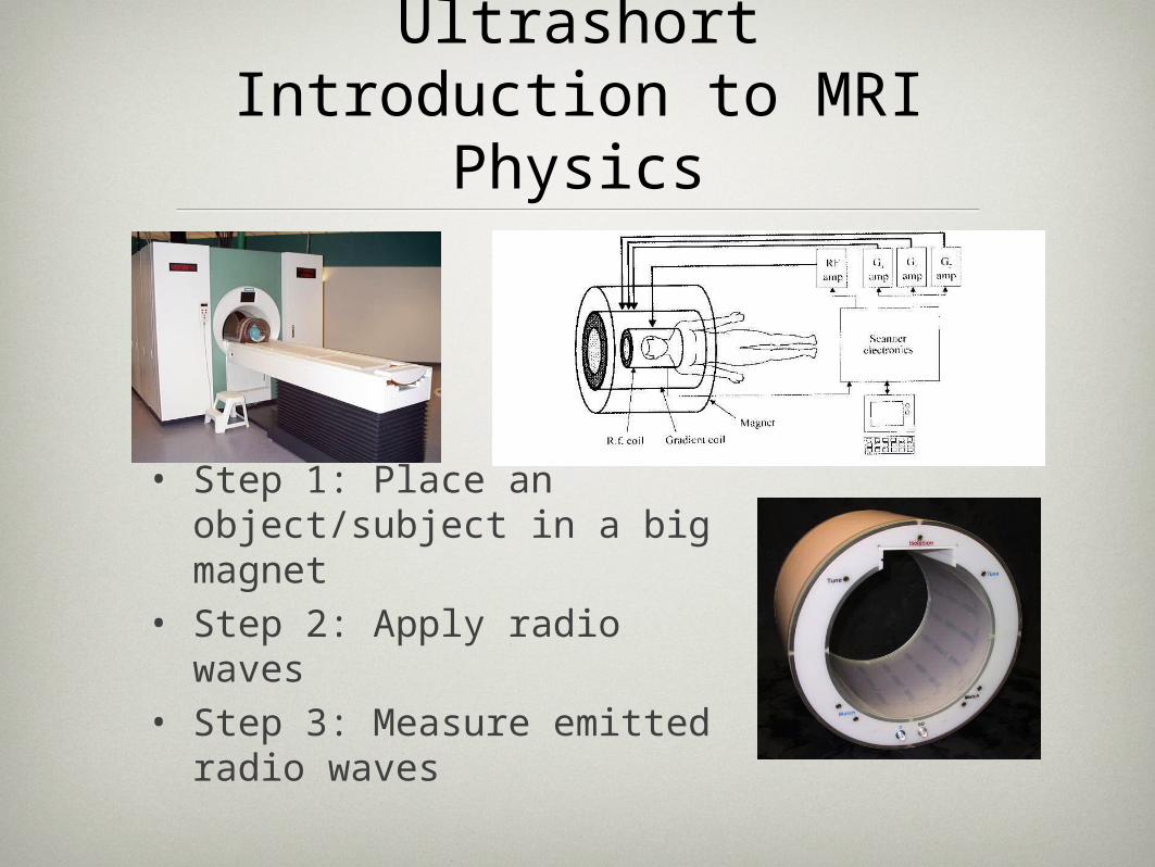
Step 3: Measure emitted radio waves (T2 or T2*)
T2 = time constant of how quickly the protons emit energy when recovering to equilibrium
T2-WEIGHTED ANATOMICAL IMAGE
fat has low signal -> dark
CSF has high signal -> bright
Images: fmri4newbies.com
T2
T1

T2* weighted images
•Two factors contribute to the decay of transverse magnetization:1) molecular interactions2) local inhomogeneities of the magnetic field (dephasing of spins)
•The combined time constant is called T2* (<T2).
•fMRI uses acquisition techniques (e.g. EPI) that are sensitive to changes in T2*.
The general principle of MRI:
–excite spins in static field by RF pulses & detect the emitted RF
–use an acquisition technique that is sensitive to local differences in T1, T2 or T2*
–construct a spatial image
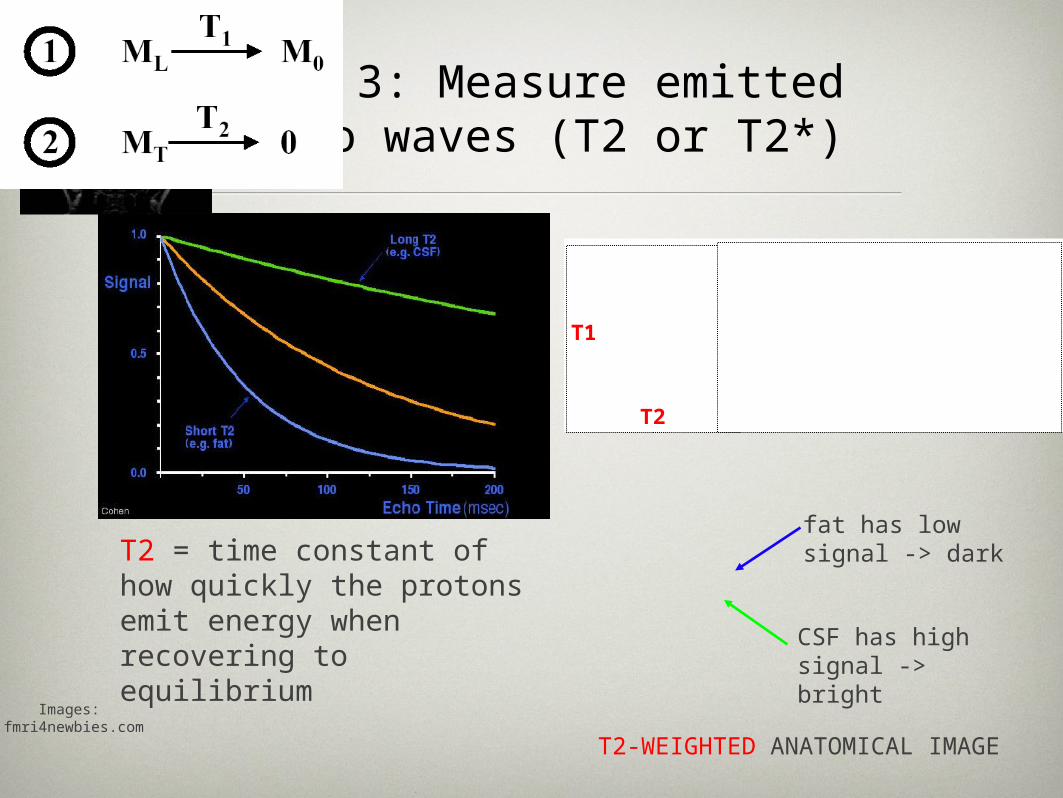
The Bold ContrastBOLD (Blood Oxygenation Level Dependent) contrast = measures inhomogeneities in the magnetic field due to changes in the level of O2 in the blood
Oxygenated blood is diamagnetic -> no signal loss
Deoxygenated blood is paramagnetic -> signal loss
High ratio deoxy/oxygenated blood -> fast decrease in MRI signal
Low ratio deoxy/oxygenated blood -> slow decrease in MRI signal
Huettel, Song, McCarthy, 2004

The BOLD contrast
Source: Jorge Jovicich, fMRIB Brief Introduction to fMRI
neural activity ↑ blood flow ↑ oxyhemoglobin ↑ T2* ↑ MR signal
REST
ACTIVITY

Summary MRI Physics• Magnetic dipole moments of hydrogen nuclei align
to magnetic field in scanner
• RF pulse causes them to spin, in phase
• Once pulse has stopped they fall back into direction of magnetic field, dephasing as they do so
• Dephasing takes various amounts of time, depending in part on inhomogeneities in magnetic field
• Inhomogeneities are caused by variable ratio of deoxygenated : oxygenated blood
• Assumption: activity in brain area lowers this ratio and thereby decreases speed of decay of MRI signal

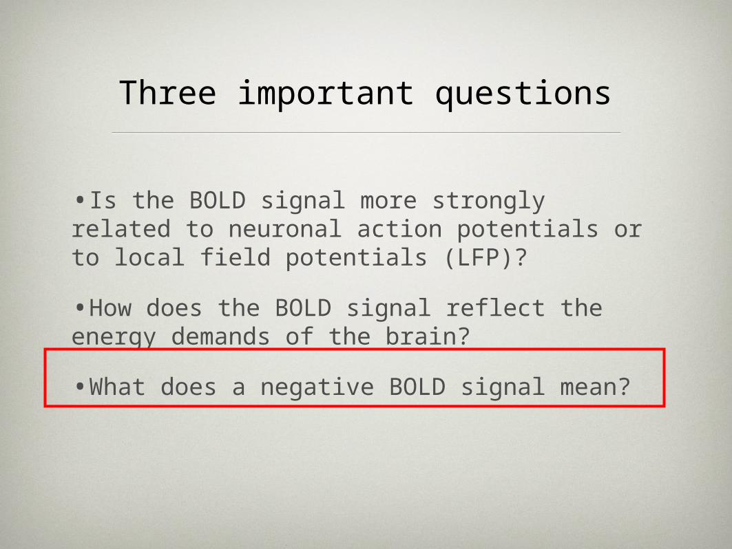
Three important questions
•Is the BOLD signal more strongly related to neuronal action potentials or to local field potentials (LFP)?
•How does the BOLD signal reflect the energy demands of the brain?
•What does a negative BOLD signal mean?

Neurophysiological basis of the BOLD signal: soma or synapse?

In early experiments comparing human BOLD signals and monkey electrophysiological data, BOLD signals were found to be correlated with action potentials.
BOLD & action potentials
Heeger et al 2000, Nat. Neurosci.
Rees et al. 2000, Nat. Neurosci.
Red curve: “average firing rate in monkey V1, as a function of contrast,estimated from a largedatabase of microelectroderecordings (333 neurons).”

Logothetis et al., 2001, Nature
Action potentials vs. postsynaptic activity
Local Field Potentials (LFP)• reflect summation of post-synaptic
potentials
Multi-Unit Activity (MUA)• reflects action potentials/spiking
Logothetis et al. (2001)• combined BOLD fMRI and
electrophysiological recordings • found that BOLD activity is more
closely related to LFPs than MUA
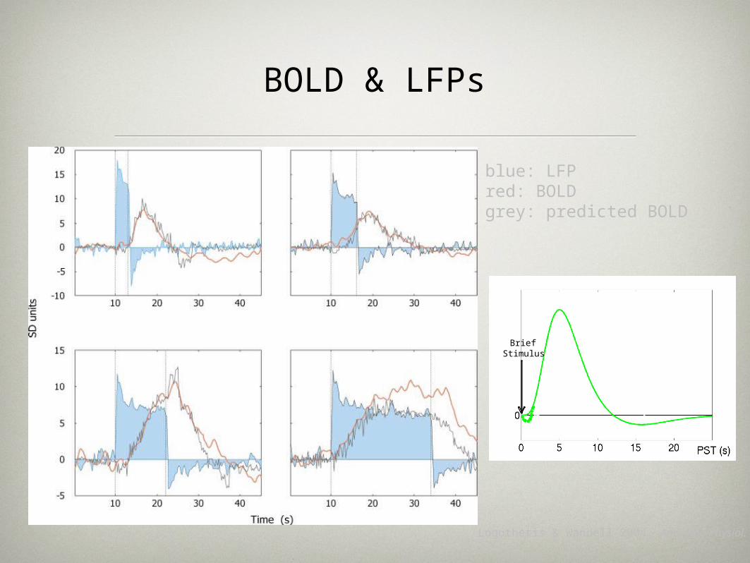
BOLD & LFPs
Logothetis & Wandell 2004, Ann. Rev. Physiol.
blue: LFPred: BOLDgrey: predicted BOLD
BriefStimulus
Undershoot
InitialUndershoot
Peak

Thomsen et al. 2004, J. Physiol.
⇒ rCBF-increase can be independent from spiking activity, but seems to be always correlated to LFPs
• GABAA antagonist picrotoxine increased spiking activity without increase in rCBF...
•... and without disturbing neurovascular coupling per se
Lauritzen et al. 2003
Dissociation between action potentials and rCBF

Current conclusion: BOLD signal seems to be more strongly correlated to
postsynaptic activity
Lauritzen 2005, Nat. Neurosci. Rev.
BOLD seems to reflect the input to a neuronal population as well as its intrinsic processing.

Three important questions
•Is the BOLD signal more strongly related to neuronal action potentials or to local field potentials (LFP)?
•How does the BOLD signal reflect the energy demands of the brain?
•What does a negative BOLD signal mean?

Is the BOLD signal driven by energy demands or synaptic processes?
synaptic activity neuronal metabolism
neurovascularcoupling
D’Esposito et al. 2003
rCBF
deoxy-Hb/oxy-Hb
? ?

Schwartz et al. 1979, Science
Localisation of neuronal energy consumption
Salt loading in rats and 2-deoxyglucose mapping
→ glucose utilization and neural activity in the posterior pituitary but not in paraventricular and supraoptic nuclei
→ neuronal energy consumption takes place at the synapses, not at the
cell body
Compatible with findings on BOLD relation to LFPs!
But does not tell us whether BOLD induction is due to energy demands or feedforward synaptic processes...

Estimated Energy Consumption
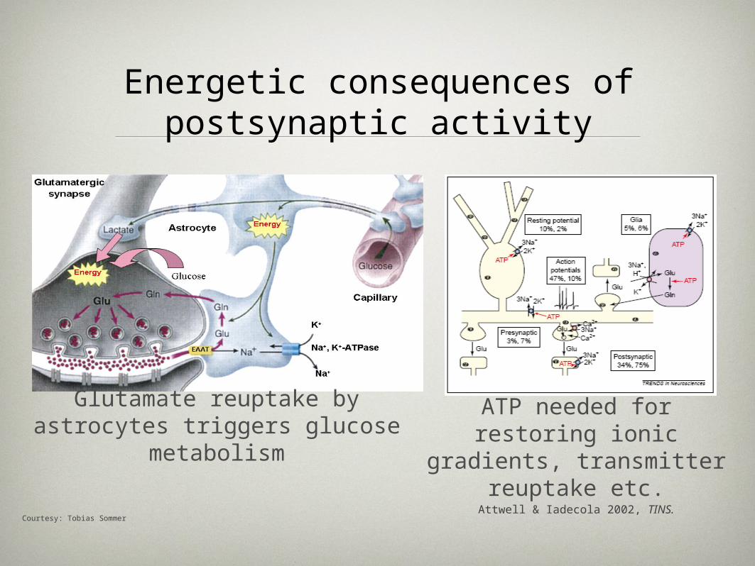
Energetic consequences of postsynaptic activity
Courtesy: Tobias Sommer
Glutamate reuptake by astrocytes triggers glucose
metabolismATP needed for restoring
ionic gradients, transmitter reuptake etc.
Attwell & Iadecola 2002, TINS.

Blood flow might be directly driven by excitatory postsynaptic
processes

Glutamatergic synapses: A feedforward system for eliciting the
BOLD signal?
Lauritzen 2005, Nat. Neurosci. Rev.
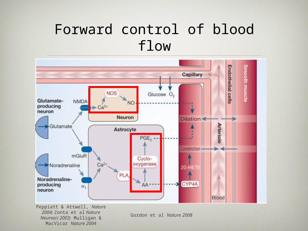
Forward control of blood flow
Peppiatt & Attwell, Nature 2004; Zonta et al Nature
Neurosci 2003; Mulligan & MacVicar Nature 2004
Gordon et al Nature 2008
Three important questions
•Is the BOLD signal more strongly related to neuronal action potentials or to local field potentials (LFP)?
•How does the BOLD signal reflect the energy demands of the brain?
•What does a negative BOLD signal mean?

Shmuel et al. 2006, Nat. Neurosci.
Negative BOLD is correlated with decreases in LFPs
positive BOLD negative BOLD

Impact of inhibitory postsynaptic potentials (IPSPs) on blood flow
Lauritzen 2005, Nat. Neurosci. Rev.

Negative BOLD signals due to IPSPs?
Lauritzen 2005, Nat. Neurosci. Rev.

From Neural Activity to fMRI Images
Neural activity
Energy consumption
Regional cerebral
blood flow
Functional & anatomical
images

BOLDBOLDcontrastcontrast
bloodbloodflowflow
bloodbloodvolumevolume
oxygenoxygenutilizationutilization
structural lesionsstructural lesions(compression)(compression)
autoregulationautoregulation(vasodilation)(vasodilation)
cerebrovascularcerebrovasculardiseasedisease
medicationsmedications
hypoxiahypoxia
anemiaanemiasmokingsmoking
hypercapniahypercapnia
degenerative diseasedegenerative disease
volume statusvolume status
anesthesia/sleepanesthesia/sleep biophysical effectsbiophysical effects
Potential physiological influences on BOLD

Summary
• The BOLD signal seems to be more strongly related to LFPs than to spiking activity.
• The BOLD signal seems to reflect the input to a neuronal population as well as its intrinsic processing, not the outputs from that population.
• Blood flow seems to be controlled in a forward fashion by postsynaptic processes leading to the release of vasodilators.
• Negative BOLD signals may result from IPSPs.
• Various drugs can interfere with the BOLD response.