Physics of Imaging Systems (ROE, CT, MRI) - uni … of Imaging Systems (ROE, CT, MRI) ... (UK, 1933...
12
1 Seite 1 RUPRECHT-KARLS- UNIVERSITY HEIDELBERG Computer Assisted Clinical Medicine Prof. Dr. Lothar Schad 12/9/2008 | Page 1 Physics of Imaging Systems (ROE, CT, MRI) Prof. Dr. Lothar Schad Chair in Computer Assisted Clinical Medicine Faculty of Medicine Mannheim University of Heidelberg Theodor-Kutzer-Ufer 1-3 D-68167 Mannheim, Germany [email protected] www.ma.uni-heidelberg.de/inst/cbtm/ckm/ Master‘s Program in Medical Physics RUPRECHT-KARLS- UNIVERSITY HEIDELBERG Computer Assisted Clinical Medicine Prof. Dr. Lothar Schad 12/9/2008 | Page 2 www.ma.uni-heidelberg.de/inst/cbtm/ckm/
Transcript of Physics of Imaging Systems (ROE, CT, MRI) - uni … of Imaging Systems (ROE, CT, MRI) ... (UK, 1933...

1
Seite 1
RUPRECHT-KARLS-UNIVERSITY HEIDELBERG
Computer Assisted Clinical MedicineProf. Dr. Lothar Schad
12/9/2008 | Page 1
Physics of
Imaging Systems (ROE, CT, MRI)
Prof. Dr. Lothar Schad
Chair in Computer Assisted Clinical MedicineFaculty of Medicine Mannheim University of HeidelbergTheodor-Kutzer-Ufer 1-3D-68167 Mannheim, GermanyLothar.Schad@MedMa.Uni-Heidelberg.dewww.ma.uni-heidelberg.de/inst/cbtm/ckm/
Master‘s Program in Medical Physics
RUPRECHT-KARLS-UNIVERSITY HEIDELBERG
Computer Assisted Clinical MedicineProf. Dr. Lothar Schad
12/9/2008 | Page 2www.ma.uni-heidelberg.de/inst/cbtm/ckm/

2
Seite 2
RUPRECHT-KARLS-UNIVERSITY HEIDELBERG
Computer Assisted Clinical MedicineProf. Dr. Lothar Schad
12/9/2008 | Page 3
Introduction
Introduction
RUPRECHT-KARLS-UNIVERSITY HEIDELBERG
Computer Assisted Clinical MedicineProf. Dr. Lothar Schad
12/9/2008 | Page 4
Nobel Prize is awarded for MRI technologylandmark achievement transformed healthcare in 20th century
Paul Lauterbur(1929 - 2007)
Sir Peter Mansfield(1933 - )
Nobel Prize 2003

3
Seite 3
RUPRECHT-KARLS-UNIVERSITY HEIDELBERG
Computer Assisted Clinical MedicineProf. Dr. Lothar Schad
12/9/2008 | Page 5
1901 W. C. Röntgen (Germany, 1845 - 1923) discovery of X-rays
2003 Paul C. Lauterbur (USA, 1929 - 2007) Peter Mansfield (UK, 1933 - ) development of magnetic resonance imaging (MRI)
1991 Richard R. Ernst (CH, 1933 - ) development of high resolution magnetic resonance spectroscopy (MRS)
1952 Felix Bloch (USA, 1905 - 1983) Edward M. Purcell (USA, 1912 - 1997) development of a new precision method of nuclear magnetism (NMR)
1979 Allan M. Cormack (USA, 1924 - 1998) Godfrey N. Hounsfield (UK, 1919 - ) development of Computer-Tomography (CT)
Diagnostic Imaging: Milestones
RUPRECHT-KARLS-UNIVERSITY HEIDELBERG
Computer Assisted Clinical MedicineProf. Dr. Lothar Schad
12/9/2008 | Page 6
“The Making of a Science”
source: ECR Newsletter 1/2003
Diagnostic Imaging: Pioneers
ROE
CT MRI
PET

4
Seite 4
RUPRECHT-KARLS-UNIVERSITY HEIDELBERG
Computer Assisted Clinical MedicineProf. Dr. Lothar Schad
12/9/2008 | Page 7
Why do we need imaging systems ?
„Addiction“ to image information ?
Motivation
source: Siemens “100 Jahre Röntgen” 1995
RUPRECHT-KARLS-UNIVERSITY HEIDELBERG
Computer Assisted Clinical MedicineProf. Dr. Lothar Schad
12/9/2008 | Page 8
- information recording of all sense organs from our surroundings: ≈ 109 bit/s
→ selection and filtering 1:10 million !
CNSsee
hear
smell
taste
108 bit/s
5×104 bit/s
102 bit/s
10 bit/sflowing capacity to short-term storage: 16 bit/s is recognized by the human awareness
information flow
source: Drischel. „Einführung in die Biokybernetik“, Akademie-Verlag 1972
self-aware data processing: 100 bit/sshort-term storage: 10 bit/slong-term storage: 1 bit/s
Information Flow of Sense Organs

5
Seite 5
RUPRECHT-KARLS-UNIVERSITY HEIDELBERG
Computer Assisted Clinical MedicineProf. Dr. Lothar Schad
12/9/2008 | Page 9
Imaging Systems are supporting the most effective
and powerful sensor !
Conclusion
RUPRECHT-KARLS-UNIVERSITY HEIDELBERG
Computer Assisted Clinical MedicineProf. Dr. Lothar Schad
12/9/2008 | Page 10
Fuchs and Sox. Health Affairs - Sept/Oct 2001
Physicians´ Ranking of Innovations 2001

6
Seite 6
RUPRECHT-KARLS-UNIVERSITY HEIDELBERG
Computer Assisted Clinical MedicineProf. Dr. Lothar Schad
12/9/2008 | Page 11
object (human, animal, plant, ...)
radiation source(s)
externalX-rays (Röntgen)ultrasound (US)radiofrequency (NMR)
internalradioactive tracers(PET, SPECT)
detector(s)
externalfilm (Röntgen)piezo-crystal (US)RF-coil (NMR)
internalRF-coil
source: http://bio.physik.uni-würzburg.de/public/medphys
Diagnostic Imaging: Principal
RUPRECHT-KARLS-UNIVERSITY HEIDELBERG
Computer Assisted Clinical MedicineProf. Dr. Lothar Schad
12/9/2008 | Page 12
To look into the object without cutting or destroying (non-invasively) !
Goal

7
Seite 7
RUPRECHT-KARLS-UNIVERSITY HEIDELBERG
Computer Assisted Clinical MedicineProf. Dr. Lothar Schad
12/9/2008 | Page 13
definition:
• interaction of energy with biological tissue in order to get spatially resolved information about the physical properties of the underlying biological structure
• energy has to penetrate through the body for interaction (absorption, scattering, …)
interpretation of imaging information:
• importance of measured physical properties with respect to differentiate between normal and diseased tissue (pathology)
• not fully understood
Diagnostic Imaging: Definition
RUPRECHT-KARLS-UNIVERSITY HEIDELBERG
Computer Assisted Clinical MedicineProf. Dr. Lothar Schad
12/9/2008 | Page 14
different energies means different interaction with tissue
PET and MRI are at the end of the spectrum
ionizing non-ionizing
HzHz
Micro-Visible Infrared Milli-
meter waveand RF
THz gap
10 15Hz 10 14 Hz 10 13 10 12 10 11Hz 10 10Hz
Ultra-violet
10 16 Hz10 17Hz
magneticresonance
imagingMRI
nuclear medicine / PET
10 18Hz10 19Hz
X-ray / CTimaging
100keV 10keV
terahertz pulseimaging (TPI)
NIRF
Frequency
TV satellitedish
THz Gap
OCT
X-ray
Diagnostic Imaging: Electromagnetic Wave

8
Seite 8
RUPRECHT-KARLS-UNIVERSITY HEIDELBERG
Computer Assisted Clinical MedicineProf. Dr. Lothar Schad
12/9/2008 | Page 15Electromagnetic Wave Penetration
RUPRECHT-KARLS-UNIVERSITY HEIDELBERG
Computer Assisted Clinical MedicineProf. Dr. Lothar Schad
12/9/2008 | Page 16Bildgebende Verfahren
without ionizingradiation
with ionizingradiation
Nuclear MagneticResonance
Ultrasound X-rays Nuclear MedicalTechniques
spectroscopy(MRS)
tomography(MRI)
tomography(CT)
planar planar emissiontomography
(PET)
Diagnostic Imaging: Overview

9
Seite 9
RUPRECHT-KARLS-UNIVERSITY HEIDELBERG
Computer Assisted Clinical MedicineProf. Dr. Lothar Schad
12/9/2008 | Page 17
image by courtesy of Helmut Newton
source: ECR Newsletter 4/2002
Diagnostic Imaging: Anatomy
RUPRECHT-KARLS-UNIVERSITY HEIDELBERG
Computer Assisted Clinical MedicineProf. Dr. Lothar Schad
12/9/2008 | Page 18Diagnostic Imaging: Pathology
10
Seite 10
RUPRECHT-KARLS-UNIVERSITY HEIDELBERG
Computer Assisted Clinical MedicineProf. Dr. Lothar Schad
12/9/2008 | Page 19Diagnostic Imaging: Animals
RUPRECHT-KARLS-UNIVERSITY HEIDELBERG
Computer Assisted Clinical MedicineProf. Dr. Lothar Schad
12/9/2008 | Page 20
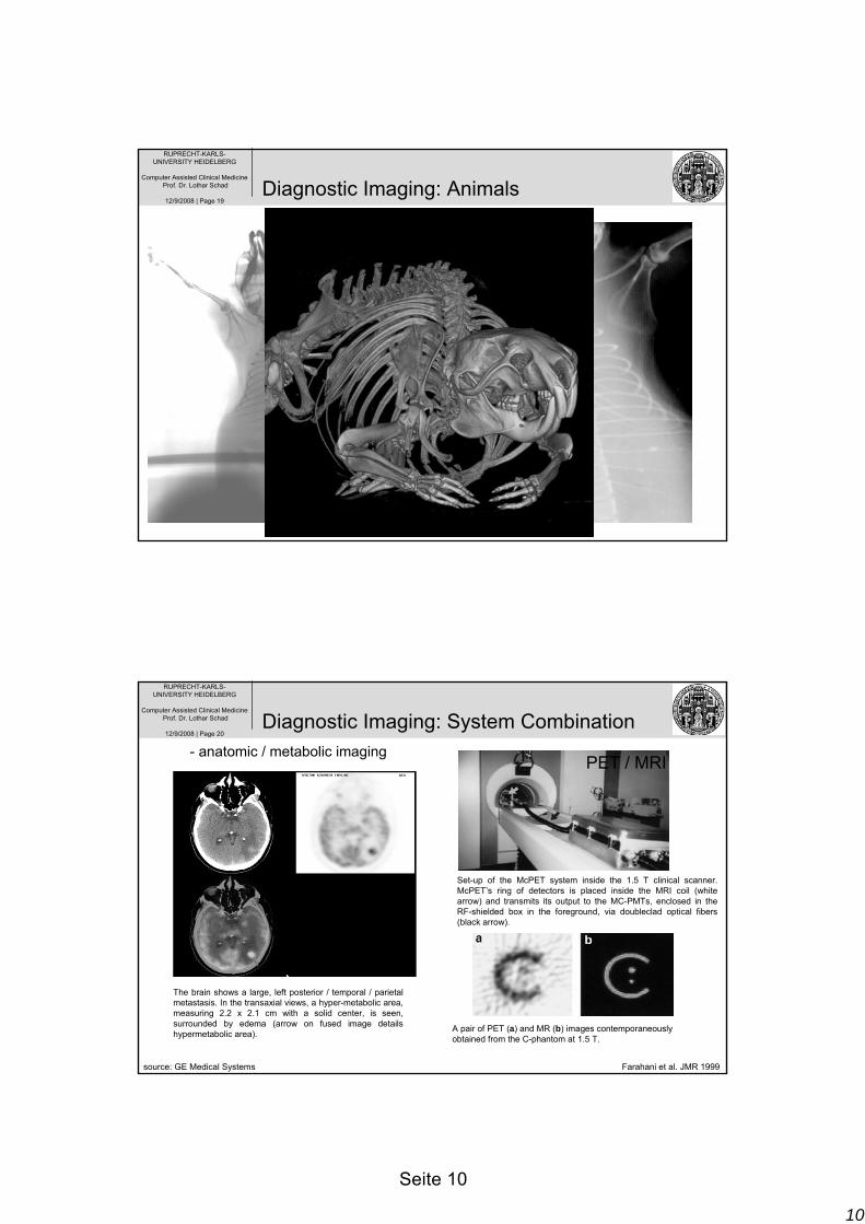
The brain shows a large, left posterior / temporal / parietal metastasis. In the transaxial views, a hyper-metabolic area, measuring 2.2 x 2.1 cm with a solid center, is seen, surrounded by edema (arrow on fused image details hypermetabolic area).
PET / CT
PET / MRI
Set-up of the McPET system inside the 1.5 T clinical scanner. McPET’s ring of detectors is placed inside the MRI coil (white arrow) and transmits its output to the MC-PMTs, enclosed in the RF-shielded box in the foreground, via doubleclad optical fibers (black arrow).
A pair of PET (a) and MR (b) images contemporaneously obtained from the C-phantom at 1.5 T.
source: GE Medical Systems Farahani et al. JMR 1999
Diagnostic Imaging: System Combination- anatomic / metabolic imaging
11
Seite 11
RUPRECHT-KARLS-UNIVERSITY HEIDELBERG
Computer Assisted Clinical MedicineProf. Dr. Lothar Schad
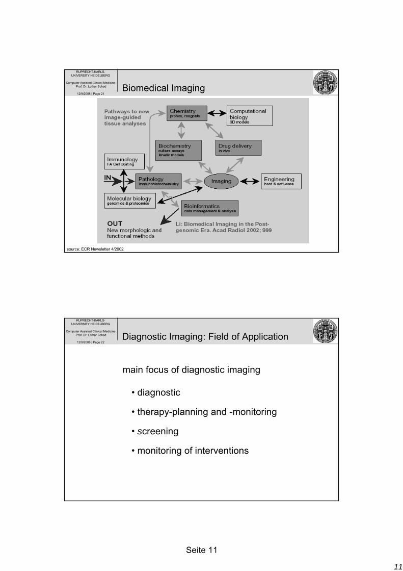
12/9/2008 | Page 21
source: ECR Newsletter 4/2002
Biomedical Imaging
RUPRECHT-KARLS-UNIVERSITY HEIDELBERG
Computer Assisted Clinical MedicineProf. Dr. Lothar Schad
12/9/2008 | Page 22
• diagnostic
• therapy-planning and -monitoring
• screening
• monitoring of interventions
main focus of diagnostic imaging
Diagnostic Imaging: Field of Application

12
Seite 12
RUPRECHT-KARLS-UNIVERSITY HEIDELBERG
Computer Assisted Clinical MedicineProf. Dr. Lothar Schad
12/9/2008 | Page 23
X-Ray CT MR US
imaging of bone soft tissue vessel function volume
+ + + – / + + + – –
+ + +
– + + –
+ +
+
+ + + + + + + +
– + +
++ +
real-time
*
+
+
+ +
psychological stress physical stress invasiv
small high no
medium
high no
high small
no
small small
no
costs (EUR)
ca. 40
ca. 100
ca. 400
ca. 10
Diagnostic Imaging: Properties