Mckenzie Approach To Treating Lumbar Radiculopathy With A ...
University of North DakotaUND Scholarly Commons
Physical Therapy Scholarly Projects Department of Physical Therapy
2019
Physical Therapy Rehabiliation for LumbarRadiculopathy: A Case ReportRebecca LynchUniversity of North Dakota
Follow this and additional works at: https://commons.und.edu/pt-grad
Part of the Physical Therapy Commons
This Scholarly Project is brought to you for free and open access by the Department of Physical Therapy at UND Scholarly Commons. It has beenaccepted for inclusion in Physical Therapy Scholarly Projects by an authorized administrator of UND Scholarly Commons. For more information,please contact [email protected].
Recommended CitationLynch, Rebecca, "Physical Therapy Rehabiliation for Lumbar Radiculopathy: A Case Report" (2019). Physical Therapy ScholarlyProjects. 680.https://commons.und.edu/pt-grad/680
PHYSICAL THERAPY REHABILIATION FOR LUMBAR RADICULOPATHY
A CASE REPORT
by
Rebecca Lynch
A Scholarly Project Submitted to the Graduate Faculty of the Department of Physical Therapy
School of Medicine University of North Dakota
in partial fulfillment of the requirements for the degree of
Doctor of Physical Therapy May,2019

This Scholarly Project, submitted by Rebecca Lynch in partial fulfillment of the requirements for the Degree of Doctor of Physical Therapy from the University of North Dakota, has been read by the Advisor and Chairperson of Physical Therapy under whom the work has been done and is hereby approved.
ii

Title
Department
Degree
PERMISSION
Physical Therapy Rehabilitation for Lumbar Radiculopathy A Case Report
Physical Therapy
Doctor of Physical Therapy
In presenting this Scholarly Project in partial fulfillment of the requirements for a graduate degree from the University of North Dakota, I agree that the Department of Physical Therapy shall make it freely available for inspection. I further agree that permission for extensive copying for scholarly purposes may be granted by the professor who supervised my work or, in her absence, by the Chairperson of the department. It is understood that any copying or publication or other use of this Scholarly Project or part thereof for financial gain shall not be allowed without my written permission. It is also understood that due recognition shall be given to me and the University of North Dakota in any scholarly use which may be made of any material in this Scholarly Project.
Signature #!k4(fF I
Date 117- 1'1- I~
iii
TABLE OF CONTENTS
Page
LIST OF TABLES ...................................................... v
ABSTRACT ............................................................ vi
CHAPTER
I. BACKGROUND AND PURPOSE ............................. .1
II. CASE DESCRIPTION ........................................ 3
Examination ............................................... .4
Evaluation, Diagnosis, Prognosis ................................ 6
III. INTERVENTION ............................................ 8
IV. OUTCOMES .............................................. 10
V. DISCUSSION .............................................. 12
Reflective Practice ........................................... 13
Conclusion ................................................ 14
REFERENCES ......................................................... 16
APPENDIX ............................................................ 18
iv

LIST OF TABLES
Page
1. Sensitivity, Specificity, and Reliability of the Special Tests ................. 6
2. Oswestry Disability Index- Rehabilitation Measures ...................... 6
3. Initial and Discharge Lumbar Spine Active Range of Motion Measurements .. .10
v
ABSTRACT
Background and Purpose. Lumbar radiculopathy is one of the most common orthopedic
conditions. This occurs when there is damage to a nerve root in the area that it exits the
spinal cord. This can be caused from a disc hemiatio14 bone spurs, trauma, or a
mechanical stretching event. There is not consistent evidence in current literature
regarding rehabilitation interventions for this condition.
Case Description. This case report describes a 62-year-old female presenting to physical
therapy with lumbar radiculopathy symptoms. She had multiple comorbidities as well as
a scheduled total knee replacement surgery. Physical therapy interventions for this patient
included manual therapy, therapeutic exercise, and patient education.
Outcomes. The patient responded well to treatment and significantly improved following
two weeks of physical therapy rehabilitation. She reported minimal pain that was
centralized to the low back. She demonstrated an increase in lumbar range of motion,
strength, and mobility.
Discussion. This patient demonstrated a significant improvement in a short period of
time. This case report may suggest intervention strategies for future research regarding
physical therapy and lumbar radiculopathy.
vi
CHAPTER I
BACKGROUND AND PURPOSE
Lumbar radiculopathy is caused when a nerve root is injured in the area where it exits the
spinal cord. The pain that radiates from the lumbar spine down into the lower extremities along
the damaged nerve is called sciatica. Along with pain, there are other symptoms that may be
present including numbness, tingling, wealmess and muscle spasms in the lower extremities.
This diagnosis can be the result of several different structural problems at the spine. These
problems include a disc herniation, a bone spur, trauma, or a mechanical stretching event. The
damage to the disc can be caused from a certain activity, an injury, or can possibly be congenital.
This nerve damage occurs when the substance located in the center of a disc escapes the outer
protective ring and puts pressure on a nerve root. MRis, CT scans, and electrodiagnosis can help
determine if the radiculopathy is disc related. Treatment options can include physical therapy,
medication, steroid injections, back supports, and surgery. The common age when symptoms
start to appear is between 30-50 years. Prognosis for lumbar radiculopathy is normally good. 14
There are several discussions in literature on theories regarding the best treatment for
lumbar radiculopathy. One issue being discussed is whether conservative or surgical treatment is
a better option for these patients. Some suggest that early surgical treatment is the best for a
faster recovery.4 However, surgery is expensive and comes with possible complications. Others
have found there to be no difference between surgery and conservative care when comparing
1' ' 1 f 12 c m1ca outcomes a ter one year.
1

Manual therapy is often used with low back pain and lumbar radiculopathy. A well
designed cohort study discussed the effectiveness of neural manual therapy interventions with
patients experiencing low back and leg pain. The goal of these passive techniques was to
mobilize the neural structures in the intervertebral foramen. This mobilization gradually
desensitizes the peripheral nervous system. The majority of the subjects in the study responded
favorably to this intervention. 20 Another study based on the conclusions from several systematic
reviews and randomized clinical trials looked at the effectiveness of manual therapy for the
treatment of several musculoskeletal conditions including sciatica and radiating leg pain.
Conclusions from this study also discussed that there is a favorable level of evidence for spinal
manipulation and mobilization for sciatica and radiating leg pain. The "favorable" level of
evidence in this study means that the evidence does not support effectiveness but if other
treatments are not effective, this intervention may be a treatment option. 2
The McKenzie protocol based on directional preference is another intervention technique
that is used with this condition. A systematic review was done studying the use of the McKenzie
therapy protocol for spinal pain. The authors concluded that the McKenzie method reduces short
term pain and disability in people suffering from low back pain over other standard treatments.
However, there was insufficient evidence on long-term outcomes. 5 Another systematic review
studying McKenzie Therapy outcomes for back pain was done that determined similar results.
The review compared McKenzie directional preference exercises to other standard treatments
including back massage, strength training, and spinal mobilizations. They found that the patients
who followed the McKenzie approach had better outcomes with decreased pain and disability.
1
This systematic review also concluded that the evidence was not strong enough to show long
term outcomes after a year with McKenzie therapy on pain or disability?
Lumbar Radiculopathy can often present similarly to piriformis syndrome. Some
similarities include pain with sitting for long periods of time, relief when standing or walking,
and pain in the buttock area. A patient with piriformis syndrome will often report trauma to the
buttock area or bowel and bladder changes during the subjective history. During the assessment,
there is often tenderness over the pirifom1is muscle, a mass in the buttock, and relief when a
traction force is applied to the leg. These findings are not commonly seen in a patient with
lumbar radiculopathy. It is important to complete a full neurological assessment in order to
correctly diagnose. 1 The FAIR test including flexion, adduction, and internal rotation is often
performed to indicate piriformis syndrome. This test has a specificity of 83.2%.11
Trochanteric bursitis can also mimic radiculopathy. Both diagnoses can cause pain on the
outside of ilie hip or in the buttock area. With trochanteric bursitis, there is commonly pain with
pressure on the outside of the hip and the pain gets worse with activities including sit-to-stand
and walking up stairs. The pain can be caused from injury to the hip, bone spurs, calcium
deposits, and overuse activities including running, walking up stairs, and standing for long
periods of time. 22
Lumbar radiculopathy is one of the most common orthopedic conditions, and there are
controversies over the best intervention strategies. More research is needed to provide the best
care to patients with this condition. The purpose of this case report was to document the fast
recovery of a patient experiencing lumbar radiculopathy in physical therapy rehabilitation. This
may be a favorable group of interventions to guide future research regarding physical therapy
and lumbar radiculopathy.
2
CHAPTER II
CASE DESCRIPTION
This case study focuses on a 62 year-old female who was referred to physical therapy
with the diagnosis of piriformis syndrome. She had been experiencing left buttock pain for the
past 3 to 4 months upon arrival to therapy. The patient did not recall any specific incident or
trauma that lead to the onset of these symptoms. She denied low back pain and symptoms such
as numbness or tingling down into her lower extremities. She reported that her symptoms
increased when she was sitting, and were relieved when she was standing or wallcing. The patient
lives with her husband in a two-story house. She is employed as a high school receptionist and
sits for approximately 80% of her workday. She described that her symptoms interfered with her
ability to sit for long periods of time at her desk and complete her work. In her free time, she
enjoys spending time with her children and grandchildren. She often takes care of her
grandchildren in the evenings. She reported having difficulty picking up her grandchildren off
the floor and playing with them due to her recent symptoms. Secondary to her buttock
symptoms, the patient explained that she was having a total knee replacement in two weeks due
to degenerative joint disease in her left knee. She stated that her knee is painful during
ambulation and when she is weight bearing on her left side. After her surgeon had suggested
losing some weight prior to surgery, she started watching her diet more closely but was not
exercising. During the systems review, she denied any constant pain, night pain, abdominal pain,
recent illnesses, or cauda equina symptoms. Her past medical history included asthma, obesity,
and type 2 diabetes. The patient reported that she monitors her asthma and diabetes closely and
3

has not had health related issues due to these comorbidities. The patient's goals were to be able
to take care of her grandchildren and work a full day without symptoms.
My clinical impression for this patient was that she was appropriate for physical therapy.
No systemic signs or symptoms were present during the subjective history taking. The subjective
history suggested a movement dysfunction, which is in the physical therapy scope of practice.
The examination plan included the assessment of her range of motion, strength, mobility, and
gait. These assessments along with palpation and special tests were performed to rule in or out
other diagnoses including lumbar radiculopathy, piriformis syndrome, and trochanteric bursitis.
Prognostic factors that could have affected her recovery included her comorbidities, family
support, and scheduled total knee replacement surgery.
Examination
Examination was based on Magee's Orthopedic Physical Assessment of the lumbar
spine. 15 The patient filled out a Modified Oswestry Disablity Index before the physical therapy
evaluation. This questionnaire gives a subjective score that rates her level of disability in
activities due to low back pain. She scored a 14% on a 0-100% scale, 0% being the best with no
restriction in function. X-ray imaging was included with the physician's referral note that
showed extensive osteoarthritis in the left knee. Upon observation, poor posture with a forward
head and rounded shoulders was documented. The patient had a slight antalgic gait pattern with a
decreased period of time in the stance phase with the left lower extremity. All directions of the
lumbar spine active range of motion were full and pain-free with the exception of flexion. Her
lumbar flexion range of motion was limited and also reproduced her symptoms in the left
buttock. She also reported feeling tightness in the left low back with this motion. Table 3
4
documents her lumbar spine range of motion measurements at the initial evaluation and at
discharge.
During palpation, she had marked tenderness and hypermobility over her lower lumbar
vertebrae and over her left sacral base. She had palpable tightness over her left lumbar paraspinal
muscles. When the piriformis muscle was palpated, she did not report any discomfort. This area
is often painful with pressure for a patient with piriformis syndrome. 1 Palpation over the greater
trochanter also did not reproduce symptoms, which is commonly a finding in patients with
trochanteric bursitis.22 Active and passive hip range of motion was within normal limits
bilaterally. She had 5/5 hip strength except for hip abduction, which was documented as 4/5. The
right lower extremity was within normal limits for strength and range of motion. The left knee
had 5/5 strength but was painful with strength testing. Passive and active range of inotion of left
lmee flexion was limited to 120 degrees secondary to pain. Crepitus was felt over both knees
during passive range of motion. The patient had no increase in muscle tone. Dermatome testing
was negative bilaterally. The Slump Test was performed to determine if the patient had nerve
root involvement. First, the patient sat in her natural posture and denied any symptoms. Then,
when she was asked to slouch, she reported having symptoms in the left buttock. The Straight
Leg Raise was a second special test performed to also determine if she had neural tissue
involvement. This test is often positive in patients with sciatica and is often negative in patients
with spinal stenosis. 16 The patient laid on her back and each leg was passively flexed off the
table with her knee extended. When her left leg reached 50 degrees, she experienced a
reproduction of her pain in the left buttock. Her left leg was then passively lowered 10 degrees,
which eliminated her symptoms. Finally, her left foot was passively dorsiflexed, which
reproduced her symptoms. Tables 1 below documents the sensitivity, specificity, and reliability
5

of the special tests performed during this examination. Table 2 below documents the Oswestry
Disability Index functional assessment rehabilitation measures.
Table 1. Sensitivity, Specificity, and Reliability of the Special Tests
Test Sensitivity Specificity Interrater Reliability
Straight Leg Raise 0.92 18 0.28 18 0.73 7
Slump Test 1.0 21 0.83 21 0.89 17
Table 2. Oswestry Disability Index- Rehabilitation Measures
MCID Test/Retest Reliability Criterion Validity MDC
12.8 6 0.94 10 0.75 8 11.1 10
Evaluation, Diagnosis, and Prognosis
The initial evaluation indicated that her symptoms were coming from her low back and
referring down to her left buttock. It is possible that she had a disc pathology that was causing
the nerve root compression. The positive special tests, her tender and hypomobile lumbar
vertebrae, and her tight left lumbar paraspinal muscles support this hypothesis along with the fact
that her symptoms increased when she was sitting and were relieved when she was standing or
walking. The piriformis muscle itself was not painful and did not reproduce her radicular
symptoms during palpation. This finding, along with the positive slump test, helped to rule out
the diagnosis of piriformis syndrome. Trochanteric bursitis was also ruled out as a diagnosis
because she did not report tenderness when lying on the affected side or with palpation over the
greater trochanter.
6

The primary rehabilitation goal created with the patient was to return to her prior level of
function, which included working at her receptionist job and taking care of her grandchildren
without symptoms. The short-term goals were to decrease her pain and for her to demonstrate
independence with her home program. These goals were to be met within a one to two week time
frame. The long-term goals were to increase lumbar spine range of motion, strength, and
mobility. These goals were to be met within a four to six week time frame after taking her
possible barriers into consideration.
Following the initial evaluation, I determined that her prognosis to reach these goals was
"fair". She had a goal-oriented and motivated personality. This suggested that she would be
compliant with her home program. She had a strong support system from her husband and
children both at the clinic and at home. Although the prognosis of patients experiencing lumbar
radiculopathy is normally good, her several comorbidities, sitting desk job, and scheduled knee
surgery were barriers that could hinder her progress and prognosis. My initial clinical impression
was confirmed after the examination, as I concluded this patient would benefit from physical
therapy interventions so she could return to her prior activities without limitations. There were
no findings that suggested this patient should be referred to another medical provider. If the
patient did not progress after two physical therapy treatments, I could re-examine and evaluate as
I could have missed something or refer if needed.
7

CHAPTER III
INTERVENTION
The patient was seen in physical therapy twice a week for 30-minute sessions.
Interventions included manual therapy and therapeutic exercise. A home exercise program was
given to the patient after demonstration and practice of each of the exercises in the clinic to
ensure that she understood them. Treatment remained fairly consistent as she was responding
well and was continually less symptomatic after every session. The patient was very compliant to
her home exercise program. Her husband was involved in the therapy sessions both in the clinic
and at home.
Manual therapy was performed on her lumbar spine at every visit. Central and left
posterior-anterior grade 3 mobilizations were applied to lumbar levels ofL2 to L5 and at the left
sacral base to promote mobility. Another manual technique used was left lumbar rotation
oscillations performed at a grade 3 while patient was in a right side-lying position to open up the
intervertebral foramen and relieve pressure off the nerve roots. The patient responded well to this
treatment.
Therapeutic exercise was provided to help strengthen, promote mobility, and increase
her range of motion in the low back. These exercises included standing back extensions, pelvic
tilts, straight leg raises, and supine bridging. Side-lying hip abductor strengthening was another
exercise given to address her limited hip abductor strength. The patient was instructed to perform
1 set of 10 repetitions of each exercise 2-3 times a day. However, we discussed that performing
8
10 repetitions of the standing back extensions every 2 hours would be beneficia1.9 No equipment
was utilized with her exercises.
Education was given to the patient and family at every therapy session. After the initial
evaluation the patient was educated on her condition. She was also advised to avoid sitting for
long periods of time. We discussed looking into a standing workstation at the school, as this
would take pressure off her aggravated nerve roots. She was informed to apply ice to the low
back to decrease inflammation and assist with pain when needed.
Since we were the only medical professionals working with her for the radicular
symptoms, communication with other disciplines was not necessary. After two weeks of
treatment, the patient had a total knee replacement surgery and came to physical therapy for her
post surgical rehabilitation. She was no longer receiving physical therapy for her low back at this
time.
9
CHAPTER IV
OUTCOMES
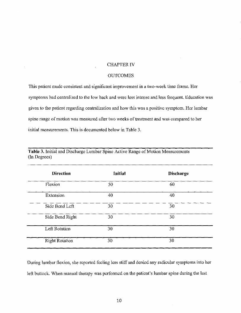
This patient made consistent and significant improvement in a two-week time frame. Her
symptoms had centralized to the low back and were less intense and less frequent. Education was
given to the patient regarding centralization and how this was a positive symptom. Her lumbar
spine range of motion was measured after two weeks of treatment and was compared to her
initial measurements. This is documented below in Table 3.
Table 3. Initial and Discharge Lumbar Spine Active Range of Motion Measurements (In Degrees)
Direction Initial Discharge
Flexion 50 60
Extension 40 40
Side Bend Left 30 30
Side Bend Right 30 30
Left Rotation 30 30
Right Rotation 30 30
During lumbar flexion, she reported feeling less stiff and denied any radicular symptoms into her
left buttock. When manual therapy was performed on the patient's lumbar spine during the last
10

physical therapy session for her back, her lumbar vertebrae had increased mobility. The patient's
hip abductor strength increased from a 4/5 to 4+/5.
After these two weeks, the patient underwent a total knee replacement surgery. She
returned to therapy following this, but was then receiving therapy for her knee rehabilitation
only. Secondary to not being able to walk and perform her exercises as frequently, her back
symptoms became slightly worse. As she made progress with her lmee rehabilitation and was
able to become more active again, her back symptoms resolve. My clinical rotation at this site
ended before the patient was discharged for her knee. The patient and her family were very
satisfied with her progress and reported that they were going to strive to live a more active
lifestyle.
11

CHAPTERV
DISCUSSION
This patient had a significantly fast recovery over two weeks and was able to return to
work and take care of her grandchildren without limitations. Some aspects that promoted her fast
recovery included finding the source of her symptoms, her compliancy to her home program and
positive attitude, and her family support. An aspect that hindered her recovery was having lmee
surgery shortly after presenting to physical therapy for her radicular symptoms.
A detailed subjective history taking and examination techniques including palpation,
lumbar special tests, and ROM assisted with identifYing the source of her symptoms. The
Oswestry Disability Index functional assessment was useful when deciding on what level the
patient was at functionally with her symptoms. Constant communication with the patient and
family helped provide a positive atmosphere and helped gain trust from the patient.
The examination process helped to rule out other pathologies including piriformis
syndrome and trochanteric bursitis. There was no trauma to the buttock area, no tenderness over
the piriformis muscle, and no mass in the buttock, which are common findings in patients with
piriformis syndrome. 1 Common findings in patients with trochanteric bursitis that were not
present with this patient included pain over the greater trochanter and increased pain with sit-to
stand motions and walking up the stairs. Trochanteric bursitis can be caused from injury and
overuse activities including running, walking up stairs, and standing for long periods oftime?2
There were no signs of these causes during the subjective history-taking portion of the
evaluation.
12

The interventions chosen for this patient addressed predominantly strengthening,
mobility, and patient education. In the systematic review discussed in the introduction regarding
McKenzie directional preference exercises for patients with spinal pain, the evidence showed
that patients had reduced short-term pain and disability. 5 My patient also demonstrated these
positive outcomes. Manual therapy was performed at every treatment in the side-lying position to
open up the intervertebral foramen and relieve pressure off of the neural structures. This
technique was used in a study discussed above that studied the effectiveness of spinal
manipulation and mobilization for sciatica or radiating leg pain. The patients in this study
responded favorably to the manual therapy, as did my patient.2 Education to the patient on how
she can make adjustments at work was important since she spends a large amount of time there
during the week.
The prognosis documented in the initial evaluation note was fair due to her age,
occupation, scheduled knee surgery, and comorbidities including obesity and diabetes mellitus.
She almost completely recovered in two weeks, which was unexpected. At this time, I would
change her prognosis to good if she keeps up with the provided exercises and recommendations.
Lifestyle changes including exercising more often and monitoring her diabetes carefully would
take additional pressure off her spine and help to continue with recovery.
Reflective Practice
My clinical instructor and I were very satisfied with our patient's fast recovery and with
the interventions chosen. The correct examination tests were completed and quality subjective
questions were asked in order to determine where her symptoms were coming from. This patient
was seen by the same therapist for both her back symptoms and her knee rehabilitation. This was
a benefit, as the patient could ask questions regarding her back during treatment for her knee.
13
There are some changes I would make in my evaluation process and plan of care for this
patient. The subjective history taking could have been more extensive. More questions could
have been asked on her current medical history, on her current living situation, and more on her
personal goals and activities that she wanted to return to. Providing more postural education and
core strengthening exercises would have been beneficial to this patient to help prevent future
lumbar radiculopathy symptoms after discharge. Educating the patient on proper body
mechanics, especially when bending down to lift her grandchildren, should have been addressed.
During examination, additional testing on the hip should have been performed to rule out
pathologies from this joint that could be causing symptoms. For example, the FAIR test would
have been beneficial as this is usually positive in patients with piriformis syndrome. 11 Myotome
and reflex testing could have also been performed to further test the neurological system. Gait
training was only performed after the patient's total knee replacement surgery, which should
have been incorporated in her plan of care previously. More extensive research regarding
therapeutic exercises that are beneficial for lumbar radiculopathy should have been done before
treating this individual.
I learned from this client that therapists need to look at the body as a whole to determine
where the problem is coming from. It is important to do a thorough evaluation even when given a
diagnosis from a physician.
Conclusion
The patient presenting with lumbar radiculopathy had a significantly fast recovery
following physical therapy rehabilitation. The interventions that were used included manual
therapy, therapeutic exercise, and patient education. The patient had minimal symptoms after
14

two weeks and her pain had centralized to the low back. This case report may suggest
intervention strategies for future research regarding physical therapy and lumbar radiculopathy.
15

REFERENCES
1. Boyajian-ONeill LA, McClain RL, Coleman MK, Thomas PP. Diagnosis and Management of Piriformis Syndrome: An Osteopathic Approach. JAm Osteopath Assoc 2008;1 08(11 ):657-664
2. Bronfort G, Haas M, Evans R, Leininger B, Triano J. Effectiveness of manual therapies: the UK evidence report. Chiropractic & Manual Therapies. https://chiromt.biomedcentral.com/articles/10.1186/1746-1340-18-3. Published February 25, 2010. Accessed June 5, 2018.
3. Busanich BM, V erscheure SD. Does McKenzie Therapy Improve Outcomes for Back Pain? Journal of Athletic Training. 2006;41(1):117-119.
4. Choi H-S, Kwak K-W, Kim SW, Ahn SH. Surgical versus Conservative Treatment for Lumbar Disc Herniation with Motor Weakness. Journal of Korean Neurosurgical Society. 20 13;54(3): 183-188. doi: 1 0.3340/jkns.2013.54.3 .183
5. Clare H A, Adams R, Maher C G. A systematic review of efficacy of McKenzie therapy for spinal pain. Australian Journal of Physiotherapy 2004; 50(4): 209-216.
6. Copay, A. G., Glassman, S.D., et al. (2008). "Minimum clinically important difference in lumbar spine surgery patients: a choice of methods using the Oswestry Disability Index, Medical Outcomes Study questionnaire Short Form 36, and pain scales." Spine J 8(6): 968-974.
7. Deyo RA, Rainville J, Kent DL. What can the history and physical examination tell us about low back pain? JAMA. 1992;268:760-765.
8. Frost, H., Lamb, S. E., et al. (2008). "Responsiveness of a patient specific outcome measure compared with the Oswestry Disability Index v2.1 and Roland and Morris Disability Questionnaire for patients with subacute and chronic low back pain." Spine (Phila Pa 1976) 33(22):2450-2457; discussion 2458.
9. Dunsford A, Kumar S, Clarke S. Integrating evidence into practice: use of McKenziebased treatment for mechanical low back pain. Journal of Multidisciplinary Healthcare. 2011;4:393-402. doi: 10.2147 /JMDH.S24733
10. Grotle, M., Garratt, A.M., et al. (2012). "Reliability and construct validity of self-report questionnaires for patients with pelvic girdle pain." Phys Ther 92(1 ): 111-123
11. Kulkarni R, Borole B, Chaudhary J, Dev S. A case of piriformis syndrome presenting as radiculopathy. Indian J Pain 2015;29:115-7
16

12. Jacobs WCH, van Tulder M, Arts M, et al. Surgery versus conservative management of sciatica due to a lumbar herniated disc: a systematic review. European Spine Journal. 2011;20( 4):513-522. doi: 10.1007 /s00586-010-1603-7.
13. Joshi V, Raiturker P. Validity and reliability of english and marathi oswestry disability index. Spine. 2013;38(11):662-668. doi: 10.1097/BRS.Ob013e31828a34c3.
14. Lumbar radiculopathy. American Association of Neuromuscular & Electrodiagnostic Medicine Web site. http://www.aanem.org/Patients/Disorders/Lumbar-Radiculopathy. Updated 2018.
15. Magee DJ. Orthopedic Physical Assessment. 6th ed. Philadelphia, PA: W.B. Saunders Co; 2014.
16. Neurodynamic Mobility and Mobilizations. In: Dutton M. eds. Dutton's Orthopaedic Examination, Evaluation, and Intervention, 4e New York, NY: McGraw-Hill; . http: I /accessp hysio therapy .mhmedical. com. ezproxylr. me d. und.edu!Content. aspx?bookid =182l§ionid=128573905. Accessed July 02, 2018.
17. Philip K, Lew P, Matias T. The inter-therapist reliability of the slump test. Australian Journal ofPhysiotherapy. 2014;35(2):89-94. doi: 10.1016/S0004-9514(14)60499-2.
18. Physiopedia contributors. Straight leg raise test. Physiopedia Web site. https://www .physio-pedia.corn/index.php?title=Straight _Leg_ Raise_ Test&oldid= 196972. Updated 2018.
19. Physiotherapy. 201 0;97(1 ):59-64. https://www .clinicalkey.es/playcontent/1-s2.0-S003194061000060X. doi: 10.1016/j.physio.2010.05.004.
20. Schafer A, Hall T, Muller G, Briffa K. Outcomes differ between subgroups of patients with low back and leg pain following neural manual therapy: a prospective cohort study. European Spine Journal. 2011;20(3):482-490. doi:l0.1007/s00586-010-1632-2.
21. Trainor K, Mark A. Reliability and diagnostic validity of the slump knee bend neurodynamic test for upper/mid lumbar nerve root compression: A pilot study. Physiotherapy. 201 0;97 ( 1 ): 59-64. https:/ /www .clinicalkey .es/playcontent/l-s2.0-S003194061000060X. doi: 10.1016/j.physio.2010.05.004.
22. Trochanteric bursitis. Cleveland Clinic Web site. https ://my. cleve Iande linic.org/health/ diseases/ 4964-trochanteric-bursitis. Updated 20 14.
23. Villano EQ, Das G, Sharma K, Rijhwani K. A Case of Piriformis Syndrome Mimicking Radiculopathy. J Recent Adv Pain 2015;1(1):24-25.
17

APPENDIX
Oswestry Low Back Pain Disability Questionnaire
Instructions
rhts t;ucs:Uc;.:m.alm has t:<Jen dcslgnod lo glvu us information as !o how YQUr back or :og patnls affecting your l..lbiUty to manage in O'.'Or)!dily !UQo, P!baso tmswcr by r;:twcking ONE box In ouch S-Oc:tlofl for tr,u
~taturnunt w'hich best appliL•:; to yo:.J. We- roali:>c you may ccnsld~:u 1.!1al two or rnoro statcmonl::i in an~· 0!1u st.n::tlon apply but please just shade o:Jt lM suot that indica los tho staterncnl w'h!ch musl clilllr\y des.cribos your problem.
Section 1- Pain lnlcnl:iity
0 Thu pulq ls faid~· ~u ... mu at ll\t! nv.:mlllr~l
0 Tho pain IS wry so ... cw at tho tnotncnl
0 The pain ;s tha '.'IOr:>t imaglnab:o utlhc n1omcnt
Su-ction 2- Pomona! care {washing, dressing etc)
0 I can look artor mysolf nomw!ly WJhout C<H.JSIIV.J extra 1-{<:li!l
0 I am looi<. artor m')'$Cif normally but it cucJSC!S cxtm pain
0 ll is pal11fu\ 10 took uftor m~·s(!lf omu I am SIO'N ar!!d C<.VIJfl.-1
-O I m .. >Otl ~omU" ll~Jip but rn<~~nago most or my fJOftiiOrtalt.-<.lH~
0 I .'\t>Ot.l htJ;tJ evory dfly in most aspects of s<:ff-caro
0 I do not got dro:.:;c:d, 1 wasll 'Nilh diffio.;lty tmd stay ;n bQd
18
Sec-tion 3 - Lifting
0 I cun lift 1\oavy woiytlJS without uxltu poin
0 ! can lift !lCa\'Y wdyill:> but It gives oxt:a pain
LJ Puin FrovontM rno lrom 11m11g houvy woigi1Js off tho floor. bull can manago ifthoy aro cOMVe!li:tlnlJy placed cg. on a labio
D Paln provonts mo from nmng I IOU\')' wolgtlts, but 1 ~n nHmilyOii1;1ht to mudium woiQ:i)lS tf thoy am corwcn:t~rllty ;:·«H>itltH"!Od
0 I cor~ lilt vory 19111 W<Jigl't"
0 l cannot fill or c<ttl)' fl"'~·thing at all
Stn:tlon 4- Walking•
0 Pwn ti-:Jts r~ot pruvunt mu walklr119 any lli:stam.:o
[J Purn ,t.HUVOnts rrlO from wu!king moro lhan 1mi!o
0 Pf.lit) provonts rrtt:r rrt>1n Wtl~king mora lhiln 112 flliiO
0 Pain prevents rno rrom watk!ng 111010 th.tin 100 yards
0 I caq only walk usJ;;g a sUuk or cr ... tchos
D l urn ln boa m~t or tho umo

Su-ction S- SlUing
U I cm1 t>llln uny V\uir us iwg us l11k.;
C] I cun on:y sd 10 m:.o tuvo.wilu <.:huir <ili !l}!'f\) £l5 f!ik.U
{] Pair. IJ(t:Wint~ rnt~- !liltfnq mom lhon Qll(} 11our
Polri jJIC~'Uflt~.* IHU lrOI'Il :litlirVJ fl11.Jf0 [l)tHl :JO rHinu!ut>
Pnln p~twoms mutrorn tirltlrXJ m01u I'H.Hl HJ rn!nu\ctj
S(fctlon & - Shmdlng
CJ I "Hir t:ll;.rrrU m; IEJIIIJ uti I wnnt vdrruut oxlrnwln
rJ I""" ti(llml an 111119 '"I wunl 0111 It ~!Von IIIC w<!m ptlln
LJ Pair\ JJWY0rtl!i nw irorn ~-iturlllllliJ rm rnw-o hun 1 htJr;lr
{] P.:~ln pnr.ocnt'\i me; from BlAnding !or mure l!um ;:lO n1inuttJ~
L 1 P<iln provcnts n1o from ui!l!l(JifliJ lor rm.m.1trmn 10 lnlt\U{Oa
SClctlon 7- Slcoplng
I My ull...\llf) iH "-JC\:HUilvml!y rJ;JJhJrbuJ !Jy pa!'1
J l3olll1UGO of !Jttill I lrti'IU kJSt:l l!u\!1 6 11\IIl.itil. !SicCJ,)
] IJoeuwow of 1-,mittl hMu ~-os~ llJ.:tlt 4 ht:<i.Jf~ slou-p
] Um:.:tlttmo Qf pairl l h~wu tOss lh1m 'J hour-'!1 sltlc-::J
[ Pair\ provuntt:, rnu lrorn Blt11Jl)it1\J •~I all
19
StiiclfQn 8- S<r• JYfo- {11t,J:tp1lcabh1)
[] My ~ux ilht Is rtutn11Ji ;mil cu:Jsu~ no ox:tru pi:>:n
[J My'"' lllu 1> '"''1111!! t>ut '""''"'Gum~ ux1m pnh
[1 My '"' 111u " r~uurl)' ""~""' ""' IS vury uulr~lul [J My'"' litu I> uovuruly ruulriCIOO by ua.n
Soetion 9- Socia! lito
r--1 My ~oGlol Ill(( is rmnntE illvd \f'VU'l trW no QA!m Di.'ll!l
I] My '"""' 1 '''" 1> '"'"""' """"""'"''" !'u GUtJI'Ctr o! Pll'"l
Ll ~IHl hils llQ u,lg~·litJ;,;anl olfocl on my soci~r rfu apdf't hX!I rirliitii11J my !!n.l'!u unt;.t~;JUUC lrilvmst-u l>g, Jl.pOrt
rJ l'uin !ms 'l"lriclud my •Oulullllu Urnll do 1101 yo {)\..{ Hiii o!tun
So(;tlon 10 -~ Tfnvoll!n~
II I r:un l•uvoli1'1ywhurolwl rt 9""' mu uxl!u pu·n
LJ Purn '" lnlo Oull "'''"llil" ~"""'"Y" "'"' lwo huvm
ll Pwn ro-~>lrlds- HltJ· h> JUt..lhtPiW ot lcilil$ trwn uJm hot;r
L] Pil-r'! m~tncl/j nH.f to ;i11Qrlnt."'.;us$aty jour'1cy~ Lil1t!Ut 30 '~WltJtl!"S
L f Pain pro~·Y11t!f- me !tom lt<J.VCJliirllJ u:KCQpllO :ucol'o'o- lront•nor't