
Physical observations assessment and the … · - Patients that have an altered level of...
51
Page 1 of 51 Do not retain a paper version of this document, always view policy / guidance documents from the desktop icon on your computer Document level: Trustwide (TW) Code: SOP3 Issue number: 2 Physical observations assessment and the management of altered levels of consciousness (including NEWS, PEWS, Pregnancy EWS, AVPU, GCS, Care and Management of the intoxicated Service User and ECG Recording Lead executive Director of Nursing Therapies Patient Partnership Authors details Deputy Director of Nursing and Therapies – 01244 397 662 Type of document Policy Target audience All clinical staff Document purpose Document purpose To provide all Clinical staff with advice and support when undertaking physical observations including NEWS, PEWS Pregnancy EWS, AVPU, GCS, Intoxicated Patient and ECG Approving meeting Patient Safety and Effectiveness Sub Committee Date 24-Feb-12 Implementation date March 2016 followed by an annual compliance review CWP documents to be read in conjunction with CP1 CP3 CP5 CP12 CP35 CP42 CP59 GR1 GR30 HR6 HS1 IC2 Admission, Discharge and Transfer Policy Health Records Policy Clinical Risk assessment Policy Searching of service users and environments, including the use pf Police Dogs policy Physical Health in Mental Health Pathway and policy Care Planning (CPA) and Standard (care) policy) Medical Device and Equipment Policy Incident reporting and management policy Decontamination of Equipment Policy Mandatory Employee Learning (MEL) Policy Waste management policy Hand decontamination policy and procedure Document change history What is different? 1. The addition of a suite of Physical Health Operational Flowcharts. 2. The addition of the NEWS / PEWS and Pregnancy EWS charts and the guidance notes for each 3. The addition and inclusion of CP27 the Care and Management of the Intoxicated Service User. 4. The addition and inclusion of Electrocardiogram (ECG) Guidelines. Appendices / electronic forms New Document
Transcript of Physical observations assessment and the … · - Patients that have an altered level of...

Page 1 of 51
Do not retain a paper version of this document, always view policy / guidance documents from the desktop icon on your computer
Document level: Trustwide (TW)
Code: SOP3 Issue number: 2
Physical observations assessment and the management of altered levels of consciousness
(including NEWS, PEWS, Pregnancy EWS, AVPU, GCS, Care and Management of the intoxicated
Service User and ECG Recording Lead executive Director of Nursing Therapies Patient Partnership Authors details Deputy Director of Nursing and Therapies – 01244 397 662
Type of document Policy Target audience All clinical staff
Document purpose Document purpose To provide all Clinical staff with advice and support when undertaking physical observations including NEWS, PEWS Pregnancy EWS, AVPU, GCS, Intoxicated Patient and ECG
Approving meeting Patient Safety and Effectiveness Sub Committee Date 24-Feb-12 Implementation date March 2016 followed by an annual compliance review
CWP documents to be read in conjunction with CP1 CP3 CP5 CP12 CP35 CP42 CP59 GR1 GR30 HR6 HS1 IC2
Admission, Discharge and Transfer Policy Health Records Policy Clinical Risk assessment Policy Searching of service users and environments, including the use pf Police Dogs policy Physical Health in Mental Health Pathway and policy Care Planning (CPA) and Standard (care) policy) Medical Device and Equipment Policy Incident reporting and management policy Decontamination of Equipment Policy Mandatory Employee Learning (MEL) Policy Waste management policy Hand decontamination policy and procedure
Document change history
What is different?
1. The addition of a suite of Physical Health Operational Flowcharts. 2. The addition of the NEWS / PEWS and Pregnancy EWS charts and the guidance notes for each 3. The addition and inclusion of CP27 the Care and Management of the Intoxicated Service User. 4. The addition and inclusion of Electrocardiogram (ECG) Guidelines.
Appendices / electronic forms New Document

Page 2 of 51
Do not retain a paper version of this document, always view policy / guidance documents from the desktop icon on your computer
Document change history
What is the impact of change?
This document will: 1. Support the introduction and management of the National Early Warning Scoring systems. 2. Support and contain aspects of CP27 The Care and Management of the Intoxicated service user, which is now contained and accessed via this SOP. 3. Contains and supports the recording of an ECG guidelines.
Training requirements
Yes - Training requirements for this policy are in accordance with the CWP Training Needs Analysis (TNA) with Learning and Development (L&D)
Document consultation East locality Who within this service have you spoken to Wirral locality Who within this service have you spoken to West locality Who within this service have you spoken to Corporate services Who within this service have you spoken to External agencies Who within this service have you spoken to
Financial resource implications None
External references 1. Dougherty, L & Lister,S (2011) The Royal Marsden Hospital Manual Of Clinical Nursing
Procedures. 8th Ed. Blackwell Publishing. Oxford. 2. Dougherty, L & Lister,S (2015) The Royal Marsden Hospital Manual Of Clinical Nursing
Procedures. 9th Ed. Blackwell Publishing. Oxford. 3. Endacott, R. Jevon, P. & Cooper, S. (2009). Clinical Nursing Skills Core and Advanced. Oxford
University Press 4. Fundamentals of nursing made incredibly easy! GMD: electronic resource Format: web URL:
http://ovidsp.ovid.com/athens/ovidweb.cgi?T=JS&NEWS=n&CSC=Y&PAGE=booktext&D=books&AN=01382814$&XPATH=/PG(0)
5. http://www.glasgowcomascale.org/recording-gcs/ 6. http://www.nice.org.uk/guidance/cg176/chapter/1-recommendations#pre-hospital-assessment-
advice-and-referral-to-hospital 7. www.gov.uk/government/organisations/medicines-and-healthcare-products-regulatory-agency) . 8. http://www.nhlbi.nih.gov/health/health-topics/topics/heartattack/signs. 9. http://www.nhs.uk/Conditions/Heart-attack/Pages/Symptoms.aspx 10. https://www.rcplondon.ac.uk/sites/default/files/documents/national-early-warning-score-
standardising-assessment-acute-illness-severity-nhs.pdf 11. http://www.institute.nhs.uk/safer_care/paediatric_safer_care/pews.html 12. http://patientsafety.health.org.uk/sites/default/files/resources/4.early_detection_of_maternal_dete
rioration_1_.pdf 13. http://www.bhsoc.org/latest-guidelines/how-to-measure-blood-pressure/
Equality Impact Assessment (EIA) - Initial assessment Yes/No Comments Does this document affect one group less or more favourably than another on the basis of: - Race No - Ethnic origins (including gypsies and travellers) No - Nationality No - Gender No - Culture No - Religion or belief No - Sexual orientation including lesbian, gay and bisexual people No - Age No

Page 3 of 51
Do not retain a paper version of this document, always view policy / guidance documents from the desktop icon on your computer
Equality Impact Assessment (EIA) - Initial assessment Yes/No Comments - Disability - learning disabilities, physical disability, sensory
impairment and mental health problems No Is there any evidence that some groups are affected differently? No If you have identified potential discrimination, are there any exceptions valid, legal and/or justifiable? N/A Is the impact of the document likely to be negative? No - If so can the impact be avoided? N/A - What alternatives are there to achieving the document without
the impact? N/A
- Can we reduce the impact by taking different action? N/A Where an adverse or negative impact on equality group(s) has been identified during the initial screening process a full EIA assessment should be conducted. If you have identified a potential discriminatory impact of this procedural document, please refer it to the human resource department together with any suggestions as to the action required to avoid / reduce this impact. For advice in respect of answering the above questions, please contact the human resource department. Was a full impact assessment required? No What is the level of impact? Low
To view the documents Equality Impact Assessment and see who the document was consulted with during the review please click here

Page 4 of 51
Do not retain a paper version of this document, always view policy / guidance documents from the desktop icon on your computer
Contents Quick Reference Flowcharts Flowchart 1 – National Early Warning Score (NEWS), Paediatric Early Warning Score (PEWS) and
Pregnancy Early Warning Score (Pregnancy EWS) ........................................................ 6 Flowchart 2 – Respirations ..................................................................................................................... 7 Flowchart 3 - Oxygen Saturations .......................................................................................................... 8 Flowchart 4 - Obtaining a digital blood pressure .................................................................................... 9 Flowchart 5 - Obtaining a manual blood pressure ................................................................................ 10 Flowchart 6 – Obtaining a temperature ................................................................................................ 11 Flowchart 7 - Monitoring levels of consciousness (AVPU) ................................................................... 12 Flowchart 8 - Procedure to be followed for patients with physical deterioration and not known to have a
head injury ...................................................................................................................... 13 Flowchart 9 - Procedure to be followed in the event of altered level of consciousness ........................ 14 Flowchart 10 - Blood glucose monitoring .............................................................................................. 15 Flowchart 11 – Alcohol / drug intoxication incident process ................................................................. 16 Flowchart 12 – ECG Procedure ............................................................................................................ 17 Section 1 - Physical Observations .................................................................................................... 18 1. Introduction ................................................................................................................................ 18 1.1 How to carry out physical observation ....................................................................................... 18 1.1.1 Respirations ............................................................................................................................... 19 1.1.2 Oxygen saturation ...................................................................................................................... 19 1.1.3 Blood pressure ........................................................................................................................... 19 1.1.4 Pulse / heart rate ....................................................................................................................... 20 1.1.5 Temperature .............................................................................................................................. 21 1.1.6 How to record AVPU (Alert, Voice, Pain, and Unresponsive) .................................................... 21 2. What are NEWS, PEWS and Pregnancy EWS? ....................................................................... 21 2.1 When to use NEWS / PEWS / Pregnancy EWS? ...................................................................... 22 2.1.1 How to Calculate Score and Action NEWS ............................................................................... 24 2.1.2 How to Calculate Score and Action PEWS ................................................................................ 27 2.1.3 How to Calculate Score and Action Pregnancy EWS ................................................................ 30 3. Glasgow Coma Scale ................................................................................................................ 33 4. Blood glucose ............................................................................................................................ 35 5. Actions required when an in-patient is suspected of being intoxicated with drugs & or Alcohol 36 Section 2 - Care and Management of the Intoxicated Service User ............................................... 38 1. Introduction ................................................................................................................................ 38 2. Scope ......................................................................................................................................... 38 3. Definitions .................................................................................................................................. 38 4. Procedure .................................................................................................................................. 38 4.1 Physical observations (see Flowchart 1 for further guidance) ................................................... 38 4.2 Ongoing assessment ................................................................................................................. 39 4.3 Assessment of substance misuse (see clinical risk policy) ........................................................ 40 4.4 Care planning (see CPA policy) ................................................................................................. 40 4.5 Actions resulting from the care plan .......................................................................................... 40 4.6 Searching of service users and environments (see searching of service users and
environments policy) .................................................................................................................. 41 4.7 Action to be taken on finding illicit substances .......................................................................... 41 5. Incident process synopsis .......................................................................................................... 41 Section 3 - Electrocardiogram (ECG) ................................................................................................ 43 1. Introduction ................................................................................................................................ 43 1.1 Scope ......................................................................................................................................... 43 1.2 Definitions / Glossary ................................................................................................................. 43 1.3 Abbreviations ............................................................................................................................. 43

Page 5 of 51
Do not retain a paper version of this document, always view from the website www.cwp.nhs.uk to ensure it is the correct version
1.4 Procedure .................................................................................................................................. 43 1.5 Service user consent ................................................................................................................. 43 2. Procedure for recording a 12 Lead ECG ................................................................................... 43 2.1 Recording and Interpreting (please see page) .......................................................................... 43 2.2 Specific guidelines on ECG monitoring for inpatients admitted to Oaktrees ............................. 44 3. Frequency of monitoring ............................................................................................................ 44 4. Common indications for ECG recording. ................................................................................... 44 5. Types of chest pain .................................................................................................................... 44 6. Equipment .................................................................................................................................. 45 6.1 Training ...................................................................................................................................... 45 7. Implementation .......................................................................................................................... 46 7.1 Audit ........................................................................................................................................... 46 7.2 Electrode placement .................................................................................................................. 46 8. Procedure for recording a 12 lead ECG and Standard Electrode positions .............................. 48 9. Drugs which require ECG monitoring by mental health services ............................................... 50

Page 6 of 51
Do not retain a paper version of this document, always view from the website www.cwp.nhs.uk to ensure it is the correct version
Flowchart 1 – National Early Warning Score (NEWS), Paediatric Early Warning Score (PEWS) and Pregnancy Early Warning Score (Pregnancy EWS)
National Early Warning Score (NEWS), Paediatric Early Warning Score (PEWS) AND Pregnancy Early Warning Score.
When to use the NEWS, PEWS and Pregnancy EWS charts
- On Admission to establish a patients normal baseline (what is normal for that patient).- For subsequent observations as documented / agreed in patients plan of care.- Patients that become physically unwell / or complain of being physically unwell.- Patients that have an altered level of consciousness or appear to have an altered level of consciousness – a full assessment Must be performed.- If the patient has an obvious wound / head injury / loss of consciousness a full assessment must be performed alongside an urgent medical assessment or (9)999 may be required.- If the patient appears to be intoxicated with alcohol / illicit substances (full assessment is required and scored with subsequent actions carried out)
A full assessment must consist of all physical observations including AVPU / GCS and blood glucose
Considerations when using NEWS, PEWS and Pregnancy EWS Charts
- Does the patient have learning disability or comprehension problem – keep any instructions and questions simple- if the patient is deaf ensure any hearing aids / devices are in working order and face the patient when assessing them.- Does the patient have any neurological problems, such as stroke, brain injury etc – check patients past medical history, check all observations on both sides of the body to establish a baseline as the patient allows and document.- if there are any abnormalities with recorded observations - is there any past medical history, this can then be care planned and monitored.- if a patient refuses observations a respiration rate and AVPU can be recorded without touching the patient, the reasons for refusal must be documented. .
Carrying out physical observations
all notations on the MEWS / News chart must be;- Legible.- Signed.- Dated - Timed- In black ink
- Wash hands in accordance with infection control policies / guidelines- Check all equipment is clean and in working order, all electronic equipment must be kept plugged in - Take the chart to the patient.- Record the patients identification - Explain the procedure to the patient, answer any questions and gain informed consent- Record all observations with a black dot in black ink and document any complications / patient refusal.- Write exact values in the boxes provided at the bottom of each parameter.- total the MEWS / NEWS score including AVPU, using the scoring Key at the bottom of the form. - Join consecutive black dots with a line over to form an easy to view graph. - clean all equipment after use plug in any electronic equipment and dispose of any single use equipment .

Page 7 of 51
Do not retain a paper version of this document, always view from the website www.cwp.nhs.uk to ensure it is the correct version
Flowchart 2 – Respirations
Obtaining respirations.
Allow the patient to settle and rest, before recording the respiration rate.
Don’t let the patient know respiration rate is being counted, as this will affect the rate once the patient is aware, try pretending to take the pulse rate, listening to breath sounds, putting a hand in a comforting gesture on the
patients back or high chest to gain an accurate respiration rate. Count the rise and fall of the chest as one respiration
Using a watch or clock with a second hand count the respiration rate for 60 seconds to account for respiration rate and watch the rise and fall of chest for
depth of respiration
Whilst counting the respiration rate note any obvious symptoms such as coughing, wheezing, the production of sputum and any sounds not associated
with normal breathing and report / document.
Wheezing is caused by a partial obstruction such as sputum, in the smaller bronchi and bronchioles this will sound like a high pitched often whistling
sound and is common in patients with asthma / COPD
Considerations
- Patients with known respiratory disease (COPD, Asthma etc), May breath rapidly and shallow, this type of breathing is difficult to count and a medical review may be required and care planned- if a patient is aware you are counting respirations, the respiration rate may become altered, this should be documented if an accurate count cannot be achieved.- An altered respiration rate may have an affect on most other observations, so be as accurate as possible.- Have an awareness of the patients medical history, if the patient is - known respiratory disease and can manage at an increased respiration rate, regular assessment is required and a management plan should be in place.- Opiates and sedation can reduce a patients respiration rate, this will require regular assessment and documentation via the MEWS / NEWS observation chart.
The Royal Marsden (2015)

Page 8 of 51
Do not retain a paper version of this document, always view from the website www.cwp.nhs.uk to ensure it is the correct version
Flowchart 3 - Oxygen Saturations
Oxygen Saturations
Blood oxygen saturation / pulse oximetry only measures capillary haemoglobin oxygen saturation it does not provide information on
ventilatory function, haemaglobin concentration or tissue oxygen delivery.
Follow manufacturers guidelines regarding probe repositioning.
Considerations
- Ensure that the site of choice is warm, as cold can have an effect on the result and lead to a reduced reading. Warm the site up by holding or rubbing the nail bed.- Ensure that the site is clean and free from Dirt, blood, and nail varnish, shellac nail varnish is difficult to remove so toes should be used, again ensure all barriers are removed as this can lead to a reduced reading.- Holding the probe in position can reduce blood flow to the finger and cause a reduced reading, if the spring in the probe is broken do not use, an alternative probe will need to be used. - If the patient has a tremor the resulting reading may be altered due to the probe moving on the finger, this altered reading must be considered before action is taken, using clinical judgement.- If a reading is lower than expected a capillary refill time (CRT) should be done to check blood flow into the nail bed – pressure should be applied to the nail bed for 6 seconds then removed a normal result is the nail bed going back to its original colour in 2 seconds or below, (if over 2 seconds the result will be altered and should be documented and repeated, in continued low CRT a medical review should be requested and the patient monitored.- If a patient becomes Hypoxic (not enough oxygen carried in the blood to the brain) the patient will show obvious signs, such as pale skin, blue tinges to the lips, ear lobes, nail beds and in later stages cheeks (Cyanosis) alongside increased confusion (Simpson 2006), oxygen must be given and urgent medical review must be requested.
Explain the procedure to the patient, let the patient settle and determine the site to be used, the site should have a good blood supply and be warm.
The usual probe site is a finger or a toe, although ear lobes and theBridge of a nose can be used with specialist probes. Thumbs / big toes should not be used as they are thicker than a finger and may alter the
reading.
Once a site is determined all barriers should be removed, such as nail varnish, dirt and blood, failure to do so may alter the reading.
Ensure the sensor is on and the red light inside the probe is visible, the
probe is then positioned on the chosen site ensuring that the probe is not held onto the nail bed.
Check that the pulse on the device corresponds with the radial pulse (wrist) before recording
Continuous use of a probe on the chosen site may cause blisters on the finger / toe pad or pressure damage to the nail bed, the probe must be
rotated to other fingers / toes at least every 4 hours (MRHA 2001) and not held on by tape or by yourself.
The Royal Marsden (2015)

Page 9 of 51
Do not retain a paper version of this document, always view from the website www.cwp.nhs.uk to ensure it is the correct version
Flowchart 4 - Obtaining a digital blood pressure
Obtaining a blood pressure
Ensure the patient has rested for at least 5 minutes before taking a blood pressure
Tight or restrictive clothing must be removed from the arm, also the cuff must be placed on bare skin, patient dignity must be maintained
Ensure the patients arm is as straight as is comfortable, is supported and positioned at heart level, palm uppermost.
Carefully choose a cuff size using the sizing scale on the cuff, a overly narrow / wide cuff can cause a false high / low reading, the cuff should be placed approx 3-4 inches above the brachial artery and tightened, but not over
tightened.
Do not obtain a blood pressure on the same side as a mastectomy, as this may compromise lymphatic circulation, increase oedema and damage the arm.
Do not obtain a blood pressure measurement on the same arm as an intravenous (IV) cannula as it may damage the cannula
The Royal Marsden (2015)
Page 10 of 51
Do not retain a paper version of this document, always view from the website www.cwp.nhs.uk to ensure it is the correct version
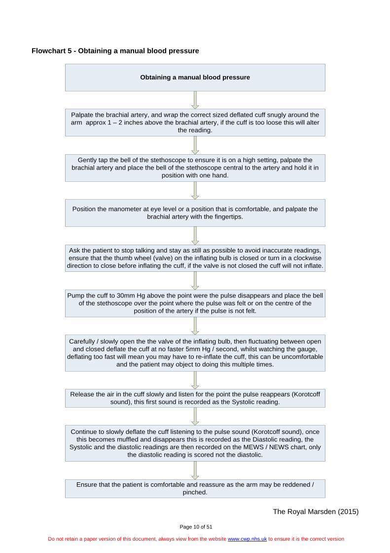
Flowchart 5 - Obtaining a manual blood pressure
Obtaining a manual blood pressure
Palpate the brachial artery, and wrap the correct sized deflated cuff snugly around the arm approx 1 – 2 inches above the brachial artery, if the cuff is too loose this will alter
the reading.
Gently tap the bell of the stethoscope to ensure it is on a high setting, palpate the brachial artery and place the bell of the stethoscope central to the artery and hold it in
position with one hand.
Position the manometer at eye level or a position that is comfortable, and palpate the brachial artery with the fingertips.
Ask the patient to stop talking and stay as still as possible to avoid inaccurate readings, ensure that the thumb wheel (valve) on the inflating bulb is closed or turn in a clockwise
direction to close before inflating the cuff, if the valve is not closed the cuff will not inflate.
Pump the cuff to 30mm Hg above the point were the pulse disappears and place the bell of the stethoscope over the point where the pulse was felt or on the centre of the
position of the artery if the pulse is not felt.
Carefully / slowly open the the valve of the inflating bulb, then fluctuating between open and closed deflate the cuff at no faster 5mm Hg / second, whilst watching the gauge,
deflating too fast will mean you may have to re-inflate the cuff, this can be uncomfortable and the patient may object to doing this multiple times.
Release the air in the cuff slowly and listen for the point the pulse reappears (Korotcoff sound), this first sound is recorded as the Systolic reading.
Continue to slowly deflate the cuff listening to the pulse sound (Korotcoff sound), once this becomes muffled and disappears this is recorded as the Diastolic reading, the
Systolic and the diastolic readings are then recorded on the MEWS / NEWS chart, only the diastolic reading is scored not the diastolic.
Ensure that the patient is comfortable and reassure as the arm may be reddened / pinched.
The Royal Marsden (2015)

Page 11 of 51
Do not retain a paper version of this document, always view from the website www.cwp.nhs.uk to ensure it is the correct version
Flowchart 6 – Obtaining a temperature
Obtaining a Temperature
Common sites for recording a temperature.
- Tympanic – Reads temperature from the tympanic membrane (ear drum). This is a core temperature (internal). - Oral – This is taken from the mouth under the tongue. This is a core temperature.- Axilla – This is taken from the centre of the arm pit (Axilla). This is not a core temperature
Tympanic Temperature recording – Ensure that the lens on the thermometer is clean and dry
Attach a disposable probe cover, this will usually turn the thermometer on (depending on the model), always follow manufacturers instructions.
Stabilise the patients head, then gently pull the ear lobe down, this slightly straightens the ear canal and provides a more accurate reading (for adults and children age 1 and over).
Insert the tip of the thermometer until the ear canal is sealed, or until the end of the thermometer stops, if the thermometer is not in the ear canal enough the reading could be
altered, and press the activation button and hold in place for 1 second, some models will beep,
The temperature will appear on the digital display, the probe cover should hen be disposed of in clinical waste.
Oral Temperature recording – explain the procedure to the patient and obtain informed consent.
Ensure the thermometer probe is placed on the probe, and the thermometer is switched on
Ask the patient to open their mouth and lift their tongue and place the probe under the tongue, ask the patient to lower their tongue onto the probe and close their mouth until the
thermometer beeps and a temperature can be recorded.
The Royal Marsden (2015)

Page 12 of 51
Do not retain a paper version of this document, always view from the website www.cwp.nhs.uk to ensure it is the correct version
Flowchart 7 - Monitoring levels of consciousness (AVPU)
Monitoring Levels of Consciousness (AVPU).
A = Alert - Fully awake - Is aware of their surroundings, are their eyes open on your approach, do they respond to your voice and have spontaneous motor function.
V = Responds to Voice - Will make some kind of response when you talk to them, which could be eye movement or motor response. - Ask short , sharp questions, ‘ are you okay’ the response could be a verbal response, grunt, moan or a movement of a limb when prompted by Voice.
P = Responds to Painful stimuli - The patient should respond to the application of pain on selected sites. - Applying short sharp pressure on the side of the knuckle of an index finger with a pen this pressure is applied by applying pressure with a pen between your finger and the patients inside knuckle. Do not use nail bed pressure as this could cause bruising. - Straight fingers onto the patients collar bone and tapping whilst applying pressure with each tap, you should focus on one area for this method to be effective.
U = Completely Unresponsive - The patient does not respond with eye movement, voice or motor responses to voice or painful stimuli
If V, P, U scores, Glasgow Coma Scale (GCS) must be activated, and the actions followed as per the GCS actions detailed in this procedure (SOP3)
Please remember that the airway is at risk in patients with a low conscious level, and can appear in patients not known to have a head injury, such as alcohol intoxication,
illicit drug use Hypoglycaemia etc as discussed within this protocol (SOP3).
The Royal Marsden (2015)

Page 13 of 51
Do not retain a paper version of this document, always view from the website www.cwp.nhs.uk to ensure it is the correct version
Flowchart 8 - Procedure to be followed for patients with physical deterioration and not known to have a head injury Procedure to be followed in the following situations: • On admission, then frequency as directed by care plan, a minimum of weekly; • The patient appears to be physically unwell; • The patient has altered level of consciousness, head injury is not suspected; • The patient not responding to requests as expected; • The patient is commencing new medication that may affect physical health; • A report from patient or witness regarding any of the above.
Immediately commence Physical Observations with NEWS / PEWS and apply GCS score as
directed by actions below
NEWS SCORE 0 PEWS SCORE 0 Continue with routine observations i.e. − Minimum of weekly
unless alternative observations are agreed as part of a care plan
− Unless patient’s condition indicates change then a care plan is required.
NEWS SCORE 1-4 PEWS SCORE 1-2 Maximum 2hourly Minimum 4 hourly. - Inform the
registered nurse who must assess the patient
- Registered nurse to decide whether to increase the monitoring frequency and/or if escalation of clinical care is required, i.e. Medical review.
- Clinical judgement and clinical decision making needs to be used when deciding whether to escalate.
NEWS SCORE 5-6 OR A SCORE OF 3 IN ANY ONE PARAMETER, PEWS SCORE 3-4. (Except AVPU, see next column). Increased frequency to a minimum of 1 hourly - Registered
nurse to urgently inform the medical team caring for the patient.
- Contact an available medic for urgent assessment within 30 minutes.
- Contact Emergency services (9)999 or crash team 2222, depending on clinical presentation, i.e. cardiac arrest.
NEWS SCORE 7 OR MORE. PEWS 5-8. Increased frequency to a minimum of 15 minute intervals. - Registered nurse to
immediately inform medical team or available medic for emergency assessment.
- Contact emergency services (9)999 or crash team, depending on clinical presentation, i.e. cardiac arrest`.
VPU SCORES 3 (NEWS 0NLY) VPU SCORES 1 (PEWS ONLY) - continue with GCS
and MEWS scoring Minimum of 30 minute intervals for 2 hours if GCS 15
- Minimum 15 minute MEWS and GCS if GCS <14 and below, follow actions shown in Flowchart 9.
Record blood glucose Below 4mmol – Hypoglycaemia Above 7mmol – Hyperglycaemia
Please Note:
Please ensure when reporting any head injury or altered level of consciousness on Datix that you include the NEWS and GCS scores
NEWS = Monitoring early warning scores GCS = Glasgow Coma Scale

Page 14 of 51
Do not retain a paper version of this document, always view from the website www.cwp.nhs.uk to ensure it is the correct version
Flowchart 9 - Procedure to be followed in the event of altered level of consciousness Procedure to be followed in the event of altered level of consciousness, including: • Patient found on floor with suspected injury; • Obvious head injury, lump, bump; • Altered level of consciousness due to possible consumption of alcohol and or illicit drugs,
potential /associated head injury? • Patient not responding to requests as expected; • Report from patient or witness.
Immediately commence physical observations with MEWS and apply GCS scores.
GCS Score = 13 or less
Call an
ambulance
15 minute NEWS observations and
GCS
Level 3 observations
GCS ≤14 WITH head injury /
suspected head injury
Call an
ambulance
15 minute NEWS observations and
GCS
Level 3 observations
GCS ≤ 14 WITHOUT head
injury 15 minute NEWS observations and
GCS
Level 3 observations
GCS = 15 NEWS
observations and GCS:
• Every 30 minutes for 2 hours
• Hourly for 4 hours
• 2 hourly until directed by Doctor
If at any time the GCS is less than
15 resume 15 minute NEWS
observations and GCS
At 2nd recording if
GCS ≤14 call an ambulance
Please Note:
Please ensure when reporting any head injury or altered level of consciousness on Datix that you include the NEWS and GCS scores
NEWS = Monitoring early warning scores GCS = Glasgow Coma Scale

Page 15 of 51
Do not retain a paper version of this document, always view from the website www.cwp.nhs.uk to ensure it is the correct version
Flowchart 10 - Blood glucose monitoring
Blood Glucose Monitoring Pre-procedure.
Turn on the machine and ensure that the onscreen date and time is correct and that there is adequate battery life
Check the unit of measurement, ensure that it is reading in mmol/L prior to each use
Before taking the monitor / test strips to the patient they need to be checked for the following:
- Ensure that the test strips are in date and have not been exposed to air- The Monitor and test strips have been calibrated together.- the monitor is recalibrated when using a new pack of test strips.- Internal quality control is carried out with both high and low solutions in accordance with trust / manufacturers guidelines.- Record the result of the internal quality control in the appropriate log book / sheet (pass / fail).- Ensure that the glucose meter is decontaminated as per local guidelines prior to use.- Ensure that the Glucose meter service record is in date in accordance with local policy.- Ensure that the screen / display is intact and that the screen safety check has been completed as per manufacturers guidelines.- Select a site that is warm, pink and free from any hard skin / calluses, burns, cuts, scars, bruises or rashes. Avoid any previous obvious puncture sites. The usual site for lancing is the palmer surface of the distal segment of the third or fourth finger, ideally of the non-dominant hand as there is usually less callusing visible.
Blood Glucose Monitoring Procedure
Ensure that the patient has washed their hands and dry thoroughly as per local infection control guidelines and that the patient is comfortable, sitting or lying down, then wash your
hands and apply gloves.
- Activate the blood glucose meter- Take a single use lancelet and ensure it has the correct depth settings (if available).- Activate the single use Lancelet as per manufacturers guidelines into the chosen site, e.g. the side of a finger and ensure that the sites are rotated to prevent the frequent use of sites.- “Milk” the fingertip from the palm of the hand to gain a large enough Droplet of blood, avoid milking the finger alone.
Insert the test strip into the blood glucose monitor and apply the first drop of blood when advised by the on screen instructions, ensuring that the correct location of the test window
is identified and is entirely covered with blood.
Place a piece of gauze over the puncture site and apply firm pressure and regularly monitor for excessive bleeding and then remove gloves and place into the clinical waste bag as per
Infection Control guidelines.
Document the result once obtained and decontaminate the glucose monitor as per local guidelines.

Page 16 of 51
Do not retain a paper version of this document, always view from the website www.cwp.nhs.uk to ensure it is the correct version
Flowchart 11 – Alcohol / drug intoxication incident process
ALCOHOL OR DRUGS SUSPECTED
TAKE PHYSICAL OBS IMMEDIATELY
MEWS/GCS
CONTACT MEDICAL ON-CALL FOR REVIEW
SERVICE USER UNRESPONSIVE YESTREAT AS MEDICAL
EMERGENCY 999
IMMEDIATELYTAKE PHYSICAL OBS
MEWS/GCS
CONTINUE TO MONITOR PHYSICAL
OBS MEWS/GCS
OBTAIN MEDICAL TROLLEY/GRAB BAG
SBAR TO PARAMEDICS
NO
REVIEW OBSERVATION LEVEL LEVEL 3 MINIMUMUNTIL PHYSICAL OBSERVATIONS IN COMPLIANCE WITH CP35 PHYSICAL HEALTH PATHWAY POLICY/
SOP3
ATTEMPT TO ENGAGE WITH SERVICE USER TO CLARIFY WHAT
THEY HAVE TAKEN/INGESTED
NO
ABNORMAL SCORESYESREFER FLOW CHART ? FOR FURTHER GUIDANCE
CONTINUE TO MONITOR PHYSICAL OBS
MEWS/GCS
REVIEW OBSERVATION LEVEL LEVEL 3 MINIMUM
UNTIL PHYSICAL OBSERVATIONS IN COMPLIANCE WITH CP35
PHYSICAL HEALTH PATHWAY POLICY/SOP3
COMMENCE CPR IF REQUIRED
POST INCIDENTNOTIFY ON-CALL BLEEP/
MANAGERCOMPLETE DATIX &
RECORD INTO ELECTRONIC RECORDS

Page 17 of 51
Do not retain a paper version of this document, always view from the website www.cwp.nhs.uk to ensure it is the correct version
Flowchart 12 – ECG Procedure
ECG Requested / Required
Wash and dry hands as per IPC policy
Full explanation of the procedure is given to the Service user
Ensure service user comfort and are relaxed (preferably lying down)
Clean Electrode site and prepare the skin (dry and shave if necessary) – Apply the 10 electodes
(appendix 3)
Attach the 10 lead cables from the ECG machine to the electrodes
Check that all leads are connected correctly to the relevant electrode and are not twisted, not pulling on the electrodes
or lying over each other
Input service user details into the ECG machine, if this facility is not available ensure the ECG is labelled
immediately following the procedure – Name, DOB, NHS number, Consultant, Location, Date &Time of
recording.
Ask the service user to relax and not move, if possible , if there is
movement during recording e.g.. due to neurological conditions, document
on the trace.
Commence recording, it will take several seconds to record, do not keep pressing
the start button
If there appears to be electrical interference or a poor recording check electrodes and
connections, keep offering the service user reassurance during the procedure
Detach and inspect the ECG trace
If despite all efforts to relax the service user, there is interference (artefact), switch on the
filter mode – this must be clearly documented on the final ECG trace
Inform the Service user the procedre is now compete and help to remove
the electodes
Discard electrodes into clinical waste, clean equipment and restock used items
Place the ECG trace in the appropriate documentation and inform nursing / medical staff, the ECG MUST be reviewed by the
requesting doctor within 12 hours of recording for inpatient services / within an
acceptable time frame for community services

Page 18 of 51
Do not retain a paper version of this document, always view from the website www.cwp.nhs.uk to ensure it is the correct version
Section 1 - Physical Observations 1. Introduction There will be occasions when patients will need an increased attention paid to the assessment and management of their physical health. This document sets out the actions that staff will need to take urgently for patients who become physically unwell, have an altered level of consciousness, head injury or suspected head injury, to prevent deterioration and save lives. To support patients during physical or neurological crisis, it is imperative that the physical and neurological assessment on admission and subsequent assessments have been completed to enable clinical and medical staff to have a base line of patient’s status using the Physical observation recording chart with National Early Warning Score (NEWS), Paediatric Early Warning Score (PEWS) AND THE Pregnancy Early Warning Score (Pages 21-31) Awake, Voice, Painful Stimulus, unresponsive (AVPU) (pages 21, 25, 28, 31) and Glasgow Coma Scale (GCS) for ALL service users (Pages 26, 29, 32, 33-35). It is necessary for staff to have competent physical and neurological observation assessment skills in order to carry out these assessments competently. If the patient has an obvious wound and / or loss of consciousness which require urgent medical attention they will need to go to Accident and Emergency for treatment – dial (9)999. Patient should be nursed in the recovery position if they have an altered level of consciousness. Monitor and record the blood glucose levels to exclude an underlying hypoglycaemia or hyperglycaemia (Page 35). A full physical assessment of the patient should be made to assess for any injury or abnormality. Consideration should always be given to patient’s allergy status. There are occasions when an in-patient may appear to be intoxicated with alcohol and or illicit substances. It is vital that these situations are assessed and managed to ensure the safety of the patient, staff and others. It is important a thorough assessment is made to rule out other conditions that may appear to be due to intoxication e.g. head injury and therefore physical and neurological assessment will be required. Acute intoxication is a serious condition which can result in death. 1.1 How to carry out physical observation The NEWS / PEWS and Pregnancy EWS are all incorporated into standardised Physical observation recording charts which utilise the National early Warning Score (NEWS) parameters and Glasgow Coma Scale (GCS) for ALL patients. Points to consider when using the physical observation recording charts with National Early Warning (NEWS), Paediatric Early Warning Score (PEWS), Pregnancy Early Warning Score (EWS) and Glasgow Coma Scale (GCS):
• Does the patient have a learning disability or comprehension problem? Keep the instructions and questions simple;
• Is the patient deaf? Make sure any hearing aids are in and in good working order, face the Patient when assessing them;
• Does the patient have a neurological problem e.g. Stroke? Check the patients’ past medical history; assess AVPU and GCS on both sides of the body.
Preparation:
• Wash hands before and after procedure as per Trust infection and prevention control policies;
• Check all equipment is clean and has been checked as fit for use.

Page 19 of 51
Do not retain a paper version of this document, always view from the website www.cwp.nhs.uk to ensure it is the correct version
All notations on patient’s Physical observation recording chart with NEWS, PEWS and Pregnancy EWS) and Glasgow Coma Scale (GCS) must be:
• Legible; • Signed; • Dated; • Timed; • In black ink. • Take chart to patient; • Record patient identification; • Explain the procedure to the patient, answer any questions and gain their consent; • Record all observations with a firm dot ● in black ink; • Write exact values in boxes provided • Join consecutive observations with a straight line over time. • Clean all equipment and store safely; • After procedure, clean and dispose of any single use items.
1.1.1 Respirations
• The best time to assess your patient’s respirations is settled and at rest, immediately after taking his pulse rate;
• Keep your fingertips over his radial artery, and don’t tell him that you’re counting respirations; otherwise, he’ll become conscious of them, and the rate may change;
• Count respirations by observing the rise and fall of the patient’s chest as he breathes. Alternatively, position the patient’s opposite arm across his chest, and count respirations by feeling its rise and fall. Consider one rise and one fall as one respiration;
• Using a watch or clock with a second hand, count the amount of breaths for 60 seconds to account for variations in respiratory rate and pattern;
• Observe chest movements for depth of respirations; • As you count respirations, note and record any obvious symptoms such as coughing,
wheezing, production of sputum wheezing, and expiratory grunting; • Wheezing is caused by partial obstruction in the smaller bronchi and bronchioles. This
high-pitched, musical sound is common in patient with emphysema or asthma. 1.1.2 Oxygen saturation
• Pulse oximetry only measures haemoglobin oxygen saturation, so does not provide information on ventilatory function, haemoglobin concentration or oxygen delivery to the tissues;
• Determine the site to be used for pulse oximetry; the site should have a good blood supply, check it is warm;
• Select probe site (usually finger, although ear lobes and bridge of nose can be used), assessing for barriers such as nail varnish, dirt, blood;
• Position the sensor securely; • Turn the pulse oximeter on; • Check that the pulse reading on the devise corresponds with their actual pulse; • Continuous use of a finger probe may cause blisters on the finger pad or pressure damage
to the skin or nail bed; • Do not use tape to hold probe in place, and re site probe at least every 4 hours, or more
frequently if stated in the manufacturers’ instructions (MDA 2001). 1.1.3 Blood pressure
• Ask the patient to rest for 5 minutes before taking their blood pressure; • Ensure tight or restrictive clothing is removed from the arm; • Ensure arm is comfortably straight, supported and positioned at heart level, palm face up; • Carefully choose a cuff of appropriate size for the patient: an excessively narrow cuff may
cause a false-high reading; an excessively wide one, a false-low reading;

Page 20 of 51
Do not retain a paper version of this document, always view from the website www.cwp.nhs.uk to ensure it is the correct version
• Do not take a blood pressure measurement on the same side as a mastectomy because it may compromise lymphatic circulation, worsen oedema, and damage the arm;
• Do not take blood pressure on the same arm as a cannula because it may damage the device.
Using a digital sphygmomanometer:
• The patient can lie in a supine position or sit erect while you measure their blood pressure;
• The patient’s arm should be extended at heart level and needs to be well supported with a pillow;
• If the artery is below heart level, you may get a false-high reading; • Make sure the patient’s is relaxed and comfortable when you measure his blood
pressure so it stays at its normal level; • Follow the manufacturers’ instructions.
Using a manual sphygmomanometer:
• Palpate the brachial artery. Centre the bell of the stethoscope over the part of the artery where you detect the strongest beats, and hold it in place with one hand;
• Wrap the deflated cuff snugly around the patient’s upper arm 1’’ (2.5cm) above the brachial pulse;
• Position the manometer at your eye level; • Instruct the patient to stop eating, talking and to stay still during the procedure as this
can cause inaccurate readings; • Palpate the brachial pulse with your fingertips while inflating the cuff; • Using the thumb and index finger of your other hand, turn the thumbscrew on the rubber
bulb of the air pump clockwise to close the valve; • Inflate the cuff to 30 mm Hg above the point where the pulse disappears; • Place the bell of your stethoscope over the point where you felt the brachial pulse; • Carefully open the valve of the air pump. Then deflate the cuff no faster than 5 mm
Hg/second, while watching the gauge; • Release the valve slowly and note the point at which you hear the pulse reappear, the
start of the pulse sound indicates the systolic pressure (Korotkoff sounds); • The sounds will become muffled and then disappear. The last Korotkoff sound you hear
is the diastolic pressure.
1.1.4 Pulse / heart rate Common areas to take the Pulse:
• Radial Artery – Located on the wrist just below the thumb; • Brachial Artery – Located on the opposite side of the elbow diagonally opposite to the
Radial artery; • Carotid Artery – Located at the side of the neck between the edge of the jaw bone and the
middle of the throat. Taking a pulse:
• Make sure the patient is comfortable and relaxed because an awkward, uncomfortable position may affect his heart rate;
• Ensure the patient is comfortable; in a sitting or supine position, with his arms at his side or across his chest;
• Gently press your index, middle, and ring fingers on the artery and apply light pressure until the pulse is felt;
• You should feel a pulse with only moderate pressure; excessive pressure may obstruct blood flow distal to the pulse site;
• Don’t use your thumb to take the patient pulse; the thumb has a strong pulse of its own and may be easily confused with the patient’s pulse;

Page 21 of 51
Do not retain a paper version of this document, always view from the website www.cwp.nhs.uk to ensure it is the correct version
• After locating the pulse, count the beats for 60 seconds to get the number of beats per minute. Counting for a full minute provides a more accurate picture of irregularities;
• While counting the rate, assess pulse rhythm and volume by noting the pattern and strength of the beats. If you detect an irregularity, repeat the count and note whether the irregularity occurs in a pattern or randomly.
1.1.5 Temperature
• Make sure the lens under the probe is clean and dry; • Attach a disposable probe cover following manufacturer’ instructions; • Stabilise the patient’s head; then gently pull his ear up and back (for adults and children
older than age 1); • Insert the thermometer until the entire ear canal is sealed; • Press the activation button, and hold for in place for 1 second; • The temperature will appear on the display.
1.1.6 How to record AVPU (Alert, Voice, Pain, and Unresponsive) Assessing conscious level involves examining simple but key components of a person’s neurological function, such as response to voice and pain. This enables an estimation of level of wakefulness and awareness at a particular time. If patient has a head injury, altered level of consciousness, including possible consumption of alcohol and / or illicit drugs, see Flowchart 8 or Flowchart 9. A = Alert
• Fully awake; • Note whether the patient has their eyes open when you approach them, will respond to
voice and have spontaneous motor function.
V = responds to Voice • Makes some kind of response when you talk to them; which could be in Eyes, Voice or
Motor; • Ask ‘Are you ok?’ The response could be a verbal response, grunt, moan or slight
movement of a limb when prompted by voice.
P = responds to Pain • The person makes a response on any of the components when pain is used on them; • Apply incremental pressure to the side of the patient’s little finger by pressing their finger
between your own finger and a pen; • Using your own straight fingers, vigorously tap the patients Collar bone (Clavicle), focusing
on one area. • Do not press the nail bed as this can cause bruising.
U = completely Unresponsive
• This is recorded when the person does not give any Eye, Voice or Motor response to voice or pain.
Remember that the airway is at risk in people with a low conscious level. There may be time when the patient has physically deteriorated and not known to have a head injury (Flowchart 8) 2. What are NEWS, PEWS and Pregnancy EWS? The National Early Warning Score (NEWS), Paediatric Early Warning Score (PEWS) and Pregnancy Early Warning Score (Pregnancy EWS) are standardised trigger scoring systems. The triggers are based on routine physical observations, Alert, Voice, Pain, Unresponsive (AVPU) and subsequently

Page 22 of 51
Do not retain a paper version of this document, always view from the website www.cwp.nhs.uk to ensure it is the correct version
Glasgow Coma Scale (GCS), are sensitive enough to detect changes in a patient’s physiology, which will be reflected in a change of score should the patient’s physical health be improving or deteriorating. All patients must have their physical observations and AVPU measured and these are converted into a score. The higher the score the more abnormal the physical observations and AVPU signs are. If the scores reach a certain threshold for example:
• NEWS score of 2 or more the senior nurse must be informed and clinical decision making should be utilised; if NEWS score of 5 or more the senior nurse must be informed and a doctor must be contacted to further assess the patient and clinical Decision making utilised (see Pages 24 - 26 ).
• PEWS Score of 1 – 2 or more the senior nurse must be informed and clinical decision making should be utilised; if PEWS scores 5 and above the senior nurse must be informed and a doctor must be contacted to further assess the patient and clinical Decision making utilised (see Page 27 – 29).
• Pregnancy EWS score of 0 – 2, Routine monitoring and scoring, Unless patient’s physical
condition indicates change – then care plan required, Score of 3 – 8, Registered nurse to urgently inform the medical team / Consultant, caring for the patient or an available medic for urgent assessment within 30mins and a score of 9 and above, Registered nurse to immediately inform medical team for emergency assessment, or Contact crash team (2222) or Emergency Services (999) (see Pages 30 - 32).
Early warning scoring systems were originally developed with two specific aims: to facilitate timely recognition of the patients with established or impending critical illness: and to empower nurses and medical staff to secure experienced help through the operation of a trigger threshold which, if reached, required mandatory attendance by a more senior member of staff within a set period of time. Use of NEWS / PEWS AND Pregnancy EWS can also:
• Improve the quality of patient’s observation and monitoring; • Improve communication within the multidisciplinary team; • Allow for timely transfer to acute assessment units; • Support good medical judgement; • Aid in securing appropriate assistance for the clinically deteriorating patient; • Give a good indication of physiological trends; • Be a sensitive indicator of abnormal physiology.
NEWS / PEWS / Pregnancy EWS are not:
• A predictor of outcome; • A comprehensive clinical assessment tool; • A replacement for clinical judgement.
NEWS Cannot:
• Be used on patients under 16 (PEWS) must be used on patients aged 13 – 18; • Be used on any patient who is pregnant Pregnancy Early Warning Score must be used.
2.1 When to use NEWS / PEWS / Pregnancy EWS? NEWS / PEWS rely on the routine assessment and charting of the physical observations and AVPU status of the patient. These are simple observations that can be performed by a nurse, doctor or other trained staff familiar with the process. All patients must have a physical assessment and AVPU within 6 hours of admission and a NEWS / PEWS score must be calculated and recorded as a benchmark. If completion of assessment has not taken place within 6 hours you must document and date each attempt, and reasons why the assessment was not completed within the time period.

Page 23 of 51
Do not retain a paper version of this document, always view from the website www.cwp.nhs.uk to ensure it is the correct version
These physical observations and AVPU observations are: • Doctor / Nurse / Family concerns (PEWS); • Respiratory rate; • Respiratory Distress (PEWS); • Oxygen saturation; • Blood pressure (Recorded, but not scored in (PEWS); • Pulse / heart rate; • Temperature; • AVPU; • GCS; • Blood Glucose.
The outcome for each observation is combined to provide a NEWS / PEWS / Pregnancy EWS score All sections of the Physical observations recording chart with National Early Warning Score (NEWS), Paediatric Early Warning Score (PEWS) and Glasgow Coma Score (GCS) chart must be completed and scored, and actions taken as described on the reverse of the chart. The frequency and specifications of all observations must be prescribed in the nursing care plan; and must be a minimum of weekly for all patients following admission. NEWS / PEWS assessment must be recommenced immediately in the following situations:
• The patient appears to be physically unwell; • The patient has fallen; • The patient has altered level of consciousness e.g. head injury; • The patient is intoxicated with alcohol or drugs; • The patient not responding to requests as expected; • The patient is commencing new medication that may affect physical health; • A report from patient or witness regarding any of the above.
NEWS / PEWS score must be updated and scored prior to any transfer / discharge to other Services or external healthcare provision. Where the patient’s multidisciplinary team decide that a full physical NEWS / PEWS assessment and scoring is not appropriate then this should be clearly documented both on the patient’s physical observation chart, with an annotation in the patients’ care notes, recording why the decision was made not to use MEWS / PEWS. This may include the following patient’s:
• The patient on palliative care pathways; • The patient for whom escalation of care is inappropriate.

Page 24 of 51
Do not retain a paper version of this document, always view from the website www.cwp.nhs.uk to ensure it is the correct version
2.1.1 How to Calculate Score and Action NEWS Please note: ≥ is greater than; ≤ is less than. Taking into account the results of the physical and AVPU observations:
• Total the NEWS score using 0-3 guide on chart. • Observation recorded in White sections score = 0 • Observation recorded in Yellow sections score = 1 • Observation recorded in Orange sections score = 2 • Observation recorded in Red sections score = 3 • Add the total observation scores and record total NEWS score in the box for NEWS.
NEWS observations should then be continued at the frequency identified on physical observation chart with NEWS pathway and must reflect the needs of the patient.
• A NEWS of 0 - Minimum of weekly NEWS, Routine monitoring and scoring, unless alternative observations are agreed as part of a care plan and if a patient’s physical condition indicates change – then a care plan is required, the care plan should be discussed with and agreed by the patient’s medical team.
• A NEWS of 1 – 4 - Maximum - 2 hourly, Minimum - 4 hourly, a registered nurse must be informed and the patient must be assessed, the registered nurse will then decide whether to increase the frequency of monitoring and if an escalation of clinical care is required, such as medical escalation.
• A NEWS of 5 – 6 or a score of 3 in any one parameter - increased frequency to a minimum of 1 hourly, Registered nurse must urgently inform the available medical team for assessment within 30 minutes or contact emergency services (9)999 or the crash team (2222 via the locality switch board).
• VPU scores 3 in one parameter – continue with GCS and NEWS scoring, at a minimum 30
minutes for 2 hours if GCS is 15 with a head injury or suspected head injury, IF GCS < 14 increase to 15 minutes observations and mews scoring and follow the actions for GCS in Flowchart 9.
• A NEWS of 7 or more - increase frequency to 5 minutes and therapeutic observations to level 3-4. Registered nurse must immediately inform medical team for emergency assessment or contact emergency services (9)999 or 2222 for the crash team. Failure of medical review or 999 to attend to a NEWS call within the acceptable timescale (i.e. within 30 minutes) the nurse in charge must complete a Datix form and inform immediate manager.
• If a patient is scoring high and a reason for this is known / suspected, this may not be deterioration, a high score may be due to patient Anxieties, pre-existing health issues, equipment etc. Clinical judgement / decision making should be utilised, this decision must be documented and discussed with the medical staff.
• If a patient is scoring high and deterioration is suspected then the actions for that score must
be followed as below.
• If patient has a head injury, altered level of consciousness, including possible consumption of alcohol and / or illicit drugs, or has an AVPU score of 3 or more, commence Glasgow Coma score (GCS) assessment, and follow the actions outlined in Flowchart 9.
• If a patient’s systolic blood pressure is recorded within the grey shaded area the nurse in
charge must be informed and then discussed with the patient’s medical team, this may then require regular observations and intervention.

Page 25 of 51
Do not retain a paper version of this document, always view from the website www.cwp.nhs.uk to ensure it is the correct version
National Early Warning Score (NEWS) Ward NHS Number Name DOB
Date Time
Respiration Rate ≥25 21-24 12-20 9-11
≤8 Record respiration rate
Oxygen Saturation ≥96 94-95 92-93
≤91 Any oxygen Given %
Record oxygen saturation %
Blood Pressure Record systolic & diastolic Inform nurse in charge if Systolic is above this line Score systolic BP only for NEWS
≥230 221-230 211-220 201-210 191-200 181-190 171-180 161-170 151-160 141-150 131-140 121-130 111-120 101-110
91-100 81-90 71-80 61-70 51-60
≤50
Record blood pressure
Pulse / Heart Rate ≥140 131-140 121-130 111-120 101-110
91-100 81-90 71-80 61-70 51-60 41-50 31-40
≤30 Record pulse / heart rate
Temperature
>39 38.1-39° 37.1-38° 36.1-37°
35.1- 36° <35°
Record temperature
Levels of Consciousness (AVPU)
Alert Voice / Pain / Unresponsive
Blood Sugar *
Calculate NEWS score using guide below* and see overleaf for actions
Staff Initials
*Only record blood sugar if the patient deteriorates, or if VPU scores 3 and GCS is activated. * NEWS key colour code for scoring 0 1 2 3 See overleaf for actions and GCS

Page 26 of 51
Do not retain a paper version of this document, always view from the website www.cwp.nhs.uk to ensure it is the correct version
How to calculate NEWS Score • Record all observations overleaf; • Note whether observation falls in shaded ‘At Risk Zone’. Score as per NEWS key; • Add points scored and record total ‘NEWS Score’ in bottom row of chart.
How to use the physical observation chart Start up Observations NEWS scores Action
1. This chart does not override clinical judgement. 2. This chart cannot be used for patients under the age of 16. 3. This chart cannot be used for patients who are pregnant. 4. Take chart to patient. 5. Record patient identification.
1. Record ALL observations with a ‘firm’ dot ● in black ink. 2. Write exact values of observations in boxes provided. 3. Join consecutive observations with a straight line over time. 4. If Systolic Blood pressure is recorded in the grey shaded box, please inform the nurse in charge.
1. Total the NEWS score including AVPU using 0 – 3 key scoring guide on the chart. 2. Record the total NEWS score in the box for NEWS.
NEWS Score Frequency of monitoring Clinical response
0
Minimum of weekly NEWS unless
alternative observations are
agreed as part of a care plan.
- Routine monitoring and scoring;
- Unless patient’s physical condition indicates change – then care plan required.
Total: 1-4
Score of 3 in
any one parameter see box below
Maximum - 2 Hourly Minimum - 4 hourly
- Inform registered nurse who must assess the patient;
- Registered nurse to decide if increased frequency of monitoring and/or escalation of clinical care required, i.e. medical review.
Total: 5-6
Or
A score of 3 in any one parameter
Increased frequency to a minimum of 1
hourly. If VPU scores 3
continue with GCS and NEWS scoring
- Minimum of every 30mins for 2hours if GCS 15.
- 15 minute NEWS and GCS if GCS < 14. Follow actions as directed in SOP3.
- Registered nurse to urgently inform the medical team caring for the patient or an available medic for urgent assessment within 30mins, if the patients’ medical team is not available.
- Contact crash team (2222) or Emergency Services (999)
Total:
7
Or
MORE
Increased frequency
to 5 minutes and
Therapeutic Observations (level
3/4)
- Registered nurse to immediately inform medical team for emergency assessment;
- Contact crash team (2222) or Emergency Services (999)
How to calculate and action GCS 15 point score: The GCS is a simple but effective way of assessing a patient’s neurological condition. It categorises the patient’s responses to certain stimuli and gives that response an overall score. It is divided into 3 main categories of response that are totalled to give an overall score.
• Score best motor, verbal and eye opening scores in the boxes provided following chart above; • Add points score and record total ‘Overall GCS score’ in the box provided.
Score and Motor Response 6 - Obeys commands 5 - Localises pain 4 - Withdrawal to pain 3 - Flexion 2 - Extension 1 - No response to pain
Score and Verbal Response 5 - Oriented 4 - Confused conversation 3 - Inappropriate words 2 - Incomprehensible sounds 1 - No verbal response
Score and Eye Opening 4 - Spontaneous 3 - Open to speech 2 - Open to pain 1 - No eye opening
Date Time Motor Response Score Verbal Response Score Eye Opening Score Overall GCS Score Staffs Initials
Action: For actions refer to Clinical Practice policy CP35 / SOP3 which incorporates the ‘Procedure to be followed in the event of altered level of consciousnesses’.

Page 27 of 51
Do not retain a paper version of this document, always view from the website www.cwp.nhs.uk to ensure it is the correct version
2.1.2 How to Calculate Score and Action PEWS Please note: ≥ is greater than; ≤ is less than. Taking into account the results of the physical and AVPU observations:
• Total the PEWS score using the guide on chart; • Observation recorded in White sections score = 0; • Observations in the shaded areas score 1 point each; • Add the total observation scores and record total PEWS score in the box for Calculate PEWS
score using guide and follow the scoring actions; • Blood pressure is not scored as part of PEWS, but must be recorded.
PEWS observations should then be continued at the frequency identified on physical observation chart with PEWS pathway and must reflect the needs of the patient.
• A PEWS of 0 - Minimum of weekly PEWS, Routine monitoring and scoring, Unless alternative observations are agreed as part of a care plan and if a patient’s physical condition indicates change – then a care plan is required, the care plan should be discussed with and agreed by the patient’s medical team;
• A PEWS of 1 - 2 - Minimum - 2 hourly, Maximum - 4 hourly, a registered nurse must be informed and the patient must be assessed, the registered nurse will then decide whether to increase the frequency of monitoring and if an escalation of clinical care is required, such as medical escalation;
• A PEWS of 3 – 4 - increased frequency to a minimum of 1 hourly, Registered nurse must
urgently inform the available medical team for assessment within 30 minutes or contact emergency services (9)999 or the crash team (2222 via the locality switch board);
• VPU scores 1 – continue with GCS and PEWS scoring, at a minimum 30 minutes for 2 hours
if GCS is 15 with a head injury or suspected head injury, IF GCS < 14 increase to 15 minutes observations and mews scoring and follow the actions for GCS in Flowchart 9;
• A PEWS of 5 - 8 - increase frequency to 5 minutes and therapeutic observations to level 3 - 4.
Registered nurse must immediately inform medical team for emergency assessment or contact emergency services (9)999 or 2222 for the crash team (only). Failure of medical review or 999 to attend to a PEWS call within the acceptable timescale (i.e. within 30 minutes) the nurse in charge must complete a Datix form and inform immediate manager;
• If a patient is scoring high and a reason for this is known / suspected, this may not be
deterioration, a high score may be due to patient Anxieties, pre-existing health issues, equipment etc. Clinical judgement / decision making should be utilised, this decision must be documented and discussed with the medical staff;
• If a patient is scoring high and deterioration is suspected then the actions for that score must
be followed as above;
• If patient has a head injury, altered level of consciousness, including possible consumption of alcohol and / or illicit drugs, or has an AVPU score of 3, commence Glasgow Coma score (GCS) assessment, and follow the actions outlined in this SOP Flowchart 9.

Page 28 of 51
Do not retain a paper version of this document, always view from the website www.cwp.nhs.uk to ensure it is the correct version
Paediatric Early Warning score (PEWS) for 13 – 18 Years Ward NHS Number Name DOB
Doctor / Nurse / Family concern
Date Time > 50 Respiratory rate (over 1 minute)
40 - 50 30 - 40 20 - 30 10 - 20 0 - 10
Record respiration rate
Respiratory Distress
Moderate - Severe None - Mild
Oxygen 93
Saturation ≤92
Receiving Oxygen L/Min
Record oxygen saturation
≥181 171 - 180 Heart Rate & Blood Pressure
161 - 170 151 - 160 141 - 150 131 - 140
121 - 130 111- 120
101 - 110 91 - 100
81 - 90 71 - 80
61 - 70 51 - 60 41 - 50 36 - 40
≤ 35
Record pulse / heart rate
Record blood pressure
Temperature °c ≥ 39.1°
38.1 - 39° 37.1 - 38° 36.1 - 37°
35.1 - 36° ≤35.9°
Record temperature
Neuro Response (AVPU)
Alert Verbal
Pain Unresponsive
Calculate PEWS score using guide below* and see overleaf for actions
Staff Initials
Total PEWS 0 1 - 2 3 - 4 5 - 8
PTO for Action: Total PEWS = Number of Entries in Shaded Boxes
How to calculate PEWS Score • Record all observations above with a firm black ● in black ink • Note whether observation falls in shaded ‘At Risk Zone as one point’. Score as per PEWS key; • Add points scored and record total ‘PEWS Score’ in bottom row of chart
BP not used to calculate PEWS, but MUST be recorded.
Score pulse
only.
Systolic BP ≥ 160 inform
N.I.C.
Systolic BP ≤ 100 inform
N.I.C.

Page 29 of 51
Do not retain a paper version of this document, always view from the website www.cwp.nhs.uk to ensure it is the correct version
How to use the physical observation chart Start up Observations PEWS
scores Action
1. This chart does not override clinical judgement. If the patient scores 3 and above and a reason for this is known this reason must be documented / careplanned and medical advice sought. 2. Take chart to patient. 3. Record patient identification.
1. Record ALL observations with a ‘firm’ dot ● in black ink. 2. Write exact values of observations in boxes provided. 3. Join consecutive observations with a straight line over time. 4. If systolic and diastolic blood pressure are above 140 or below 100 (the two black lines) – inform the Nurse in charge.
1. Total the PEWS score including AVPU using 0 – 3 key scoring guide on the chart. 2. Record the total PEWS score in the box for PEWS.
PEWS Score
Frequency of monitoring Clinical response
0
Minimum of weekly PEWS unless
alternative observations are
agreed as part of a care plan.
- Routine monitoring and scoring;
- Unless patient’s physical condition indicates change – then care plan required.
Total: 1 - 2
Minimum - 2 Hourly Maximum - 4 hourly
- Inform registered nurse who must assess the patient;
- Registered nurse to decide if increased frequency of monitoring and/or escalation of clinical care required, i.e. medical review.
Total: 3 - 4
Increased frequency to a minimum of 1
hourly. If VPU scores in the shaded area continue with GCS and PEWS scoring - Minimum of
every 30mins for 2hours if GCS 15.
- 15 minute PEWS and GCS if GCS < 14. Follow actions as directed in SOP3.
- Registered nurse to urgently inform the medical team / Consultant, caring for the patient or an available medic for urgent assessment within 30mins, if the patients’ medical team is not available, call
- Emergency Services (999) or ring (2222)
Total: 5 - 8
Increased frequency
to 5 minutes and
Therapeutic Observations (level
3/4)
- Registered nurse to immediately inform medical team / Consultant for emergency assessment, or
- Contact Emergency Services (999) or (2222)
How to calculate and action GCS 15 point score: The Glasgow Coma Scale is a simple but effective way of assessing a patient’s neurological condition. It categorises the patient’s responses to certain stimuli and gives that response an overall score. It is divided into 3 main categories of response that are totalled to give an overall score.
• Score best motor, verbal and eye opening scores in the boxes provided following chart above; • Add points score and record total ‘Overall GCS score’ in the box provided.
Score and Motor Response 6 - Obeys commands 5 - Localises pain 4 - Withdrawal to pain 3 - Flexion 2 - Extension 1 - No response to pain
Score and Verbal Response 5 - Oriented 4 - Confused conversation 3 - Inappropriate words 2 - Incomprehensible sounds 1 - No verbal response
Score and Eye Opening 4 - Spontaneous 3 - Open to speech 2 - Open to pain 1 - No eye opening
Date Time Motor Response Score Verbal Response Score Eye Opening Score Overall GCS Score Staffs Initials

Page 30 of 51
Do not retain a paper version of this document, always view from the website www.cwp.nhs.uk to ensure it is the correct version
2.1.3 How to Calculate Score and Action Pregnancy EWS Please note: ≥ is greater than; ≤ is less than. Taking into account the results of the physical and AVPU observations:
• Total the Pregnancy EWS score using the guide on chart; • Observation recorded in White sections score = 0; • Observations in the Red or Yellow shaded areas score 1 point each; • If the patient scores 1 or more point in the red or 2 or more in the yellow the medics
should be contacted for early intervention – unless otherwise documented; • Add the total observation scores and record total Pregnancy EWS score in the box for total
score using guide and follow the scoring actions. Pregnancy EWS observations should then be continued at the frequency identified on physical observation chart with Pregnancy EWS pathway and must reflect the needs of the patient.
• A Pregnancy EWS of 0 - 2 = Minimum of 12 hourly, Routine monitoring and scoring, unless the patients physical condition indicates change – then a care plan is required, that should include maternity services input, the care plan should be discussed with and agreed by the patient’s medical team;
• A Pregnancy EWS of 3 - 5 = 1 - 4 hourly, a registered nurse must be informed and the
patient must be assessed, the registered nurse will then urgently inform the patient’s medical team or an available medic for urgent assessment within 30 minutes, or contact the crash team (2222) or (9)999, unless a plan of care has been formulated and agreed by the patients care team;
• A Pregnancy EWS of 6 – 8, a registered nurse must be informed and the patient must be
assessed, the registered nurse will then urgently inform the patient’s medical team or an available medic for urgent assessment within 30 minutes, or contact the crash team or (9)999, unless a plan of care has been formulated and agreed by the patients care team;
• A Pregnancy EWS score 0f 9 and above, Registered nurse to immediately inform the
medical team for emergency assessment OR contact the crash team (2222) or Emergency services (9)999;
• AVPU scores in red or yellow – continue with GCS and Pregnancy EWS scoring, at a
minimum 30 minutes for 2 hours if GCS is 15 with a head injury or suspected head injury, IF GCS < 14 increase to 15 minutes observations and mews scoring and follow the actions for GCS in Flowchart 9;
• If a patient is scoring high and a reason for this is known / suspected, this may not be
deterioration, a high score may be due to patient Anxieties, pre-existing health issues, equipment etc. Clinical judgement / decision making should be utilised, this decision Must be documented and discussed with the medical staff;
• If a patient is scoring high and deterioration is suspected then the actions for that score
Must be followed as above;
• If patient has a head injury, altered level of consciousness, including possible consumption of alcohol and / or illicit drugs, or has an AVPU score of 3, commence Glasgow Coma score (GCS) assessment, and follow the actions outlined in Flowchart 9.

Page 31 of 51
Do not retain a paper version of this document, always view from the website www.cwp.nhs.uk to ensure it is the correct version
Pregnancy Early Warning Score Chart
Ward NHS Number Name DOB
Date Time Respirations (over I minute)
>30 21 - 30 11 - 20 0 - 10
Record respiration rate 95 – 100%
≤95% Record oxygen saturation %
Oxygen administered L/min Blood Pressure Record systolic & diastolic Score systolic BP only for Pregnancy EWS
≥191 181-190 171-180 161-170 151-160 141-150 131-140 121-130 111-120 101-110
91-100 81-90 71-80 61-70
≤60
Record blood pressure
Pulse / Heart Rate
≥141 131-140 121-130 111-120 101-110
91-100 81-90 71-80 61-70 51-60 41-50 31-40
≤30 Record pulse / heart rate
Temperature ≥39.1° 38.6 - 39°
38.1- 38.5° 37.6 - 38°
37.1- 37.5° 36.6 - 37°
36.1- 36.5° 35.6 - 36°
35.1- 35.5° 34.6 - 35°
34.1- 34.5° ≤34°
Record temperature Neuro Response (AVPU)
Alert Verbal
Pain Unresponsive
Total Yellow Score Total Red Score Total score – see actions overleaf
Staff Signature
Contact medics for early intervention if patient scores One or more in Red or Two or more Yellow at any one time - unless otherwise documented.

Page 32 of 51
Do not retain a paper version of this document, always view from the website www.cwp.nhs.uk to ensure it is the correct version
How to calculate Pregnancy EWS Score • Record all observations overleaf; • Note whether observation falls in the Red or yellow boxes, Score one point per box. • Add points scored – in red and yellow boxes and record total if patient scores one point in red or
two points in yellow contact the medical team unless this has already been discussed and care planned.
• AVPU - If a point is scored in red or yellow – commence GCS.
How to use the physical observation chart Start up Observations P - EWS
scores Action
1. This chart does not override clinical judgement. 2. Record patient identification 3. This chart is to be used on pregnant patients only 4. Take chart to patient.
1. Record ALL observations with a ‘firm’ dot ● in black ink. 2. Write exact values of observations in boxes provided. 3. Join consecutive observations with a straight line over time.
1. Total the P - EWS score including AVPU using 2. Record the total P- EWS score in the box for total score.
P -EWS Score
Frequency of monitoring Clinical response
0 - 2 12 Hourly - unless
alternative observations are agreed as part of a
care plan.
- Routine monitoring and scoring;
- Unless patient’s physical condition indicates change – then care plan required.
Total: 3 - 5
1 – 4 Hourly - Inform registered nurse who must assess the
- Registered nurse to urgently inform the medical team caring for the patient or an available medic for urgent assessment within 30mins, if the patients’ medical team is not available.
- Contact crash team (2222) or Emergency Services (999)
Total: 6 - 8
Increased frequency to a minimum of 1 – 2
hourly. If VPU scores continue with GCS and P - EWS
scoring - Minimum of every
30mins for 2hours if GCS 15.
- 15 minute P - EWS and GCS if GCS < 14. Follow actions as directed in SOP3.
Total:
>9
Increased frequency to
30 minutes and
Therapeutic Observations (level 3/4)
- Registered nurse to immediately inform medical team for emergency assessment;
- Contact crash team (2222) or Emergency Services (999)
How to calculate and action GCS 15 point score: The Glasgow Coma Scale is a simple but effective way of assessing a patient’s neurological condition. It categorises the patient’s responses to certain stimuli and gives that response an overall score. It is divided into 3 main categories of response that are totalled to give an overall score.
• Score best motor, verbal and eye opening scores in the boxes provided following chart above; • Add points score and record total ‘Overall GCS score’ in the box provided.
Action: For actions refer to Clinical Practice policy CP35 / SOP3 which incorporates the ‘Procedure to be followed in the event of altered level of consciousnesses’
Score and Motor Response 6 - Obeys commands 5 - Localises pain 4 - Withdrawal to pain 3 - Flexion 2 - Extension 1 - No response to pain
Score and Verbal Response 5 - Oriented 4 - Confused conversation 3 - Inappropriate words 2 - Incomprehensible sounds 1 - No verbal response
Score and Eye Opening 4 - Spontaneous 3 - Open to speech 2 - Open to pain 1 - No eye opening
Date Time Motor Response Score Verbal Response Score Eye Opening Score Overall GCS Score Staffs Initials

Page 33 of 51
Do not retain a paper version of this document, always view from the website www.cwp.nhs.uk to ensure it is the correct version
3. Glasgow Coma Scale The Glasgow Coma Scale (GCS) is the most commonly used tool for evaluating conscious level. The GCS evaluates conscious level in three areas: motor response; verbal response and eye opening. The GCS categorises the person’s responses to stimuli and gives the responses a score; these scores are then added up to give an overall score; the total sum gives a score out of 15. A score of 15 indicates a fully alert and responsive person, whereas a score of 3 (the lowest possible score) indicates unconsciousness and critical state. The GCS and NEWS / PEWS and Pregnancy EWS observations procedure must be commenced immediately in the event of:
• The patient appears to be physically unwell; • The patient has fallen; • The patient has altered level of consciousness e.g. head injury; • The patient is intoxicated with alcohol or drugs; • The patient not responding to requests as expected; • The patient is commencing new medication that may affect physical health; • A report from patient or witness regarding any of the above.
In all cases of head injury or suspected head injury or altered level of consciousness NEWS / PEWS and Pregnancy EWS and GCS observations must be assessed, recorded and actioned at the frequency identified on NEWS / PEWS and Pregnancy EWS pathway for a minimum of 4 hours. A GCS of 8 or less indicates severe head injury, a GCS of 9-12 moderate head injury, and a GCS score of 13-15 is obtained when the head injury is minor. How to Calculate and Score the Glasgow Coma Scale:
• Explain the procedure to the patient, whether conscious or not, answer any questions and gain their consent;
• Talk to the patient. Note whether they are alert and giving full attention or restless, lethargic and drowsy;
• Ask the patient to perform a simple tasks e.g. raise your arm (include both sides of the body), stick out your tongue;
• If the patient does not respond apply painful stimuli (i.e. Apply incremental pressure to the side of the patient’s little finger by pressing their finger between your own finger and a pen;
• Score each category as per chart below; • Add up category scores to give a total score out of 15; • Record total GCS score on the GCS chart i.e.15/15.
Assessment of score motor response Score and motor response Number Response Explanation
6 Obeys commands The person does simple things you ask e.g. raise your arm, stick out your tongue
5 Localises pain A purposeful movement towards changing painful stimuli is a localizing response.
4 Withdrawal to pain Abnormally pulls limb away from painful stimulus.
3 Flexion Stimulus causes abnormal flexion of limbs (adduction of arm, internal rotation of shoulder, pronation of forearm, wrist flexion - decorticate posture.
2 Extension The stimulus causes limb extension (abduction, internal rotation of shoulder, pronation of forearm, wrist extension) - decerebrate posture.
1 No response to pain No response, flaccid limbs

Page 34 of 51
Do not retain a paper version of this document, always view from the website www.cwp.nhs.uk to ensure it is the correct version
Assessment of score and verbal response Score and verbal response Number Response Explanation
5 Orientated Patient ‘knows who he is, where he is and why, the year, season, and month.
4 Confused conversation
Patient responds to questions in a conversational manner but some disorientation and confusion.
3 Inappropriate words Random or exclamatory articulated speech, but no conversational exchange.
2 Incomprehensible sounds Moaning but no words.
1 No verbal response No verbal response despite verbal or other stimuli Assessment of score and eye opening Score and eye opening Number Response Explanation
4 Spontaneous Eyes open spontaneously without stimulation
3 Open to speech Eye opening in response any speech (or shout, not necessarily request to open eyes);
2 Open to Pain Eyes open with painful stimulus. 1 No eye opening No eye opening regardless of stimulation
Procedures to be followed in the event of a head injury or altered level of consciousness see Flowchart 9 http://www.glasgowcomascale.org/recording-gcs/ Patients who have sustained a head injury must be referred to a hospital Accident and Emergency department if any of the Risk factors listed below are present: Risk factors:
• GCS less than 15 on initial assessment; • Any loss of consciousness as a result of the injury; • Any focal neurological deficit since the injury (examples include problems understanding,
speaking, reading or writing; decreased sensation; loss of balance; general weakness; visual changes; abnormal reflexes; and problems walking);
• Any suspicion of a skull fracture or penetrating head injury since the injury (for example, clear fluid running from the ears or nose, black eye with no associated damage around the eyes, bleeding from one or both ears, new deafness in one or both ears, bruising behind one or both ears, penetrating injury signs, visible trauma to the scalp or skull of concern to the professional);
• Amnesia for events before or after the injury. The assessment of amnesia will not be possible in pre-verbal children and is unlikely to be possible in any child aged under 5 years;
• Persistent headache since the injury; • Any vomiting episodes since the injury; • Any seizure since the injury; • Any previous cranial neurosurgical interventions; • A high-energy head injury (for example, pedestrian struck by motor vehicle, occupant
ejected from motor vehicle, fall from a height of greater than 1 m or more than five stairs, diving accident, high-speed motor vehicle collision, rollover motor accident, accident involving motorized recreational vehicles, bicycle collision, or any other potentially high-energy mechanism);
• History of bleeding or clotting disorder; • Current anticoagulant therapy such as warfarin;
Page 35 of 51
Do not retain a paper version of this document, always view from the website www.cwp.nhs.uk to ensure it is the correct version
• Current drug or alcohol intoxication; • Age 65 years or older.
(NICE guidelines CG176 http://www.nice.org.uk/guidance/cg176/chapter/1recommendations#prehospital-assessment-advice-and-referral-to-hospital ) 4. Blood glucose Normal blood glucose levels – In a healthy individual, the body regulates the blood glucose to be maintained between 4 and 7 mmols (Nice 2008). Blood glucose levels outside of the normal range may cause altered levels of consciousness; for this reason if a patient falls into one of the following categories a peripheral blood glucose sample must be obtained and using a BM machine, obtain a blood glucose level, and recorded:
• The patient appears to be physically unwell; • The patient has altered level of consciousness e.g. head injury; • The patient is intoxicated with alcohol or drugs; • The patient not responding to requests as expected; • AVPU score 3; • A report from patient or witness regarding any of the above.
Blood Glucose Monitoring
• Explain the procedure to the patient and gain verbal consent; • Encourage patient, assisting where necessary, to wash their hands with soap and water
drying them thoroughly afterwards if skin is contaminated; • Ensure patient is sitting/lying down and is comfortable; • Decontaminate hands as per the CWP hand decontamination policy and procedure and
don disposable plastic apron and non-sterile gloves; • Select site of piercing**. Ensure the site used is rotated to reduce the risk of infection from
multiple stabbing; • Using a disposable lancet pierce the skin at the side of the finger; • Encourage bleeding by use of gravity or by ‘milking’ to form a droplet of blood; • Dispose of lancet immediately after use in sharps disposal box; • Apply blood to test strip ensuring required coverage of pad; • Proceed as per device instructions; • Apply gauze if necessary to punctured area to stop bleeding; • Obtain result and record immediately; • Correctly dispose of waste as per the CWP waste management policy; • Remove and dispose of gloves and apron as per the CWP waste management policy.
Decontaminate hands as per the CWP hand decontamination policy and procedure; • Assess patient for any adverse reactions or bleeding; • Report any abnormal results immediately to the nurse in charge and/or duty doctor; • Document results in the patient’s notes; • The skin at the sample site should be clean and dry, otherwise results can be affected.
Avoid use of alcohol wipes / rub as they can affect the result, however if used, allow skin to dry before proceeding.
(The Royal Marsden 2015)

Page 36 of 51
Do not retain a paper version of this document, always view from the website www.cwp.nhs.uk to ensure it is the correct version
5. Actions required when an in-patient is suspected of being intoxicated with drugs and or Alcohol The priorities for patient care are:
• Airway management; • Physical and neurological assessments and; • Protection from cold.
If it is suspected that a patient might be intoxicated, the actions below must be followed and recorded in the patient’s health records.
No Action required Rationale
1
Conduct assessment of patients’ physical and neurological observations using the Physical observation recording chart with National Early Warning Score (NEWS), Paediatric Early Warning Score (PEWS), Pregnancy EWS, AVPU and Glasgow Coma Score (GCS) for ALL patients.
Pulse May be full and bounding, but become weak and rapid. Respirations May have deep, noisy respirations which become less frequent and shallow Temperature It is likely that body temperature falls in response to alcohol intoxication. The skin appears cold and clammy. A head injury can be missed if a patient appears intoxicated with alcohol.
2 Establish what substance the patient has consumed, in what amounts and over what span of time
To ensure appropriate monitoring and treatment is implemented. The larger the amount of alcohol consumed and the shorter span of time might indicate that the patient’s condition could deteriorate rapidly as blood alcohol levels increase.
3
A qualified nurse (where possible, who knows the patient) will be responsible for assessment and monitoring of the patients physical condition
To ensure that any changes in the patients physical condition are monitored and acted upon in a timely manner.
Do not use an Alco meter to assess levels of intoxication
They are only recommended to show that alcohol has been consumed and not the level of intoxication.
4
Increase level of observation to a minimum of level 2 15 minute intermittent
To ensure patient is observed so that any deterioration in health can be acted upon immediately.
Where it is necessary to put a patient to bed level 3 observations must be conducted and the patient must be nursed in the recovery position
To ensure that the risk of vomiting and/or inhalation of gastric content or obstructed airway are rapidly recognised and medical staff alerted if required.
5 If there are any changes to patients physical observations, AVPU or GCS the duty doctor must be contacted to conduct a physical assessment
To ensure a full physical examination is conducted and to asses level of intoxication and if any intervention in an acute trust is required.

Page 37 of 51
Do not retain a paper version of this document, always view from the website www.cwp.nhs.uk to ensure it is the correct version
No Action required Rationale
6
Note - Snoring should be regarded as a warning sign of possible respiratory difficulty (Stridor) If snoring is heard the nurse in charge should be informed immediately. They will assess the need for further actions that could include different levels of observations, nursing the patient in the recovery position and requesting a medical assessment
Snoring is an indication of restricted respiration and needs immediate assessment and (possible) medical intervention
7
If patient is conscious and swallowing normally encourage extra fluids (preferably water) Note - Lack of vomiting is not an indication that the patient has only consumed small amounts of alcohol
To prevent the patient from becoming dehydrated.
8 Do not increase fluids if patient’s level of consciousness is impaired according GCS score
To reduce the change of patient vomiting or inhaling gastric contents
9 Do not administer any medication without consulting medical staff
To prevent overdose or interaction with alcohol
10 Observe for signs of hypoglycaemia This can be a result of alcohol intoxication and can cause coma
11 If the patient requires transfer to acute trust they must be accompanied by a member of ward staff and would need to be transported by ambulance
To ensure the acute trust can assess level of intoxication with additional information provided by ward staff.
Physical and neurological assessment and scores is essential for responding appropriately to their deteriorating physical and / or neurological health. All escalation of NEWS, PEWS and Pregnancy action must be communicated to Senior Clinicians / GP using the SBAR communication / handover tool in the admission, discharge and transfer policy:
• Situation; • Background; • Assessment; • Recommendation.
This can be recorded using the SBAR documentation – this can be accessed via the trusts admission, discharge and transfer policy or Via CareNotes under assessments. CWP recognises that the effective recognition and appropriate early management of the deteriorating patient is a key objective for the safety and wellbeing of patients and will take all reasonable steps to achieve this:
• Training in the use of NEWS / PEWS / Pregnancy EWS for all nursing staff using the documentation;
• Establishing algorithms for each clinical area / virtual ward that recognise individual clinical circumstances encountered on these sites;
• The use of SBAR as a communication template in handover, and care escalation situations throughout CWP;
• The establishment of robust mechanisms for accessing emergency assistance either on site or externally;
• The development of transfer protocols agreed with the ambulance service and receiving hospital;
• The audit of all NEWS / PEWS / Pregnancy EWS activations and emergency transfer against current clinical standards.

Page 38 of 51
Do not retain a paper version of this document, always view from the website www.cwp.nhs.uk to ensure it is the correct version
Section 2 - Care and Management of the Intoxicated Service User 1. Introduction CWP recognises that the support of all persons who present with mental health conditions which require an effective response to promote wellbeing and recovery. The effects of alcohol and or substance use on an individual are not predictable and any individual using substances may not be optimising their mental and physical health. Due to the psychoactive nature of many substances their use makes accurate diagnosis and treatment difficult. There are occasions when a person may appear to be intoxicated with alcohol and or illicit substances. It is vital that these situations are managed to ensure the safety of the service user, staff and others. It is important to rule out other conditions that may appear to be intoxication i.e. head injury and therefore physical assessment may be required. Mental Health practitioners need to assist service users with complex needs in a way that is effective, respectful to the individuals’ human rights and within the law. To support this, the Trust does not permit the use of any harmful substances including, alcohol, illicit or unknown substances, solvents or tobacco. This applies to service users, visitors and staff, anywhere on the Trust’s premises. Appropriate action will be taken when individuals do not observe this policy. Service users are also asked not to use any prescribed over the counter medicines without seeking the advice of the ward team. 2. Scope This guidance refers to any service user within clinical inpatient services, however the guidance can be used by all services when required to safeguard a person’s physical wellbeing. 3. Definitions Acute intoxication is a serious condition which can result in death. If it is suspected that a patient might be intoxicated, the actions below must be followed and recorded in the patient’s health records. Substances refers to anything which known or suspected to have been ingested. This includes alcohol, illicit substances and legal highs 4. Procedure On admission service users will to be advised by the admitting nurse not to take any substances that are not prescribed for them, the possibility of interactions with medications and potential health consequences should be outlined to them. Where service users have had or are having treatment from substance misuse agencies, where appropriate these agencies will be updated through any agreed pathways. When a service user is suspected of being intoxicated with drugs and or alcohol, the immediate priority will be to assess and maintain airway management, physical observations and neurological assessments and protection from cold. Concurrent mental health problems and substance misuse have been associated with the following;
• Increased likelihood of suicide; • More severe mental health problems; • Increased risk of being violent; • Increased risk of victimisation; • More contact with the criminal justice system; • Family problems; • History of childhood abuse (sexual/physical); • More likely to slip through the net of care; • Less likely to be compliant with medication and other treatment (Banerjee, Clancy, Crome
2002). 4.1 Physical observations (see Flowchart 1 for further guidance) CWP physical observations are recorded on National Early Warning Score (NEWS) / Paediatric Early warning Score and Pregnancy Early Warning Score forms and are track and trigger scoring systems.

Page 39 of 51
Do not retain a paper version of this document, always view from the website www.cwp.nhs.uk to ensure it is the correct version
The triggers are based on routine physical observations and Alert, Voice, Pain, Unresponsive (AVPU) and are sensitive enough to detect changes in a patient’s physiology, which will be reflected in a change of score should the patient’s physical health be improving or deteriorating. Patients who have sustained a head injury must be referred to a hospital Accident and Emergency department if any of the Risk factors listed below are present: Risk factors:
• GCS less than 15 on initial assessment; • Any loss of consciousness as a result of the injury; • Any focal neurological deficit since the injury (examples include problems understanding,
speaking, reading or writing; decreased sensation; loss of balance; general weakness; visual changes; abnormal reflexes; and problems walking);
• Any suspicion of a skull fracture or penetrating head injury since the injury (for example, clear fluid running from the ears or nose, black eye with no associated damage around the eyes, bleeding from one or both ears, new deafness in one or both ears, bruising behind one or both ears, penetrating injury signs, visible trauma to the scalp or skull of concern to the professional);
• Amnesia for events before or after the injury. The assessment of amnesia will not be possible in pre-verbal children and is unlikely to be possible in any child aged under 5 years;
• Persistent headache since the injury; • Any vomiting episodes since the injury; • Any seizure since the injury; • Any previous cranial neurosurgical interventions; • A high-energy head injury (for example, pedestrian struck by motor vehicle, occupant
ejected from motor vehicle, fall from a height of greater than 1 m or more than five stairs, diving accident, high-speed motor vehicle collision, rollover motor accident, accident involving motorized recreational vehicles, bicycle collision, or any other potentially high-energy mechanism);
• History of bleeding or clotting disorder; • Current anticoagulant therapy such as warfarin; • Current drug or alcohol intoxication; • Age 65 years or older.
4.2 Ongoing assessment The Model of Care for Substance Misuse Treatment 2006 document produced by the National Treatment Agency highlights the importance of substance misuse assessment for all services coming into contact with people using substances:
“Effective use of treatment modalities and appropriate matching of treatment type and intensity to presenting needs depends crucially on adequate assessment of the individual substance misuser. There are important elements of assessment that can be carried out by non-substance misuse specialists or less highly trained personnel that can help individuals into appropriate, more specialised treatment” (NTA 2006).
• Service users, who are suspected or known to have been using illicit substances or alcohol, must receive a comprehensive physical assessment, with particular attention to possible physical consequences of substance use;
• Nursing staff must immediately assess the need for enhanced observation levels; • Nursing staff must immediately assess physical observation levels i.e. MEWS / GCS and take
appropriate action in compliance with this policy; • Assessment by medical team for referral to appropriate specialist services for a comprehensive
assessment of substance use.

Page 40 of 51
Do not retain a paper version of this document, always view from the website www.cwp.nhs.uk to ensure it is the correct version
4.3 Assessment of substance misuse (see clinical risk policy) In accordance with National Treatment Agency for Substance Misuse [2006] it is best practice to carry out risk assessment as part of screening, triage and comprehensive assessment. Risk assessment aims to identify whether the individual has, or has had at some point in the past, certain experiences or displayed certain behaviours that might lead to harm to self or others. The main areas of risk requiring assessment are:
• Physical health deterioration; • Suicide or self-harm [including unintentional harm to self; • Harm to others (including harm to treatment staff, harm to children and domestic violence); • Harm from others (including domestic violence); • Self-neglect.
A management plan must be developed and actioned to mitigate any identified risk. As with comprehensive assessment, risk assessment is an ongoing process and requires integration into care planning. Issues of risk highlight the need for appropriate information sharing across services and therefore the need for cross-agency policies and plans, and for clarity with a client around the limits of confidentiality. All staff undertaking assessment of service users must have awareness of substance misuse including the ability to recognise signs of intoxication and withdrawal. 4.4 Care planning (see CPA policy) Following admission all service users known to have dual diagnosis must have care plans which focus on the management of their physical health and safety. Advice must be sought from external specialist agencies or existing harm reduction community workers in the development of any care plan. Specifically service users known to have histories of substance misuse carry an increased risk of physical withdrawal syndromes and staff must identify these specific signs into the development of care plans. This will include observation for signs of seizures, delirium tremens, regular blood pressure, pulse and temperature recording and administration of medication where appropriate. Some obvious signs of physical withdrawal may in some cases require transfer to general medical services due to the severity of risk for harm for that service user. 4.5 Actions resulting from the care plan The Trust recognises that service users who use substances are a high -risk group who may require specialist support in addition to inpatient care. These services must be delivered in a non- discriminatory and non-judgmental manner and based on individual assessment:
• All areas must provide up to date and appropriate sources of health information about various substances that is accessible to service users. Information should also be provided about locally available services;
• All care plans to be reviewed at regular intervals with the service user, carers and appropriate members of the multi-disciplinary team;
• All professionals involved in the care of the service user and carers should meet regularly to discuss appropriate methods of treatment.
Action to be taken on suspicion of possession of drugs and or alcohol
• Inform nurse in charge and bleep-holder; • The consultant should be informed as soon as possible; • Service user must be placed on level 3 observation as a minimum; • Staff must immediately take the service users physical observations;
Physical observations assessment and the management of altered levels of consciousness (including MEWS, AVPU, GCS).

Page 41 of 51
Do not retain a paper version of this document, always view from the website www.cwp.nhs.uk to ensure it is the correct version
4.6 Searching of service users and environments (see searching of service users and environments policy) The Mental Health Act 2015 confers no specific powers to authorise personal searching of service users and / or their property other than particular obligations to ensure the safety of service users or others. The application of this policy will apply equally to detained and informal service users. All searching of a service user and / or property is a last resort and must only be done when verbal attempts to cooperate / volunteer / to give up an object or be searched have failed. The searching of service users is not expected to be a common routine occurrence. There are however, circumstances in which there is justification in law and preservation of professional duty for conducting personal searches. Staff must take into account the need to maintain a balance between the interests of treatment, care, the security / safety of the environment and the human rights of the service users, to the extent that these may conflict. 4.7 Action to be taken on finding illicit substances If illicit substances or alcohol are found:
• A Datix incident form must be completed and an entry made in the service user’s care notes detailing the incident and also the rational and actions taken;
• All suspected substances must be stored securely and disposed of in accordance with medicines policy;
• All alcohol removed must be stored securely and the service user advised that they must be encouraged to agree to its disposal or return to nearest relative at the earliest opportunity. All discussions and outcomes must be recorded into CareNotes.
5. Incident process synopsis ACTION REQUIRED RATIONALE
1) Establish what substance the service user has consumed, in what amounts and over what span of time
To ensure appropriate monitoring and treatment is implemented. The larger the amount of alcohol consumed and the shorter span of time might indicate that the patient’s condition could deteriorate rapidly as blood alcohol levels increase. When assessing service user for suspected ingestion of a substance other than alcohol the priority would be to try and establish how the person has taken the substance i.e. nasally, smoked, swallowed or injected it. This would then establish the peak time of effect and actions to be taken accordingly.
2) Conduct assessment of patients’ physical and neurological observations using the Physical observation recording chart with Monitoring Early Warning Score (MEWS), AVPU and Glasgow Coma Score (GCS) for ALL patients.
Temperature It is likely that body temperature falls in response to alcohol intoxication. The skin appears cold and clammy. Pulse May be full and bounding, but become weak and rapid. Respirations May have deep, noisy respirations which become less frequent and shallow. A head injury can be missed if a patient appears intoxicated with alcohol or ingested substance.

Page 42 of 51
Do not retain a paper version of this document, always view from the website www.cwp.nhs.uk to ensure it is the correct version
ACTION REQUIRED RATIONALE 3) A qualified nurse
(where possible, who knows the patient) will be responsible for assessment and monitoring of the patients physical condition
To ensure that any changes in the patients physical condition are monitored and acted upon in a timely manner. Do not use an Alco meter to assess levels of intoxication - They are only recommended to show that alcohol has been consumed and not the level of intoxication.
4) Increase level of observation to a minimum of level 3 until medical review has been conducted.
To ensure service user is observed so that any deterioration in health can be acted upon immediately. To ensure that the risk of vomiting and/or inhalation of gastric content or obstructed airway are rapidly recognised and medical staff alerted if required. Where it is necessary to put a patient to bed level 3 observations must be conducted and the patient must be nursed in the recovery position.
5) Note - Snoring should be regarded as a warning sign of possible respiratory difficulty
Snoring is an indication of restricted respiration and needs immediate assessment and (possible) medical intervention If snoring is heard the nurse in charge should be informed immediately. They will assess the need for further actions that could include different levels of observations, nursing the patient in the recovery position and requesting a medical assessment.
6) Conduct assessment of service users level of consciousness and record in health records
Glasgow coma scale (GCS) may be used and as levels of alcohol intoxication or ingested substance increase the pupils will become more dilated and sluggish in response to light. A head injury can be missed if a patient appears intoxicated with alcohol.
7) If patient is conscious and swallowing normally encourage extra fluids (preferably water)
To prevent the patient from becoming dehydrated. Note - Lack of vomiting is not an indication that the patient has only consumed small amounts of alcohol or ingested substance.
8) Do not increase fluids if patients level of consciousness is impaired using GCS
To reduce the change of patient vomiting or inhaling gastric contents
9) Do not administer any medication without consulting medical staff
To prevent overdose or interaction with alcohol or ingested substance.
10) Observe for signs of hypoglycaemia
This can be a result of alcohol intoxication or ingested substance and can cause coma.
11) If the patient requires transfer to acute trust they must be accompanied by a member of ward staff and would need to be transported by ambulance
To ensure the acute trust can assess level of intoxication with additional information provided by ward staff. Staff must use SBAR handover tool as part of this process.

Page 43 of 51
Do not retain a paper version of this document, always view from the website www.cwp.nhs.uk to ensure it is the correct version
Section 3 - Electrocardiogram (ECG) 1. Introduction The Electrocardiogram (ECG) records the changes in magnitude and direction of the electrical activity of the heart. The electrodes placed in standard positions on the body detect the electric current generated by depolarisation and repolarisation of the atria and ventricles. The voltages generated are amplified and recorded on ECG paper as waves and complexes. 1.1 Scope This clinical guideline applies to all clinical staff employed by CWP Trust or working on sites where the Trust provides services and who may be required to perform and/or interpret an ECG. The main aim of this document is to set standards in practice to ensure ECG‟s are carried out safely and accurately. Staff who are required to perform ECGs must ensure they have received adequate training to meet the trust Standardisation on the accurate recording of an ECG. 1.2 Definitions / Glossary An ECG is used to monitor and amplify the hearts electrical activity and provide a graphical representation of the hearts electrical conduction and myocardial excitation. The 12 lead ECG is a routine clinical examination that can be performed by a range of healthcare professionals and is a valuable diagnostic tool used to ascertain information regarding the electrophysiology of the heart. Electrical changes take place as the cardiac muscle contracts and relaxes, the 12 lead ECG then records the electrical activity of the heart through 12 leads that form viewpoints through 10 cables attached to electrodes correctly placed onto the patient’s chest and limbs. The 12 differing views of cardiac electrical activity then show the three dimensional electrical activity that occurs within the heart. (The Royal Marsden 2015) 1.3 Abbreviations ECG = Electrocardiogram. BNF = British National Formulary. https://www.evidence.nhs.uk/formulary/bnf/current SPC = Summary of Product Characteristics http://www.mhra.gov.uk/spc-pil/ MHRA = Medicines and Healthcare Products Regulatory Agency. https://www.gov.uk/government/organisations/medicines-and-healthcare-products-regulatory-agency
1.4 Procedure Universal precautions should be adhered to as stated in the Standard (Universal) Infection Prevention and Control Precautions Policy No. IC3. See Guidelines for Decontamination of Equipment Policy and Medical Devices Policy. 1.5 Service user consent Valid consent is required by the service user. Please refer to Policy for Consent to Treatment Part IV AND IVA. A service user may lack capacity to make decisions and give informed consent. Please refer to Policy for Mental Capacity Act 2005. Service users who require an ECG as part of the wards standard assessment criteria should have any refusal fully documented into CareNotes. 2. Procedure for recording a 12 Lead ECG An ECG may be requested for several reasons; it is mainly used as a baseline assessment and to monitor service users who are receiving medication that may affect the service user’s cardiovascular function. It is called a 12 Lead ECG as it takes 12 different views of the heart despite only having 10 leads attached to the patient. 2.1 Recording and Interpreting (please see page) All clinical staff should be aware of their own responsibility and accountability when recording or interpreting an ECG and always adhere to professional codes of practice and ensure their clinical competence is maintained.

Page 44 of 51
Do not retain a paper version of this document, always view from the website www.cwp.nhs.uk to ensure it is the correct version
• It is the responsibility of the person who is performing the ECG to ensure they provide the Medical staff with a legible and accurate ECG recording. This will avoid misinterpretation of the ECG and also prevent any mismanagement of care provided for the service user;
• The staff performing the ECG must ensure the Medical staff are aware the ECG has been done and needs assessing. This should be documented on CareNotes;
• Interpretation of the ECG reading is the responsibility of the Medical staff not the nursing staff;
• All completed recordings must be seen by medical staff within the inpatient setting within 12 hours;
• All completed recordings in a community setting must be seen by a doctor within an acceptable time frame;
• Medical staff who are unsure of how to interpret the ECG reading must seek advice from another Doctor who is competent before administrating any treatment;
• Medical staff wishing to change service users medications prescribed by another consultant following an ECG should seek advice from an available consultant first;
• Medical staff must sign off and document any ECG recording they have checked even if no action has been taken.
2.2 Specific guidelines on ECG monitoring for inpatients admitted to Oaktrees Northwest centre for eating disorders
• All patients should have an ECG recorded on admission; • All patients with BMI of less than 12.5 or classed as high risk according to MARSIPAN
guidelines should have ECG recorded daily; • During, re-feeding, all patients should have ECG recorded monthly; • All patients should have an ECG recorded prior to discharge; • An ECG should be recorded for any patient who develops electrolyte abnormalities; • An ECG should be recorded if a patient’s condition causes concern e.g. develops mild
chest pain, collapses, as part of the Airway, Breathe, Circulation, and Disability Exposure (ABCDE) assessment if Early Warning Score (EWS) is elevated or as requested by attending Doctor.
3. Frequency of monitoring The frequency of monitoring a 12 lead ECG will be dependent on the service user’s condition and prescribed medication and be requested by medical staff, nursing staff can also record an ECG dependant on a patients presentation, or when otherwise indicated. 4. Common indications for ECG recording.
• Patients with a history of or suspected cardiovascular disease, i.e. chest pain, history of collapse, suspected arrhythmias, fainting or palpitations;
• Signs of haemodynamic instability present on patient assessment; • Patient presenting with a history of disease or condition affecting the cardiovascular
system, e.g. electrolyte imbalance, hypothermia; • Patients receiving medication that affects cardiovascular function; • Routine investigation to establish a baseline, if necessary.
5. Types of chest pain Cardiac sounding chest pain Staff are expected to perform an ECG if a patient complains of chest pain; common signs and symptoms of cardiac sounding chest pain / Heart attack pain are described as:
• Chest pain – a sensation of pressure, tightness or squeezing in the centre of the chest; • Pain in other parts of the body – it can feel as if the pain is travelling from the chest to the
arms (usually the left arm is affected, but it can affect both arms), jaw, neck, back and abdomen;

Page 45 of 51
Do not retain a paper version of this document, always view from the website www.cwp.nhs.uk to ensure it is the correct version
• Feeling lightheaded or dizzy; • Sweating; • Shortness of breath; • Feeling sick (nausea) or being sick (vomiting); • An overwhelming sense of anxiety (similar to having a panic attack); • Coughing or wheezing; • Any sudden, new symptoms or a change in the pattern of symptoms you already have (for
example, if your symptoms become stronger or last longer than usual); • Although the chest pain is often severe, some people may only experience minor pain,
similar to indigestion. In some cases, there may not be any chest pain at all, especially in women, the elderly and people with diabetes. (National Heart lung and Blood institute 2015).
Non Cardiac chest pain
• Consider non-cardiac causes of chest pain, including recent trauma, past medical history, and current medications;
• Pleuritic pain (pain is aggravated during inspiration and when coughing) may indicate a respiratory or musculoskeletal cause of pain. Musculoskeletal pain is usually associated with tenderness of the chest wall;
• Gastrointestinal chest pain may be very difficult to distinguish from cardiac chest pain, especially in patients with oesophageal spasm;
• Screen for panic disorder: If any service use complains of localised chest pain the patient must be reviewed by the medical team and an ECG recording obtained and reviewed. In all other cases of chest pain or collapse, a Doctor must be consulted immediately. If an ECG is considered necessary this should be performed within 1 hour of the event, but should not preclude admission to acute hospital for assessment if this is indicated, if a doctor is not available the patient must be sent to an acute general hospital in an ambulance via a (9)999 call. Service users, who have been prescribed medications that are known to affect the electrical activity of the heart, should be closely monitored and have regular ECGs performed to ensure patient safety. A detailed guide on these medications can be found on Page 50-51. 6. Equipment CWP currently uses different types of ECG machines that will eventually be replaced following the trusts equipment standardisation programme that is currently under development. These devices should be maintained and used alongside ECG paper, a pack of electrodes applicable to each machine, a pack of abrasive pads, a user manual, a control box with electrode leads, a pack of electrode clips connected to the electrode leads, a power cable, and an ECG machine trolley. This equipment needs to be maintained and reordered via the Trust re-ordering system. The Medical Devices and Equipment policy must be followed when commissioning these devices. All ECG machines will be maintained through the annual schedule of planned maintenance which is managed by CWP Estates department (BCAS). 6.1 Training A two hour ECG training workshop is currently available through Education CWP. This training is intended to be a one off workshop for all staff who have attained the required competencies. Candidates will be required to carry out a number of ECG procedures in practice under the supervision of a competent and trained assessor. The assessor will then need to complete and sign off a competency assessment sheet for the participant to keep as evidence; the competency will then also be entered onto the trusts ESR system as competent. Competencies must be achieved within 3 months of attending the training workshop. This training should only be accessed by staff that are required to perform ECGs as part of their working role to ensure competencies are maintained.

Page 46 of 51
Do not retain a paper version of this document, always view from the website www.cwp.nhs.uk to ensure it is the correct version
7. Implementation Managers and Heads of Service should ensure that all staff are made aware of the guidelines and its contents. If training is identified as part of the implementation process this can be arranged via the Training and Education department. 7.1 Audit Ward/Unit Managers will ensure training records are maintained and competence is reviewed annually at appraisal. 7.2 Electrode placement Numerical values for the ECG waves will be calculated to the top left of the ECG graph, these will be summarised as measurement results. QT information will be labelled as: ‘QT/QTcB: __/__Ms’ Highlighting the QT interval in milliseconds first/then the QTc interval calculated according to heart rate, following Bazetts formula. Ongoing maintenance will include an annual check by medical engineering staff. Ensure the ECG machine is set to the following standardised calibration settings:
• (denoted on the ECG recording) • Speed 25mm/second • Voltage 10mm/ 1millivolt
The 10 ECG Electrodes must be placed in standardised positions in order to obtain a good quality ECG trace. Refer to Table 3.ECG Electrode placement and Appendix 3. Diagram of ECG electrode placement across the chest, for further information When recording an ECG from female patients, the lateral chest electrodes (V4, V5, and V6) are placed beneath the left breast. ECG Electrode placement Electrode Position RA (R) limb lead Right forearm, proximal to wrist LA (L) limb lead Left forearm, proximal to the wrist LL (F) limb lead Left lower leg, proximal to ankle RL (N) limb lead Right lower leg, proximal to ankle V1 or C1 chest lead Fourth intercostal space at the right sternal edge V2 or C2 chest lead Fourth intercostal space at the left sternal edge V3 or C3 chest lead Midway between V2 and V4 V4 or C4 chest lead Fifth intercostal space in the mid-clavicular line V5 or C5 chest lead Left anterior axillary line at the same horizontal level as V4 V6 or C6 chest lead Left mid-axillary lie at the same horizontal level as V4 & V5.

Page 47 of 51
Do not retain a paper version of this document, always view from the website www.cwp.nhs.uk to ensure it is the correct version
ECG Electrode placement

Page 48 of 51
Do not retain a paper version of this document, always view from the website www.cwp.nhs.uk to ensure it is the correct version
8. Procedure for recording a 12 lead ECG and Standard Electrode positions Equipment needed:
• ECG Machine stocked with ECG paper and with chest and limb leads labelled; • Disposable electrodes; • Swabs saturated with 70% isopropyl alcohol; • Abrasive pads; • Safety razor – locked in a store cupboard; • Paper towels.
No Action Rationale
1. Wash and dry hands correctly To minimise the risk of infection or cross contamination
2. Explain to the patient that the ECG is to be taken, that it is not a painful procedure and it will be useful to aid diagnosis
To ensure the patient understands the procedure and gives his/her valid consent.
3.
Ensure the environment is warm and the patient is comfortably positioned, and relaxed ideally lying on a bed with 1-2 pillows under their head and shoulders (semi-recumbent) If the patient cannot be positioned semi-recumbent for any reason, please record any variance on the ECG trace
To ensure optimal recording and comfort of the patient. To ensure the ECG interpreter is aware of any variances to the standard procedure
4.
Clean each electrode site, prior to electrode application and prepare skin as necessary, this may include clipping, shaving hairs and/or abrading skin. Apply the 10 electrodes as described in the Table 3 and highlighted in chest placement diagram in Appendix 3.
To ensure good grip and therefore good contact between skin and electrode, this results in fewer artefacts. To obtain the ECG recording from vertical and horizontal planes
5 Attach the 10 lead cables from the ECG machine to the electrodes To obtain the ECG recording
6 Check that the leads are connected correctly and to the relevant electrode.
To ensure the correct polarity in the ECG recording
7 Ensure the leads are not twisted or pulling on the electrodes or lying over each other.
To reduce electrical artefact and to obtain a good ECG recording.
8.
Input the required service user data into the ECG machine (If this facility is not available, ensure the ECG is labelled immediately after the procedure) Data to include: name, DOB, NHS number, consultant, location, date and time of recording
To ensure the ECG is accurately labelled with all the relevant information that is required.
9.
Ask the patient to relax and refrain from movement. (this may not be possible for all patients, for example: neurological conditions such as Parkinson’s disease causing tremor – annotate the trace with an appropriate explanation
To obtain the optimal recording by the reduction of artefact from muscular movement.
10. Commence 12 lead recording, it will take several seconds to obtain patient data so do not keep pressing the start button.
Obtain ECG
11. In the case of electrical interference or poor recording, check electrodes and connections. To ensure optimal recording
12. During the procedure give reassurance to the patient
Ensure the patient feels informed and reassured.
13. Detach and inspect ECG To ensure a good quality ECG has been recorded, if the ECG is of poor quality, a repeat ECG may need to be recorded

Page 49 of 51
Do not retain a paper version of this document, always view from the website www.cwp.nhs.uk to ensure it is the correct version
No Action Rationale
14
If, despite all efforts to relax the patient, there is still interference on the ECG, switch on the filter mode if available and repeat the ECG recording. Use of the filter mode MUST be clearly identified on the final ECG trace.
The filter mode will reduce the interference, but it will also distort the ECG –ONLY use this facility when absolutely necessary, when all attempts to eliminate interference have failed.
15. Inform the service user the procedure is now complete and help remove the electrodes.
To ensure that the service user can relax and the electrodes are removed.
16. Discard electrodes into household waste, clean equipment following manufacturer’s instructions, restock used items as necessary
Minimise the risk of infection and cross contamination.
17.
Mount the ECG in the appropriate documentation and inform nursing and medical staff that an ECG has been recorded; the ECG MUST be reviewed and reported by the requesting Doctor within 12 hours of recording.
Ensure the recording does not get lost, and to ensure relevant medical staff are aware of ECG data and can utilise this information in their care planning & treatment.

Page 50 of 51
Do not retain a paper version of this document, always view from the website www.cwp.nhs.uk to ensure it is the correct version
9. Drugs which require ECG monitoring by mental health services
Drug / Group Why? When? Risk factors Reference guidance Antipsychotics (other than pimozide & sertindole – see below)
Advisory Before treatment in all patients, including for rapid tranquilisation Every 6 months if risk factors exist If cardiac symptoms develop
Existing heart disease Known QT prolongation Concurrent drugs that may prolong QT interval (see below) “High dose” therapy (HDAT) Uncorrected electrolyte abnormality, e.g. hypokalaemia, hypocalcaemia, hypomagnesaemia
BNF CWP Guidance on Safe Transfer of Prescribing CWP Rapid Tranquilisation Policy SPC (Orap®)
Citalopram / Escitalopram
Advisory Before treatment in all patients Every 6 months & after each medicine or dose change if risk factors exist If cardiac symptoms develop
Existing heart disease Known QT prolongation (contra-indication) Concurrent drugs that may prolong QT interval (see below) Dose > recommended maximum (see BNF/SPC) Uncorrected electrolyte abnormality, e.g. hypokalaemia, hypocalcaemia, hypomagnesaemia
BNF SPC MHRA CWP guidance on prescribing / monitoring of citalopram & escitalopram
Lithium Advisory Before treatment if risk factors exist If cardiac symptoms develop
Existing heart disease Known QT prolongation Concurrent drugs that may prolong QT interval (see below) Uncorrected electrolyte abnormality, e.g. hypokalemia, hypocalcaemia, hypomagnesaemia
CWP Safe Lithium Therapy & Shared Care Guidelines

Page 51 of 51
Do not retain a paper version of this document, always view from the website www.cwp.nhs.uk to ensure it is the correct version
Drug / Group Why? When? Risk factors Reference guidance Methadone Advisory In patients with risk factors:
- Before treatment - Once dose stabilised, or >100mg
daily - Every 12 months
- Dose >100mg daily - Existing heart or liver disease - Known QT prolongation - Concurrent treatment with CYP3A4
inhibitors (see below) - Concurrent drugs that may prolong QT
interval (see below) - Uncorrected electrolyte abnormality, e.g.
hypokalemia, hypocalcaemia, hypomagnesaemia
- BNF - CWP Prescribing
Guidance – Drug Misuse
- UK Drug Misuse Guidelines (2007)
Other drugs which may prolong QT interval (not exhaustive): CYP 3A4 inhibitors: • Antiarrhythmics – amiodarone, disopyramide, dronedarone, flecainide, procainamide,
quinidine, sotalol • Antibiotics – azithromycin, clarithromycin, erythromycin, metronidazole, moxifloxacin • Antifungals – fluconazole, ketoconazole • Antimalarials – chloroquine, mefloquine • Tricyclic antidepressants – amitriptyline, clomipramine, dosulepin, imipramine,
lofepramine • Domperidone • Mizolastine
• Antibiotics – clarithromycin, erythromycin • Antifungals – itraconazole, ketoconazole,
voriconazole • Calcium-channel blockers – diltiazem, nicardipine,
verapamil • Protease inhibitors – atazanavir, darunavir,
fosamprenavir, indinavir, lopinavir, nelfinavir, ritonavir, saquinavir, tipranavir
• Cimetidine • Grapefruit juice


