Physical Activity in Association with Prognostic ...
128
Physical Activity in Association with Prognostic Determinants Across Heart Failure Continuum by Milad Yavari A thesis submitted in partial fulfillment of the requirements for the degree of Doctor of Philosophy in Rehabilitation Science Faculty of Rehabilitation Medicine University of Alberta © Milad Yavari, 2020
Transcript of Physical Activity in Association with Prognostic ...
Physical Activity in Association with Prognostic Determinants Across Heart Failure Continuum
by
Milad Yavari
A thesis submitted in partial fulfillment of the requirements for the degree of
Doctor of Philosophy
in
Rehabilitation Science
Faculty of Rehabilitation Medicine University of Alberta
© Milad Yavari, 2020

ii
Abstract
Heart Failure (HF) is a complex syndrome that greatly contributes to declining physical
function in older adults and causes a substantial economic burden for health care systems. Older
adults at risk of developing HF typically have other comorbidities. In addition, patients with HF
often experience periods of exacerbations as the disease progresses. Despite recent
improvements in medical treatments, the prognosis of older adults diagnosed with HF is poor
and their quality of life deteriorates quickly. Near half of the patients with HF have preserved
ejection fraction (HFPEF) with an increasing prevalence compared to those with HF and reduced
ejection fraction (HFREF). Although the underlying mechanisms to develop the two phenotypes
are different, the reduced aerobic capacity is one of the important clinical features in both. While
historically exercise training has been prescribed to improve exercise intolerance in patients with
HF, there is no consensus regarding the characteristics of an effective training program.
Moreover, the low rate of referrals to some exercise-based rehabilitation programs and poor
compliance may not bode well for a long-term behavior change. Some investigators suggest daily
physical activity (PA) as a more practical substitute in older adults with HF.
The role of PA in promoting cardiovascular health and improving symptoms, function and
health-related quality of life in patients at risk of with HF (At-risk) has been understudied. In
addition, for many HF patients daily PA may be closely linked to clinical prognosis. With the
advancements of technology reliable devices capable of monitoring PA in a broader range of
intensities are available now. However, the objectively measured PA in the two phenotypes of
HF and those at risk of HF compared to healthy controls are unknown. Therefore, the purpose of
the first study was to assess daily PA across the HF continuum. The findings showed that patients

iii
with HFPEF had the lowest volume of PA across the four groups. Also, patients with HFREF spent
a higher amount of time in bouts of moderate-vigorous PA than patients with HFPEF. In addition,
our results suggested the steps/day as the most robust measure in evaluating PA in this
population.
The second and third studies in this thesis aimed not only to investigate the association
between daily PA (i.e., steps/day) and prognostic determinants in patients with HF but also to
assess if these associations are different across the HF continuum. Evaluating the associations
between the markers of PA, aortic distensibility (AD), and myocardial stress biomarkers could
also help in the development of pathways leading to earlier diagnosis, more precise
classifications, and a better prognosis for patients with HF.
The results of the second study showed that the association between steps/day and AD
may not be similar across the continuum of HF. The findings showed there was a direct
relationship, such that a higher range of steps/day was associated with a higher AD, but only in
our small HFREF group. The findings of the third study also indicated the association between
steps/day and BNP or NT-proBNP were not comparable across groups, from healthy controls, to
those At-risk and with HFPEF. In fact, the association between steps/day and biomarkers were
more prominent in the At-risk group compared to HFPEF group.
In summary, the findings of three studies in this thesis suggest that the majority of patients
at risk or with HF have a sedentary lifestyle. In addition, the daily PA performed by the majority
of patients with HF might not reach the minimum volume required to improve AD or reduce
biomarkers. The information provided by the objective assessment of PA could be used as an
important tool to establish realistic rehabilitation goals and design individualized programs.

iv
Although the ultimate goal in this population should be to meet the current recommendations,
for a majority of patients it appears to be achievable only through a tailored increase in the
volume of PA. In addition, the association between a single marker of PA such as steps/day and
important prognostic determinant of HF underscores the importance of regular assessment of
this behavior. Despite the different mechanisms by which PA benefits individuals across HF
continuum, it is critical to recognize risk factors associated with a sedentary lifestyle and proper
strategies to tackle this issue.

v
Preface
The introduction in Chapter 1 and the literature review in chapter 2, as well as the general
discussion and conclusion in chapter 6 of this thesis, are the original work by Milad Yavari.
Chapter 3, 4 and 5 are sub-studies of the Heart Failure Etiology and Analysis Research
Team (HEART) research project which has received research ethics approval from the University
of Alberta Health Research Ethics Board, Project Name “AHFMR Interdisciplinary Team Grant on
Understanding and Treating Diastolic Heart Failure: Novel Mechanisms, Diagnostics and Potential
Therapeutics ”, No. Pro00007105, March 28, 2012.
Chapter 3 is a collaborative project and has been published as Milad Yavari, Mark
Haykowsky, Anamaria Savu, Padma Kaul, Jason Dyck, Robert Haennel, “ Volume and Patterns of
Physical Activity Across the Health and Heart Failure Continuum” Canadian Journal of Cardiology.
2017 Nov 1;33(11):1465-71. I was responsible for concept formation, data collection, and
processing, as well as manuscript composition and revision. R. Haennel was the supervisory
author and was involved with the concept formation and manuscript edits. M. Haykowsky and J.
Dyck contributed to manuscript edits. A. Savu and P. Kaul were involved with data analysis.

vi
Dedication
To my parents whose sacrifice made it possible for me to follow my dreams. Thank you
for your support and encouragement from miles away.
To my amazing wife, Nasim, for her endless love and inspiration. You have always been a
tremendous support for me.
To my little boy, Taha, whose laughs always fill my heart with joy and love.

vii
Acknowledgments
Throughout my Ph.D. program and during the course of writing this dissertation I have
received a great deal of support and assistance. First, I would like to express my sincere gratitude
to my supervisor, Dr. Robert Haennel, whose continuous support and guidance made this journey
possible. His knowledge and expertise, patience, and enthusiasm have helped me to grow
professionally and personally.
I am deeply grateful for the opportunity that the Alberta HEART research team and their
training program created for me to learn from respected experts and complete my research
studies.
I would also like to acknowledge my committee members Dr. Mark Haykowsky, Dr.
Patricia Manns, Dr. Ian Paterson, and Dr. Kerry Mummery for their insightful advice and guidance.
Finally, there are all my colleagues and friends, who were of great support in deliberating
over our problems and findings, as well as providing a happy distraction to rest my mind outside
of my research.

viii
Table of Contents Chapter 1 Introduction ................................................................................................................... 1
1.1. Introduction and Purpose .................................................................................................... 1
1.2. Hypotheses .......................................................................................................................... 4
Chapter 2 Literature Review ........................................................................................................... 6
2.1. Heart Failure ........................................................................................................................ 6
2.1.1. Heart failure with reduced ejection fraction ................................................................ 9
2.1.2. Heart failure with preserved ejection fraction ........................................................... 10
2.2 Physical Activity Assessment .............................................................................................. 12
2.2.1. Criterion methods ....................................................................................................... 14
2.2.2. Subjective Methods .................................................................................................... 14
2.2.3. Objective Methods...................................................................................................... 15
2.2.4. Physical Activity Measures .......................................................................................... 16
2.3. Heart Failure and Physical Activity .................................................................................... 19
2.4. Aortic Distensibility ............................................................................................................ 22
2.5. Aortic Distensibility and Physical Activity .......................................................................... 23
2.6. Myocardial stress Biomarkers ............................................................................................ 25
2.7. Myocardial Stress Biomarkers and Physical Activity ......................................................... 26
Chapter 3 ....................................................................................................................................... 28

ix
3.1. Introduction ....................................................................................................................... 28
3.2. Methods ............................................................................................................................. 29
3.2.1. Study Design ............................................................................................................... 29
3.2.2. Study Participants ....................................................................................................... 30
3.2.3. Outcome Measures ..................................................................................................... 31
3.2.4. Statistical Analysis ....................................................................................................... 32
3.3. Results ................................................................................................................................ 33
3.3.1. Physical Activity .......................................................................................................... 33
3.4. Discussion ........................................................................................................................... 34
3.4.1. Limitations................................................................................................................... 38
3.5. Conclusion .......................................................................................................................... 38
Chapter 4 ....................................................................................................................................... 42
4.1. Introduction ....................................................................................................................... 42
4.2. Methods ............................................................................................................................. 44
4.2.1. Study Participants ....................................................................................................... 44
4.2.2. Outcome Measures ..................................................................................................... 45
4.2.3.1. Physical Activity. ...................................................................................................... 45
4.2.3.2. Aortic Distensibility. ................................................................................................. 46
4.2.3. Statistical Analysis ....................................................................................................... 46

x
4.3. Results ................................................................................................................................ 47
4.3.1. Participants Demographics across Groups ................................................................. 47
4.3.2. Physical Activity across Groups ................................................................................... 47
4.3.3. Aortic Distensibility across Groups ............................................................................. 48
4.3.4. The relation between Physical Activity and Aortic Distensibility ............................... 48
4.4. Discussion ........................................................................................................................... 49
4.4.1. Limitations................................................................................................................... 53
4.5. Conclusion .......................................................................................................................... 53
Chapter 5 ....................................................................................................................................... 57
5.1. Introduction ....................................................................................................................... 57
5.2. Methods ............................................................................................................................. 59
5.2.1. Study participants ....................................................................................................... 59
5.2.2. Outcome Measures ..................................................................................................... 60
5.2.2.1. Physical Activity. ...................................................................................................... 60
5.2.2.2. Myocardial Stress Biomarkers. ................................................................................ 60
5.2.3. Statistical Analysis ....................................................................................................... 61
5.3. Results ................................................................................................................................ 62
5.3.1. Participant Demographics across Groups ................................................................... 62
5.3.2. Physical Activity across Groups ................................................................................... 62
xi
5.3.3. Myocardial Stress Biomarkers across Groups ............................................................ 63
5.3.4. The relation between Physical Activity and Biomarkers ............................................ 63
5.4. Discussion ........................................................................................................................... 64
5.4.1. Limitations................................................................................................................... 67
5.5. Conclusion .......................................................................................................................... 67
Chapter 6 ....................................................................................................................................... 70
General Discussion and Conclusion .............................................................................................. 70
6.1. General Discussion ............................................................................................................. 70
6.2. Limitations.......................................................................................................................... 77
6.3. Recommendations ............................................................................................................. 78
Comprehensive Bibliography .................................................................................................... 80
Appendix A .................................................................................................................................. 112
Appendix B .................................................................................................................................. 113
xii
List of Tables
TABLE 3-1. DESCRIPTION OF CHARACTERISTICS. 39
TABLE 3-2. DAILY MEASURES OF PHYSICAL ACTIVITY ACROSS THE FOUR GROUPS. 41
TABLE 4-1. CHARACTERISTICS OF STUDY PARTICIPANTS 55
TABLE 5-1. CHARACTERISTICS OF STUDY PARTICIPANTS 68
1
Chapter 1 Introduction
1.1. Introduction and Purpose
Despite improvements in medical interventions and longevity of cardiac patients, heart
failure (HF) remains a significant burden to our health care system (1, 2). Within the Canadian
population, HF affects 1.5 to 2.0% of all Canadians, with 50,000 new patients diagnosed each
year (3, 4).
The American Heart Association (AHA) defined HF as “a complex clinical syndrome resulting
from any structural or functional impairment of ventricular filling or ejection of blood” (5-page
246). The previously known form of HF has been characterized by reduced ejection fraction
(HFREF) and different degrees of left ventricular (LV) enlargement (5). However, up to 50% of
patients diagnosed with HF have a preserved ejection fraction (EF) and are categorized as
diastolic HF (HFPEF) (6-8).
Until the 1980s, physical activity (PA) restriction was the treatment of choice for patients
with HF. However, it was subsequently proven that exercise capacity in patients with LV
dysfunction was not related to ventricular filling pressures and EF (9). Since that time, there has
been a gradual move towards incorporating PA and exercise training into the therapeutic plan
for patients with HF. Several recent studies have demonstrated the beneficial effects of exercise
training on patients with HF (10-12). Unfortunately, exercise training may not be feasible for all
patients with HF and their adherence to exercise training can be problematic (12-14). Indeed,
some investigators suggest that daily PA may be considered as a viable substitute to regimented

2
exercise training, as it includes occupational, leisure time, household activities and can be
accomplished throughout the day (15, 16). For example, one study showed that by increasing
daily walking duration, both exercise capacity and the general well-being of patients with HF is
improved (15).
Monitoring daily PA may also offer additional useful information about functional status in
patients with HF because performance is evaluated in terms of routine activities as opposed to
test-specific expectations and abilities. Moreover, assessing free-living PA allows day-to-day
fluctuations to be taken into account, which periodical cardiopulmonary exercise stress tests
does not. As several studies have shown, PA is a relatively strong predictor of clinical status,
prognosis, and survival in patients with HF. Therefore, daily PA may be a valuable therapeutic
tool in the management of HF (17, 18).
Most PA guidelines encourage elderly individuals and patients to be active for at least 30
minutes on most days of the week at a moderate to vigorous intensity (i.e., PA ≥ 3 METs) (19-22).
Patients who do not meet these time and intensity thresholds historically have been labeled as
sedentary (i.e., inactive). However, due to recent advancements in PA measurement devices
researchers can now monitor a broader range of intensities of activity that may occur throughout
the day (23). Hence sedentary behavior has been redefined as “any waking behavior
characterized by an energy expenditure ≤ 1.5 METs and a sitting or reclining posture” (24-page
540). Also, the lower end of PA continuum that is recognized as light intensity PA (i.e., 1.6-3 METs)
has recently gained attention. There is evidence supporting the beneficial effects of light
intensity PA in elderly individuals and those with heart diseases (25-27).

3
Sedentary behavior has been extensively studied in healthy populations; however, there
are scarce data in patients with HF, especially patients with HFPEF. In healthy populations, several
studies have demonstrated excess television viewing to be negatively associated with metabolic
risk factors (28). Based on a recent meta-analysis, higher levels of sedentary behavior appear to
be associated with a 112% increase in the risk ratio for diabetes, 147% increase in the risk ratio
for cardiovascular (CV) events, 90% increase in the risk for CV mortality and a 49% increase in the
risk of all-cause mortality (29). One unique aspect of this thesis is the objective measurement of
sedentary behavior in patients with both types of HF and the comparison with healthy individuals
(Control) and those at risk of developing HF (At-risk).
In a recent study, Dontje et al. reported considerable variability in daily PA for patients with
HF (30) and only 50% were performing equally or above the range of step/day (∼ 5,000
steps/day) recommended for older adults with disabilities, such as HF (31). In addition, for many
HF patients daily PA may be closely linked to clinical prognosis (16-18). Aortic dispensability (AD)
and cardiac natriuretic peptides are well-documented determinants of prognosis in patients with
HF (32-35). The association between daily PA and these determinants of prognosis across the
whole continuum of HF is unknown.
The initial study in this thesis examines the daily volume and patterns of PA in two
phenotypes of HF and contrast it with At-risks and Controls. The second study will evaluate the
association between markers of PA and AD across the HF continuum. The final study examines
the relationship between a marker of PA and brain natriuretic peptides (BNP) and N-terminal
pro-brain natriuretic peptide (NT-pro-BNP) levels across three groups of patients with HFPEF,
those at risk of developing HF and healthy controls.
4
From the clinical standpoint, this series of studies could provide a clearer picture of the
functional status of individuals At-risk of HF versus those patients suffering from HF. In addition,
having objectively measured daily PA across the continuum of HF patients might help to adjust
exercise training targets and establish realistic rehabilitation goals that may, in turn, result in
more motivated patients and improved outcomes. Moreover, investigating the associations
between daily PA and AD and cardiac natriuretic peptides could help in the development of
pathways leading to earlier diagnosis, more precise classifications, and better prognosis of HF.
The results might also provide a better understanding as to the extent of sedentary behavior in
patients with HF and the possible contribution of inactivity to the prognosis of patients across
the HF continuum.
1.2. Hypotheses
The lack of comprehensive and objectively measured daily PA and sedentary behavior in
patients with HF and those At-risk of HF is the rationale for the first study (30, 36). In this study
we will use a multi-sensor activity monitor to objectively quantify daily PA across the HF
continuum. Although some reports have suggested the low levels of daily PA in patients with HF
(16, 37) most of these studies had several limitations including; small sample sizes, self-reported
measures of PA or combining patients with HFPEF and HEFREF into a single group (30, 38). For
the first study it is hypothesized that there is a difference between the volume of PA and
sedentary time across the HF continuum, from healthy control participants to patients at risk of
HF and those patients with HFREF or HFPEF. Further, there is an emerging trend of evidence
5
suggesting patients with HFPEF would benefit more from PA than their HFREF peers (39).
Therefore, we hypothesize that the daily PA of patients with HFPEF and HFREF will differ.
A primary symptom of both phenotypes of HF is exercise intolerance (40), and while
exercise is a powerful intervention to improve peak VO2, there is evidence suggesting different
mechanisms and pathways by which patients with HFPEF and HFREF respond to exercise (39, 41).
However, there is no evidence of how patients with different level of daily PA would benefit in
terms of changes in prognostic determinates of HF. Thus, our second study is based on the
premise that investigating the relationship between daily PA and markers of arterial stiffness
across the continuum of HF may help with our understanding of the mechanisms and pathways
associated with HFREF and HFPEF. It is hypothesized that the association between daily PA with
AD will differ across the continuum of HF.
Several reports have shown different levels of blood biomarkers between patients with HF
and healthy controls (42, 43). In addition, Jehn et. al., (18) showed that, in patients with HF,
objectively measured PA is closely linked to prognostic parameters such as NT-pro-BNP level.
However, the differences between the association of daily PA and cardiac natriuretic peptides
across the HF spectrum is unknown. Thus, the third study hypothesizes that there will be a
difference in the association between daily PA and the BNP, NT-pro-BNP across patients with
HFPEF, those at-risk of HF and healthy controls.
6
Chapter 2 Literature Review
2.1. Heart Failure
The lack of a universal definition of HF based on objective and verifiable measures has led
to diagnosis of patients largely based on clinical interpretation of subjective criteria (44). Heart
Failure has been defined by American Heart Association as “a clinical syndrome that can result
from any structural or functional cardiac disorder that impairs the ability of the ventricle to fill or
eject blood” (45-page 397). Recently, Canadian Cardiovascular Society has also defined HF as “a
complex clinical syndrome in which abnormal heart function results in, or increase subsequent
risk of , clinical symptoms and signs of reduced cardiac output and/or pulmonary or systemic
congestion at rest or stress (46).Despite medical advancements and improved prognosis of CV
diseases, HF remains a major healthcare problem with high morbidity and mortality rate (47, 48).
The 5-year mortality of HF is almost 50%, and in the US, 30-day mortality has been reported to
be as high as 10-12% (48). Furthermore, the healthcare burden of HF is high, with more than 75%
of the costs attributed to hospitalization (48).
Heart failure is not simply a single disease but rather a complex multi-system clinical
syndrome with a collection of symptoms resulted from the impairment of several organs and
physiologic systems. The main clinical symptoms of HF include limited exercise tolerance,
dyspnea, and fluid retention. In the latter stages of the disorder, pulmonary or splanchnic
congestion and/or peripheral edema are also common (5). The clinical manifestations of HF are
diverse, and the diagnosis is largely based on history and medical examination (5). Usually,

7
regardless of ejection fraction (EF), abnormalities of systolic and diastolic function are present
and often coexist (5) The complex syndrome of HF affects primarily the elderly (about 10% of
men and 8% of women) and its prevalence increases with age (48). The prevalence of HF is
projected to increase by 46% from 2012 to 2030 (47).
It may seem contradictory that, the prevalence of HF is growing while there are striking
improvements in the prognosis of individual cardiac conditions, such as acute coronary
syndrome, valvular and congenital heart diseases. There are several possible explanations for this
paradox (48). First, for the majority of the individuals with a cardiac condition, mortality is
declining but morbidity remains high. Second, the frequency of myocyte death and its various
adverse cardiac consequences increases with aging. Lastly, the slow slope in the decline of the
age-adjusted mortality rate will lead to an increased prevalence of this condition.
The clinical-hemodynamic profile of patients with HF appears to be changing (48). Patients
with HFPEF can account for up to 50% of HF hospital admissions, and their prevalence is rising,
but the outcomes in both phenotypes are comparable (49). The American Heart Association has
categorized the development of HF in four stages consisting of asymptomatic and symptomatic
phases (5, 45). This staging system recognizes that HF has established risk factors and structural
prerequisites (45).
• Stage A: At risk for HF but without structural heart disorder or symptom of HF
• Stage B: Structural heart disease but without sign or symptoms of HF
• Stage C: Structural heart disease with prior or current symptoms of HF
• Stage D: refractory HF required specialized interventions
Heart Failure is a progressive and complex syndrome, and usually, once the it advanced to
later stages it has been shown to be associated with reduced 5-years survival, and it is rare to
8
regress to an earlier stage (5). While therapeutic interventions aim to modify the risk profile of
patients in stage A, the primary goal in stage B is treating the structural heart disease. In the more
severe stages (C and D) treatment strategies focus on reducing morbidity and delaying mortality
(5).
Many comorbidities or conditions are related to the increased propensity for structural
heart disease. However, hypertension, diabetes mellitus, metabolic syndrome, and
atherosclerotic disease are the most common risk factors (5). Other modifiable risk factors with
indirect effects include a sedentary lifestyle (50), smoking, diet, and alcohol consumption (5). In
addition, the latest update of AHA’s guideline for the management of HF recommends screening
of natriuretic peptides and, if elevated, early intervention may prevent or significantly delay the
onset of HF (51).
Patients with a history of signs and symptoms of HF are often referred to as patients with
“chronic HF” (52). A stable patient is defined as someone whose signs and symptoms remained
unchanged for at least a month. Whenever a patient with chronic stable HF deteriorates, that
patient may be described as “decompensated” and the patient is said to have “Acute HF” (52).
The severity of signs and symptoms of patients with HF may be graded by the New York
Heart Association’s (NYHA) four functional levels (52).
• Class I: No limitation of PA. Ordinary PA does not cause undue breathlessness, fatigue, or palpitations.
• Class II: Slight limitation of PA. Comfortable at rest, but ordinary PA results in symptoms of HF.
• Class III: Marked limitation of PA. Comfortable at rest, but less than normal PA results in symptoms of HF.
9
• Class IV: Unable to carry on any PA without discomfort. Symptoms of HF present at rest.
It should be noted that symptom severity (i.e., NYHA Classifications) is poorly correlated
with ventricular function (52). Moreover, symptoms can suddenly change, and acute
decompensation attacks and rapid worsening of the symptoms are important determinants of
prognosis in patients with HF (52). Due to the nature of the HF syndrome which involves several
physiologic systems, even patients with lower NYHA classes and no symptoms could have poor
outcomes (53). In fact, in a large trial with patients with HFREF, sudden cardiac death found
responsible for the greatest proportion of cardiovascular death in patients with milder HF
symptoms (54). In addition, the NYHA functional classes are assigned based on physician’s
consultation and have poor reproducibility (53). Also, the patient’s perception of symptoms is
influenced by variety of none-HF factors such as deconditioning, motivation and tolerance of
discomfort (55). Other researchers also have suggested that factors beyond LV function such as
musculoskeletal, vascular, pulmonary end even metabolic system might play a role in severity of
symptoms presented in patients with HF (56). Therefore, it could be possible that dissociation of
hemodynamic properties and symptoms be more prominent in patients with HFPEF.
2.1.1. Heart failure with reduced ejection fraction
Individuals with HF and an EF ≤ 40% are classified as patients with reduced EF or HFREF
(5). In addition to conventional risk factors of HF, coronary artery disease (CAD) with prior
myocardial infarction is the primary cause of HFREF (50). In many patients with HF abnormalities
of systolic and diastolic dysfunction coexist, irrespective of EF. In patients with systolic
dysfunction, maladaptive changes occurring in surviving myocytes and extracellular matrix after

10
myocardial injury lead to pathophysiological remodeling of the ventricle with dilatation and
impaired contractility (50). Stroke volume (SV) is usually maintained by an increase in end-
diastolic volume (EDV) that is due to LV enlargement (50). The more severe the systolic
dysfunction, the greater the reduction in EF and generally, the higher the EDV and end-systolic
volume (ESV) (50). Reoccurrence of further myocyte death and systemic responses, such as
neurohormonal activation induced by systolic dysfunction are responsible for the progressive
worsening of ventricular changes (50).
The HFREF phenotype has been studied extensively in terms of pathophysiology and
treatment (50). In fact, it is only in patients with HFREF that beneficial therapies have been
demonstrated (5). This might be due to a better understanding of HFREF phenotype and more
complex and heterogeneous nature of HFPEF.
2.1.2. Heart failure with preserved ejection fraction
In elderly populations HF with preserved EF is becoming more prevalent (57). Patients
with HFPEF are usually older obese females (52). They are less likely to have a history of CAD and
are more likely to suffer from hypertension and atrial fibrillation (52). One possible explanation
is that, increased adiposity promotes inflammation, hypertension and insulin resistance which all
contribute to the pathophysiology of HFPEF (58). According to the Law of Laplace, LV wall stress
is related to the pressure and chamber radius and is inversely related to wall thickness (59).
Unlike HFREF, this eccentric enlargement happens and induces greater wall tension, increased
wall thickness, and concentric hypertrophy. This is the typical compensation observed in HFPEF
to maintain the forward flow (59). The criteria for diagnosing HFPEF include: a) Clinical signs and
11
symptoms of HF; b) Evidence of preserved or normal EF (≥ 50%); and c) Evidence of abnormal LV
diastolic dysfunction. The latter can be determined by Doppler echocardiography or cardiac
catheterization (or with surrogate markers of diastolic LV dysfunction such as LV hypertrophy,
left atrial (LA) enlargement, atrial fibrillation (AF), or elevated plasma natriuretic peptides (NP)
levels) (5).
In addition, the diagnosis of HFPEF is more difficult than HEREF since it needs the
exclusion of other potential non-cardiac causes of symptoms suggestive of HF (5). This poor
prognosis is likely because of the complex nature of HF in general, including multisystem
involvement (e.g., skeletal muscle, vascular dysfunction, pulmonary hypertension, renal failure,
anemia, and atrial fibrillation) (60). Despite the alarming increase of HFPEF’s prevalence, the
pathophysiological mechanisms and diagnostic or therapeutic strategies have remained unclear
(60).
Recently, patients with EF between 41%-49% have been categorized into HF with mid-
range EF or, HFMEF (46). These patients might represent many different phenotypes, including
patients transitioning to and from HFPEF (46). An emerging term of “recovered EF” has also been
introduced in recent years referring to patients who had an EF ≤ 40% and now might be classified
as HFMEF or FHPEF. The evidence regarding the management and prognosis of HFMEF and those
with recovered EF is still scarce (46).
12
2.2 Physical Activity Assessment
PA has been defined by Caspersen (61-page 126) as “any bodily movement produced by
skeletal muscles that result in energy expenditure” (EE), whereas exercise, is a sub-category of
PA that is structured, repetitive and planned to maintain or improve an element of physical
fitness (PF). The American Academy of Physical Education defined PF as “the ability to carry out
daily tasks within vigour and alertness, without undue fatigue and with ample energy to engage
in leisure-time pursuits and to meet unforeseen emergencies” (61-page 128). For this thesis, the
focus will be on daily PA or non-exercise related PA for several reasons. First, the AHA
recommended a regular holistic assessment of all domains of PA (including occupational,
domestic, transportation and leisure time activity) since health-enhancing PA may occur in any
of these domains (62). It is important to recognize that the entire spectrum of PA intensities is
essential and potentially beneficial (63, 64). Second, the majority of evidence suggests that the
volume of activity as defined by the product of intensity multiplied by frequency and duration
(62) is closely linked to the comprehensive health-benefits and reduced mortality (65). Third,
exercise training especially at higher intensities, that may induce structural changes (66), is not
always feasible for patients with chronic conditions (e.g., HF) (52). Furthermore, even among
patients who are engaging in exercise interventions compliance might be low and sustaining the
behavior may be problematic (12). Finally, sedentary behavior is a distinct behavior separate
from that of merely not exercising or being physically inactive (67). Historically, sedentary have
been attributed to those individuals with insufficient exercise (23). However, with the recent
advancements in technology PA monitoring devices can now objectively assess the EE of the
13
entire range of activities during daily living. Pate et al., have defined sedentary behaviors as
waking activities that do not increase EE noticeably above resting level (e.g., watching television,
sitting, laying down) (23). Technically, sedentary behavior includes activities at energy level
between 1-1.5 metabolic equivalent units (METs) while one MET is the energy cost of resting
quietly or oxygen uptake of 3.5 ml/kg/min. Sedentary behavior ranks together with tobacco,
alcohol, and obesity as a leading cause of reducing healthy life expectancy (68). In healthy adult
sedentary time has been shown to be associated with increased risk of diabetes, CV disease, and
CV or all-cause mortality (29). In addition, a recent longitudinal study that followed more than
6,800 individuals for approximately 11 years reported that every hour increase in sedentary time
was associated with a 3% increased risk of HF (69). Also, in patients with HF, sedentary lifestyle
has been shown to be strongly linked with all-cause mortality (70).
One of the most important lifestyle factors for the maintenance of health is PA (71).
Despite the evidence supporting the importance of PA for health, the patterns and the threshold
of PA in older adults associated with health benefits are not clearly defined (71). One of the main
purposes of evaluating PA in individuals with chronic conditions (e.g., HF), is to develop an
accurate treatment plan to improve their long-term behavior (72). A valid, reliable and practical
method to quantify PA is essential in determining the goals of a treatment plan, the effectiveness
of an intervention, and prognosis of patients. However, usually there is not a holistic method
encompassing all these characteristics, and often an instrument is advantageous in one aspect
while it tends to lack in another (73). There are three main methods of PA assessments: criterion
methods, objective methods, and subjective methods (74).
14
2.2.1. Criterion methods
Criterion methods are used to validate other techniques for estimating PA (75). Based on
the definition of PA, calorimetry is often considered the gold standard for validating field
methods of assessing PA. As direct calorimetry (i.e., measuring EE by measuring heat production
or heat loss) is not practical, indirect calorimetry and doubly labeled water (DLW) have become
accepted criterion methods for the validation of both laboratory and field studies (74). The
principle of DLW is to measure the difference in elimination rate of two ingested stable isotope
(2H and 18O) to estimate EE (74). Although the DLW has been shown to accurately measure EE in
different populations, it is expensive and therefore only suitable as a gold standard for assessing
PA (74).
2.2.2. Subjective Methods
Activity questionnaires primarily assess purposeful movements in terms of exercise or
transport and focus predominantly on activities with moderate to heavy intensities (76). Low-
intensity occupational or routine activities such as household chores, gardening, walking, or
standing are usually only superficially accounted for (62, 77). Although questionnaires have been
composed specially for patients with functional limitations (78, 79), it is difficult to compose
questions that are general enough to target a large group of people yet, at the same time, capture
sufficient detail about ubiquitous, nonspecific activities (80, 81).
The most common subjective method to measure daily PA is a questionnaire. Several
studies have used questionnaires to assess PA under controlled and free-living conditions in
various patient populations (82, 83). While questionnaires cover a broad spectrum of activities
15
and are relatively simple to use, they are memory dependent, which can be especially
problematic in the elderly (84). Moreover, these subjective tools are highly influenced by social
desirability and recall bias (74, 85).
2.2.3. Objective Methods
Over the past decade, motion sensors such as pedometers and accelerometers became
more popular for objective measurement of PA. Pedometers are easy to use and low in cost, but
there are several limitations associated with their use. For example, they are unreliable at
detecting steps during slow and irregular walking (37, 86, 87). In addition, pedometers only
record walking-related activities, and they are not able to measure correctly upper body
movements, cycling, swimming or other activities such as carrying a load or moving on a graded
surface (74). Unlike pedometers, accelerometers provide more detailed information about
exercise intensities and time spent in the activity (85). They also reduce human errors in reporting
bias and PA recalls (88). The typical tri-axis accelerometer uses a piezoelectric transducer and
microprocessors to quantify the magnitude and direction of acceleration (74). Although,
accelerometers have solved many shortcomings of pedometers, some limitations of recording
complex movements (e.g., upper body, graded surface, cycling) still remain (74). Therefore, by
incorporating multiple physiologic sensors to accelerometers pattern recognition monitors have
emerged to address these limitations (89). The SenseWear armband mini ™ (SWA; Body Media
Inc., Pittsburg, USA) is an excellent example of a pattern recognition device which integrates
information from physiologic sensors (heat flux, temperature, galvanic skin response) with a tri-
axes accelerometer and user demographics to estimate EE, duration of PA in various intensities
and step counts. The SWA measurement has been validated against doubly labeled water
16
technique and shown to be reliable in the elderly population and during exercise (89-91). In
addition, the reports show that the EE estimation of walking by SWA are comparable to other
triaxial accelerometers (92-95). In non-weight bearing activities such as cycling which has been a
common weakness of triaxial accelerometers, SWA appears to provide a more precise estimate
(96). Studies that compared the EE estimates of several activity monitors within free-living
condition against indirect calorimetry have concluded SWA is a valid tool for quantifying EE
during low-intensity activities (97, 98). Another study by Ryan et al. compared three activity
monitors against indirect calorimetry and determined SWA provided the most accurate estimate
of EE across a range of PA intensities (99). Other investigation on the validity of SWA in patients
with chronic conditions such as COPD reported SWA to be a sensitive device that provides
repeatable measurements (100).
2.2.4. Physical Activity Measures
Daily PA is closely associated to exercise capacity and clinical prognosis in patients with HF
(101, 102). A recent report suggests the impact of inactivity is similar to smoking in relation to
the burden of non-communicable diseases (103). The authors estimated about 9% of premature
mortality could be related to inactivity and by changing sedentary lifestyle to active, people could
expect about 1.3 to 3.7 years added to their life (103). The objective measurement of daily PA
could be used to identify those at risk of a sedentary lifestyle and thus with worse prognosis
(104). In fact, a recent guideline of the American Heart Association in 2013 (62) recommended
PA assessment as regular as other major risk factors.

17
PA causes an increase in EE above resting level and the rate of EE is directly associated with
the intensity of PA (62). PA is commonly quantified by the amount of energy expended in
kilocalories or metabolic equivalent (MET) of the task. One MET is equivalent to the resting EE
during quiet rest when someone is in the waking state. Another routine method of quantifying
PA is to assess the amount of time spent in different intensities per day. Therefore, the most
common markers of accelerometer-based motion sensors like SWA are physical activity energy
expenditure (PAEE), sedentary behavior (i.e. <1.5 METs), Light PA (i.e. 1.6- 2.9 MET) and
moderate-to-vigorous PA (MVPA, ≥ 3 MET). In addition, daily step count (e.g., steps/day) has
been used to estimate the volume of PA.
Despite the former belief of a “minimum intensity threshold” associated with health
benefits of PA (64), there has been accumulating evidence suggesting the biggest drop in the risk
of all-cause mortality belongs to those sedentary individuals who decide to start an active
lifestyle (65, 105). Indeed, some activity is preferred over being sedentary, but more is better
(65). Manini et al. showed that the daily PAEE regardless of intensity was associated with a lower
risk of mortality in healthy elderly (105). They reported that every 287 kcal/day in free-living PAEE
could lower the risk of mortality by 30%.
Perhaps one of the most prevalent measures of PA is simply the amount of time spent in
MVPA (62). In 1986, the American College of Sports Medicine recommended moderate intensity
PA (i.e., 40-60% peak VO2) as the minimum requirement to improve physical fitness, (64). It is
now clear that PA volume may not fully compensate for intensity across the range (65). For
example, regular walking cannot develop the physiologic capacities of which regular fast running
18
requires, no matter how much regular walking you do. Therefore, although the value of light PA
needs to be recognized, MVPA remains important. In the recently published PA guideline a few
interesting points have been raised (106). The assumption is that for any given level of MVPA,
the time spent in light intensity PA (i.e. 1.6 – 3 MET) and the time spent on sedentary behavior is
reciprocal. In other words, the amount of time spent on waking sedentary will displace light PA
and vice versa. In those with PA patterns representing a relatively lower volume of MVPA, even
replacing sedentary behavior with light PA reduces the risk of premature mortality. Performing
regular activities at intensities > 3 METs (i.e., MVPA) is essential to reach the greatest all-cause
mortality risk reduction. For an equal risk reduction, increasing the volume of MVPA is
considerably more cost-efficient (i.e., less time) than increasing light PA. Finally, for those with
PA patterns at the relatively highest volume of MVPA (about 35-38 Met-h/week), the negative
effect of time spent in sedentary behavior becomes negligible (106).
The intensity of a given aerobic power can be described in absolute or relative terms.
Absolute intensity typically refers to the energy expenditure during the activity regardless of a
person’s cardiorespiratory fitness or aerobic power (107). The standard unit of absolute intensity
is metabolic equivalent (MET) of the task. Relative intensity is the level of effort required to do a
task relative to a person’s aerobic capacity (107). The two measures of intensity (absolute vs
relative) are not linearly related. For instance, an activity which is defined as light in terms of
absolute intensity may, in fact, be perceived as moderate or even vigorous PA in elderly
populations or those with chronic conditions.
19
From all types of daily PA, walking is probably the most popular form of leisure time PA
(108). That is perhaps why tracking the number of steps/day has been one of the early tools for
screening, prescription, and promoting PA and evaluating a patient’s progress while participating
in a rehabilitation intervention (108). For instance, steps/day has been widely used as the
principal marker of daily PA to identify the minimum required volume of PA associated with
health benefits or risk of adverse outcomes. Schmidt et al. showed that those individuals who
take ≥ 5,000 steps/day had a significantly lower prevalence of cardiometabolic risk factors (109).
In addition, in a chronically ill population known with a lower level of PA, steps/day correlates
with fitness (110). For healthy older adults (>50 years) it has been recommended that they strive
to achieve between 6,000-8,500 steps/day in order to achieve the health benefits associated with
PA (111). For elderly adults living with a disability or chronic diseases the expected step count
range has been reported to be 3,500-5,500 steps/day (112). It is noteworthy that, the
recommended step range for HF population is between 7,100-8,000 steps/day (111). Step counts
inherently are not able to differentiate PA intensity whereas speed could be used to determine
the intensity during ambulation (113). For example, Tudor-Locke suggested a minimum of 100
steps/min as the absolute minimum value for moderate-intensity walking in healthy adults (114).
Recently use of cadence as a marker of PA intensity has been growing and studies showed clinical
and practical values of providing both volume and cadence (i.e. intensity) targets (115, 116).
2.3. Heart Failure and Physical Activity
The 2018 PA guideline of US department of Health and Human Services reported strong to
moderate evidence that PA provides health benefits to elderly people with frail health (117). The
20
growing number of older adults with HF relates largely to the high prevalence of traditional CV
risk factors in this population (57). Data from several nation-wide or community–based studies
show that coronary heart disease, hypertension, obesity, and diabetes are responsible for a
substantial percentage of HF incidence (57, 118, 119). While even a slight improvement in the
management of these risk factors could result in a significant reduction in the incidence of HF
(57), it is well documented that the various risk factors associated with HF could be improved by
PA (64, 107, 120, 121). Indeed, in a recent longitudinal study of the Framingham cohort,
investigators suggested that there is a causal relationship between a lower volume of PA and a
higher risk of the HF incident (120). The underlying mechanisms by which increasing PA could
prevent incident HF are multifaceted. The most apparent effect is reducing the risk factor profile
of patients who are at risk of developing HF (e.g. lower BMI, lower blood pressure and better
glycemic profile). In addition, the positive impact of PA on the heart itself is another possible
pathway. Examples of beneficial effects of PA on the heart include better systolic and diastolic
function and prevention of age-related cardiac remodeling. Finally, PA induces beneficial changes
on the vasculature including improved endothelial function and enhanced angiogenesis (120).
While the beneficial effects of PA on HF have been known for a long time, a common
limitation of many studies has been self-reported PA data. The objective evaluation of daily PA
in HF population goes back to the early 90s (122) and until now PA has been reported to be a
strong determinant of mortality in patients with HF (16, 123). In fact, Loprinzi found that for every
60 min of additional PA at any intensity above the sedentary level, the risk of all-cause mortality
in HF population drops by 35% (123). The protective effect of PA on the development of HF

21
appears to be linear, hence the greatest volume of MVPA is associated with the lowest risk of HF
(117).
For patients with chronic conditions such as HF, guidelines recommend the same volume
and intensity of PA as is recommended for healthy adults (approximately 150 to 300 minutes a
week of MVPA) adding that the emphasis should be on moving more and sitting less (107). While
studies have documented the beneficial effects of exercise training for patients with HF
adherence to such programs may be problematic (12, 13). As an alternative to exercise training
some investigators suggest that simple daily PA may be a practical alternative as it encompasses
occupational, leisure time, household activities and can be accomplished throughout the day (15,
16). Corvera-Tindel et al. showed that by increasing daily walking duration, both the exercise
capacity and general well-being of patients with HF was improved (15). In addition, patients with
HF have severely reduced exercise tolerance. Therefore, activities of daily living may require
near-maximal effort for them (40). Encouraging patients with HF to perform daily PA may reduce
mortality, hospitalizations, and risk for other comorbidities (19). Moreover, daily PA may
decrease disease progression and improve function, independency, and quality of life (10, 12).
Several studies have reported low daily PA with wide variability in HF population (30, 122,
124). The National Health and Nutrition Examination Survey indicated that for healthy adults age
>60 years, sedentary behavior accounts for 60-65% of waking time (125). A review by Healy et
al. reported that patients with various chronic diseases spent between 75-88% of their time
sedentary (126). In older adults (>60 years) light activity accounts for approximately 30% of
waking time (127). Indeed, participation in light PA throughout the day has been shown to both
reduce sedentary time and result in greater engagement in overall daily activity (128).

22
2.4. Aortic Distensibility
One of the key functions of large arteries, such as the aorta, is to act as an elastic reservoir
for blood flow, thereby reducing the afterload imposed on the heart (129, 130). With aging, the
elastic reserve of the aorta decreases due to diminished distensibility of the aortic wall and this
may consequently cause systolic hypertension in the elderly (131). Arterial stiffness is the primary
determinant of age-related systolic and pulse pressure increases, a major predictor of stroke and
myocardial infarction, and it has also been associated with HF (132-134). The aorta accounts for
most of the global arterial stiffening and is a key player in the atherosclerosis beginning and its
subsequent complications (135). In other words, aortic stiffness is a sensitive marker of arterial
ageing and in time may lead to arterial dysfunction, atherosclerosis and potentially
cardiovascular abnormalities. It is well documented that arterial stiffens is associated with
increased CV morbidity and mortality (136). In patients at risk of developing HF (e.g.,
hypertension or diabetes) aortic distensibility (AD) is lower compared to age-matched individuals
(137, 138). In patients with HFREF impaired ventricular-vascular coupling and endothelial
function has been associated with abnormal aortic elastic properties and hence reduced aortic
distensibility (i.e., increased stiffness) (35, 139) whereas, for patients with HFPEF, central arterial
changes may occur as a consequence of hypertension (140). Several studies have confirmed that
reduction of AD, beyond normal aging, is associated with exercise intolerance in patients with HF
and can be positively modified by PA/exercise interventions (136, 139, 141).
Arterial compliance (C) is the absolute change in area or diameter whereas distensibility is
the relative change in area for a given pressure at a fixed vessel length (142). Assuming that the
23
vessel area or wall thickness is approximately stable, the speed which the pulse wave travels
through a certain vessel length could be indicative of vessel stiffness, which is called the Pulse
Wave Velocity (PWV) (142). There is an inverse relationship between PWV and vascular
compliance and distensibility.
Magnetic resonance imaging (MRI) is a validated technique that can combine the
assessment of ventricular and aortic geometry and provide a reliable non-invasive measurement
of aortic strain, distensibility and PWV (135, 143). Using MRI has several advantages over other
methods such as PWV or Ultrasound (142). It enables researchers to detect more subtle changes
in regional stiffness and provides a 3-dimensional visualization of the vessel. In addition, with
MRI, velocity data is recorded simultaneously in 2 aortic locations and the distance between
them can be measured with high precision (142).
2.5. Aortic Distensibility and Physical Activity
The complex interaction between vascular smooth muscle cells and the extracellular
matrix is an important determinant of aortic stiffness (142). Moreover, mechanical stress and
repetitive stretching of the elastic vessel wall contributes to structural changes such as increased
collagen deposition and calcification which consequently lead to arterial stiffness (142).
Deposition of oxidized low-density lipoprotein in the subendothelial space of arterial walls is an
important starting point for atherosclerosis because it contributes to foam cell generation,
inflammatory processes, and endothelial dysfunction (144). The positive effect of regular PA on
slowing atherosclerosis development is widely recognized (145-147). The preventive effect of PA
on the aging of the arterial wall has also been demonstrated (129, 148). Performing regular PA

24
and avoiding sedentary behavior has been reported to be associated with better arterial
distensibility (149-151). In a recent longitudinal study, investigators showed that over time,
increasing PA is associated with slower progression of aortic stiffness (149). These authors also
concluded that spending more time at MVPA and less time being sedentary were each associated
with a slower age-related aortic stiffness, independent of traditional vascular risk factors. Other
studies have also shown the beneficial effect of light intensity PA on arterial stiffness but only in
older adults (152, 153). One of the possible pathways accounting for the positive impact of MVPA
on arterial stiffness is cardiorespiratory fitness. Boreham et al. found that the inverse relationship
between cardiorespiratory fitness and arterial stiffness was independent of PA (154). The
investigators suggested that the beneficial effects of MVPA on arterial stiffness are most likely to
accumulate if the PA is designed to improve cardiorespiratory fitness. Other plausible
mechanisms through which PA is related to arterial stiffness are improving vascular compliance,
arterial remodeling and improved endothelial function (149). Other studies have also speculated
that metabolic factors such as blood glucose and adiposity could influence the relation between
PA and arterial stiffness (153, 155).
There is some evidence suggesting a minimum threshold of volume and/or intensity of PA
required to positively affect arterial stiffness. For example, Aoyagi et al. found a graded
relationship between steps/day and central arterial stiffness at a minimum volume of
approximately 6,600 steps/day or 16 minutes of MVPA/day (156). The results of a recent
systematic review of 20 studies reported that for each additional 1,000 steps/day the PWV was
reduced by 0.18 m/s which translates to approximately a 3% decrease in vascular events and
mortality (155). In addition, the systematic review showed that the association between PA and
25
arterial stiffness is curvilinear, with a small PWV reduction associated with sedentary behavior (<
5,000 steps/day) and light PA (5,000 – 7,499 steps/day ) but a larger reduction when PA reached
the level of 7,499 to 9,999 steps/day (i.e., active).
2.6. Myocardial stress Biomarkers
B-type natriuretic peptide (BNP) was first discovered in the brain, (which explains why it is
also coined brain natriuretic peptide), however, the majority of BNP is produced in the heart
ventricles (157). The BNP and its inactive N-terminal fragment (NT-proBNP) are synthesized in
response to myocyte stretch and/or pressure overloads such as dilated or hypertrophic
ventricles, or increased wall tension (34, 157). The net physiologic effects of natriuretic peptides
(NPs) are the reduction of pre-load and after-load by inducing the intravascular fluid to move into
the interstitial space and reducing smooth muscle tone of the vasculature (157). However, the
kidneys clear natriuretic peptides and their secretion increases by hypervolemia and
hypertension characteristic of renal failure (34). Previous studies have reported a higher level of
NPs in those with either hypertension, diabetes or CAD compared to healthy controls (158-160).
Some studies suggest that NPs have a mediating role in metabolic and endocrine systems in those
people with lower fittness level (161).
The NPs has been used over a decade to help in the diagnosis of HF (162). Furthermore, as
ventricular systolic dysfunction is typically associated with larger chamber radius and greater wall
stress, it has been documented that patients with HFREF typically have a higher level of NPs than
those with HFPEF (163, 164). Also, individual and inter-individual variations of NPs both in healthy

26
and stable patients with HF have been noticed by several studies (157, 162, 165). The level of NPs
are inversely associated with visceral fat, BMI, waist circumference, and serum insulin level .(166)
2.7. Myocardial Stress Biomarkers and Physical Activity
Both PA and myocardial stress biomarkers have been shown to be predictors of prognosis
in patients with HF (18, 34). In addition, a few studies reported a negative association between
PA and the level of NPs in healthy elderly and HF populations (167, 168). On the other hand, the
findings of studies investigating the effect of exercise training on improvements in biomarker
levels are controversial (169). A systematic review of 9 small randomized controlled studies
showed a favorable effect of exercise on BNP and NT-proBNP in patients with HF (170). However,
in a large prospective study Ahmed et al., found that exercise training did not lead to meaningful
changes in the level of NT-proBNP in patients with HF (169). These findings may imply that the
relationship between PA and biomarkers reported in observational studies might not be a cause-
and-effect relationship (169). Also, there is some recent evidence suggesting abnormal changes
of NPs in response to PA in those with metabolic diseases such as diabetes, dyslipidemia, and
obesity (160, 171). For instance, Hamasaki et.al. reported even light intensity PA was positively
associated with plasma BNP in individuals with glucose intolerance and type 2 diabetic patients
(172).
The effects of duration and intensity of PA on changes in NPs are not extensively studied.
The plasma level of NT-proBNP has been shown to be a function of the duration of exercise rather
than intensity in healthy adults (166). In addition, Benda et. al. recently showed that the exercise
induced changes in BNP were similar between 30-min isocaloric endurance exercise and single

27
bout high intensity interval exercise in both HFREF and healthy controls (42). However, they
reported that the changes were larger in HFREF compared to controls. The plasma level of NPs in
the elderly and those with metabolic diseases might be more susceptible to cardiac stress (e.g.
PA and exercise) depending on the intensity of PA and physical fitness of the individuals (166).
In summary, the volume and pattern of daily PA in patients with HFPEF and HFREF against
those at risk of developing HF and healthy elderly adults are not clear. Moreover, assessment of
association of PA and other determinants of prognosis and mortality in HF population, such as
AD and myocardial stress biomarkers, could provide a better understanding of the different
mechanism through which PA may affect patients across the HF continuum. Further, the results
of the current series of studies may provide more information toward designing a rehabilitation
program with long-term effects.

28
Chapter 3
Volume and Patterns of Physical Activity across the Health and Heart
Failure Continuum
3.1. Introduction
Heart failure (HF) is one of the most prevalent cardiovascular syndromes worldwide and is
associated with high morbidity and mortality rates (173). Patients with HF have severely reduced
exercise tolerance; therefore, activities of daily living may require near-maximal effort (40).
Encouraging patients with HF to perform daily physical activity (PA) may reduce mortality,
hospitalizations, and the risk of other comorbidities (19). Moreover, daily PA may decrease
disease progression and improve function, independence, and quality of life (10, 12).
For patients with HF, PA guidelines typically recommend at least 30 min of moderate
intensity PA on most days of the week or exercise training at a moderate to vigorous intensity
(i.e., PA >3 METs) (174). While studies have documented the beneficial effects of exercise training
for patients with HF, adherence to such programs may be problematic (12, 13). As an alternate
to exercise training, some investigators suggest that simple daily PA may be a viable substitute
as it encompasses occupational, leisure time, household activities and can be accomplished
throughout the day (15, 16). For example, Corvera-Tindel et al., showed that by increasing daily
walking duration, both the exercise capacity and general well-being of patients with HF was
improved (15). For many patients with HF daily PA may also be closely linked to clinical prognosis
(16, 102). Several reports suggest that, for patients with HF, daily PA may be as strong a predictor
29
of survival as is peak oxygen uptake (16, 175). Walsh et al., reported that daily PA, or more
specifically inactivity in daily life (i.e., sedentary living), was a better indicator of disease prognosis
and mortality than cardiopulmonary exercise testing (16). The authors speculated that daily PA
unlike lab-based exercise test, are more likely to reflect day-to-day fluctuation of symptoms and
psychological factors.
Nearly half of the growing population of patients with HF have a preserved ejection fraction
(HFPEF) (40), the rest present with reduced ejection fraction (HFREF). The quantity and quality of
PA in patients with HFPEF and indeed those at risk of developing HF has not been well studied.
In addition, mechanisms of exercise intolerance in patients with HFPEF has been shown to be
different than patients with HFREF (40). Thus, the purpose of the present study was to explore
the volume and patterns of daily PA in patients with HF and preserved (HFPEF) versus reduced
(HFREF) ejection fraction (EF) and to contrast these findings with those from healthy participants
and patients at risk of developing HF.
3.2. Methods
3.2.1. Study Design
This was a cross-sectional observational study. The sample size was determined based on
a power of 0.80, and an α of 0.05 using Cohen’s d of 0.867 (f=0.4335) for daily energy expenditure
between patients with HF and healthy controls (176). A total sample size of 55 was calculated.
However, since this was a sub-study of the larger Alberta HEART research program (177), (a
prospective observational cohort study aimed to define new diagnostic criteria for patients with
HF with preserved EF), we were able to recruit 157 participants. The study was approved by the
30
University’s health ethics research board and all participants provided their written informed
consent.
3.2.2. Study Participants
All participants were enrolled in the Alberta HEART and were assigned into groups
according to their baseline clinical criteria (177). The control group (Controls) was healthy age
and gender-matched participants with no evidence of hypertension, coronary artery disease
(CAD), diabetes mellitus or any organ diseases and had no evidence of inflammatory or
autoimmune conditions and were not taking any cardiac medications. The At-risk group consisted
of patients with preserved left ventricular (LV) function but had one or more of the following;
hypertension (≥3 medications or LV hypertrophy on ECG or LV mass index > gender-matched
upper limit normal on an imaging test); DM (>45 years of age); atrial fibrillation; or obesity (body
mass index >30); confirmed acute coronary syndrome >2weeks; chronic CAD with chest
pain/shortness of breath or COPD but with no signs of HF. The HF groups included patients with
signs and symptoms of HF along with preserved LVEF (HFPEF) or reduced LVEF (HFREF). The
HFPEF diagnosis was based on the clinical phenotype of symptoms consistent with HF (including
dyspnea, fatigue, exertional intolerance) and a LVEF >45% while HFREF has HF symptoms with
LVEF <45%. Exclusion criteria included age <18 years; known malignancy with expected survival
<1 year; pregnant or recent pregnancy <6 months; recent event (<2 weeks since Acute Coronary
Syndrome, HF or other admissions); severe mitral or aortic stenosis or severe pulmonary
hypertension (>60mmHg) (177).
31
3.2.3. Outcome Measures
Daily PA was assessed objectively using the SenseWear™ Mini Armband (SWA; Body
Media, Inc. Pittsburgh, PA). The SWA is a three-axis accelerometer that incorporates data from
three additional sensors (heat flux, galvanic skin response, and skin temperature) into its
estimations of EE. The SWA data were used to quantify measures of daily energy expenditure
(DEE), physical activity energy expenditure (PAEE), daily step counts, and time spent in different
intensities of PA (i.e., light, moderate to vigorous, MVPA) and sedentary time. The estimates of
EE using the SWA have been validated against the doubly labeled water technique and has also
been shown to be valid and reliable in elderly populations and during exercise (90, 91).
Participants were instructed to wear the SWA for a minimum of four consecutive days, except
during bathing or swimming. Upon completion of the 4 days collection period, the SWA device
was returned and the data were analyzed using the manufacturer provided software. To ensure
an accurate representation of daily PA, valid days were entered into average when wearing time
was >80% of the day (30).
Daily energy expenditure was determined by averaging the minute-by-minute kilocalories
expended over each day. Sedentary time was defined as EE <1.5 METs and represented as minute
per day (178). PA was reported as steps/day, calculated by averaging the total daily steps from
each day over the four-day recording period. Time spent at different PA intensities was estimated
by subdivided PA into light PA, consisting of activities requiring >1.5-2.9 METs (e.g., activities of
daily living; ADL) and MVPA for PA >3 METs. In addition to defining PAEE as >3 METs (PAEEMV),
we assessed the PAEE >1.5 METs (PAEEAll); which has been suggested is an appropriate threshold
for sedentary behavior (versus activity) for older adults especially those with chronic conditions
32
such as HF (178). Finally, The PAEEAll was calculated as the sum of PAEE associated with light and
MVPA.
To achieve the health benefits associated with PA guidelines recommend that MVPA
should be accumulated in bouts of >10 minutes (174). Therefore, distinguishing between
continuous MVPA accumulated in bouts of >10 min (bout) from sporadic activities could help
depict a more explicit picture of activity behavior for our study population. We developed an
algorithm that first identifies minutes of MVPA >3 METs and then calculated the number of bouts
within each day. Further, bout length (i.e., min) and the EE (Kcal) for bouts were also calculated.
To be defined as a bout, two criteria had to be met; 1) it started and ended with a minute of
activity >3 METs, and 2) it was >10 min in duration with only up to 2 minutes of activity below 3
METs in that period (179).
3.2.4. Statistical Analysis
Continuous variables are presented through median with interquartile range (unless
otherwise stated) and discrete variables are presented through counts with proportions. The
median was chosen as our marker of central tendency because it has been shown that there is a
wide PA variability in healthy elderly and patients with HF (30, 111). All statistical analyses were
carried out using SAS software, version 9.4 (SAS Institute Inc., Cary, NC, USA). Descriptive
statistics were compared across all 4 groups using the Kruskal-Wallis test and Chi-square for
continuous and discrete variables, respectively.
Quantile regression models were used to compare the medians of the 12 PA outcomes
across all 4 groups after adjustment for age, gender, BMI and sleeping time. These variables were
33
included in the model as adjustment variables since they were significantly different across
groups.
A similar analysis was repeated to compare 4 activity outcomes of waking sedentary time,
time spent at light PA, time spent in MVPA and time spent in bouts between HFPEF and HFREF
groups. All tests of significance were two-sided. The level of significance was set at 0.05 for all
unadjusted tests of baseline characteristics, valid days, sleeping time, hospitalization and
emergency department visits. Bonferroni method was used to adjust the significance levels of
comparison tests for activity outcomes and ensure a maximum experiment-wise error rate of
0.05. Specifically, the level of significance was corrected at 0.05/12=0.0042 for the 12 unadjusted
tests and 12 adjusted tests for the comparison of 12 activity outcomes across all 4 groups. Also,
the level of significance was corrected at 0.05/4=0.0125 for the unadjusted tests comparing 4
activity outcomes between HFPEF and HFREF, since there were no significant differences
between the two groups regarding age, gender, BMI or sleeping time.
3.3. Results
3.3.1. Physical Activity
One hundred and fifty-one participants (age 72 [64-78] years, 46% female) were
recruited. Data from 6 participants were removed from the analysis because of their inability to
comply with the minimum required SWA recording time. Participants wore the SWA for 4 [3-4]
days and 23.8 [23.4-23.9] hours/day. Age, gender, BMI and sleeping time were significantly
different across groups (Table 3.1) and therefore considered as covariates to be controlled in the
model. Because LVEF is a factor defining the group of HF, controlling for that would challenge the
34
observatory nature of this study although it was significantly different across the groups
[p<0.0001]. Based on the unadjusted results (Table 3.2), the groups were comparable in the total
wearing time of the SWA (p=0.46), DEE (p=0.15), waking sedentary time (p=0.0125), and PAEEAll
(p=0.0158). Across the four groups, sedentary time accounted for 66% of waking time for the
Controls and up to 78-79% of waking time with the HF groups. The Control group was the most
active followed by At-risk and HFREF groups whereas the HFPEF group was the least active. For
the unadjusted data significant differences across four groups were observed for steps/day
(p<0.0001), time spent at light (p=0.0003) and MVPA (p=0.0003), PAEEMV (p=0.002), and the
number, duration, and energy expenditure of bouts (p<0.0001). However, when covariates were
controlled for only steps/day remained significantly different across groups (Table 3.2).
Results demonstrate that waking sedentary time and time spent completing light PA and
MVPA were comparable across the two HF groups (p=.04, p=0.6, and p=0.0169 respectively;
Table 3.2). The one notable difference between the HFREF and HFPEF groups was the time spent
in bouts of PA. The HFREF group completed significantly more time/bout (26 [3.7-46.8] min/day
vs 2.4 [0-13.5] min/day) (p=0.007).
3.4. Discussion
To the best of our knowledge, this is the first study to compare PA in healthy, at risk for
HF, and patients with HFREF and HFPEF. The major new findings are: i) steps/day was the most
robust activity outcome to evaluate daily PA in this population; ii) patients with HFPEF completed
the lowest volume of daily PA defined as time in light and MVPA; iii) the only significant difference
between HFPEF and HFREF was the time spent at bouts of MVPA.
35
Step count is a common method to assess PA in both healthy and chronic disease
populations. The expected range for healthy older adults (>50 years) has been reported to be
6,000-8,500 steps/day (111). Our control participants step count was consistent with this range,
but the At-risk group was not (Table 3.2). For elderly adults living with disability or chronic
diseases the expected step count range has been reported to be 3,500-5,500 steps/day (112). In
the present study, both HF groups reported significantly fewer steps/day (Table 3.2). It is
noteworthy that, the recommended step range for HF population is between 7,100-8,000
steps/day (111).
Despite the low step counts, results demonstrated that across the various PA measures
only steps/day remained significant after adjusting for covariates. This finding suggests that for
older and overweight individuals regardless of gender, simply monitoring steps/day may provide
the best estimate of daily PA as opposed to other measures of PA.
Present results suggest that patients with HFPEF completed the lowest volume of PA (i.e.,
mild and MVPA) across the four groups. Further, both HF groups completed a lower volume of
PA than the Control group. Previous studies have reported that both HF groups have marked
reduction in exercise capacity (e.g., 3.9 METs and 3.4 METs, respectively) (180). Thus, for these
patients the burden of a given task may require a greater percentage of their peak aerobic power,
leading to the early onset of fatigue, termination of a given activity, and a reduction in the overall
volume of daily activity.
With respect to continuous MVPA, only the patients with HFREF achieve 30 minutes per
day whereas the HFPEF did virtually no continuous MVPA. This finding may be attributed to
36
differences in medical history. It has been reported that patients with HFREF have a greater
prevalence of myocardial infarction and/or previous cardiovascular procedures (181, 182).
Indeed, the medical history of our HFREF cohort appears to be consistent with this observation
(Table 3.1). Given the medical history of our HFREF cohort is it conceivable that these patients
received more advice regarding the importance of exercise for their condition, both as inpatients
and outpatients. In a recent study among of 100,000 patients with HF (48% HFREF, 52% HFPEF)
who were eligible for cardiac rehabilitation (CR), only 12% with HFREF and 9% with HFPEF were
referred to CR at discharge (183). In the same paper, by reviewing approximately 10 years of
hospital records a higher trend of CR referrals for HFREF versus HFPEF was noted. In addition,
evidence-based treatment strategies for HFREF are far more established which may have helped
them to develop the skills necessary to cope better with their condition and sustain a more active
lifestyle (184). By contrast, the complex and heterogeneous nature of HFPEF makes it harder to
be diagnosed in early stages and hence it is difficult to ascertain if they received the same level
of exposure to exercise recommendations as their HFREF peers (185). In other words, it may be
that patients with HFPEF received insufficient PA education and do not perceive the need to be
more active beyond ADL. This may partially explain why the HFREF group recorded more time in
bouts of PA. In terms of motivation, it is not likely that wearing the SWA affected participants,
because it does not have a display and does not provide any feedback (186).
One of the unique aspects of this study was the examination of sedentary behavior. It has
been suggested that sedentary behavior is in fact distinctly different than the absence of PA (67).
The National Health and Nutrition Examination Survey indicated that, for healthy adults age ≥ 60
years, sedentary behavior accounts for 60-65% of waking time (125). In this study, our patients
37
with HF spent 80% of their waking time sedentary (Table 3.2). A review by Healy et al., reported
that patients with various chronic diseases spent between 75-88% of their time sedentary (126).
The sedentary time observed in our patients with HF may partially be attributed to a reduced
exercise capacity typically seen in patients with HF or perhaps apprehension associated with
performing PA given their cardiac history and comorbidities (187). This underscores the need to
educate all patients with HF (regardless of phenotype) on the importance of reducing sedentary
time and performing mild or MVPA throughout the day.
Light PA encompasses predominately activities of daily living. In elderly adults (>60 years)
light activity accounts for approximately 30% of waking time (127). Despite the inevitability of
having to perform ADL, both of our HF groups appeared to spend considerably less time
performing light PA (Table 3.2). It has been shown that for every 60 min increase of light PA,
independent of MVPA, there is 16% reduction in all-cause mortality (188). Therefore, for
patients with HF one of the strategies may be to simply encourage them to engage in higher
volumes of light PA. Indeed, participation in light PA throughout the day has been shown to both
reduces sedentary time and results in greater engagement in overall daily activity (128).
With respect to the At-risk group, our results show that although they do not have HF,
their daily PA was both lower than what was observed in the healthy controls and the
recommended levels daily PA (189). The inverse dose-response relationship between PA and risk
of HF has been well documented (120, 189). Interestingly, a recent meta-analysis by Pandey et
al. reported that participants who engaged in PA at twice or 4 times the basic guideline-
recommended levels (500 MET-min/week) had 19% and 35% lower risk of developing HF,

38
respectively (189). The PA volume of patients at risk of developing HF observed in this study (380
MET-min/week) failed to achieve the basic guideline-recommended level which highlights the
importance of addressing this issue in practice. However, despite the well-documented dose-
response relationship between intensity and volume of PA and health benefits, recent evidence
suggests that extensive benefits are achievable with a much lower dosage than what has been
recommended in the guidelines (190).
3.4.1. Limitations
Some studies suggest that more than 4 days of monitoring is required to ascertain daily
PA (191). However, Rowe et al., noted that two days of monitoring is sufficient for older
populations (i.e., >60 years) (192). In addition, our understanding of the HFPEF phenotype is still
insufficient to fully explain underlying mechanisms of exercise intolerance and activity behavior
(193). Finally, we were not able to assess clinical outcomes on this study and therefore further
longitudinal research with a larger sample size of HF spectrum is needed.
3.5. Conclusion
In the present study step count was the most robust outcome in evaluating daily PA. By
monitoring daily PA, we determined that patients with HF appear to be habitually sedentary, and
those with HFPEF did virtually no continuous MVPA. While PA guidelines for HF patients typically
recommend at least 30 min of moderate intensity PA on most of the week, our findings suggest
that a more realistic initial goal for patients with HF would be to focus on reducing sedentary
time and encouraging them to increase the volume daily activity though light PA spread
throughout the day.
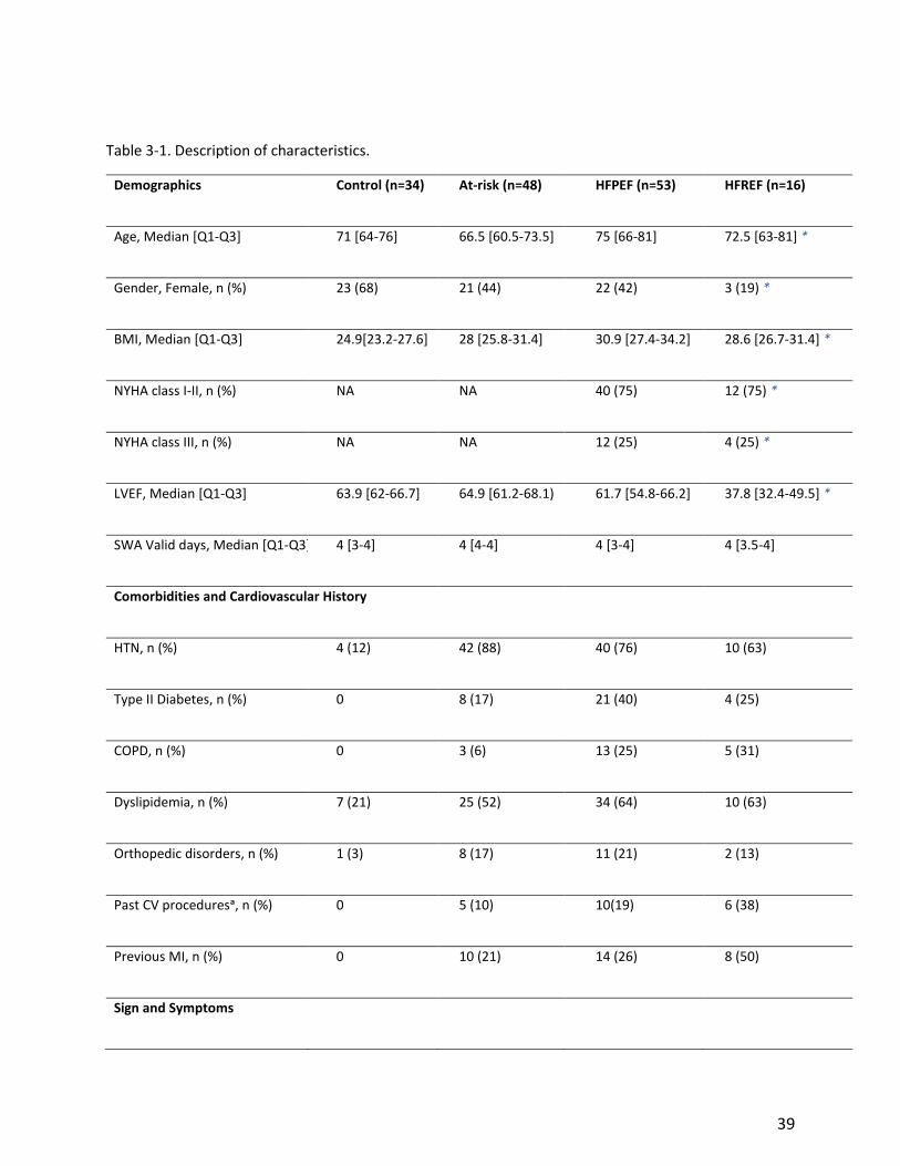
39
Table 3-1. Description of characteristics.
Demographics Control (n=34) At-risk (n=48) HFPEF (n=53) HFREF (n=16)
Age, Median [Q1-Q3] 71 [64-76] 66.5 [60.5-73.5] 75 [66-81] 72.5 [63-81] *
Gender, Female, n (%) 23 (68) 21 (44) 22 (42) 3 (19) *
BMI, Median [Q1-Q3] 24.9[23.2-27.6] 28 [25.8-31.4] 30.9 [27.4-34.2] 28.6 [26.7-31.4] *
NYHA class I-II, n (%) NA NA 40 (75) 12 (75) *
NYHA class III, n (%) NA NA 12 (25) 4 (25) *
LVEF, Median [Q1-Q3] 63.9 [62-66.7] 64.9 [61.2-68.1) 61.7 [54.8-66.2] 37.8 [32.4-49.5] *
SWA Valid days, Median [Q1-Q3] 4 [3-4] 4 [4-4] 4 [3-4] 4 [3.5-4]
Comorbidities and Cardiovascular History
HTN, n (%) 4 (12) 42 (88) 40 (76) 10 (63)
Type II Diabetes, n (%) 0 8 (17) 21 (40) 4 (25)
COPD, n (%) 0 3 (6) 13 (25) 5 (31)
Dyslipidemia, n (%) 7 (21) 25 (52) 34 (64) 10 (63)
Orthopedic disorders, n (%) 1 (3) 8 (17) 11 (21) 2 (13)
Past CV proceduresᵃ, n (%) 0 5 (10) 10(19) 6 (38)
Previous MI, n (%) 0 10 (21) 14 (26) 8 (50)
Sign and Symptoms

40
Dyspnea, n (%) 0 7 (15) 28 (53) 6 (38)
Fatigue, n (%) 2 (6) 8 (17) 25 (47) 9 (56)
Leg Edema, n (%) 0 3 (6) 12 (23) 3 (19)
At-risk, patients at risk of developing Heart failure; HFPEF, heart failure with preserved ejection fraction; HFREF, Heart failure with reduced ejection fraction; BMI, Body mass index; NYHA, New York heart association functional classification; LVEF, left ventricle ejection fraction; SWA, SenseWear Armband; HTN, Hypertension; COPD, chronic obstructive pulmonary disease; CV,
cardiovascular; MI, Myocardial infarction. Star (*) indicates significant difference across groups, P<0.05.ᵃ Previous Cardiovascular
procedures including; Pacemaker or Implantable Cardioverter Defibrillator, Coronary Artery Bypass Grafting, or Valve surgery
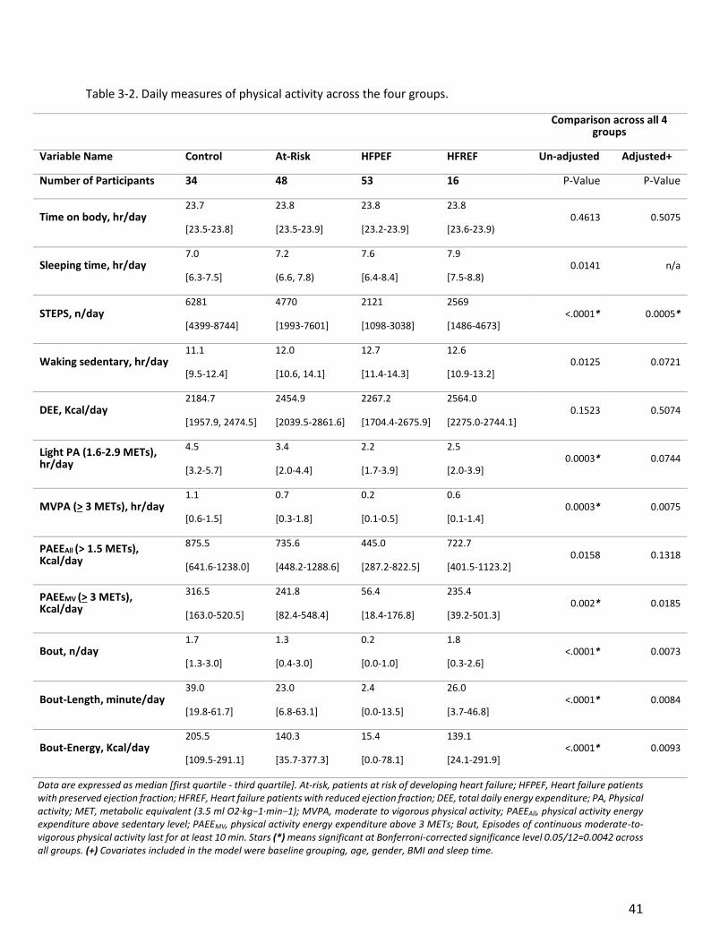
41
Table 3-2. Daily measures of physical activity across the four groups.
Comparison across all 4
groups
Variable Name Control At-Risk HFPEF HFREF Un-adjusted Adjusted+
Number of Participants 34 48 53 16 P-Value P-Value
Time on body, hr/day 23.7
[23.5-23.8]
23.8
[23.5-23.9]
23.8
[23.2-23.9]
23.8
[23.6-23.9) 0.4613 0.5075
Sleeping time, hr/day 7.0
[6.3-7.5]
7.2
(6.6, 7.8)
7.6
[6.4-8.4]
7.9
[7.5-8.8) 0.0141 n/a
STEPS, n/day 6281
[4399-8744]
4770
[1993-7601]
2121
[1098-3038]
2569
[1486-4673] <.0001* 0.0005*
Waking sedentary, hr/day 11.1
[9.5-12.4]
12.0
[10.6, 14.1]
12.7
[11.4-14.3]
12.6
[10.9-13.2] 0.0125 0.0721
DEE, Kcal/day 2184.7
[1957.9, 2474.5]
2454.9
[2039.5-2861.6]
2267.2
[1704.4-2675.9]
2564.0
[2275.0-2744.1] 0.1523 0.5074
Light PA (1.6-2.9 METs), hr/day
4.5
[3.2-5.7]
3.4
[2.0-4.4]
2.2
[1.7-3.9]
2.5
[2.0-3.9] 0.0003* 0.0744
MVPA (> 3 METs), hr/day 1.1
[0.6-1.5]
0.7
[0.3-1.8]
0.2
[0.1-0.5]
0.6
[0.1-1.4] 0.0003* 0.0075
PAEEAll (> 1.5 METs), Kcal/day
875.5
[641.6-1238.0]
735.6
[448.2-1288.6]
445.0
[287.2-822.5]
722.7
[401.5-1123.2] 0.0158 0.1318
PAEEMV (> 3 METs), Kcal/day
316.5
[163.0-520.5]
241.8
[82.4-548.4]
56.4
[18.4-176.8]
235.4
[39.2-501.3] 0.002* 0.0185
Bout, n/day 1.7
[1.3-3.0]
1.3
[0.4-3.0]
0.2
[0.0-1.0]
1.8
[0.3-2.6] <.0001* 0.0073
Bout-Length, minute/day 39.0
[19.8-61.7]
23.0
[6.8-63.1]
2.4
[0.0-13.5]
26.0
[3.7-46.8] <.0001* 0.0084
Bout-Energy, Kcal/day 205.5
[109.5-291.1]
140.3
[35.7-377.3]
15.4
[0.0-78.1]
139.1
[24.1-291.9] <.0001* 0.0093
Data are expressed as median [first quartile - third quartile]. At-risk, patients at risk of developing heart failure; HFPEF, Heart failure patients with preserved ejection fraction; HFREF, Heart failure patients with reduced ejection fraction; DEE, total daily energy expenditure; PA, Physical activity; MET, metabolic equivalent (3.5 ml O2·kg−1·min−1); MVPA, moderate to vigorous physical activity; PAEEAll, physical activity energy expenditure above sedentary level; PAEEMV, physical activity energy expenditure above 3 METs; Bout, Episodes of continuous moderate-to-vigorous physical activity last for at least 10 min. Stars (*) means significant at Bonferroni-corrected significance level 0.05/12=0.0042 across all groups. (+) Covariates included in the model were baseline grouping, age, gender, BMI and sleep time.
42
Chapter 4
Association of Daily Physical Activity and Aortic Distensibility across the
Heart Failure Continuum
4.1. Introduction
Approximately half of the patients with heart failure (HF) are reported to have
preserved ejection fraction (HFPEF) with similar or higher morbidity and mortality rate than
those patients with HF and reduced ejection fraction (HFREF) (194). This is partly due to the fact
that diastolic dysfunctions could remain asymptomatic for years and that may result in delayed
diagnosis of patients with HFPEF. In addition, HFPEF is more complex and all effective
treatments of HFREF seems to be ineffective in this phenotype.
Vascular afterload is an important aspect of ventricular function, especially for patients
with heart failure (HF) (195). A major determinant of the vascular afterload is arterial stiffness,
which typically increases with advancing age and has a pathological role in the etiology of
cardiovascular disease (CVD) and HF (136, 195). Increased arterial stiffness is also linked to
increased cardiovascular mortality in older populations (196). In a prospective study by Demir et
al., arterial stiffness was found to be a strong predictor of mortality in patients with HF. These
authors also suggested that monitoring arterial stiffness could be used to assess the efficiency of
treatments and potentially reduce HF-related hospitalizations (35).
Patients with HF have limited aerobic power, which in turn has been shown to be
associated with increased arterial stiffness, or more specifically, decreased aortic distensibility
43
(AD) (139, 141). In healthy individuals, exercise and in general, physical activity (PA) has been
shown to preserve and improve arterial compliance (151, 197). Tanaka et al. have suggested that
PA could favorably affect arterial stiffness independently of aerobic power (197). It is unknown
whether such findings could be extended to patients with preserved or reduced ejection fraction
(i.e., HFPEF or HFREF respectively). Conversely, what is known is that these patients with HF
typically fail to achieve the minimum recommended level of daily PA (30, 198).
The dose-response effect of physical activity (PA) on mortality in healthy adults and
patients with heart disease, including patients with HF, has been well documented (199-201).
Recent evidence suggests that there are benefits associated with light-intensity physical activity
that result in improved health-related quality of life in HF for patients (26, 202). A study by Izawa
et al. investigated the association of several determinants of prognosis with survival in a HF
population and showed that only step count was a strong and independent predictor of survival
in HF outpatients (201).
The relationship between PA and arterial compliance, measured as AD, has not been
evaluated across the HF continuum. Therefore, the purpose of this study was to compare the
relationship between PA and AD across two phenotypes of patients with HF (i.e., HFPEF and
HFREF) and compare these findings with those from patients at risk of developing HF (At-risk)
and healthy controls (Controls). We hypothesize that this association will differ across the HF
continuum.

44
4.2. Methods
This was a cross-sectional substudy of the Heart Failure Etiology and Analysis Research
Team (HEART) research project (177). The HEART project was a prospective observational cohort
study aimed to define new diagnostic criteria for patients with HF with preserved EF. The present
study was approved by the University of Alberta’s health research ethics board, and all
participants provided written informed consent. Sample size calculation was performed based
on a power of 0.80 and α of 0.05. For the effect size we used was correlation coefficient of r=-
0.21 (156). Aoyagi et al. investigated the association of yearlong objectively measured step count
with arterial stiffness (i.e. PWV) in elderly individuals. The total required sample size was
calculated to be 176 participants.
4.2.1. Study Participants
All participants were enrolled in the HEART project and were categorized into one of four
groups based on their baseline clinical criteria (177). The Control group included healthy age and
gender-matched participants with no evidence of hypertension, coronary artery disease (CAD),
diabetes mellitus, any organ diseases, or evidence of inflammatory or autoimmune conditions.
Further, the participants in the Control group were not taking any cardiac medications. The At-
risk group consisted of patients with preserved left ventricular (LV) systolic function but who had
one or more of the following conditions; hypertension (≥3 medications or LV hypertrophy on ECG
or LV mass index > gender-matched upper limit normal on an imaging test); diabetes; atrial
fibrillation; or obesity (body mass index >30); confirmed acute coronary syndrome >2 weeks;
chronic CAD with chest pain/shortness of breath or chronic obstructive pulmonary disease but

45
with no signs of HF. The two HF groups included patients with signs and symptoms of HF along
with either preserved LVEF (HFPEF) or reduced LVEF (HFREF). The diagnosis of HFPEF was based
on the clinical phenotype of symptoms consistent with HF (including dyspnea, fatigue, exertional
intolerance) and LVEF >45% while HFREF was classified by HF symptoms with LVEF <45%.
Exclusion criteria included age <18 years; known malignancy with expected survival <1 year;
pregnant or recent pregnancy <6 months; recent event (<2 weeks since Acute Coronary
Syndrome, HF or another admission); severe mitral or aortic stenosis or severe pulmonary
hypertension (>60 mmHg) (177). For this study, we were able to include 132 participants
categorized into the four groups who had both aortic distensibility measurements and PA data.
4.2.2. Outcome Measures
4.2.3.1. Physical Activity. Daily PA was assessed objectively using the SenseWear™
Mini Armband (SWA; Body Media, Inc. Pittsburgh, PA). In the previous work by our group on the
HEART cohort, results indicated that steps/day was the most robust measure to evaluate PA in
HF population (198). Therefore, we used steps/day (from the SWA) as the single measure of PA.
Step count was collected for a minimum of 4 consecutive days and averaged over the number of
days with valid recording to represent daily PA (198). We categorized step counts into three
ranges; low (≤ 3,500 steps/day), moderate (3,500-7,100 steps/day) or high (≥ 7,100 steps/day).
The normative data in the literature suggested 3,500 – 5,500 step/day as an expected level of PA
for older adults with disabilities such as HF (111). Also, 7,100 steps/day has been proposed as the
equivalent of the minimum PA recommended by the public health guideline for older adults with
46
chronic illnesses (e.g., HF population) (111). The time spent in the cadence of >100 steps/min
was calculated as a measure of moderate intensity walking (114).
4.2.3.2. Aortic Distensibility. Cardiac MRI assessment was performed using a 1.5-T
magnetic resonance scanner. Images were acquired in supine using 12-element phased array coil.
Aortic distensibility (AD) was measured using the fractional change in cross-sectional area of the
ascending aorta over the cardiac cycle, (Amax-Amin) / Amin, normalized by the pulse pressure,
ΔP, the difference in systolic and diastolic blood pressure (AD = (Amax-Amin) /(Amin×ΔP)) (142).
Amax and Amin are the maximal and minimal cross-sectional area of the aorta over the cardiac
cycle, respectively.
4.2.3. Statistical Analysis
The normality of the data was analyzed using the Kolmogorov–Smirnov test. Continuous
variables are presented as median with interquartile range (IQR) for skewed variables and mean
with standard deviation (SD) when the distribution of the variables was normal. Also, discrete
variables are presented as the frequency with proportions unless otherwise noted. To examine
the differences in the dependent and descriptive variables across all groups analysis of variance
(ANOVA), Kruskal-Wallis and Chi-square were used for continuous and normally distributed,
continuous with skewed distribution and discrete variables, respectively. Both AD and step count
were log-transformed because of skewed distribution and linear regression analysis was
performed to assess the relationship between AD (dependent measure) and step count
(independent measure) after adjusting for potential confounding variables including age, gender,
BMI, and pulse pressure (PP). Residual analyses were conducted to examine the normality
47
(residuals) and linearity assumptions of linear regression. A sensitivity analysis of excluding
outliers which was determined based on standardized residuals and covariance ratio (203), was
performed to evaluate the impact of outliers on the robustness of regression models. Since the
results were not different, we are reporting the results of all participants in the cohort. For all of
the analyses, a 2-tailed P value of <0.05 was required for significance test. All statistical analyses
were performed with SPSS 24.0 (SPSS, Inc. Chicago, IL, USA).
4.3. Results
4.3.1. Participants Demographics across Groups
Demographics, comorbidities and clinical characteristics of the participants are shown in
Table 4.1. All 132 participants (age, 69.3 ± 11.1 years, 51.5% female) were included in the
analyses. Age, BMI, gender, and LVEF were significantly different across groups (p <0.05), with
HFPEF group being the oldest and with the highest BMI. The majority of HFPEF or HFREF patients
were classified into NYHA class I or II (76% and 83%, respectively).
4.3.2. Physical Activity across Groups
The comparison of the mean across groups showed that the number of steps/day was not
comparable. Post-hoc analysis revealed that patients with HFPEF took significantly fewer
steps/day than either the At-risk and Control groups (p < 0.05; Table 4.1). However, there was no
significant difference between the daily step counts of the HFPEF and HFREF groups (2,852 ±
2,771 vs. 3,831 ± 2,592, steps/day, respectively).

48
In the present study, there was a significant difference across the four groups regarding
the average time per day spent walking in >100 steps/min (p< 0.05). The HFPEF group spent
significantly less time walking at a pace >100 steps/min than both Control and At-risk groups
(Table 4.1). While the HFREF group completed 0.9 [0-14.3] min at >100 steps/min, this time was
not significantly different from the HFPEF group (p >0.05).
4.3.3. Aortic Distensibility across Groups
Comparison of the AD’s median across the four groups demonstrated a significant
difference between the Control and At-risk groups (p< 0.05; Table 4.1). However, when we
controlled for age, there were no significant differences between any of the groups. When we
tested for differences in AD across the three ranges of step counts (i.e., low, moderate, high),
there were no significant differences in AD in the 132 participants (p=0.083), HFPEF group (p=
0.172) or the non-HF participants (i.e., Controls and At-risk combined; p=0.476). However, those
patients with HFREF who achieved a high step count range, had a significantly higher AD (median
3.7 [3.7-5.2] 10-3 mmHg-1) than those categorized into the low step count range (1.3 [0.9-2.5] 10-
3 mmHg-1, p<0.05).
4.3.4. The relation between Physical Activity and Aortic Distensibility
Simple linear regression analyses and stepwise model selection revealed that BMI was
not significantly associated with AD (p>0.01), and therefore it was excluded from further analysis.
A multiple linear regression analysis, which included all participants, did not yield any significant
interaction between groups and steps/day. However, the results of the interaction analysis
suggested that for every 1,000 steps taken, the correspondent change of AD was

49
disproportionately larger in the HFREF group compared to HFPEF and At-risk groups.
Furthermore, the small sample in the HFREF group (12 participants) could partially be responsible
for an underpowered analysis of interaction analysis. Therefore, we performed a group-based
analysis adjusting for age, gender, and PP, which indicated a significant association between step
count and AD in the HFREF groups (p < 0.05). In addition, in a cluster analysis of AD across step
categories, those with HFREF who achieved ≥ 7,100 steps/day had a significantly higher AD than
patients with HFREF who averaged ≤ 3,500 steps/day.
4.4. Discussion
The primary purpose of this study was to determine if there was a relationship between
PA and AD across the HF continuum. The major new finding is that the association between daily
step count and AD may not be similar across the continuum of HF. The results showed there was
a direct relationship, such that a higher range of steps/day was associated with a higher AD, but
only in our small HFREF group. The demographic and clinical characteristics of our HF patients
were almost consistent with the literature. The HFREF group had a reduced LVEF (39 ± 9%) and
close to 60% of the participants in this group had a history of myocardial infarction.
In a previous study, our group showed that the daily step count was the most robust
measure of PA in patients with HF (198). When we examined the step count across the
continuum of HF groups we noted that patients with HFPEF took significantly fewer steps/day
than either At-risk and Control groups. However, there was no difference in the daily step counts
between the HFPEF and HFREF groups. The step counts observed in At-risk and HF groups were
lower than what has been reported (30, 111). Previous studies report an average between 3,500
50
to 5,000 steps/day for patients with HF, while healthy older adults have been shown to take 6,000
to 8,500 steps/day (30, 112). The daily step count across groups would categorized participants
from our two HF groups as sedentary, while our two non-HF groups would be placed in the low
active category (111, 204).
When aortic distensibility (AD) was examined we noted that our Control group had a AD
that was lower than what has been reported previously. In a review of 6 studies, the average AD
for healthy older adults (age ~63 years) was found to be 1.7 10-3 mmHg-1 (205). Hundley et al.
noted that the AD in healthy older adults (age ~72 years) was 2.2 ± 0.4 10-3 mmHg-1 (141). We
can only speculate that subtle differences in SBP and PP may have been a contributing factor to
the difference in the AD observed in our Control group. For patients with HFREF, AD is often
observed to be diminished, compared to age-matched healthy control participants. This lower
AD that is often seen in patients with HFREF could be attributed to abnormal aortic elastic
properties as a consequence of impaired ventricular-vascular coupling and endothelial function
(35, 139). However, the AD of our HFREF participants was higher than what has been reported
previously (139, 205). In addition, our HFREF group had higher ejection fraction than what is
normally observed in previous reports (35, 139). For our patients with HFPEF, we noted that their
AD was also higher than what has been reported (141, 205). Desai et al. have suggested that AD
is often adversely impacted by chronic hypertension, which is typically seen in patients with
HFPEF (140). We can only speculate that discrepancies observed in the AD of our HF groups
compared to previous reports might be due to lower SPB and PP (26, 139, 141).

51
A number of studies have examined the relationship between arterial stiffness and PA
(206, 207). For instance, Tanaka et al. noted an association between higher levels of habitual PA
and reduced arterial stiffness (197). Laurent et al. made a similar observation when they
compared sedentary and aerobically trained individuals (208). Both Laursen et al., and Aoyagi et
al., have reported that higher levels of daily PA (e.g., daily PA energy expenditure or steps/day)
were associated with lower arterial stiffness in elderly adults (156, 209). Finally, the findings of
Tanaka et al., suggest that PA could diminish arterial stiffness independent of aerobic capacity
(197). When we examined the relationship between PA and AD across the HF continuum, a
significant association in the HFREF groups was found. Also, when we analyzed AD across the
categories of steps (i.e., low, medium and high), we noted that those with HFREF and a high daily
step count (i.e., ≥ 7,100 steps/day) had higher AD than those HFREF with low step count.
However, given the small sample size of our HFREF group, these results should be interpreted
cautiously. This observation may imply that a higher volume of PA is needed to achieve higher
levels of AD. The overall low step count observed in our HF participants may imply this volume
of daily activity may simply not be feasible for the majority of patients with HF (129).
Tanaka et al., (197) suggested that regular moderate to vigorous-intensity PA (MVPA) may
be required to favorably affect arterial compliance. They speculated that exercise could “stretch”
collagen fibers and modify their cross-link through increase of pulse pressure and mechanical
distension, thereby increasing arterial compliance. Authors also suggested that modulation of
the sympathetic-adrenergic tone of smooth muscle cells in the arterial wall could play a role in
positive effects of MVPA on arterial compliance (197). To gain a greater understanding as to
whether or not the step count observed in the present study constitutes MVPA we examine the
52
intensity of the activity. Tudor-Locke suggested a stepping rate of 100 steps/min as the absolute
minimum value for moderate-intensity walking in healthy adults (114). In 2005–2006, the U.S.
National Health and Nutrition Examination Survey noted that older adults (i.e., > 60 years old)
spend the majority of their waking time below this threshold, with an average cadence of 60
steps/min (113). Indeed, it has been reported that patients with HF spend the vast majority of
their waking time in activities below what is defined as moderate intensity (i.e., 100 steps/min)
(30, 198). Present findings are consistent with these previous reports, as both our HFPEF and
HFPEF groups completed less that one min/day MVPA per day (i.e., >100 steps/min). Further, all
four of our groups failed to achieve a duration of 10 minutes or more at a rate of 100 steps/min
(see Table 1). This implies that the majority of our study participants were indeed sedentary and
their daily PA would be classified in the light category (i.e., activities of daily living) as opposed to
the MVPA which may be needed to enhance AD.
In summary, while the current clinical management of HF might not routinely include
assessment of AD, PA is among the few interventions proven to have a positive impact on
exercise intolerance in HF (210). Therefore, assessment of daily PA level could provide a surrogate
meaure for assessment of AD, especially in patients with HFREF. While the evidence suggests a
direct relation between central arteries stiffness and exercise intolerance in patients with HF, the
positive association between central arteries distensibility and peak VO2 in patients with HFREF
is noticeably greater than those with HFPEF (136, 139). The findings of the present study align
with previous reports suggest that there are differences in underlying mechanisms between the
two phenotypes of HF in responding to PA (41, 211). Although the generalizability of the current

53
study is limited, current results suggest that the volume of PA may play a significant role in
determining the extent of arterial changes induced by PA.
4.4.1. Limitations
The cross-sectional design of this study and its relatively small cohort may limit the
generalizability of the findings. Also, to calculate pulse pressure a non-invasive brachial cuff blood
pressure measurement was incorporated instead of invasive assessment. Plus, at the time of the
cardiac MRI, blood pressure calculation may have varied as a result of anxiety. For these reasons,
our calculation of AD should be considered an approximation. Second, most of the patients At-
risk or with HF were taking medication at the time of testing, which could affect the AD
measurements, especially in patients HF (212, 213). These could partially be responsible for our
finding of a non-significance difference in AD in other groups.
4.5. Conclusion
The association between daily PA and AD may not be similar across the continuum of HF.
The comparison of AD in each group across the three ranges of steps/day showed a significant
difference only in patients with HFREF, such that patients who recorded the higher range of
steps/day had higher AD. The results of the current study might be an additional piece of the
puzzle that underscores the different pathways by which PA can impact each of the HF
phenotypes. These findings also reinforce previous work which reported a higher volume and
intensity of PA may be required to impact the arterial properties in patients with HF.
Acknowledgment

54
We would like to acknowledge the support of the Consultation and Research Services
Platform at The Alberta' SPOR SUPPORT Unit in statistics analysis service.

55
Table 4-1. Characteristics of study participants
Demographics Control (n=32) At-risk (n=42) HFPEF (n=46) HFREF (n=12)
Age, Mean± SD 69 ± 10 * 65 ± 13 74 ± 9 68 ± 9
Gender, Female, n (%) 22 (69) * 22 (69) 21 (46) 3 (25)
BMI, Mean± SD 25 ± 3.4 * 28 ± 4 30.1 ± 5.8 28.3 ± 3.6
NYHA class I-II/III, n (%) NA NA 35 (76)/11 (24) 10 (83)/2 (17) *
LVEF, Mean± SD 63.5 ± 4.5 * 63.9 ± 6.5 60.1 ± 10.3 38.9 ± 8.5
AD, Median [IQR] in 10-3 mmHg-1 0.86 [0.66-2.6] * 2 [1.2-2.8] 1.2 [0.95-2.4] 1.9 [1.1-3.1]
SWA wearing time (Hrs.), Mean± SD 23.6 ± 0.4 23.6 ± 0.4 23.6 ± 0.6 23.6 ± 0.3
Steps/ day, Median [IQR]
Time >100 Steps/day ᵃ, Median [IQR]
Low step range b, n (%)
Medium step range, n (%)
High step range, n (%)
6281 [4514-8676] *
4.5 [1-18] *
7 (22)
10 (31)
15 (47)
4770 [2443-7891]
3 [0-12]
14 (33)
15 (36)
13 (31)
2177 [1023-3125]
0 [0-0.6]
37 (80)
6 (13)
3 (7)
2949 [1824-6134]
0.9 [0-14]
7 (58)
3 (25)
2 (17)
SBP Mean± SD 133 ± 18 131 ± 19 126 ± 18 122 ± 28
DBP, Mean± SD 72 ± 11 73 ± 12 72 ± 14 69 ± 6
PP, Mean± SD 61 ± 15 58 ± 16 54 ± 18 53 ± 26
Comorbidities and Cardiovascular History
HTN, n (%) 4 (13) * 37 (80) 36 (80) 8 (67)
Type II Diabetes, n (%) 0 * 6 (14) 20 (43) 2 (17)
COPD, n (%) 0 * 3 (7) 9 (20) 4 (33)
Past CV procedures C, n (%) 0 * 4 (10) 9 (20) 3 (25)
Previous MI, n (%) 0 * 9 (21) 12 (26) 7 (58)
Sign and Symptoms
Dyspnea, n (%) 0 * 5 (12) 25 (54) 5 (42)
Fatigue, n (%) 1 (3) * 7 (17) 22 (48) 7 (58)

56
Leg Edema, n (%) 0 * 3 (7) 11 (24) 2 (17)
Medications
ACE inhibitors, n (%) 3 (9) 21 (50) 25 (54) 9 (75)
Angiotensin receptor blockers, n (%) 1 (3) 14 (33) 11 (24) 2 (17)
Anti-Arrhythmic, n (%) 0 1 (2) 10 (22) 1 (8)
Diuretics, n (%) 1 (3) 20 (48) 37 (80) 10 (83)
Β-Blockers, n (%) 1 (3) 19 (45) 39 (85) 10 (83)
Calcium channel blockers, n (%) 0 8 (19) 12 (26) 1 (8)
Nitrates, n (%) 1 (3) 1 (2) 9 (20) 4 (33)
Statins, n (%) 6 (19) 19 (45) 32 (70) 8 (67)
AD, Aortic distensibility (10-3 mmHg-1); IQR, Interquartile range; At-risk, patients at risk of developing Heart failure; HFPEF, heart
failure with preserved ejection fraction; HFREF, Heart failure with reduced ejection fraction; BMI, Body mass index; NYHA, New
York heart association functional classification; LVEF, left ventricle ejection fraction; SBP, systolic blood pressure (mmHg); DBP,
Diastolic blood pressure (mmHg); PP, Pulse pressure (mmHg); HTN, Hypertension; COPD, chronic obstructive pulmonary disease;
CV, cardiovascular; MI, Myocardial infarction. Star (*) indicates significant difference across groups, P<0.05. a Time in minutes
spent in cadence > 100 Steps/day. b ≤ 3,500 steps/day, between 3,500 and 7,100 steps/day, and ≥ 7,100 steps/day were
categorized as low, medium and high step ranges, respectively. C Previous Cardiovascular procedures including; Pacemaker or
Implantable Cardioverter Defibrillator, Coronary Artery Bypass Grafting, or Valve surgery

57
Chapter 5
The Relationship between Daily Physical Activity and Myocardial Stress
Biomarkers in Patients with Heart Failure and Preserved Ejection Fraction
5.1. Introduction
At first, natriuretic peptides (NPs) as biomarkers were introduced for the diagnosis of
heart failure (HF), however; the usage of both B-type natriuretic peptide (BNP) and N-terminal
pro-B-type natriuretic peptide (NT-proBNP) has expanded to include prognosis for patients with
HF (214). There is also evidence advocating the use of NPs in screening asymptomatic individuals
who are at risk of developing HF (At-risk), such as the elderly with hypertension, diabetes or
asymptomatic coronary artery disease (34). The primary stimuli for the synthesis and release of
the NPs precursor are ventricular dilatation, hypertrophy or increased wall tension (34). As
ventricular systolic dysfunction is typically associated with larger chamber radius and greater wall
stress, it has been documented that patients with HF and reduced ejection fraction (i.e., HFREF)
typically have a higher level of NPs than those with HF and preserved ejection fraction (i.e.,
HFPEF) (163, 164). It is well documented that exercise is beneficial for heart health and due to
recent advancements in biomarker analyses, new mechanisms by which exercise can impede the
progression of cardiovascular (CV) diseases such as HF have been uncovered (215).
A systematic review of 9 studies showed moderate intensity exercise training significantly
reduced BNP and NT-proBNP level in HF patients (170). The evidence suggests the volume,
intensity and length of the exercise program might play a role in determining the changes of NPs
58
in patients with HF whereas the frequency and duration of exercise sessions appeared to have
no effect (170). However, the referral rate of patients with HF to cardiac rehabilitation is
dramatically low (9% for HFPEF vs 12% for HFREF) (183) and even for those who attend CR,
adherence to the exercise program has proven to be difficult (12, 13). In fact, one of the major
trials on the effects of exercise training in the HF population, HF-ACTION trial (216), failed to show
the positive impact on decreasing the rate of adverse CV events (217). The investigators reported
less than 70% adherence to complete all exercise sessions and only 25% of patients in exercise
treatments arm continued to perform any exercise after two-thirds of trial length (217). On the
other hand, due to the complex and heterogeneous nature of HFPEF, early-stage diagnosis is
difficult. Therefore, it is unlikely that patients with HFPEF acquire enough awareness and
education regarding the importance of being physically active (183, 185). Indeed, promoting daily
PA has been suggested as a suitable alternative to structured exercise regimens (16, 218).
Daily physical activity (PA) has been shown to be associated with favorable cardiac
structural changes in a healthy adult, elder population (219, 220) and patients with HF (221,
222).deFilippi et al., in a prospective study of older adults free of HF, showed the probability of
an increase in NT-proBNP level over 2 to 3 years was inversely related to the level of PA (167).
The same negative association was reported between PA and NT-proBNP level in patients with
HFPEF (168). The association between daily PA and the level of circulating myocardial stress
biomarkers across the healthy controls to those at risk or with HF could highlight the importance
of non-exercise daily PA in the prevention and management of patients with HFPEF. Therefore,
the purpose of this study is to investigate the association between PA and myocardial stress
biomarkers in patients with HFPEF and contrast it with At-risk and healthy controls.

59
5.2. Methods
5.2.1. Study participants
This was a cross-sectional substudy of the Heart Failure Etiology and Analysis Research Team
(HEART) research project (177). The HEART project was a prospective observational cohort study
aimed to define new diagnostic criteria for patients with HF with preserved EF. The present study
was approved by the University of Alberta’s health research ethics board, and all participants
provided written informed consent. Sample size calculation was performed based on a power of
0.80 and α of 0.05. For the effect size we used correlation coefficient of r=-0.26 (18). Jehn et al.
investigated the association between the amount of time/day spent walking measured by
accelerometer and NT-proBNP in elderly patients with HF. The total required sample size was
calculated to be 114 participants.
All participants were enrolled in the HEART project and were categorized into one of three
groups based on their baseline clinical criteria (177). The control group included healthy, age and
gender-matched participants with no evidence of hypertension, coronary artery disease (CAD),
diabetes mellitus, any organ diseases, or inflammatory or autoimmune conditions. Further, the
participants in the control group were not taking any cardiac medications. The At-risk group
consisted of patients with preserved left ventricular (LV) systolic function but who had one or
more of the following conditions; hypertension (≥3 medications or LV hypertrophy on ECG or LV
mass index > gender-matched upper limit normal on an imaging test); diabetes; atrial fibrillation;
or obesity (body mass index >30); confirmed acute coronary syndrome >2 weeks; chronic CAD
with chest pain/shortness of breath or chronic obstructive pulmonary disease but with no signs
60
of HF. The HF group included patients with signs and symptoms of HF along with preserved LVEF
(HFPEF). The diagnosis of HFPEF was based on the clinical phenotype of symptoms consistent
with HF (including dyspnea, fatigue, exertional intolerance) and a LVEF >45%. Exclusion criteria
included age <18 years; known malignancy with expected survival <1 year; pregnant or recent
pregnancy <6 months; recent event (<2 weeks since Acute Coronary Syndrome, HF or another
admission); severe mitral or aortic stenosis or severe pulmonary hypertension (>60 mmHg) (177).
For this study, we were able to include 122 participants categorized into the 4 groups who had
both biomarkers measurements and PA data.
5.2.2. Outcome Measures
5.2.2.1. Physical Activity. Daily PA was assessed objectively using the SenseWear™
Mini Armband (SWA; Body Media, Inc. Pittsburgh, PA). In the previous work by our group on the
HEART cohort, results indicated that steps/day was the most robust measure to evaluate PA in a
HF population (198). Therefore, we used steps/day (from the SWA) as the single measure of PA.
Step count was collected for a minimum of 4 consecutive days and averaged over the number of
days with valid recording to represent daily PA (198).
5.2.2.2. Myocardial Stress Biomarkers. Plasma BNP levels were assessed using a
Biosite Triage reagent pack (Biosite Inc., San Diego, CA, USA) as previously described in detail
(223). In addition, plasma NT-proBNP analysis was performed with the commercially available
immunoassay using the Elecsys proBNP assay (Roche Diagnostics GmbH, Manheim, Germany)
read in an automated access immunoanalyzer (Beckman-Coulter, Fullerton,CA, USA) at Alberta
Health Services Laboratory Services, Edmonton, Alberta.
61
5.2.3. Statistical Analysis
Descriptive analysis (i.e., mean (standard deviation), median (range), or frequency
(proportion)) was used to summarize and compare patients’ clinical and demographic
characteristics between groups.
Univariable analysis was performed by using the Chi-square test or Fisher’s exact test
(when small frequencies were present) for categorical variables, while independent T-test was
used for continuous variables to assess whether the differences of variables between groups
were statistically significant. Assumptions were checked before performing the tests.
Due to the skewed distribution of biomarkers (BNP and NT-proBNP), we logged
transformed the dependent variables (i.e. Log BNP and Log NT-proBNP). Univariable linear
regression and mixed effect model, treating the group as a random effect, were used to analyze
the relationship of Log BNP/Log NT-proBNP with each variable individually. The aim was to
identify the variables needed to be adjusted.
To assess the relationship between biomarkers and steps/day, and the interaction
between groups and steps, the multivariable analysis was performed while adjusting other
covariates. The variables representing demographics (i.e., age, gender, BMI) medications,
medical history and common sign and symptoms of HF (e.g., dyspnea, fatigue and etc.) that
showed to be statistically significant (P-value < 0.05) at Univariable level or clinically significant
were used as main covariates in the multivariable linear regression model.
The equation used in order to calculate the magnitude of the association between
biomarkers and daily activity (for every 1,000 steps/day) was:

62
Log Biomarker = (β1×1,000) + β2 + (β3 × 1,000)
Where β1 is the coefficient of steps, β2 is the coefficient of the selected group and β3 is the
coefficient of the interaction between the selected group and steps.
All assumptions of the models were satisfied, and P-value < 0.05 was considered as
statistically significant. The performance of models was measured by R Square. The analysis was
performed by using R 3.4.0 (Vienna, Austria; https://www.R-project.org/) and SAS 9.4 software
(SAS Institute Inc. Cary, NC, USA).
5.3. Results
5.3.1. Participant Demographics across Groups
Demographics, comorbidities and medical history of participants are shown in Table 5.1.
The HFPEF group was the oldest, and the At-risk group were the youngest. Also, the HFPEF group
was more obese than controls (P<0.0001) and there was a similar trend in comparison with At-
risk (P=0.0589). In both HFPEF and At-risk groups almost 60% of participants were females in
contrast to the control group with only 31% female. In the HFPEF group 78% of patients were
classified in NYHA class I-II and their LVEF was 60% and 37%, respectively. The distribution of
dyspnea, fatigue, leg edema, atrial fibrillation or flutter, and type II diabetes were higher in HFPEF
group compared with both At-risk and control groups.
5.3.2. Physical Activity across Groups
The average daily step count of all participants was 4,523 ± 3,446 steps/day. Across the
three groups, the HFPEF group had the lowest step count (2,755 ± 2,682 steps/day compared to
63
control (6,475 ± 3,344 steps/day, p<0.0001) and the At-risk group (5,262 ± 3,384 steps/day, p
<0.001) (Table 5.1). The difference in the daily step count between the At-risk and control groups
was not statistically significant.
5.3.3. Myocardial Stress Biomarkers across Groups
Across the groups, there was a significantly higher concentration of both biomarkers in
the HFPEF versus the non-HF groups (P<0.001) (Table 5.1). The median levels of BNP and NT-
proBNP were not significantly different between the At-risk and controls.
5.3.4. The relation between Physical Activity and Biomarkers
The results of the multivariable regression models revealed that the association of steps
and Log BNP in the At-risk group was significantly different from that in HFPEF group ( P-value,
0.025) (Appendix B-Table 5-2). In fact, there was a negative association between daily steps and
log BNP in At-risk group such that for every 1,000 steps/day corresponded with 1.136 units
reduction in Log BNP. In the HFPEF group, every 1,000 steps/day corresponded to 0.2 units
decrease in log BNP. Meanwhile, we did not find statistical evidence to show the effect of
steps/day on Log BNP within the control group is significantly different from the HFPEF group (P-
value is 0.274).
When the association between steps/day and the Log NT-proBNPwas examined, we did
not observe any differences when we compared the HFPEF group with the At-risk or control
groups (P-values 0.083 and 0.8202, respectively). However, results did suggest a trend in the At-
risk group. There was a negative association between daily steps and log NT-proBNP in the At-
risk group in which every 1,000 steps/day corresponded with 1.454 units reduction in Log NT-

64
proBNP. This reduction was only 0.13 units of Log NT-proBNP for every 1,000 steps/day in HFPEF
group.
5.4. Discussion
The purpose of the present study was to evaluate the association between daily PA and
myocardial stress biomarkers in patients with HFPEF and contrast them with those from the at-
risk of HF group and healthy controls. A few studies have documented such a relationship in the
elderly population (158, 167) or in patients with HFPEF (168), however; to the best of our
knowledge, this is the first attempt to assess this relationship across the three groups (i.e., HFPEF,
At-risk, and healthy controls). The findings of the present study are: a) The level of both BNP and
NT-proBNP were higher in HFPEF group compared to control and At-risk groups, b) Both non-HF
groups took significantly more steps per day than patients with HFPEF and c) In comparison to
the HFPEF group, those in the At-risk group had a greater reduction of their BNP level for every
1,000 steps/day completed.
A higher level of NPs in patients with HF compared to the healthy control group is
consistent with previous reports (42, 164). However, to the best of our knowledge, this is the first
comparison to include an At-risk group. Previous studies have reported a higher level of NPs in
those with either hypertension, diabetes or CAD compared to healthy controls (158-160). The
result from the present study showed that the level of both myocardial stress biomarkers was
comparable for the At-risk and control groups. One explanation could be the fact that our At-risk
group had a higher BMI than controls. The inverse relation between NPs and obesity has been
shown previously (160, 224).
65
The findings of the current study showed that for every increment of 1,000 step/day, the
reduction in BNP level in the At-risk group was higher than what was observed in the HFPEF
group. The association of NPs with daily PA has not been investigated extensively and few studies
have used objective measures of PA. In a recent study, Snipelsky et al. reported an inverse
relationship between NT-proBNP and both measures of activity they recorded (i.e., average daily
accelerometer units and hours active per day) in patients with HFPEF at baseline (168). However,
with over a 6 weeks nitrate therapy, there were no significant associations between changes in
measures of PA and changes in NT-proBNP. The investigators suggested, compared indices of
HF’s severity (e.g., 6MWT, NT-proBNP, or NYHA classification) objectively measured PA is more
sensitive to the impact of an intervention on the functional status of patients with HFPEF. At 2004
Meyer et al. reported changes in anaerobic threshold of patients with HF after 12 weeks of
endurance training was not associated with changes in NT-proBNP (225). Later, Nilsson et al.
found no significant association between changes of NT-proBNP and changes in 6MWT after 4
months of high-intensity interval training or at 12-months follow-up in HF patients (226).
Another study by Parsons et al. used objectively measured PA data in more than 1,000
British men without a history of CV disease and found the relationship between NT-proBNP and
PA was not linear (158). There was a significant negative association between the level of NT-
proBNP and steps/day; however, this relationship was only present when the daily step count
was below 4,000. Higher levels of PA did not yield significant associations. In the present study,
there was a significant association between Log BNP and PA and a meaningful but not significant
association between Log NT-proBNP and PA in At-risk group despite thier 5,262 ± 3,384 steps/day
activity level.
66
Another observation from Parsons’s study was that, by stratifying based on hypertension
status, the association between PA and NT-proBNP only existed in those with hypertension (158).
In addition, these authors reported a higher proportion of those with hypertension were taking
statins and anticoagulants, had diabetes, higher BMI and took fewer steps/day compared to
normotensive individuals. These characteristics are consistent with our At-risk group and the
findings of the present study that showed the relationship between PA and Log BNP was stronger
in those At-risk. Also, there was a clinically significant association between NT-proBNP and
steps/day in our results in the At-risk group with a trend toward statistical significance (P-value
0.0825). The association between changes in BNP and changes in NT-proBNP in the HF population
has shown to be around 52% and up to 58% in those without chronic kidney disease (227).
The importance of these key findings in the clinical setting is multidimensional. First, these
results confirm the potential preventive impact of daily PA on reducing the risk of developing HF.
Secondly, the improvement of PA could impede the decline in cardiac function and recover
detrimental structural changes in patients with HF (228). It is well documented that many
established HF risk factors such as obesity, hypertension, diabetes, CAD and etc. are influenced
by improving daily PA level (229). Now the recent evidence is emerging that shows promoting
daily PA has great positive effects on not only patients with HF but those at-risk of developing HF
(158, 168). In fact, recently, Florido et al. in a six-year follow-up study showed even for those with
a sedentary lifestyle, each 512 MET×min/week higher PA (e.g., 30 minutes of brisk walking 4
times a week) corresponded with 11% lower risk of HF (230).
67
5.4.1. Limitations
The interpretation of our findings should be with caution due to the cross-sectional design
of this study with a relatively small cohort. However, to the best of our knowledge, this is the first
attempt to compare the association of natriuretic peptides with objectively-measured daily PA
in healthy controls and those at-risk of HF in comparison to patients with HFPEF. In addition, even
though the accelerometer-measured PA is far more valid and reliable than self-reported PA,
different devices used in recording and different methods of data transformation might be
partially responsible for discrepancies observed in the results of studies (62). High interindividual
and intraindividual variations have been reported for NPs in different populations (43, 231, 232),
which makes it even more difficult to compare the result of studies.
5.5. Conclusion
Across those with HFPEF, at risk of developing HF or healthy controls, completing more
volume of PA have the greatest impact on myocardial stress biomarkers in the At-risk group. As
the famous quote says, “prevention is better than cure”. Encouraging those patients A-risk of HF
could be more rewarding and practical than asking patients with HF to be more active. Therefore,
objective assessment of daily PA simply by recording daily step count could provide a strong tool
for identifying those at higher risk and a great benchmark for planning a rehabilitation
intervention.
Acknowledgment
We would like to acknowledge the support of the Consultation and Research Services
Platform at The Alberta' SPOR SUPPORT Unit in statistics analysis service.
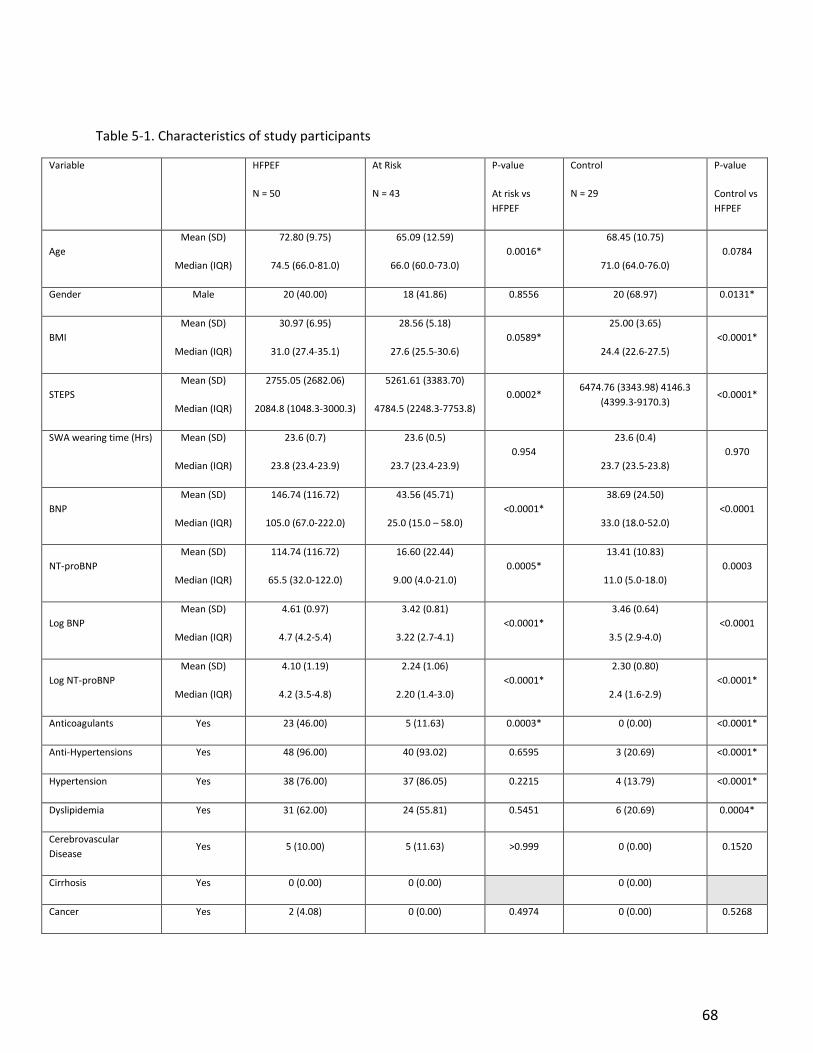
68
Table 5-1. Characteristics of study participants
Variable HFPEF
N = 50
At Risk
N = 43
P-value
At risk vs
HFPEF
Control
N = 29
P-value
Control vs
HFPEF
Age
Mean (SD)
Median (IQR)
72.80 (9.75)
74.5 (66.0-81.0)
65.09 (12.59)
66.0 (60.0-73.0)
0.0016*
68.45 (10.75)
71.0 (64.0-76.0)
0.0784
Gender Male 20 (40.00) 18 (41.86) 0.8556 20 (68.97) 0.0131*
BMI
Mean (SD)
Median (IQR)
30.97 (6.95)
31.0 (27.4-35.1)
28.56 (5.18)
27.6 (25.5-30.6)
0.0589*
25.00 (3.65)
24.4 (22.6-27.5)
<0.0001*
STEPS
Mean (SD)
Median (IQR)
2755.05 (2682.06)
2084.8 (1048.3-3000.3)
5261.61 (3383.70)
4784.5 (2248.3-7753.8)
0.0002* 6474.76 (3343.98) 4146.3
(4399.3-9170.3) <0.0001*
SWA wearing time (Hrs)
Mean (SD)
Median (IQR)
23.6 (0.7)
23.8 (23.4-23.9)
23.6 (0.5)
23.7 (23.4-23.9)
0.954
23.6 (0.4)
23.7 (23.5-23.8)
0.970
BNP
Mean (SD)
Median (IQR)
146.74 (116.72)
105.0 (67.0-222.0)
43.56 (45.71)
25.0 (15.0 – 58.0)
<0.0001*
38.69 (24.50)
33.0 (18.0-52.0)
<0.0001
NT-proBNP
Mean (SD)
Median (IQR)
114.74 (116.72)
65.5 (32.0-122.0)
16.60 (22.44)
9.00 (4.0-21.0)
0.0005*
13.41 (10.83)
11.0 (5.0-18.0)
0.0003
Log BNP
Mean (SD)
Median (IQR)
4.61 (0.97)
4.7 (4.2-5.4)
3.42 (0.81)
3.22 (2.7-4.1)
<0.0001*
3.46 (0.64)
3.5 (2.9-4.0)
<0.0001
Log NT-proBNP
Mean (SD)
Median (IQR)
4.10 (1.19)
4.2 (3.5-4.8)
2.24 (1.06)
2.20 (1.4-3.0)
<0.0001*
2.30 (0.80)
2.4 (1.6-2.9)
<0.0001*
Anticoagulants Yes 23 (46.00) 5 (11.63) 0.0003* 0 (0.00) <0.0001*
Anti-Hypertensions Yes 48 (96.00) 40 (93.02) 0.6595 3 (20.69) <0.0001*
Hypertension Yes 38 (76.00) 37 (86.05) 0.2215 4 (13.79) <0.0001*
Dyslipidemia Yes 31 (62.00) 24 (55.81) 0.5451 6 (20.69) 0.0004*
Cerebrovascular
Disease Yes 5 (10.00) 5 (11.63) >0.999 0 (0.00) 0.1520
Cirrhosis Yes 0 (0.00) 0 (0.00) 0 (0.00)
Cancer Yes 2 (4.08) 0 (0.00) 0.4974 0 (0.00) 0.5268

69
History of Alcoholism Yes 1 (2.00) 0 (0.00) >0.999 0 (0.00) >0.999
Atrial fibrillation/Flutter Yes 26 (52.00) 7 (16.28) 0.0003* 0 (0.00) <0.0001*
Smoking Current 4 (8.00) 3 (6.98) 0.9652 0 (0.00) 0.0495*
Former 18 (36.00) 18 (41.86) 6 (20.69)
Never 25 (50.00) 20 (46.51) 23 (79.31)
Unknown 3 (6.00) 2 (4.65) 0 (0.00)
Previous MI Yes 12 (24.00) 10 (23.26) 0.9329 0 (0.00) 0.0029*
Dyspnea Yes 26 (52.00) 7 (16.28) 0.0003* 0 (0.00) <0.0001*
Fatigue Yes 24 (48.00) 8 (18.60) 0.0029* 1 (3.45) <0.0001*
Leg Edema Yes 11 (22.00) 2 (4.65) 0.0161* 0 (0.00) 0.0056*
Other CV procedures Yes 8 (16.00) 5 (11.63) 0.5444 0 (0.00) 0.0237*
NYHA I-II / III-IV 49 (78)/ 11 (22) 0 (0.00) 0 (0.00)
COPD Yes 12 (24.00) 3 (6.98) 0.0621 0 (0.00) 0.0029*
T2D Yes 19 (38.00) 6 (13.95) 0.0091* 0 (0.00) 0.0001*
T1D Yes 1 (2.00) 2 (4.65) 0.5941 0 (0.00) >0.999
At-risk, patients at risk of developing Heart failure; HFPEF, heart failure with preserved ejection fraction; BMI, Body mass index;
BNP, brain natriuretic peptide (pg/ml); NT-proBNP, N-terminal pro-brain natriuretic peptide (pmol/L); NYHA, New York heart
association functional classification; LVEF, left ventricle ejection fraction; COPD, chronic obstructive pulmonary disease; CV,
cardiovascular; MI, Myocardial infarction. Star (*) indicates significant difference, P<0.05. a Previous Cardiovascular procedures
including; Pacemaker or Implantable Cardioverter Defibrillator, Coronary Artery Bypass Grafting, or Valve surgery. b Antihypertensive medications including beta-blockers, ACE inhibitors, calcium channel blockers, angiotensin II receptor blockers,
diuretics, and etc.
70
Chapter 6
General Discussion and Conclusion
6.1. General Discussion
One of the key manifestations of heart failure (HF) is diminished exercise capacity meaning
the maximum ability to perform physical activity (PA) is reduced. However, having the capacity
does not necessarily translate to performing daily PA. Historically, rehabilitation programs
targeting patients with HF have addressed reduced aerobic capacity with exercise training.
However, there is no consensus on the best mode, volume, and intensity of an exercise program
for patients with HF (12). In addition, adherence to these exercise training programs seems to be
problematic for the HF population, and hence the sustainability of achieved benefits might
decrease or dissipate over time (233). As an alternative to exercise training some investigators
suggest that encouraging daily PA might be a viable substitute because it encompasses
occupational, leisure, and household activities and can be accomplished throughout the day.
Promoting regular PA in patients with HF has shown to reduce hospitalization, disease
progression, and mortality while improving function, independence, and quality of life (10, 12,
19). In fact, every 60 min increase in total non-sedentary ambulatory-based movements (i.e., all
intensities > 1.5 METs) in patients with HF has been shown to be associated with a 35% reduction
in the risk of all-cause mortality (123).
To the best of our knowledge, there is no study that has compared the daily PA of patients
with HF and reduced ejection fraction (HFREF) with those with HF and preserved ejection fraction

71
(HFPEF). Furthermore, the daily volume and patterns of PA in both HF phenotypes contrasted to
those at risk of developing HF (At-risk) and healthy controls are unknown. Having a better
understanding of the daily volume of PA across the HF continuum could provide a holistic view
of the changes in daily PA while the capacity to perform PA diminishes. Identifying the patterns
of PA could shed light on daily fluctuations and distribution of PA energy expenditure (PAEE)
across the HF continuum. Lack of objective information about the volume and patterns of daily
PA across the HF continuum could lead to negative aftereffects including late diagnosis and worst
prognosis, higher costs of treatments or failed intervention. In contrast, staring a routine
objective assessment of PA as early as the appearance of risk factors such as higher blood
pressure, overweight, or myocardial infarction could help to identify those at higher risk of HF
sooner. It could also assist health care providers in designing a tailored program for these
patients. In addition, daily PA data could provide a perfect foundation for an effective and long-
lasting behavioral change intervention which in return may result in lower costs and better
quality of life.
Despite the recommendation of several health agencies, such as the American Heart
Association, to regularly assess the daily PA as an important risk factor (62), this practice is still
far away from reality. The 2018 PA guideline of the American Medical Association reported that
almost 80% of US adults and adolescents are insufficiently active compared to the recommended
level of PA (107). This is unfortunate since recent advancement in the technology of monitoring
devices allows for an accurate assessment of a number of PA measures. Further, these devices
are accessible and affordable for the public. There have been a variety of measures reported in
the literature that yields objective information about different dimensions of PA (e.g., intensity,

72
duration, etc.). Although each of these measures adds to the collective information about the
volume and patterns of PA, using multiple measures has some drawbacks. For example, it may
be harder to compare the results of studies that used different measures (e.g., steps vs counts).
In addition, gathering data from several domains of PA would require a more complex and
expensive device. Also, using different measures makes it harder to translate the findings to the
general audience. Since walking is the most popular form of PA and a functional part of our daily
life (108), almost all tracking devices available in the market report the number of steps/day as
the most common measure of PA. In fact, the result of our first study revealed step count
compared to other measures that represent time or energy spent in different intensities is the
most robust measure of PA for patients at risk or with HF. Steps/day has been widely used as a
measure to promote PA in healthy and patient populations because it makes it easy to set a goal,
track progress and provide feedback for different target populations (155).
For years investigators have tried to calculate the minimum volume and intensity of PA
required to achieve different positive health benefits. According to the recent PA guidelines,
typically, the higher the intensity and volume of PA the greater the benefits (107). However, it is
not clear if in patients with HF performing activities of daily living (i.e. ADL) is associated with
favorable changes in prognostic determinants.
The second and third studies in this thesis aimed to not only investigate the association
between steps/day and prognostic determinant in patients with HF but also to assess if these
associations are different across the HF continuum. Evaluating the associations between the
marker of daily PA and aortic distensibility (AD) and myocardial stress biomarkers could also help

73
in the development of pathways leading to earlier diagnosis, more precise classifications, and
better prognosis of patients with HF.
The results of the second study showed that the association between daily step count and
AD may not be similar across the continuum of HF. The findings showed there was a direct
relationship, such that a higher range of steps/day was associated with a higher AD, but only in
our small HFREF group. The fact that the association was only seen in the HFREF group suggest
that larger studies are needed to assess the association between AD and steps/day across the
spectrum of HF. In fact, the results of a 2019 meta-analysis showed that in older adults, there is
a statistically weak but clinically significant correlation between objectively measured daily step
count and pulse wave velocity (PWV) (155). Their results showed that for every 1,000 steps/day
increase in PA, a 0.18 m/s reduction in PWV was observed (155). This association appeared to be
curvilinear whereby the greatest effects on arterial stiffness were observed between those with
low-active to an active lifestyle (i.e., from 5,000 to 7,499 steps/day) and those with an active to
a very active lifestyle (i.e., from 7,500 to 9,999 steps/day).
The findings of the third study also indicated the associations between steps/day and BNP
or NT-proBNP were not comparable across groups; from healthy controls, to those At-risk and
with HFPEF. In fact, the association between steps/day and natriuretic peptides (NPs) were more
prominent in the At-risk group compared to patients with HFPEF. To the best of our knowledge,
this is the first study investigating the relationship between a marker of objectively measured
daily PA and NPs while including an At-risk group. We could only find one study assessing the
association of daily PA with NT-proBNP in patients with HFPEF (168). In that study, investigators
used two markers of accelerometer-measured PA. The first marker was representative of both
74
intensity and duration (e.g., average daily accelerometer units) while the second marker was the
only representative of the total active time. The authors noted that while at baseline, those with
higher NT-proBNP showed lower PA in both markers, there were no significant associations
between changes in the two markers of PA and changes in NT-proBNP. Systematic reviews about
the effects of aerobic exercise training on NPs in patients with HF suggested exercise intensity
and the total volume of PAEE during exercise may determine the extent of favorable changes in
NPs (170, 234). Smart et al., suggested that a minimum aerobic exercise training volume of
approximately 460 Kcal/week might be required to elicit changes in NPs in patients with HF (234).
In summary, the volume of PA, as defined by the product of intensity multiplied by
frequency and duration (62), seems to play a crucial role in determining the extent of health
benefits in those At-risk or with HF. The results of our first study revealed that the majority of
our cohort had a sedentary lifestyle. While those with HF may benefit the most by being more
active, according to our results they were the least active across the HF continuum followed by
the At-risk group. The daily PA performed by the majority of patients with HF might not reach
the minimum volume required to improve AD or reduce NPs. This could partially explain why in
the 2nd and 3rd studies we did not find significant associations where previous reports did.
To conclude, the main objective of this thesis was to provide a clearer picture of the
functional status of individuals At-risk of HF versus those patients with HF. This information could
be used to adjust exercise training targets and establish realistic rehabilitation goals that may, in
turn, result in long-term behavior change of the patients and improved outcomes. Generally,
behavior change is a function of three components ability, motivation, and trigger (235). To put
75
it in context, for changing PA behavior of patients At-risk or with HF, not only they should be able
to perform the PA tasks but also should be motivated and be prompted to do that.
Therefore, the first question should be, is this target population able to complete the
current recommended level of PA? The answer to this question appears to depend on the part
of the HF continuum we are targeting. For instance, the results of our first study showed that
patients with HF completed the least volume of PA compared to those At-risk and healthy
controls. The first study also provided some important insights regarding the patterns of daily PA
of two phenotypes of HF. While the energy and time that each HF group spent in light intensity
and all moderate-to-vigorous intensity PA (MVPA) (i.e., not limited to bouts of ≥10 min) were
comparable, those with HFREF were actually performing 6 times more continuous MVPA lasting
for at least 10-minutes than patients with HFPEF. These findings may have implications for
individualized strategies to promote PA. First, for patients with HF who are currently engaged in
some daily MVPA, increasing the amount of time spent at ≥ 3 METs should be encouraged. A
higher volume of MVPA will lead to a greater drop in the risk of mortality and extended health
benefits (106). However, insisting on this approach for those patients who are spending the
majority of their waking time sedentary or light PA (e.g., ADL), may not bode well for a long-term
behavior change. For these patients increasing the volume of the light intensity PA and
decreasing the sedentary time should be prioritized over completing MVPA. Reducing the risk of
mortality and improving the quality of life is achievable by reducing sedentary time and
increasing light intensity PA (106). Secondly, those at risk of developing HF are performing below
the recommended level of PA which may soon join the HF population if not encouraged to
decrease their sedentary time and spend more time in regular MVPA.

76
The element of ability was discussed by introducing feasible and progressive strategies to
promote PA in this population. Thus, the remaining part of the puzzle is understanding what it
takes to get them to do that hence motivation and trigger. In brief, patients should receive clear
and customized advice from health care providers regarding why and how to be active as an
important part of their standard care (236, 237). Helping patients through the process of
adopting positive health behavior by breaking the goals down into smaller achievable steps,
correcting patient’s perceived costs and benefits, and improving their self-confidence could
increase their motivation. In addition, increasing social support and self-efficacy in patients with
HF in response to patient’s endeavors could help in forming triggers (236, 237). Although the
ultimate goal in this population should be meet the current PA recommendations, for a majority
of patients it appears to be achievable only through a tailored increase in the volume of PA and
continuous support (e.g. reducing barriers, increasing accessibility, social support) to boost their
confidence.
The findings of this thesis could further advance our understanding of the significance of PA
across the HF continuum. Routine objective assessment of PA is vital. Longitudinal objective data
on PA could provide a proper basis for individualized behavioral interventions. In addition, it
could be used to motivate and empower patients with detailed feedback for guideline-directed
self-care.
Moreover, these findings could provide additional evidence that the response to PA
conveys through different pathways across the HF continuum. In other words, PA appears to have
a different impact on different groups from healthy to At-risk and HF. One obvious reason is the
relative intensity which reflects the different aerobic capacity of these groups. However, despite
77
an equal reduction of the capacity in the two phenotypes of HF, our results suggest that the
effects of PA could differ across HFPEF and HFREF. In fact, there is an emerging trend of evidence
suggesting patients with HFPEF would benefit more from PA than their HFREF peers (39). Also,
Omar et al. reported a heterogeneous response to exercise training in patients with HF that calls
for more individualized approaches in PA promotion (238).
The combined message from the results of these three studies is that regular assessment
of PA could be used as an important tool to identify those with a higher risk of HF incidence and
to design individualized rehabilitation programs to improve function and prognosis. Although the
underlying mechanisms were not studied in this thesis, several other studies have noticed a
considerable risk reduction after adopting an active lifestyle (120, 230). In addition, the
association between a single marker of PA such as steps/day and important prognostic
determinant of HF underscores the importance of regular assessment of this behavior. Despite
the different mechanisms by which PA benefits individuals across the HF continuum, it is critical
to recognize risk factors associated with a sedentary lifestyle and proper strategies to tackle this
issue.
6.2. Limitations
This thesis was a sub-study of a larger Alberta Heart Failure Etiology and Analysis Research
Team (HEART) research program, a prospective observational study aimed to define new
diagnostic criteria for patients with HFPEF. One of the main limitations of the present thesis was
the delayed start, which impacted the sample size of participants, especially for the HFREF group.
In addition, although the nature of the Alberta HEART was observational, it still took

78
approximately 2 years to collect our PA data. Consequently, during that period we recorded the
PA data for some participants at Alberta HEART’s baseline and for some others at Alberta HEART’s
follow-up. Although, we used corresponded dates for all variables included in our analysis, it is
possible that internal (e.g., signs and symptoms, comorbidities) and external factors (e.g., phases
of Alberta HEART, time of year) affected the PA of the some of the participants. One of the
challenges of this thesis was the interpretation of the results of 2nd and 3rd studies knowing the
majority of the cohort were sedentary. The daily PA performed by the majority of patients with
HF may not have reached the minimum volume required to improve AD or reduce NPs.
6.3. Recommendations
Using simple and reasonably accurate activity trackers that can objectively report the
number of steps/day should be encouraged in future longitudinal studies. Having information
about PA over several time-points along other prognostic markers could provide us with the
trend of changes in daily performance over time. The application of universal measures of PA
could help to compare the results of different studies and perform stronger systematic reviews
or meta-analysis. Having the aerobic capacity of participants could significantly add to the value
of the information provided by PA assessment. For example, the relative intensity of daily PA
performed could be calculated as the percentage of their VO2 peak. Our original design included
assessments of aerobic capacity and cardiac output to gain a better sense of the functional and
cardiac capacity of participants. However, in order to measure both VO2 peak and cardiac output
during maximal bike test, we used an Inert Gas Rebreathing machine called Innocor® which
required patients to go through complex and difficult procedures. For example, the mouth-piece
79
was connected to a proportionally heavy apparatus. In order to help participants tolerate this
weight, we suspended the apparatus by a ring to a modified serum stand which resulted in
restricted head movements. Moreover, saliva retention in the mouth-piece caused discomfort
and breathing difficulties. Thus, sub-maximal termination of the test and daily fluctuations of the
patient’s sign and symptoms might have contributed to not recording enough usable data during
exercise tests.
For future studies, comparing the long-term effects of daily PA on prognostic determinants
such as AD and NPs across both phenotypes of HF categorized in sedentary and active patients is
recommended.
80
Comprehensive Bibliography
1. Lee DS, Johansen H, Gong Y, Hall RE, Tu JV, Cox JL, et al. Regional outcomes of heart
failure in Canada. The Canadian journal of cardiology. 2004;20(6):599-607.
2. Johansen H, Strauss B, Arnold JM, Moe G, Liu P. On the rise: The current and projected
future burden of congestive heart failure hospitalization in Canada. The Canadian journal of
cardiology. 2003;19(4):430-5.
3. Ross H, Howlett J, Arnold JMO, Liu P, O’Neill B, Brophy J, et al. Treating the right
patient at the right time: access to heart failure care. Canadian journal of Cardiology.
2006;22(9):749-54.
4. Arnold JMO, Liu P, Demers C, Dorian P, Giannetti N, Haddad H, et al. Canadian
Cardiovascular Society consensus conference recommendations on heart failure 2006: diagnosis
and management. Canadian Journal of Cardiology. 2006;22(1):23-45.
5. Yancy CW, Jessup M, Bozkurt B, Butler J, Casey DE, Drazner MH, et al. 2013
ACCF/AHA guideline for the management of heart failure: a report of the American College of
Cardiology Foundation/American Heart Association Task Force on Practice Guidelines. Journal
of the American College of Cardiology. 2013;62(16):e147-e239.
6. Redfield MM, Jacobsen SJ, Burnett JC, Jr., Mahoney DW, Bailey KR, Rodeheffer RJ.
Burden of systolic and diastolic ventricular dysfunction in the community: appreciating the scope
of the heart failure epidemic. The Journal of the American Medical Association.
2003;289(2):194-202.

81
7. Owan TE, Hodge DO, Herges RM, Jacobsen SJ, Roger VL, Redfield MM. Trends in
prevalence and outcome of heart failure with preserved ejection fraction. The New England
Journal of Medicine. 2006;355(3):251-9.
8. Ezekowitz JA, Lee DS, Tu JV, Newman AM, McAlister FA. Comparison of one-year
outcome (death and rehospitalization) in hospitalized heart failure patients with left ventricular
ejection fraction >50% versus those with ejection fraction <50%. American Journal of
Cardiology. 2008;102(1):79-83.
9. Thompson PD. Exercise and physical activity in the prevention and treatment of
atherosclerotic cardiovascular disease. Arteriosclerosis, Thrombosis, and Vascular Biology.
2003;23(8):1319-21.
10. Davies EJ, Moxham T, Rees K, Singh S, Coats AJ, Ebrahim S, et al. Exercise training for
systolic heart failure: Cochrane systematic review and meta-analysis. European Journal of Heart
Failure. 2010;12(7):706-15.
11. McMurray JJ, Adamopoulos S, Anker SD, Auricchio A, Bohm M, Dickstein K, et al.
ESC Guidelines for the diagnosis and treatment of acute and chronic heart failure 2012: The
Task Force for the Diagnosis and Treatment of Acute and Chronic Heart Failure 2012 of the
European Society of Cardiology. Developed in collaboration with the Heart Failure Association
(HFA) of the ESC. European Heart Journal. 2012;33(14):1787-847.
12. Piepoli MF, Conraads V, Corra U, Dickstein K, Francis DP, Jaarsma T, et al. Exercise
training in heart failure: from theory to practice. A consensus document of the Heart Failure
Association and the European Association for Cardiovascular Prevention and Rehabilitation.
European Journal of Heart Failure. 2011;13(4):347-57.

82
13. Conraads VM, Deaton C, Piotrowicz E, Santaularia N, Tierney S, Piepoli MF, et al.
Adherence of heart failure patients to exercise: barriers and possible solutions: a position
statement of the Study Group on Exercise Training in Heart Failure of the Heart Failure
Association of the European Society of Cardiology. European Journal of Heart Failure.
2012;14(5):451-8.
14. van der Wal MH, van Veldhuisen DJ, Veeger NJ, Rutten FH, Jaarsma T. Compliance
with non-pharmacological recommendations and outcome in heart failure patients. European
Heart Journal. 2010;31(12):1486-93.
15. Corvera-Tindel T, Doering LV, Woo MA, Khan S, Dracup K. Effects of a home walking
exercise program on functional status and symptoms in heart failure. American Heart Journal.
2004;147(2):339-46.
16. Walsh JT, Charlesworth A, Andrews R, Hawkins M, Cowley AJ. Relation of daily
activity levels in patients with chronic heart failure to long-term prognosis. American Journal of
Cardiology. 1997;79(10):1364-9.
17. Belardinelli R. Monitoring skeletal muscle oxygenation during exercise by near infrared
spectroscopy in chronic heart failure. Congest Heart Fail. 1999;5(3):116-9.
18. Jehn M, Schmidt-Trucksass A, Hanssen H, Schuster T, Halle M, Koehler F. Association
of physical activity and prognostic parameters in elderly patients with heart failure. Journal of
aging and physical activity. 2011;19(1):1-15.
19. Corra U, Piepoli MF, Carre F, Heuschmann P, Hoffmann U, Verschuren M, et al.
Secondary prevention through cardiac rehabilitation: physical activity counselling and exercise
training: key components of the position paper from the Cardiac Rehabilitation Section of the
83
European Association of Cardiovascular Prevention and Rehabilitation. European heart journal.
2010;31(16):1967-74.
20. Briffa T, Maiorana A, Allan R. On behalf of the Executive Working Group and National
Forum Participants. National Heart Foundation of Australia physical activity recommendations
for people with cardiovascular disease Sydney: National Heart Foundation of Australia. 2006.
21. Nelson ME, Rejeski WJ, Blair SN, Duncan PW, Judge JO, King AC, et al. Physical
activity and public health in older adults: recommendation from the American College of Sports
Medicine and the American Heart Association. Medicine and science in sports and exercise.
2007;39(8):1435-45.
22. Rahl RL. Physical activity and health guidelines: recommendations for various ages,
fitness levels, and conditions from 57 authoritative sources: Human Kinetics; 2010.
23. Pate RR, O'Neill JR, Lobelo F. The evolving definition of" sedentary". Exercise and sport
sciences reviews. 2008;36(4):173-8.
24. Sedentary Behaviour Research N. Letter to the Editor: Standardized use of the terms
“sedentary” and “sedentary behaviours”. Applied Physiology, Nutrition, and Metabolism.
2012;37(3):540-2.
25. Wannamethee SG, Shaper AG, Walker M. Physical activity and mortality in older men
with diagnosed coronary heart disease. Circulation. 2000;102(12):1358-63.
26. Loprinzi PD. Implications of light-intensity physical activity in improving health-related
quality of life among congestive heart failure patients. International Journal of Cardiology.
2016;212:16-7.
84
27. Loprinzi PD, Lee H, Cardinal BJ. Evidence to support including lifestyle light-intensity
recommendations in physical activity guidelines for older adults. American journal of health
promotion : AJHP. 2015;29(5):277-84.
28. Katzmarzyk PT, Church TS, Craig CL, Bouchard C. Sitting time and mortality from all
causes, cardiovascular disease, and cancer. Medicine and science in sports and exercise.
2009;41(5):998-1005.
29. Wilmot EG, Edwardson CL, Achana FA, Davies MJ, Gorely T, Gray LJ, et al. Sedentary
time in adults and the association with diabetes, cardiovascular disease and death: systematic
review and meta-analysis. Diabetologia. 2012;55(11):2895-905.
30. Dontje ML, van der Wal MH, Stolk RP, Brugemann J, Jaarsma T, Wijtvliet PE, et al.
Daily physical activity in stable heart failure patients. J Cardiovasc Nurs. 2014;29(3):218-26.
31. Tudor-Locke C, Craig CL, Cameron C, Griffiths JM. Canadian children's and youth's
pedometer-determined steps/day, parent-reported TV watching time, and overweight/obesity: the
CANPLAY Surveillance Study. The international journal of behavioral nutrition and physical
activity. 2011;8:66.
32. Bonapace S, Rossi A, Cicoira M, Targher G, Valbusa F, Benetos A, et al. Increased aortic
pulse wave velocity as measured by echocardiography is strongly associated with poor prognosis
in patients with heart failure. Journal of the American Society of Echocardiography.
2013;26(7):714-20.
33. Berezin AE. Prognostication in Different Heart Failure Phenotypes: The Role of
Circulating Biomarkers. Journal of Circulating Biomarkers. 2016;5:6.
34. Braunwald E. Biomarkers in Heart Failure. New England Journal of Medicine.
2008;358(20):2148-59.
85
35. Demir S, Akpinar O, Akkus O, Nas K, Unal I, Molnar F, et al. The prognostic value of
arterial stiffness in systolic heart failure. Cardiology Journal. 2013;20(6):665-71.
36. Hegde SM, Claggett B, Shah AM, Lewis EF, Anand I, Shah SJ, et al. Physical Activity
and Prognosis in the TOPCAT Trial (Treatment of Preserved Cardiac Function Heart Failure
With an Aldosterone Antagonist). Circulation. 2017;136(11):982-92.
37. Jehn M, Schmidt-Trucksaess A, Schuster T, Hanssen H, Weis M, Halle M, et al.
Accelerometer-based quantification of 6-minute walk test performance in patients with chronic
heart failure: applicability in telemedicine. Journal of Cardiac Failure. 2009;15(4):334-40.
38. Guimarães GV, Carvalho VO, Torlai V, Bocchi EA. Physical activity profile in heart
failure patients from a Brazilian tertiary cardiology hospital. Cardioliology Journal.
2010;17(2):143-5.
39. Pandey A, Kitzman DW, Brubaker P, Haykowsky MJ, Morgan T, Becton JT, et al.
Response to Endurance Exercise Training in Older Adults with Heart Failure with Preserved or
Reduced Ejection Fraction. Journal of the American Geriatrics Society. 2017;65(8):1698-704.
40. Haykowsky MJ, Tomczak CR, Scott JM, Paterson DI, Kitzman DW. Determinants of
exercise intolerance in patients with heart failure and reduced or preserved ejection fraction.
Journal of Applied Physiology (1985). 2015;119(6):739-44.
41. Kitzman DW, Brubaker PH, Herrington DM, Morgan TM, Stewart KP, Hundley WG, et
al. Effect of endurance exercise training on endothelial function and arterial stiffness in older
patients with heart failure and preserved ejection fraction: a randomized, controlled, single-blind
trial. Journal of the American College of Cardiology. 2013;62(7):584-92.

86
42. Benda NMM, Eijsvogels TMH, Van Dijk APJ, Hopman MTE, Thijssen DHJ. Changes in
BNP and cardiac troponin I after high-intensity interval and endurance exercise in heart failure
patients and healthy controls. International journal of cardiology. 2015;184:426-7.
43. Meijers WC, van der Velde AR, Muller Kobold AC, Dijck-Brouwer J, Wu AH, Jaffe A,
et al. Variability of biomarkers in patients with chronic heart failure and healthy controls.
European Journal of Heart Failure. 2017;19(3):357-65.
44. Cleland JGF, Pellicori P, Clark AL. Prevention or Procrastination for Heart Failure?:
Why We Need a Universal Definition of Heart Failure∗. Journal of the American College of
Cardiology. 2019;73(19):2398-400.
45. Hunt SA, Abraham WT, Chin MH, Feldman AM, Francis GS, Ganiats TG, et al. 2009
Focused update incorporated into the ACC/AHA 2005 Guidelines for the Diagnosis and
Management of Heart Failure in Adults A Report of the American College of Cardiology
Foundation/American Heart Association Task Force on Practice Guidelines Developed in
Collaboration With the International Society for Heart and Lung Transplantation. Journal of the
American College of Cardiology 2009;53(15):e1-e90.
46. Ezekowitz JA, O'Meara E, McDonald MA, Abrams H, Chan M, Ducharme A, et al. 2017
Comprehensive Update of the Canadian Cardiovascular Society Guidelines for the Management
of Heart Failure. The Canadian journal of cardiology. 2017;33(11):1342-433.
47. Mozaffarian D, Benjamin EJ, Go AS, Arnett DK, Blaha MJ, Cushman M, et al. Heart
disease and stroke statistics--2015 update: a report from the American Heart Association.
Circulation. 2015;131(4):e29-322.
48. Braunwald E. Heart failure. JACC Heart Fail. 2013;1(1):1-20.
87
49. MacDonald MR, Wee PP, Cao Y, Yang DM, Lee S, Tong KL, et al. Comparison of
Characteristics and Outcomes of Heart Failure Patients With Preserved Versus Reduced Ejection
Fraction in a Multiethnic Southeast Asian Cohort. American Journal of Cardiology.
2016;118(8):1233-8.
50. Young DR, Reynolds K, Sidell M, Brar S, Ghai NR, Sternfeld B, et al. Effects of
physical activity and sedentary time on the risk of heart failure. Circulation Heart Failure.
2014;7(1):21-7.
51. Yancy CW, Jessup M, Bozkurt B, Butler J, Casey DE, Jr., Colvin MM, et al. 2017
ACC/AHA/HFSA Focused Update of the 2013 ACCF/AHA Guideline for the Management of
Heart Failure: A Report of the American College of Cardiology/American Heart Association
Task Force on Clinical Practice Guidelines and the Heart Failure Society of America. Journal of
Cardiac Failure. 2017;23(8):628-51.
52. McMurray JJ, Adamopoulos S, Anker SD, Auricchio A, Bohm M, Dickstein K, et al.
ESC guidelines for the diagnosis and treatment of acute and chronic heart failure 2012: The Task
Force for the Diagnosis and Treatment of Acute and Chronic Heart Failure 2012 of the European
Society of Cardiology. Developed in collaboration with the Heart Failure Association (HFA) of
the ESC. European Journal of Heart Failure. 2012;14(8):803-69.
53. Papadimitriou L, Moore CK, Butler J, Long RC. The Limitations of Symptom-based
Heart Failure Management. Cardiac Failure Review. 2019;5(2):74-7.
54. Desai AS, McMurray JJ, Packer M, Swedberg K, Rouleau JL, Chen F, et al. Effect of the
angiotensin-receptor-neprilysin inhibitor LCZ696 compared with enalapril on mode of death in
heart failure patients. European Heart Journal. 2015;36(30):1990-7.
88
55. Guglin M, Patel T, Darbinyan N. Symptoms in heart failure correlate poorly with
objective haemodynamic parameters. International Journal of Clinical Practice.
2012;66(12):1224-9.
56. Shah MR, Hasselblad V, Stinnett SS, Kramer JM, Grossman S, Gheorghiade M, et al.
Dissociation between hemodynamic changes and symptom improvement in patients with
advanced congestive heart failure. European Journal of Heart Failure. 2002;4(3):297-304.
57. Dharmarajan K, Rich MW. Epidemiology, Pathophysiology, and Prognosis of Heart
Failure in Older Adults. Heart Failure Clinics. 2017;13(3):417-26.
58. Kitzman DW, Lam CSP. Obese Heart Failure With Preserved Ejection Fraction
Phenotype. Circulation. 2017;136(1):20-3.
59. Velagaleti RS, Gona P, Pencina MJ, Aragam J, Wang TJ, Levy D, et al. Left ventricular
hypertrophy patterns and incidence of heart failure with preserved versus reduced ejection
fraction. American Journal of Cardiology. 2014;113(1):117-22.
60. Borlaug BA, Paulus WJ. Heart failure with preserved ejection fraction: pathophysiology,
diagnosis, and treatment. European Heart Journal. 2011;32(6):670-9.
61. Caspersen CJ, Powell KE, Christenson GM. Physical activity, exercise, and physical
fitness: definitions and distinctions for health-related research. Public health reports.
1985;100(2):126.
62. Strath SJ, Kaminsky LA, Ainsworth BE, Ekelund U, Freedson PS, Gary RA, et al. Guide
to the assessment of physical activity: Clinical and research applications: a scientific statement
from the American Heart Association. Circulation. 2013;128(20):2259-79.

89
63. Hamilton MT, Healy GN, Dunstan DW, Zderic TW, Owen N. Too Little Exercise and
Too Much Sitting: Inactivity Physiology and the Need for New Recommendations on Sedentary
Behavior. Current cardiovascular risk reports. 2008;2(4):292-8.
64. Blair SN, Kohl HW, Gordon NF, Paffenbarger RS, Jr. How much physical activity is
good for health? Annual Review Public Health. 1992;13:99-126.
65. Powell KE, Paluch AE, Blair SN. Physical activity for health: What kind? How much?
How intense? On top of what? Annual Review Public Health. 2011;32:349-65.
66. Stoylen A, Conraads V, Halle M, Linke A, Prescott E, Ellingsen O. Controlled study of
myocardial recovery after interval training in heart failure: SMARTEX-HF--rationale and
design. European Journal of Preventive Cardiology. 2012;19(4):813-21.
67. Harvey JA, Chastin SF, Skelton DA. How Sedentary are Older People? A Systematic
Review of the Amount of Sedentary Behavior. Journal of aging and physical activity.
2015;23(3):471-87.
68. Lee IM, Shiroma EJ, Lobelo F, Puska P, Blair SN, Katzmarzyk PT, et al. Effect of
physical inactivity on major non-communicable diseases worldwide: an analysis of burden of
disease and life expectancy. Lancet. 2012;380(9838):219-29.
69. Rariden BS, Boltz AJ, Brawner CA, Pinkstaff SO, Richardson MR, Johnson TM, et al.
Sedentary Time and Cumulative Risk of Preserved and Reduced Ejection Fraction Heart Failure:
From the Multi-Ethnic Study of Atherosclerosis. Journal of Cardiac Failure. 2019;25(6):418-24.
70. Park LG, Dracup K, Whooley MA, McCulloch C, Lai S, Howie-Esquivel J. Sedentary
lifestyle associated with mortality in rural patients with heart failure. European Journal of
Cardiovascular Nursing. 2019;18(4):318-24.
90
71. Hamer M, de Oliveira C, Demakakos P. Non-Exercise Physical Activity and Survival:
English Longitudinal Study of Ageing. American journal of preventive medicine.
2014;47(4):452-60.
72. Van Remoortel H, Giavedoni S, Raste Y, Burtin C, Louvaris Z, Gimeno-Santos E, et al.
Validity of activity monitors in health and chronic disease: a systematic review. The international
journal of behavioral nutrition and physical activity. 2012;9:84.
73. Melanson E, Freedson, PS. Physical activity assessment: a review of the methods.
Critical Reviews in Food Science and Nutrition.1996;36:385-96.
74. Vanhees L, Lefevre J, Philippaerts R, Martens M, Huygens W, Troosters T, et al. How to
assess physical activity? How to assess physical fitness? European Journal of Cardiovascular
Prevention & Rehabilitation. 2005;12(2):102-14.
75. Westerterp KR. Physical activity and physical activity induced energy expenditure in
humans: measurement, determinants, and effects. Frontiers in physiology. 2013;4:90.
76. Lee IM, Buchner DM. The importance of walking to public health. Medicine and science
in sports and exercise. 2008;40(7 Suppl):S512-8.
77. Jacobs DR, Jr., Ainsworth BE, Hartman TJ, Leon AS. A simultaneous evaluation of 10
commonly used physical activity questionnaires. Medicine and science in sports and exercise.
1993;25(1):81-91.
78. Bonnefoy M, Normand S, Pachiaudi C, Lacour JR, Laville M, Kostka T. Simultaneous
validation of ten physical activity questionnaires in older men: a doubly labeled water study.
Journal of the American Geriatrics Society. 2001;49(1):28-35.
91
79. Garet M, Barthelemy JC, Degache F, Costes F, Da-Costa A, Isaaz K, et al. A
questionnaire-based assessment of daily physical activity in heart failure. European Journal of
Heart Failure. 2004;6(5):577-84.
80. Bassett DR, Jr., Cureton AL, Ainsworth BE. Measurement of daily walking distance-
questionnaire versus pedometer. Medicine and science in sports and exercise. 2000;32(5):1018-
23.
81. Pereira MA, FitzerGerald SJ, Gregg EW, Joswiak ML, Ryan WJ, Suminski RR, et al. A
collection of Physical Activity Questionnaires for health-related research. Medicine and science
in sports and exercise. 1997;29(6 Suppl):S1-205.
82. Kochersberger G, Hielema F, Westlund R. Rehabilitation in the nursing home: how
much, why, and with what results. Public Health Reports. 1994;109(3):372-6.
83. Pitta F, Troosters T, Spruit MA, Probst VS, Decramer M, Gosselink R. Characteristics of
physical activities in daily life in chronic obstructive pulmonary disease. American Journal of
Respiratory and Critical Care Medicine. 2005;171(9):972-7.
84. Steele BG, Holt L, Belza B, Ferris S, Lakshminaryan S, Buchner DM. Quantitating
physical activity in COPD using a triaxial accelerometer. Chest. 2000;117(5):1359-67.
85. Ainsworth BE. How do I measure physical activity in my patients? Questionnaires and
objective methods. British Journal of Sports Medicine. 2009;43(1):6-9.
86. Cyarto EV, Myers AM, Tudor-Locke C. Pedometer accuracy in nursing home and
community-dwelling older adults. Medicine and science in sports and exercise. 2004;36(2):205-
9.
87. Le Masurier GC, Tudor-Locke C. Comparison of pedometer and accelerometer accuracy
under controlled conditions. Medicine and science in sports and exercise. 2003;35(5):867-71.
92
88. Vanhees L, Geladas N, Hansen D, Kouidi E, Niebauer J, Reiner Z, et al. Importance of
characteristics and modalities of physical activity and exercise in the management of
cardiovascular health in individuals with cardiovascular risk factors: recommendations from the
EACPR. Part II. European Journal of Preventive Cardiology. 2012;19(5):1005-33.
89. Johannsen DL, Calabro MA, Stewart J, Franke W, Rood JC, Welk GJ. Accuracy of
armband monitors for measuring daily energy expenditure in healthy adults. Medicine and
science in sports and exercise. 2010;42(11):2134-40.
90. Mackey DC, Manini TM, Schoeller DA, Koster A, Glynn NW, Goodpaster BH, et al.
Validation of an armband to measure daily energy expenditure in older adults. The journals of
gerontology Series A, Biological sciences and medical sciences. 2011;66(10):1108-13.
91. Jakicic JM, Marcus M, Gallagher KI, Randall C, Thomas E, Goss FL, et al. Evaluation of
the SenseWear Pro Armband to assess energy expenditure during exercise. Medicine and science
in sports and exercise. 2004;36(5):897-904.
92. Campbell KL, Crocker PR, McKenzie DC. Field evaluation of energy expenditure in
women using Tritrac accelerometers. Medicine and science in sports and exercise.
2002;34(10):1667-74.
93. Jakicic JM, Winters C, Lagally K, Ho J, Robertson RJ, Wing RR. The accuracy of the
TriTrac-R3D accelerometer to estimate energy expenditure. Medicine and science in sports and
exercise. 1999;31(5):747-54.
94. Levine JA, Baukol PA, Westerterp KR. Validation of the Tracmor triaxial accelerometer
system for walking. Medicine and science in sports and exercise. 2001;33(9):1593-7.

93
95. Welk GJ, Blair SN, Wood K, Jones S, Thompson RW. A comparative evaluation of three
accelerometry-based physical activity monitors. Medicine and science in sports and exercise.
2000;32(9 Suppl):S489-97.
96. Fruin ML, Rankin JW. Validity of a multi-sensor armband in estimating rest and exercise
energy expenditure. Medicine and science in sports and exercise. 2004;36(6):1063-9.
97. Wetten AA, Batterham M, Tan SY, Tapsell L. Relative validity of 3 accelerometer
models for estimating energy expenditure during light activity. Journal of physical activity &
health. 2014;11(3):638-47.
98. Calabro MA, Lee JM, Saint-Maurice PF, Yoo H, Welk GJ. Validity of physical activity
monitors for assessing lower intensity activity in adults. The international journal of behavioral
nutrition and physical activity. 2014;11:119.
99. Ryan J, Gormley J. An evaluation of energy expenditure estimation by three activity
monitors. European Journal of Sport Science. 2013;13(6):681-8.
100. Hill K, Dolmage TE, Woon L, Goldstein R, Brooks D. Measurement properties of the
SenseWear armband in adults with chronic obstructive pulmonary disease. Thorax.
2010;65(6):486-91.
101. Belardinelli R, Georgiou D, Cianci G, Purcaro A. Randomized, controlled trial of long-
term moderate exercise training in chronic heart failure: effects on functional capacity, quality of
life, and clinical outcome. Circulation. 1999;99(9):1173-82.
102. Jehn M, Schmidt-Trucksass A, Schuster T, Weis M, Hanssen H, Halle M, et al. Daily
walking performance as an independent predictor of advanced heart failure: Prediction of
exercise capacity in chronic heart failure. American heart journal. 2009;157(2):292-8.
94
103. Lee IM, Shiroma EJ, Lobelo F, Puska P, Blair SN, Katzmarzyk PT. Effect of physical
inactivity on major non-communicable diseases worldwide: an analysis of burden of disease and
life expectancy. The Lancet. 2012;380(9838):219-29.
104. Jehn M, Schmidt-Trucksass A, Hanssen H, Schuster T, Halle M, Koehler F. Association
of physical activity and prognostic parameters in elderly patients with heart failure. Journal of
aging and physical activity. 2011;19(1):1-15.
105. Manini TM, Everhart JE, Patel KV, Schoeller DA, Colbert LH, Visser M, et al. Daily
activity energy expenditure and mortality among older adults. JAMA. 2006;296(2):171-9.
106. DiPietro L, Buchner DM, Marquez DX, Pate RR, Pescatello LS, Whitt-Glover MC. New
scientific basis for the 2018 U.S. Physical Activity Guidelines. Journal of Sport and Health
Science. 2019;8(3):197-200.
107. Piercy KL, Troiano RP, Ballard RM, Carlson SA, Fulton JE, Galuska DA, et al. The
physical activity guidelines for Americans. The Journal of the American Medical Association.
2018;320(19):2020-8.
108. Tudor-Locke C. Steps to Better Cardiovascular Health: How Many Steps Does It Take to
Achieve Good Health and How Confident Are We in This Number? Current cardiovascular risk
reports. 2010;4(4):271-6.
109. Schmidt MD, Cleland VJ, Shaw K, Dwyer T, Venn AJ. Cardiometabolic risk in younger
and older adults across an index of ambulatory activity. American journal of preventive
medicine. 2009;37(4):278-84.
110. Savage PD, Ades PA. Pedometer step counts predict cardiac risk factors at entry to
cardiac rehabilitation. Journal of Cardiopulmonary Rehabilitation and Prevention.
2008;28(6):370-7; quiz 8-9.

95
111. Tudor-Locke C, Craig CL, Aoyagi Y, Bell RC, Croteau KA, De Bourdeaudhuij I, et al.
How many steps/day are enough? For older adults and special populations. The international
journal of behavioral nutrition and physical activity. 2011;8:80.
112. Tudor-Locke CE, Myers AM. Methodological considerations for researchers and
practitioners using pedometers to measure physical (ambulatory) activity. Research quarterly for
exercise and sport. 2001;72(1):1-12.
113. Tudor-Locke C, Camhi SM, Leonardi C, Johnson WD, Katzmarzyk PT, Earnest CP, et al.
Patterns of adult stepping cadence in the 2005-2006 NHANES. Preventive medicine.
2011;53(3):178-81.
114. Tudor-Locke C, Hatano Y, Pangrazi RP, Kang M. Revisiting "How Many Steps Are
Enough?". Medicine and science in sports and exercise. 2008;40(7):S537-S43.
115. Tudor-Locke C, Schuna JM, Jr., Han HO, Aguiar EJ, Green MA, Busa MA, et al. Step-
Based Physical Activity Metrics and Cardiometabolic Risk: NHANES 2005-2006. Medicine and
science in sports and exercise. 2017;49(2):283-91.
116. Tudor-Locke C, Han H, Aguiar EJ, Barreira TV, Schuna JM, Jr., Kang M, et al. How fast
is fast enough? Walking cadence (steps/min) as a practical estimate of intensity in adults: a
narrative review. British Journal of Sports Medicine. 2018;52(12):776-88.
117. Committee PAGA. Physical activity guidelines advisory committee scientific report.
Washington, DC: US Department of Health and Human Services. 2018.
118. Blair JEA, Huffman M, Shah SJ. Heart failure in North America. Current Cardiology
Reviews. 2013;9(2):128-46.

96
119. Gottdiener JS, Arnold AM, Aurigemma GP, Polak JF, Tracy RP, Kitzman DW, et al.
Predictors of congestive heart failure in the elderly: the Cardiovascular Health Study. Journal of
the American College of Cardiology. 2000;35(6):1628-37.
120. Kraigher-Krainer E, Lyass A, Massaro JM, Lee DS, Ho JE, Levy D, et al. Association of
physical activity and heart failure with preserved vs. reduced ejection fraction in the elderly: the
Framingham Heart Study. European Journal of Heart Failure. 2013;15(7):742-6.
121. Wang Y, Tuomilehto J, Jousilahti P, Antikainen R, Mähönen M, Katzmarzyk PT, Hu G.
Lifestyle factors in relation to heart failure among Finnish men and women. Circulation: Heart
Failure. 2011 Sep;4(5):607-12.
122. Oka RK, Stotts NA, Dae MW, Haskell WL, Gortner SR. Daily physical activity levels in
congestive heart failure. American Journal of Cardiology. 1993;71(11):921-5.
123. Loprinzi PD. The effects of free-living physical activity on mortality after congestive
heart failure diagnosis. International Journal of Cardiology. 2016;203:598-9.
124. Asakuma S, Ohyanagi M, Iwasaki T. Simple methods of assessing physical activity in
patients with chronic heart failure. Congestive Heart Failure. 2000;6(5):250-5.
125. Matthews CE, Chen KY, Freedson PS, Buchowski MS, Beech BM, Pate RR, et al.
Amount of time spent in sedentary behaviors in the United States, 2003–2004. American journal
of epidemiology. 2008;167(7):875-81.
126. Healy GN, Matthews CE, Dunstan DW, Winkler EAH, Owen N. Sedentary time and
cardio-metabolic biomarkers in US adults: National Health and Nutrition Examination Survey.
2003–062011 2011-01-12 00:08:58.

97
127. Buman MP, Hekler EB, Haskell WL, Pruitt L, Conway TL, Cain KL, et al. Objective
light-intensity physical activity associations with rated health in older adults. American Journal
of Epidemiology. 2010;172(10):1155-65.
128. Jakicic JM. Is recommending breaks in sedentary behavior effective for improving
health-related outcomes? Obesity. 2015;23(9):1739.
129. Schmitz KH, Arnett DK, Bank A, Liao D, Evans GW, Evenson KR, et al. Arterial
distensibility and physical activity in the ARIC study. Medicine and science in sports and
exercise. 2001;33(12):2065-71.
130. Kakiyama T, Koda Y, Matsuda M. Effects of physical inactivity on aortic distensibility in
visually impaired young men. European Journal of Applied Physiology and Occupational
Physiology. 1999;79(3):205-11.
131. Kakiyama T, Sugawara J, Murakami H, Maeda S, Kuno S, Matsuda M. Effects of short-
term endurance training on aortic distensibility in young males. Medicine and science in sports
and exercise. 2005;37(2):267-71.
132. Mattace-Raso FU, van der Cammen TJ, Hofman A, van Popele NM, Bos ML,
Schalekamp MA, et al. Arterial stiffness and risk of coronary heart disease and stroke: the
Rotterdam Study. Circulation. 2006;113(5):657-63.
133. Giannattasio C, Achilli F, Failla M, Capra A, Vincenzi A, Valagussa F, et al. Radial,
carotid and aortic distensibility in congestive heart failure: effects of high-dose angiotensin-
converting enzyme inhibitor or low-dose association with angiotensin type 1 receptor blockade.
Journal of the American College of Cardiology. 2002;39(8):1275-82.

98
134. Boutouyrie P, Tropeano AI, Asmar R, Gautier I, Benetos A, Lacolley P, et al. Aortic
stiffness is an independent predictor of primary coronary events in hypertensive patients - A
longitudinal study. Hypertension. 2002;39(1):10-5.
135. Redheuil A, Yu WC, Wu CO, Mousseaux E, de Cesare A, Yan R, et al. Reduced
ascending aortic strain and distensibility: earliest manifestations of vascular aging in humans.
Hypertension. 2010;55(2):319-26.
136. Kitzman DW, Herrington DM, Brubaker PH, Moore JB, Eggebeen J, Haykowsky MJ.
Carotid arterial stiffness and its relationship to exercise intolerance in older patients with heart
failure and preserved ejection fraction. Hypertension. 2013;61(1):112-9.
137. Mitchell GF, Guo CY, Benjamin EJ, Larson MG, Keyes MJ, Vita JA, et al. Cross-
sectional correlates of increased aortic stiffness in the community: the Framingham Heart Study.
Circulation. 2007;115(20):2628-36.
138. Eren M, Gorgulu S, Uslu N, Celik S, Dagdeviren B, Tezel T. Relation between aortic
stiffness and left ventricular diastolic function in patients with hypertension, diabetes, or both.
Heart. 2004;90(1):37-43.
139. Rerkpattanapipat P, Hundley WG, Link KM, Brubaker PH, Hamilton CA, Darty SN, et
al. Relation of aortic distensibility determined by magnetic resonance imaging in patients > or
=60 years of age to systolic heart failure and exercise capacity. American Journal of Cardiology.
2002;90(11):1221-5.
140. Desai AS, Mitchell GF, Fang JC, Creager MA. Central Aortic Stiffness is Increased in
Patients With Heart Failure and Preserved Ejection Fraction. Journal of Cardiac Failure.
2009;15(8):658-64.

99
141. Hundley WG, Kitzman DW, Morgan TM, Hamilton CA, Darty SN, Stewart KP, et al.
Cardiac cycle-dependent changes in aortic area and distensibility are reduced in older patients
with isolated diastolic heart failure and correlate with exercise intolerance. Journal of the
American College of Cardiology. 2001;38(3):796-802.
142. Cavalcante JL, Lima JAC, Redheuil A, Al-Mallah MH. Aortic Stiffness: Current
Understanding and Future Directions. Journal of the American College of Cardiology.
2011;57(14):1511-22.
143. Metafratzi ZM, Efremidis SC, Skopelitou AS, De Roos A. The clinical significance of
aortic compliance and its assessment with magnetic resonance imaging. Journal of
cardiovascular magnetic resonance : official journal of the Society for Cardiovascular Magnetic
Resonance. 2002;4(4):481-91.
144. Kawamoto R, Ninomiyax D, Kusunoki T, Kasai Y, Ohtsuka N, Kumagi T. Oxidative
stress is associated with increased arterial stiffness in middle-aged and elderly community-
dwelling persons. Journal of Clinical Gerontology and Geriatrics. 2016;7(4):136-40.
145. Arnett DK, Evans GW, Riley WA. Arterial stiffness: a new cardiovascular risk factor?
American Journal of Epidemiology. 1994;140(8):669-82.
146. Despres JP. Visceral obesity, insulin resistance, and dyslipidemia: contribution of
endurance exercise training to the treatment of the plurimetabolic syndrome. Exercise and Sport
Sciences Reviews. 1997;25:271-300.
147. Durstine JL, Haskell WL. Effects of exercise training on plasma lipids and lipoproteins.
Exercise and Sport Sciences Reviews. 1994;22:477-521.
148. Kakiyama T, Matsuda M, Koseki S. Effect of Physical Activity on the Distensibility of
the Aortic Wall in Healthy Males. Angiology. 1998;49(10):749-57.
100
149. Ahmadi-Abhari S, Sabia S, Shipley MJ, Kivimaki M, Singh-Manoux A, Tabak A, et al.
Physical Activity, Sedentary Behavior, and Long-Term Changes in Aortic Stiffness: The
Whitehall II Study. Journal of the American Heart Association. 2017;6(8).
150. Ashor AW, Lara J, Siervo M, Celis-Morales C, Mathers JC. Effects of exercise
modalities on arterial stiffness and wave reflection: a systematic review and meta-analysis of
randomized controlled trials. PLoS One. 2014;9(10):e110034.
151. Endes S, Schaffner E, Caviezel S, Dratva J, Autenrieth CS, Wanner M, et al. Long-term
physical activity is associated with reduced arterial stiffness in older adults: longitudinal results
of the SAPALDIA cohort study. Ageing. 2016;45(1):110-5.
152. Fantin F, Rossi A, Morgante S, Soave D, Bissoli L, Cazzadori M, et al. Supervised
walking groups to increase physical activity in elderly women with and without hypertension:
effect on pulse wave velocity. Hypertension Research. 2012;35(10):988-93.
153. Gando Y, Yamamoto K, Murakami H, Ohmori Y, Kawakami R, Sanada K, et al. Longer
Time Spent in Light Physical Activity Is Associated With Reduced Arterial Stiffness in Older
Adults. Hypertension. 2010;56(3):540-6.
154. Boreham Colin A, Ferreira I, Twisk Jos W, Gallagher Alison M, Savage Maurice J,
Murray Liam J. Cardiorespiratory Fitness, Physical Activity, and Arterial Stiffness.
Hypertension. 2004;44(5):721-6.
155. Cavero-Redondo I, Tudor-Locke C, Alvarez-Bueno C, Cunha PG, Aguiar EJ, Martinez-
Vizcaino V. Steps per Day and Arterial Stiffness. Hypertension. 2019;73(2):350-63.
156. Aoyagi Y, Shephard RJ. Habitual physical activity and health in the elderly: the
Nakanojo Study. Geriatrics & gerontology international. 2010;10 Suppl 1:S236-43.

101
157. Maries L, Manitiu I. Diagnostic and prognostic values of B-type natriuretic peptides
(BNP) and N-terminal fragment brain natriuretic peptides (NT-pro-BNP). Cardiovascular Journal
of Africa. 2013;24(7):286-9.
158. Parsons TJ, Sartini C, Welsh P, Sattar N, Ash S, Lennon LT, et al. Objectively measured
physical activity and cardiac biomarkers: A cross sectional population based study in older men.
International Journal of Cardiology. 2018;254:322-7.
159. Huang FY, Peng Y, Deng XX, Huang BT, Xia TL, Gui YY, et al. The influence of
metabolic syndrome and diabetes mellitus on the N-terminal pro-B-type natriuretic peptide level
and its prognostic performance in patients with coronary artery disease. Coronary Artery
Disease. 2017;28(2):159-65.
160. Olsen MH, Hansen TW, Christensen MK, Gustafsson F, Rasmussen S, Wachtell K, et al.
N-terminal pro brain natriuretic peptide is inversely related to metabolic cardiovascular risk
factors and the metabolic syndrome. Hypertension. 2005;46(4):660-6.
161. Schlueter N, de Sterke A, Willmes DM, Spranger J, Jordan J, Birkenfeld AL. Metabolic
actions of natriuretic peptides and therapeutic potential in the metabolic syndrome.
Pharmacology & Therapeutics. 2014;144(1):12-27.
162. Maisel AS, Shah KS, Barnard D, Jaski B, Frivold G, Marais J, et al. How B-Type
Natriuretic Peptide (BNP) and Body Weight Changes Vary in Heart Failure With Preserved
Ejection Fraction Compared With Reduced Ejection Fraction: Secondary Results of the HABIT
(HF Assessment With BNP in the Home) Trial. Journal of Cardiac Failure. 2016;22(4):283-93.
163. O'Donoghue M, Chen A, Baggish AL, Anwaruddin S, Krauser DG, Tung R, et al. The
effects of ejection fraction on N-terminal ProBNP and BNP levels in patients with acute CHF:

102
analysis from the ProBNP Investigation of Dyspnea in the Emergency Department (PRIDE)
study. Journal of Cardiac Failure. 2005;11(5 Suppl):S9-14.
164. Santhanakrishnan R, Chong JPC, Ng TP, Ling LH, Sim D, Toh G. Leong K, et al.
Growth differentiation factor 15, ST2, high-sensitivity troponin T, and N-terminal pro brain
natriuretic peptide in heart failure with preserved vs. reduced ejection fraction. European Journal
of Heart Failure. 2012;14(12):1338-47.
165. Burke MA, Cotts WG. Interpretation of B-type natriuretic peptide in cardiac disease and
other comorbid conditions. Heart Failure Reviews. 2007;12(1):23-36.
166. Hamasaki H. The Effects of Exercise on Natriuretic Peptides in Individuals without Heart
Failure. Sports (Basel). 2016;4(2).
167. deFilippi CR, de Lemos JA, Tkaczuk AT, Christenson RH, Carnethon MR, Siscovick
DS, et al. Physical activity, change in biomarkers of myocardial stress and injury, and subsequent
heart failure risk in older adults. Journal of the American College of Cardiology.
2012;60(24):2539-47.
168. Snipelisky D, Kelly J, Levine JA, Koepp GA, Anstrom KJ, McNulty SE, et al.
Accelerometer-Measured Daily Activity in Heart Failure With Preserved Ejection Fraction:
Clinical Correlates and Association With Standard Heart Failure Severity Indices. Circulation:
Heart Failure. 2017;10(6):e003878.
169. Ahmad T, Fiuzat M, Mark DB, Neely B, Neely M, Kraus WE, et al. The effects of
exercise on cardiovascular biomarkers in patients with chronic heart failure. American heart
journal. 2014;167(2):193-202.e1.

103
170. Smart NA, Steele M. Systematic review of the effect of aerobic and resistance exercise
training on systemic brain natriuretic peptide (BNP) and N-terminal BNP expression in heart
failure patients International Journal of Cardiology. 2010;140(3):260-5.
171. Chainani-Wu N, Weidner G, Purnell DM, Frenda S, Merritt-Worden T, Kemp C, et al.
Relation of B-type natriuretic peptide levels to body mass index after comprehensive lifestyle
changes. American Journal of Cardiology. 2010;105(11):1570-6.
172. Hamasaki H, Yanai H, Kakei M, Noda M, Ezaki O. The association between daily
physical activity and plasma B-type natriuretic peptide in patients with glucose intolerance: a
cross-sectional study. BMJ Open. 2015;5(1):e006276.
173. Mosterd A, Hoes AW. Clinical epidemiology of heart failure. Heart. 2007;93(9):1137-46.
174. Haskell WL, Lee IM, Pate RR, Powell KE, Blair SN, Franklin BA, et al. Physical activity
and public health: updated recommendation for adults from the American College of Sports
Medicine and the American Heart Association. Circulation. 2007;116(9):1081-93.
175. Myers J, Kaykha A, George S, Abella J, Zaheer N, Lear S, et al. Fitness versus physical
activity patterns in predicting mortality in men. The American Journal of Medicine.
2004;117(12):912-8.
176. Toth MJ, Gottlieb SS, Fisher ML, Poehlaman ET. Daily energy requirements in heart
failure patients. Metabolism. 1997;46(11):1294-8.
177. Ezekowitz JA, Becher H, Belenkie I, Clark AM, Duff HJ, Friedrich MG, et al. The
Alberta Heart Failure Etiology and Analysis Research Team (HEART) study. BMC
cardiovascular disorders. 2014;14:91.

104
178. Dogra S, Stathokostas L. Sedentary behavior and physical activity are independent
predictors of successful aging in middle-aged and older adults. Journal of Aging Research.
2012;2012:190654.
179. Ward DS, Evenson KR, Vaughn A, Rodgers AB, Troiano RP. Accelerometer use in
physical activity: best practices and research recommendations. Medicine and science in sports
and exercise. 2005;37(11 Suppl):S582-8.
180. Dhakal BP, Malhotra R, Murphy RM, Pappagianopoulos PP, Baggish AL, Weiner RB, et
al. Mechanisms of exercise intolerance in heart failure with preserved ejection fraction: the role
of abnormal peripheral oxygen extraction. Circulation Heart Failure. 2015;8(2):286-94.
181. Lenzen MJ, Scholte op Reimer WJ, Boersma E, Vantrimpont PJ, Follath F, Swedberg K,
et al. Differences between patients with a preserved and a depressed left ventricular function: a
report from the EuroHeart Failure Survey. European Heart Journal. 2004;25(14):1214-20.
182. Steinberg BA, Zhao X, Heidenreich PA, Peterson ED, Bhatt DL, Cannon CP, et al.
Trends in patients hospitalized with heart failure and preserved left ventricular ejection fraction:
prevalence, therapies, and outcomes. Circulation. 2012;126(1):65-75.
183. Golwala H, Pandey A, Ju C, Butler J, Yancy C, Bhatt DL, et al. Temporal Trends and
Factors Associated With Cardiac Rehabilitation Referral Among Patients Hospitalized With
Heart Failure: Findings From Get With The Guidelines-Heart Failure Registry. Journal of the
American College of Cardiology. 2015;66(8):917-26.
184. Yancy CW, Lopatin M, Stevenson LW, De Marco T, Fonarow GC, Committee ASA, et
al. Clinical presentation, management, and in-hospital outcomes of patients admitted with acute
decompensated heart failure with preserved systolic function: a report from the Acute
105
Decompensated Heart Failure National Registry (ADHERE) Database. Journal of the American
College of Cardiology. 2006;47(1):76-84.
185. Maurer MS, Hummel SL. Heart failure with a preserved ejection fraction: what is in a
name? Journal of the American College of Cardiology. 2011;58(3):275-7.
186. Clemes SA, Matchett N, Wane SL. Reactivity: an issue for short-term pedometer studies?
British Journal of Sports Medicine. 2008;42(1):68-70.
187. Tierney S, Elwers H, Sange C, Mamas M, Rutter MK, Gibson M, et al. What influences
physical activity in people with heart failure?: a qualitative study. International Journal of
Nursing Studies. 2011;48(10):1234-43.
188. Loprinzi PD. Light-Intensity Physical Activity and All-Cause Mortality. American
journal of health promotion : American Journal of Health-System Pharmacy. 2016.
189. Pandey A, Garg S, Khunger M, Darden D, Ayers C, Kumbhani DJ, et al. Dose-Response
Relationship Between Physical Activity and Risk of Heart Failure: A Meta-Analysis.
Circulation. 2015;132(19):1786-94.
190. Lavie CJ, Arena R, Swift DL, Johannsen NM, Sui X, Lee D-c, et al. Exercise and the
Cardiovascular System. Circulation Research. 2015;117(2):207.
191. Scheers T, Philippaerts R, Lefevre J. Variability in physical activity patterns as measured
by the SenseWear Armband: how many days are needed? European Journal of Applied
Physiology. 2012;112(5):1653-62.
192. Rowe DA, Kemble CD, Robinson TS, Mahar MT. Daily walking in older adults: day-to-
day variability and criterion-referenced validity of total daily step counts. Journal of Physical
Activity & Health. 2007;4(4):434-46.

106
193. Bove AA. Exercise and Heart Disease. Methodist DeBakey Cardiovascular Journal.
2016;12(2):74-5.
194. Haykowsky MJ, Kitzman DW. Exercise Physiology in Heart Failure and Preserved
Ejection Fraction. Heart Failure Clinics. 2014;10(3):445-52.
195. Ooi H, Chung W, Biolo A. Arterial stiffness and vascular load in heart failure.
Congestive Heart Failure. 2008;14(1):31-6.
196. Vlachopoulos C, Terentes-Printzios D, Stefanadis C. Arterial stiffness and carotid intima-
media thickness: together they stand. Hypertension Research. 2010;33(4):291-2.
197. Tanaka H, Dinenno FA, Monahan KD, Clevenger CM, DeSouza CA, Seals DR. Aging,
habitual exercise, and dynamic arterial compliance. Circulation. 2000;102(11):1270-5.
198. Yavari M, Haykowsky MJF, Savu A, Kaul P, Dyck JRB, Haennel RG. Volume and
Patterns of Physical Activity Across the Health and Heart Failure Continuum. Canadian Journal
of Cardiology. 2017.
199. Lear SA, Hu W, Rangarajan S, Gasevic D, Leong D, Iqbal R, et al. The effect of physical
activity on mortality and cardiovascular disease in 130 000 people from 17 high-income, middle-
income, and low-income countries: the PURE study. The Lancet. 2017;390(10113):2643-54.
200. Arem H, Moore SC, Patel A, et al. Leisure time physical activity and mortality: A
detailed pooled analysis of the dose-response relationship. Journal of the American Medical
Association Internal Medicine. 2015;175(6):959-67.
201. Izawa KP, Watanabe S, Oka K, Hiraki K, Morio Y, Kasahara Y, et al. Usefulness of Step
Counts to Predict Mortality in Japanese Patients With Heart Failure. The American Journal of
Cardiology. 2013;111(12):1767-71.
107
202. Miyahara S, Fujimoto N, Dohi K, Sugiura E, Moriwaki K, Omori T, et al. Postdischarge
Light-Intensity Physical Activity Predicts Rehospitalization of Older Japanese Patients With
Heart Failure. Journal of Cardiopulmonary Rehabilitation and Prevention. 2018;38(3):182-6.
203. Belsley D, Kuh E, RE W. Regression diagnostic: Identifying influential data and sources
of collinearity.: New York: John Wiley & Son Inc; 1980.
204. Tudor-Locke C, Craig CL, Thyfault JP, Spence JC. A step-defined sedentary lifestyle
index: <5000 steps/day. Applied physiology, nutrition, and metabolism = Physiologie appliquee,
nutrition et metabolisme. 2013;38(2):100-14.
205. Meena N. Aortic stiffness across the heart failure continuum [Thesis Manuscript]:
University of Alberta; 2014.
206. Tanaka H, DeSouza CA, Seals DR. Absence of age-related increase in central arterial
stiffness in physically active women. Arteriosclerosis, Thrombosis, and Vascular Biology.
1998;18(1):127-32.
207. Vaitkevicius PV, Fleg JL, Engel JH, O'Connor FC, Wright JG, Lakatta LE, et al. Effects
of age and aerobic capacity on arterial stiffness in healthy adults. Circulation. 1993;88(4 Pt
1):1456-62.
208. Laurent P, Marenco P, Castagna O, Smulyan H, Blacher J, Safar ME. Differences in
central systolic blood pressure and aortic stiffness between aerobically trained and sedentary
individuals. Journal of American Society of Hypertension. 2011;5(2):85-93.
209. Laursen AS, Hansen AL, Wiinberg N, Brage S, Sandbaek A, Lauritzen T, et al. Higher
physical activity is associated with lower aortic stiffness but not with central blood pressure: the
ADDITION-Pro Study. Medicine. 2015;94(5):e485.
108
210. Kitzman DW, Haykowsky MJ. Vascular Dysfunction in Heart Failure with Preserved
Ejection Fraction. Journal of Cardiac Failure. 2016;22(1):12-6.
211. Fu TC, Yang NI, Wang CH, Cherng WJ, Chou SL, Pan TL, et al. Aerobic Interval
Training Elicits Different Hemodynamic Adaptations Between Heart Failure Patients with
Preserved and Reduced Ejection Fraction. American Journal of Physical Medicine &
Rehabilitation. 2016;95(1):15-27.
212. Safar ME, Blacher J. Thiazide-like/calcium channel blocker agents: a major combination
for hypertension management. American journal of cardiovascular drugs : drugs, devices, and
other interventions. 2014;14(6):423-32.
213. Ito H, Ishii K, Kihara H, Kasayuki N, Nakamura F, Shimada K, et al. Adding thiazide to
a renin-angiotensin blocker improves left ventricular relaxation and improves heart failure in
patients with hypertension. Hypertens Research. 2012;35(1):93-9.
214. Januzzi JL, Jr. Natriuretic peptides, ejection fraction, and prognosis: parsing the
phenotypes of heart failure. Journal of the American College of Cardiology. 2013;61(14):1507-9.
215. O'Connor CM, Ahmad T. Can we prevent heart failure with exercise? Journal of the
American College of Cardiology. 2012;60(24):2548-9.
216. O'Connor CM, Whellan DJ, Lee KL, Keteyian SJ, Cooper LS, Ellis SJ, et al. Efficacy
and safety of exercise training in patients with chronic heart failure: HF-ACTION randomized
controlled trial. Journal of the American Medical Association. 2009;301(14):1439-50.
217. Stone JA, Arena R, Aggarwal S. Failing at Heart Failure Therapies: Are Health
Behaviours to Blame? The Canadian journal of cardiology. 2017;33(11):1462-4.
109
218. Corvera-Tindel T, Doering LV, Woo MA, Khan S, Dracup K. Effects of a home walking
exercise program on functional status and symptoms in heart failure. American heart journal.
2004;147(2):339-46.
219. Shah AM, Claggett B, Hegde SM, Cheng S, Solomon SD, Gonçalves A, et al. Cardiac
structure and function and leisure-time physical activity in the elderly: The Atherosclerosis Risk
in Communities Study. European Heart Journal. 2016;37(32):2544-51.
220. Andersson C, Lyass A, Larson MG, Spartano NL, Vita JA, Benjamin EJ, et al. Physical
activity measured by accelerometry and its associations with cardiac structure and vascular
function in young and middle-aged adults. Journal of the American Heart Association.
2015;4(3):e001528.
221. Hambrecht R, Gielen S, Linke A, et al. Effects of exercise training on left ventricular
function and peripheral resistance in patients with chronic heart failure: A randomized trial.
Journal of the American Medical Association. 2000;283(23):3095-101.
222. Vegh EM, Kandala J, Orencole M, Upadhyay GA, Sharma A, Miller A, et al. Device-
Measured Physical Activity Versus Six-Minute Walk Test as a Predictor of Reverse Remodeling
and Outcome After Cardiac Resynchronization Therapy for Heart Failure. American Journal of
Cardiology. 2014;113(9):1523-8.
223. Bionda C, Bergerot C, Ardail D, Rodriguez-Lafrasse C, Rousson R. Plasma BNP and
NT-proBNP assays by automated immunoanalyzers: analytical and clinical study. Annals of
Clinical & Laboratory Science. 2006;36(3):299-306.
224. Baldassarre S, Fragapani S, Panero A, Fedele D, Pinach S, Lucchiari M, et al. NTproBNP
in insulin-resistance mediated conditions: overweight/obesity, metabolic syndrome and diabetes.
The population-based Casale Monferrato Study. Cardiovascular Diabetology. 2017;16(1):119.

110
225. Meyer T, Schwaab B, Gorge G, Scharhag J, Herrmann M, Kindermann W. Can serum
NT-proBNP detect changes of functional capacity in patients with chronic heart failure?
Zeitschrift Fur Kardiologie. 2004;93(7):540-5.
226. Nilsson BB, Westheim A, Risberg MA, Arnesen H, Seljeflot I. No effect of group-based
aerobic interval training on N-terminal pro- B-type natriuretic peptide levels in patients with
chronic heart failure. Scandinavian Cardiovascular Journal. 2010;44(4):223-9.
227. Farnsworth CW, Bailey AL, Jaffe AS, Scott MG. Diagnostic concordance between NT-
proBNP and BNP for suspected heart failure. Clinical Biochemistry. 2018;59:50-5.
228. Vegh EM, Kandala J, Orencole M, Upadhyay GA, Sharma A, Miller A, et al. Device-
measured physical activity versus six-minute walk test as a predictor of reverse remodeling and
outcome after cardiac resynchronization therapy for heart failure. American Journal of
Cardiology. 2014;113(9):1523-8.
229. Nayor M, Vasan RS. Preventing heart failure: the role of physical activity. Current
Opinion in Cardiology. 2015;30(5):543-50.
230. Florido R, Kwak L, Lazo M, Nambi V, Ahmed HM, Hegde SM, et al. Six-Year Changes
in Physical Activity and the Risk of Incident Heart Failure: ARIC Study. Circulation.
2018;137(20):2142-51.
231. Bruins S, Fokkema MR, Römer JWP, DeJongste MJL, van der Dijs FPL, van den
Ouweland JMW, et al. High Intraindividual Variation of B-Type Natriuretic Peptide (BNP) and
Amino-Terminal proBNP in Patients with Stable Chronic Heart Failure. Clinical chemistry.
2004;50(11):2052-8.

111
232. Nordenskjold AM, Ahlstrom H, Eggers KM, Frobert O, Venge P, Lindahl B. Short- and
long-term individual variation in NT-proBNP levels in patients with stable coronary artery
disease. International Journal of Clinical Chemistry. 2013;422:15-20.
233. Chase JA. Systematic Review of Physical Activity Intervention Studies After Cardiac
Rehabilitation. The Journal of cardiovascular nursing. 2011.
234. Smart NA, Meyer T, Butterfield JA, Faddy SC, Passino C, Malfatto G, et al. Individual
patient meta-analysis of exercise training effects on systemic brain natriuretic peptide expression
in heart failure. European Journal of Preventive Cardiology. 2012;19(3):428-35.
235. Fogg BJ, Hreha J, editors. Behavior Wizard: A Method for Matching Target Behaviors
with Solutions. Persuasive Technology; 2010 2010//; Berlin, Heidelberg: Springer Berlin
Heidelberg.
236. Albert NM, Forney J, Slifcak E, Sorrell J. Understanding physical activity and exercise
behaviors in patients with heart failure. Heart & lung : the journal of critical care. 2015;44(1):2-
8.
237. Tierney S, Elwers H, Sange C, Mamas M, Rutter MK, Gibson M, et al. What influences
physical activity in people with heart failure? A qualitative study. International Journal of
Nursing Studies. 2011;48(10):1234-43.
238. Omar W, Pandey A, Haykowsky MJ, Berry JD, Lavie CJ. The Evolving Role of
Cardiorespiratory Fitness and Exercise in Prevention and Management of Heart Failure. Current
heart failure reports. 2018;15(2):75-80.

112
Appendix A
Ethics Approval
Date: March 28, 2012
Principal Investigator:
Justin Ezekowitz
Study ID: Pro00007105
Study Title: AHFMR Interdisciplinary Team Grant on Understanding and Treating Diastolic Heart Failure: Novel Mechanisms, Diagnostics and Potential Therapeutics
Sponsor/Funding Agency:
AHFMR - Alberta Heritage Foundation for Medical Research AHFMR
Prevention of Organ Failure Centre (PROOF)
Approval Expiry Date:
September 1, 2012
Thank you for submitting an amendment request to the Health Research Ethics Board - Biomedical Panel. The following has been reviewed and approved on behalf of the committee: Vascular substudy (without the use of contrast agent) utilizing pulse arterial tonometry and the associated Vascular Substudy Informed Consent Form, 27 Mar 2012; addition of daily energy expenditure measured captured by use of an armband; revised main study Informed Consent Form, 27 Mar 2012; Permission to contact potential participant form, 27 Mar 2012; and Recruitment poster, file dated Feb 2012. Note: Approval for an amendment does not change the original approval date of a study.
Sincerely,
S.K.M. Kimber, MD, FRCPC Chair, HREB Biomedical
Note: This correspondence includes an electronic signature (validation and approval via an online system).

113
Appendix B
Table 5-2. The results of multivariable linear regression analysis showing the interaction between groups and steps/day, and the relationship between biomarkers and steps/day while adjusting for covariates
Variable Coefficient (S.E) (C.I) EXP Coefficient (S.E) (C.I) P-value
Log BNP
Continuous Variable
Age 0.0205 (0.0073)
0.0060 – 0.0349
0.0059
BMI 0.0070 (0.0152)
-0.0233 – 0.0372
0.6488
STEPS -0.0002 (0.0001)
-0.0233 - -0.0001
0.0018
Categorical variable
Groups At-risk -1.068 (0.2903)
-1.6437 - -0.4914
0.3439
0.1933 – 0.6118
0.0004
Control -0.5161 (0.4696)
-1.4473 – 0.4151
0.5968
0.2352 – 1.5146
0.2741
HFPEF Reference
Groups:STEPS
Interaction
At-risk:STEPS 0.000132 (0.00006)
0.00001687 – 0.0002487
1.00012
1.0000169 – 1.0002487
0.0252
Control:STEPS 0.000102 (0.00006)
-0.00002491 – 0.0002253
1.00010
0.9999751 – 1.0002253
00.115
HFPEF:STEPS Reference
ANTICOAGULANT Yes 0.1081 (0.3317)
-0.5500 – 0.7221
1.1142
0.5769 – 2.1518
0.7451
No Reference
Anti HTN b Yes 0.1903 (0.2680)
-0.3415 – 0.7221
1.2096
0.7107 – 2.1518
0.4793

114
No Reference
Gender Male 0.0271 (0.1643)
-0.2991 – 0.3532
1.0274
0.7415 – 1.4236
0.8696
Female Reference
Atrial fibrillation flutter Yes 0.2909 (0.3120)
-0.3283 – 0.9100
1.3376
0.7202 – 2.4843
0.3535
No Reference
History of Smoking Yes 0.1608 (0.1522)
-0.1411 – 0.4628
1.1745
0.8684 – 1.5885
0.2931
No Reference
Previous MI Yes -0.0066 (0.2232)
-0.4495 – 0.4364
0.9934
0.6379 – 1.5471
0.9766
No Reference
Dyspnea Yes 0.0902 (0.2171)
-0.3405 – 0.5210
1.0944
0.7114 – 1.6836
0.6786
No Reference
Fatigue Yes -0.0527 (0.1891)
-0.4279 – 0.3225
0.9486
0.6518 – 1.3806
0.7809
No Reference
Leg Edema Yes 0.5910 (0.2722)
0.0508 – 1.1313
1.8058
1.0521 – 3.0997
0.0324
No Reference
Other CV procedures a Yes 0.4487 (0.3484)
-0.0443 – 0.9165
1.5663
0.9567 – 2.5642
0.0740
No Reference
T2D Yes 0.0377 (0.2077)
-0.3744 – 0.4498
1.0384
0.6877 – 1.5680
0.8563
No Reference
R-square 0.5672
Log NTproBNP

115
Continuous Variable
Age 0.02854 (0.0084)
0.01183 – 0.0452
0.0010
BMI -0.0285 (0.0174)
-0.04745 – 0.0217
0.4611
STEPS -0.00013 (0.0000548)
-0.0002353 – -0.00001786
0.0229
Categorical variable
Groups atrisk -1.442 (0.9450)
-0.5186 – 3.2305
0.2364
0.1217 – 0.4592
<0.0001
Control -0.1598 (0.3346)
-2.1060 – -0.7782
0.8523
0.2906 – 2.4993
0.7688
HFPEF Reference
Groups:STEPS
Interaction
At-risk:STEPS 0.0001185 (0.0001185)
-0.00001556 – 0.0002527
1.0001186
0.9999844 – 1.0002527
0.0825
Control:STEPS 0.0000166 (0.0000166)
-0.00012812 – 0.0001614
1.0000166
0.9998719 – 1.001614
0.8202
HFPEF:STEPS Reference
ANTICOAGULANT Yes 0.8265 (0.3664)
0.09941 – 1.5536
2.2853
1.1045 – 4.7284
0.0263
No Reference
Anti HTN b Yes 0.5153(0.3095)
-0.09886 – 1.1295
1.6742
0.9059 – 3.0942
0.0991
No Reference
Gender Male 0.1405(0.1807)
-0.02181 – 0.4992
1.1509
0.8040 – 1.6474
0.4387
Female Reference
Atrial fibrillation flutter Yes 0.1390(0.3504)
-0.5563 – 0.8342
1.1491
0.5733 – 2.3030
0.6925
No Reference

116
History of Smoking Yes 0.1616(0.1756)
-0.1868 – 0.5100
1.1754
0.8296 – 1.6654
0.3597
No Reference
Dyspnea Yes 0.2827(0.2435)
-0.2006 – 0.7659
1.3267
0.8183 – 2.1510
0.2485
No Reference
Fatigue Yes 0.0800 (0.2172)
-0.3510 – 0.5109
1.0833
0.7040 – 1.6668
0.7134
No Reference
Leg Edema Yes 0.1922 (0.3147)
-0.4322 – 0.8166
1.2119
0.6491 – 2.2629
0.5427
No Reference
Other CV procedures. a Yes 0.3145 (0.2801)
-0.2412 – 0.8703
1.3696
0.7857 – 2.3875
0.2641
No Reference
T2D Yes 0.1285 (0.2387)
-0.3452 – 0.6023
1.1372
0.7081 – 1.8262
0.5915
No Reference
R-square 0.6885
At-risk, patients at risk of developing Heart failure; HFPEF, heart failure with preserved ejection fraction; BMI, Body mass index;
BNP, brain natriuretic peptide (pg/ml); NT-proBNP, N-terminal pro-brain natriuretic peptide (pmol/L); NYHA, New York heart
association functional classification; LVEF, left ventricle ejection fraction; COPD, chronic obstructive pulmonary disease; CV,
cardiovascular; MI, Myocardial infarction. a Other Cardiovascular procedures including; Pacemaker or Implantable Cardioverter
Defibrillator, Coronary Artery Bypass Grafting, or Valve surgery. b Antihypertensive medications including beta-blockers, ACE
inhibitors, calcium channel blockers, angiotensin II receptor blockers, diuretics, and etc.