Physalis peruviana-Derived Physapruin A (PHA) Inhibits ...
14
antioxidants Article Physalis peruviana-Derived Physapruin A (PHA) Inhibits Breast Cancer Cell Proliferation and Induces Oxidative-Stress-Mediated Apoptosis and DNA Damage Tzu-Jung Yu 1,† , Yuan-Bin Cheng 1,2,† , Li-Ching Lin 3,4,5 , Yi-Hong Tsai 1 , Bo-Yi Yao 1 , Jen-Yang Tang 6,7 , Fang-Rong Chang 1 , Chia-Hung Yen 1 , Fu Ou-Yang 8, * and Hsueh-Wei Chang 9,10,11,12,13, * Citation: Yu, T.-J.; Cheng, Y.-B.; Lin, L.-C.; Tsai, Y.-H.; Yao, B.-Y.; Tang, J.-Y.; Chang, F.-R.; Yen, C.-H.; Ou-Yang, F.; Chang, H.-W. Physalis peruviana-Derived Physapruin A (PHA) Inhibits Breast Cancer Cell Proliferation and Induces Oxidative-Stress-Mediated Apoptosis and DNA Damage. Antioxidants 2021, 10, 393. https://doi.org/10.3390/ antiox10030393 Academic Editors: Dimitrios Stagos and Maria Kourti Received: 10 February 2021 Accepted: 1 March 2021 Published: 5 March 2021 Publisher’s Note: MDPI stays neutral with regard to jurisdictional claims in published maps and institutional affil- iations. Copyright: © 2021 by the authors. Licensee MDPI, Basel, Switzerland. This article is an open access article distributed under the terms and conditions of the Creative Commons Attribution (CC BY) license (https:// creativecommons.org/licenses/by/ 4.0/). 1 Graduate Institute of Natural Products, Kaohsiung Medical University, Kaohsiung 80708, Taiwan; [email protected] (T.-J.Y.); [email protected] (Y.-H.T.); [email protected] (B.-Y.Y.); [email protected] (F.-R.C.); [email protected] (C.-H.Y.) 2 Department of Marine Biotechnology and Resources, National Sun Yat-sen University, Kaohsiung 80424, Taiwan; [email protected] 3 Department of Radiation Oncology, Chi-Mei Foundation Medical Center, Tainan 71004, Taiwan; [email protected] 4 School of Medicine, Taipei Medical University, Taipei 11031, Taiwan 5 Chung Hwa University Medical Technology, Tainan 71703, Taiwan 6 School of Post-Baccalaureate Medicine, Kaohsiung Medical University, Kaohsiung 80708, Taiwan; [email protected] 7 Department of Radiation Oncology, Kaohsiung Medical University Hospital, Kaohsiung 80708, Taiwan 8 Division of Breast Surgery, Department of Surgery, Kaohsiung Medical University Hospital, Kaohsiung 80708, Taiwan 9 Center for Cancer Research, Kaohsiung Medical University, Kaohsiung 80708, Taiwan 10 Cancer Center, Kaohsiung Medical University Hospital, Kaohsiung 80708, Taiwan 11 Institute of Medical Science and Technology, National Sun Yat-sen University, Kaohsiung 80424, Taiwan 12 Department of Medical Research, Kaohsiung Medical University Hospital, Kaohsiung 80708, Taiwan 13 Department of Biomedical Science and Environmental Biology, Ph.D. Program in Life Sciences, College of Life Sciences, Kaohsiung Medical University, Kaohsiung 80708, Taiwan * Correspondence: [email protected] or [email protected] (F.O.-T.); [email protected] (H.-W.C.); Tel.: +886-7-312-1101 (ext. 8105) (F.O.-Y.); +886-7-312-1101 (ext. 2691) (H.-W.C.) † These authors contribute equally. Abstract: Breast cancer expresses clinically heterogeneous characteristics and requires multipurpose drug development for curing the different tumor subtypes. Many withanolides have been isolated from Physalis species showing anticancer effects, but the anticancer function of physapruin A (PHA) has rarely been investigated. In this study, the anticancer properties of PHA in breast cancer cells were examined by concentration and time-course experiments. In terms of cellular ATP content, PHA inhibited the proliferation of three kinds of breast cancer cells: MCF7 (estrogen receptor (ER)+, progesterone receptor (PR)+/-, human epidermal growth factor receptor 2 (HER2)-), SKBR3 (ER-/PR-/HER2+), and MDA-MB-231 (triple-negative). Moreover, PHA induced G2/M arrest in MCF7 and MDA-MB-231 cells. In terms of flow cytometry, PHA induced the generation of reactive oxygen species (ROS), the generation of mitochondrial superoxide, mitochondrial membrane potential depletion, and γH2AX-detected DNA damage in breast cancer MCF7 and MDA-MB-231 cells, which were suppressed by the ROS inhibitor N-acetylcysteine (NAC). In terms of flow cytometry and Western blotting, PHA induced apoptotic expression (annexin V, and intrinsic and extrinsic apoptotic signaling), which was suppressed by NAC and an apoptosis inhibitor (Z-VAD-FMK), in breast cancer cells. Therefore, PHA is a potential anti-breast-cancer natural product that modulates the oxidative-stress response, cell-cycle disturbance, apoptosis, and γH2AX-detected DNA damage. Keywords: withanolide; breast cancer; apoptosis; oxidative stress; DNA damage Antioxidants 2021, 10, 393. https://doi.org/10.3390/antiox10030393 https://www.mdpi.com/journal/antioxidants
Transcript of Physalis peruviana-Derived Physapruin A (PHA) Inhibits ...
antioxidants
Article
Physalis peruviana-Derived Physapruin A (PHA) InhibitsBreast Cancer Cell Proliferation and InducesOxidative-Stress-Mediated Apoptosis and DNA Damage
Tzu-Jung Yu 1,†, Yuan-Bin Cheng 1,2,† , Li-Ching Lin 3,4,5, Yi-Hong Tsai 1, Bo-Yi Yao 1, Jen-Yang Tang 6,7 ,Fang-Rong Chang 1 , Chia-Hung Yen 1 , Fu Ou-Yang 8,* and Hsueh-Wei Chang 9,10,11,12,13,*
�����������������
Citation: Yu, T.-J.; Cheng, Y.-B.; Lin,
L.-C.; Tsai, Y.-H.; Yao, B.-Y.; Tang, J.-Y.;
Chang, F.-R.; Yen, C.-H.; Ou-Yang, F.;
Chang, H.-W. Physalis
peruviana-Derived Physapruin A
(PHA) Inhibits Breast Cancer Cell
Proliferation and Induces
Oxidative-Stress-Mediated Apoptosis
and DNA Damage. Antioxidants 2021,
10, 393. https://doi.org/10.3390/
antiox10030393
Academic Editors: Dimitrios Stagos
and Maria Kourti
Received: 10 February 2021
Accepted: 1 March 2021
Published: 5 March 2021
Publisher’s Note: MDPI stays neutral
with regard to jurisdictional claims in
published maps and institutional affil-
iations.
Copyright: © 2021 by the authors.
Licensee MDPI, Basel, Switzerland.
This article is an open access article
distributed under the terms and
conditions of the Creative Commons
Attribution (CC BY) license (https://
creativecommons.org/licenses/by/
4.0/).
1 Graduate Institute of Natural Products, Kaohsiung Medical University, Kaohsiung 80708, Taiwan;[email protected] (T.-J.Y.); [email protected] (Y.-H.T.); [email protected] (B.-Y.Y.);[email protected] (F.-R.C.); [email protected] (C.-H.Y.)
2 Department of Marine Biotechnology and Resources, National Sun Yat-sen University,Kaohsiung 80424, Taiwan; [email protected]
3 Department of Radiation Oncology, Chi-Mei Foundation Medical Center, Tainan 71004, Taiwan;[email protected]
4 School of Medicine, Taipei Medical University, Taipei 11031, Taiwan5 Chung Hwa University Medical Technology, Tainan 71703, Taiwan6 School of Post-Baccalaureate Medicine, Kaohsiung Medical University, Kaohsiung 80708, Taiwan;
[email protected] Department of Radiation Oncology, Kaohsiung Medical University Hospital, Kaohsiung 80708, Taiwan8 Division of Breast Surgery, Department of Surgery, Kaohsiung Medical University Hospital,
Kaohsiung 80708, Taiwan9 Center for Cancer Research, Kaohsiung Medical University, Kaohsiung 80708, Taiwan10 Cancer Center, Kaohsiung Medical University Hospital, Kaohsiung 80708, Taiwan11 Institute of Medical Science and Technology, National Sun Yat-sen University, Kaohsiung 80424, Taiwan12 Department of Medical Research, Kaohsiung Medical University Hospital, Kaohsiung 80708, Taiwan13 Department of Biomedical Science and Environmental Biology, Ph.D. Program in Life Sciences,
College of Life Sciences, Kaohsiung Medical University, Kaohsiung 80708, Taiwan* Correspondence: [email protected] or [email protected] (F.O.-T.); [email protected] (H.-W.C.);
Tel.: +886-7-312-1101 (ext. 8105) (F.O.-Y.); +886-7-312-1101 (ext. 2691) (H.-W.C.)† These authors contribute equally.
Abstract: Breast cancer expresses clinically heterogeneous characteristics and requires multipurposedrug development for curing the different tumor subtypes. Many withanolides have been isolatedfrom Physalis species showing anticancer effects, but the anticancer function of physapruin A (PHA)has rarely been investigated. In this study, the anticancer properties of PHA in breast cancer cellswere examined by concentration and time-course experiments. In terms of cellular ATP content,PHA inhibited the proliferation of three kinds of breast cancer cells: MCF7 (estrogen receptor(ER)+, progesterone receptor (PR)+/−, human epidermal growth factor receptor 2 (HER2)−), SKBR3(ER−/PR−/HER2+), and MDA-MB-231 (triple-negative). Moreover, PHA induced G2/M arrestin MCF7 and MDA-MB-231 cells. In terms of flow cytometry, PHA induced the generation ofreactive oxygen species (ROS), the generation of mitochondrial superoxide, mitochondrial membranepotential depletion, and γH2AX-detected DNA damage in breast cancer MCF7 and MDA-MB-231cells, which were suppressed by the ROS inhibitor N-acetylcysteine (NAC). In terms of flow cytometryand Western blotting, PHA induced apoptotic expression (annexin V, and intrinsic and extrinsicapoptotic signaling), which was suppressed by NAC and an apoptosis inhibitor (Z-VAD-FMK), inbreast cancer cells. Therefore, PHA is a potential anti-breast-cancer natural product that modulatesthe oxidative-stress response, cell-cycle disturbance, apoptosis, and γH2AX-detected DNA damage.
Keywords: withanolide; breast cancer; apoptosis; oxidative stress; DNA damage
Antioxidants 2021, 10, 393. https://doi.org/10.3390/antiox10030393 https://www.mdpi.com/journal/antioxidants

Antioxidants 2021, 10, 393 2 of 14
1. Introduction
Breast cancer was the top female cancer type for the recorded new cancer cases in Tai-wan [1] and the United States [2] in 2020. Breast cancer expresses clinically heterogeneouscharacteristics that show differential expression for estrogen receptor (ER), human epider-mal growth factor receptor 2 (HER2), and progesterone receptor (PR) [3]. For breast cancertreatments, targeted therapies for ER-positive [4], HER2-positive [5], and PR-positive [6]subtypes have been well reviewed. However, the subtype triple-negative breast cancer(TNBC) [3,7] is characterized by a lack of ER, HER2, and PR expression and, therefore,shows no response during therapy targeted towards these receptors.
Chemotherapy provides an alternative strategy for curing all subtypes of breast cancercells. However, different subtypes have different therapeutic responses to chemotherapy,which may partly be explained by the finding that different subtypes show differentcombinations of negative and positive expression for ER, HER2, and PR [8]. Chemotherapyfor curing breast cancer is occasionally associated with side effects [9] or drug resistance [10].Accordingly, drug development for breast cancer therapy remains a challenge.
Physalis peruviana L. belongs to the family Solanaceae, containing at least 120 species [11].Many Physalis species are used for traditional medicinal applications in Asia and SouthAmerica [12] since they contain the bioactive compound class withanolides. Withanolidesinclude more than 300 natural C-28 steroidal lactones [12], and some exhibit anticancerfunctions [13–20]. For example, withaferin A [17,19], 4β-hydroxywithanolide [18,19], andwithanone [13] have demonstrated anti-breast-cancer effects. However, the anticancereffects of several kinds of withanolides have not been fully explored. The further identifica-tion of the anticancer effects of withanolides is warranted.
Although several withanolides are reported in anticancer studies, this does not holdfor physapruin A (PHA), which was firstly isolated from Physalis peruviana [21] in 1993.Recently, PHA was shown to demonstrate antiproliferative abilities for prostate (LNCaP)and renal (ACHN) cancer cells [22]. However, anticancer functions for other types ofcancer cells have rarely been reported. Particularly, the detailed anticancer mechanismsof PHA have not been investigated for breast cancer cells yet. Herein, we investigated itsantiproliferative effects and explored its mechanisms of action for the case of PHA-treatedbreast cancer cells.
2. Materials and Methods2.1. Plant Material, Extraction, and Compound Isolation
Specimens of Physalis peruviana and their roots were harvested in Chiayi county,Taiwan, in July 2017. Prof. Yuan-Bin Cheng recognized the specimens (no. KMU-ppr1),and the specimens were stored in the Department of Marine Biotechnology and Resources,National Sun Yat-sen University, Kaohsiung.
The roots of P. peruviana (20.0 kg) were extracted by ethanol thrice. After removingthe organic solvent, a crude extract (361.9 g) was obtained. The crude extract was dividedinto EtOAc-soluble and H2O-soluble portions (45.2 g). The EtOAc-soluble layer was thendivided into hexane-soluble and 75% EtOH-soluble portions. The 75% EtOH-solubleportion (116.0 g) was separated by a silica gel flash column (hexane–EtOAc–MeOH, 2:1:0to 0:0:1) to produce eight fractions (Fr.1–Fr.8). Fr.5 (20.4 g) was separated on a Si gel opencolumn, eluted with CH2Cl2–MeOH (40:1 to 0:1) to yield a triterpenoid-enriched fractionF5A–F5F. F5C (4.1 g) was subjected to an RP-C18 column, eluted with H2O–MeOH toprovide subfractions F5C1–F5C8. F5C4 (3.4 g) was purified by a silica gel column, stepwiseeluted with hexane–acetone–MeOH (4:1:0 to 0:0:1), yielding subfractions F5C4A–F5C4E.F5C4A (1.8 g) was re-separated by a silica gel column, stepwise eluted with hexane–acetone–MeOH (4:1:0 to 0:0:1) to yield fractions F5C4A1–F5C4A8. Fraction F5C4A7 (587.3 mg) wasfurther isolated by a silica gel column, stepwise eluted with CH2Cl2–EtOAc–MeOH (3:1:0to 0:0:1) to afford fractions F5C4A7A–F5C4A7F. F5C4A7D (287.1 mg) was purified by asilica gel column, stepwise eluted with CH2Cl2–MeOH (35:1 to 0:1) to obtain fractionF5C4A7D6 (83.8 mg). This fraction was finally purified by NP–HPLC (Phenomenex CN;

Antioxidants 2021, 10, 393 3 of 14
10 × 250 mm; flow rate, 2.0 mL/min; n-hexane–EtOAc–MeOH, 15:10:1) to provide thePHA compound (26.5 mg), which was further investigated here. The purity of each sample(>99%) was verified by analytical HPLC before the bioassays.
2.2. PHA Chemical Profile
PHA was obtained as a white solid and identified based on its electrospray ionizationmass spectrometer (ESI MS) and NMR characteristics. The molecular formula of PHA wasdetermined to be C28H38O7 based on the ESIMS spectrum, which showed a parent ion peakat m/z 509 [M+Na]+. The IR spectrum revealed the presence of hydroxy (3367 cm−1) andcarbonyl (1647 and 1719 cm−1) functionalities. The proton NMR spectrum of PHA revealedfive methyl singlets (δH 1.94, 1.87, 1.44, 1.41, and 1.12), three olefinic methines (δH 6.77 dd,J = 10.0, 4.6 Hz; δH 5.94 m; δH 5.93 d, J = 10.0 Hz), and two oxymethines (δH 4.94 dd,J = 10.8, 4.6 Hz and δH 4.62 d, J = 4.6 Hz). In the 13C-NMR and DEPT spectra, 28 carbonsignals were observed, consisting of one ketone (δC 204.2), one ester carbonyl (δC 166.8),three olefinic methines (δC 146.1, 130.4, and 128.6), three olefinic nonprotonated carbons(δC 150.8, 139.4, and 121.5), two oxymethines (δC 81.7 and 69.2), three oxygen-bearingquaternary carbons (δC 88.5, 81.7, and 79.4), and five methyls (δC 22.6, 21.3, 20.2, 19.7, and12.6). On the basis of the above data, the structure of PHA was confirmed.
2.3. Reagents and Antibody Information
Pretreatment with an oxidative-stress inhibitor, 10 mM N-acetylcysteine (NAC) (Sigma-Aldrich; St. Louis, MO, USA), for 1 h [23,24] was applied to confirm the involvement ofoxidative stress in the PHA post-treatment experiments. For the drug treatments, PHAstock (20 mM) was made in dimethyl sulfoxide (DMSO). An NAC stock (400 mM) was madein double-distilled water. The apoptosis inhibitor Z-VAD-FMK (ZVAD) (Selleckchem.com;Houston, TX, USA) was made in DMSO. In the Western blotting analysis, cleaved formtypes of primary antibodies (1:1000) for poly (ADP-ribose) polymerase (c-PARP) andcaspases 3, 8, and 9 (c-Cas 3, 8, and 9) (Cell Signaling Technology; Danvers, MA, USA) aswell as a control primary antibody (1:5000) for β-actin (Sigma-Aldrich) were chosen [25]. Ap-Histone H2A.X (γH2AX) primary antibody (Santa Cruz Biotechnology; Santa Cruz, CA,USA) and secondary antibody labeled with Alexa 488 (Cell Signaling Technology) wereused for the flow cytometry experiments.
2.4. Cell Culture and ATP Assay
Three ATCC (Manassas, VA, USA) human breast cancer cell lines (SKBR3, MCF7, andMDA-MB-231) were chosen. These cells were grown with a mixed medium (Dulbecco’sModified Eagle Medium (DMEM) and F12) formulated in the ratio of 3:2 and supplementedwith 10% bovine serum and cell culture antibiotics (Gibco, Grand Island, NY, USA) forcommon cell culture use (otherwise at 5% CO2 and 37 ◦C). The cell viability analysis wasperformed using an ATP commercial kit (PerkinElmer Life Sciences, Boston, MA, USA) [26].
2.5. Cell-Cycle Assay
Cells were processed with 1 µg/mL of the DNA dye 7-aminoactinomycin D (7AAD)(Biotium Inc., Hayward, CA, USA) for 30 min of treatment at 37 ◦C for the cell-cycle assayas described previously [15]. The cell-cycle assay was performed using a flow cytometer(Guava® easyCyteTM; Luminex, TX, USA). Each cell-cycle phase was analyzed using theFlowJo tool (Becton-Dickinson; Franklin Lakes, NJ, USA).
2.6. Annexin V/7AAD Dual Staining for Apoptosis and Necrosis Detection
Cells were harvested, washed, and mixed with an annexin V/7AAD dual staining kit(Strong Biotech Corp., Taipei, Taiwan) for the apoptosis assay as described previously [27]using flow cytometry (Guava® easyCyteTM) and the FlowJo software (Becton-Dickinson).The 7AAD (+/−)/annexin V (+) (%) ratio is regarded as the apoptosis (%), and the 7AAD(+)/annexin V (−) (%) ratio is regarded as the necrosis (%) [28,29].

Antioxidants 2021, 10, 393 4 of 14
2.7. ROS Detection
Cells were processed with 10 µM 2′,7′-dichlorodihydrofluorescein diacetate (H2DCF-DA) dye (Sigma-Aldrich) for 30 min of treatment at 37 ◦C in the ROS assay as describedpreviously [16] using flow cytometry (Guava® easyCyteTM). The ROS intensity was ana-lyzed using the FlowJo tool.
2.8. Mitochondrial Superoxide (MitoSOX) Detection
Cells were processed with 50 nM MitoSOX™ Red dye (Thermo Fisher Scientific,Carlsbad, CA, USA) for 30 min of treatment at 37 ◦C for the MitoSOX assay as describedpreviously [30] using flow cytometry (Guava® easyCyteTM). The MitoSOX intensity wasanalyzed using the FlowJo tool.
2.9. Mitochondrial Membrane Potential (MitoMP) Detection
Cells were processed with 5 nM MitoProbeTM DiOC2(3) (Thermo Fisher Scientific,Carlsbad, CA, USA) for 20 min of treatment at 37 ◦C for the MitoMP assay as described previ-ously [31] using flow cytometry (Guava® easyCyteTM) and the FlowJo tool (Becton-Dickinson).
2.10. Quantitative RT-PCR
The total RNA extraction, cDNA reverse transcription, and quantitative RT-PCR aswell as the primer information for the glutathione-disulfide reductase (GSR) and GAPDHgenes were described previously [32]. GSR mRNA expression is presented as fold activation(log2 scale) relative to the control, GAPDH.
2.11. γ. H2AX DNA-Damage Detection
The γH2AX primary antibody (1:500; 1 h, 4 ◦C) and Alexa 488-modified secondaryantibody incubation (30 min, 37 ◦C), and 5 µg/mL of 7AAD staining (30 min, 37 ◦C) wereperformed for the γH2AX assay as previously described [33] using flow cytometry (Guava®
easyCyteTM) and the FlowJo tool (Becton-Dickinson).
2.12. Statistical Analysis
Statistical analysis was performed by using the analysis of variance (ANOVA) associatedwith the JMP®12-based HSD post-hoc test when the multiple comparisons were considered.
3. Results3.1. PHA Provided Antiproliferative Effects for Breast Cancer Depending on ROS
In an ATP assay for 24 h, the cell viability of breast cancer (SKBR3, MCF7, and MDA-MB-231) cells was inhibited by PHA (Figure 1A). Pretreatments with NAC or ZVAD couldmostly or moderately suppress the PHA-induced antiproliferative effects against the threetypes of breast cancer cells (Figure 1B).
3.2. PHA Changed Cell-Cycle Progression of Breast Cancer Cells
The variations in the cell-cycle phases in the breast cancer cells following PHA treat-ment were detected by flow cytometric analysis (Figure 2A). Different PHA concentrationsdecreased the G1 population, decreased the S population, and increased G2/M arrest inbreast cancer cells, although the subG1 population was small (Figure 2B).

Antioxidants 2021, 10, 393 5 of 14
Figure 1. Physapruin (PHA) decreased ATP- detected cell viabilities of breast cancer cells. (A) Concentration effect of PHAin an ATP assay. As indicated, three breast cancer cell lines were incubated with 0 (0.05% DMSO as control), 0.5, 1, 2.5, 5,and 10 µM PHA for 24 h. (B) NAC or ZVAD pretreatment responses in ATP changes in PHA-treated breast cancer cells.Cells pretreated with either NAC (10 mM for 1 h) or ZVAD (100 µM for 2 h) and post-treated with 0 (control with 0.05%DMSO) and 10 µM PHA for 24 h, i.e., NAC/PHA or ZVAD/PHA. Data, means ± SDs (n = 3 independent experiments).Experiments without the same top-labeled letters indicate significant changes according to a multiple ANOVA comparison(p < 0.05 to 0.0001).
Figure 2. PHA redistributed the cell-cycle phases of two breast cancer cell lines. (A,B) Cell-cycle phase patterns andstatistical analysis for concentration responses to PHA. Cells were incubated with PHA (0 (0.05% DMSO as control), 2.5,5, and 10 µM) for 24 h, i.e., control, PHA 2.5, PHA 5, and PHA 10. Data, means ± SDs (n = 3 independent experiments).Experiments without the same top-labeled letters indicate significant changes according to multiple ANOVA comparisons(p < 0.05 to 0.0001).
3.3. PHA Triggered More Apoptosis Than Necrosis in Breast Cancer Cells
The changes in the annexin V/7AAD patterns at different concentrations and exposuretimes of PHA-treated breast cancer cells were detected by flow cytometry (Figure 3A,C).PHA induced concentration- and time-dependent increases in apoptosis (annexin V (+)) (%)in the breast cancer cells (MCF7 and MDA-MB-231) (Figure 3B,D). For Western blotting, theapoptosis expression was further examined at different time intervals. Apoptosis-signalingproteins such as c-PAPR, c-Cas 9, c-Cas 8, and c-Cas 3 were mildly increased at 12 h anddramatically increased at 24 h of PHA treatment in the breast cancer cells (Figure 3E).Moreover, annexin V (−)/7AAD (+)-defined necrosis [28,29] in the breast cancer cellswas induced by PHA (Figure 3B). PHA induced more apoptosis than necrosis in breastcancer cells.

Antioxidants 2021, 10, 393 6 of 14
Antioxidants 2021, 10, x FOR PEER REVIEW 6 of 14
apoptosis (annexin V) in the breast cancer cells at 12 and 24 h (Figure 3D). The NAC
pretreatment suppressed the PHA-induced overexpression of apoptosis-signaling
proteins in the breast cancer cells (Figure 3E). Moreover, the PHA-induced apoptosis
(annexin V) was suppressed by the apoptosis inhibitor ZVAD at 24 h (Figure 3D). The
PHA-induced overexpression of apoptosis-signaling proteins was suppressed by ZVAD
(Figure 3E), confirming the apoptosis induction by PHA.
Figure 3. PHA triggers apoptosis and activates caspases in breast cancer cells. (A,B) Annexin V/7AAD patterns and
statistical analysis for concentration responses to PHA. Cells were treated with PHA (0 (0.05% DMSO as control), 2.5, 5,
and 10 μM) for 24 h. 7AAD (+/−)/annexin V (+) (%) ratio is regarded as the apoptosis (%), and 7AAD (+)/annexin V (−) (%)
ratio is regarded as the necrosis (%) [28,29]. (C,D) Pattern and statistical analysis for NAC or ZVAD pretreatment
responses in annexin V changes in PHA-post-treated breast cancer cells. Following pretreatment with either NAC (10 mM
for 1 h) or ZVAD (100 μM for 2 h), cells were post-treated with 0 (0.05% DMSO as control) and 10 μM PHA for 0, 12, and
24 h, i.e., NAC/PHA or ZVAD/PHA. Data, means ± SDs (n = 3 independent experiments). Experiments without the same
top-labeled letters indicate significant changes based on multiple comparisons (p < 0.05 to 0.0001). (E) Apoptosis-signaling
expression of PHA-treated breast cancer cells. Cleaved PARP (c-PARP) and cleaved caspase 3, 8, and 9 (c-Cas 3, 8, and 9)
expression is compared with reference to β-actin expression.
3.4. PHA Substantially Upregulated ROS Generation in Breast Cancer Cells
The changes in the ROS generation patterns with different concentrations and
exposure times of PHA-treated breast cancer cells were detected by flow cytometry
(Figure 4A,C). PHA induced concentration- and time-course-dependent increases in the
ROS (+) (%) in the breast cancer cells (Figure 4B,D).
To validate the function of ROS, NAC was used to pretreat PHA-treated breast cancer
cells. The NAC pretreatment suppressed the PHA-induced ROS overexpression in the
breast cancer cells (Figure 4D).
Figure 3. PHA triggers apoptosis and activates caspases in breast cancer cells. (A,B) Annexin V/7AAD patterns andstatistical analysis for concentration responses to PHA. Cells were treated with PHA (0 (0.05% DMSO as control), 2.5, 5,and 10 µM) for 24 h. 7AAD (+/−)/annexin V (+) (%) ratio is regarded as the apoptosis (%), and 7AAD (+)/annexin V(−) (%) ratio is regarded as the necrosis (%) [28,29]. (C,D) Pattern and statistical analysis for NAC or ZVAD pretreatmentresponses in annexin V changes in PHA-post-treated breast cancer cells. Following pretreatment with either NAC (10 mMfor 1 h) or ZVAD (100 µM for 2 h), cells were post-treated with 0 (0.05% DMSO as control) and 10 µM PHA for 0, 12, and24 h, i.e., NAC/PHA or ZVAD/PHA. Data, means ± SDs (n = 3 independent experiments). Experiments without the sametop-labeled letters indicate significant changes based on multiple comparisons (p < 0.05 to 0.0001). (E) Apoptosis-signalingexpression of PHA-treated breast cancer cells. Cleaved PARP (c-PARP) and cleaved caspase 3, 8, and 9 (c-Cas 3, 8, and 9)expression is compared with reference to β-actin expression.
To validate the role of ROS in the apoptosis induction, NAC was used to pretreat thePHA-treated breast cancer cells. The NAC pretreatment suppressed the PHA-inducedapoptosis (annexin V) in the breast cancer cells at 12 and 24 h (Figure 3D). The NACpretreatment suppressed the PHA-induced overexpression of apoptosis-signaling proteinsin the breast cancer cells (Figure 3E). Moreover, the PHA-induced apoptosis (annexin V)was suppressed by the apoptosis inhibitor ZVAD at 24 h (Figure 3D). The PHA-inducedoverexpression of apoptosis-signaling proteins was suppressed by ZVAD (Figure 3E),confirming the apoptosis induction by PHA.
3.4. PHA Substantially Upregulated ROS Generation in Breast Cancer Cells
The changes in the ROS generation patterns with different concentrations and expo-sure times of PHA-treated breast cancer cells were detected by flow cytometry (Figure 4A,C).PHA induced concentration- and time-course-dependent increases in the ROS (+) (%) inthe breast cancer cells (Figure 4B,D).

Antioxidants 2021, 10, 393 7 of 14
Figure 4. PHA triggered ROS generation in two breast cancer cell lines. (A,B) ROS patterns and statistical analysis forconcentration responses to PHA. Cells were incubated with PHA (0 (0.05% DMSO as control), 2.5, 5, and 10 µM) for 24 h. (+)indicates ROS (+) (%). (C,D) Pattern and statistical analysis for NAC pretreatment responses in ROS intensity changes inPHA-post-treated breast cancer cells. Following pretreatment with NAC, cells were processed with post-treatment with0 (control with 0.05% DMSO) and 10 µM PHA for 0, 12, and 24 h, i.e., PHA and NAC/PHA. Data, means ± SDs (n = 3independent experiments). Experiments without the same top-labeled letters indicate significant changes according tomultiple ANOVA comparison (p < 0.05 to 0.0001).
To validate the function of ROS, NAC was used to pretreat PHA-treated breast cancercells. The NAC pretreatment suppressed the PHA-induced ROS overexpression in thebreast cancer cells (Figure 4D).
3.5. PHA Substantially Upregulated MitoSOX in Breast Cancer Cells
The changes in the MitoSOX generation patterns with different concentrations andexposure times of PHA-treated breast cancer cells were detected by flow cytometry(Figure 5A,C). Following PHA treatment, the breast cancer cells exhibited concentration-and time-course-dependent overexpression for MitoSOX (+) (%) (Figure 5B,D).
Figure 5. PHA triggered MitoSOX generation in two breast cancer cell lines. (A,B) MitoSOX patterns and statistical analysisfor concentration responses to PHA. Cells were incubated with PHA (0 (0.05% DMSO as control), 2.5, 5, and 10 µM) for24 h. (+) indicates MitoSOX (+) (%). (C,D) Pattern and statistical analysis for NAC pretreatment responses in MitoSOXintensity changes in PHA-post-treated breast cancer cells. Following pretreatment with NAC, cells were processed withpost-treatment for 0 (control with 0.05% DMSO) and 10 µM PHA for 0, 12, and 24 h, i.e., PHA and NAC/PHA. Data,means ± SDs (n = 3 independent experiments). Experiments without the same top-labeled letters indicate significantchanges according to multiple ANOVA comparisons (p < 0.05 to 0.0001).
Antioxidants 2021, 10, 393 8 of 14
To validate the role of ROS in the MitoSOX generation, NAC was used to pretreatPHA-treated breast cancer cells. The pretreatments with NAC suppressed PHA-inducedMitoSOX overexpression in the breast cancer cells (Figure 5D).
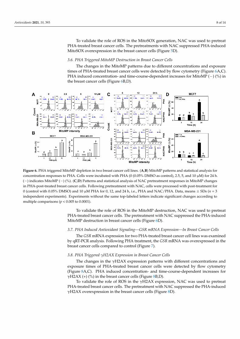
3.6. PHA Triggered MitoMP Destruction in Breast Cancer Cells
The changes in the MitoMP patterns due to different concentrations and exposuretimes of PHA-treated breast cancer cells were detected by flow cytometry (Figure 6A,C).PHA induced concentration- and time-course-dependent increases for MitoMP (−) (%) inthe breast cancer cells (Figure 6B,D).
Figure 6. PHA triggered MitoMP depletion in two breast cancer cell lines. (A,B) MitoMP patterns and statistical analysis forconcentration responses to PHA. Cells were incubated with PHA (0 (0.05% DMSO as control), 2.5, 5, and 10 µM) for 24 h.(−) indicates MitoMP (−) (%). (C,D) Patterns and statistical analysis of NAC pretreatment responses in MitoMP changesin PHA-post-treated breast cancer cells. Following pretreatment with NAC, cells were processed with post-treatment for0 (control with 0.05% DMSO) and 10 µM PHA for 0, 12, and 24 h, i.e., PHA and NAC/PHA. Data, means ± SDs (n = 3independent experiments). Experiments without the same top-labeled letters indicate significant changes according tomultiple comparisons (p < 0.005 to 0.0001).
To validate the role of ROS in the MitoMP destruction, NAC was used to pretreatPHA-treated breast cancer cells. The pretreatment with NAC suppressed the PHA-inducedMitoMP destruction in breast cancer cells (Figure 6D).
3.7. PHA Induced Antioxidant Signaling—GSR mRNA Expression—In Breast Cancer Cells
The GSR mRNA expression for two PHA-treated breast cancer cell lines was examinedby qRT-PCR analysis. Following PHA treatment, the GSR mRNA was overexpressed in thebreast cancer cells compared to control (Figure 7).
3.8. PHA Triggered γH2AX Expression in Breast Cancer Cells
The changes in the γH2AX expression patterns with different concentrations andexposure times of PHA-treated breast cancer cells were detected by flow cytometry(Figure 8A,C). PHA induced concentration- and time-course-dependent increases forγH2AX (+) (%) in the breast cancer cells (Figure 8B,D).
To validate the role of ROS in the γH2AX expression, NAC was used to pretreatPHA-treated breast cancer cells. The pretreatment with NAC suppressed the PHA-inducedγH2AX overexpression in the breast cancer cells (Figure 8D).

Antioxidants 2021, 10, 393 9 of 14
Figure 7. PHA induced GSR mRNA expression in breast cancer cells. Cells were incubated with PHA(0 (0.05% DMSO as control), 5 (PHA 5), and 10 (PHA 10) µM) for 24 h. Data, means ± SDs (n = 3independent experiments). Experiments without the same top-labeled letters indicate significantchanges according to multiple comparisons (p < 0.005 to 0.0001).
Figure 8. PHA triggered γH2AX overexpression in two breast cancer cell lines. (A,B) γH2AX patterns and statisticalanalysis for concentration responses of PHA. Cells were incubated with PHA (0 (0.05% DMSO as control), 2.5, 5, and 10 µM)for 24 h. Small box indicates γH2AX (+) (%). (C,D) Patterns and statistical analysis for NAC pretreatment responses inγH2AX intensity changes in PHA-post-treated breast cancer cells. Following pretreatment with NAC, cells were processedwith post-treatment for 0 (control with 0.05% DMSO) and 10 µM PHA for 0, 12, and 24 h, i.e., PHA and NAC/PHA. Data,means ± SDs (n = 3 independent experiments). Experiments without the same top-labeled letters indicate significantchanges according to multiple comparisons (p < 0.05 to 0.0001).
4. Discussion
PHA, one of the P. peruviana-derived natural products, has rarely been investigated,especially for its anticancer effect. In the current study, the antiproliferative, cell-cycle-disturbing, oxidative-stress-inducing, and DNA-damaging effects of PHA were validated indose-dependence and time-course experiments with breast cancer cells. The detailed drug-acting mechanisms of PHA-induced anti-breast-cancer effects are discussed as follows.
4.1. PHA Is a Potential Antiproliferative Natural Product for Breast Cancer Cells
Recently, PHA was reported to provide antiproliferative effects against prostate(LNCaP) and renal (ACHN) cancer cells but induce less damage to human foreskin fi-

Antioxidants 2021, 10, 393 10 of 14
broblasts (HEF) [22], i.e., the IC50 concentrations at 72 h in an MTS assay were 0.11, 1.0, and>2 µM, respectively. However, these studies focused on the structure–activity relationshipand reported the IC50 values of PHA without investigating the detailed mechanisms.
Breast cancer cells are reported to have several subtypes that respond differently tochemotherapy [8]. In the current study, the IC50 concentrations for PHA at 24 h in theATP study for the three breast cancer cell lines ranged from 3.12 to 6.15 µM (Figure 1A),suggesting that PHA has cell-killing effects against different kinds of breast cancer cells,including those classified [3] as luminal A positive, HER2 positive, and Claudin-low(TNBC). The examination of possible killing effects on other types of breast cancer cells suchas luminal B and basal are warranted in the future. Moreover, PHA may have the potentialfor combinatorial treatment with radiation and immunologically active compounds in atargeted therapy due to the immunoprofile characteristics of MCF7 (ER+, PR+/−, HER2−)and SKBR3 (ER−, PR−, HER2+) cells.
To provide a comparison with the clinically used anticancer drug cisplatin, the IC50value for cisplatin at 24 h in the MTS assay for SKBR3 cells was 49.8 µM [34]. The IC50concentrations for 48 h cisplatin in the ATP assay were 4.9, 17.9, and 26.9 µM for SKBR3,MCF7, and MDA-MB-231 cells, respectively [35]. For comparison, the IC50 concentrationsfor 24 h PHA in the ATP assay were 4.18, 3.12, and 6.15 µM for SKBR3, MCF7, and MDA-MB-231 cells, respectively (Figure 1A). Therefore, PHA has a higher potency to inhibitthe proliferation of breast cancer cells than cisplatin. Although PHA exhibits low cellcytotoxicity to normal foreskin fibroblasts [22], it warrants a survival comparison of PHAwith cisplatin in relation to more normal cell lines. The potential therapeutic index of PHAin terms of selectivity towards cancer cells needs to be examined in the future.
4.2. PHA Generates Oxidative Stress in Breast Cancer Cells
ROS-modulating strategies are commonly used for anticancer drug development [13,36,37].ATP-production ability is proportional to mitochondrial function. When mitochondriashow dysfunction, MitoSOX generation may show substantial upregulation. For example,manoalide induces ATP depletion, associated with MitoSOX generation and cell death inoral cancer cells [38]. PHA induces ATP depletion (Figure 1) and triggers oxidative-stressresponses, including ROS and MitoSOX overexpression (Figures 4 and 5), as well as MitoMPdepolarization (Figure 6), in breast cancer cells. Accordingly, PHA is a ROS-modulatingnatural product with a substantial anti-breast-cancer effect.
Cellular redox homeostasis is regulated by both oxidative stress and antioxidant ma-chinery. Cellular antioxidant machinery shows either activation or inactivation responsesto oxidative-stress environments [39]. For example, a transient oxidative stress may in-duce a ROS detoxification response by activating superoxide dismutase (SOD) or catalase(CAT) [40]. By contrast, sustained oxidative stress may induce cancer cell death [40]. Cultur-ing oocytes under a high-O2 condition induces GSR, glutathione peroxidase 1 (GPX1), CAT,SOD1, and SOD2 mRNA overexpression [41]. Exogenous C8-ceramide induces ROS andapoptosis in lung cancer H1299 cells by upregulating mitochondrion-located SOD2 [42].Similarly, PHA treatments for two breast cancer cell lines induce the GSR mRNA expression(Figure 7) associated with oxidative stress. It is possible that the GSR gene is activatedin response to PHA-induced oxidative stress, but its ROS-scavenging capacity fails tocounteract the high induction of oxidative stress in the end.
Some natural products generate ROS by themselves, such as naphthoquinones. Forexample, β-lapachone, a naphthoquinone derived from the lapacho tree’s bark, is a redoxrecycler [43] and a ROS-generating chemical [44]. β-lapachone can generate ROS by itself,oxidizing catalytic cysteine’s thiol group to the sulfinic acid form [43]. Whether PHAitself is a ROS-generating molecule remains unclear. This warrants the investigation of theROS-generating ability of PHA in the future.

Antioxidants 2021, 10, 393 11 of 14
4.3. PHA Induces G2/M-Phase Arrest, Apoptotic Change, and γH2AX-Detected DNA Damage inBreast Cancer Cells
By activating cell-cycle checkpoints, the proliferation of cancer cells can be inhib-ited [45]. Several G2/M-arresting drugs have been developed to inhibit cancer cell prolifer-ation, such as withaferin A in glioblastoma cells [46], sinularin in oral and breast cancercells [27,47], genistein in colon cancer cells [48], and pevonedistat in breast cancer cells [49].Similarly, PHA induced oxidative stress and G2/M arrest and resulted in apoptosis (ac-cording to flow cytometry and Western blotting) in breast cancer cells in spite of the lowpopulation of subG1 cells (Figures 2 and 3).
SubG1 accumulation is not essential for apoptosis. Drugs may induce G2/M-phasearrest and trigger apoptosis but induce no subG1 accumulation with 24 h treatments, suchas withametelin treatment for lung cancer A549 cells [50], (−)-anonaine treatment for lungcancer H1299 cells, and sinularin treatment for oral cancer Ca9-22 cells [47]. When cellsarrested in G2/M phases become apoptotic, their DNA contents may degrade, and theymay shift from G2/M to S or G1 phases without subG1 movement. In some cases, drugswith longer exposure may induce greater apoptosis compared to short exposure [47,51,52].For example, subG1 accumulation was weak at 24 and 48 h of treatment but moderate at72 h of treatment with the chalcone derivative Ch1 [52] and (−)-anonaine [47].
Several ROS-modulating drugs [30,38,53] were reported to activate both extrinsic(Cas-8) and intrinsic (Cas-9) apoptotic pathways; both pathways trigger apoptosis throughthe cleavage of downstream signaling molecules such as Cas-3. Similarly, PHA activates,in a similar manner, Cas-9, -8, and -3 in breast cancer cells, suggesting that PHA inducesgeneric oxidative stress that activates apoptotic caspases.
After 24 h of treatment with 10 µM PHA, the various breast cancer cells showedstrong cytotoxicity (about 80%) (Figure 1A); however, about 40% apoptosis was detected(Figure 3B). This suggests that PHA also induces another type of cell death, such asnecrosis, which was detectable for 20% in terms of annexin V (−)/7AAD (+) analysis [28,29](Figure 3B). Therefore, PHA induces more apoptosis than necrosis in breast cancer cells.Knowing the amount of necrosis allows discriminating nonspecific anticancer effects. Itwarrants a detailed investigation exploring the role of necrosis in PHA cytotoxicity tobreast cancer cells in the future.
Moreover, oxidative stress is also a DNA-damage-inducing factor [24,54,55]. Thiswas supported by our finding that PHA induced oxidative-stress responses such asROS/MitoSOX overproduction and MitoMP depletion. Therefore, it caused DNA damagein breast cancer cells as detected by γH2AX (Figure 8).
4.4. NAC Suppresses PHA-Induced Antiproliferative Effects and ROS-Associated Changes inBreast Cancer Cells
All the PHA-induced changes such as antiproliferative effects, oxidative-stress in-duction, apoptosis, and DNA damage were recovered by NAC pretreatment. This heldfor ATP depletion, ROS/MitoSOX overproduction, MitoMP depletion, annexin V- andWestern-blot-detected extrinsic and intrinsic apoptosis, and γH2AX-detected DNA damage.Accordingly, the PHA-induced antiproliferative effects, apoptosis, and DNA damage weremediated by oxidative-stress induction in the breast cancer cells.
5. Conclusions
Many bioactive compounds belonging to the withanolides isolated from severalPhysalis species show anticancer effects, but the action mechanisms have rarely beeninvestigated. In the current study, we showed that PHA inhibits the proliferation ofthree kinds of breast cancer cells: MCF7 (ER+, PR+/−, HER2−), SKBR3 (ER−, PR−,HER2+), and MDA-MB-231 (TNBC). This antiproliferative effect of PHA against the breastcancer cells was proven to be oxidative-stress-dependent by NAC pretreatment. The cell-killing mechanisms of PHA include cell-cycle G2/M arrest, oxidative-stress induction,and DNA damage. Both the apoptosis and DNA damage were proven to be oxidative-

Antioxidants 2021, 10, 393 12 of 14
stress-dependent. Therefore, PHA represents a potential anti-breast-cancer natural product,and its cell-killing mechanism is associated with the modulation of the oxidative-stressresponse, cell-cycle disturbance, apoptosis, and DNA damage.
Author Contributions: Conceptualization, H.-W.C. and F.O.-Y.; data curation, T.-J.Y.; formal analysis,T.-J.Y. and B.-Y.Y.; methodology, T.-J.Y., Y.-B.C., L.-C.L., Y.-H.T., B.-Y.Y., J.-Y.T., F.-R.C., and C.-H.Y.; su-pervision, F.O.-Y. and H.-W.C.; writing—original draft, T.-J.Y., Y.-B.C., and H.-W.C.; writing—reviewand editing, F.O.-Y. and H.-W.C. All authors have read and agreed to the published version ofthe manuscript.
Funding: This work was partly supported by funds from the Ministry of Science and Technology(MOST 108-2320-B-037-015-MY3, MOST 109-2628-B-110-004, MOST 108-2314-B-037-080, and MOST109-2314-B-037-018), the National Sun Yat-sen University–Kaohsiung Medical University (KMU) JointResearch Project (#NSYSUKMU 110-P016), the Kaohsiung Medical University Hospital (KMUH109-9R35), the Kaohsiung Medical University Research Center (KMU-TC108A04), and the Health andWelfare Surcharge of Tobacco Products, the Ministry of Health and Welfare, Taiwan (MOHW 109-TDU-B-212-134016). The authors thank their colleague Hans-Uwe Dahms for editing the manuscript andthe Natural Product Libraries and High-Throughput Screening Core (NPS) for the high-throughputscreening and technical support. The NPS Core is funded by the Ministry of Science and Technology(MOST 109-2740-B-037-001).
Institutional Review Board Statement: Not applicable.
Informed Consent Statement: Not applicable.
Data Availability Statement: Data is contained within the article.
Conflicts of Interest: The authors declare that there are no conflict of interest among them.
References1. Kuo, C.N.; Liao, Y.M.; Kuo, L.N.; Tsai, H.J.; Chang, W.C.; Yen, Y. Cancers in Taiwan: Practical insight from epidemiology,
treatments, biomarkers, and cost. J. Formos. Med. Assoc. 2020, 119, 1731–1741. [CrossRef]2. Siegel, R.L.; Miller, K.D.; Jemal, A. Cancer statistics, 2020. CA Cancer J. Clin. 2020, 70, 7–30. [CrossRef]3. Holliday, D.L.; Speirs, V. Choosing the right cell line for breast cancer research. Breast Cancer Res. 2011, 13, 215. [CrossRef]4. Ariazi, E.A.; Ariazi, J.L.; Cordera, F.; Jordan, V.C. Estrogen receptors as therapeutic targets in breast cancer. Curr. Top. Med. Chem.
2006, 6, 181–202. [CrossRef]5. Oh, D.Y.; Bang, Y.J. HER2-targeted therapies—A role beyond breast cancer. Nat. Rev. Clin. Oncol. 2020, 17, 33–48. [CrossRef]6. Giulianelli, S.; Molinolo, A.; Lanari, C. Targeting progesterone receptors in breast cancer. Vitam. Horm. 2013, 93, 161–184.7. Foulkes, W.D.; Smith, I.E.; Reis-Filho, J.S. Triple-negative breast cancer. N. Engl. J. Med. 2010, 363, 1938–1948. [CrossRef] [PubMed]8. Rouzier, R.; Perou, C.M.; Symmans, W.F.; Ibrahim, N.; Cristofanilli, M.; Anderson, K.; Hess, K.R.; Stec, J.; Ayers, M.; Wagner, P.;
et al. Breast cancer molecular subtypes respond differently to preoperative chemotherapy. Clin. Cancer Res. 2005, 11, 5678–5685.[CrossRef] [PubMed]
9. Partridge, A.H.; Burstein, H.J.; Winer, E.P. Side effects of chemotherapy and combined chemohormonal therapy in women withearly-stage breast cancer. J. Natl. Cancer Inst. Monogr. 2001, 2001, 135–142. [CrossRef]
10. Saeki, T.; Tsuruo, T.; Sato, W.; Nishikawsa, K. Drug resistance in chemotherapy for breast cancer. Cancer Chemother. Pharmacol.2005, 56 (Suppl. 1), 84–89. [CrossRef] [PubMed]
11. Zhang, W.N.; Tong, W.Y. Chemical constituents and biological activities of plants from the genus Physalis. Chem. Biodivers 2016,13, 48–65. [CrossRef] [PubMed]
12. Chen, L.X.; He, H.; Qiu, F. Natural withanolides: An overview. Nat. Prod. Rep. 2011, 28, 705–740. [CrossRef] [PubMed]13. Widodo, N.; Priyandoko, D.; Shah, N.; Wadhwa, R.; Kaul, S.C. Selective killing of cancer cells by Ashwagandha leaf extract and
its component Withanone involves ROS signaling. PLoS ONE 2010, 5, e13536. [CrossRef] [PubMed]14. Samadi, A.K. Chapter three—Potential anticancer properties and mechanisms of action of withanolides. In The Enzymes; Bathaie,
S.Z., Tamanoi, F., Eds.; Academic Press: Cambridge, MA, USA, 2015; Volume 37, pp. 73–94.15. Chang, H.W.; Li, R.N.; Wang, H.R.; Liu, J.R.; Tang, J.Y.; Huang, H.W.; Chan, Y.H.; Yen, C.Y. Withaferin A induces oxidative
stress-mediated apoptosis and DNA damage in oral cancer cells. Front. Physiol. 2017, 8, 634. [CrossRef] [PubMed]16. Chiu, C.C.; Haung, J.W.; Chang, F.R.; Huang, K.J.; Huang, H.M.; Huang, H.W.; Chou, C.K.; Wu, Y.C.; Chang, H.W. Golden
berry-derived 4beta-hydroxywithanolide E for selectively killing oral cancer cells by generating ROS, DNA damage, and apoptoticpathways. PLoS ONE 2013, 8, e64739. [CrossRef] [PubMed]
17. Royston, K.J.; Paul, B.; Nozell, S.; Rajbhandari, R.; Tollefsbol, T.O. Withaferin A and sulforaphane regulate breast cancer cell cycleprogression through epigenetic mechanisms. Exp. Cell Res. 2018, 368, 67–74. [CrossRef]

Antioxidants 2021, 10, 393 13 of 14
18. Peng, C.Y.; You, B.J.; Lee, C.L.; Wu, Y.C.; Lin, W.H.; Lu, T.L.; Chang, F.C.; Lee, H.Z. The roles of 4beta-hydroxywithanolide E fromPhysalis peruviana on the Nrf2-anti-oxidant system and the cell cycle in breast cancer cells. Am. J. Chin. Med. 2016, 44, 617–636.[CrossRef]
19. Wang, H.C.; Hu, H.H.; Chang, F.R.; Tsai, J.Y.; Kuo, C.Y.; Wu, Y.C.; Wu, C.C. Different effects of 4beta-hydroxywithanolide E andwithaferin A, two withanolides from Solanaceae plants, on the Akt signaling pathway in human breast cancer cells. Phytomedicine2019, 53, 213–222. [CrossRef] [PubMed]
20. Machin, R.P.; Veleiro, A.S.; Nicotra, V.E.; Oberti, J.C.; MPadrón, J. Antiproliferative activity of withanolides against human breastcancer cell lines. J. Nat Prod 2010, 73, 966–968. [CrossRef]
21. Shingu, K.; Miyagawa, M.; Yahara, S.; Nohara, T. Physapruins A and B, two new withanolides from Physalis pruinosa Bailey. Chem.Pharm. Bull 1993, 41, 1873–1875. [CrossRef]
22. Xu, Y.M.; Wijeratne, E.M.K.; Babyak, A.L.; Marks, H.R.; Brooks, A.D.; Tewary, P.; Xuan, L.J.; Wang, W.Q.; Sayers, T.J.; Gunatilaka,A.A.L. Withanolides from aeroponically grown Physalis peruviana and their selective cytotoxicity to prostate cancer and renalcarcinoma cells. J. Nat. Prod. 2017, 80, 1981–1991. [CrossRef] [PubMed]
23. Huang, C.H.; Yeh, J.M.; Chan, W.H. Hazardous impacts of silver nanoparticles on mouse oocyte maturation and fertilization andfetal development through induction of apoptotic processes. Environ. Toxicol. 2018, 33, 1039–1049. [CrossRef] [PubMed]
24. Wang, T.S.; Lin, C.P.; Chen, Y.P.; Chao, M.R.; Li, C.C.; Liu, K.L. CYP450-mediated mitochondrial ROS production involved inarecoline N-oxide-induced oxidative damage in liver cell lines. Environ. Toxicol. 2018, 33, 1029–1038. [CrossRef] [PubMed]
25. Tang, J.Y.; Shu, C.W.; Wang, C.L.; Wang, S.C.; Chang, M.Y.; Lin, L.C.; Chang, H.W. Sulfonyl chromen-4-ones (CHW09) shows anadditive effect to inhibit cell growth of X-ray irradiated oral cancer cells, involving apoptosis and ROS generation. Int. J. Radiat.Biol. 2019, 95, 1226–1235. [CrossRef] [PubMed]
26. Chen, C.Y.; Yen, C.Y.; Wang, H.R.; Yang, H.P.; Tang, J.Y.; Huang, H.W.; Hsu, S.H.; Chang, H.W. Tenuifolide B from Cinnamomumtenuifolium stem selectively inhibits proliferation of oral cancer cells via apoptosis, ROS generation, mitochondrial depolarization,and DNA damage. Toxins 2016, 8, 319. [CrossRef]
27. Huang, H.W.; Tang, J.Y.; Ou-Yang, F.; Wang, H.R.; Guan, P.Y.; Huang, C.Y.; Chen, C.Y.; Hou, M.F.; Sheu, J.H.; Chang, H.W.Sinularin selectively kills breast cancer cells showing G2/M arrest, apoptosis, and oxidative DNA damage. Molecules 2018, 23,849. [CrossRef]
28. Zingue, S.; Michel, T.; Cisilotto, J.; Tueche, A.B.; Ndinteh, D.T.; Mello, L.J.; Njamen, D.; Creczynski-Pasa, T.B. The hydro-ethanolicextract of Acacia seyal (Mimosaceae) stem barks induced death in an ER-negative breast cancer cell line by the intrinsic pathwayof apoptosis and inhibited cell migration. J. Ethnopharmacol. 2018, 223, 41–50. [CrossRef]
29. Crowley, L.C.; Marfell, B.J.; Scott, A.P.; Waterhouse, N.J. Quantitation of apoptosis and necrosis by annexin V binding, propidiumiodide uptake, and flow cytometry. Cold Spring Harb. Protoc. 2016. [CrossRef] [PubMed]
30. Tang, J.Y.; Wu, C.Y.; Shu, C.W.; Wang, S.C.; Chang, M.Y.; Chang, H.W. A novel sulfonyl chromen-4-ones (CHW09) preferentiallykills oral cancer cells showing apoptosis, oxidative stress, and DNA damage. Environ. Toxicol. 2018, 33, 1195–1203. [CrossRef]
31. Yen, C.Y.; Chiu, C.C.; Chang, F.R.; Chen, J.Y.; Hwang, C.C.; Hseu, Y.C.; Yang, H.L.; Lee, A.Y.; Tsai, M.T.; Guo, Z.L.; et al.4beta-Hydroxywithanolide E from Physalis peruviana (golden berry) inhibits growth of human lung cancer cells through DNAdamage, apoptosis and G2/M arrest. BMC Cancer 2010, 10, 46. [CrossRef] [PubMed]
32. Yu, T.J.; Tang, J.Y.; Ou-Yang, F.; Wang, Y.Y.; Yuan, S.F.; Tseng, K.; Lin, L.C.; Chang, H.W. Low concentration of withaferin A inhibitsoxidative stress-mediated migration and invasion in oral cancer cells. Biomolecules 2020, 10, 777. [CrossRef]
33. Tang, J.Y.; Peng, S.Y.; Cheng, Y.B.; Wang, C.L.; Farooqi, A.A.; Yu, T.J.; Hou, M.F.; Wang, S.C.; Yen, C.H.; Chan, L.P.; et al. Ethylacetate extract of Nepenthes adrianii x clipeata induces antiproliferation, apoptosis, and DNA damage against oral cancer cellsthrough oxidative stress. Environ. Toxicol. 2019, 34, 891–901. [CrossRef] [PubMed]
34. Ou-Yang, F.; Tsai, I.H.; Tang, J.Y.; Yen, C.Y.; Cheng, Y.B.; Farooqi, A.A.; Chen, S.R.; Yu, S.Y.; Kao, J.K.; Chang, H.W. Antiproliferationfor breast cancer cells by ethyl acetate extract of Nepenthes thorellii x (ventricosa x maxima). Int. J. Mol. Sci. 2019, 20, 3238. [CrossRef][PubMed]
35. Yu, T.J.; Tang, J.Y.; Lin, L.C.; Lien, W.J.; Cheng, Y.B.; Chang, F.R.; Ou-Yang, F.; Chang, H.W. Withanolide C inhibits proliferation ofbreast cancer cells via oxidative stress-mediated apoptosis and DNA damage. Antioxidants 2020, 9, 873. [CrossRef] [PubMed]
36. Lee, J.C.; Hou, M.F.; Huang, H.W.; Chang, F.R.; Yeh, C.C.; Tang, J.Y.; Chang, H.W. Marine algal natural products with anti-oxidative, anti-inflammatory, and anti-cancer properties. Cancer Cell Int. 2013, 13, 55. [CrossRef] [PubMed]
37. Tang, J.Y.; Ou-Yang, F.; Hou, M.F.; Huang, H.W.; Wang, H.R.; Li, K.T.; Fayyaz, S.; Shu, C.W.; Chang, H.W. Oxidative stress-modulating drugs have preferential anticancer effects—Involving the regulation of apoptosis, DNA damage, endoplasmicreticulum stress, autophagy, metabolism, and migration. Semin. Cancer Biol. 2019, 58, 109–117. [CrossRef]
38. Wang, H.R.; Tang, J.Y.; Wang, Y.Y.; Farooqi, A.A.; Yen, C.Y.; Yuan, S.F.; Huang, H.W.; Chang, H.W. Manoalide preferentiallyprovides antiproliferation of oral cancer cells by oxidative stress-mediated apoptosis and DNA damage. Cancers 2019, 11, 1303.[CrossRef] [PubMed]
39. Schieber, M.; Chandel, N.S. ROS function in redox signaling and oxidative stress. Curr. Biol. 2014, 24, R453–R462. [CrossRef]40. Espinosa-Diez, C.; Miguel, V.; Mennerich, D.; Kietzmann, T.; Sanchez-Perez, P.; Cadenas, S.; Lamas, S. Antioxidant responses and
cellular adjustments to oxidative stress. Redox Biol. 2015, 6, 183–197. [CrossRef] [PubMed]41. Salavati, M.; Ghafari, F.; Zhang, T.; Fouladi-Nashta, A.A. Effects of oxygen concentration on in vitro maturation of canine oocytes
in a chemically defined serum-free medium. Reproduction 2012, 144, 547–556. [CrossRef] [PubMed]
Antioxidants 2021, 10, 393 14 of 14
42. Chang, Y.C.; Fong, Y.; Tsai, E.M.; Chang, Y.G.; Chou, H.L.; Wu, C.Y.; Teng, Y.N.; Liu, T.C.; Yuan, S.S.; Chiu, C.C. ExogenousC(8)-ceramide induces apoptosis by overproduction of ROS and the switch of superoxide dismutases SOD1 to SOD2 in humanlung cancer cells. Int. J. Mol. Sci. 2018, 19, 3010. [CrossRef]
43. Ohayon, S.; Refua, M.; Hendler, A.; Aharoni, A.; Brik, A. Harnessing the oxidation susceptibility of deubiquitinases for inhibitionwith small molecules. Angew Chem. Int. Ed. Engl. 2015, 54, 599–603. [CrossRef]
44. Gopinath, P.; Mahammed, A.; Ohayon, S.; Gross, Z.; Brik, A. Understanding and predicting the potency of ROS-based enzymeinhibitors, exemplified by naphthoquinones and ubiquitin specific protease-2. Chem. Sci. 2016, 7, 7079–7086. [CrossRef] [PubMed]
45. Kastan, M.B.; Bartek, J. Cell-cycle checkpoints and cancer. Nature 2004, 432, 316–323. [CrossRef]46. Tang, Q.; Ren, L.; Liu, J.; Li, W.; Zheng, X.; Wang, J.; Du, G. Withaferin A triggers G2/M arrest and intrinsic apoptosis in
glioblastoma cells via ATF4-ATF3-CHOP axis. Cell Prolif. 2020, 53, e12706. [CrossRef]47. Chang, Y.T.; Wu, C.Y.; Tang, J.Y.; Huang, C.Y.; Liaw, C.C.; Wu, S.H.; Sheu, J.H.; Chang, H.W. Sinularin induces oxidative
stress-mediated G2/M arrest and apoptosis in oral cancer cells. Environ. Toxicol. 2017, 32, 2124–2132. [CrossRef]48. Zhang, Z.; Wang, C.Z.; Du, G.J.; Qi, L.W.; Calway, T.; He, T.C.; Du, W.; Yuan, C.S. Genistein induces G2/M cell cycle arrest and
apoptosis via ATM/p53-dependent pathway in human colon cancer cells. Int. J. Oncol. 2013, 43, 289–296. [CrossRef] [PubMed]49. Chen, Y.; Du, M.; Yusuying, S.; Liu, W.; Tan, Y.; Xie, P. Nedd8-activating enzyme inhibitor MLN4924 (Pevonedistat), inhibits
miR-1303 to suppress human breast cancer cell proliferation via targeting p27(Kip1). Exp. Cell Res. 2020, 392, 112038. [CrossRef][PubMed]
50. Rao, P.C.; Begum, S.; Jahromi, M.A.; Jahromi, Z.H.; Sriram, S.; Sahai, M. Cytotoxicity of withasteroids: Withametelin induces cellcycle arrest at G2/M phase and mitochondria-mediated apoptosis in non-small cell lung cancer A549 cells. Tumour. Biol. 2016, 37,12579–12587. [CrossRef]
51. Chen, B.H.; Chang, H.W.; Huang, H.M.; Chong, I.W.; Chen, J.S.; Chen, C.Y.; Wang, H.M. (-)-Anonaine induces DNA damage andinhibits growth and migration of human lung carcinoma h1299 cells. J. Agric. Food Chem. 2011, 59, 2284–2290. [CrossRef]
52. Kello, M.; Drutovic, D.; Pilatova, M.B.; Tischlerova, V.; Perjesi, P.; Mojzis, J. Chalcone derivatives cause accumulation of coloncancer cells in the G2/M phase and induce apoptosis. Life Sci. 2016, 150, 32–38. [CrossRef] [PubMed]
53. Park, G.B.; Choi, Y.; Kim, Y.S.; Lee, H.K.; Kim, D.; Hur, D.Y. ROS and ERK1/2-mediated caspase-9 activation increases XAF1expression in dexamethasone-induced apoptosis of EBV-transformed B cells. Int. J. Oncol. 2013, 43, 29–38. [CrossRef] [PubMed]
54. Wu, C.F.; Lee, M.G.; El-Shazly, M.; Lai, K.H.; Ke, S.C.; Su, C.W.; Shih, S.P.; Sung, P.J.; Hong, M.C.; Wen, Z.H.; et al. Isoaaptamineinduces T-47D cells apoptosis and autophagy via oxidative stress. Mar. Drugs 2018, 16, 18. [CrossRef]
55. Hung, J.H.; Chen, C.Y.; Omar, H.A.; Huang, K.Y.; Tsao, C.C.; Chiu, C.C.; Chen, Y.L.; Chen, P.H.; Teng, Y.N. Reactive oxygenspecies mediate Terbufos-induced apoptosis in mouse testicular cell lines via the modulation of cell cycle and pro-apoptoticproteins. Environ. Toxicol. 2016, 31, 1888–1898. [CrossRef] [PubMed]