Phthalocyanine Photodynamic Therapy: Disparate Effects of Pharmacologic Inhibitors on Cutaneous...
7
Photochemistry and Photobiology, 1997, 65(5): 895-901 Phthalocyanine Photodynamic Therapy: Disparate Effects of Pharmacologic Inhibitors on Cutaneous Photosensitivity and on Tumor Regression Cathy Anderson, Sharon Hrabovsky, Yvonne McKinley, Karen Tubesing, Hai-Ping Tang, Robert Dunbar, Hasan Mukhtar and Craig A. Elmets' Skin Diseases Research Center, Departments of Dermatology and Chemistry, Case Western Reserve University, Cleveland, OH, USA Received 11 October 1996; accepted 9 February 1997 ABSTRACT The phthalocyanines are promising second-generation photosensitizers that are being evaluated for the photo- dynamic therapy (PDT) of malignant tumors. In vivo studies with the silicon phthalocyanine Pc 4 have shown that it is highly effective at causing regression of RIF-1 tumors in C3H/HeN mice in PDT protocols. Because cu- taneous photosensitivity is the major complication of photosensitizers used for PDT, experiments were per- formed to evaluate the effect of inhibitors of the inflam- matory response (cyproheptadine, dexamethasone, pen- toxifylline, and tumor necrosis factor alpha [TNF-a] an- tibodies) on Pc 4-induced cutaneous photosensitivity and tumor regression. The C3WHeN mice were injected with either Pc 4 or Photofrin and were exposed to 86 J/cm2 of filtered radiation emitted from a solar simulator. Ani- mals were irradiated at 1, 3, 7, 10, 14 and 28 days post- injection. Cutaneous photosensitivity was assessed using the murine ear-swelling response. Cyproheptadine, dexa- methasone, pentoxifyllineand TNF-a antibodies were ad- ministered prior to illumination to assess their ability to block Pc 4-induced cutaneous photosensitivity and to evaluate whether such treatment adversely influenced Pc 4 PDT-induced tumor regression. Compared to Photo- frin, Pc 4 produced cutaneous photosensitivity that was transient, resolving within 24 h, and that could be elicited for only 10 days after administration. In contrast, Pho- tofrin caused photosensitivity that required 4 days to re- solve and could be elicited for at least 1 month after it was administered. The Pc 4-induced cutaneous photosen- sitivity could be blocked by corticosteroids and an inhib- itor of vasoactive amines (cyproheptadine). The TNF-a gene transcription was found to increase in keratinocytes following treatment with Pc 4 and light. The anti-TNF-a antibodies and pentoxifylline, an inhibitor of cytokine transcription, also prevented cutaneous photosensitivity, ~~ *To whom correspondence should be addressed at: Skin Diseases Research Center, Department of Dermatology, University Hospi- tals of Cleveland, I I100 Euclid Ave., Cleveland, OH 44106, USA. Fax: 2 16-844-8993; e-mail: [email protected]. 0 1997 American Society for Photobiology 003 I -8655/97 $5.00+0.00 implicating TNF-a in the pathogenesis of Pc 4-induced cutaneous photosensitivity. None of these agents had any effect on Pc 4 PDT-induced tumor regression. Cyprohep- tadine, dexamethasone, pentoxifylline and TNF-a anti- bodies may be valuable pharmacologic agents in the management of cutaneous photosensitivity associated with PDT without altering the efficacy of this new ther- apeutic modality. The findings suggest that it should be possible to devise PDT protocols that block cutaneous photosensitivity without impairing the anti-tumor re- sponse to the agents. INTRODUCTION Photodynamic therapy (PDT)? is a promising new therapeu- tic modality for the management of a variety of solid tumors (1-5). Cutaneous squamous cell and basal cell carcinomas, squamous cell carcinomas of the head and neck, esophageal malignancies and transitional carcinomas of the bladder have all been shown to respond favorably in many animal tumor models and in clinical trials in both humans and domestic animals (1-7). The process consists of local or systemic ad- ministration of photosensitizing chemicals that preferentially accumulate within tumors. Tumors are then illuminated with specific wavelengths of visible light that are absorbed by the photosensitizing compound. As the energy transferred from light to the photosensitizer is dissipated, damage to tumor cells and to the tumor vasculature occurs, which ultimately leads to widespread tumor destruction. The photosensitizer that has received the most extensive evaluation in PDT protocols has been Photofrin. This agent, which is a mixture of several different porphyrins, is ap- proved for palliative therapy for solid tumors in the United States and several other countries. Although effective, Pho- tofrin has a number of undesirable features that make it less than ideal as a photosensitizing agent. These include the fact TAbbreviurions: BSA, bovine serum albumin; ICAM, intercellular adhesion molecule; im., intramuscular; i.p., intraperitoneal; i.v., intravenous; PBS, phosphate-buffered saline; Pc 4, silicon phthal- ocyanine 4; PDT, photodynamic therapy; RT-PCR, reverse tran- scriptase polymerase chain reaction; s.c., subcutaneous; SDS, so- dium dodecyl sulfate; TNF-a, tumor necrosis factor alpha. 895
-
Upload
cathy-anderson -
Category
Documents
-
view
212 -
download
0
Transcript of Phthalocyanine Photodynamic Therapy: Disparate Effects of Pharmacologic Inhibitors on Cutaneous...

Photochemistry and Photobiology, 1997, 65(5): 895-901
Phthalocyanine Photodynamic Therapy: Disparate Effects of Pharmacologic Inhibitors on Cutaneous Photosensitivity and on Tumor Regression
Cathy Anderson, Sharon Hrabovsky, Yvonne McKinley, Karen Tubesing, Hai-Ping Tang, Robert Dunbar, Hasan Mukhtar and Craig A. Elmets' Skin Diseases Research Center, Departments of Dermatology and Chemistry, Case Western Reserve University, Cleveland, OH, USA
Received 11 October 1996; accepted 9 February 1997
ABSTRACT
The phthalocyanines are promising second-generation photosensitizers that are being evaluated for the photo- dynamic therapy (PDT) of malignant tumors. In vivo studies with the silicon phthalocyanine Pc 4 have shown that it is highly effective at causing regression of RIF-1 tumors in C3H/HeN mice in PDT protocols. Because cu- taneous photosensitivity is the major complication of photosensitizers used for PDT, experiments were per- formed to evaluate the effect of inhibitors of the inflam- matory response (cyproheptadine, dexamethasone, pen- toxifylline, and tumor necrosis factor alpha [TNF-a] an- tibodies) on Pc 4-induced cutaneous photosensitivity and tumor regression. The C3WHeN mice were injected with either Pc 4 or Photofrin and were exposed to 86 J/cm2 of filtered radiation emitted from a solar simulator. Ani- mals were irradiated at 1, 3, 7, 10, 14 and 28 days post- injection. Cutaneous photosensitivity was assessed using the murine ear-swelling response. Cyproheptadine, dexa- methasone, pentoxifylline and TNF-a antibodies were ad- ministered prior to illumination to assess their ability to block Pc 4-induced cutaneous photosensitivity and to evaluate whether such treatment adversely influenced Pc 4 PDT-induced tumor regression. Compared to Photo- frin, Pc 4 produced cutaneous photosensitivity that was transient, resolving within 24 h, and that could be elicited for only 10 days after administration. In contrast, Pho- tofrin caused photosensitivity that required 4 days to re- solve and could be elicited for at least 1 month after it was administered. The Pc 4-induced cutaneous photosen- sitivity could be blocked by corticosteroids and an inhib- itor of vasoactive amines (cyproheptadine). The TNF-a gene transcription was found to increase in keratinocytes following treatment with Pc 4 and light. The anti-TNF-a antibodies and pentoxifylline, an inhibitor of cytokine transcription, also prevented cutaneous photosensitivity,
~~
*To whom correspondence should be addressed at: Skin Diseases Research Center, Department of Dermatology, University Hospi- tals of Cleveland, I I100 Euclid Ave., Cleveland, OH 44106, USA. Fax: 2 16-844-8993; e-mail: [email protected].
0 1997 American Society for Photobiology 003 I -8655/97 $5.00+0.00
implicating TNF-a in the pathogenesis of Pc 4-induced cutaneous photosensitivity. None of these agents had any effect on Pc 4 PDT-induced tumor regression. Cyprohep- tadine, dexamethasone, pentoxifylline and TNF-a anti- bodies may be valuable pharmacologic agents in the management of cutaneous photosensitivity associated with PDT without altering the efficacy of this new ther- apeutic modality. The findings suggest that it should be possible to devise PDT protocols that block cutaneous photosensitivity without impairing the anti-tumor re- sponse to the agents.
INTRODUCTION
Photodynamic therapy (PDT)? is a promising new therapeu- tic modality for the management of a variety of solid tumors (1-5). Cutaneous squamous cell and basal cell carcinomas, squamous cell carcinomas of the head and neck, esophageal malignancies and transitional carcinomas of the bladder have all been shown to respond favorably in many animal tumor models and in clinical trials in both humans and domestic animals (1-7). The process consists of local or systemic ad- ministration of photosensitizing chemicals that preferentially accumulate within tumors. Tumors are then illuminated with specific wavelengths of visible light that are absorbed by the photosensitizing compound. As the energy transferred from light to the photosensitizer is dissipated, damage to tumor cells and to the tumor vasculature occurs, which ultimately leads to widespread tumor destruction.
The photosensitizer that has received the most extensive evaluation in PDT protocols has been Photofrin. This agent, which is a mixture of several different porphyrins, is ap- proved for palliative therapy for solid tumors in the United States and several other countries. Although effective, Pho- tofrin has a number of undesirable features that make it less than ideal as a photosensitizing agent. These include the fact
TAbbreviurions: BSA, bovine serum albumin; ICAM, intercellular adhesion molecule; im . , intramuscular; i.p., intraperitoneal; i.v., intravenous; PBS, phosphate-buffered saline; Pc 4, silicon phthal- ocyanine 4; PDT, photodynamic therapy; RT-PCR, reverse tran- scriptase polymerase chain reaction; s.c., subcutaneous; SDS, so- dium dodecyl sulfate; TNF-a, tumor necrosis factor alpha.
895
896 Cathy Anderson et a/.
o-ki-/ \ I I
3 0x105
2 5 ~ 1 0 ~
I ~ 0 ~ 1 0 5
$ E 1 5 ~ 1 0 ~ u - 0
10x10‘
5 0x10‘
0 0
3uu 400 500 600 7w BW I
Wavelength (nm)
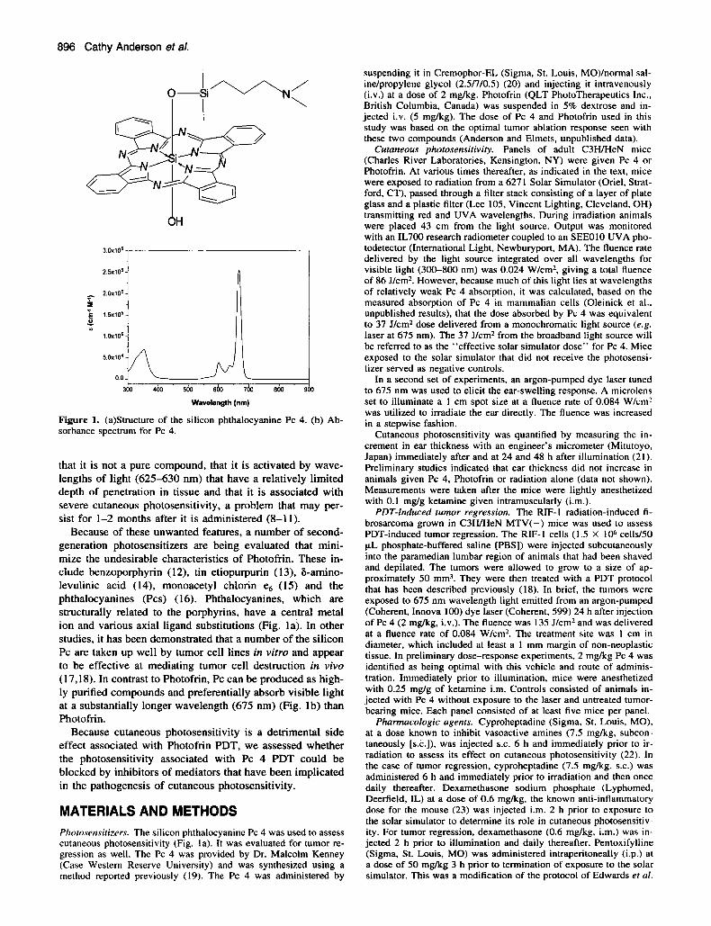
Figure 1. (a)Structure of the silicon phthalocyanine Pc 4. (b) Ab- sorbance spectrum for Pc 4.
that it is not a pure compound, that it is activated by wave- lengths of light (625-630 nm) that have a relatively limited depth of penetration in tissue and that it is associated with severe cutaneous photosensitivity, a problem that may per- sist for 1-2 months after it is administered (8-1 1).
Because of these unwanted features, a number of second- generation photosensitizers are being evaluated that mini- mize the undesirable characteristics of Photofrin. These in- clude benzoporphyrin (12), tin etiopurpurin (13), &amino- levulinic acid (14), monoacetyl chlorin e6 (15) and the phthalocyanines (Pcs) (16). Phthalocyanines, which are structurally related to the porphyrins, have a central metal ion and various axial ligand substitutions (Fig. la). In other studies, it has been demonstrated that a number of the silicon Pc are taken up well by tumor cell lines in vitro and appear to be effective at mediating tumor cell destruction in vivo (17,18). In contrast to Photofrin, Pc can be produced as high- ly purified compounds and preferentially absorb visible light at a substantially longer wavelength (675 nm) (Fig. lb) than Photofrin.
Because cutaneous photosensitivity is a detrimental side effect associated with Photofrin PDT, we assessed whether the photosensitivity associated with Pc 4 PDT could be blocked by inhibitors of mediators that have been implicated in the pathogenesis of cutaneous photosensitivity.
MATERIALS AND METHODS Photosensitizers. The silicon phthalocyanine Pc 4 was used to assess cutaneous photosensitivity (Fig. la). It was evaluated for tumor re- gression as well. The Pc 4 was provided by Dr. Malcolm Kenney (Case Western Reserve University) and was synthesized using a method reported previously (19). The Pc 4 was administered by
suspending it in Cremophor-EL (Sigma, St. Louis, M0)lnormal sal- inelpropylene glycol (2.5/7/0.5) (20) and injecting it intravenously (i.v.) at a dose of 2 mg/kg. Photofrin (QLT PhotoTherapeutics Inc., British Columbia, Canada) was suspended in 5% dextrose and in- jected i.v. (5 mgkg). The dose of Pc 4 and Photofrin used in this study was based on the optimal tumor ablation response seen with these two compounds (Anderson and Elmets, unpublished data).
Cutaneous photosensitivity. Panels of adult C3WHeN mice (Charles River Laboratories, Kensington, NY) were given Pc 4 or Photofrin. At various times thereafter, as indicated in the text, mice were exposed to radiation from a 6271 Solar Simulator (Oriel, Strat- ford, CT), passed through a filter stack consisting of a layer of plate glass and a plastic filter (Lee 105, Vincent Lighting, Cleveland, OH) transmitting red and UVA wavelengths. During irradiation animals were placed 43 cm from the light source. Output was monitored with an IL700 research radiometer coupled to an SEEOIO UVA pho- todetector (International Light, Newburyport, MA). The fluence rate delivered by the light source integrated over all wavelengths for visible light (300-800 nm) was 0.024 Wlcm2, giving a total fluence of 86 J/cm2. However, because much of this light lies at wavelengths of relatively weak Pc 4 absorption, it was calculated, based on the measured absorption of Pc 4 in mammalian cells (Oleinick et al., unpublished results), that the dose absorbed by Pc 4 was equivalent to 37 J/cm2 dose delivered from a monochromatic light source (e.g. laser at 675 nm). The 37 Jlcm2 from the broadband light source will be referred to as the “effective solar simulator dose” for Pc 4. Mice exposed to the solar simulator that did not receive the photosensi- tizer served as negative controls.
In a second set of experiments, an argon-pumped dye laser tuned to 675 nm was used to elicit the ear-swelling response. A microlens set to illuminate a 1 cm spot size at a fluence rate of 0.084 Wlcm’ was utilized to irradiate the ear directly. The fluence was increased in a stepwise fashion.
Cutaneous photosensitivity was quantified by measuring the in- crement in ear thickness with an engineer’s micrometer (Mitutoyo, Japan) immediately after and at 24 and 48 h after illumination (21). Preliminary studies indicated that ear thickness did not increase in animals given Pc 4, Photofrin or radiation alone (data not shown). Measurements were taken after the mice were lightly anesthetized with 0.1 mg/g ketamine given intramuscularly (i.m.).
PDT-induced rumor regression. The RIF-I radiation-induced fi- brosarcoma grown in C3WHeN MTV(-) mice was used to assess PDT-induced tumor regression. The RIF-I cells (1.5 X IOb cellsl50 p,L phosphate-buffered saline [PBS]) were injected subcutaneously into the paramedian lumbar region of animals that had been shaved and depilated. The tumors were allowed to grow to a size of ap- proximately 50 mm3. They were then treated with a PDT protocol that has been described previously (18). In brief, the tumors were exposed to 675 nm wavelength light emitted from an argon-pumped (Coherent, Innova 100) dye laser (Coherent, 599) 24 h after injection of Pc 4 (2 mg/kg, i.v.). The fluence was 135 Jlcm2 and was delivered at a fluence rate of 0.084 W/cmZ. The treatment site was 1 cm in diameter, which included at least a 1 mm margin of non-neoplastic tissue. In preliminary dose-response experiments, 2 mgkg Pc 4 was identified as being optimal with this vehicle and route of adminis- tration. Immediately prior to illumination, mice were anesthetized with 0.25 mglg of ketamine i.m. Controls consisted of animals in- jected with Pc 4 without exposure to the laser and untreated tumor- bearing mice. Each panel consisted of at least five mice per panel.
Pharmacologic agents. Cyproheptadine (Sigma, St. Louis, MO), at a dose known to inhibit vasoactive amines (7.5 mgkg, subcon- taneously [s.c.]), was injected S.C. 6 h and immediately prior to ir- radiation to assess its effect on cutaneous photosensitivity (22). In the case of tumor regression, cyproheptadine (7.5 mgkg, s.c.) was administered 6 h and immediately prior to irradiation and then once daily thereafter. Dexamethasone sodium phosphate (Lyphomed, Deerfield, IL) at a dose of 0.6 mgkg, the known anti-inflammatory dose for the mouse (23) was injected i.m. 2 h prior to exposure to the solar simulator to determine its role in cutaneous photosensitiv- ity. For tumor regression, dexamethasone (0.6 mgkg, im . ) was in- jected 2 h prior to illumination and daily thereafter. Pentoxifylline (Sigma, St. Louis, MO) was administered intraperitoneally (i.p.) at a dose of 50 mgkg 3 h prior to termination of exposure to the solar simulator. This was a modification of the protocol of Edwards et a[.

Photochemistry and Photobiology, 1997, 65(5) 897
10
’ T (24), and is known to inhibit the in vivo activity of tumor necrosis factor alpha (TNF-a). For tumor regression, pentoxifylline admin- istration was initiated 18 h and 3 h prior to illumination and was given every 12 h thereafter. Rabbit anti-mouse TNF-a polyclonal antibody (Genzyme, Boston, MA) was injected i.p. (antibody neu- tralizes approximately 2000 units of mouse TNF-a bioactivity in the standard L929 cytotoxicity assay) 2 h prior to irradiation with the solar simulator. For tumor regression, this dose was repeated daily thereafter.
Reverse transcriptuse po1,vmerase chain reaction (RT-PCR). A C3H/HeN mouse keratinocyte cell line was employed to assess the effect of PDT on steady-state levels of TNF-a RNA. The Pc 4 sus- pended in dimethylformamide was added to culture medium to attain a final concentration of 0.5 pA4 Pc 4. The samples were then incu- bated at 37°C for 18 h in the dark. The plates were washed three times with cold calcium- and magnesium-free PBS and were then exposed to 1 kJ/mS broad-spectrum light emitted from a 500 W tungsten-halogen lamp mounted in a reflector housing equipped with a heat-absorbing glass plate 74 cm below plate glass and a Lee 105 plastic filter transmitting red and UVA wavelengths (Vincent Lighting, Cleveland, OH) and a dichroic filter-4009 (Edmund Sci- entific.). Cultures were washed and then placed back in culture me- dium. Samples were collected at 6 h in guanidine. They were quick frozen and subsequently stored at -80°C until assayed.
Semiquantitative PCR for mouse TNF-a was performed on a Per- kin-Elmer Gene Amp PCR 9600 (Norwalk, CT) according to the procedure described by Dallman er a/. (25). Briefly, samples were denatured at 94°C for 1 min, annealed for 1 min at 60°C and exten- sion was completed at 72°C for I min for 25 cycles. The TNF-a primers were used at a final concentration of 1 pM. The primer sequence was 5’ TTC TGT CTA CTG AAC TTC GGG GTG ATC GGT CC 3’ (5’ primer) and 5’ GTA TGA GAT AGC AAA TCG GCT GAC GGT GTG GG 3’ (3’ primer).
Southern blot unal.vsis of PCR products. Southern blot hybridiza- tion was performed for quantitation and 10 verify PCR products. Twenty microliters aliquots of PCR product were electrophoresed on a 1.5% agarose gel (Gibco-BRL) in 0.5X TBE for 1 h at 150 V. The gel was then denatured, washed and neutralized and then trans- ferred to nylon membranes (Amersham. United Kingdom) via Southern blotting. The DNA was crosslinked to the membrane with a U V crosslinker after which filters were vacuum dried for 2 h at 80°C. Blots were prehybridized with 10 mL/100 cm? 5 X SCC, 0.1% hybridization buffer (Amersham, Amersham, UK), 0.02% sodium dodecyl sulfate (SDS), and 0.5% fine powdered milk for 30 rnin at 42°C. An ECL 3-oligolabeling and detection system (Amersham) was employed for hybridization and was performed according to the manufacturer’s instructions. Briefly, 5 ng of fluorescein-conjugated probe nested between the primers employed for RT-PCR was added to the blot and was allowed to hybridize overnight. The sequence of the internal probe was 5‘ CAG CGC GCC AAC GCC CTC CTC CTG GCC AAC GGC 3’. The blots were washed two times in 5 X SCC, 0.1 % SDS for 5 min at room tcmperature with agitation, then two times in I X SCC, 0.1% SDS for 15 rnin at 45°C. Blots were then incubated with a 1: 1000 dilution of anti-fluorescein horseradish peroxidase containing 0.5% bovine serum albumin (BSA) for 30 rnin at room temperature. They were then developed by adding solutions containing hydrogen peroxide and luminol for 1 rnin at room tem- perature. Blots were then exposed to Kodak XAR film (Eastman Kodak, Rochester, NY). Autoradiographs were quantitated by den- sitometric scanning.
RESULTS Cutaneous photosensitivity produced by Pc 4 relative to Photofrin
To determine the extent to which Pc 4 produced cutaneous photosensitivity, panels of mice were given Pc 4 and 24 h later were exposed to 86 Jkm? of filtered solar-simulated radiation. Preliminary dose-response studies indicated that, when combined with light, a dose of 2 mgkg was the largest amount that could be administered without causing excessive mortality in animals (data not shown). The cutaneous pho-
0 24 48 72 96 Hours post-expasure
94 T
0 J 1 3 7 10 14 28
Days post-injection
Figure 2. The Pc 4 produces less cutaneous phototoxicity that Pho- tofrin after exposure to a solar simulator. (a) To determine the mag- nitude of cutaneous phototoxicity, Pc 4 (0) injected at a dose of 2 mg/kg i.v. into C3HWeN mice. Similarly, Photofrin (0) was in- jected i.v. at a dose of 5 mgkg. Experimental findings were com- pared to an untreated control (m). Twenty-four hours after injection, animals were exposed to solar simulator radiation at a dose of 86 J/cm2. Ear thickness was measured immediately prior, immediately after, 24, 48 and in the case of Photofrin, 72 and 96 h postexposure. (n = 4). (b) To assess the course of the cutaneous photosensitivity response of Pc 4 compared to Photofrin, Pc 4 (0) was injected at a dose of 2 mgkg i.v. or Photofrin (0) was injected at a dose of 5 mg/kg i.v, into C3HWeN mice. At I , 3, 7, 14 or 28 days postinjec- tion animals were exposed to filtered solar simulator radiation at a dose of 86 Jlcm?. Cutaneous photosensitivity was assessed as the change in ear thickness. Ear thickness was measured immediately prior to and immediately postexposure (n = 4).
tosensitivity produced by Pc 4 returned to background levels by 24 h (Fig. 2a). These findings with respect to Pc 4 con- trasted with what was observed in panels of mice treated with Photofrin. When those animals were exposed to the same dose of filtered solar-simulated light cutaneous pho- tosensitivity was still present at 72 h (Fig. 2a).
The duration of the animals’ susceptibility to cutaneous photosensitivity following administration of Pc 4 was ex- amined by administering Pc 4 and then delaying exposure to the solar simulator for periods of time ranging from 1 day to 4 weeks (Fig. 2b). Cutaneous photosensitivity could still be demonstrated at day 10 but had returned to background levels by day 14 post-Pc 4 injection. In these experiments as well, separate panels of mice were treated with Photofrin. Cutaneous photosensitivity was still present in animals treat- ed with this photosensitizer at 28 days (Fig. 2b).
Inhibition of cutaneous photosensitivity A number of soluble mediators have been shown to partic- ipate in the cutaneous porphyrin photosensitivity response

898 Cathy Anderson et a/.
Table 1. ing response
Cutaneous photosensitivity so measured by the ear-swell-
TNFa Dose Change in ear Percent
Treatment* (mgkg) thickness? suppression
Negative control 0.1 ? 0.4 NIA Positive control 8.0 2 0.5 N/A CyproheptadineS 7.5 0.0 ? 0.4 100 Dexamethasones 0.6 0.0 2 0.6 100
*The C3WHeN mice were injected with Pc 4 suspended in cremo- phor (2 m g k g , i.v.) 24 h prior to exposure to filtered solar sim- ulator radiation (37 J/cm2) (n = 4).
?Change in ear thickness represents the difference between the ear thickness immediately prior and immediately postexposure to a solar simulator mm -+ SEM).
ZCyproheptadine (7.5 mgkg) was injected S.C. 6 h and immediately prior to illumination (n = 4).
PDexamethasone (0.6 mg/kg, i.v.) was injected i.m. 2 h prior to exposure to a solar simulator (n = 4).
that accompanies PDT treatment. Included among these are superoxide anion (26), vasoactive amines (serotonin and his- tamine) (22,27), prostaglandins (27) and the complement component C5a (28). In the next series of experiments, two pharmacologic inhibitors of some of these mediators-cy- proheptadine and dexamethasone-were selected to investi- gate their efficacy in blocking the cutaneous photosensitivity response produced by Pc 4. When dexamethasone was ad- ministered to PcCtreated animals 2 h prior to exposure to solar-simulated radiation and every 24 h thereafter, complete inhibition of the ear-swelling response was observed (Table 1). Similarly, cyproheptadine, an inhibitor of vasoactive amines, completely blocked cutaneous photosensitivity com- pared to controls receiving Pc 4-PDT (Table 1).
Although a cause and effect relationship has not been es- tablished between TNF-a and the cutaneous photosensitivity response that accompanies PDT treatment, this cytokine is known to have a number of known proinflammatory effects and is produced in vivo and in vitro in response to Photofrin and light (29). Studies were conducted to determine whether it might be involved in the cutaneous- photosensitivity re- sponse caused by Pc 4. To investigate this issue, cultured keratinocytes from C3wHeN mice were exposed to 0.5 phf Pc 4 and 1 kJ/m2 broad-spectrum light emitted from a 500 W tungsten halogen lamp through plate glass, a L105 plastic filter and a dichroic filter. At 6 h, RNA was extracted from these cells and was subjected to semiquantitative RT-PCR employing murine TNF-a-specific oligonucleotides. The PCR products were then hybridized with an oligonucleotide probe nested between the primers employed for PCR. The hybridization products were then quantified by dosimetry. There was a consistent increase in TNF-a mRNA levels at 6 h (Fig. 3). The presence of vehicle alone and Pc 4 without photoactivation caused no up-regulation of TNF-a mRNA while vehicle and illumination caused a discernible PCR sig- nal. However, this value was much less than that seen with Pc 4 and irradiation.
When animals were treated with antibodies to TNF-a im- mediately before exposure to solar-simulated radiation and every 24 h thereafter, a 77% reduction in the ear-swelling response was observed (Fig. 4). No such reduction was ob-
HGPRT
Figure 3. The Pc 4 and light augment steady state levels of TNF-a. The C3H keratinocytes were treated with 0.5 pA4 Pc 4 and then were exposed to broad-spectrum light 18 h later. Samples were col- lected at 6 h postexposure and were then subjected to semiquanti- tative RT-PCR as described in the Materials and Methods. The first lane was the negative control. Lane 2 received vehicle only. Lane 3 received Pc 4 only. Lane 4 received 1 k J h 2 of broadband illumi- nation. Lane 5 received vehicle and 1 kJ/m2 of broadband illumi- nation. Lane 6 received 0.5 pA4 Pc 4 and 1 kJlm2 of broadband illumination.
served when animals were given anti-BSA antibodies. Pen- toxifylline, an inhibitor of TNF-a transcription, was also ex- amined for its effect on the cutaneous photosensitivity re- sponse. This agent also inhibited ear swelling when it was administered to Pc 4-treated animals prior to irradiation with the solar simulator (Fig. 4).
Tumor ablation. The same compounds that were used to block Pc 4- and light-induced cutaneous photosensitivity were evaluated for their influence on Pc 4 PDT-mediated tumor regression. The Pc 4 was administered to RIF-1 tu- mor-bearing C3H mice. Twenty-four hours later the tumors were treated with 135 J/cm2 675 nm light emitted from a argon-pumped tunable dye laser. This protocol was deter- mined in preliminary studies to be optimal for Pc 4-induced tumor regression. Animals were treated with cyproheptadine, dexamethasone, TNF-a or pentoxifylline prior to illumina- tion in a manner identical to that which was found to block cutaneous photosensitivity. None of the agents was found to retard Pc 4 PDT-induced tumor regression (Fig. 5a,b).
Elicitation of Pc 4 cutaneous photosensitivity with 675 nm light from an argon-pumped dye laser
It was of interest to consider why the pharmacological in- hibitors that were successful in blocking cutaneous photo- sensitivity did not adversely influence Pc 4-induced tumor
Figure 4. Pentoxifylline and anti-TNF-a antibodies inhibit Pc 4-in- duced cutaneous photosensitivity. The C3H mice were injected with 2 mgkg of Pc 4 i.v. Twenty-four hours later they were pretreated with 50 mgkg of pentoxifylline or 20 pL of TNF-a antibody prior to exposure to 86 J/cm2 of filtered light emitted from a solar simu- lator. Results were compared to animals that received Pc 4 and light only.

Posllve conliol (Pc 4 and light)
500-
1 4oo- E t 3 300-
z m-
100-
+
2 3 4 S 6 7 0 9 1011 1213 D.y.~.ill"niNbbn
qprohepladins
dexamelhasone 2 3 4 S 6 7 0 9 1011 1213
D.y.~.ill"niNbbn
80% S"PpTBSSl0"
80% suppresr,on
p r e t m t l 2 3 4 5 6 7 8 9 10 Days post-illumination
I I 1400- B 100
1200- ; ;;
E a 10 E 20
2 3 4 5 6 7 0 9 lOIl1213 T I
pretmt 1 2 3 4 5 6 7 8 9 10 1 1 12 13 14 15 16 Days post-illumination
Figure 5. Inhibitors of Pc 4-induced cutaneous photosensitivity do not inhibit Pc 4 PDT-induced tumor regression. (a) The RIF-I tu- mor-bearing mice were injected with 2 mgkg of Pc 4 i.v. Twenty- four hours later anesthetized animals were exposed to an argon- pumped dye laser (675 nm, 135 J/cm*, 0.084 Wlcm'). Mice were pretreated with pharmacologic agents as described in the Materials and Methods; cyproheptadine (A) (n = 7). dexamethasone (0) (n = 7) and pentoxifylline (0) (n = 8). Tumor volumes were compared to a control that received Pc 4 and illumination (+ ) or Pc 4 without illumination (0). The inset shows the duration of the tumor-free interval for each panel of mice expressed as a percentage; cypro- heptadine (A), dexamethasone (0). pentoxifylline (0) and Pc 4 and illumination (W). (b) The RIF-I tumor-bearing mice were injected with 2 mgkg of Pc 4 i.v. Twenty-four hours later anesthetized an- imals were exposed to an argon-pumped dye laser (675 nm, 135 J/cm?. 0.084 W/cm'). An experimental panel of C3H/HeN mice were pretreated with anti-TNF-a antibodies (0) (n = 5) as described in the Materials and Methods. Injections were repeated once daily. The control panel (W) (n = 5) received PBS only on a similar injection schedule. The inset shows the duration of the tumor-free interval for each panel of mice expressed as a percentage.
regression. It seemed likely that this might be due to the larger light dose absorbed by Pc 4 in the laser-irradiation tumor ablation experiments compared with the filtered solar simulator irradiation photosensitivity experiments. It was calculated that the light dose in the tumor ablation experi- ments (135 J/cm2) was 3.6 times larger than the effective solar simulator dose in the photosensitivity experiments (37 J/cm*; this is equivalent to 86 J/cm2 incident light dose [see Materials and Methods]).
To examine this possibility further, a set of experiments was performed in which the cutaneous photosensitivity re-
0 0 5 1 1 5 2 2'5 3'5
Change mearlhicknssa (r10~2rnrn)
I Poslire conlro IPc 4 and ghl)
k Negalive conlrol (light only)
71% Suppeaion
71% suppressim
50% supppreaion
cyprohepladlne
dexarmulasone
penlorilylline
I 0 5 10 I5 20 25
Changs in em lhickness ( ~ 1 0 . ~ mm) 0
Positive control (Pc 4 and IigM)
Negative control (light only)
cyproheptadine
doXamelha6one OX suppression
penloxitylline
i 5 10 15 20 25 30
Figure 6. The Pc 4 cutaneous photosensitivity can be inhibited by pharmacologic agents at low fluences of 675 nm light but not at high fluences. The C3H mice were injected with 2 mgkg i.v. of Pc 4. Twenty-four hours later they were pretreated with cyproheptadine, dexamethasone or pentoxifylline as described in the Materials and Methods. The ear-swelling response was measured immediately postirradiation (n = 4). (a) In this experiment the animals were ex- posed to 15 J/cm', the minimal fluence necessary to elicit an ear- swelling response, of red light emitted from an argon-pumped dye laser tuned to 675 nm 24 h after injection of Pc 4. (b) In experiment 2, Pc 4-pretreated mice were exposed to 45 J/cm' of red light emitted from an argon-pumped dye laser tuned to 675 nm. (c) In the third study, mice are treated with 135 J/cm' of red light emitted from an argon-pumped dye laser tuned to 675 nm 24 h postinjec- tion of Pc 4.
Change In ear ulickness (x1O2 mm)
sponse was evoked by exposing animals to various doses of light from the argon-pumped tunable dye laser. The Pc 4-in- duced cutaneous photosensitivity was demonstrable at a flu- ence of 15 J/cm2. At this light dose the cutaneous phototox- icity was suppressed by 90% when animals were pretreated with dexamethasone or cyproheptadine and by 67% when they received pentoxifylline (Fig. 6a). At 45 J/cm2, the phar- macologic agents still significantly reduced the ear-swelling response by 7 1 %, while pentoxifylline caused a 58% reduc-

900 Cathy Anderson et a/.
tion in this endpoint (Fig. 6b). The antagonists did not sig- nificantly inhibit the ear-swelling response at fluence values greater than or equal to 70 J/cm2. In particular, when 135 J/cm*, which is the dose of light used for tumor regression, was administered, there was profound ear swelling that was not suppressed by any of the inhibitors (Fig. 6c). These re- sults suggest that the reason that the pharmacologic agents employed in this study were able to suppress cutaneous pho- tosensitivity but did not influence tumor regression was due to the difference in the light doses delivered.
DISCUSSION
Photodynamic therapy is an effective therapeutic modality for a variety of solid tumors (1-5). However, cutaneous pho- tosensitivity is a debilitating adverse reaction of this form of therapy that is otherwise generally well tolerated by patients (8-1 1). In order to circumvent this problem, a concerted ef- fort has been made to identify photosensitizers that perform well in PDT protocols but which have less phototoxic po- tential for the skin and to devise methods to prevent or treat the cutaneous photosensitivity when it does occur. In this study, we found that Pc 4 produced substantially less cuta- neous photosensitivity in C3H mice than Photofrin, the only photosensitizer currently approved for clinical use. Animals that were treated with Pc 4 exhibited a phototoxic response that was transient and resolved within 24 h following ex- posure to solar-simulated radiation. In contrast, mice treated with Photofrin took more than twice as long for the reaction to resolve. Also, in animals that had been treated with Pc 4, cutaneous photosensitivity could only be elicited for 10 days after photosensitizer administration. On the other hand, fol- lowing Photofrin treatment, cutaneous phototoxicity could be elicited for at least 4 weeks. These results indicate that Pc 4 has a favorable side effect profile with respect to cu- taneous photosensitivity when compared to Photofrin. There are several reasons why Pc 4 produces less cutaneous pho- tosensitivity than Photofrin. This could be caused by in- creased uptake of Photofrin into the skin relative to Pc 4. Alternatively, it could be due to slower clearance times of Photofrin from the skin than Pc 4.
One of the interesting features of our study was that in- hibitors of cutaneous phototoxicity do not necessarily block tumor regression. This indicates that it should be possible to devise strategies to reduce cutaneous photosensitivity with- out interfering with tumor regression. Cyproheptadine and dexamethasone were selected for these experiments because they have been shown to inhibit the cutaneous photosensi- tivity response to other photosensitizers (26,29) and because they are used clinically, making it possible to incorporate them into PDT protocols almost immediately. Cyprohepta- dine acts by inhibiting the activity of vasoactive amines pro- duced by mast cells, platelets and possibly keratinocytes (30). Dexamethasone is a potent anti-inflammatory agent that among other things is known to inhibit phospholipase A, (31,32), an initiator of the arachidonic acid cascade. It also down-regulates the IgE receptor (33) and inhibits the production of TNF-a (34).
The fact that differences were noted in the pharmacolog- ical effects of dexamethasone, pentoxifylline, TNF-a and cy- proheptadine on cutaneous photosensitivity and tumor re-
gression would suggest that the pathogenesis of the two bi- ological responses is dissimilar. Additional support for this concept comes from mechanistic studies in which Photofrin was employed as the photosensitizer. In those experiments, the major reactive oxygen intermediate responsible for por- phyrin-induced cutaneous photosensitivity is superoxide an- ion (35), whereas singlet oxygen is primarily responsible for tumor regression (36).
It should be noted that in our experiments the light sources employed for cutaneous photosensitivity and for tumor re- gression were different. Thus, the emission spectra and the dose of light delivered for cutaneous photosensitivity and tumor regression were not the same. In experiments in which cutaneous photosensitivity was produced by exposing ears to light from the laser under conditions that were identical to those employed for tumor regression, no reduction in the ear-swelling response could be produced. However, when the fluence was reduced, it was possible to inhibit phototox- icity with the pharmacologic agents. This indicates that the dose of light was responsible for these differences between the two reactions. It is possible that, at the higher energy dose, additional mediators are released that are not suscep- tible to the agents that were employed in these studies to block cutaneous photosensitivity.
Although it has previously been identified as a mediator produced following exposure to photosensitizers and light (29). the importance of TNF-a in the pathogenesis of PDT- induced cutaneous photosensitivity has not been appreciated. We were prompted to consider the role of TNF-(w in this response because of the known biological effects of this mul- tifunctional cytokine. Among other things, TNF-a augments PGEz release (37), enhances neutrophil and macrophage che- motaxis (38,39), and induces the adhesion molecules E-se- lectin and ICAM-1 on endothelial cells and intercellular cell adhesion molecule (1CAM)- 1 on fibroblasts and keratino- cytes (38). Both antibodies to TNF-a and the drug pentox- ifylline, which inhibits TNF-a transcription (34). were found to inhibit cutaneous photosensitivity. Precisely which of these biological effects is necessary for production of the cutaneous photosensitivity response is open to speculation at this time.
To summarize, Pc 4 is one of a new generation of pho- tosensitizers being evaluated for the PDT of tumors. The cutaneous photosensitivity that it produces is much less se- vere than that which is found following Photofrin adminis- tration. Moreover, the cutaneous photosensitivity response can be blocked by a variety of pharmacologic agents and inhibitors including those to TNF-a without adversely influ- encing its effectiveness in producing tumor regression.
Acknowledgements-The excellent technical assistance of John Mul- vihill is greatly appreciated. This work was supported by NIH grants AR39750, CA57643, CA48735, CA73096 and CA5 1802. C.A. is the recipient of an NIH-sponsored Postdoctoral Fellowship 5-T32- AR07569 and a fellowship award from the Dermatology Foundation.
REFERENCES 1. Doughtery, T. J. (1993) Photodynamic therapy. Photochem.
2. Doughtery, T . J. and S. L. Marcus (1992). Photodynamic ther-
3. Comer, C. J. (1991) Preclinical examination of first and second
Phorobiol. 58, 895-900.
apy. Eur. J. Cancer 28A, 1734-1742.

Photochemistry and Photobiology, 1997, 65(5) 901
4.
5.
6.
7.
8.
9.
10.
11.
12.
13.
14.
15.
generation photosensitizers used in photodynamic therapy. Pho- tochem. Photohiol, 54, 1093-1 107. Pass, H. I. (1993) Photodynamic therapy in oncology. J. Natl. Cuncer Inst. 85. 443456. Henderson, B. W. and T. J. Doughtery (1992) How does pho- todynamic therapy work? Photochem. Photobiol. 55, 145-157. Roberts, W. G . , M. K. Klein. M. Loomis, S. Weldy and M. W. Berns (1992) Photodynamic therapy of spontaneous cancers in felines, canines, and snakes with chloro-aluminum sulfonated phthalocyanine. J. Natl. Cancer Inst. 83. 18-23. Beck, E. R. (1992) Lasers in veterinary medicine. In Kirk’s Cur- rent Veterinan) Therqv X I in Smull Animal Practice (Edited by R. W. Kirk and J. D. Bonagura), pp. 414-418. W. B. Saun- ders, Philadclphia. Bellnier, D. A. and T. I . Doughtery (1989) The time course of cutaneous porphyrin photosensitization in the murine ear. Pho- tochem. Photohiol. 49. 369-372. Richter, A. N., S. Yip, E. Waterfield, P. M. Logan, C. E. Slo- necker and J. G . Levy (1991) Mouse skin photosensitization with benzoporphyrin derivatives and Photofrin: macroscopic and microscopic evaluation. Photochem. Photobiol. 53, 28 1- 286. Roberts, W. G., K. M. Smith, J. L. McCullough and M. W. Berns ( 1989) Skin photosensitivity and photodestruction of sev- eral potential photodynamic sensitizers. Photochem. Photobiol. 49. 43 1438 . Tralau, C. J., A. R. Young, N. P. J. Walker, D. 1. Vernon, A. J. MacRobert, S. B. Brown and S. G. Brown (1989) Mouse skin photosensitivity with dihaeniatoporphyrin ether (DHE) and alu- minum sulphonated phthalocyanine (AISPc). Photochem. Pho- tohiol. 49, 305-3 12. Marcus, J.. E. Glassberg. L. Dimino-Emine, R. Yamamato, R. L. Moy, S. G. Vai, T. Papaioannou, V. R. Pergadia, W. J. Sny- der and W. S. Grundfest (1994) Photodynamic therapy for the treatment of squamous cell carcinoma using benzoporhyrin de- rivative. J. Dermutol. Surg. Oncol. 20, 375-382. Morgan, A. R., G. M. Garbo, R. W. Keck and S. H. Selman ( 1988) New photosensitizers for photodynamic therapy: com- bined effect of metalloporphyrin derivatives and light on trans- plantable bladder tumors. Cancer Res. 48, 194-198. Caundruff, F., N. R. Stringer, E. J. Hudson, D. V. Ash and S. B. Brown ( I 994) Superficial photodynamic therapy with topical 5-aminolaevulinic acid for superficial primary and secondary skin cancer. Br. J . Cancer 69, 605-608. Pandcy, R. K., D. A. Bellnier, K. M. Smith and T. J. Doughtery ( I 99 I ) Chlorin and porphyrin derivativcs as potential photosen- sitizers in photodynamic therapy. Photochem. Photobiol. 53, L C -11 UJ)-- IL.
16. Agarwal R., M. Athar, C. A. Elmets, D. Bickers and H. Mukhtar ( 1 992) Photodynamic therapy of chemically and ultraviolet B radiation-induced murine skin papillomas by chloroaluminum phthalocyanine tetrasulfonate. Photochem. Photobiol. 56, 43- 50.
17. Zaidi, S. 1. A,. R. Agarwal, G. Eichler, B. D. Rihter, M. E. Kenney and H. Mukhtar (1993) Photodynamic effects of new silicon phthalocyanines: in virro studies utilizing rat hepatic mi- crosomes and human erythrocyte ghosts as model membrane sources. Photochem. Photohiol. 58, 204-2 10.
18. Zaidi S. I. A,, N. L. Oleinick, M. T. Zaim and H. Mukhtar (1993) Apoptosis during photodynamic therapy-induced abla- tion of RIF-I tumors in C3H mice: electron microscopic, his- topathologic and biochemical evidence. Photochem. Photobiol. 58, 77 1-776.
19. Oleinick, N. L., A. R. Antunez, M. E. Clay, B. D Rihter and M. E. Kenney (1993) New phthalocyanines photosensitizers for photodynamic therapy. Photochem. Photobiol. 57, 242-247.
20. Kessel, D., K. M. Smith. R. K. Pandey, F. Y. Shiau and B. Henderson ( 1993) Photosensitization with bacteriochlorins. Photochem. Photobiol. 58, 200-203.
21. Hawkins, C. W., D. R. Bickers, H. Muhktar and C. A. Elmets
( 1986) Cutaneous porphyrin photosensitization: murine ear swelling as a marker of the acute response. J. Invest. Dermatol. 86, 638442.
22. Elmets, C. A. (1985) Induction of cutaneous photosensitivity in mast cell deficient mice. Clin. Res. 32, 635A.
23. Schuchman, S. M. (1989) Individual care and treatment of rab- bits, mice, rats, guinea pigs, hamsters and gerbils. Kirk’s Cur- rent Veterinary Therupv X in Small Animal Practice (Edited by R. W. Kirk), pp. 738-764. W. B. Saunders, Philadelphia.
24. Edwards, M. J., B. T. Heinford. E. A. Klar. K. W. Doak and F. N. Miller (1990) Pentoxyfilline inhibits interleukin-2-induced toxicity in C57BL/6 mice but preserves antitumor efficacy. J. Clin. Invest. 90, 637-64 1.
25. Dallman, M. J., C. P. Larsen and P. J. Morris (1991) Cytokine gene transcription in vascularized organ grafts: analysis using semiquantitative polymerase chain reaction. J. Exp. Med. 174, 493496.
26. Athar, M., H. Mukhtar. C. A. Elmets, M. T. Zaim, J. R. Lloyd and D. R. Bickers (1988) In situ evidence for the involvement of superoxide anions in cutaneous porphyrin photosensitization. Biochem. Biophys. Res. Comm. 151. 1054-1059.
27. He, D., N. A. Soter and H. W. Lim (1989) The late phase of hematoporphyrin derivative-induced phototoxicity in mice: re- lease of histamine and histologic changes. Photochem. Photo-
28. Lim, H. W., D. He, S. Esquenazi-Behar, K. B. Yancey and N. A. Soter (1991) C5a, cutaneous mast cells and inflammation: in vitro and in vivo studies in a murine model. J. Invest. Dermatol.
29. Evans, S., W. Matthews, R. Perry, D. Fraker, J. Norton and H. I. Pass (1990) Effect of photodynamic therapy on tumor necrosis factor production by murine macrophages. J . Nut1 Cancer Inst. 82, 34-39.
30. Elmets, C. A. (1985) Mechanisms of porphyrin photosensitivity in vivo. Clin. Res. 33, 742A.
31. Goppelt-Struebe, M., D. Wolter and K. Resch (1989) Gluco- corticoids inhibit prostaglandin synthesis not only at the level of phospholipase Az but also at the level of cyclo- oxygenasePGE isomerase. Br. J. Pbarmacol. 98, 1287-1 295.
32. Johns, R. A., N. J. Izzo, P. J. Milner, A. L. Loeb and M. J. Peach (1988) Use of cultured cells to study the relationship between arachidonic acid and endothelium-derived relaxing fac- tor. Am. J. Med. Sci. 295. 287-292.
33. Collado-Escobar. D., J. R. Cunha-Melo and M. A. Beaven ( 1990) Treatment with dexamethasone down-regulates IgE Re- ceptor-mediated signals and up-regulates adenosine-receptor- mediated signals in a rat mast cell (RBL-2H3) line. J. Immunol. 144, 244-25 I .
34. Han, J., P. Thompson and B. Beutler (1990) Dexamethasone and pentoxifylline inhibit endotoxin-induced cachectidtumor necrosis factor synthesis at separate points in the signaling path- way. J. Exp. Med. 172, 391-394.
35. Athar, M., C. A. Elmets, D. R. Bickers and H. Mukhtar (1989) A novel mechanism for the generation of superoxide anions in hematoporphyrin derivative-mediated cutaneous photosensiti- zation. Action of the xanthine oxidase pathway. J. Clin. Invest.
36. Agarwal, R., M. Athar, S. A. Urban, D. R. Bickers and H. Mu- khtar (1991) Involvement of singlet oxygen in chloroaluminum phthalocyanine tetrasulfonate-mediated photoenhancement of lipid peroxidation in rat epidermal microsomes. Cancer Lett. 56,
37. Burch, R. M. and C. W. Tiffany (1989) Tumor necrosis factor causes amplification of arachidonic acid metabolism in response to interleukin I , bradykinin, and other agonists. J . Cell Physiol.
38. Frei, E. and D. Spriggs (1989) Tumor necrosis factor: still a promising agent. J . Clin. Oncol. 7, 291-294.
39. Wakefield, P. E., W. D James, C. P. Samlaska and M. S. Meltzer (1990) Tumor necrosis factor. J . Am. Acad. Dermatol. 24, 675- 685.
bid. 50, 91-95.
97, 305-31 1.
83, 1137-1 143.
125-129.
141, 85-89.


![Index [link.springer.com]978-1-4684-8312-3/1.pdf · Index Bacteriophage, photodynamic inactivation, 129 Basal cell carcinoma ... definition, 426-427 in liver disease, 431 and photosensitivity,](https://static.fdocuments.in/doc/165x107/5c6882b709d3f2f5638ba941/index-link-978-1-4684-8312-31pdf-index-bacteriophage-photodynamic-inactivation.jpg)