Photosynthetic Generation of Oxygen - Rutgers University
18
1 Photosynthetic Generation of Oxygen James Barber Division of Molecular Biosciences, Imperial College London, London SW7 2AZ, UK The oxygen in the atmosphere is derived from the light driven oxidation of water at a catalytic centre contained within a multi-subunit enzyme known as Photosystem II (PSII). PSII is located in the photosynthetic membranes of plants, algae and cyanobacteria and its oxygen evolving centre (OEC) consists of four manganese ions and a calcium ion surrounded by a highly conserved protein environment. Recently the structure of PSII was elucidated by X-ray crystallography thus revealing details of the molecular architecture of the OEC. This structural information, coupled with an extensive knowledge base derived from a wide range of biophysical, biochemical and molecular biological studies, has provided a framework for understanding the chemistry of photosynthetic oxygen generation as well as opening up debate about its evolutionary origin. Keywords: oxygen-evolving centre; water oxidation, structure; photosystem II; manganese cluster; evolution. Author for correspondence ([email protected]) 1. PHOTOSYSTEM II (PSII): WHERE ATMOSPHERIC OXYGEN IS GENERATED Some where in the region of 2.5 billion years ago an enzyme emerged which would dramatically change the chemical composition of our atmosphere and set in motion an unprecedented explosion in biological activity. This enzyme used solar energy to power the thermodynamically and chemically demanding reaction of water splitting. In so doing it provided biology with an unlimited supply of ‘hydrogen’ (in the form of reducing equivalents) needed to convert carbon dioxide, initially into sugars (CH 2 O), and then into the other organic molecules of life . 2H 2 O 4 hν > O 2 + 4e + 4H + . 4e + 4H + .+ CO 2 > (CH 2 O) + H 2 O Prior to this biology had been dependent on hydrogen/electron donors, such as H 2 S, NH 3 , organic acids and Fe 2+ , which were in limited supply compared with the ‘oceans’ of water with which planet Earth is blessed. The by-product of the water splitting reaction is molecular oxygen. The release of this gas also had dramatic consequences for biology since it converted our planet from being anaerobic to aerobic and led ultimately to the formation of the ozone layer. With oxygen available, the efficiency of metabolism
Transcript of Photosynthetic Generation of Oxygen - Rutgers University

1
Photosynthetic Generation of Oxygen
James Barber Division of Molecular Biosciences, Imperial College London, London SW7 2AZ, UK
The oxygen in the atmosphere is derived from the light driven oxidation of water at a catalytic
centre contained within a multi-subunit enzyme known as Photosystem II (PSII). PSII is located in the
photosynthetic membranes of plants, algae and cyanobacteria and its oxygen evolving centre (OEC)
consists of four manganese ions and a calcium ion surrounded by a highly conserved protein
environment. Recently the structure of PSII was elucidated by X-ray crystallography thus revealing
details of the molecular architecture of the OEC. This structural information, coupled with an extensive
knowledge base derived from a wide range of biophysical, biochemical and molecular biological
studies, has provided a framework for understanding the chemistry of photosynthetic oxygen generation
as well as opening up debate about its evolutionary origin.
Keywords: oxygen-evolving centre; water oxidation, structure; photosystem II;
manganese cluster; evolution. Author for correspondence ([email protected])
1. PHOTOSYSTEM II (PSII): WHERE ATMOSPHERIC OXYGEN IS GENERATED
Some where in the region of 2.5 billion years ago an enzyme emerged which would dramatically change
the chemical composition of our atmosphere and set in motion an unprecedented explosion in biological
activity. This enzyme used solar energy to power the thermodynamically and chemically demanding
reaction of water splitting. In so doing it provided biology with an unlimited supply of ‘hydrogen’ (in
the form of reducing equivalents) needed to convert carbon dioxide, initially into sugars (CH2O), and
then into the other organic molecules of life .
2H2O 4 hν > O2 + 4e + 4H+.
4e + 4H+.+ CO2 > (CH2O) + H2O
Prior to this biology had been dependent on hydrogen/electron donors, such as H2S, NH3, organic acids
and Fe2+, which were in limited supply compared with the ‘oceans’ of water with which planet Earth is
blessed. The by-product of the water splitting reaction is molecular oxygen. The release of this gas also
had dramatic consequences for biology since it converted our planet from being anaerobic to aerobic and
led ultimately to the formation of the ozone layer. With oxygen available, the efficiency of metabolism

2
increased dramatically since aerobic respiration provides in the region of twenty times more cellular
energy than anaerobic respiration. It was probably this improved efficiency due to aerobic metabolism,
which drove the subsequent evolution of eukaryotic cells and multicellular organisms. The
establishment of the ozone layer provided a shield against harmful UV radiation allowing organisms to
explore new habitats and especially to fully exploit the terrestrial environment. In a nutshell, when
biology learnt to split water using sunlight it simply had solved its energy problem allowing life to
prosper and diversify on an enormous scale as witnessed by the fossil records and by the extent and
variety of living organisms on our planet today. The enzyme that gave rise to this ‘big bang of
evolution’ is known as Photosystem II (PSII) and is therefore an enzyme of global and evolutionary
significance. It is a multiprotein complex contained within the thylakoid membranes of all types of
plants, algae and cyanobacteria (Diner & Babcock 1996, Barber 2003, Wydryznski & Satoh 2005). In
contrast to chemical and electrochemical water splitting, which are thermodynamically highly
demanding, the PSII-catalyzed biological water-splitting mechanism is truly remarkable since it
proceeds with very little driving force and requires only moderate activation energies.
2. THE REACTIONS OF PSII
Over the years, a wide range of biochemical and biophysical techniques have provided a good
understanding of the events that power the oxidation of water and the resulting generation of molecular
oxygen (reviewed in the various chapters of Wydryznski & Satoh (2005)). These processes are initiated
through the absorption of light energy by the many chlorophyll and other pigment molecules associated
with PSII. The nature of these PSII light harvesting systems varies under different growth conditions
and with different types of organisms. However, within the PSII core complex only chlorophyll a (Chl
a) and β-carotene are found, bound mainly to the CP43 and CP47 proteins. In total there are about 36
Chl a and 11 β-carotene per PSII core based on biochemical (Barbato et al 1991) and structural analyses
(Zouni et al 2001, Kamiya & Shen 2003, Ferreira et al 2004, Loll et al 2005) (but see Footnote).
The excitation energy absorbed by these pigments is transferred to the reaction centre (RC) composed of
the D1 and D2 proteins. Together these RC proteins bind all the redox active cofactors involved in the
energy conversion process and the following sequence of reactions occurs. A special form of Chl a,
denoted P, functions as an exciton trap and is converted to a strong reducing agent after excitation (P*).
P* reduces a pheophytin molecule (Pheo) within a few picoseconds to form the radical pair state
P•+Pheo•-. Within a few hundred picoseconds, Pheo•- reduces a firmly bound plastoquinone molecule
protein (QA) to produce P•+PheoQA-.
Footnote : In fact in the case of the unusual cyanobacterium Acaryochloris marina, Chl a is replaced almost entirely by
Chl d (Miyashita et al 1996).

3
The radical cation, P•+, has a very high redox potential (>1V) and oxidises a tyrosine residue (TyrZ) to
form TyrZ•PPheoQA
- on a nanosecond time scale. The oxidation of TyrZ is dependent on the
deprotonation of its phenolic group to generate a neutral radical (TryZ•). In the millisecond time domain
QA- reduces a second plastoquinone (QB) to form Tyr•PPheoQAQB
-. At about the same time Tyr• extracts
an electron from a cluster of four manganese ions and a calcium ion (Mn4Ca-cluster) that binds two
substrate water molecules. A second photochemical turnover reduces QB- to QB
2- which is then
protonated to plastoquinol and released from PSII into the lipid bilayer where it is available to be
oxidised by photosystem I (PSI) via the cytochrome b6f complex. Two further photochemical turnovers
provide the four oxidising equivalents required to oxidise two water molecules to form dioxygen. Each
oxidation state generated in the oxygen evolving centre(OEC) is represented as an intermediate of the S-
state cycle (Joliot et al 1969, Kok et al 1970) of which there are five (S0 to S4). In addition to these
reactions, side reactions can occur under some conditions, including the oxidation of a high potential
cytochrome bound within the PSII core complex (Cyt b559), a β-carotene molecule and a Chl a
molecule (ChlZ) (Tracewell & Brudvig 2003, Faller et al 2001, Telfer 2002, Stewart & Brudvig 1998).
These side reactions occur on the tens of millisecond time scale and therefore do not compete with the
electron transfer pathway leading to water oxidation. Indeed, they probably only occur when the rate of
water oxidation becomes limited and thus provide a protective mechanism against the detrimental
reactions resulting from the very high redox potential of the long lived P radical cation.
3. THE STRUCTURE OF PSII AND ITS OEC
A more detailed understanding of the reactions leading to dioxygen production has come from
elucidating the structure of PSII. It had been shown by electron microscopy (EM) that the PSIIRC core
complex of plants and cyanobacteria was dimeric (reviewed in Hankamer et al 1997). In fact the early
EM studies of my research group, employing both electron crystallography (Rhee et al 1997, 1998,
Hankamer et al 1999, 2001) and single particle analyses (Nield et al 2000a,b, 2002), had also revealed
the relative positions of the D1, D2, CP43 and CP47 proteins within each monomer of the dimeric PSII
RC core complex isolated from higher plants (spinach) and suggested how their transmembrane helices
were arranged (Barber 2002). The best resolution obtained was 8 Å and thus densities could be
tentatively assigned to Chls bound within the CP47 protein as well as those which were contained within
the D1/D2 heterodimer (Rhee et al 1998). For the same reason it was possible to speculate which of the
various single transmembrane helices also contained in the map could be assigned to those of PsbE and
PsbF since the haem of Cyt b559, which they ligate, is equivalent in size to that of the tetrapyrrole head
group of Chl (about 7 Å in width). Although the EM studies had provided the first glimpse of the

4
structural organisation of PSII they did not give the high resolution information necessary to describe
the exact positioning of cofactors and the nature of their protein environments. This was provided later
by several X-ray diffraction analyses yielding crystals structures at different degrees of completion
(Zouni et al 2001, Kamiya & Shen 2003, Ferreira et al 2004, Loll et al 2005). Using PSII isolated from
the cyanobacterium Thermosynechococcus elongatus Zouni et al (2001) were able to grow 3D crystals
and obtain a structural model at a resolution of 3.8 Å. However in this initial study the tracing of C-
alpha backbones of some subunits was not completed and amino acids were not assigned, other than a
tentative identification of D1Tyr161 (TyrZ) and D2Tyr160 (TyrD). Despite this, Zouni et al (2001) did
provide information on the positioning of cofactors involved in excitation energy transfer and charge
separation. Most importantly the analysis of the diffraction data revealed the first direct hints of the
position of the Mn4-cluster within PSII which was found to be towards the lumenal surface of PSII on
the D1-side of the pseudo-two-fold axis relating the transmembrane helices of the D1- and D2- reaction
centre proteins and the cofactors they bind. In this way Zouni et al (2001) confirmed the expectation that
the location of the Mn-cluster breaks the pseudo-two-fold symmetry of the PSII reaction centre and
therefore placed it in the vicinity of TyrZ and close to the surface helix located in the loop joining the
lumenal ends of the C- and D-transmembrane helix of the D1-protein (CD-helix). The electron density
had a “pear-shape” and Mn ions were tentatively positioned in the three bulges of this density to form an
isosceles triangle with a fourth Mn ion placed above the centre of the triangle. Another important
outcome of this work was confirmation that Cyt b559 was located on the D2 side of the reaction centre.
The 3 + 1 organisation of the four Mn ions modelled by Zouni et al (2001) gave support to the
arrangement previously suggested by Peloquin et al (2000) and also by Hasegawa et al (1999), and was
a feature of the crystallographic model of the Mn-cluster derived by Kamiya and Shen (2003). Using
PSII isolated from the Thermosynechococcus vulcanus, a cyanobacterium closely related to T.
elongatus, these workers obtained a crystal structure at 3.7 Å and provided additional information to that
revealed by Zouni et al (2001). The tracing of the main chains was more complete and there was some
effort made to assign amino acids, particularly those of the D1- and D2-proteins, as well as to some
regions of the chlorophyll-binding proteins, CP43 and CP47. The positioning of cofactors was
essentially the same as that reported by Zouni et al (2001). Kamiya and Shen confirmed that electron
density for the Mn-cluster was “pear-like” in shape and modelled the four Mn ions in approximately the
same positions as Zouni et al (2001) except for the central Mn ion. Importantly, Kamiya and Shen’s map
contained electron density connecting to that of the Mn-cluster tentatively assigned to side chains of the
D1-protein including those previously identified by site directed mutagenesis (Diner 2001, Debus 2001).

5
As in the case of the earlier crystal structure (Zouni et al 2001), the model of Kamiya and Shen did not
include a Ca2+ bound close to the Mn-cluster.
A breakthrough came in 2004 with the publication of the first complete and refined structure of PSII
(Ferreira et al 2004) where over 5000 amino acids were assigned in the dimeric complex. Consequently
this 3.5 Å model identified and gave the structures of 19 different subunits of the T. elongatus PSII
complex, except for one low molecular weight intrinsic subunit tentatively assigned to PsbN, which is
almost certainly PsbYcf12 (Kashino et al 2007). It also provided the first reliable information about the
protein environments of all the major cofactors of PSII, not only those of the OEC but also for those
involved in energy capture, excitation transfer and charge separation. The ‘pear-shaped’ Mn anomalous
difference map of Ferreira et al (2004) correlated with one metal in the small domain and three in the
large globular domain, whereas the Ca2+ anomalous difference map suggested Ca2+ in the large domain.
In this way three Mn ions and the Ca2+ were modelled as a trigonal pyramid with the Ca2+ at its apex, all
located in the large domain. The fourth Mn ion was placed in the small domain (see Fig. 1A).
Fig. 1 (A,B) The water splitting site as reported by Ferreira et al (2004). (A) The Mn4Ca2+-cluster positioned within the Mn-anomalous difference map with amino acid side chains. (B) Schematic representation of the amino acid ligation pattern for model in (A) with distance less than 3 Å shown by connecting lines. (C,D) Remodelling the water splitting site using the native electron density maps of Ferreira et al (2004) and Loll et al (2005) and Mn-anomalous difference map of Ferreira et al (2004), keeping the Mn3Ca2+O4 cubane of Ferreira et al but with Mn4 linked to it via a single 3.3 Å mono-µ-oxo bridge. (C) Structure of the water splitting site assuming a single mono-µ-oxo bridge between Mn4 (dangler Mn) and Mn3 of the Mn3Ca2+O4-cubane fitted into the Mn-anomalous difference map by real-space refinement using the molecular graphics programme, Coot (Emsley & Cowtan, 2004). The amino acid assignments and the positioning of their side chains are based on the native electron density maps of Ferreira et al (2004) and Loll et al (2005) (see text). (D) Schematic representation of the amino acid ligation pattern for model in (C) with distance less than 2.8 Å shown by connecting lines. The Mn-anomalous difference map is shown in red and contoured at 5 σ. The arrow indicates the direction of the normal to the membrane plane. Taken from Barber & Murray (2007). The much improved quality of the electron density map obtained by Ferreira et al (2004), led to the
assignment of virtually all the amino acids in the PSII complex and in particular those in close proximity
to the Mn4Ca-cluster. The positioning of side chains, the analyses of the anomalous diffraction data and
recognition that the ions were likely to be bridged by oxo-bonds at distances suggested by Extended X-

6
ray Absorption Fine-edge Spectroscopy (EXAFS) resulted in a detailed model for the Mn4Ca-cluster
(Fig.1A/B). However, at the resolution of the diffraction data, the precise positioning of the metal ions
could not be determined. Nevertheless, calculations using Density Function Theory (Lundberg &
Siegbahn 2004), state-of-the-art Quantum Mechanic/Molecular Mechanic (QM/MM) methodologies
(Sproviero et al 2006) and chemical synthesis of a mixed Mn/Ca complex (Misra et al 2005) indicated
that the model suggested by Ferreira et al (2004) is chemically feasible despite there being no known
similar structure in biology. Moreover, the model has provided an important basis for developing
detailed mechanisms for the water splitting reaction leading to dioxygen formation (McEvoy & Brudvig
2004, 2006, Siegbahn and Lundberg 2005, Siegbahn 2006). Based on this organisation of the metal ions,
the Mn3CaO4 cubane had four protein side chains as ligands: D1Asp342 to Mn1, D1Glu189 and
D1His332 for Mn2 and CP43Glu354 for Mn3 (see Fig.1A/B). Identification of the glutamate of CP43 as
a Mn-ligand was a surprise and is a residue of a conserved motif Gly-Gly-Glu-Thr-Met-Arg-Phe-Trp-
Asp which forms a 310 helix in the large extrinsic loop joining the lumenal ends of transmembrane
helices V and V1 of this protein. In addition to these four apparent protein ligands it was noted that the
C-terminal residue of the D1 protein, D1Ala344 is located close to Ca2+ and that D1His337 could be
hydrogen bonded to one of the bridging oxo-bonds of the cubane. Two side chain densities were
available as ligands for the “dangler” Mn4 outside the cubane cluster. These were identified as
D1Asp170 and D1Glu333 and also noted was that D1Asp61 might function as a ligand via a bridging
water molecule. Because the coordination number is usually six or seven for Ca2+ and five or six for Mn,
then according to the Ferreira et al model (Fig A/D) there would need to be additional non-protein
ligands present, such as water molecules or hydroxides. Nevertheless Ferreira et al (2004) did emphasise
that there was non-protein electron density in the vicinity of Mn(4) and Ca2+ which they tentatively
assigned to a carbonate ion that formed bridging ligands between the two metals. The positioning of this
carbonate ion adjacent to TyrZ suggested that this could be the site for the binding of two substrate water
molecules involved in the formation of dioxygen. Also located in this potential catalytic site are
D1Gln165 and CP43Arg357 which may provide hydrogen bonding networks for deprotonation of the
substrate water molecules during the catalytic cycle, while D1Asp61 is strategically located at the mouth
of a polar channel which probably functions to facilitate the exit of protons to the lumenal surface. This
channel is about 30 Å long and composed of side chains of the D1 protein (Asp61, Glu65), D2 protein
(Lys317, Glu312) and the extrinsic PsbO protein (Asp158, Asp222, Asp223, Asp224, His228 and
Glu114). As well as acting as a pathway for removing protons, it probably also provides a route for
supplying water molecules to the active site where both functions are aided by a Ca2+ bound at the
lumenal end of the channel ligated by PsbO residues (Murray and Barber 2006). Recently, a closer
analysis of the Ferreira et al structure (Murray and Barber, 2007) indicated two additional channels

7
leading from the OEC to the lumenal surface; one sufficiently polar to also act as a H+/water channel
while the less polar nature of the other suggests that it may promote rapid oxygen diffusion from the
catalytic site. The work of Ferreira et al (2004) also established that D1His190 was in hydrogen bonding
distance to D1Tyr161(TyrZ) as required, and predicted, for the oxidation of the latter by P•+ to generate
the neutral tyrosine radical (Tyr•) (Hoganson and Babcock 1997).
As mentioned above, the model proposed by Ferreira et al (2004) has recently been analysed in
considerable depth using QM/MM analysis (Sproviero et al 2006, 2007a,b). The calculations assumed
that the carbonate, tentatively identified in the X-ray structure, is replaced by a chloride ion in the active
S1 state of the water splitting catalytic cycle and that the assigned protein ligands were complemented by
water and hydroxyl ligands to satisfy the coordination requirements of the five metal ions. Despite these
adjustments the calculated model for the metal cluster was remarkably similar to that proposed by
Ferreira et al (2004) and confirmed that it is a chemically stable structure even in the absence of protein
ligands
Despite the good correlation between the Ferreira et al structural model of the OEC and theoretical
calculations there are inconsistencies with distance and angular information derived from EXAFS.
Indeed polarised EXAFS studies conducted on single crystals of PSII isolated from T. elongatus, give at
least four different arrangements for the Mn4-Ca2+ cluster while a more recently determined crystal
structure (Loll et al 2005) suggests yet another organisation. Radiation damage during the collection of
X-ray diffraction data has been implied as being the cause for inconsistencies between the different
models (Yano et al 2006). Recently Barber and Murray (2007) have attempted to rationalise the existing
data to provide a series of working models of the OEC. The differences between them are not large
having the same amino acid environments as first defined by Ferreira et al (2004). Fig. 1C/D shows one
such model in which Mn4 is linked to the Mn3CaO4-cubane via one of its Mn (Mn3) rather than by a
bridging oxygen of the cubane. As a consequence this new arrangement has one rather than two mono-
µ- oxo bonds and is more compatible with EXAFS analyses (Yachandra 2002). The adjustment of the
linkage between Mn4 and the cubane requires a repositioning of the other Mn ions and therefore
changes in the amino acid ligation pattern as emphasised by comparing Figs. 1A/B with Fig. 1C/D.
4. OXYGEN EVOLVING MECHANISM
Although the precise geometry of the Mn4 Ca -cluster and its exact ligand field characteristics are not yet
known precisely, either for its relaxed S1-state or for higher S-state conditions, the models available do
provide a basis for developing chemical mechanisms for water oxidation and dioxygen formation. The

8
location of one Mn ion (Mn4 or dangler Mn) adjacent to the Ca2+ and their positioning towards the side
chains of several key amino acids, including the redox active TyrZ suggests that they provide the
‘catalytic surface’ for the binding of two substrate water molecules and their subsequent oxidation. One
well championed mechanism (Messinger et al 1995, Pecoraro et al 1998, Messinger 2004, McEvoy &
Brudvig 2004, 2006, Brudvig 2007) suggests that the substrate water associated with Mn4 is
deprotonated during the S-state cycle and is converted to a high oxidation state (possible Mn(V)) during
progression to the S4-state just prior to O-O bond formation. The other three Mn ions are also driven into
high valency states (Mn(IV) by S4 and act as an ‘oxidising battery’ for the oxo-Mn4 complex. In this
way the oxo is highly electrophilic, so much so that it makes an ideal target for a nucleophilic attack by
the oxygen of the second substrate water bound within the coordination sphere of the Ca2+ (see Fig. 2A).
An alternative mechanism suggests that the deprotonated water molecule on Mn4 forms a radical and
that this attacks an oxygen atom linking Ca2+ with a Mn (Siegbahn 2006, 2007) or the oxygen of a water
molecule coordinated to the Ca2+ to form the O-O bond (Sproviero et al 2007a,b) (see Fig 2B). For an in
depth discussion of these mechanisms see Betley et al (2007).
5. EVOLUTIONARY ORIGIN OF PSII AND THE OEC
The evolutionary path which gave rise to the OEC of PSII remains a mystery. Despite this there are
several features of PSII that link it back to anaerobic photosynthesis.
(a) Reaction centres.
When the genes for the L and M subunits of the reaction centre of purple photosynthetic bacteria were
first sequenced it became immediately clear that they were homologous with those that encode the D1
and D2 proteins of PSII (Youvan et al 1984, Williams et al 1986, Barber 1987, Michel & Diesenhofer
1988). This remarkable similarity of PSII reaction centres and their bacterial counterpart has been
reinforced over the years and has led to their classification as Type II RCs. X-ray crystallography has
shown that the arrangement of cofactors on the acceptor side is essentially identical in the aerobic and
Fig. Fig. 2. Possible mechanisms for the formation of dioxygen during the S4 to S0 transition. In B the oxyl radical attack could be on the oxygen of a water molecule located in the coordination sphere of Ca2+ or on a bridging oxo as shown.
9
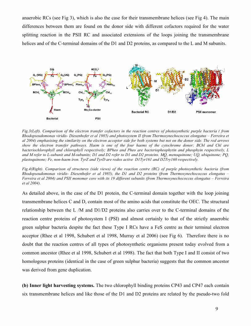
anaerobic RCs (see Fig 3), which is also the case for their transmembrane helices (see Fig 4). The main
differences between them are found on the donor side with different cofactors required for the water
splitting reaction in the PSII RC and associated extensions of the loops joining the transmembrane
helices and of the C-terminal domains of the D1 and D2 proteins, as compared to the L and M subunits.
Fig.3(Left). Comparison of the electron transfer cofactors in the reaction centres of photosynthetic purple bacteria ( from Rhodopseudomonas viridis- Diesenhofer et al 1985) and photosystem II (from Thermosynechococcus elongatus – Ferreira et al 2004) emphasising the similarity on the electron acceptor side for both systems but not on the donor side. The red arrows show the electron transfer pathways. Haem is one of the four haems of the cytochrome donor; BChl and Chl are bacteriochlorophyll and chlorophyll respectively; BPheo and Pheo are bacteriopheophytin and pheophyin respectively, L and M refer to L-subunit and M-subunits; D1 and D2 refer to D1 and D2 proteins. MQ, menoquinone; UQ, ubiquinone; PQ, plastoquinone; Fe, non-haem iron. TyrZ and TyrD are redox active D1Tyr161 and D2Try160 respectively. Fig.4(Right). Comparison of structures (side views) of the reaction centre (RC) of purple photosynthetic bacteria (from Rhodopseudomonas viridis- Diesenhofer et al 1985), the D1 and D2 proteins (from Thermosynechococcus elongatus – Ferreira et al 2004) and PSII monomer core with its 19 different subunits (from Thermosynechococcus elongatus – Ferreira et al 2004). As detailed above, in the case of the D1 protein, the C-terminal domain together with the loop joining
transmembrane helices C and D, contain most of the amino acids that constitute the OEC. The structural
relationship between the L /M and D1/D2 proteins also carries over to the C-terminal domains of the
reaction centre proteins of photosystem I (PSI) and almost certainly to that of the strictly anaerobic
green sulphur bacteria despite the fact these Type I RCs have a FeS centre as their terminal electron
acceptor (Rhee et al 1998, Schubert et al 1998, Murray et al 2006) (see Fig 6). Therefore there is no
doubt that the reaction centres of all types of photosynthetic organisms present today evolved from a
common ancestor (Rhee et al 1998, Schubert et al 1998). The fact that both Type I and II consist of two
homologous proteins (identical in the case of green sulphur bacteria) suggests that the common ancestor
was derived from gene duplication.
(b) Inner light harvesting systems. The two chlorophyll binding proteins CP43 and CP47 each contain
six transmembrane helices and like those of the D1 and D2 proteins are related by the pseudo-two fold

10
axis relating the cofactors involved in primary and secondary electron transfer. Again there is a
remarkable structural similarity between them and the PSI reaction centre proteins, this time at their N-
terminus (Rhee et al 1998, Schubert et al 1998, Murray et al 2006) (see Figs 5 and 6). The most striking
difference being the presence of the large loops joining the lumenal ends of helices V and VI of the PSII
proteins, where some amino acids of the CP43 loop make up a part of the OEC as mentioned above. The
large loop of CP47 also has a conserved region containing bulky side chains including several
phenylalanines which occupy a cavity symmetrically related to the OEC (Ferreira et al 2004).
Fig. 5(Right). Overlay of the structures of (A) carbon backbones of CP43 (yellow), CP47 (brown), PsaA (green) and PsaB (blue) and of (B) the conserved 12 Chls of CP43 (yellow), CP47 (purple), PsaA (green) and PsaB based on the crystal structures of PSII (Ferreira et al 2004) and PSI (Jordan et al 2001).
Fig. 6(Left). Top stromal views derived from X-ray crystallography of (a) PSII (Ferreira et al 2004) and (b) PSI Jordon et al 2001) to emphasise the similarity in the organisation of transmembrane helices of CP43/D1 and CP47/D2 with those of PsaA and PsaB.
As emphasised in Fig 5B, X-ray crystallography (Jordan et al 2001, Ferreira et al 2004, Murray et al
2006) has shown that many of the Chl-binding sites in CP47 and CP43 are conserved in the PSI RC
proteins. Curiously this basic six transmembrane Chl-binding helical protein unit is not found in purple
photosynthetic bacteria but its probable existence in the RC of anaerobic green sulphur bacteria suggests
that it has a long evolutionary origin. Moreover it is the basis of the Pcb-protein of prochlorophytes and
of the iron stressed induced (IsiA) protein of cyanobacteria (Chen et al 2005, Murray et al 2006,) and
clearly is a basic building block found in most photosynthetic organisms.
(c) The extrinsic proteins of the OEC. As far as we know the components of the OEC which are
located in membrane spanning proteins are highly conserved across the complete range of oxygenic
photosynthetic organisms. All the amino acids identified in the D1, D2 and CP43 proteins as being
functionally important for the water splitting reaction are conserved in all genomes sequenced to date.

11
Also the PsbO protein is ubiquitous to PSII in all types of oxygenic photosynthetic organisms. In
contrast the PsbV and PsbU extrinsic proteins of the OEC of cyanobacteria are not found in higher
plants or green algae (red algae contain PsbV). Instead the latter contain the PsbP and PsbQ proteins
which show little or no homology with PsbV and PsbU. However PsbP-like and PsbQ-like proteins
seem to exist in cyanobacteria (Kashino et al 2002) although as yet they have not been present in
isolated PSII complexes used for crystallography.
In principle, these extrinsic OEC proteins should give hints as to the evolutionary origin of the OEC.
However detailed analyses have not revealed any indications to this effect (De Las Rivas et al 2004, De
Las Rivas & Barber 2004). The PsbV protein of cyanobacteria is a low potential cytochrome (c-type)
with no active role in the catalytic activity of the OEC. It therefore seems to be an evolutionary relic of
an electron transport system comparable with the donor side of purple bacterial reaction centres. It is
clear however from crystallography (Ferreira et al 2004, Loll et al 2005) and model building based on
electron microscopy (Morris et al 1997, Nield & Barber 2006) that these extrinsic proteins form a ‘cap’
over the OEC and therefore prevent reductants other than water reacting with the catalytic centre.
Since the PsbO protein is ubiquitous to all types of oxygenic photosynthetic organisms it should be the
best candidate for tracing back the evolutionary origin of the OEC. The main body of the protein is a
β−barrel composed of eight antiparallel β-strands. A very large loop joining β-strands 1 and 2 provide a
head domain which binds to the lumenal surfaces of the D1, D2, CP43 and CP47 proteins. The β-barrel
is not hollow but full of bulky side chains. A search of the data base revealed a few proteins with
structural homology with the β-barrel. Of these the quinohaemoprotein amine dehydrogenase (QHNDH)
(pdb 1jju-A1) being perhaps the most interesting. This bacterial enzyme is an amine-quinone
oxidoreductase in bacteria and has a β-barrel as one of its three subunits. Interestingly it also contains
two Cyt c haems. It is a matter of debate whether PsbO is a relic of this oxidase system.
(e) Cytochrome b559. Cyt b559 is a universal component of PSII. It is characterised by having a high
redox potential (~0.4V) with the ability to protect PSII reaction centres from oxidation by acting as an
electron donor to P•+ when the rate of water oxidation is limiting. The haem is ligated by two co-axial
histidines contained in the apoproteins, PsbE (α-subunit) and PsbF (β-subunit). Despite having an
unusual structure and high redox potential its evolutionary origin is not obvious.
(f) The Mn4Ca-cluster
There are a number of enzymes that contain one or two Mn ions at their catalytic centres but the cluster
of four found in the OEC is unique. Blankenship and Hartman (1998) argued that perhaps the OEC was

12
derived from the duplication of a two Mn-cluster of the type found in catalase which would be expected
to give a symmetrical arrangement something like the dimer-of-dimer model favoured from earlier
EXAFS analyses (see Yachandra 2002). However the Mn3CaO4 cubane organisation with a dangler Mn
attached to it does not fit comfortably with this hypothesis although the idea that PSII evolved from a
hydrogen peroxide oxidase is interesting. Russell and Hall (2001) proposed the OEC may have evolved
from precipitates of manganese oxides similar to rancieite (Mn4CaO9.3H2O) an idea which has been
explored in depth by Sauer and Yachandra (2002).
The presence of five metal ions in a single catalytic centre is also very unusual. However there is one
interesting example, carbon monoxide dehydrogenase found in anaerobic bacteria such as
Carboxdothermus hydrogenoforman (Dobbek et al 2001). This enzyme contains four Fe ions and a Ni
ion with S-bridges. The arrangement of the ions is remarkably like those proposed for the metal centre
of the OEC by Ferreira et al (2004), a Fe3Ni-cubane with the fourth Fe ion linked to the cubane as a
“dangler.” The enzyme catalyses the water shift reaction to generate protons (H+) and electrons (e) from
water
CO + H2O > CO2 + 2H+ + 2e .
The mechanism for the oxygen atom transfer involves CO bound to Ni with the substrate water probably
associated with the “dangler” Fe. The similarities between this reaction and the water oxidation reaction
of PSII and between the geometries of the catalytic centres is interesting but it seems unlikely that there
is a direct evolutionary link. Nevertheless carbon monoxide could have played a major role in
bioenergetics prior to the evolution of the OEC (Sleep & Bird 2007). A plentiful supply of CO could
have come from the photochemically (UV) driven disproportionation reaction between methane and
carbon dioxide.
CH4 + CO2 > CO + H2
6. CONCLUSION
X-ray crystallography together with a wide range of biophysical, biochemical and molecular biological
techniques have provided much detail of the molecular properties of PSII. In so doing we are very close
to revealing the precise chemical mechanism of the water splitting reaction by which photosynthetic
organisms generated the oxygenic atmosphere of our planet. The reaction is powered by light driven
charge separation across the RC of PSII and it is clear that this RC evolved from the same ancestor as
for the RC of PSI, purple photosynthetic bacteria and green sulphur bacteria. Moreover the six
transmembrane helical chlorophyll-binding unit typified by the CP43 and CP47 is also found in PSI and
green sulphur bacteria, again providing evidence of a common evolutionary origin for anaerobic and

13
aerobic photosynthesis. At present, however, there is no obvious indication of the evolutionary origin of
the OEC. One clear message, however, is that based on current knowledge it seems that the catalytic site
of the OEC is fully conserved in all types of oxygenic photoautotrophs. Therefore we can assume that
the reaction giving rise to molecular oxygen in the atmosphere was invented only once and that the
structural characteristics of its catalytic centre and mechanism have been conserved since its conception.
7. ACKNOWLEDGEMENTS
I particularly wish to thank Dr James Murray and Dr Javier De Las Rivas for their contributions to the
analyses of various aspects of PSII structure and are referred to in the paper. I also acknowledge
financial support from the Biotechnology and Biological Research Council (BBSRC).
8. REFERENCES
Barbato, R., Race, H.L., Friso, G. & Barber, J. 1991 Chlorophyll levels in the pigment binding proteins
of PSII: A study based on the chlorophyll to cytochrome ratio in different PSII preparations. FEBS Lett.
286, 86-90.
Barber J 1987 Photosynthetic reaction centres: A common link. Trends Biochem Sci. 12, 321-326
Barber, J. 2002 Photosystem II: a multisubunit membrane protein that oxidises water. Curr. Opinions
Structural Biology 12, 523-530
Barber, J. 2003 Photosystem II: The engine of life. Biophys. Quart. Revs. 36, 71-89.
Barber, J. & Murray, J.W. 2007 The structure of the Mn4Ca2+-cluster of Photosystem II and its protein
environment as revealed by X-ray crystallography. Phil. Trans. R. Soc. B.363.
(doi:10.1098/rsb.2007.2208)
Betley, T.A., Surendranarth, Y., Childress, M.V., Alliger, G.E., G.E., Fu, R., Cummins, C.C. & Nocera,
D.G. 2007 A ligand field chemistry of oxygen generation by the oxygen evolving complex and synthetic
active sites. Phil. Trans. R. Soc. B.363. (doi:10.1098/rsb.2007.2226)
Blankenship, R.E. & Hartman, H. 1998 The origin and evolution of oxygenic photosynthesis. Trends
Biochem. Sci., 23, 94-97.
Brudvig, G.W. 2007 Water oxidation chemistry of photosystem II. Phil. Trans. R. Soc. B.363.
(doi:10.1098/rsb.2007.2217)
Chen, M., Hillier, R.G., Howe, C.J. & Larkum, A.W. 2005 Unique origin and lateral transfer of
prokaryotics chlorophyll-b and chlorophyll-d light-harvesting systems. Mol. Biol. and Evol. 22, 21-28.
De Las Rivas, J., Balsera, M. & Barber, J. 2004 Evolution of oxygenic photosynthesis: genome-wide
analysis of the OEC extrinsic proteins. Trends Plant Sci. 9, 18-25.

14
De Las Rivas, J. & Barber, J. 2004 Analyses of the structure of the PsbO protein and its implications.
Photosyn. Res. 81, 329-343.
Debus, R. J. 2001 Amino acid residues that modulate the properties of tyrosine Y-Z and the manganese
cluster in the water oxidizing complex of photosystem II. Biochim. Biophys. Acta 1503, 164-186.
Deisenhofer, J., Epp, O, Miki, K., Huber, R. & Michel, H. 1985 Structure of the protein subunits in the
photosynthetic reaction centre Rhodopseudomonas viridis at 3.8 Å resolution. Nature 318: 618-624
Diner, B.A.2001 Amino acid residues involved in the coordination and assembly of the manganese
cluster of photosystem II. Proton-coupled electron transport of the redox-active tyrosines and its
relationship to water oxidation. Biochim Biophys Acta 1503, 147-163.
Diner, B.A. & Babcock, G.T. 1996 Structure, dynamics and energy conversion efficiency in
photosystem II. In Oxygenic Photosynthesis. The Light Reactions (Ort, D.R. and Yocum, C.F., eds.), pp.
213-247, Kluwer Academic Pubs., Dordrecht, The Netherlands.
Dobbek, H., Svetlitchnyi, V., Gremer, L., Huber, R. & Meyer, O. 2001 Crystal structure of a carbon
monoxide dehydrogenase reveals a [Ni-4Fe-5S] cluster. Science 293, 1281-1285.
Emsley, P. & Cowtan, K. 2004 Coot: model-building for molecular graphics. Acta Cryst. Sec. D, Biol.
Cryst. 60, 2126-2132.
Faller, P. Pascal, A. & Rutherford, A.W. 2001 β-carotene redox reactions in photosystem II: Electron
Transfer Pathway. Biochemistry 40, 6431-6440.
Ferreira, K.N., T.M. Iverson, K. Maghlaoui, J. Barber, & S. Iwata. 2004 Architecture of the
Photosynthetic Oxygen-Evolving Center. Science 303, 1831-1838.
Joliot, P., Barbieri, G. & Chabaud, R. 1969 Un nouveau modele des centres photochimiques du systeme
II. Photochem. Photobiol. 10, 309-329.
Jordan, P. et al. 2001 Three-dimensional structure of cyanobacterial photosystem I at 2.5 Å resolution.
Nature 411, 909-917
Hankamer, B., Barber, J. & Boekema, E.J. 1997 Structure and membrane organisation of PSII in green
plants. Annu. Rev. Plant Phys. Mol. Biol. 48, 641-671
Hankamer, B., Morris, E.P. & Barber, J. 1999 Cryoelectron microscopy of photosystem two shows that
CP43 and CP47 are located on opposite sides of the D1/D2 reaction centre proteins. Nature Structural
Biology 6, 560-564
Hankamer, B., Morris, E.P., Nield, J., Gerle, C. & Barber, J. (2001) Three-dimensional structure of
photosystem II core dimer of higher plants determined by electron microscopy. J. Struct. Biol. 135, 262-
269.
15
Hasegawa, K., Ono, T.A., Inoue, Y. & Kusunoki, M. 1999 Spin-exchange interactions in the S2-state
manganese tetramer in photosynthetic oxygen-evolving complex deduced from g=2 multiline EPR
signal. Chem. Phys Letts. 300, 9-19.
Hoganson, C.W. & Babcock, G.T. 1997 A metalloradical mechanism for the generation of oxygen from
water in photosynthesis. Science 277, 1953-1956.
Kamiya, N. & Shen J.R. 2003 Crystal structure of oxygen-evolving photosystem II from
Thermosynechococcus vulcanus at 3.7-Å resolution. Proc. Natl. Acad. Sci. USA 100, 98-103.
Kashino, Y., Koike, H., Yoshio, M., Egashira, H., Ikeuchi, M., Pakrasi, H.B. & Satoh, K. 2002 Low-
molecular-mass polypeptide components of a Photosystem II preparation from the thermophilic
cyanobacterium Thermosynechococcus vulcanus. Plant Cell Physiol. 43, 1366-1373
Kashino, Y., Takahashi, T., Inoue-Kashino, N.; Ban, A., Yohei Ikeda, Y., Satoh, K., Sugiura, M. 2007
Ycf12 is a core subunit in the Photosystem II complex. Biochim. Biophys. Acta 1767, 1269-1275
Kok, B. Forbush, B. & McGloin, M. 1970Cooperation of charges in photosynthetic O2 evolution. 1. A
linear four step mechanism. Photochem. Photobiol. 11, 457-475
Loll, B., Kern, J., Saenger, W., Zouni, A. & Biesiadka, J. 2005 Towards complete cofactor arrangement
in the 3.0 Å resolution structure of photosystem II. Nature 438, 1040-1044.
Lundberg, M. & Siegbahn, P.E.M. 2004 Theorectical investigations of the structure and mechanism of
the oxygen-evolving complex in PSII Phys. Chem. Chem. Phys. 6, 4772-4780.
Messinger, J., Badger, M. & Wydrzynski, T. 1995 Detection of one slowly exchanging substrate water
molecule in the S3 State of Photosystem II. Proc. Natl. Acad. Sci. 92, 3209-3213.
Messinger, J. 2004 Evaluation of different mechanistic proposals for water oxidation in photosynthesis
on the basis of Mn4OxCa structures for the catalytic site and spectroscopic data. Phys. Chem. Chem.
Phys. 6, 4764-4771.
McEvoy, J. P. & Brudvig, G. W. 2004 Structure-based mechanism of photosynthetic water oxidation.
Phys. Chem. Chem. Phys. 6, 4754-4763.
McEvoy, J. P. & Brudvig, G. W. 2006 Water-splitting chemistry of photosystem II. Chem. Rev. 106,
4455-4483.
Michel, H. & Deisenhofer, J. 1988 Relevance of the photosynthetic reaction center of purple bacteria to
the structure of photosystem II. Biochemistry 27,1-7
Misra, A., Wernsdorfer, W., Abboud, K.A. & Christou, G. 2005 The first high oxidation state
manganese-calcium cluster: Relevance to the water oxidizing complex of photosynthesis. Chem.
Commun.(Camb.) 54-56.

16
Miyashita, H., Ikemoto, H., Kurano, N., Adachi, K., Chihara, M. & Miyachi, S. 1996 A niche for
cyanobacteria containing chlorophyll d. Nature 383, 402.
Morris, E.P., Hankamer, B., Zheleva, D., Friso, G. & Barber, J. 1997 The 3-D structure of a
photosystem II core complex determined by electron crystallography. Structure 5, 837-849
Murray J.W. & Barber, J 2006 Identification of a calcium-binding site in the PsbO protein of
photosystem II. Biochemistry. 45, 4128-4130.
Murray, J.W. & Barber, J 2007 Structural characteristics of channels and pathways in Photosystem II
including the identification of an oxygen channel. J.Struct. Biol. 159, 228-237.
Murray, J.W., Duncan, J. & Barber, J. 2006 CP43-like chlorophyll binding proteins: structural and
evolutionary implications. Trends Plant Sci. 11, 152-158.
Neild, J & Barber, J. 2006 Refinement of the structural model of the Photosystem II supercomplex of
higher plants. Biochim.Biophys.Acta 1757, 353-361.
Nield, J., Orlova, E., Morris, E., Gowen, B., van Heel, M. & Barber, J. 2000a 3D map of the plant
photosystem two supercomplex obtained by cryoelectron microscopy and single particle analysis.
Nature Structural Biology 7, 44-47
Nield, J., Kruse, O., Ruprecht, J., Da Fonseca, P, Büchel, C. & Barber, J. 2000b 3D structure of
Chlamydomonas reinhardtii and Synechococcus elongatus photosystem II complexes allow for
comparison of their OEC organisation. J. Biol. Chem. 275, 27940-27946
Nield, J., Balsera, M., De Las Rivas, J. & Barber, J. 2002 3D cryo-EM study of the extrinsic domains of
the oxygen evolving complex of spinach. Assignment of the PsbO protein. J. Biol. Chem. 277, 15006-
15012.
Pecoraro, V. L., Baldwin, M. J., Caudle, M. T., Hsieh, W.-Y. & Law, N. A. 1998 A proposal for the
water oxidation in Photosystem II. Pure and Appl. Chem. 70, 925-929.
Peloquin, J.M., Campbell, K.A., Randall, D.W., Evanchik, M.A., Pecoraro, V.L., Armstrong, W.H. &
Britt, R.D. 2000. 55Mn ENDOR of the S2-state multiline EPR signal of Photosystem II: Implications on
the structure of the tetranuclear Mn cluster. J. Amer. Chem. Soc. 122, 10926-10942.
Rhee, K-H., Morris, E.P, Zheleva, D., Hankamer, B., Kühlbrandt, W. & Barber, J. (1997) Two-
dimensional structure of plant photosystem II at 8Å resolution. Nature 389, 522-526
Rhee, K.-H., Morris, E.P., Barber, J. & Kühlbrandt, W. (1998) Three-dimensional structure of the
photosystem II reaction centre at 8Å resolution. Nature 396, 283-286.

17
Russell, M.J. & Hall, A.J. 2001The onset of life and the dawn of oxygenic photosynthesis: Respective
roles of cubane core structures [Fe4S4] and transient [Mn4O4]4+[OCaO]2. Sixth International Congress
on Carbon Dioxide Utilization, Sept. 9-14, Breckenridge, Colorado, abstr. p. 49
Sauer, K. & Yachandra, V.K. 2002 A possible evolutionary origin for the Mn4 cluster of the
photosynthetic water oxidation complex from natural MnO2 precipitates in the early oceans.
Proc. Nat. Acad. Sci. USA 99, 8631-8636.
Schubert, W-D., Klukas, O., Saenger, W., Witt, H.T., Fromme, P. & Krausz, N. 1998 A common
ancestor for oxygenic and anoxygenic photosynthetic systems: a comparison based on the structural
model of Photosystem I. J. Mol. Biol. 280, 297-341
Siegbahn, P.E.M. & Lundberg, M. 2005 The mechanism for dioxygen formation in PSII studied by
quantum chemical methods. Photochem. Photobiol. Sci. 4, 1035-1043.
Siegbahn, P.E.M. 2006 O-O bond formation in the S4-state of the oxygen evolving complex in
Photosystem II. Chemistry-A European Journal 12, 9217-9237.
Siegbahn, P.E.M. 2007 Mechanism and energy diagram for O-O bond formation in the oxygen-evolving
complex in Photosystem II. Phil. Trans. R. Soc. B.363. (doi:10.1098/rsb.2007.2218)
Sleep, N.H. & Bird, D.K. 2007 Niches of the pre-photosynthetic biosphere and geologic preservation of
Earth’s earliest ecology. Geobiology. 5, 101-117.
Sproviero, E. M., Gascon, J. A., McEvoy, J. P., Brudvig, G. W. & Batista, V. S. 2006 QM/MM models
of the O2-evolving complex of photosystem II. J. Chem. Theory Comput. 2, 1119-1134.
Sproviero, E. M., Gascon, J. A., McEvoy, J. P., Brudvig, G. W. & Batista, V. S. 2007a Quantum
mechanics/molecular mechanics structural models of the oxygen-evolving complex of photosystem II.
Curr. Opin. Struct. Biol. 17, 173-180
Sproviero, E. M., Shinopolous, K., Gascon, J. A., McEvoy, J. P., Brudvig, G. W. & Batista, V.S. 2007b
QM/MM computational studies of substrate water binding to the oxygen-evolving complex of
Photosystem II. Phil. Trans. R. Soc. B.363. (doi:10.1098/rsb.2007.2210).
Stewart, D.H. & Brudvig, G.W. 1998 Cytochrome b559 of Photosystem II. Biochim. Biophys. Acta
1367, 63-87.
Telfer, A 2002 What is β-carotene doing in the Photosystem two reaction centre? Phil. Trans. R. Soc. B
357, 1431-1440
Tracewell, C.A. & Brudvig, G.W. 2003 Two redox active β-carotene molecules in Photosystem II.
Biochemistry 42, 9127-9136
Williams, J.C., Steiner, L.A. & Feher, G. 1986 Primary structures of the reaction center from
Rhodopseudomonas spheroids. Proteins 1, 312-325

18
Wydrzynski, T.J. and Satoh, K. 2005 Photosystem II. The Light-Driven Water:Plastoquinone
Oxidoreductase, Vol. 22 Advances in Photosynthesis and Respiration pp1-786. Pub. Springer,
Dordrecht, The Netherlands
Yano, J., Kern, J., Irrgang, K.-D., Latimer, M.J., Bergmann, U., Glatzel, P., Pushkar, Y., Biesiadka,
J., Loll, B., Sauer, K., Messinger, J., Zouni, A. & Yachandra, V.K. 2005 X-ray damage to the Mn4Ca
complex in photosystem II crystals : A case study for metallo-protein X-ray crystallography. Proc. Natl.
Acad. Sci. USA, 102 (2005) 12047-12052
Yachandra V K (2002) Structure of the Mn complex in Photosystem II: Insights from X-ray
spectroscopy. Phil Trans Roy Soc. B 357, 1347-1358
Yano, J., Kern, J., Sauer, K., Latimer, M.J., Pushkar, Y., Biesiadka, J., Loll, B., Saenger, W.,
Messinger, J., Zouni, A. & Yachandra, V.K., 2006 Where water is oxidised to dioxygen : Structure of
the photosynthetic Mn4Ca cluster. Science 314 , 821-825.
Youvan, D.C., Bylina, E.J., Alberti, M., Begusch, H. & Hearst, J.E. 1984 Nucleotide and deduced
polypeptide sequences of the photosynthetic reaction center, B870 antennae and flanking polypeptides
from Rps. capsulate. Cell 37, 949-957.
Zouni, A., Witt, H. T., Kern, J., Fromme, P., Krauss, N., Saenger, W. & Orth, P. 2001 Crystal structure
of photosystem II from Synechococcus elongatus at 3.8 Å resolution. Nature 409, 739-743.