Photochemical Restoration of Visual Responses in … club materials/CBI JC_Eron_McCarron.pdf ·...
12
Neuron Article Photochemical Restoration of Visual Responses in Blind Mice Aleksandra Polosukhina, 1 Jeffrey Litt, 2,6 Ivan Tochitsky, 2,6 Joseph Nemargut, 3 Yivgeny Sychev, 3 Ivan De Kouchkovsky, 2 Tracy Huang, 1 Katharine Borges, 2 Dirk Trauner, 5 Russell N. Van Gelder, 3,4 and Richard H. Kramer 1,2, * 1 Vision Science Graduate Group 2 Department of Molecular and Cell Biology University of California, Berkeley, Berkeley, CA 94720, USA 3 Department of Ophthalmology 4 Department of Biological Structure University of Washington, Seattle, WA 98195, USA 5 Department of Chemistry and Biochemistry, University of Munich, D-81377 Munich, Germany 6 These authors contributed equally to this work *Correspondence: [email protected] http://dx.doi.org/10.1016/j.neuron.2012.05.022 SUMMARY Retinitis pigmentosa (RP) and age-related macular degeneration (AMD) are degenerative blinding diseases caused by the death of rods and cones, leaving the remainder of the visual system intact but largely unable to respond to light. Here, we show that AAQ, a synthetic small molecule photo- switch, can restore light sensitivity to the retina and behavioral responses in vivo in mouse models of RP, without exogenous gene delivery. Brief applica- tion of AAQ bestows prolonged light sensitivity on multiple types of retinal neurons, resulting in synapti- cally amplified responses and center-surround antagonism in arrays of retinal ganglion cells (RGCs). Intraocular injection of AAQ restores the pupillary light reflex and locomotory light avoidance behavior in mice lacking retinal photoreceptors, indi- cating reconstitution of light signaling to brain circuits. AAQ and related photoswitch molecules present a potential drug strategy for restoring retinal function in degenerative blinding diseases. INTRODUCTION Inherited degenerative diseases of the retina including retinitis pigmentosa (RP) affect 1 in 3,000 people worldwide. As differen- tiation of rods and cones ceases soon after birth in mammals, disorders resulting in photoreceptor degeneration lead to a permanent visual deficit. At present, there is no effective treat- ment for preventing this degenerative process and without some means of restoring photoreception, patients with advanced RP face the prospect of irreversible blindness. Retinal ganglion cells (RGCs) are the sole output neurons of the retina. Hence, all of the visual information that reaches the brain is encoded by the spatial and temporal pattern of RGC action potentials. Several strategies have been advanced to enable light to alter RGC firing in the absence of rods and cones, with the goal of restoring visual function after the photoreceptors are lost (Jime ´ nez et al., 1996; Marc et al., 2003; Punzo and Cepko, 2007; Strettoi and Pignatelli, 2000). First, biomedical engineers have developed surgically implanted retinal ‘‘chip’’ prosthetics (Chader et al., 2009; Gerding et al., 2007; Shire et al., 2009) that can be electronically controlled by an external camera to enable optical stimuli to trigger RGC firing. Retinal implants have restored simple shape discrimination to blind patients (Humayun et al., 2003; Yanai et al., 2007), indicating that artificial stimulation of RGCs in vivo can create a useful visual experience. Second, genes encoding optogenetic tools, including light-activated ion channels (Bi et al., 2006; Lagali et al., 2008; Tomita et al., 2010), transporters (Busskamp et al., 2010), or receptors (Caporale et al., 2011; Lin et al., 2008), can be introduced with viruses to bestow light-sensitivity on retinal neurons that survive after the natural photoreceptive cells have degenerated. Expression of optogenetic proteins in RGCs (Caporale et al., 2011; Tomita et al., 2010), bipolar cells (Lagali et al., 2008), and remnant cones (Busskamp et al., 2010) can reinstate light-elicited behavioral responses in mouse models of RP. Third, embryonic stem cells can be differentiated into photoreceptor progenitors in vitro (Lamba et al., 2006). Injecting these progenitors into blind animals results in integration of photoreceptors in the retina and restoration of some electrical activity in response to light (Lamba et al., 2009). Each of these strategies has shown promise for restoring visual function, but they all require highly invasive and/or irre- versible interventions that introduce hurdles to further develop- ment as a therapeutic approach. Implantation of retinal chips or stem cell-derived photoreceptors requires invasive surgery, while exogenous expression of optogenetic tools leads to permanent genetic alterations in retinal neurons. Retinal chip prosthetics rely on extracellular electrical stimulation of RGCs, which can be cytotoxic when excessive (Winter et al., 2007). Stem cell therapies carry potential for teratoma formation (Chaudhry et al., 2009). Viruses that deliver optogenetic tools can have off-target effects and may elicit inflammatory responses (Beltran et al., 2010). While the potential permanence Neuron 75, 271–282, July 26, 2012 ª2012 Elsevier Inc. 271
Transcript of Photochemical Restoration of Visual Responses in … club materials/CBI JC_Eron_McCarron.pdf ·...

Neuron
Article
Photochemical Restorationof Visual Responses in Blind MiceAleksandra Polosukhina,1 Jeffrey Litt,2,6 Ivan Tochitsky,2,6 Joseph Nemargut,3 Yivgeny Sychev,3 Ivan De Kouchkovsky,2
Tracy Huang,1 Katharine Borges,2 Dirk Trauner,5 Russell N. Van Gelder,3,4 and Richard H. Kramer1,2,*1Vision Science Graduate Group2Department of Molecular and Cell BiologyUniversity of California, Berkeley, Berkeley, CA 94720, USA3Department of Ophthalmology4Department of Biological StructureUniversity of Washington, Seattle, WA 98195, USA5Department of Chemistry and Biochemistry, University of Munich, D-81377 Munich, Germany6These authors contributed equally to this work*Correspondence: [email protected]://dx.doi.org/10.1016/j.neuron.2012.05.022
SUMMARY
Retinitis pigmentosa (RP) and age-related maculardegeneration (AMD) are degenerative blindingdiseases caused by the death of rods and cones,leaving the remainder of the visual system intactbut largely unable to respond to light. Here, weshow that AAQ, a synthetic small molecule photo-switch, can restore light sensitivity to the retina andbehavioral responses in vivo in mouse models ofRP, without exogenous gene delivery. Brief applica-tion of AAQ bestows prolonged light sensitivity onmultiple types of retinal neurons, resulting in synapti-cally amplified responses and center-surroundantagonism in arrays of retinal ganglion cells(RGCs). Intraocular injection of AAQ restores thepupillary light reflex and locomotory light avoidancebehavior in mice lacking retinal photoreceptors, indi-cating reconstitution of light signaling to braincircuits. AAQ and related photoswitch moleculespresent a potential drug strategy for restoring retinalfunction in degenerative blinding diseases.
INTRODUCTION
Inherited degenerative diseases of the retina including retinitispigmentosa (RP) affect 1 in 3,000 people worldwide. As differen-tiation of rods and cones ceases soon after birth in mammals,disorders resulting in photoreceptor degeneration lead toa permanent visual deficit. At present, there is no effective treat-ment for preventing this degenerative process and without somemeans of restoring photoreception, patients with advanced RPface the prospect of irreversible blindness.Retinal ganglion cells (RGCs) are the sole output neurons of
the retina. Hence, all of the visual information that reaches thebrain is encoded by the spatial and temporal pattern of RGCaction potentials. Several strategies have been advanced to
enable light to alter RGC firing in the absence of rods and cones,with the goal of restoring visual function after the photoreceptorsare lost (Jimenez et al., 1996; Marc et al., 2003; Punzo andCepko, 2007; Strettoi and Pignatelli, 2000). First, biomedicalengineers have developed surgically implanted retinal ‘‘chip’’prosthetics (Chader et al., 2009; Gerding et al., 2007; Shireet al., 2009) that can be electronically controlled by an externalcamera to enable optical stimuli to trigger RGC firing. Retinalimplants have restored simple shape discrimination to blindpatients (Humayun et al., 2003; Yanai et al., 2007), indicatingthat artificial stimulation of RGCs in vivo can create a usefulvisual experience. Second, genes encoding optogenetic tools,including light-activated ion channels (Bi et al., 2006; Lagaliet al., 2008; Tomita et al., 2010), transporters (Busskamp et al.,2010), or receptors (Caporale et al., 2011; Lin et al., 2008), canbe introduced with viruses to bestow light-sensitivity on retinalneurons that survive after the natural photoreceptive cells havedegenerated. Expression of optogenetic proteins in RGCs(Caporale et al., 2011; Tomita et al., 2010), bipolar cells (Lagaliet al., 2008), and remnant cones (Busskamp et al., 2010) canreinstate light-elicited behavioral responses in mouse modelsof RP. Third, embryonic stem cells can be differentiated intophotoreceptor progenitors in vitro (Lamba et al., 2006). Injectingthese progenitors into blind animals results in integration ofphotoreceptors in the retina and restoration of some electricalactivity in response to light (Lamba et al., 2009).Each of these strategies has shown promise for restoring
visual function, but they all require highly invasive and/or irre-versible interventions that introduce hurdles to further develop-ment as a therapeutic approach. Implantation of retinal chipsor stem cell-derived photoreceptors requires invasive surgery,while exogenous expression of optogenetic tools leads topermanent genetic alterations in retinal neurons. Retinal chipprosthetics rely on extracellular electrical stimulation of RGCs,which can be cytotoxic when excessive (Winter et al., 2007).Stem cell therapies carry potential for teratoma formation(Chaudhry et al., 2009). Viruses that deliver optogenetic toolscan have off-target effects and may elicit inflammatoryresponses (Beltran et al., 2010). While the potential permanence
Neuron 75, 271–282, July 26, 2012 ª2012 Elsevier Inc. 271

of optoelectronic, stem cell, or optogenetic interventions couldbe favorable in the absence of complications, any deleteriouseffects of these treatments could be very difficult or impossibleto reverse.
Here, we report an alternative strategy for restoring visualfunction, based on a small molecule ‘‘photoswitch’’ that bestowslight sensitivity onto neurons without requiring exogenous geneexpression. The photoswitch is injected into the vitreous cavityof the eye, but unlike the other strategies, it does not requirehighly invasive surgical interventions and its actions are revers-ible. We used acrylamide-azobenzene-quaternary ammonium(AAQ), a K+ channel photoswitch that enables optical control ofneuronal excitability (Banghart et al., 2009; Fortin et al., 2008).AAQ was originally thought to conjugate to K+ channels (Fortinet al., 2008), but recent work shows that the molecule interactsnoncovalently with the cytoplasmic side of the channels, similarto the mechanism of action of local anesthetics (Banghart et al.,2009). The trans form of AAQ blocks K+ channels and increasesexcitability, whereas photoisomerization to the cis form withshort wavelength light (e.g., 380 nm) unblocks K+ channels anddecreases excitability. Relaxation from cis to trans occurs slowlyin darkness but much more rapidly in longer-wavelength light(e.g., 500 nm), enabling rapid bi-directional photocontrol ofneuronal firing with different wavelengths.
We show that AAQ confers robust light responses in RGCs inretinas from mutant mice that lack rods and cones. Moreover,after a single intraocular injection, AAQ restores light-drivenbehavior in blind mice in vivo. Because it is a rapid and reversibledrug-like small molecule, AAQ represents a class of compoundsthat has potential for the restoration of visual function in humanswith end-stage photoreceptor degenerative disease.
RESULTS
Imparting Light Sensitivity on rd1 Mouse Retinawith AAQWe tested whether AAQ can impart light sensitivity on retinasfrom 6-month-old rd1mice, a murine model of RP. The homozy-gous rd1 mouse (rd1/rd1) has a mutation in the gene encodingthe b-subunit of cGMP phosphodiesterase-6, essential for rodphototransduction. Rods and cones in these mice degeneratenearly completely within 3 months after birth, leading to a lossof electrical and behavioral light responses (Sancho-Pelluzet al., 2008). We placed the rd1 mouse retina onto a multi-elec-trode array (MEA) that enables simultaneous extracellularrecording from many RGCs (Meister et al., 1994). Before AAQapplication, light generated nomeasurable change in RGC firing.However, after 30 min of treatment with AAQ, nearly all RGCsresponded to light (Figure 1A). Photosensitization increasedwith AAQ concentration (Figure S1; Table S1 available online),but we used 300 mM for our standard ex vivo treatment. Lightresponses slowly diminished but were still robust for >5 hr afterremoving AAQ from the bathing medium (Figure S2a). Lightresponses could also be detected in three of four recordingsfrom retinas removed from rd1 mice that had received in vivointravitreal AAQ injections 12 hr previously (Figure S2b). Thedegree of photosensitivity varied, reflecting inaccurate injectionin the small intravitreal volume of the mouse eye (2–3 ml).
Most RGCs exhibited an increase in firing rate in response to380 nm light and a decrease in 500 nm light, opposite to AAQ-mediated light responses in neurons in culture (Fortin et al.,2008). To quantify the effects of light, we calculated a photoswitchindex (PI), representing the normalized change in firing rate uponswitching from darkness to 380 nm light. Positive or negative PIvalues reflect an increase or decrease, respectively, of firing.Before AAQ treatment, RGCs had almost no light response(median PI = 0.02); but after treatment, nearly all were activatedby 380 nm light (median PI = 0.42) (Figure 1B). The rare lightresponses before AAQ treatment might result from melanopsin-containing intrinsically photosensitive RGCs (ipRGCs), whichaccount for !3% of the RGCs in the adult mouse retina (Hattaret al., 2002). Significant photosensitization was observed in eachof 21 AAQ-treated retinas. On average, we observed an !3-foldincrease in RGC firing rate in response to 380 nm light, with indi-vidual retinas showing up to an 8-fold increase (Figure 1C).
AAQ Acts on RGCs, Bipolar, and Amacrine Cellsin rd1 RetinasWe were surprised that 380 nm light stimulated RGC firingbecause this wavelength unblocks K+ channels, which shouldreduce neuronal excitability. However, since RGCs receiveinhibitory input from amacrine cells, RGC stimulation might beindirect, resulting from amacrine cell-dependent disinhibition.To test this hypothesis, we applied antagonists of receptors forGABA and glycine, the two inhibitory neurotransmitters releasedby amacrine cells. Photosensitization of RGCs by AAQ persistedafter adding inhibitors of GABAA, GABAC, and glycine receptors(Figure 2A), but the polarity of photoswitching was reversed, withnearly all neurons inhibited rather than activated by 380 nmlight (Figure 2B). These results indicate that photoregulation ofamacrine cells is the dominant factor that governs the AAQ-mediated light response of RGCs.After blocking amacrine cell synaptic transmission, the re-
maining light response could result from photoregulation of K+
channels intrinsic to RGCs and/or photoregulation of excitatoryinputs from bipolar cells. To explore the contribution of intrinsicK+ channels, we obtained whole-cell patch clamp recordingsfrom RGCs and pharmacologically blocked nearly all synapticinputs (glutamatergic, GABAergic, and glycinergic). Depolarizingvoltage steps activated outward K+ currents that were smallerand decayed more rapidly in 500 nm light than in 380 nm light(Figure 2C). Comparison of current versus voltage (I-V) curvesshows that the current was reduced by !50% in 500 nm light(Figure 2D), similar to previous results (Fortin et al., 2008).However, MEA recordings indicate that photoregulation ofRGC firing was nearly eliminated by blocking all excitatory andinhibitory synaptic inputs (Figure S3), suggesting that the lightresponse is driven primarily by photoregulation of upstreamneurons synapsing with RGCs.To examine directly the contribution of retinal bipolar cells to
the RGC light response, we blocked RGCK+ channels with intra-cellular Cs+ and added GABA and glycine receptor antagoniststo block amacrine cell inputs. Flashes of 500 nm light triggeredexcitatory postsynaptic currents (EPSCs) in RGCs, and 380 nmlight suppressed these events (Figures 2E and 2F). Blockingglutamate receptors eliminated these events, and bipolar cells
Neuron
Restoration of Visual Responses in Blind Mice
272 Neuron 75, 271–282, July 26, 2012 ª2012 Elsevier Inc.
provide the only known glutamatergic input to RGCs. Hence, weconclude that inputs from amacrine cells, bipolar cells, and toa lesser extent, the intrinsic K+ conductances of RGCs, allcombine to shape and amplify the AAQ-mediated RGC lightresponse.
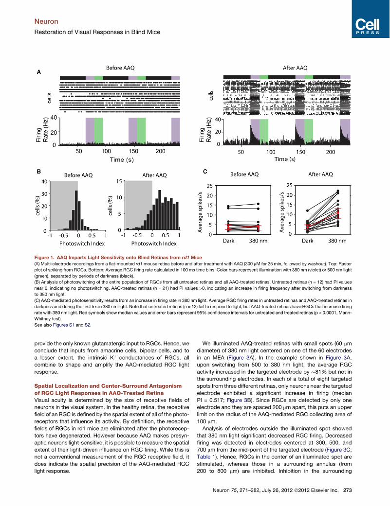
Spatial Localization and Center-Surround Antagonismof RGC Light Responses in AAQ-Treated RetinaVisual acuity is determined by the size of receptive fields ofneurons in the visual system. In the healthy retina, the receptivefield of an RGC is defined by the spatial extent of all of the photo-receptors that influence its activity. By definition, the receptivefields of RGCs in rd1 mice are eliminated after the photorecep-tors have degenerated. However because AAQ makes presyn-aptic neurons light-sensitive, it is possible to measure the spatialextent of their light-driven influence on RGC firing. While this isnot a conventional measurement of the RGC receptive field, itdoes indicate the spatial precision of the AAQ-mediated RGClight response.
We illuminated AAQ-treated retinas with small spots (60 mmdiameter) of 380 nm light centered on one of the 60 electrodesin an MEA (Figure 3A). In the example shown in Figure 3A,upon switching from 500 to 380 nm light, the average RGCactivity increased in the targeted electrode by !81% but not inthe surrounding electrodes. In each of a total of eight targetedspots from three different retinas, only neurons near the targetedelectrode exhibited a significant increase in firing (medianPI = 0.517; Figure 3B). Since RGCs are detected by only oneelectrode and they are spaced 200 mm apart, this puts an upperlimit on the radius of the AAQ-mediated RGC collecting area of100 mm.Analysis of electrodes outside the illuminated spot showed
that 380 nm light significant decreased RGC firing. Decreasedfiring was detected in electrodes centered at 300, 500, and700 mm from the mid-point of the targeted electrode (Figure 3C;Table 1). Hence, RGCs in the center of an illuminated spot arestimulated, whereas those in a surrounding annulus (from200 to 800 mm) are inhibited. Inhibition in the surrounding
Figure 1. AAQ Imparts Light Sensitivity onto Blind Retinas from rd1 Mice(A) Multi-electrode recordings from a flat-mounted rd1mouse retina before and after treatment with AAQ (300 mM for 25 min, followed by washout). Top: Raster
plot of spiking from RGCs. Bottom: Average RGC firing rate calculated in 100 ms time bins. Color bars represent illumination with 380 nm (violet) or 500 nm light
(green), separated by periods of darkness (black).
(B) Analysis of photoswitching of the entire population of RGCs from all untreated retinas and all AAQ-treated retinas. Untreated retinas (n = 12) had PI values
near 0, indicating no photoswitching, AAQ-treated retinas (n = 21) had PI values >0, indicating an increase in firing frequency after switching from darkness
to 380 nm light.
(C) AAQ-mediated photosensitivity results from an increase in firing rate in 380 nm light. Average RGC firing rates in untreated retinas and AAQ-treated retinas in
darkness and during the first 5 s in 380 nm light. Note that untreated retinas (n = 12) fail to respond to light, but AAQ-treated retinas have RGCs that increase firing
rate with 380 nm light. Red symbols show median values and error bars represent 95% confidence intervals for untreated and treated retinas (p < 0.0001, Mann-
Whitney test).
See also Figures S1 and S2.
Neuron
Restoration of Visual Responses in Blind Mice
Neuron 75, 271–282, July 26, 2012 ª2012 Elsevier Inc. 273

RGCs implies that a sign-inverting synapse from a laterally-pro-jecting neuron is involved in transmitting information from thecenter illuminated area to the surround. Amacrine cells areknown to form a mutually inhibitory network, making them thelikely source of the inhibitory signal.
Spectral Requirements of AAQ-Mediated LightResponsesWe determined the optimal wavelength for turning off RGC firingwhen the AAQ photoswitch is driven from the cis to the transconfiguration. First, a conditioning 380 nm stimulus was usedto turn on firing and then we measured suppression of firing inresponse to test flashes of different wavelengths. We foundthat 500 nm light is best at suppressing activity (Figure 4A), asexpected from previous results (Fortin et al., 2008). To determinewhich wavelengths are best at triggering firing when AAQ photo-isomerizes from trans to cis, we again applied test flashes ofdifferent wavelengths, but to ensure that the photoswitch startedmaximally in the trans configuration, the stimulation protocolbegan with a reset flash of 500 nm light followed by a period ofdarkness. We found that the optimal wavelength for stimulatingfiring was 380 nm under these conditions. However, robust firingcould also be activated with 420 or 460 nm light (Figure 4B), andeven 500 nm light could trigger an increase in firing frequency if
the preceding dark interval was sufficiently long. The historydependence of photoswitching is a consequence of the initialratio of the cis and trans photoisomers. Starting with all mole-cules in the trans state, even 500 nm light can increase the frac-tion of cismolecules. Hence, UV light is not essential for elicitingretinal responses. We also found that broad spectrum white lightcan trigger an increase in firing frequency in RGCs (Figures 4Cand 4D).We measured the absolute light intensity required to photore-
gulate AAQ-treated retinas from rd1 mice. The threshold inten-sity required to induce RGC firing was 2.6 3 1015 photons/cm2/s of 380 nm light (Figure 4E). The RGC firing rate increasedprogressively with brighter light, up to 1017 photons/cm 2/s, buteven this intensity did not saturate the response. By comparison,retinas from rd1 mice expressing ChR2 in bipolar cells (Lagaliet al., 2008) have RGCs that exhibit a firing threshold of 6 31015 photons/cm2/s.
Restoring Behavioral Light Responses In Vivo with AAQGiven that AAQ can bestow photosensitivity onto blind retinasex vivo, we asked whether it can confer light-induced behaviorin blind mice in vivo. Although rd1 mice lose all morphologicallyrecognizable rods and cones, a small fraction of cones withaltered morphology can survive, allowing correct performance
Figure 2. Multiple Types of Retinal NeuronsContribute to the AAQ-Mediated LightResponse of RGCs(A) Amacrine cell-mediated synaptic inhibition
dominates the RGC light response. MEA recording
with antagonists of GABAA (gabazine; 4 mM),
GABAC (TPMPA; 10 mM), and glycine receptors
(strychnine; 10 mM) is shown. Top: Raster plot of
RGC spiking. Bottom: Average RGC firing rate.
(B) After blocking inhibition, PI values show
a decrease in firing frequency upon switching from
darkness to 380 nm light (n = 11 retinas).
(C) Endogenous K+ channels contribute to the
RGC light response. Whole-cell patch clamp
recording from an RGC. Currents were evoked by
voltage steps from "80 to +40mV in 20mV incre-
ments in 380 nm and 500 nm light. Inhibitory
GABAergic and glycinergic inputs were blocked as
in (A), and excitatory glutamatergic inputs were
blocked with DNQX (10 mM) and AP5 (50 mM).
(D) Photoregulation of endogenous K+ channels
evaluated in steady-state I-V curves obtained in
380 and 500 nm light (n = 5 RGCs). Current is
normalized to themaximal value at +40mV (380 nm
light). Variability among data is expressed as
mean ± SEM.
(E) Bipolar cell-mediated synaptic excitation also
contributes to the RGC light response. Whole-cell
patch clamp recording from an RGC. Blockade of
inhibitory synaptic inputs (as in A) and endogenous
RGC K+ channels (as in C) reveals photoregulation
of EPSC rate. Note the disappearance of EPSCs
after perfusion with glutamate receptor antago-
nists DNQX (10 mM) and AP5 (50 mM). Holding
potential = "60mV.
(F) Average EPSC rate in 380 nm and 500 nm light. Note the significant increase in EPSC rate in 500 nm light (p < 0.05, Mann-Whitney test; n = 9 cells). Red
symbols show median values and error bars represent 95% confidence intervals.
See also Figure S3.
Neuron
Restoration of Visual Responses in Blind Mice
274 Neuron 75, 271–282, July 26, 2012 ª2012 Elsevier Inc.
of a visual discrimination task under some illumination conditions(Thyagarajan et al., 2010). Rd1 mice also exhibit a pupillary lightreflex (PLR), but this behavior is completely absent from rd1micelacking melanopsin, the photopigment found in the smallpercentage (!3%) of RGCs that are intrinsically photosensitive(ipRGCs) (Hattar et al., 2002; Panda et al., 2003). Therefore, wetested the PLR of adult rd1 mice lacking the melanopsin gene(opn4"/" rd1/rd1) (Panda et al., 2003). After 3 months of age,no PLR could be elicited in any of the mice that we tested,even with the brightest light available (Figure 5A). However, ina subset of these mice (9 out of 25), intravitreal injection ofAAQ resulted in a substantial PLR, with a maximal pupillaryconstriction of!65% as large as wild-type. Control experimentsshowed no restoration of the PLR following sham injection ofvehicle alone (n = 4; Figure S4). The AAQ-mediated responsewas attributable to the retina, as direct application of AAQ tothe isolated iris in vitro did not produce light-elicited constriction.In the remaining mice, suboptimal intravitreal placement orleakage resulting from puncture damage may have reducedhow much AAQ reached the retina, precluding effectivephotosensitization.
The AAQ-mediated PLR in opn4"/" rd1/rd1mice could be trig-gered by photopic irradiance levels normally encountered duringdaytime, but the PLR threshold was 2 to 3 log units higher thanthe normal PLR in wild-typemice (Figure 5B). The AAQ-mediatedPLR was slower than in wild-type mice (see Movie S1), and AAQinduced some basal pupillary constriction in darkness. Nonethe-less, these results show that light responses in AAQ-treatedretina can drive brain circuits, leading to a behavioral responsethat is absent from untreated blind animals.We next tested whether locomotory light-avoidance behavior
(Johnson et al., 2010; Kandel et al., 1987) could be restored inblind opn4"/" rd1/rd1 mice treated with a unilateral intravitrealinjection of AAQ. We placed a mouse into a narrow cylindricaltransparent tube and recorded behavior with an infrared videocamera (Figure 6A). An automated image analysis system wasused to detect the mouse and measure how quickly it movedaway from the illuminated end of the tube, toward the center.The latency to movement was significantly shorter in light thanin darkness in wild-type mice (n = 13, 26 trials, p < 0.01) butnot in opn4"/" rd1/rd1 mice (n = 7, 14 trials), indicating lightavoidance in the wild-type mice but not in the mutant mice.AAQ reinstated the light versus dark latency difference,measured 2 hr after injection (n = 7, 14 trials, p < 0.02), indicatingrestoration of light avoidance. At 24 hr after AAQ injection, therewas no difference in latency in light versus darkness, consistentwith dissipation of the AAQ. These results indicate that an activelight-avoidance behavior can be elicited by AAQ followinga single injection into the eye.Wild-type mice exhibit a decrease in open-field locomotion in
response to light, which corresponds to a decrease in explor-atory drive (Bourin and Hascoet, 2003). In contrast, rd1 mice
Figure 3. The AAQ-Treated Retina Gener-ates Spatially Precise Light Responses(A) Targeted illumination of a portion of the retina
centered on a single MEA electrode (top). The
target (electrode E6) was exposed to 3 s flashes of
alternating 380 and 500 nm light. Spot size =
60 mm in radius, inter-electrode spacing = 200 mm.
Only the targeted electrode records an increase in
RGC firing in response to 380 nm light (bottom).
PI values are color-coded (scale at left) and also
represented by bar height. The red bar is electrode
E6 (PI = 0.812; n = 1 cell), and blue electrodes are
the surround (PI = "0.209; n = 56 cells). Empty
squares are electrodes on which no action
potentials were recorded.
(B) Targeted illumination results from three retinas,
displayed in a box plot. PI values for the target and
the surround RGCs are significantly different from
one another (p < 0.005, Mann-Whitney test).
Whiskers denote 1.5 times the interquartile range
from the 25th and 75th percentile.
(C) Targeted illumination elicits opposite
responses in center and surround RGCs (n = 11
cells and n = 385 cells, respectively, from three
retinas). PI values of RGCs (open circles) as
a function of distance from the target electrode,
displayed in 200 mm bins. The red diamonds indi-
cate the median plus or minus the bootstrapped
95% confidence intervals. See Table 1 for values.
Table 1. Center and Surround RGC Responses under TargetedIllumination
Distance (um) No. of Cells Median PI 95% Confidence Interval
Target 11 0.517 0.455 to 0.812
200–400 95 "0.165 "0.239 to "0.090
400–600 143 "0.213 "0.284 to "0.150
600–800 97 "0.256 "0.294 to "0.206
800–1,200 50 "0.296 "0.626 to 0.034
Neuron
Restoration of Visual Responses in Blind Mice
Neuron 75, 271–282, July 26, 2012 ª2012 Elsevier Inc. 275

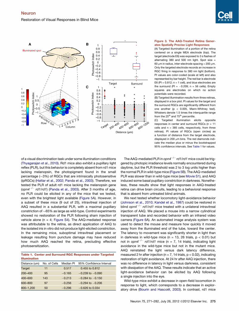
exhibit no change in locomotion over at least a 10 min period ofillumination (Lin et al., 2008). In order to determine if AAQ cansupport light modulated exploratory behavior in rd1 mice, wecarried out open field experiments. We placed a mouse into
a circular test chamber and monitored movement during 5 minin darkness followed by 5 min in 380 nm light. Figures 7A and7B show an example of the effect of AAQ on one rd1 mouse(see also Movies S2 and S3). Before AAQ, light had no effect
Figure 4. Spectral and Illuminance Sensitivity of AAQ-Mediated Photocontrol of RGC Firing(A) Spectral sensitivity of light-elicited suppression of RGC firing. Top: Light stimulation protocol. AAQ was first driven into its cis configuration with 380 nm light
(5 s), and various test wavelengths triggered photoisomerization to the trans configuration. Bottom: PI values reveal the effectiveness of different wavelengths in
suppressing RGC firing (n = 5 retinas). Error bars represent mean ± SEM.
(B) Spectral sensitivity of light-elicited activation of RGC firing. Top: Light stimulation protocol. AAQ was first driven into its trans configuration with 500 nm light
(15 s). After an additional dark period (45 s) various test wavelengths triggered photoisomerization to the cis configuration. Bottom: PI values reveal the effec-
tiveness of different wavelengths in stimulating RGC firing (n = 5 retinas). For (A) and (B), the PI was measured over the first 1 s after applying the test wavelength.
Error bars represent mean ± SEM.
(C) Stimulation of RGC firing in an AAQ-treated retina with white light. Top: Raster plot of spiking from RGCs. Bottom: Average RGC firing rate.
(D) Box plot representation of increased firing rate in white light versus 500 nm. White light significantly increases peak firing rate (p < 0.05, Mann-Whitney test,
n = 5). Whiskers denote 1.5 times the interquartile range from the 25th and 75th percentile.
(E) Light intensity-response relationship for AAQ-treated rd1 mouse retinas exposed to different intensities of 380 nm light. Minimum light intensity needed for
photoswitching is 2.6 3 1015 photons/cm2/s. Error bars represent mean ± SEM.
Figure 5. AAQ Restores the Pupillary Light Reflexin Mice Lacking All Retinal Photoreceptors(A) Pupillary light responses to 5.5 3 104 mW/m2 white
light in opn4"/" rd/rdmice, before (left) and 3 hr after (right)
intravitreal injection of AAQ (1 ml of 80 mM in DMSO). Dark
images taken 5 s before light stimulus; light images
represent maximal pupillary constriction during 30 s light
exposure. Images were taken with an infrared-sensitive
camera under infrared illumination.
(B) Irradiance-dependence of pupillary light responses to
white light. Irradiance response for wild-type mice (plotted
as mean ± STD, n = 5) (A) and four opn4"/" rd/rd mice
injected with AAQ (plotted individually: CB;D). Data
were fitted with a three parameter Hill equation.
See also Figure S4 and Movie S1.
Neuron
Restoration of Visual Responses in Blind Mice
276 Neuron 75, 271–282, July 26, 2012 ª2012 Elsevier Inc.

on the movement trajectory (Figure 7A) or total distance traveled(Figure 7B). After AAQ, light caused an almost immediatedecrease in exploratory behavior, quantified as diminisheddistance traveled. Average data from eight rd1 mice showedno light versus dark difference in movement before AAQ (Fig-ure 7C). However, after AAQ, there was a decrease in movementthat occurred within 30 s of light onset. This decrease was sus-tained throughout the illumination period. Before AAQ, there wasno statistically significantly change in the speed of locomotion inlight as compared to darkness (Figure 7D), but after AAQ injec-tion, light caused a significant 40% slowing of locomotion.Sham injections with vehicle alone elicited no significant changein light modulated behavior (n = 4, p > 0.6). Further analysis of theeight mice showed that seven of them exhibited significant light-evoked slowing of locomotion after AAQ injection (Figure 7E).After termination of the behavioral test, mice were sacrificed
and retinas were placed on the MEA for electrophysiological
analysis. In five cases, we successfully obtainedMEA recordingsand were able to directly compare the AAQ-mediated photosen-sitization of the retina ex vivo with the behavioral responsesin vivo. The one mouse that failed to exhibit light-modulatedbehavior (mouse A in Figures 7E and 7F) also failed to exhibitlight-sensitive retinal responses. For all of the other four mice,light-elicited behavior corresponded with a light-elicited changein firing rate.Rd1 mice possess ipRGCs, which should respond to the light
used in this behavioral test. However, previous studies (Lin et al.,2008) show that ipRGCs do not mediate short-term light-elicitedchanges in exploratory behavior. Moreover, in our open fieldexperiments, mice exhibited no light-modulated behavior priorto AAQ injections, confirming that alone, the ipRGCs are notsufficient to evoke this behavior.
DISCUSSION
The ultimate goal of vision restoration research is to recreate asclosely as possible the activity of the entire population of RGCs inresponse to a natural visual scene. Since only a small fraction ofRGCs are intrisically light-sensitive (Ecker et al., 2010; Pandaet al., 2003), photosensitivity must be conferred artificially bydirectly or indirectly making the neurons sensitive to light. Ideally,the kinetics and absolute sensitivity to light should be equivalentto natural RGC responses. The healthy retina has a remarkablybroad operating range owing to light-adaptation mechanisms,so the artificial system should include gain adjustment and rangeextension capabilities. Ideally, the system would replicatenormal encoding of contrast and color and highlight movement,with certain RGCs being directionally selective. All of this shouldbe accomplished with a minimally invasive and safe technology.To date, no restorative technology is close to meeting thesecriteria, but new developments are providing reason foroptimism.Broadly, three approaches have been suggested for restoring
visual function to the eye in the absence of rods and cones:optoelectronic engineering with retinal chip prosthetics; geneticengineering with viral-mediated delivery of optogenetic tools;and cellular engineering, with rod or cone progenitors differenti-ated from stem cells in vitro. We now describe a fourth approach:photochemical engineering with a small molecule photoswitch.The following functional considerations suggest that thephotoswitch approach compares favorably with other methodsfor restoringvisual functionandoffers somepractical advantages.
KineticsAAQ-mediated retinal light responses are rapid. MEA recordingsshow that the median response latency of RGC spiking is 45 msin the AAQ-treated rd1 mouse retina, compared to !50 ms(Farrow and Masland, 2011) to several hundred ms (Carcieriet al., 2003) for photopic light responses from RGCs in wild-type retina. Retinal chips electrically stimulate RGCs directly,and therefore can elicit spikes with latencies of several millisec-onds. For optogenetic tools, depending onwhich retinal cell typeexpresses the tool, the response latency of RGCs ranges fromseveral milliseconds to 150 ms (Bi et al., 2006; Busskampet al., 2010; Lagali et al., 2008). Stem cell-based therapies would
Figure 6. AAQ Restores Active Light Avoidance Behavior in MiceLacking All Retinal Photoreceptors(A) Schematic diagram of the locomotory light-avoidance test chamber.
(B) Restoration of light avoidance behavior in opn4"/" rd/rd mice following
AAQ injection. Bars represent mean latency of movement from the ‘‘East’’ to
the ‘‘Center’’ third of the tube (plotted as ± STD).
Neuron
Restoration of Visual Responses in Blind Mice
Neuron 75, 271–282, July 26, 2012 ª2012 Elsevier Inc. 277
presumably restore wild-type kinetics, assuming the differenti-ated rods and cones have full function.
SensitivityMEA recordings in vitro and PLR measurements in vivo indicatethat the AAQ-treated rd1 mouse retina responds under brightphotopic conditions, comparable to levels achieved in naturaloutdoor illumination. This is similar to light sensitivity conferredonto RGCs by optogenetic tools (Bi et al., 2006; Thyagarajanet al., 2010). Exogenous expression of NpHR in cone remnantscan result in higher light sensitivity (Busskamp et al., 2010).However, it is unclear whether many patients with advancedRP have sufficient cone remnants to allow this to be a broadlyapplicable approach (Milam et al., 1998). High sensitivity canalso be conferred by exogenously expressing melanopsin inRGCs that are not normally light-sensitive (Lin et al., 2008), butthe responses are variable and slow (on the order of seconds).Stem cell-based therapies in theory might recapitulate thewild-type sensitivity of rods and cones. However, the humanretina normally contains >100,000,000 rods and cones, and
Figure 7. AAQ Restores Light-ModulatedLocomotor Behavior in an Open-Field Test(A) Paths traveled by an rd1 mouse before and
after injection with AAQ in darkness and with
380 nm illumination.
(B) Cumulative distance traveled by the mouse in
darkness and in 380 nm light, before and after
AAQ.
(C) Average cumulative distance traveled of all
mice in darkness and 380 nm light, before and after
AAQ. Closed squares represent time spent in
darkness while open squares represent time spent
in 380 nm light. (mean ± SEM, n = 8).
(D) Mean locomotory velocity in light normalized to
basal velocity in darkness. Velocity decreases
significantly in light (n = 8, p < 0.0006).
(E) Light evoked change in the velocity of each of
the eight mice before and after AAQ. The red line
shows the mean light evoked change before and
after AAQ.
(F) Light-induced behavior is correlated with the
light-induced change in firing rate. Data were from
the five mice for which both in vivo behavioral
measurement and ex vivo retinal MEA recordings
were obtained (as labeled a–e in panel E). The
light-induced percent change in firing rate was
calculated from the aggregate light response for all
units recorded with the MEA upon switching from
darkness to 380 nm light. The light-induced
behavior represents percent change in velocity
upon switching from darkness to 380 nm light.
See also Movies S2 and S3.
whether a significant fraction can berestored with stem cells remains unclear.
Spatial Resolution and Extentof Retinal Functional RestorationAAQ-mediated retinal responses havea high spatial resolution. Our spot illumi-
nation experiments places a 100 mm radius upper limit on theAAQ-mediated receptive field size. Amacrine cells, whichpredominate in driving RGC responses, can project over severalhundred mm, but mutual inhibition between these cells presum-ably spatially constrains RGC responses to a smaller area.Because AAQ is a diffusible small molecule, in principle it shouldreach the entire retina and confer light sensitivity on all RGCs. Inpractice, we observed robust light responses in almost all RGCswhen AAQ was applied in vitro, but intravitreal injections in vivowere less effective, with only 25%–36% of injections resultingin behavioral responses to light. Drug delivery via intravitrealinjections in mice can be unreliable because of the very smallvitreal volume (20 ml), which is 250-fold less than the vitrealvolume of the human eye (5.5 ml). Further experiments usinganimals with larger vitreal volumes are needed to better testand optimize the effectiveness of intravitreal AAQ administration.In contrast to the relatively high spatial resolution that could be
conferred by AAQ, the spatial resolution of a retinal chip is limitedby the relatively large size of the stimulating electrodes and thespread of current emanating from each electrode. While the
Neuron
Restoration of Visual Responses in Blind Mice
278 Neuron 75, 271–282, July 26, 2012 ª2012 Elsevier Inc.
healthy human retina contains !1.2 million RGCs, current retinalchips have 16–64 electrodes spaced 100–200 mm apart (Winteret al., 2007). Chips with electrodes more densely packed exhibitcrosstalk between electrodes, limiting their effectiveness. Atpresent, the highest resolution that could be provided by retinalchip stimulation is several orders of magnitude lower than thetheoretical limits imposed by RGC density in the macula, theregion crucial for high-acuity vision. The area of RGC stimulationis limited by the physical size of the chip implant, which typicallycovers only the central 20 degrees of vision in the macula(Chader et al., 2009). Larger chips are possible, but there arechallenges in power delivery and achieving stable adherenceto the retina.Similar to photoswitches, the spatial resolution conferred by
optogenetic tools is defined by the size of the cell type targetedfor expressing a given light-activated protein. In principle, thesmaller the cell type and the more densely they are packedtogether, the higher the spatial resolution. In practice, viral trans-duction with current vectors has resulted in expression of opto-genetic tools in a minority of targeted cells (e.g., !5% of bipolarcells in mice [Lagali et al., 2008] and 5%–10% of RGCs inmarmosets [Ivanova et al., 2010]), but it is possible that new viralvectors will be developed that improve transduction efficiency(Vandenberghe et al., 2011). Viral transduction of NpHR hasresulted in more efficient transduction (50%–75%) of remnantcones in blind mice (Busskamp et al., 2010), but this approachis only appropriate for the few patients thought to possessremnant cones. Viral transduction of cones requires subretinalinjection, which involves local detachment of a portion of theretina from the underlying retinal pigment epithelium. Effectiveviral gene transfer is limited to the detached area (Hauswirthet al., 2008).Stem cell approaches offer the potential for greater spatial
resolution, but this is dependent on having a high density ofdifferentiated photoreceptor cells that form functional andanatomically correct synapses with appropriate retinal neuronpartners, and at present, only a very low density of cells hasbeen achieved (Lamba et al., 2009).
ON and OFF Retinal Output ChannelsOptogenetic tools have the advantage of being genetically-targetable to particular types of neurons to generate the appro-priate stimulation or inhibition of firing, for example to ON- orOFF-RGCs (Busskamp et al., 2010; Lagali et al., 2008). More-over, ChR2 and NpHR can be co-expressed in the same RGCand trafficked to different compartments to restore antagonisticcenter-surround responses (Greenberg et al., 2011). In contrast,all RGCs in AAQ-treated retina respond with the same polaritylight response. While this pattern of responsiveness is differentthan the normal retina, it may not preclude a useful visual expe-rience. Behavioral studies in primates demonstrate that theselective pharmacological blockade of ON neurons does notseverely impair recognition of shapes or detection of light decre-ments (Schiller et al., 1986). Moreover, in RP patients, electronicretinal prosthetics can restore shape recognition, even thoughthe devices stimulate ON- and OFF-RGCs indiscriminately(Sekirnjak et al., 2009). Hence, while two channels of visualinformation flow are important for normal vision, simultaneous
activation of ON- and OFF-pathways is sufficient for visualperception. AAQ treatment enables RGCs surrounding an illumi-nated area to respond with the opposite polarity to those in thecenter. Since all RGCs respond with the same polarity lightresponse to full-field illumination (Figure 1A), the opposite centerversus surround responses to spot illumination suggests thatinhibitory neurons that project laterally invert the sign of theresponse. It seems likely that the opposite center versussurround responsewould enhance perception of spatial contrastand facilitate edge detection in downstream visual regions of thebrain. But ultimately, the evaluation of the quality of imagesproduced by photoswitch activation of retinal cells will requirestudy in primates or human patients.
Spectral SensitivityIn AAQ-treated retinas, RGCs respond most strongly to shortwavelength light, consistent with the photochemical propertiesof the molecule (Fortin et al., 2008). Although 380 nm light isoptimal for enhancing firing frequency, longer wavelengths (upto 500 nm) can still generate excitatory light responses, reflectingthe spectral range of trans to cis azobenzene photoisomeriza-tion. This is important, because unlike in the mouse, the humanlens minimally transmits 380 nm light (Kessel et al., 2010).Newly-developed red-shifted azobenzene derivatives allow K+
channel regulation with even longer wavelengths of light andchemical modification of the azobenzene moiety results incompounds with improved quantum efficiency (Mourot et al.,2011). Ideally, second-generation AAQ derivatives would enablephotostimulation of the retina with intensities and wavelengthsexperienced during normal photopic vision. Alternatively,a head-mounted optoelectronic visual aid (Degenaar et al.,2009) designed to intensify and transform the palette of visualscenes to a blue-shifted wavelength could enhance the effec-tiveness of AAQ and related agents. Such a device might alsoallow switching of individual RGCs ON and OFF by rapid modu-lation of shorter- and longer-wavelength light.Except for some of the optogenetic tools, the other vision
restoration methods pose no particular spectral challenges.NpHR and ChR2 respond optimally to 580 and 470 nm light,respectively (Nagel et al., 2003; Zhang et al., 2007), but newlydiscovered red-shifted homologs (Govorunova et al., 2011)expand the toolkit for potential use for photosensitizing retinalneurons. Since they are driven by images captured by anexternal camera, retinal chip prosthetics can be engineered tooperate over the entire visual spectrum. Similarly, assumingstem cell-derived photoreceptors express the full complementof cone opsins, these should be responsive to a broad rangeof wavelengths.
Invasiveness, Safety, and ReversibilityThe phototswitch approach has the advantage of being relativelynoninvasive and readily reversible. We envision photoswitchmolecules being administered therapeutically by intravitrealinjection, a safe and frequent procedure for treating maculardegeneration with anti-vasoproliferative agents. Because AAQphotosensitization dissipates within 24 hr, it may be possibleto titrate the most effective dose with repeated intravitreal injec-tions. The reversibility of AAQ will allow for ‘‘upgrades’’ as newer
Neuron
Restoration of Visual Responses in Blind Mice
Neuron 75, 271–282, July 26, 2012 ª2012 Elsevier Inc. 279

agents become available, perhaps with improved spectral orkinetic properties. Longer-term therapy would require anextended release formulation. We estimate that a several monthsupply of AAQ could be packaged into an intravitreal device likethose currently used for long-term steroid treatment of ocularinflammation (London et al., 2011).
In contrast, retinal chip prosthetics require invasive intraocularsurgery. Optogenetic treatment of remnant cones and stem celltherapy both require subretinal injection, a risky procedure thatbegins with iatrogenic retinal detachment, which could furtherdamage the retina. These three approaches are essentially irre-versible. Should they produce undesired effects (such as chronicphotophobia or disturbing visual sensations) there is no readymeans for reversal of either stem cell implantation or genetherapy, and removal of chip prosthetics would require addi-tional significant surgery.
Both retinal chip prosthetics and human gene replacementtherapy have received investigational new device/drug statusand have been tested in human patients under research proto-cols (Ahuja et al., 2011; Benav et al., 2010) without significanttoxicity. However, microbial optogenetic tools would requiretrans-species gene therapy, which is unprecedented. Viralgene expression in the eye can elicit late-onset inflammation,indicating an immune reaction (Beltran et al., 2010). Becausethe unitary conductance of ChR2 and NpHR is quite small(Feldbauer et al., 2009; Sjulson and Miesenbock, 2008; Zhanget al., 2007), photosensitivity requires very high levels of exoge-nous expression, raising concerns about an immune response tothe microbially-derived protein or cytotoxicity. While long-termsafety of AAQ or similar compounds will require toxicologystudies, to date, we have not seen acute toxicity of AAQ onneural function in vitro (Fortin et al., 2008) or in vivo (Figure S2).The pathway for evaluating photoswitch compounds for toxicityis straightforward and will mirror those that have been followedfor other approved, intravitreal agents.
Finally, in addition to its potential clinical use, AAQ has utility asa scientific tool for understanding normal retinal function anddevelopment. Using AAQ, the firing activity of single cells orsmall regions of the retina can be controlled with high temporaland spatial resolution. This may be useful for better under-standing information processing by the retina and for studyingdevelopmental plasticity in animals before rods and cones arefunctional (Huberman et al., 2008). AAQ-mediated photocontrolof retinal neurons also provides a unique way to investigatecircuit remodeling after the rods and cones have degeneratedin mouse models of RP (Marc et al., 2003).
EXPERIMENTAL PROCEDURES
AnimalsWild-type mice (C57BL/6J strain, Jackson Laboratories) and homozygous rd1
mice (C3H/HeJ strain, Charles River Laboratories) >3 months old were used
for the experiments. All animal use procedures were approved by the UC
Berkeley or University of Washington Institutional Animal Care and Use
Committee (see Supplemental Experimental Procedures).
Electrophysiology and PharmacologyMouse retinaswere dissected and kept in physiological saline at 36#C contain-
ing (in mM) 119 NaCl, 2.5 KCl, 1 KH2PO4, 1.3 MgCl2, 2.5 CaCl2, 26.2 NaHCO3,
and 20 D-glucose, aerated with 95% O2/5% CO2. For extracellular recording,
the retina was placed ganglion cell layer down onto a multielectrode array
system (model number MEA 1060-2-BC, Multi-Channel Systems).
The MEA electrodes were 30 mm in diameter and arranged on an 83 8 rect-
angular grid. Extracellular spikes were high-pass filtered at 200 Hz and digi-
tized at 20 kHz. A spike threshold of 4SD was set for each channel. Typically,
each electrode recorded spikes from one to three RGCs. Principal component
analysis of spike waveforms was used for sorting spikes generated by indi-
vidual cells (Offline Sorter; Plexon). Only cells with interspike intervals of
<1 ms were included in the analysis.
Borosilicate glass electrodes of 6–11 MU were used for whole-cell voltage-
clamp recordings. Current records were low-pass filtered at 2 kHz. For
measuring voltage-gated K+ currents, electrodes contained (in mM) 98.3 K+
gluconate, 1.7 KCl, 0.6 EGTA, 5 MgCl2, 40 HEPES, 2 ATP-Na, and 0.3 GTP-
Na (pH = 7.25). For recording glutamatergic EPSCs, electrodes contained
(in mM) 125 Cs+ sulfate, 10 TEA-Cl, 5 EGTA, 0.85 MgCl2, 10 HEPES,
2 QX-314, and 4 ATP-Na2 (pH = 7.25). Neurotransmitter receptor antagonists
were used to evaluate synaptic contributions of different retinal neurons to
RGC light responses (see Supplemental Experimental Procedures).
Light StimulationIn MEA recordings, we used a 100 Wmercury arc lamp filtered through 380 or
500 nm narrow-pass filters (Chroma, Inc.) and switched wavelengths with an
electronically-controlled shutter and filter wheel (SmartShutter, Sutter Instru-
ments). Unless otherwise indicated, the standard incident light intensity at
the retina was 13.4 mW/cm2 (2.56 3 1016 photons/cm2/s) for 380 nm and
11.0 mW/cm2 (2.77 3 1016 photons/cm2/s) for 500 nm.
PLR MeasurementMicewere sedatedwith an intraperitoneal injection of ketamine (6.7mg/ml) and
xylazine (0.45 mg/ml) in saline. A glass micropipette was inserted through the
sclera into the vitreous cavity to inject a 1 ml bolus of AAQ (80 mM in a saline
solution containing 40% DMSO).
Videos of pupillary light responses of mice were recorded before and 3 hr
after AAQ injection. White light was derived from halogen dissecting lamp,
and intensity was controlled with neutral density filters. Animals were dark-
adapted for at least 20min prior to testing. An infrared (IR) illuminator and video
camera (focused 15 cm from the objective) was used to measure pupil dilation,
as described (Van Gelder, 2005).
Locomotory Light AvoidanceWild-type or opn4"/" rd/rdmice injected with 80 mM AAQ were dark-adapted
and placed into a transparent tube. The tube was illuminated with IR light and
mouse movement was recorded with an IR video camera and stored for offline
analysis. During testing, the face of the mouse was illuminated with 385 nm
light (log irradiance 15.7) and at 5 s intervals flashes of 480 nm light (log irradi-
ance 15.2) were superimposed. For each mouse, we recorded position in the
tube preinjection, and 2 hr and 24 hr postinjection. Analysis was conducted
with automated image-analysis software.
Open-Field TestRd1 mice were placed in a 190 mm 3 100 mm circular UV-transparent
chamber. The chamber was surrounded by six panels of 380 nm LEDs
(Roithner Laserteknik), providing uniform illumination with a light intensity
of !7 mW/cm2.
Themice were dark-adapted in their cages for 1 hr prior to each experiment.
The mice were placed in the experimental chamber and allowed to acclimate
for 5 min. The behavior was then recorded using an IR sensitive video camera
(Logitech C310) for 5 min in darkness under IR illumination. After 5 min, the
chamber was illuminated by the 380 nm LEDs, and behavior was monitored
for an additional 5 min. The apparatus was cleaned and thoroughly dried prior
to each experiment.
After the open-field test, each mouse was given an intravitreal injection of
AAQ (20 mM AAQ, 9:1 saline: DMSO) and were allowed to recover for !6 hr
on a heating pad with open access to food and water in their cage located
in the dark room followed by a second round of behavioral testing. The videos
were analyzed utilizing motion tracking video analysis software (Tracker) in
Neuron
Restoration of Visual Responses in Blind Mice
280 Neuron 75, 271–282, July 26, 2012 ª2012 Elsevier Inc.

order to quantify the average velocity of the mice, the trajectory of motion
throughout the test, and the total distance traveled.
Data Analysis and StatisticsLight-elicited changes in firing rate during test flashes were normalized with
respect to initial firing rate and expressed as a PI, defined as follows: PI =
(test firing rate – initial firing rate) / (test firing rate + initial firing rate).
Relative pupillary light responses were calculated as 1 " (pupil area
minimum during thirty seconds of the light stimulus) / (pupil area minimum
during five seconds preceding the stimulus). Relative response data for wild-
type and opn4"/" rd/rd mice were fitted with a three parameter Hill equation
(SigmaPlot, Systat Software, Inc.). Data are expressed asmean ± SEM, unless
otherwise indicated. The p values for open-field experiments were calculated
using the two tailed unpaired Students t test.
Latencies were calculated for every cell with a PI greater than 0.011, the
upper median confidence interval PI of our control experiments (n = 13 retinas;
n = 409 cells). For each cell, firing rate was averaged over the first two light
periods (dark and 380 nm light), with a 10 ms bin size. Basal firing rate was
calculated from theuppermedianconfidence interval in 500nm light.Response
latencywas then calculated as the timedifference between the onset of 380 nm
light and the first bin with a firing rate greater than the cell’s basal activity. The
median response latency was 45 ms (n = 10 retinas; n = 368 cells).
All statisticswereperformedwithMATLAB (Mathworks) algorithms.Distribu-
tionswere first tested for normality using the Shapiro-Wilk test. For non-normal
distributions, the Wilcoxon rank sum test was used for pairwise comparisons.
The 95% confidence intervals for medians were generated by resampling the
original distributions and applying the bias-corrected percentile method (Efron
and Tibshirani, 1986). Results with p < 0.05 were considered significant.
For all box plots, box limits represent the 25th and 75th percentile, respec-
tively. The red line represents themedian andwhiskers denote 1.5 times the in-
terquartile range from the limits of the box. Outliers are marked by red + signs.
SUPPLEMENTAL INFORMATION
Supplemental Information includes four figures, two tables, Supplemental
Experimental Procedures, and three movies and can be found with this article
online at http://dx.doi.org/10.1016/j.neuron.2012.05.022.
ACKNOWLEDGMENTS
We thank A. Anishchenko and J. Elstrott for helpful comments and discus-
sions; Trevor Lee, Andrew Noblet, R. Montpetit, T. Lamprecht, and X. Qiu for
technical and experimental assistance; and J. Flannery and K. Greenberg for
valuable suggestions. This work was supported by the National Eye Institute
(NEI), which provided research Grant EY018957 to R.H.K., Core Grant
P30 EY003176 to R.H.K., and Core Grant P30 EY001730 to R.V.G. This
work was also supported by Beckman Foundation for Macular Research
(R.H.K.) and a Research to Prevent Blindness award to Y.S. and R.V.G. and
an Ezell Fellowship to A.P. The NEI also funded the Nanomedicine Develop-
ment Center (PN2 EY018241), which supported this interdisciplinary project.
R.H.K. and D.T. are SAB members and consultants of Photoswitch Biosci-
ence, Inc., which is developing commercial uses for chemical photoswitches.
A.P., J.L., I.T., J.N., Y.S., T.H., I.D.K., andK.B. conducted the in vitro and in vivo
experiments. D.T. designed and synthesized chemical reagents. R.H.K. and
R.V.G. coordinated the research and wrote the manuscript. R.H.K. initiated
the research and supervised the program.
Accepted: April 30, 2012
Published: July 25, 2012
REFERENCES
Ahuja, A.K., Dorn, J.D., Caspi, A., McMahon, M.J., Dagnelie, G., Dacruz, L.,
Stanga, P., Humayun, M.S., and Greenberg, R.J.; Argus II Study Group.
(2011). Blind subjects implanted with the Argus II retinal prosthesis are able
to improve performance in a spatial-motor task. Br. J. Ophthalmol. 95,
539–543.
Banghart, M.R., Mourot, A., Fortin, D.L., Yao, J.Z., Kramer, R.H., and Trauner,
D. (2009). Photochromic blockers of voltage-gated potassium channels.
Angew. Chem. Int. Ed. Engl. 48, 9097–9101.
Beltran, W.A., Boye, S.L., Boye, S.E., Chiodo, V.A., Lewin, A.S., Hauswirth,
W.W., and Aguirre, G.D. (2010). rAAV2/5 gene-targeting to rods:dose-depen-
dent efficiency and complications associated with different promoters. Gene
Ther. 17, 1162–1174.
Benav, H., Bartz-Schmidt, K.U., Besch, D., Bruckmann, A., Gekeler, F.,
Greppmaier, U., Harscher, A., Kibbel, S., Kusnyerik, A., Peters, T., et al.
(2010). Restoration of useful vision up to letter recognition capabilities using
subretinal microphotodiodes. In Engineering in Medicine and Biology
Society (EMBC), 2010 Annual International Conference of the IEEE, pp.
5919–5922.
Bi, A., Cui, J., Ma, Y.P., Olshevskaya, E., Pu, M., Dizhoor, A.M., and Pan, Z.H.
(2006). Ectopic expression of a microbial-type rhodopsin restores visual
responses in mice with photoreceptor degeneration. Neuron 50, 23–33.
Bourin, M., and Hascoet, M. (2003). The mouse light/dark box test. Eur. J.
Pharmacol. 463, 55–65.
Busskamp, V., Duebel, J., Balya, D., Fradot, M., Viney, T.J., Siegert, S.,
Groner, A.C., Cabuy, E., Forster, V., Seeliger, M., et al. (2010). Genetic reacti-
vation of cone photoreceptors restores visual responses in retinitis pigmen-
tosa. Science 329, 413–417.
Caporale, N., Kolstad, K.D., Lee, T., Tochitsky, I., Dalkara, D., Trauner, D.,
Kramer, R., Dan, Y., Isacoff, E.Y., and Flannery, J.G. (2011). LiGluR restores
visual responses in rodent models of inherited blindness. Mol. Ther. 19,
1212–1219.
Carcieri, S.M., Jacobs, A.L., and Nirenberg, S. (2003). Classification of retinal
ganglion cells: a statistical approach. J. Neurophysiol. 90, 1704–1713.
Chader, G.J., Weiland, J., and Humayun, M.S. (2009). Artificial vision: needs,
functioning, and testing of a retinal electronic prosthesis. Prog. Brain Res.
175, 317–332.
Chaudhry, G.R., Fecek, C., Lai, M.M., Wu, W.C., Chang, M., Vasquez, A.,
Pasierb, M., and Trese, M.T. (2009). Fate of embryonic stem cell derivatives
implanted into the vitreous of a slow retinal degenerative mouse model.
Stem Cells Dev. 18, 247–258.
Degenaar, P., Grossman, N., Memon, M.A., Burrone, J., Dawson, M.,
Drakakis, E., Neil, M., and Nikolic, K. (2009). Optobionic vision—a new genet-
ically enhanced light on retinal prosthesis. J. Neural Eng. 6, 035007.
Ecker, J.L., Dumitrescu, O.N., Wong, K.Y., Alam, N.M., Chen, S.-K., LeGates,
T., Renna, J.M., Prusky, G.T., Berson, D.M., and Hattar, S. (2010). Melanopsin-
expressing retinal ganglion-cell photoreceptors: cellular diversity and role in
pattern vision. Neuron 67, 49–60.
Efron, B., and Tibshirani, R. (1986). Bootstrap methods for standard errors,
confidence intervals, and other measures of statistical accuracy. Stat. Sci. 1,
54–75.
Farrow, K., and Masland, R.H. (2011). Physiological clustering of visual
channels in the mouse retina. J. Neurophysiol. 105, 1516–1530.
Feldbauer, K., Zimmermann, D., Pintschovius, V., Spitz, J., Bamann, C., and
Bamberg, E. (2009). Channelrhodopsin-2 is a leaky proton pump. Proc. Natl.
Acad. Sci. USA 106, 12317–12322.
Fortin, D.L., Banghart, M.R., Dunn, T.W., Borges, K., Wagenaar, D.A., Gaudry,
Q., Karakossian, M.H., Otis, T.S., Kristan, W.B., Trauner, D., and Kramer, R.H.
(2008). Photochemical control of endogenous ion channels and cellular excit-
ability. Nat. Methods 5, 331–338.
Gerding, H., Benner, F.P., and Taneri, S. (2007). Experimental implantation of
epiretinal retina implants (EPI-RET) with an IOL-type receiver unit. J. Neural
Eng. 4, S38–S49.
Govorunova, E.G., Spudich, E.N., Lane, C.E., Sineshchekov, O.A., and
Spudich, J.L. (2011). New channelrhodopsin with a red-shifted spectrum
and rapid kinetics from Mesostigma viride. MBio 2, e00115–e11.
Greenberg, K.P., Pham, A., and Werblin, F.S. (2011). Differential targeting of
optical neuromodulators to ganglion cell soma and dendrites allows dynamic
control of center-surround antagonism. Neuron 69, 713–720.
Neuron
Restoration of Visual Responses in Blind Mice
Neuron 75, 271–282, July 26, 2012 ª2012 Elsevier Inc. 281
Hattar, S., Liao, H.W., Takao, M., Berson, D.M., and Yau, K.W. (2002).
Melanopsin-containing retinal ganglion cells: architecture, projections, and
intrinsic photosensitivity. Science 295, 1065–1070.
Hauswirth, W.W., Aleman, T.S., Kaushal, S., Cideciyan, A.V., Schwartz, S.B.,
Wang, L.L., Conlon, T.J., Boye, S.L., Flotte, T.R., Byrne, B.J., and Jacobson,
S.G. (2008). Treatment of leber congenital amaurosis due to RPE65 mutations
by ocular subretinal injection of adeno-associated virus gene vector: short-
term results of a phase I trial. Hum. Gene Ther. 19, 979–990.
Huberman, A.D., Feller, M.B., and Chapman, B. (2008). Mechanisms under-
lying development of visual maps and receptive fields. Annu. Rev. Neurosci.
31, 479–509.
Humayun,M.S., Weiland, J.D., Fujii, G.Y., Greenberg, R., Williamson, R., Little,
J., Mech, B., Cimmarusti, V., Van Boemel, G., Dagnelie, G., and de Juan, E.
(2003). Visual perception in a blind subject with a chronic microelectronic
retinal prosthesis. Vision Res. 43, 2573–2581.
Ivanova, E., Hwang, G.S., Pan, Z.H., and Troilo, D. (2010). Evaluation of
AAV-mediated expression of Chop2-GFP in the marmoset retina. Invest.
Ophthalmol. Vis. Sci. 51, 5288–5296.
Jimenez, A.J., Garcıa-Fernandez, J.M., Gonzalez, B., and Foster, R.G. (1996).
The spatio-temporal pattern of photoreceptor degeneration in the aged rd/rd
mouse retina. Cell Tissue Res. 284, 193–202.
Johnson, J., Wu, V., Donovan, M., Majumdar, S., Renterıa, R.C., Porco, T.,
Van Gelder, R.N., and Copenhagen, D.R. (2010). Melanopsin-dependent light
avoidance in neonatal mice. Proc. Natl. Acad. Sci. USA 107, 17374–17378.
Kandel, G., Bedell, H., Walker, R., and Wolf, B. (1987). Negative phototaxis in
pigmented, albinotic and RCS rat pupsmeasured with a new technique. Vision
Sci. 1, 357–366.
Kessel, L., Lundeman, J.H., Herbst, K., Andersen, T.V., and Larsen, M. (2010).
Age-related changes in the transmission properties of the human lens and their
relevance to circadian entrainment. J. Cataract Refract. Surg. 36, 308–312.
Lagali, P.S., Balya, D., Awatramani, G.B., Munch, T.A., Kim, D.S., Busskamp,
V., Cepko, C.L., and Roska, B. (2008). Light-activated channels targeted to ON
bipolar cells restore visual function in retinal degeneration. Nat. Neurosci. 11,
667–675.
Lamba, D.A., Gust, J., and Reh, T.A. (2009). Transplantation of human embry-
onic stem cell-derived photoreceptors restores some visual function in Crx-
deficient mice. Cell Stem Cell 4, 73–79.
Lamba, D.A., Karl, M.O., Ware, C.B., and Reh, T.A. (2006). Efficient generation
of retinal progenitor cells from human embryonic stem cells. Proc. Natl. Acad.
Sci. USA 103, 12769–12774.
Lin, B., Koizumi, A., Tanaka, N., Panda, S., and Masland, R.H. (2008).
Restoration of visual function in retinal degeneration mice by ectopic expres-
sion of melanopsin. Proc. Natl. Acad. Sci. USA 105, 16009–16014.
London, N.J., Chiang, A., and Haller, J.A. (2011). The dexamethasone drug
delivery system: indications and evidence. Adv. Ther. 28, 351–366.
Marc, R.E., Jones, B.W., Watt, C.B., and Strettoi, E. (2003). Neural remodeling
in retinal degeneration. Prog. Retin. Eye Res. 22, 607–655.
Meister, M., Pine, J., and Baylor, D.A. (1994). Multi-neuronal signals from the
retina: acquisition and analysis. J. Neurosci. Methods 51, 95–106.
Milam, A.H., Li, Z.Y., and Fariss, R.N. (1998). Histopathology of the human
retina in retinitis pigmentosa. Prog. Retin. Eye Res. 17, 175–205.
Mourot, A., Kienzler, M.A., Banghart, M.R., Fehrentz, T., Huber, F.M.E., Stein,
M., Kramer, R.H., and Trauner, D. (2011). Tuning photochromic ion channel
blockers. ACS Chemical Neuroscience, in press.
Nagel, G., Szellas, T., Huhn, W., Kateriya, S., Adeishvili, N., Berthold, P., Ollig,
D., Hegemann, P., and Bamberg, E. (2003). Channelrhodopsin-2, a directly
light-gated cation-selective membrane channel. Proc. Natl. Acad. Sci. USA
100, 13940–13945.
Panda, S., Provencio, I., Tu, D.C., Pires, S.S., Rollag, M.D., Castrucci, A.M.,
Pletcher, M.T., Sato, T.K., Wiltshire, T., Andahazy, M., et al. (2003).
Melanopsin is required for non-image-forming photic responses in blind
mice. Science 301, 525–527.
Punzo, C., and Cepko, C. (2007). Cellular responses to photoreceptor death in
the rd1 mouse model of retinal degeneration. Invest. Ophthalmol. Vis. Sci. 48,
849–857.
Sancho-Pelluz, J., Arango-Gonzalez, B., Kustermann, S., Romero, F.J.,
van Veen, T., Zrenner, E., Ekstrom, P., and Paquet-Durand, F. (2008).
Photoreceptor cell death mechanisms in inherited retinal degeneration. Mol.
Neurobiol. 38, 253–269.
Schiller, P.H., Sandell, J.H., andMaunsell, J.H. (1986). Functions of theONand
OFF channels of the visual system. Nature 322, 824–825.
Sekirnjak, C., Hulse, C., Jepson, L.H., Hottowy, P., Sher, A., Dabrowski, W.,
Litke, A.M., and Chichilnisky, E.J. (2009). Loss of responses to visual but not
electrical stimulation in ganglion cells of rats with severe photoreceptor
degeneration. J. Neurophysiol. 102, 3260–3269.
Shire, D.B., Kelly, S.K., Chen, J., Doyle, P., Gingerich, M.D., Cogan, S.F.,
Drohan, W.A., Mendoza, O., Theogarajan, L., Wyatt, J.L., and Rizzo, J.F.
(2009). Development and implantation of a minimally invasive wireless subre-
tinal neurostimulator. IEEE Trans. Biomed. Eng. 56, 2502–2511.
Sjulson, L., and Miesenbock, G. (2008). Photocontrol of neural activity:
biophysical mechanisms and performance in vivo. Chem. Rev. 108, 1588–
1602.
Strettoi, E., andPignatelli, V. (2000).Modifications of retinal neurons in amouse
model of retinitis pigmentosa. Proc. Natl. Acad. Sci. USA 97, 11020–11025.
Thyagarajan, S., van Wyk, M., Lehmann, K., Lowel, S., Feng, G., and Wassle,
H. (2010). Visual function in mice with photoreceptor degeneration and trans-
genic expression of channelrhodopsin 2 in ganglion cells. J. Neurosci. 30,
8745–8758.
Tomita, H., Sugano, E., Isago, H., Hiroi, T., Wang, Z., Ohta, E., and Tamai, M.
(2010). Channelrhodopsin-2 gene transduced into retinal ganglion cells
restores functional vision in genetically blind rats. Exp. Eye Res. 90, 429–436.
Van Gelder, R.N. (2005). Nonvisual ocular photoreception in the mammal.
Methods Enzymol. 393, 746–755.
Vandenberghe, L.H., Bell, P., Maguire, A.M., Cearley, C.N., Xiao, R., Calcedo,
R., Wang, L., Castle, M.J., Maguire, A.C., Grant, R., et al. (2011). Dosage
thresholds for AAV2 and AAV8 photoreceptor gene therapy in monkey. Sci.
Transl. Med. 3, 88ra54.
Winter, J.O., Cogan, S.F., and Rizzo, J.F., 3rd. (2007). Retinal prostheses:
current challenges and future outlook. J. Biomater. Sci. Polym. Ed. 18,
1031–1055.
Yanai, D., Weiland, J.D., Mahadevappa, M., Greenberg, R.J., Fine, I., and
Humayun, M.S. (2007). Visual performance using a retinal prosthesis in three
subjects with retinitis pigmentosa. Am. J. Ophthalmol. 143, 820–827.
Zhang, F., Wang, L.P., Brauner, M., Liewald, J.F., Kay, K., Watzke, N., Wood,
P.G., Bamberg, E., Nagel, G., Gottschalk, A., and Deisseroth, K. (2007).
Multimodal fast optical interrogation of neural circuitry. Nature 446, 633–639.
Neuron
Restoration of Visual Responses in Blind Mice
282 Neuron 75, 271–282, July 26, 2012 ª2012 Elsevier Inc.