Photobiomodulation for the Treatment of Retinal Injury and
11
. Eells et al 1 Photobiomodulation for the Treatment of Retinal Injury and Retinal Degenerative Diseases Janis T. Eells 1 , Kristina D. DeSmet 1 , Diana K. Kirk 2 , Margaret Wong-Riley 3 , Harry T. Whelan 4 , James Ver Hoeve 5 , T. Michael Nork 5 , Jonathan Stone 2 and Krisztina Valter 2 1 Department of Health Sciences, University of Wisconsin-Milwaukee, Milwaukee, Wisconsin 53201 2 CNS Stability and Degeneration Group,Research School of Biological Sciences, Australian National University Canberra, Australia 3 Department of Cell Biology, Neurobiology and Anatomy, Medical College of Wisconsin, Milwaukee, Wisconsin 53226 4 Department of Pediatric Neurology, Medical College of Wisconsin, Milwaukee, Wisconsin 53226 5 Department of Ophthalmology, University of Wisconsin, Madison, Madison, Wisconsin 53792 Proceedings ofLight Activated Tissue Regeneration and Therapy Conference. eds R.W. Waynant and D.B. Tata pp 39-51, Springer, New York, 2008. ACKNOWLEDGEMENTS: We gratefully acknowledge Quantum Devices, Inc. (Barneveld, WI) for providing light emitting diode arrays with Quantum’s High-Emissivity Aluminiferous Light-Emitting Substrate (HEALS®) technology for this project. This research was supported by an Individual Investigator Grant TA-NE-0606-0348-UWI and TA-NP-1107-0435 from the Foundation Fighting Blindness (J.T.E.); The ARC Center of Excellence in Vision Science (D.K, K.V., J.S.); The Defense Advanced Research Projects Agency Grant DARPA N66001-01-1-8969 (H.T.W.); The National Institute of Environmental Health Sciences Grant ES06648 (J.T.E); The National Eye Institute Core Grant P30-EY01931 (J.T.E., M.W.R.) and RO1 Grant EY05439 (M.W.R.) and the Bleser Foundation Endowed Professorship (H.T.W.)
Transcript of Photobiomodulation for the Treatment of Retinal Injury and

. Eells et al
1
Photobiomodulation for the Treatment of Retinal Injury and Retinal Degenerative Diseases
Janis T. Eells1, Kristina D. DeSmet1, Diana K. Kirk2, Margaret Wong-Riley3, Harry T. Whelan4, James Ver Hoeve5, T. Michael Nork5, Jonathan Stone2 and Krisztina Valter2
1Department of Health Sciences, University of Wisconsin-Milwaukee, Milwaukee, Wisconsin 53201
2CNS Stability and Degeneration Group,Research School of Biological Sciences, Australian National University
Canberra, Australia
3Department of Cell Biology, Neurobiology and Anatomy, Medical College of Wisconsin, Milwaukee, Wisconsin 53226
4Department of Pediatric Neurology, Medical College of Wisconsin, Milwaukee, Wisconsin 53226
5Department of Ophthalmology, University of Wisconsin, Madison, Madison, Wisconsin 53792
Proceedings ofLight Activated Tissue Regeneration and Therapy Conference.
eds R.W. Waynant and D.B. Tata pp 39-51, Springer, New York, 2008.
ACKNOWLEDGEMENTS: We gratefully acknowledge Quantum Devices, Inc. (Barneveld, WI) for providing light emitting diode arrays with Quantum’s High-Emissivity Aluminiferous Light-Emitting Substrate (HEALS®) technology for this project. This research was supported by an Individual Investigator Grant TA-NE-0606-0348-UWI and TA-NP-1107-0435 from the Foundation Fighting Blindness (J.T.E.); The ARC Center of Excellence in Vision Science (D.K, K.V., J.S.); The Defense Advanced Research Projects Agency Grant DARPA N66001-01-1-8969 (H.T.W.); The National Institute of Environmental Health Sciences Grant ES06648 (J.T.E); The National Eye Institute Core Grant P30-EY01931 (J.T.E., M.W.R.) and RO1 Grant EY05439 (M.W.R.) and the Bleser Foundation Endowed Professorship (H.T.W.)

. Eells et al
2
Abstract
Retinal injury and retinal degenerative diseases are a leading causes of visual impairment in the
developed world. Mitochondrial dysfunction and oxidative stress play key roles in the pathogenesis of retinal injury and disease. The development and testing of strategies designed to improve mitochondrial function and attenuate oxidative stress are essential for combating retinal disease. One strategy involves the use of photobiomodulation. Photobiomodulation, low-energy photon irradiation by light in the far-red to near-infrared (NIR) range using low energy lasers or light-emitting diode (LED) arrays, has been applied clinically in the treatment soft tissue injuries and acceleration of wound healing for more than 30 years. The therapeutic effects of photobiomodulation have been hypothesized to be mediated by intracellular signaling mechanisms triggered by the interaction of far-red to NIR photons with the mitochondrial photoacceptor molecule cytochrome oxidase which culminate in improved mitochondrial energy metabolism, increased synthesis of cytoprotective factors and cell survival.
The therapeutic potential of 670 nm LED photobiomodulation administered once per day at a fluence
of 4 J/cm2 was investigated in established experimental models of retinal injury, retinal toxicity and retinal disease. Photobiomodulation stimulated retinal wound healing following high-intensity laser-induced retinal injury. Photobiomodulation not only enhanced the rate of wound healing, it also prevented the loss of retinal and cortical visual function induced by laser-induced retinal injury. In a rodent model of retinal mitochondrial toxicity, photobiomodulation preserved retinal function and prevented photoreceptor damage. Moreover, molecular studies revealed that photobiomodulation induced significant upregulation of gene expression pathways involved in mitochondrial energy production and cytoprotection in the retina. Retinitis pigmentosa is the leading cause of vision loss due to retinal degeneration. Photobiomodulation administered during the critical period of photoreceptor development in a rat model of retinitis pigmentosa increased retinal mitochondrial cytochrome oxidase activity, upregulated the production of retinal antioxidants, increased the production of retinal neurotrophic factors and prevented photoreceptor cell death.
The molecular, biochemical and functional insights obtained from this research provide crucial
information needed for a comprehensive FDA approval for the use of photobiomodulation in the treatment of retinal diseases. From a basic science perspective, they substantiate previous in vitro investigations and support the hypothesis that photobiomodulation augments mitochondrial function and stimulates cytoprotective pathways to prevent retinal damage. From a clinical perspective, they document the therapeutic potential of 670 nm photon therapy in experimental models of retinal injury, retinal toxicity and retinitis pigmentosa, thus setting the stage for clinical trials of photobiomodulation in human disease. Key Words: photobiomodulation, mitochondrial dysfunction, oxidative stress, retinal injury, retinal degenerative disease,

. Eells et al
3
Introduction
Oxidative stress and mitochondrial dysfunction are central in retinal aging, retinal injury and in the pathogenesis of retinal degenerative diseases (2,3,24,25). Oxidative stress plays a key role in high intensity laser burn injury and retinal mitochondrial dysfunction is causative in methanol toxicity (12). Retinal injury and retinal degenerative diseases are leading causes of visual impairment in the developed world (1,9). Although age-related macular degeneration is responsible for the vast majority of the cases of retinal degeneration, other important retinal dystrophies include, retinitis pigmentosa, cone-rod dystrophies and Stargardt disease. The pathogenesis of these diseases is incompletely understood and no efficient therapy or prevention exists to date. Therefore, the development of simple long-term strategies designed to improve mitochondrial function and attenuate oxidative stress are essential for combating neurodegenerative and retinal degenerative disease. One newly developed strategy involves the use of photobiomodulation.
Low energy photon irradiation by light in the far-red to near-infrared (NIR) range (630-1000 nm) using low energy lasers or light-emitting diode (LED) arrays (collectively termed photobiomodulation) has been shown to accelerate wound healing, improve recovery from ischemic injury in the heart and attenuate degeneration in the injured retina and optic nerve (5,12). Moreover, red to near-infrared light therapy has been applied clinically in the treatment soft tissue injuries and to accelerate wound healing for more than 30 years (5,12). The therapeutic effects of red to near infrared light have been hypothesized to be mediated by intracellular signaling mechanisms triggered by the interaction of far-red to near-infrared light with the mitochondrial photoacceptor molecule cytochrome oxidase which culminate in improved cellular mitochondrial energy metabolism and antioxidant production (Figure 1). In support of this hypothesis, we have demonstrated in primary neuronal cells that far-red to near-infrared LED photo-irradiation (4 J/cm2) increases the production of cytochrome oxidase in cultured primary neurons, reverses the reduction of cytochrome oxidase activity produced by metabolic inhibitors and attenuates cyanide-induced apoptosis (20,21). We have also shown that the action spectrum of NIR light for stimulation of cytochrome oxidase activity parallels the near-infrared absorption spectrum of the oxidized form of cytochome oxidase (Figure 2) (21).
Figure 1: Hypothesized Signal Transduction Pathway for Photobiomodulation
670 nm LED
nucleus
gene transcription
mitochondrion
Protein Synthesis CNTF, MnSOD, GSH
Cytochrome oxidase
Activation of transcription factors
Photons
Cell

. Eells et al
4
Figure 2: Recovery of Neuronal Cytochrome Oxidase Activity and Cellular ATP Content Correlates with Cytochrome Oxidase Action Spectrum)
670 nm LED Treatment Promotes Retinal Wound Healing: Stimulation of wound healing by photobiomodulation is an attractive technology for instances where the lesion is located in tissue that might be compromised by more invasive approaches. Lesions in tissues such as the retina or brain thus might benefit from photobiomodulation as a primary or an adjunctive therapeutic approach. One such wound is a lesion in the retina that results from exposure to a laser beam. Lasers are increasingly used by the military and in industrial applications. At present, these types of retinal injuries are increasing, a trend that is expected to continue. (30).
We have initiated studies of laser retinal injury in a nonhuman primate model. In each experiment one monkey was lased without LED treatment and one lased with LED treatment (670 nm, 4 J/cm2). A laser grid (128 spots delivered to the macula and perimacula) was created in the central retina of right eye of each animal. This grid consisted of grade I and II burns, photocoagulating the photoreceptors and outer nuclear layer of the retina. Multifocal ERG was performed to assess the functional state of the retina. In the first experiment, the LED-treated monkey was treated at 1, 24 ,72 and 96 h post injury. ERG amplitude in both LED treated and untreated monkeys was temporarily increased shortly after laser injury and this increase was greater in the LED-treated monkey (30). Assessment of the severity of the laser burn in LED treated and untreated animal demonstrated a greater that 50% improvement in the degree of retinal healing at 1 month post-laser in the LED-treated monkey (Figure 3). In addition, the thickness of the retina measured at the fovea by optical coherence tomography did not differ from the pre-laser thickness in the LED-treated animal whereas it was 50% thinner in the untreated animal (Figure 3). Importantly, LED treatment prevented the loss of cytochrome oxidase staining in the lateral geniculate nucleus (30) clearly showing that the brain was responding to visual input from the “healed” retina in the LED-treated animal much more effectively than in the untreated animal.
Figure 3: Laser grid appearance and foveal thickness in control (left) and NIR-LED (right) treated monkey
retina
0
0.25
0.5
0.75
1
1.25
600 700 800 900 1000 1100
Wavelength (nm)
Cytochrome Oxidase Activity
ATP Content
Cytochrome Oxidase Absorption Spectrum
50
70
90
110
130
150
170
190
No LED LED
Pre Laser
Post Laser
. Eells et al
5
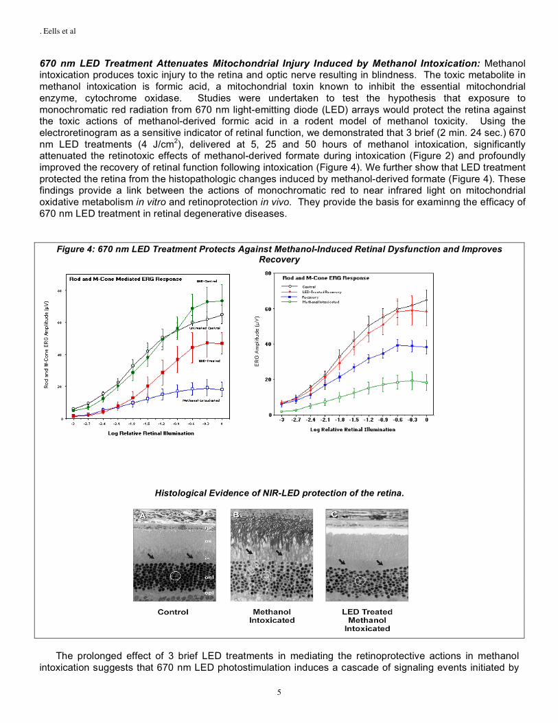
670 nm LED Treatment Attenuates Mitochondrial Injury Induced by Methanol Intoxication: Methanol intoxication produces toxic injury to the retina and optic nerve resulting in blindness. The toxic metabolite in methanol intoxication is formic acid, a mitochondrial toxin known to inhibit the essential mitochondrial enzyme, cytochrome oxidase. Studies were undertaken to test the hypothesis that exposure to monochromatic red radiation from 670 nm light-emitting diode (LED) arrays would protect the retina against the toxic actions of methanol-derived formic acid in a rodent model of methanol toxicity. Using the electroretinogram as a sensitive indicator of retinal function, we demonstrated that 3 brief (2 min. 24 sec.) 670 nm LED treatments (4 J/cm2), delivered at 5, 25 and 50 hours of methanol intoxication, significantly attenuated the retinotoxic effects of methanol-derived formate during intoxication (Figure 2) and profoundly improved the recovery of retinal function following intoxication (Figure 4). We further show that LED treatment protected the retina from the histopathologic changes induced by methanol-derived formate (Figure 4). These findings provide a link between the actions of monochromatic red to near infrared light on mitochondrial oxidative metabolism in vitro and retinoprotection in vivo. They provide the basis for examinng the efficacy of 670 nm LED treatment in retinal degenerative diseases.
Figure 4: 670 nm LED Treatment Protects Against Methanol-Induced Retinal Dysfunction and Improves Recovery
Histological Evidence of NIR-LED protection of the retina.
The prolonged effect of 3 brief LED treatments in mediating the retinoprotective actions in methanol
intoxication suggests that 670 nm LED photostimulation induces a cascade of signaling events initiated by

. Eells et al
6
the initial absorption of light by cytochrome oxidase. We have compared gene expression profiles in the neural retina of untreated rats with those from the neural retina of methanol-intoxicated rats and LED-treated methanol-intoxicated rats (ref). Results from these studies indicate that methanol intoxication and LED treatment altered the retinal expression of nearly 80 genes. At least 26 of these genes that were up-regulated in the retinas of methanol intoxicated rats were correspondingly down-regulated in the retinas of LED treated methanol intoxicated rats and the converse (Figure 5). Several functional subcategories of genes regulated by 670 nm-LED treatment were identified in retinal samples including those encoding DNA repair proteins, antioxidant defense enzymes, molecular chaperones, protein biosynthesis enzymes, and trafficking and degradation proteins. It is important to note that a number of genes associated with protection against macular degeneration, including glutathione S-transferase (GST), are upregulated by 670 nm LED treatment and genes associated with retinal damage, including TIMP3, are down regulated. (31)
Figure 5: Differential Regulation of Retinal Genes by Mitochondrial Inhibition and 670 nm LED Treatment
-20
-10
0
10
20
Retinal Genes Up or Down Regulated by Methanol Intoxication +/- LED Treatment
LED-Treated Methanol-Intoxicated
Methanol-Intoxicated
670 nm LED therapy for the treatment of Retinitis Pigmentosa: Retinitis pigmentosa (RP) is the leading cause of vision loss due to retinal degeneration. RP is a heterogenous group of retinal dystrophies characterized by photoreceptor death (8). Autosomal dominant forms of RP, which cause a majority of RP cases, result from mutations in the rhodopsin gene. Rhodopsin is the visual pigment located within the outer segment of rod photoreceptor cells and is involved in phototransduction. The exact mechanism of photoreceptor degeneration is unclear but it is thought that the rhodopsin mutation alters the tertiary structure of the protein thus altering its function. Altered rhodopsin structure leads to misfolded opsin protein. Opsin is present in the photoreceptor inner segment where mitochondria are located. Opsin binds cis-retinal chromophore forming active rhodopsin which is subsequently translocated to the photoreceptor outer segment away from the mitochondria. It is thought that misfolded opsin does not sufficiently sequester cis-retinal chromophore which can initiate free radical reactions, increase oxidative stress, and leave the photoreceptor cells vulnerable to apoptosis (1). The P23H-3 rat is a well-established rodent model of human disease. The transgene in the P23H-3 rat mimics a rhodopsin mutation found in humans in North America, and photoreceptor death is excessive, most markedly during development (P15-30). (3,17,19). Our studies describe the effect of 670 nm LED treatment administered during the critical period of photoreceptor cell loss in this animal model of RP.
P23H-3 rat pups were treated daily for 5 days during the critical period of photoreceptor development from postnatal day 16-20 with 670 nm LED array (GaAlAs LED arrays, 670 + 20 nm at 50% power; Quantum Devices, Inc., Barneveld, WI). Rats pups were hand-held by the investigator and the 10 cm2 670 nm LED array was positioned directly over the animal’s head at a distance of 2 cm exposing both eyes. Treatment consisted of irradiation at 670 nm for 3 minutes resulting in a power intensity of 50mW/ cm2

. Eells et al
7
and an energy density of 4 joules/cm2. These stimulation parameters have been previously demonstrated to stimulate cell survival and cytochrome oxidase activity in cultured neurons (20-21) to promote wound healing clinically and in experimental animal models (4,6) and to promote the survival and functional recovery of the retina and optic nerve in vivo after acute injury by a mitochondrial toxin (2). Sham treatment involved holding the animals for 3 minutes with the LED array positioned over the eyes, but not illuminated. On postnatal day 21, animals were euthanized. One eye from each animal was prepared for immunohistochemical evaluation and the retina was dissected from the other eye and flash-frozen in liquid nitrogen for biochemical analysis.
670 nm LED-induced increases in Cytochrome Oxidase Concentrations in the P23H retina: 670 nm LED treatment significantly increased the concentration of cytochome oxidase (CO), in the retina of the P23H-3 rat ( N = 6, p < 0.001). Marked increases in CO concentrations were most apparent in inner segments of the photoreceptor cells consistent with the large numbers of mitochondria present in this region of the retina. These findings are consistent with our in vitro observations and support our hypothesis that upregulation of cytochrome oxidase activity is a critical component in the neuroprotective actions of photon therapy (5,12). Moreover, recent studies have shown that cytochrome oxidase is the rate-limiting step in mitochondrial respiration (28,29) and that increased cytochrome oxidase activity improves respiratory efficiency under conditions of oxidative stress, thus attenuating the production of reactive oxygen species (29).
Figure 6: 670 nm LED Treatment Increases Cytochrome Oxidase Activity in the P23H-3 Retina
CP22 Cyotchrome oxidase Outer Segments to Outer Plexiform LayerImmunohistochemistry
P23H Sham P23H Treat 0.00.51.01.52.02.53.03.54.04.55.05.56.06.57.07.58.08.5
Mean
In
ten
sit
y o
f im
mu
no
flo
ure
sen
ce
(arb
itra
ry u
nit
s)
Control Treated
Photobiomodulation-induced increases in Cytoprotective Enzymes and Cofactors in the P23H retina: In addition to the increase in cytochrome oxidase activity and oxidative capacity, 670 nm LED treatment also significantly increased the concentrations of two important neuroprotective factors in the retina, the mitochondrial Mn-dependent form of superoxide dismutase, MnSOD, and ciliary neurotrophic factor (CNTF).
As shown in figure 7, 670 nm LED treatment significantly increased retinal concentrations of MnSOD (N = 6, p<0.001). MnSOD is the mitochondrial form of superoxide dismutase, an essential antioxidant enzyme responsible for the conversion of the highly reactive superoxide anion to the less reactive hydrogen peroxide. Mitochondria are the major source of superoxide production and are subjected to direct attack of reactive oxygen species (ROS). Mitochondria generate superoxide through a series of electron carriers arranged spatially according to their redox potentials. Mitochondrial dysfunction itself can lead to increased production of ROS, which can increase oxidative stress if the defense mechanisms of the cell are overwhelmed. ROS generated by mitochondria are considered to be integral factors in the retinal degenerative disease.

. Eells et al
8
Figure 7: 670 nm LED Treatment Increases MnSOD Activity in the P23H-3 Retina
Control Treated
Figure 8 shows that 670 nm LED treatment significantly increases the concentration of CNTF in the developing P23H retina (N = 6, p<0.001). We interpret these findings to indicate that 670 nm LED treatment upregulates the production of this neuroprotective factor by a mitochondrial mediated signaling mechanism. CNTF was first identified as a survival factor in studies involving ciliary ganglion neurons in the chick eye (9). CNTF is a member of the IL-6 family of cytokines and acts through a heterotrimeric receptor complex composed of CNTF receptor plus two signal-transducing transmembrane subunits. CNTF receptor is located on Müller glial membranes (9) and on rod and cone photoreceptors (9). In animal studies, CNTF has been shown to be effective at retarding retinal degeneration in at least 13 RP models, including transgenic rats expressing the P23H mutation (9). More recently, clinical studies have documented the potential of CNTF as therapeutic agent for RP (9)
Figure 8: 670 nm LED Treatment Increases the Protective Factor CNTF in the P23H-3 Retina
Control Treated
Photobiomoduation attenuates the loss of photoreceptors in the P23H rat during the critical period:
Our working hypothesis is that dysfunction of P23H-3 photoreceptors leads to a rise in oxygen tension in outer retina, which is toxic to photoreceptors [8], particularly during early development. We postulate that photobiomodulation acts to improve mitochondrial function and to increase antioxidant protection. Photobiomodulation-induced improvement of mitochondrial function would increase oxygen consumption in the P23H retina, reduce oxygen tension and thus reduce oxygen toxicity (8). Secondly the

. Eells et al
9
photobiomodulation-induced increased production of cytoprotective factors and antioxidants would act in concert to protect the retina.
Figure 9 shows sections of P23H-3 retinas labeled for normal DNA (blue) and for the DNA fragmentation characteristic of apoptosis (red). The frequency of TUNEL+ (dying) cells is an order of magnitude higher in the P23H-3 than in control (SD) animals (ref). 670 nm LED treatment reduced apoptotic photoreceptor cell death by more than 70% in the P23H-3 rat. These findings link the upregulation of antioxidants and cytoprotective factors to the prevention of photoreceptor apoptosis in the developing P23H-3 retina.
Figure 9: 670 nm LED Treatment Attenuates Photoreceptor Apoptosis in the P23H-3 Retina
Control Treated
In summary, we have demonstrated that 670 nm LED treatment administered once per day for 5
days during the critical period of photoreceptor development in the P23H-3 rat increases retinal mitochondrial cytochrome oxidase activity, upregulates the production of antioxidant protective enzymes and cofactors in the retina, increases the production of retinal neuroprotective neurotrophic factors and prevents apoptotic photoreceptor cell death. These findings have profound implications for the use of photobiomodulation in the treatment of retinal degenerative diseases. They document the therapeutic potential of 670 nm photon therapy in a well-established rodent model of retinitis pigmentosa, thus setting the stage for clinical trials of photobiomodulation in human disease. The molecular, biochemical and functional insights obtained from this research will provide crucial information needed for a comprehensive FDA approval for the use of far-red to near-infrared LED devices in the treatment of retinal degenerative diseases. From a basic science perspective, they substantiate our previous findings in vitro and in vivo and strongly support our hypothesis that photobiomodulation augments mitochondrial function and stimulates antioxidant protective pathways in the neural retina to protect against retinal degeneration Finally, we believe that photobiomodulation has the potential to revolutionize the delivery of health care. In contrast to traditional medical approaches that rely on surgical or pharmacological intervention, photobiomodulation harnesses the cells’ own potential for repair and provides an innovative and non-invasive therapeutic approach for the prevention and treatment of retinal dystrophies and degenerative disease.

. Eells et al
10
Literature Cited
1. Hafezi, F., Grimm, C., Simmen, B.C., Wenzel, A., Reme. C.E (2000). Molecular Ophthalmology: an update on
animal models for retinal degenerations and dystrophies. Br. J. Ophthalmol. 84: 922-927 2. Kanwar, M., Chan P.S., Kern T.S., Kowluru, R.A (2007). Oxidative damage in the retinal mitochondria of
diabetic mice: possible protection by superoxide dismutase. Invest. Ophthalmol. Vis. Sci. 48: 3805-3811. 3. Ranchon, I., LaVail, L.M., Koyake, Y., Anderson, R.E (2003). Free radical trap phenyl-N-tert-butylnitrone
protects against light damage but does not rescue P23H and S334ter rhodopsin transgenic rats from inherited retinal degeneration. J. Neurosci. 23: 6050-6057.
4. Liang, H.L., Whelan, H.T., Eells, J.T., Meng, H., Buchmann, E., Lerch-Gaggl, A., Wong-Riley, M (2006).
Photobiomodulation partially rescues visual cortical neurons from cyanide-induced apoptosis. Neuroscience. 139(2): 639-49.
5. Desmet, K.D., Paz, D.A., Corry, J.J., Eells, J.T., Wong-Riley, M.T., Henry, M.M., Buchmann, E.V., Connelly,
M.P., Dovi, J.V., Liang, H.L., Henshel, D.S., Yeager, R.L., Millsap, D.S., Lim, J., Gould, L.J., Das, R., Jett, M., Hodgson, B.D., Margolis, D., Whelan, H.T (2006). Clinical and experimental applications of NIR-LED photobiomodulation. Photomed. Laser Surg. 24(2): 121-8.
6. Wong-Riley, M.T., Liang, H.L, Eells, J.T., Chance, B., Henry, M.M., Buchmann, E., Kane, M., Whelan, H.T
(2005). Photobiomodulation directly benefits primary neurons functionally inactivated by toxins: role of cytochrome c oxidase. J. Biol. Chem. 280(6): 4761-71.
7. Eells, J., Henry, M.M., Summerfelt, P., Wong-Riley, M.T., Buchmann, E.V., Kane, M., Whelan, N.T., Whelan,
H.T (2003). Therapeutic photobiomodulation for methanol-induced retinal toxicity. Proc. Natl. Acad. Sci. 100(6): 3439-3444.
8. Stone, J., Maslim, J., Valter-Kocsi, K., Mervin, K., Bowers, F., Chu, Y., Barnett, N., Provis, J., Lewis, G., Fisher,
S.K., Bisti, S., Gargini, C., Cervetto, L., Merin, S., Peer, J (1999). Mechanisms of photoreceptor death and survival in mammalian retina. Prog. Ret. Eye. Res. 18: 689-735.
9. Sieving, P. et al., Ciliary neurotropic factor (CNTF) for human retinal degeneration: phase 1 trial of CNTF
delivered by encapsulated cell intraocular implants (2006). Proc. Natl. Acad. Sci. 103: 3896-3901 10. Gargini, C., Bisti, S., Demontis, G.C., Valter, K., Stone, J., Cervetto, L (2004). Electroretinogram changes
associated with retinal upregulation of trophic factors: observations following optic nerve section. Neuroscience. 126(3): 775-83.
11. Valter, K., Bisti, S., Gargini, C., Di Loreto, S., Maccarone, R., Cerbetto, L., Stone, J (2005). Time course of
neurotrophic factor upregulation and retinal protection against light-induced damage after optic nerve section. Invest. Ophthalmol. Vis. Sci. 46(5): 1748-54.
12. Eells, J.T., Wong-Riley, M.T., VerHoeve, J., Henry, M., Buchmann, E.V., Kane, M.P., Gould, L.J., Das, R., Jett.
M., Hodgson B.D, Margolis, D., WHelan, H.T (2004). Mitochondrial signal transduction in accelerated wound and retinal healing by near-infrared light therapy. Mitochondrion. 4(5-6): 559-67.
13. Karu, T (1999). Primary and secondary mechanisms of action of visible to near-IR radiation on cells. J.
Photochem. Photobiol. B. 49: 1-17. 14. Eells, J., Kirk, D.A., Cribb, J., Valter, K., DeSmet, K.D., VerHoeve, J.N., Whelna, H.T., Stone, J. et al., Near-
infrared light therapy for retinitis pigmentosa. Invest. Ophthalmol. Vis. Sci. 2006, 46: ARVO Abstract 1022. 15. Geller, S., Krowka, R., Valter, K., Stone, J. Toxicity of hyperoxia to the retina: evidence from the mouse, in
Retinal Degenerative Disease, J. Hollyfield, G. Anderson, and M. LaVail, Editors. 2005, Springer: 425-438. 16. Algvere, P.V., Marshall, J., Seregard, S (2006). Age-related maculopathy and the impact of blue light hazard.
Acta. Ophthalmol. Scand. 84(1): 4-15.

. Eells et al
11
17. Komeima, K., Roger, B.S, Campochiaro, P.A (2006). Antioxidants reduce cone cell death in a model of retinitis
pigmentosa. Proc. Natl. Acad. Sci. 103(30): 11300-5. 18. Wellard, J., Lee, D., Valter, K., Stone, J (2005). Photoreceptors in the rat retina are specifically vulnerable to
both hypoxia and hyperoxia. Vis. Neurosci. 22(4): 501-7. 19. Yu, D., Cringle, S., Valter, K., Walsh, N., Lee, D., Stone, J (2004). Photoreceptor Death, Trophic Factor
Expression, Retinal Oxygen Status, and Photoreceptor Function in the P23H Rat. Invest. Ophthalmol. Vis. Sci. 45(6): 2013-2019.
20. Valter, K., Maslim, J., Bowers, F., Stone, J (1998). Photoreceptor dystrophy in the RCS rat: Roles of oxygen,
debris and bFGF. Invest. Ophthal. Vis. Sci. 39: 2427-2442. 21. Nixon, P.J., Bui, B.V., Armitage, J.A., Vingrys, A.J (2001). The contribution of cone responses to rat
electroretinograms. Clin. Experimental Ophthalmol. 29(3): 193-6. 22. Stone, J., Mervin, K., Walsh, N., Valter, K., Provis, J., Penfold, P. Photoreceptor stability and degeneration in
mammalian retina: lessons from the edge, in Macular Degeneration: Science and Medicine in Practice, P. Penfold and J. Provis, Editors. 2005, Springer Verlag: 149-165.
23. Bravo-Nuevo, A., Williams, N., Geller, S., Stone, J. Mitochondrial deletions in normal and degenerating rat
retina, in Retinal Degenerations: Mechanisms and Experimental Therapy, M. La Vail, J. Hollyfield, and G. Anderson, Editors. 2003, Kluwer Academic/Plenum Publishers: New York, 241-248.
24. Qi, X., Lewin, A.S., Sun, L., Hauswirth, W.W., Guy, J (2006). Mitochondrial protein nitration primes
neurodegeneration in experimental autoimmune encephalomyelitis. J. Biol. Chem. 281(42): 31950-31962. 25. Shen, J., Yang, X., Dong, A., Petters, R.M., Peng, Y.W., Wong, F., Campochiaro, P.A (2005). Oxidative
Damage is a Potential Cause of Cone Cell Death in Retinitis Pigmentosa. J. Cell. Physiol. 203: 457-464. 26. Maslim, J., Valter, K., Egensperger, R., Hollander, H., Stone, J (1997). Tissue oxygen during a critical
developmental period controls the death and survival of photoreceptors. Invest. Ophthal. Vis. Sci. 38: 1667-1677.
27. Machida, S., Kondo, M., Jamison, J.A., Khan, N.W., Kononen, L.T., Sugawara, T., Bush, R.A., Sieving, P.A
(2000). P23H rhodopsin transgenic rat: correlation of retinal function with histopathology. Invest. Ophthalmol. Vis. Sci. 41(10): 3200-9.
28. Campian, J.L., Oian, M., Gao, X., Eaton, J.M (2004). Oxygen tolerance and coupling of mitochondrial electron
transport. J. Biol. Chem. 279(45): 46580-7. 29. Villani, G., Greco, M., Papai, S., Attardi, G (1998). Low reserve of cytochrome c oxidase capacity in vivo in the
respiratory chain of a variety of human cell types. J. Biol. Chem. 273(48):31829-36. 30. Whelan, HT, Wong-Riley, MTT, Eells, JT, VerHoeve, JN, Das, R, Jett, M (2005). DARPA soldier self care:
rapid healing of laser eye injuries with light-emitting diode technology. NATO RTO-MP-HFM 109: 1-18.