Photo-crosslinked synthetic biodegradable polymer networks ...
31
Full Terms & Conditions of access and use can be found at https://www.tandfonline.com/action/journalInformation?journalCode=tbsp20 Journal of Biomaterials Science, Polymer Edition ISSN: 0920-5063 (Print) 1568-5624 (Online) Journal homepage: https://www.tandfonline.com/loi/tbsp20 Photo-crosslinked synthetic biodegradable polymer networks for biomedical applications Bas van Bochove & Dirk W. Grijpma To cite this article: Bas van Bochove & Dirk W. Grijpma (2019) Photo-crosslinked synthetic biodegradable polymer networks for biomedical applications, Journal of Biomaterials Science, Polymer Edition, 30:2, 77-106, DOI: 10.1080/09205063.2018.1553105 To link to this article: https://doi.org/10.1080/09205063.2018.1553105 © 2019 The Author(s). Published by Informa UK Limited, trading as Taylor & Francis Group. Accepted author version posted online: 29 Nov 2018. Published online: 12 Jan 2019. Submit your article to this journal Article views: 1427 View related articles View Crossmark data Citing articles: 7 View citing articles
Transcript of Photo-crosslinked synthetic biodegradable polymer networks ...

Full Terms & Conditions of access and use can be found athttps://www.tandfonline.com/action/journalInformation?journalCode=tbsp20
Journal of Biomaterials Science, Polymer Edition
ISSN: 0920-5063 (Print) 1568-5624 (Online) Journal homepage: https://www.tandfonline.com/loi/tbsp20
Photo-crosslinked synthetic biodegradablepolymer networks for biomedical applications
Bas van Bochove & Dirk W. Grijpma
To cite this article: Bas van Bochove & Dirk W. Grijpma (2019) Photo-crosslinked syntheticbiodegradable polymer networks for biomedical applications, Journal of Biomaterials Science,Polymer Edition, 30:2, 77-106, DOI: 10.1080/09205063.2018.1553105
To link to this article: https://doi.org/10.1080/09205063.2018.1553105
© 2019 The Author(s). Published by InformaUK Limited, trading as Taylor & FrancisGroup.
Accepted author version posted online: 29Nov 2018.Published online: 12 Jan 2019.
Submit your article to this journal
Article views: 1427
View related articles
View Crossmark data
Citing articles: 7 View citing articles

REVIEW ARTICLE
Photo-crosslinked synthetic biodegradable polymernetworks for biomedical applications
Bas van Bochovea and Dirk W. Grijpmaa,b
aDepartment of Biomaterials Science and Technology, Faculty of Science and Technology, TechnicalMedical Centre University of Twente, Enschede, The Netherlands; bDepartment of BiomedicalEngineering, W. J. Kolff Institute, University Medical Centre, University of Groningen, Groningen, TheNetherlands
ABSTRACTPhoto-crosslinked synthetic biodegradable polymer networks arehighly interesting materials for utilization in biomedical applica-tions such as drug delivery, cell encapsulation and tissue engin-eering scaffolds. Varying the architecture, chemistry, degree offunctionalization and molecular weight of the macromer precursormolecules results in networks with a wide range of physical- andmechanical properties, crosslinking densities, degradation charac-teristics and thus in potential applications. Photo-crosslinked net-works can easily be prepared and have the possibility to entrapa wide range of (biologically active) substances and cells.Additionally, spatial and temporal control over the crosslinking pro-cess when using additive manufacturing processes, allows for thepreparation of network structures with complex shapes. Photo-crosslinked networks have been used to prepare drug delivery devi-ces, as these networks allow for drug delivery in a controlled wayover a prolonged period of time. Furthermore, additive manufactur-ing techniques such as extrusion-based additive manufacturing andstereolithography have been used to prepare photo-crosslinked tis-sue engineering scaffolds. This allows for the preparation ofdesigned porous structures with precise control over the pore sizeand pore architecture and optimal mechanical properties. In par-ticular for stereolithography, a wide variety of resins based on bio-degradable photo-crosslinkable macromers has been developed.
ARTICLE HISTORYReceived 20 September 2018Accepted 25 November 2018
KEYWORDSPhoto-crosslinking; bio-degradable polymernetworks; biomedicalapplications;stereolithography
Photo-crosslinked synthetic biodegradable polymer networks
In polymer networks, the macromolecular chains are attached to each other by cova-lent bonds. In these materials viscous flow is not possible and creep is restricted.Especially in polymeric materials with low glass transition temperatures, such as
CONTACT Dirk W. Grijpma [email protected] University of Twente, PO Box 217, 7500 AE Enschede,The Netherlands
� 2019 The Author(s). Published by Informa UK Limited, trading as Taylor & Francis Group.
This is an Open Access article distributed under the terms of the Creative Commons Attribution-NonCommercial-NoDerivatives License (http://creativecommons.org/licenses/by-nc-nd/4.0/), which permits non-commercial re-use, dis-tribution, and reproduction in any medium, provided the original work is properly cited, and is not altered, trans-formed, or built upon in any way.
JOURNAL OF BIOMATERIALS SCIENCE, POLYMER EDITION2019, VOL. 30, NO. 2, 77–106https://doi.org/10.1080/09205063.2018.1553105

elastomers, it is important to prevent creep and so ensure form-stability of the mater-ial. Biodegradable elastomers have gained much attention in the biomedical field forapplication as flexible tissue engineering scaffolds and controlled drug delivery sys-tems [1]. Biodegradable materials are preferred, as these avoid a long-term foreignbody response [2]. In tissue engineering, biodegradability allows for the growing tis-sue to slowly replace the scaffolding material and in drug delivery systems the drugrelease characteristics can be controlled by the degradation process [3]. Covalentlycrosslinked biodegradable elastomers have been prepared by reactions of end-func-tionalized polymers or oligomers. For example by free addition reactions or step poly-merization reactions [4]. In some cases, biodegradable polymers have beencrosslinked by actinic radiation such as by gamma irradiation [5].
A very effective method to prepare such polymer networks is by photo-crosslink-ing oligomers that contain photo-polymerizable groups. Three specific photo-poly-merization reactions can be distinguished [6]:
1. [2þ 2] cyclo dimerization reactions using end-groups such as cinnamate-, cou-marin- or thymine end-groups[6–8],
2. radical recombination reactions leading to inter- and intramolecular crosslinkingutilizing end-groups such as phenyl azide-, dithiocarbamate- and benzophenoneend-groups, and
3. radical polymerization reactions using end-groups such as styryl-, fumarate- or(meth)acrylate end-groups (see Figure 1A) [6,9]. (Note that fumarate groups canbe incorporated into the main chain [10].
Figure 1. (A) Photo-polymerizable (meth)acrylate- and fumarate end-groups. (B) Network formationby photo-crosslinking.
78 B. VAN AND D. BOCHOVEGRIJPMA

Radical photo-polymerizations have been used most often to polymerize the end-groups of these oligomers (macromers) and form a covalent network [9]. In the pro-cess of photo-crosslinking, a photo-initiator dissociates upon illumination and formsone or more radicals. These radicals can react with the double bonds of macromers,forming non-degradable carbon-carbon chains that act as multifunctional crosslin-kages [11]. This is schematically presented in Figure 1(B). (Although thermal-cross-linking would also be possible, the radical initiator is then formed upon heating,relatively high temperatures and reaction times are required [12]. Photo-crosslinkingis relatively rapid and efficient and can be done at low temperatures, making it thusmore advantageous compared to thermal-crosslinking).
Oligomers with fumarate (end-)groups are interesting materials for preparing bio-degradable networks. Fumaric acid is found in the human body and therefore it isexpected that residual fumarate groups will be biocompatible and non-toxic [9,13].However, compared to (meth)acrylate functionalized oligomers, the reactivity offumarate-functionalized oligomers is relatively low and therefore the use of reactivediluents is required [14–16]. The use of such reactive diluents will lead to an increasein the non-degradable part of the networks. For applications in medicine where bio-degradability of the implant is desired the non-degradable content of the implantshould be as low as possible [11]. Thus, for such applications (meth)acrylate function-alized oligomers are preferred.
Polymers and oligomers used in the preparation of photo-crosslinked syntheticbiodegradable networks
There are many biodegradable polymers and oligomers that have been used to pre-pare photo-crosslinkable macromers for biodegradable polymer networks. Examplesinclude poly(D,L-lactide) (PDLLA) [17], poly(e-caprolactone) (PCL) [18,19], poly(tri-methylene carbonate) (PTMC) [20], poly(ethylene carbonate) (PEC) [21], and blockcopolymers containing poly(ethylene glycol) (PEG), poly(propylene glycol) (PPG) orpoly(tetramethylene glycol) (PTMG) and poly(glycolide) (PGA), PDLLA or PCL seg-ments [22,23]. Most of these polymers can readily synthesized by the ring openingpolymerization of their cyclic monomers. The polymerization is usually initiated byan alcohol and catalyzed by stannous octoate [24] This reaction is usually performedin the melt at temperatures between 90 and 180 �C [1]. By adjusting the amount andthe functionality of the hydroxyl-group terminated alcohols used as initiator, themolecular weight and architecture of the synthesized oligomers can be precisely con-trolled [25].
An overview the networks described below and their properties is given inTable 1.
PolyestersPolyesters are a group of polymers that contain an ester group in their main chain.Biodegradable polyesters used to prepare synthetic biodegradable networks includePDLLA and PCL.
JOURNAL OF BIOMATERIALS SCIENCE, POLYMER EDITION 79
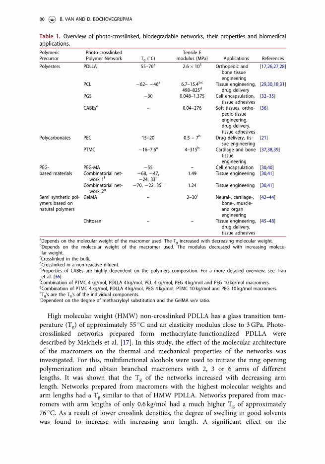
High molecular weight (HMW) non-crosslinked PDLLA has a glass transition tem-perature (Tg) of approximately 55 �C and an elasticity modulus close to 3GPa. Photo-crosslinked networks prepared form methacrylate-functionalized PDLLA weredescribed by Melchels et al. [17]. In this study, the effect of the molecular architectureof the macromers on the thermal and mechanical properties of the networks wasinvestigated. For this, multifunctional alcohols were used to initiate the ring openingpolymerization and obtain branched macromers with 2, 3 or 6 arms of differentlengths. It was shown that the Tg of the networks increased with decreasing armlength. Networks prepared from macromers with the highest molecular weights andarm lengths had a Tg similar to that of HMW PDLLA. Networks prepared from mac-romers with arm lengths of only 0.6 kg/mol had a much higher Tg of approximately76 �C. As a result of lower crosslink densities, the degree of swelling in good solventswas found to increase with increasing arm length. A significant effect on the
Table 1. Overview of photo-crosslinked, biodegradable networks, their properties and biomedicalapplications.PolymericPrecursor
Photo-crosslinkedPolymer Network Tg (�C)
Tensile Emodulus (MPa) Applications References
Polyesters PDLLA 55–76a 2.6� 103 Orthopedic andbone tissueengineering
[17,26,27,28]
PCL �62– �46a 6.7–15.4b,c
498–825dTissue engineering,
drug delivery[29,30,18,31]
PGS �30 0.048–1.375 Cell encapsulation,tissue adhesives
[32–35]
CABEse – 0.04–276 Soft tissues, ortho-pedic tissueengineering,drug delivery,tissue adhesives
[36]
Polycarbonates PEC 15–20 0.5 – 7b Drug delivery, tis-sue engineering
[21]
PTMC �16–7.6a 4–315b Cartilage and bonetissueengineering
[37,38,39]
PEG-based materials
PEG-MA �55 – Cell encapsulation [30,40]Combinatorial net-
work 1f�68, �47,�24, 33h
1.49 Tissue engineering [30,41]
Combinatorial net-work 2g
�70, �22, 35h 1.24 Tissue engineering [30,41]
Semi synthetic pol-ymers based onnatural polymers
GelMA – 2–30i Neural-, cartilage-,bone-, muscle-and organengineering
[42–44]
Chitosan – – Tissue engineering,drug delivery,tissue adhesives
[45–48]
aDepends on the molecular weight of the macromer used. The Tg increased with decreasing molecular weight.bDepends on the molecular weight of the macromer used. The modulus decreased with increasing molecu-lar weight.
cCrosslinked in the bulk.dCrosslinked in a non-reactive diluent.eProperties of CABEs are highly dependent on the polymers composition. For a more detailed overview, see Tranet al. [36].fCombination of PTMC 4 kg/mol, PDLLA 4 kg/mol, PCL 4 kg/mol, PEG 4 kg/mol and PEG 10 kg/mol macromers.gCombination of PTMC 4 kg/mol, PDLLA 4 kg/mol, PEG 4 kg/mol, PTMC 10 kg/mol and PEG 10 kg/mol macromers.hTg’s are the Tg’s of the individual components.iDependent on the degree of methacryloyl substitution and the GelMA w/v ratio.
80 B. VAN AND D. BOCHOVEGRIJPMA

mechanical properties of the networks was not observed, all networks having proper-ties similar to those of HMW PDLLA.
Poly(e-caprolactone) is a semi-crystalline, highly biocompatible polymer with a lowTg of approximately �60 �C, a melting point close to 65 �C and an elasticity modulusof approximately 260MPa [19,25,49]. The thermal properties of photo-crosslinkednetworks prepared from methacrylated PCL have been described by Elomaa et al. andZant et al. [29,30]. Interestingly, the PCL networks were found to be amorphous. Fornetworks prepared from macromers with a low molecular weight (below 4 kg/mol)the Tg is 10–15 �C higher than that of their respective macromers. For networks pre-pared from macromers with molecular weights of 4 kg/mol and higher, Tg is similarto that of linear PCL. Elomaa et al. further evaluated the swelling ratios and mechan-ical properties of the prepared PCL networks [29]. As can be expected, the swellingratio of PCL networks in good solvents increases with increasing molecular weight ofthe macromer used to prepare the networks. The networks behaved in a rubber-likemanner and showed elastic deformation. With increasing molecular weights, the elas-tic modulus of the networks decreased while their elongation at break increased.
Amsden and coworkers synthesized a series of poly(e-caprolactone-co-D,L-lactide)macromers [50–53] and prepared the corresponding networks by photo-crosslinking.The glass transition temperature of networks prepared from poly(e-caprolactone-co-D,L-lactide) macromers with a 50:50 molar ratio composition were close to �3 �Cand independent of the molecular weight of the macromers [50].
Poly(glycerol sebacate) (PGS) is a semicrystalline polymer with low Tg between�52 and �18 �C [54]. At 37 �C it is completely amorphous [54,55]. Photo-crosslinkednetworks of PGS have been prepared by crosslinking acrylated PGS [32–35]. Themechanical properties of these networks a linearly depend on the degree of acryla-tion [32]. With increasing degree of acrylation the modulus increases, as does theultimate tensile strength. The moduli varied from 0.048MPa to 1.375MPa with thehighest ultimate tensile strength of 0.498MPa. Increasing the macromer molecularweight results in increasing strains at break [34]. Potential applications include cellencapsulations devices [33] and tissue adhesives [35].
Citrate based biodegradable elastomers (CABEs) are polyesters prepared by thereaction of a diol with citric acid [36]. Polymers with a wide range of properties canbe obtained by varying the diol length, chemical composition of the diol, and partialreplacement of the citric acid with other diacids. Acrylate- and fumerate functional-ized CABEs have been developed to allow for photo-crosslinking, which resulted instrengthened networks and allowed fine tuning of the mechanical and degradationproperties of the materials [56–58]. Proposed applications of these type of networksinclude coatings, films, and devices for tissue engineering and drug delivery.
PolycarbonatesPolycarbonates are a group of polymers that contain a carbonate ester group in theirmain chain. Polycarbonates that have been used to prepare synthetic biodegradablenetworks are PEC and PTMC.
Poly(ethylene carbonate) networks have been prepared by the thermal degradationof high molecular weight PEC, subsequent acrylation an photo-crosslinking [21].
JOURNAL OF BIOMATERIALS SCIENCE, POLYMER EDITION 81

These networks had a Tg of approximately 20 �C meaning the networks were rubberyat room temperature. The elastic modulus of the networks decreased with increasingmacromer molecular weight. Potential applications include drug delivery and tissueengineering scaffolds.
Poly(trimethylene carbonate) is an amorphous polymer with a low Tg of approxi-mately �16 �C [37,59]. The mechanical properties of PTMC are strongly dependenton its molecular weight [5]. Non-crosslinked, low molecular weight (LMW) PTMC issoft and gummy, and has very low modulus and tensile strength. As a result, TMCwas mainly used as comonomer to reduce the modulus of lactide and glycolide poly-mers. Non-crosslinked, high molecular weight (HMW) PTMC is tough, flexible andto some extent shows rubber-like recovery after mechanical deformation. By prepar-ing networks from methacrylate-functionalized PTMC oligomers, creep resistant net-works with excellent mechanical properties could be obtained [37].
Figure 2 shows stress-strain curves of PTMC networks prepared from macromers(methacrylate end-functionalized) of different molecular weights. Networks preparedfrom macromers with molecular weights lower than 1800 g/mol were rigid and brittle.In contrast, networks prepared from macromers with molecular weights higher than10 kg/mol were rubber-like with elastic moduli of approximately 5MPa. The max-imum tensile strengths and elongations at break of the networks increased withincreasing molecular weight of the macromers used. As was the case for PCL, theswelling ratios of the networks in a good solvent increased with increasing molecularweights. Interestingly, the Tg values of networks prepared from very low molecularweight macromers were relatively high (the Tg of networks prepared from a macro-mer with Mn of 1 kg/mol was 7.6 �C). With an increase in the molecular weight of
Figure 2. Stress-strain curves of PTMC networks prepared by photo-crosslinking PTMC macromers(methacrylate end-functionalized) of different molecular weights. In the figure, the molecularweights of the macromers used to prepare the networks are shown with the corresponding stress-strain curves. Reprinted with permission from [37].
82 B. VAN AND D. BOCHOVEGRIJPMA

the macromers, the Tg of the corresponding networks approached the Tg value ofHMW PTMC.
PTMC networks have been investigated for a variety of medical applications, whichinclude cartilage tissue engineering [60], annulus fibrosus tissue engineering [61,62],meniscus tissue engineering [38], preparation of microvascular networks [63] andorbital floor implants [39].
To allow tuning of the mechanical- and degradation properties, copolymer net-works of TMC with DLLA and/or e-CL have been extensively investigated [7,9,50,64].Copolymerizing TMC and DLLA, subsequent functionalization with methacrylate end-groups to yield poly(trimethylene carbonate-co-D,L-lactide) macromers, andphoto-polymerization allows the formation of copolymer networks in which theglass transition temperature depends on the ratio of the co-monomers [9,16]. Inthis way networks with a wide range of mechanical properties can be obtained.For example, Sharifi et al. used such networks to prepare structures with shapememory behavior: the temporary shape of the structure is fixed at temperaturesbelow Tg of the copolymer, it then returns to its original permanent shape uponheating to body temperature. Surgically implantable devices prepared from thesephoto-crosslinked poly(trimethylene carbonate-co-D,L-lactide) macromers, can beused in minimal invasive surgery [65,66]. An example of such an implant is shownin Figure 3.
PTMC degrades without the formation of acidic degradation products [51,67].Therefore, preparing biodegradable networks from functionalized TMC and DLLAcopolymers instead of from DLLA homo-polymers may be beneficial in applicationssuch as drug delivery or bone tissue engineering [68].
Copolymerizing TMC and e-CL to obtain poly(trimethylene carbonate-co-e-caprolactone) macromers results in networks with low glass transition tempera-tures ranging from �23 to �50 �C, depending on the e-CL content [9,51,69].These networks are rubbery and amorphous at room temperature, with relativelylow elastic moduli [51,69]. Copolymer networks of poly(TMC-co-e-caprolactone-co-D,L-lactide) macromers were prepared as well [64]. These networks wereinvestigated for drug release purposes and were able to show sustained release formore than 10 days.
Figure 3. Shape recovery of a 3D structure prepared from photo-crosslinked P(DLLA-co-TMC) mac-romers. (A) Temporary shape of the structure at 0 �C. (B) Transient shape of the structure duringheating at 37 �C. (C) Completely recovered structure at 37 �C. Reprinted with permission from [66].
JOURNAL OF BIOMATERIALS SCIENCE, POLYMER EDITION 83

PEG-based materialsIn contrast to natural hydrogels, synthetic hydrogels can readily be prepared, proc-essed and tailored. Hydrogels are interesting for biomedical applications, as they pro-vide highly swollen 3D environments which are similar to soft tissues [70].Furthermore, they allow easy diffusion of nutrients, waste products and drugs.
One of the major hydrophilic synthetic polymers used to prepare hydrogels forbiomedical applications is poly(ethylene glycol) (PEG) [70]. PEG is a biocompatible,non-toxic and water-soluble polymer [23]. As a result, PEG is used in wide range ofbiomedical applications including drug delivery, tissue engineering and implant sur-face modification.
The end groups of PEG oligomers and polymers are hydroxyl groups. These canbe reacted to yield other functional end groups, which include (meth)acrylates thatallow photo-crosslinking into hydrogel networks. Photo-crosslinking is the most com-mon method to make PEG hydrogel networks [70]. Hydrogels prepared from PEG-methacrylate have a low Tg of approximately �55 �C, and high water uptake of900–1700wt% [30]. Networks prepared from (meth)acrylated PEG are not readilydegradable in vivo [71], but below a molecular weight of approximately 30 kg/mol itcan be excreted from the body via the renal pathway [72]. However, by using PEG asinitiator in the ring opening polymerization reaction of polyesters or TMC, biodegrad-able hydrogel networks containing high amounts of PEG can be prepared [23,73]. Bycarefully adjusting the molecular weight of the PEG and the composition of the TMC/ester (co)polymer segment, the degradation behavior and drug release profile of suchnetworks can be controlled.
Basic PEG hydrogels have resistance to protein adsorption [74], and in general alack of cell specific adhesion [70]. For tissue engineering purposes, the lack of celladhesion is a major limitation. Modification of PEG hydrogels with cell adhesive pep-tides (CAPs) derived from the extracellular matrix have been researched extensivelyto overcome these limitations. Photo-polymerization of PEG diacrylates and acrylatedpeptides, peptide monoacrylates [75] and peptide diacrylates [76] resulted in networkswith significantly higher cell adhesion and spreading compared to basicPEG hydrogels.
In a combinatorial approach, Zant et al. used mixtures of homo-polymeric macro-mers based on methacrylated DLLA, TMC, CL and PEG to prepare 255 differentphoto-crosslinked networks in solution [30]. After extraction and drying, thesemixed-macromer networks were evaluated with regard to their physical and biologicalcharacteristics in a high throughput manner. Two macromer combinations consistingof i) PTMC 4 kg/mol, PDLLA 4 kg/mol, PCL 4 kg/mol, PEG 4 kg/mol and PEG 10 kg/mol and ii) PTMC 4 kg/mol, PDLLA 4 kg/mol, PEG 4 kg/mol, PTMC 10 kg/mol andPEG 10 kg/mol, showed interesting properties. These networks had high water uptake(approximately 190%), showed excellent cell adhesion, and at the same time possessedexcellent mechanical properties. The elastic moduli where up to 1.49MPa and thenetworks were very resistant to tearing. Porous structures prepared from these macro-mer combinations could be compressed up to 80% without failure. It was hypothe-sized that the excellent properties of these networks were due to phase separation ofthe different macromers [41]. Phase separation was shown by the presence of glass
84 B. VAN AND D. BOCHOVEGRIJPMA

transition temperatures that corresponded to the individual macromer components inDSC, as well as by AFM and XRD.
Semi-synthetic polymersSemi synthetic polymers are chemically modified natural polymers. Some examplesinclude modified gelatin and chitosan.
Gelatin is a substance consisting of denatured and partially hydrolyzed natural col-lagen [77]. Gelatin is an attractive material for tissue engineering and drug delivery asit is biocompatible, biodegradable, can easily be manipulated and can be used at lowcost [78,79]. However, gelatin is instable at body temperature and therefor it needs tobe covalently crosslinked for use as biomaterial [80]. Gelatin modified with photo-crosslinkable side groups, gelatin-methacryloyl (gelMA), allows for crosslinking with ahigh degree of control of the properties of the obtained hydrogel network [78]. Thecrosslinking of gelMA leads to hydrogel that is stiffer as compared to its non-cross-linked counterpart [81]. The mechanical properties of photo-crosslinked gelMA wereshown to be directly proportional to the degree of methacryloyl substitution (DS) andthe gelMA mass/volume ratio [42–44]. When the DS increased from approximately50 to 73%, the compression modulus of gelMA hydrogels increased from 2.0 to4.5 kPa [43]. Likewise, for gelMA with a DS of 54% the compression moduli of net-work with a w/v ratio of 5, 10 and 15% were 2, 10 and 22 kPa respectively while net-works prepared from gelMA with a DS of 81% and the same w/v ratios had moduliof 3, 16 and 30 kPa [44]. GelMA hydrogels have been shown to be highly adaptableand have been researched for a wide range of biomedical applications includingneural, cartilage, bone, muscle and organ engineering [42].
Polysaccharide-based hydrogels are materials with interesting properties for bioma-terials as they are non-toxic, low cost in use, biocompatible and biodegradable [82].One of these polysaccharides, chitosan, is a well-researched polysaccharide obtainedfrom the alkaline hydrolysis of chitin [83,84]. On the chitosan chain, many amineand hydroxyl groups are available for reaction with photo-crosslinkable functionalgroups to prepare photo-crosslinked hydrogel networks [85,86]. These networks havebeen used in tissue engineering and drug delivery systems [45]. Methacrylated glycolchitosan could be crosslinked with blue light initiators [46]. These materials had bet-ter mechanical properties with increasing crosslinking time and the stability and deg-radation were depending on the mechanical properties. An injectable, photo-crosslinkable, chitosan based hydrogel was developed based on chitosan, PEGDA andN,N-dimethylacrylamide (DMMA) [47]. This material had excellent mechanical prop-erties, was thermally stable, showed no cytotoxicity and was able to promote celladhesion and proliferation. A photo-crosslinkable chitosan adhesive showed superiorstrength compared to fibrin glue, potentially due to covalent binding with tissue pro-teins [48].
Degradation and erosion of synthetic biodegradable networks
To successfully apply the previously described networks in the biomedical field, it isessential to understand the degradation and erosion behavior of the networks.
JOURNAL OF BIOMATERIALS SCIENCE, POLYMER EDITION 85

Degradation is defined as the process in which polymer chains are cleaved, while ero-sion is defined as the loss of material mass as a result of dissolution and diffusion ofthe soluble low molecular weight compounds that are formed upon degradation [87].
Degradation can occur by a variety of mechanisms, including hydrolysis, thermoly-sis and mechanical or oxidative stress [12]. Hydrolyzable bonds such as ester-, anhyd-ride-, amide- and carbonate bonds can be found in the main chains of manysynthetic biodegradable polymers. These bonds can be cleaved upon reaction withwater, either enzymatically or non-enzymatically. Factors that influence the rate ofdegradation are glass transition temperature, hydrophilicity, crosslinking density, pH,presence of proteins, nature of the labile bond and accessibility of the bonds to wateror enzymes.
Biodegradable polymers and polymer networks can be categorized as surface- orbulk eroding materials [12]. Erosion is a complex process that depends on polymerdegradation, polymer molecular weight, swelling, and diffusion of water, monomersand oligomers [88]. Surface eroding polymers lose material from the surfaceonly [12]. Therefore, the rate of the loss of mass and the change in dimensions of thepolymeric device depend on its surface area. As the molecular weight of the remain-ing polymer remains essentially the same, the strength of the material essentiallyremains unchanged. This is shown in Figure 4(A). In bulk degradation, the mass andthe dimensions of the material remain unchanged for relatively long times. However,the molecular weight of the material decreases significantly [12]. Upon reaching acritical low molecular weight the material loses it mechanical strength, potentiallywith dramatic mechanical failure of the implant as a result. Rapid release of degrad-ation products then also occurs. This is shown in Figure 4(B).
Although most biodegradable polymers and polymer networks degrade by bulkerosion, surface eroding materials are to be preferred [12]: in medical implants and
Figure 4. Schematic illustration of the processes of surface erosion (A) and bulk erosion (B). Theeffect of degradation on strength, molecular weight and mass of the remaining material is shown.Reprinted with permission from [89].
86 B. VAN AND D. BOCHOVEGRIJPMA

tissue engineering scaffolds the mechanical properties and structural integrity of theimplants are maintained during the functional life time of the implant. For drugdelivery devices, the predictability of the surface erosion process allows for well-con-trolled release. The sequential release of the bioactive component takes place at a con-stant release rate (zero-order release).
The degradation and erosion behavior of photo-crosslinked networks has beenstudied extensively [26,52,64,90]. PDLLA networks degraded hydrolytically inapproximately 40weeks via bulk erosion [26]. The networks were form-stable andshowed very little mass loss in the first 6months. The mechanical propertiesremained unchanged for approximately 15weeks, then the materials failed catastroph-ically with near complete mass loss in a very short time.
Interestingly, the degradation mechanism of copolymeric poly(e-caprolactone-co-D,L-lactide) networks appeared to depend on the crosslink density [52]. Networksprepared from end-functionalized macromers with low molecular weights (i.e. highcrosslink density) degraded via surface erosion, while networks prepared form highermolecular weight macromers degraded via bulk erosion.
While the degradation of many polyester networks such as PDLLA and PCL havebeen extensively researched, the degradation products of the networks have not beenextensively analyzed. However, polyester degradation is in general characterized bythe formation of acidic compounds [91,92]. These acidic compounds may be harmfulin applications such as bone tissue engineering [39]. Analyses of the degradationproducts of these PDLLA networks showed that the ester bonds with the poly(lactide)chains were much more prone to hydrolyses than the ester bonds between the lactideand the methacrylates [26]. As a result, the degradation products are lactic acid, lowmolecular weight oligomers and poly(lactic acid methacrylate) chains. The averagelength of these poly(lactic acid methacrylate) chains was between 1.1 and 3.5 kg/mol,falling within the range of effective and fast renal clearance.
PGS degrades via surface erosion [54,93], primarily due to cleavage of the esterlinkages [55]. PGS networks degrade at a slower rate, indicating that the alkyl cross-links formed by the acrylate groups are less susceptible to degradation [32]. No evi-dence of inflammation or necrosis was observed upon PGS implantation anddegradation in vivo [34]. CABEs degrade through hydrolysis of the esterlinkages [36,56] where shorter, more hydrophilic diols used in the polymerizationresulted in faster degradation [36,57].
PEC networks degrade in vivo via surface erosion [21]. Macrophages are heavilyinvolved in the degradation. It was shown that increased crosslinking densitiesdecreased the degradation rates.
PTMC networks degrade by enzymatic surface erosion. The degradation rate ofnetworks prepared from PTMC macromers was found to depend on the molecularweight of the macromers used to prepare these networks [90]. Other studies showedthat networks prepared by photo-crosslinking linear HMW PTMC in mixtures withlow molecular weight PTMC macromers as a cross-linker also degrade via surfaceerosion [94,95]. In vivo, the surface erosion of PTMC may be mediated by macro-phages. It was shown that after culturing macrophages on PTMC network films, pitshad formed on the surface and loss of mass was observed [95]. Degradation
JOURNAL OF BIOMATERIALS SCIENCE, POLYMER EDITION 87

experiments on linear high molecular weight PTMC showed that PTMC degradedwithout producing acidic degradation products [67].
An oxidative degradation mechanism was proposed for PTMC [51] and PEC [21].This degradation proceeds by the formation of ionic end groups via a nucleophilicattack of superoxide ions. This is then followed by a chain unzipping reaction ofwhich TMC and EC monomers are among the degradation products. In vivo, thedegradation products of PTMC may also include 1,3-propanediol and CO2 [51].
Poly(TMC-co-e-caprolactone-co-D,L-lactide) macromers were used to prepare net-works that had higher degradation rates than poly(trimethylene carbonate-co-D,L-lac-tide) networks, but released minimal amounts of acidic degradation products [64].
Gelatin is prepared from collagen extracted from bovine- or porcine skin underacidic or basic conditions [78]. The bioactive sequences for cell attachment and met-alloproteinase (MMP) sensitive degradation sites in the collagen remain present inthe gelatin backbone. In vitro, enzymatic degradation of GelMA networks isobserved [42,96,97]. The degradation products are particularly oligomethacrylates [78,98].GelMA hydrogels prepared from gelatin obtained under basic conditions showed goodbiocompatibility. In contrast, GelMA hydrogels prepared from gelatin prepared underacidic condition showed inflammatory reactions, possibly due to high concentrations ofendotoxins in this type of gelatin. Polysaccharides such as chitosan degrade into non-toxic oligosaccharides which can be excreted or incorporated into glycosaminoglycansand glycoproteins [82].
Biomedical applications of synthetic biodegradable networks
Photo-crosslinked biodegradable networks form an interesting group of materials forbiomedical applications [99]. This interest relates to:
1. the ease of preparation (also in vivo),2. the possibility to entrap a wide range of substances and even cells in the net-
works [100], and3. the spatial and temporal control over the polymerization process which allows
for the preparation of network structures with complex shapes [101].
As a result, photo-crosslinked biodegradable networks have been studied for a var-iety of applications such as drug delivery [102] and tissue engineering [3].
Drug delivery devices
Controlled and sustained delivery greatly improves the therapeutic efficacy and safetyof drugs. [103]. Ideally, implantable drug delivery devices are biodegradable as theywill not need to be removed after the drug has been delivered [104].
Photo-crosslinked biodegradable polymer networks are an interesting group ofmaterials for application in drug delivery devices [105]. Through photo-crosslinking,drugs can easily be entrapped in the networks by dissolving or dispersing the drugsinto the macromer solution prior to crosslinking [73,106]. This allows for large
88 B. VAN AND D. BOCHOVEGRIJPMA

amounts of drugs to be loaded into the devices at high efficiencies. As photo-cross-linking is fast and can be performed with minimal heat generation, heat-sensitivecompounds such as proteins can be incorporated as well. Detrimental reactions ofproteins with free radicals [107] are avoided, as in the photo-crosslinking the macro-mers act as free radical scavengers [108].
Photo-crosslinked biodegradable polymer networks allow control over the rate ofrelease of the incorporated compounds by variation of the crosslink density and com-position of the networks [64,68,73,105,106,109]. Different studies showed that lessdensely crosslinked networks released incorporated compounds faster than moredensely crosslinked networks [106]. Furthermore, several studies showed that morehydrophilic networks lead to more rapid release [105,106,109]. In block-copolymerichydrogel networks, variation of the hydrophilicity of the networks by varying thechemical composition if the hydrophobic segment allowed good control of drugrelease profiles [73].
In addition, poly(ester anhydride) networks prepared from PCL show great versa-tility for drug delivery [18,110–113]. Hydroxyl group-terminated PCL oligomers areacid-functionalized with an anhydride compound to yield acid-terminated oligomers.Subsequent methacrylate-functionalization results in photo-crosslinkable methacrylatefunctionalized oligomers (macromers). Regular PCL networks degrade veryslowly. The addition of the labile anhydride bond results in much faster degradation.PCL-anhydride networks prepared from low molecular weight precursors degrade in48 hours [110]. The addition of the alkenyl chain in the anhydride bond result inslower degradation of 64 to 72 hours for alkenyl chains containing 12 or 18 carbonsrespectively [111]. The degradation can be further slowed down by increasing themolecular weight of the PCL precursors [113]. The control over the degradation timeof these types of polymer networks results in control over the drug release time.These networks have been investigated for the controlled release of drugsand peptides.
For drug delivery devices, it is important that the formation of large amounts ofacidic degradation products is avoided [64,68]. In PLGA release systems, is wasshown that the degradation of such systems resulted in a drop of the pH within thedevice due to acidic monomeric and oligomeric degradation products [114,115]. Thiscan result in the denaturation of acid labile proteins such as VEGF in about7–10 days [68]. Cleland et al. reported that VEGF released from PLGA lost approxi-mately 13% of its heparin binding affinity in 8 days [116] and Kim et al. showed thatreleased VEGF lost 25% if its bioactivity [117].
Tissue engineering scaffolds
In tissue engineering, biodegradable scaffolds are used in combination with cells and/or biologically active compounds to induce the (re)generation of tissues in vitro or invivo [118]. Scaffolds are porous implants intended to provide temporary support forcells and the formed tissues. Such scaffolds ideally have a high porosity, good poreinterconnectivity and optimal pore sizes for an intended application [119–121]. Thescaffolds need to be biocompatible, biodegradable at a rate which matches the tissue
JOURNAL OF BIOMATERIALS SCIENCE, POLYMER EDITION 89
replacement, and have mechanical properties that are compatible with those of thetissues that are to be regenerated [119,122,123].
Conventional techniques used to fabricate tissue engineering scaffolds include solv-ent casting, particulate porogen leaching, phase separation, membrane lamination,melt molding, injection molding and freeze drying [119,120,124]. Several of thesetechniques have also been used to prepare photo-crosslinked porous struc-tures [125–127]. For example, porous tubular scaffolds for vascular tissue engineeringhave been prepared by photo-crosslinking a mixture of photo-crosslinkable PTMCmacromers and salt particles, followed by leaching of the salt [125]. Porous photo-crosslinked scaffolds have also been prepared by employing temperature-inducedphase separation [126,127]. Upon cooling macromer solutions ethylene carbonate (acrystallizable solvent), subsequent photo-crosslinking of the matrix and extraction ofthe dispersed ethylene carbonate crystals with water, a porous photo-crosslinkedstructure is obtained.
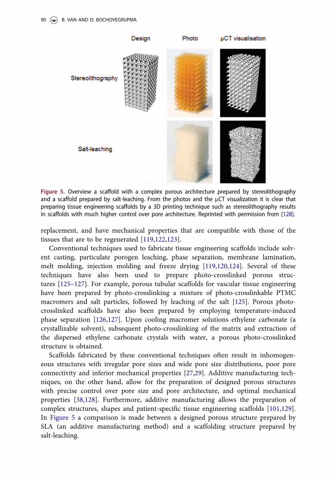
Scaffolds fabricated by these conventional techniques often result in inhomogen-eous structures with irregular pore sizes and wide pore size distributions, poor poreconnectivity and inferior mechanical properties [27,29]. Additive manufacturing tech-niques, on the other hand, allow for the preparation of designed porous structureswith precise control over pore size and pore architecture, and optimal mechanicalproperties [38,128]. Furthermore, additive manufacturing allows the preparation ofcomplex structures, shapes and patient-specific tissue engineering scaffolds [101,129].In Figure 5 a comparison is made between a designed porous structure prepared bySLA (an additive manufacturing method) and a scaffolding structure prepared bysalt-leaching.
Figure 5. Overview a scaffold with a complex porous architecture prepared by stereolithographyand a scaffold prepared by salt-leaching. From the photos and the mCT visualization it is clear thatpreparing tissue engineering scaffolds by a 3D printing technique such as stereolithography resultsin scaffolds with much higher control over pore architecture. Reprinted with permission from [128].
90 B. VAN AND D. BOCHOVEGRIJPMA

Cell encapsulation devices
A most interesting use of photo-crosslinked networks is in the preparation of cellencapsulation devices. In such devices, cells are encapsulated in a support structureduring its formation rather than seeded onto prefabricated tissue engineering scaf-folds [130]. Although the number of photo-crosslinkable biomaterials suited for cellencapsulation is limited due to the required cytocompatibility of the encapsulationprocess, use of cell encapsulation devices can be highly advantageous. First, injectablesystems with cells suspended in liquid precursor solutions can be used, and second,by curing the material in situ, enhanced adhesion of the implant to the tissues can beachieved without the use of glues or sutures.
Hydrogels are attractive materials for this application as they provide a highlyhydrated tissue-like environment for cells and tissues. In addition, they are easy tohandle and can be formed in situ. Several studies aiming at engineering cartilage tis-sue have made use of PEG-based hydrogels to encapsulate the cells [100,131].Uniform cell seeding was easy to achieve and chondrocyte cell viability could readilybe maintained in these hydrogels [132]. It has been shown that the mechanical prop-erties of the hydrogels and the incorporation tissue-specific molecules can have aneffect on ECM production [100], chondrocyte metabolism and gene expression [133].
The illumination needed to initiate the photo-crosslinking process can be donewith ultraviolet (UV) and visible light (VIS). While the use of UV light is not a prob-lem as long as no biological content is involved, when cells and/or proteins are incor-porated into the material prior to photo-crosslinking caution with UV light isnecessary [134]. The free radicals formed upon irradiation not only react into the net-work, but can also react with proteins, cell membranes and DNA. Reactive oxygenspecies that indirectly cause cellular damage can be formed as a result of this inter-action of the radicals with the cells. Therefore, careful selection of the wavelength ofthe light and a photo-initiator that reacts to that wavelength is essential.
Other applications
Other biomedical applications of photo-crosslinked networks include tissue adhesives,tissue barriers and dental composites.
Photo-crosslinkable tissue adhesives have been developed from natural materialssuch as chitosan and mussel proteins, and from synthetic methacrylate-functionalizedblock copolymers containing PEG and DLLA or TMC segments [135–137]. Uponirradiation with light, such synthetic adhesives not only crosslink but at the sametime also adhere to the tissue [136] as the (meth)acrylate groups can covalently bindto amine groups present in the tissue [138].
Photo-crosslinkable hydrogels have been investigated for use as resorbable tissuebarriers to prevent postoperative adhesions [139,140]. These systems were based onPEG and lactide block copolymers that are end-functionalized with methacrylic acid.In situ photo-crosslinking allows the formation of the barriers that prevent adhe-sions [139]. These barriers could also be loaded with drugs [140].
Low molecular weight, multifunctional (meth)acrylates have been used in the prep-aration of photo-crosslinkable resins for dental applications [141–143]. While metal
JOURNAL OF BIOMATERIALS SCIENCE, POLYMER EDITION 91

alloys were the standard, drawback such as mercury content and its associated tox-icity, high costs and poor aesthetics prompted the need for other systems [142].These materials were mixtures of low molecular weight acrylates and methacrylatesand could be crosslinked by UV- and visible light. The exact composition of the mix-tures is an important factor in determining the crosslinking density of thenetworks [141,142]. High degrees of crosslinking need to be achieved to preventshrinkage of the network and leaching of unreacted (meth)acrylates.
Additive manufacturing
Additive manufacturing is a very interesting method to prepare photo-crosslinkednetworks for most of the aforementioned application as it allows for the preparationof highly complex, designed 3D structures with optimized properties and patient-spe-cific shapes [38]. Photo-crosslinking has been employed in several additive manufac-turing techniques. While the most widely used additive manufacturing technique toprepare photo-crosslinked structures and tissue engineering scaffolds is stereolithog-raphy (SLA) [17,144], extrusion-based additive manufacturing [91,94,145] and a com-bination of these methods [146] have also been done.
Extrusion-based additive manufacturing
Extrusion-based additive manufacturing methods are interesting for the preparationof designed structures. These methods are based on the extrusion of a material atpre-defined locations in a layer-by-layer manner to form 3D structures with specificinternal and external geometries [91]. A commonly used extrusion-based additivemanufacturing technique is fused deposition modeling (FDM) [147,148]. Aliphaticpolyesters such as PLLA and PCL are very well suited for FDM, as they flow in themelt at elevated temperatures and readily solidify after extrusion. Polymers that donot crystallize or only slowly solidify are more difficult to process as they will not beform-stable [37,59,91]. An example of such a polymer is PTMC, which is amorphousand has a low glass transition temperature. Nevertheless, this polymer could be proc-essed by an extrusion-based additive manufacturing method when the polymer wasdissolved in a crystallizable solvent. Using low-temperature extrusion-based additivemanufacturing (LTEAM) [91,94] the materials was extruded at a temperature abovethe melting point of the solvent. By cooling the material after extrusion, a scaffoldthat was form stable until photo-crosslinking could be performed was obtained.
A new approach to prepare porous TE scaffolds is melt electrospinning writing(MEW) [145,149–151]. MEW is essentially applying an extrusion-based additive man-ufacturing approach to melt electrospinning [152]. An electrified polymer melt isextruded through a nozzle onto a grounded, translating and/or rotating platform. Asthe electrified molten jet rapidly cools in the air and on the platform, well-definedporous structures can be prepared. Furthermore, polymer fibers with diameterssmaller than 1mm can be prepared [150]. Chen et al. used MEW to prepare scaffoldsfrom poly(L-lactide-co-e-caprolactone-co-acryloyl carbonate) macromers which werephoto-crosslinked to prevent creep and a decrease in the elasticity modulus upon
92 B. VAN AND D. BOCHOVEGRIJPMA

hydration [145]. Furthermore, crosslinking increased the average elasticity modulus ofthe fibers and improved their toughness. The crosslinked scaffolds could be exposedto cyclic strains of 10% elongation for 200,000 cycles without failure, whereas 4 outof 6 non-crosslinked scaffolds failed under the same conditions.
Stereolithography
Stereolithography makes use of a light source to photo-crosslink a polymer resin in alayer-by-layer manner [101,153]. As can be seen in Figure 6, a 3D design of an implant(for example a patient-specific meniscus implant based on a render from CT imagingdata) is virtually sliced into 2D layers. The thickness of these layers corresponds to thethickness of the layers in the additive manufacturing process. The data is then uploadedto the control computer and the structures are fabricated by SLA. Of all 3D printingtechniques, SLA is the most accurate additive manufacturing technique allowing build-ing of designed structures at the highest resolution. Whereas commercially availableSLA setups allow building constructs with details of 20mm in size, other additivemanufacturing techniques allow building structures with details in the range of50–200mm [101]. High resolution micro-SLA setups have been developed that allowbuilding at resolutions even lower than 20mm, with details from 5mm [154–156] toclose to 40–500 nm in size [157–161]. More recently, an apparatus in which SLA andextrusion are combined has been developed by Shanjani et al. [146].
In general, two types of SLA systems are used to prepare designed tissue engineeringstructures [38,162]: laser-based SLA and digital light processing SLA (DLP-SLA). Inlaser-based SLA, a layer of a photo-crosslinkable resin is illuminated at the surface using
Figure 6. From a 3D design towards a porous meniscus implant manufactured by stereolithog-raphy (SLA). A 3D design based on a 3D render from CT imaging data with a gyroid porous net-work architecture is made. 2D slices with thicknesses corresponding to the build layers are thenmade and converted into pixel patterns. The structure is then manufactured by SLA in a layer-by-layer manner.
JOURNAL OF BIOMATERIALS SCIENCE, POLYMER EDITION 93

a computer controlled laser beam. The structures are prepared layer-by-layer by movingthe build platform down into the resin. In DLP-SLA, a UV or blue light pattern of pixelsis projected into the resin through a transparent and non-adherent resin container frombelow. In this case, the build platform moves upwards and out of the resin.
3D bioprinting
A novel approach to the use of additive manufacturing is 3D bioprinting. 3D bio-printing is based on the layer-by-layer manufacturing of 3D structures consisting ofnatural and synthetic polymers, living cells, drugs, growth factor and genes with spa-tial control over the placement of these components [163,164]. In 3D bioprinting, tis-sues, organs and other biological systems are prepared in vitro [165]. Theseconstructs can be used ad 3D models to replace 2D in vitro cell culture and animalmodels. In addition, bone and cartilage tissues have been prepared for musculoskel-etal injury repair [164].
The main additive manufacturing methods used for 3D bioprinting are extrusion-based additive manufacturing, inkjet printing, laser assisted bioprinting and stereolithog-raphy [163,164]. Of these techniques, extrusion-based additive manufacturing [166–169],stereolithography [170], and a combination of these two [146] have been used to pre-pare 3D bioprinted structures by photo-crosslinking. The concerns with regards to theadverse effect of the irradiation wavelength on the cell viability [171] were in a few casesaddressed by using visible light [170] and a water filter [146]. Cell viabilities between 80and 95% were reported in these studies [146,166–168,170].
Photo-crosslinkable systems for additive manufacturing
As additive manufacturing is such an important method to prepare photo-crosslinkedbiodegradable polymer networks, it is important to discuss the photo-crosslinkable sys-tems that have been developed for use in additive manufacturing. Photo-crosslinkablesystems based on fumarate-functionalized oligomers were developed [10,129,172–175].These systems were based on PDLLA, PCL or poly(propylene fumarate) (PPF). Thedisadvantage of these materials is that they need a reactive diluent such as N-vinyl-2-pyrrolidone (NVP) [173,175] and diethyl fumarate [129,172,174]. As described earlier,this increases the non-degradable content of the resulting polymer networks.Resins based on (meth)acrylated macromers are therefore perhaps a more suitedalternative [176,177], as these are more reactive. In the case of photo-crosslinkablesystems for stereolithography (resins), non-reactive diluents can be used to adjustthe viscosity of the resin to allow its processing [17,38]. (Note that this non-react-ive diluent needs to be extracted from the built structure.)
Photo-crosslinkable systems based on poly(D,L-lactide)
Methacrylate-functionalized PDLLA oligomers were one of the early photo-reactivecompounds used for the preparation of biodegradable tissue engineering scaffolds bySLA [17]. As non-crosslinked HMW PDLLA was already used in bone tissue
94 B. VAN AND D. BOCHOVEGRIJPMA

engineering, Melchels et al. proposed to prepare scaffolds for bone tissue engineeringby SLA [17]. Using a non-reactive diluent, they developed resins based on suchphoto-crosslinkable PDLLA macromers. The mechanical properties of the networksand porous scaffolds prepared by SLA were similar to those prepared using HMWPDLLA. In the further development of PDLLA bone tissue engineering scaffolds,nano-hydroxyapatite was incorporated into the resin. As the chemistry of hydroxy-apatite is similar to the calcium phosphate mineral phase present in hard tissues, thismay lead to composite scaffolds that enhance bone formation [27,28]. The incorpor-ation of nano-hydroxyapatite into the polymer matrix also resulted in increasing thecompressive- and tensile moduli of the networks [27,28].
Photo-crosslinkable systems based on fumarate-functionalized PDLLA oligomerscontaining 35wt% NVP reactive diluent were developed for SLA [173]. Highly porousstructures, closely matching the designs could be prepared.
Photo-crosslinkable systems based on poly(e-caprolactone)
Designed tissue engineering scaffolds have also been prepared using SLA resins basedon methacrylated PCL macromers with relatively low molecular weights [29,178]. Inthis case, no diluents were required as the resins had sufficiently low viscosity atroom temperature or after moderate heating to allow their processing. The scaffoldscould be manufactured very accurately, closely resembling the geometry, porosity andpore architecture of the designs [29].
Liquid coumarin end-functionalized copolymers based on CL and TMC were usedto prepare microstructured films and surfaces by stereolithography [7,8]. Multilayeredfilms containing three different copolymers in one single construct were preparedwith these materials. The polymers crosslinked upon UV irradiation.
A photo-crosslinkable system for SLA based on divinyl-fumarate poly(e-caprolac-tone) was used to prepare tissue engineering scaffolds [175]. Initially, resin were pre-pared with either a non-reactive solvent (N-methyl-2-pyrrolidone, NMP) or withNVP as a reactive diluent. As the best crosslinking was achieved with the reactivediluent under UV light the tissue engineering scaffolds were prepared using thatresin. The obtained scaffolds closely resembled the designs.
Photo-crosslinkable systems based on poly(trimethylene carbonate)
For processing in extrusion-based additive manufacturing, PTMC was dissolved inethylene carbonate (melting point 37 �C) and used in (LTEAM) [91,94]. After extru-sion of the fibers at 60 �C, the ethylene carbonate was crystallized at a temperaturebelow the melting temperature of the solvent. This provided the required form-stabil-ity when building the structure. The prepared structures were then photo-crosslinked,and after subsequent extraction of ethylene carbonate, the manufactured structuresremained form-stable. Interestingly, this use of crystallizing ethylene carbonateresulted in porous scaffolds with an additional micro-porosity. It has been suggestedthat these micro-pores may have a beneficial effect on the regenerative capacity of thescaffolds [91].
JOURNAL OF BIOMATERIALS SCIENCE, POLYMER EDITION 95

Methacrylate-functionalized PTMC macromers have been extensively used in thepreparation of designed structures and tissue engineering scaffolds by SLA[38,39,60–63,179]. Non-reactive diluents were used to decrease the viscosity and allowprocessing. It was shown that the mechanical properties, and especially the moduli, ofporous scaffolds strongly depended on porosity and not on the molecular weight ofthe macromer [179] or on pore size [38]. Furthermore, it was shown that incorporat-ing nano hydroxyapatite into the resin to create composites resulted in increased ten-sile strength and toughness [39].
Photo-crosslinkable systems based on poly(ethylene glycol)
Photo-crosslinkable systems based on meth(acrylated) PEG have been developed aswell [124,180,181]. These resins could also contain cells, making the preparation ofcell encapsulating scaffolds by SLA possible [40]. As described previously, PEG-basednetworks are only biodegradable when PEG is block co-polymerized with a bio-degradable component. Scaffolds using a resin based on tri-block copolymer ofPDLLA and PEG were prepared by SLA [182]. These scaffolds were hydrogels andhighly flexible and the structures matched their design precisely. Furthermore, themechanical properties of these scaffolds in compression experiments were similar tothe properties of soft tissues. In a combinatorial approach PEG was one of the com-ponents in hydrogel mixtures which further included PTMC, PDLLA and PCL toprepare mixed-macromer network scaffolds by SLA[41]. These scaffolds had compres-sion moduli up to 170 kPa.
For 3D bioprinting, a photo-crosslinkable system based on methacrylated PEG wasdeveloped [146]. The cells were encapsulated in the PEG subsequently crosslinkedafter deposition while non-crosslinkable PCL was used as a rigid component.
Photo-crosslinkable systems based on semi-synthetic polymers
A stereolithography resin based on GelMA was developed by Gauvin et al [183]. Theresin was prepared to contain calcium carbonate micrioparticles and further con-tained photo-initiator, UV absorber and a solution quencher. Resins based on GEL-MA modified with PEG or methoxy-PEG has been evaluated in preparing designed3D structures at high resolutions. These resins contained reactive diluents [177] oraqueous solutions of co-monomers [184].
Photo-crosslinkable systems based on gelMA and cells (bioinks) have been widelyutilized as systems for 3D bioprinting [166–170]. GelMA was mixed with alginate,photo-initiator and cells [167]. The alginate was used to obtain physically crosslinkedfibers that acted as a structural template to prevent the collapsing of the printedstructures. After printing the GelMA ws crosslinked by UV irradiation. A bioink withthe same components but also containing a 4-arm PEG-tetra-acrylate (PEGTA) wasalso developed [166]. The addition of PEGTA resulted in increased compressionmoduli of the obtained structures. Similarly, a GelMA based bioink was developedwith PEG, but without the alginate [170]. In addition, bioinks based on GelMA con-taining methacrylated hyaluronic acid have been investigated [168,169].
96 B. VAN AND D. BOCHOVEGRIJPMA

Photo-crosslinkable systems based on other polymers
Photo-crosslinkable systems based on other polymeric biomaterials have been investi-gated as well.
Resins for m-SLA based on vinylesters and vinylcarbonates have been investigated[156,185]. These resin contained mixtures of monomers, photo-initiator and a UV-absorber. Using these resins, highly accurate designed 3D structures could be prepared.
To prepare designed 3D ceramic structures, photo-crosslinkable composite resinshave been developed as well [186,187]. In these resins, a ceramic powder is dispersedin a solution of acrylate-based monomers. After fabrication of the designed structuresby SLA, heating of the green body to elevated temperatures leads to decomposition ofthe polymer network phase and sintering of the ceramic particles.
Conclusions
In this review, a short overview is given of the field of photo-crosslinked syntheticbiodegradable polymer networks. These materials can be prepared form a wide rangeof materials such as polyesters, polycarbonates, PEG-based materials and semi-syn-thetic polymers based on natural polymers. Photo-crosslinking these materials resultsin enormously versatile materials as varying in materials, crosslink density and mac-romer molecular weight results in networks with diverse mechanical properties, deg-radation properties and applications. These applications include scaffolds for tissueengineering, drug delivery devices and cell encapsulation.
An interesting method to prepare photo-crosslinked networks for use in these applica-tions is additive manufacturing. Additive manufacturing allows for the 3D preparation ofphoto-crosslinked constructs at a high resolution and in complex structures. Additive man-ufacturing is a relatively new technique which has prospect in many fields from medicineand engineering to life sciences and has become much more affordable and accessible inrecent years. Combined with the high versatility of photo-crosslinked networks, and forexample the recent developments in 3D bioprinting, this may result in significant break-throughs in the improvement of current, and development of novel generation constructsthat deliver drugs at a controlled rate and replace and repair damaged tissues.
Disclosure statement
No potential conflict of interest was reported by the authors.
Funding
This work was supported by the Netherlands Organization for Scientific Research (NWO)under Stichting voor de Technische Wetenschappen project number 12410.
References
[1] Amsden B. Curable, Biodegradable elastomers: emerging biomaterials for drug deliveryand tissue engineering. Soft Matter. 2007;3:1335–1348.
JOURNAL OF BIOMATERIALS SCIENCE, POLYMER EDITION 97

[2] Kaihara S, Vacanti JP. Tissue engineering: toward new solutions for transplantationand reconstructive surgery. Arch Surg. 1999;134:1184–1188.
[3] Ifkovits JL, Burdick JA. Review: photopolymerizable and degradable biomaterials fortissue engineering applications. Tissue Eng. 2007;13:2369–2385.
[4] Pereira MJN, Ouyang B, Sundback CA, et al. A highly tunable biocompatible andmultifunctional biodegradable elastomer. Adv Mater. 2013;25:1209–1215.
[5] Pego AP, Grijpma DW, Feijen J. Enhanced mechanical properties of 1,3-trimethylenecarbonate polymers and networks. Polymer. 2003;44:6495–6504.
[6] Matsuda T, Magoshi T. Preparation of vinylated polysaccharides and photofabricationof tubular scaffolds as potential use in tissue engineering. Biomacromolecules. 2002;3:942–950.
[7] Matsuda T, Mizutani M, Arnold SC. Molecular design of photocurable liquid bio-degradable copolymers. 1. Synthesis and photocuring characteristics. Macromolecules.2000;33:795–800.
[8] Mizutani M, Matsuda T. Liquid photocurable biodegradable copolymers: in vivo deg-radation of photocured poly(epsilon-caprolactone-co-trimethylene carbonate). JBiomed Mater Res. 2002;61:53–60.
[9] Grijpma DW, Hou Q, Feijen J. Preparation of biodegradable networks by photo-cross-linking lactide, epsilon-caprolactone and trimethylene carbonate-based oligomers func-tionalized with fumaric acid monoethyl ester. Biomaterials. 2005;26:2795–2802.
[10] Cooke MN, Fisher JP, Dean D, et al. Use of stereolithography to manufacture critical-sized 3D biodegradable scaffolds for bone ingrowth. J Biomed Mater Res. 2003;64:65–69.
[11] Jansen J, Ghaffar A, van der Horst TN, et al. Controlling the kinetic chain length ofthe crosslinks in photo-polymerized biodegradable networks. J Mater Sci: Mater Med.2013;24:877–888.
[12] Bat E, Zhang Z, Feijen J, et al. Biodegradable elastomers for biomedical applicationsand regenerative medicine. Regen Med. 2014;9:385–398.
[13] Shin H, Temenoff JS, Mikos AG. In vitro cytotoxicity of unsaturated oligo[poly(ethy-lene glycol) fumarate] macromers and their cross-linked hydrogels.Biomacromolecules. 2003;4:552–560.
[14] Domb AJ, Mathiowitz E, Ron E, et al. Polyanhydrides. 4. Unsaturated and cross-linkedpolyanhydrides. J Polym Sci A Polym Chem. 1991;29:571–579.
[15] He S, Timmer MD, Yaszemski MJ, et al. Synthesis of biodegradable poly(propylenefumarate) networks with poly(propylene fumarate)-diacrylate macromers as crosslink-ing agents and characterization of their degradation products. Polymer. 2001;42:1251–1260.
[16] Storey RF, Warren SC, Allison CJ, et al. Methacrylate-endcapped poly(d,l-lactide-co-trimethylene carbonate) oligomers. Network formation by thermal free-radical curing.Polymer. 1997;38:6295–6301.
[17] Melchels FPW, Feijen J, Grijpma DW. A poly(D,L-lactide) resin for the preparation oftissue engineering scaffolds by stereolithography. Biomaterials. 2009;30:3801–3809.
[18] Helminen AO, Korhonen H, Seppala JV. Crosslinked poly(ester anhydride)s based onpoly(epsilon-caprolactone) and polylactide oligomers. J Polym Sci A Polym Chem.2003;41:3788–3797.
[19] Turunen MPK, Korhonen H, Tuominen J, et al. Synthesis, characterization and cross-linking of functional star-shaped poly(epsilon-caprolactone). Polym Int. 2002;51:92–100.
[20] Matsuda T, Kwon IK, Kidoaki S. Photocurable biodegradable liquid copolymers:Synthesis of acrylate-end-capped trimethylene carbonate-based prepolymers, photocur-ing, and hydrolysis. Biomacromolecules. 2004;5:295–305.
[21] Cornacchione LA, Qi B, Bianco J, et al. Photo-cross-linked poly(ethylene carbonate)elastomers: synthesis, in vivo degradation, and determination of in vivo degradationmechanism. Biomacromolecules. 2012;13:3099–3107.
98 B. VAN AND D. BOCHOVEGRIJPMA

[22] Kim BS, Hrkach JS, Langer R. Biodegradable photo-crosslinked poly(ether-ester) net-works for lubricious coatings. Biomaterials. 2000;21:259–265.
[23] Sawhney AS, Pathak CP, Hubbell JA. Bioerodible hydrogels based on photopolymer-ized poly(ethylene glycol)-co-poly(alpha-hydroxy acid) diacrylate macromers.Macromolecules. 1993;26:581–587.
[24] Storey RF, Hickey TP. Degradable polyurethane networks based on d,l-lactide, glyco-lide, e-caprolactone, and trimethylene carbonate homopolyester and copolyester triols.Polymer. 1994;35:830–838.
[25] Korhonen H, Helminen A, Seppala JV. Synthesis of polylactides in the presence of co-initiators with different numbers of hydroxyl groups. Polymer. 2001;42:7541–7549.
[26] Melchels FPW, Velders AH, Feijen J, et al. Photo-cross-linked poly(DL-lactide)-basednetworks. structural characterization by HR-MAS NMR spectroscopy and hydrolyticdegradation behavior. Macromolecules. 2010;43:8570–8579.
[27] Ronca A, Ambrosio L, Grijpma DW. Preparation of designed poly(D,L-lactide)/nano-sized hydroxyapatite composite structures by stereolithography. Acta Biomater. 2013;9:5989–5996.
[28] Ronca A, Ambrosio L, Grijpma DW. Design of porous three-dimensional PDLLA/nano-hap composite scaffolds using stereolithography. JABFM. 2012;10:249–258.
[29] Elomaa L, Teixeira S, Hakala R, et al. Preparation of poly(epsilon-caprolactone)-basedtissue engineering scaffolds by stereolithography. Acta Biomater. 2011;7:3850–3856.
[30] Zant E, Grijpma DW. Synthetic biodegradable hydrogels with excellent mechanicalproperties and good cell adhesion characteristics obtained by the combinatorial syn-thesis of photo-cross-linked networks. Biomacromolecules. 2016;17:1582–1592.
[31] Zant E, Grijpma D. Tough biodegradable mixed-macromer networks and hydrogels byphoto-crosslinking in solution. Acta Biomater. 2016;31:80–88.
[32] Nijst CLE, Bruggeman JP, Karp JM, et al. Synthesis and characterization of photocura-ble elastomers from poly(glycerol-co-sebacate). Biomacromolecules. 2007;8:3067–3073.
[33] Gerecht S, Townsend SA, Pressler H, et al. A porous photocurable elastomer for cellencapsulation and culture. Biomaterials. 2007;28:4826–4835.
[34] Ifkovits JL, Padera RF, Burdick JA. Biodegradable and radically polymerized elasto-mers with enhanced processing capabilities. Biomed Mater. 2008;3:034104.
[35] Mahdavi A, Ferreira L, Sundback C, et al. A biodegradable and biocompatible gecko-inspired tissue adhesive. Proc Natl Acad Sci USA. 2008;105:2307–2312.
[36] Tran RT, Yang J, Ameer GA. Citrate-based biomaterials and their applications inregenerative engineering. Annu Rev Mater Res. 2015;45:277–310.
[37] Schuller-Ravoo S, Feijen J, Grijpma DW. Flexible, elastic and tear-resistant networksprepared by photo-crosslinking poly(trimethylene carbonate) macromers. ActaBiomater. 2012;8:3576–3585.
[38] van Bochove B, Hannink G, Buma P, et al. Preparation of designed poly(trimethylenecarbonate) meniscus implants by stereolithography: challenges in stereolithography.Macromol Biosci. 2016;16:1853–1863.
[39] Geven MA, Varjas V, Kamer L, et al. Fabrication of patient specific composite orbitalfloor implants by stereolithography. Polym Adv Technol. 2015;26:1433–1438.
[40] Lu Y, Mapili G, Suhali G, et al. A digital micro-mirror device-based system for themicrofabrication of complex, spatially patterned tissue engineering scaffolds. J BiomedMater Res A. 2006;77: 396–405.
[41] Przeradzka MA, van Bochove B, Bor TC, et al. Phase-separated mixed-macromerhydrogel networks and scaffolds prepared by stereolithography. Polym Adv Technol.2017;28:1212–1218.
[42] Yue K, Trujillo-de Santiago G, Alvarez MM, et al. Synthesis, properties, and biomed-ical applications of gelatin methacryloyl (GelMA) hydrogels. Biomaterials. 2015;73:254–271.
[43] Chen Y, Lin R, Qi H, et al. Functional human vascular network generated in photo-crosslinkable gelatin methacrylate hydrogels. Adv Funct Mater. 2012;22:2027–2039.
JOURNAL OF BIOMATERIALS SCIENCE, POLYMER EDITION 99

[44] Nichol JW, Koshy ST, Bae H, et al. Cell-laden microengineered gelatin methacrylatehydrogels. Biomaterials. 2010;31:5536–5544.
[45] Zhou Y, Ma G, Shi S, et al. Photopolymerized water-soluble chitosan-based hydrogelas potential use in tissue engineering. Int J Biol Macromol. 2011;48:408–413.
[46] Hu J, Hou Y, Park H, et al. Visible light crosslinkable chitosan hydrogels for tissueengineering. Acta Biomaterialia. 2012;8:1730–1738.
[47] Ma G, Yang D, Li Q, et al. Injectable hydrogels based on chitosan derivative/polyethyl-ene glycol dimethacrylate/N,N-dimethylacrylamide as bone tissue engineering matrix.Carbohyrdate Polymers. 2010;79:620–627.
[48] Ono K, Saito Y, Yura H, et al. Photocrosslinkable chitosan as a biological adhesiveKatsuaki. J Biomed Mater Res. 2000;49:289–295.
[49] Engelberg I, Kohn J. Physico-mechanical properties of degradable polymers used inmedical applications: a comparative study. Biomaterials. 1991;12:292–304.
[50] Amsden BG, Misra G, Gu F, et al. Synthesis and characterization of a photo-cross-linked biodegradable elastomer. Biomacromolecules. 2004;5:2479–2486.
[51] Chapanian R, Tse MY, Pang SC, et al. The role of oxidation and enzymatic hydrolysison the in vivo degradation of trimethylene carbonate based photocrosslinkable elasto-mers. Biomaterials. 2009;30:295–306.
[52] Amsden BG, Tse MY, Turner ND, et al. In vivo degradation behavior of photo-cross-linked star-poly(epsilon-caprolactone-co-D,L-lactide) elastomers. Biomacromolecules.2006;7:365–372.
[53] Ilagan BG, Amsden BG. Macroporous photocrosslinked elastomer scaffolds containingmicroposity: preparation and in vitro degradation properties. J Biomed Mater Res A.2010;93:211–218.
[54] Wang Y, Ameer GA, Sheppard BJ, et al. A tough biodegradable elastomer. NatBiotechnol. 2002;20:602–606.
[55] Rai R, Tallawi M, Grigore A, et al. Synthesis, properties and biomedical applicationsof poly(glycerol sebacate) (PGS): A review. Prog Polym Sci. 2012;37:1051–1078.
[56] Zhao H, Ameer GA. Modulating the mechanical properties of poly(diol citrates) viathe incorporation of a second type of crosslink network. J Appl Polym Sci. 2009;114:1464–1470.
[57] Gyawali D, Tran RT, Guleserian KJ, et al. Citric-acid-derived photo-cross-linked bio-degradable elastomers. J Biomater Sci Polym Ed. 2010;21:1761–1782.
[58] Wang Y, Kibbe MR, Ameer GA. Photo-crosslinked biodegradable elastomers for con-trolled nitric oxide delivery. Biomater Sci. 2013;1:625–632.
[59] Zhu KJ, Hendren RW, Jensen K, et al. Synthesis, properties, and biodegradation ofpoly(1,3-trimethylene carbonate). Macromolecules. 1991;24:1736–1740.
[60] Schuller-Ravoo S, Teixeira SM, Feijen J, et al. Flexible and elastic scaffolds for cartilagetissue engineering prepared by stereolithography using poly(trimethylene carbonate)-based resins. Macromol Biosci. 2013;13:1711–1719.
[61] Blanquer SB, Sharifi S, Grijpma DW. Development of poly(trimethylene carbonate)network implants for annulus fibrosus tissue engineering. JABFM. 2012;10:177–184.
[62] Blanquer SBG, Gebraad AWH, Miettinen S, et al. Differentiation of adipose stem cellsseeded towards annulus fibrosus cells on a designed poly(trimethylene carbonate) scaf-fold prepared by stereolithography. J Tissue Eng Regen Med. 2017;11:2752–2762.
[63] Schuller-Ravoo S, Zant E, Feijen J, et al. Preparation of a designed poly(trimethylenecarbonate) microvascular network by stereolithography. Adv Healthcare Mater. 2014;3:2004–2011.
[64] Chapanian R, Amsden BG. Combined and sequential delivery of bioactive VEGF165and HGF from poly(trimethylene carbonate) based photo-cross-linked elastomers . JControl Release. 2010;143:53–63.
[65] Sharifi S, Blanquer S, Grijpma DW. Polymeric microstructures with shape-memoryproperties for biomedical use built by stereolithography. JABFM. 2012;10:280–286.
100 B. VAN AND D. BOCHOVEGRIJPMA

[66] Sharifi S, Grijpma DW. Resilient amorphous networks prepared by photo-crosslinkinghigh-molecular-weight D,L-lactide and trimethylene carbonate macromers: mechanicalproperties and shape-memory behavior. Macromol Biosci. 2012;12:1423–1435.
[67] Zhang Z, Kuijer R, Bulstra SK, et al. The in vivo and in vitro degradation behavior ofpoly(trimethylene carbonate). Biomaterials. 2006;27:1741–1748.
[68] Chapanian R, Amsden BG. Osmotically driven protein release from photo-cross-linkedelastomers of poly(trimethylene carbonate) and poly(trimethylene carbonate-CO-D,L-lactide). Eur J Pharm Biopharm. 2010;74:172–183.
[69] Timbart L, Tse MY, Pang SC, et al. Tissue response to, and degradation rate of, pho-tocrosslinked trimethylene carbonate-based elastomers following intramuscularimplantation. Materials. 2010;3:1156–1171.
[70] Zhu J. Bioactive modification of poly(ethylene glycol) hydrogels for tissue engineering.Biomaterials. 2010;31:4639–4656.
[71] van Bochove B, Rongen JJ, Hannink G, et al. Grafting a lubricious coating ontophoto-crosslinked poly(trimethylene carbonate). Polym Adv Technol. 2015;26:1428–1432.
[72] Yamaoka T, Tabata Y, Ikada Y. Distribution and tissue uptake of poly(ethylene glycol)with different molecular weights after intravenous administration to mice. J PharmSci. 1994;83:601–606.
[73] Jansen J, Mihov G, Feijen J, et al. Photo-crosslinked biodegradable hydrogels preparedfrom fumaric acid monoethyl ester-functionalized oligomers for protein delivery.Macromol Biosci. 2012;12:692–702.
[74] Lee JH, Lee HB, Andrade JD. Blood compatibility of polyethylene oxide surfaces. ProgPolym Sci. 1995;20:1043–1079.
[75] Hern DL, Hubbell JA. Incorporation of adhesion peptides into nonadhesive hydrogelsuseful for tissue resurfacing. J Biomed Mater Res. 1998;39:266–276.
[76] Zhu J, Beamish JA, Tang C, et al. Extracellular matrix-like cell-adhesive hydrogelsform RGD-containing poly(ethylene glycol) diacrylate. Macromolecules. 2006;39:1305–1307.
[77] Young S, Wong M, Tabata Y, et al. Gelatin as a delivery vehicle for the con- trolledrelease of bioactive molecules. J. Controlled Release. 2005;109:256–274.
[78] Klotz BJ, Gawlitta D, Rosenberg AJWP, et al. Gelatin-methacryloyl hydrogels: towardsbiofabrication-based tissue repair. Trends Biotechnol. 2016;34:394–407.
[79] LaI J, Li Y. Functional assessment of cross-linked porous gelatin hydrogels for bioengi-neered cell sheet carriers. Biomacromolecules. 2010;11:1387–1397.
[80] Reddy N, Reddy R, Jiang Q. Crosslinking biopolymers for biomedical applications.Trends Biotechnol. 2015;33:362–369.
[81] Van den Bulcke AI, Bogdanov B, de Rooze N, et al. Structural and rheological proper-ties of methacrylamide modified gelatin hydrogels. Biomacromolecules. 2000;1:31–38.
[82] Pell�a MCG, Lima-Ten�orio MK, Ten�orio-Neto ET, et al. Chitosan-based hydrogels:From preparation to biomedical applications. Carbohydr Polym. 2018;196:233–245.
[83] Soares PAG, Bourbon AI, Vicente AA, et al. Development and characterization ofhydrogels based on natural polysaccharides: policaju and chitosan. Mater Sci Eng C.2014;42:219–226.
[84] Xu J, Ma L, Liu Y, et al. Design and characterization of antitumor drug paclitaxel-loaded chitosan nanoparticles by W/O emulsions. Int J Biol Macromol. 2012;50:438–443.
[85] Pujana MA, P�erez-�Alvarez L, Iturbe LCC, et al. Biodegradable chitosan nanogels cross-linked with genipin. Carbohyd Polym. 2013;94:836–842.
[86] Xiao C, You R, Fan Y, et al. Tunable functional hydrogels formed from a versatilewater-soluble chitosan. Int J Biol Macromol. 2016;85:386–390.
[87] Gopferich A. Mechanisms of polymer degradation and erosion. Biomaterials. 1996;17:103–114.
JOURNAL OF BIOMATERIALS SCIENCE, POLYMER EDITION 101

[88] Gopferich A, Langer R. Modeling of polymer erosion. Macromolecules. 1993;26:4105–4112.
[89] van Dijkhuizen-Radersma R, Moroni L, van Apeldoorn A, et al. Degradable polymersfor tissue engineering. In: van Blitterswijk CA, editor. Tissue engineering. London(UK): Elsevier Academic Press; 2008. p. 193–221.
[90] Rongen JJ, van Bochove B, Hannink G, et al. Degradation behavior of, and tissueresponse to photo-crosslinked poly(trimethylene carbonate) networks. J Biomed MaterRes A. 2016;104: 2823–2832.
[91] Geven MA, Sprecher C, Guillaume O, et al. Micro-porous composite scaffolds ofphoto-crosslinked poly(trimethylene carbonate) and nano-hydroxyapatite prepared bylow-temperature extrusion-based additive manufacturing. Polym Adv Technol. 2017;28:1226–1232.
[92] Wu L, Ding J. In vitro degradation of three-dimensional porous poly(D,L-lactide-co-glycolide) scaffolds for tissue engineering. Biomaterials. 2004;25:5821–5830.
[93] Wang Y, Kim YM, Langer R. In vivo degradation characteristics of poly(glycerol seba-cate). J Biomed Mater Res A. 2003;66:192–197.
[94] Bat E, Kothman BH, Higuera GA, et al. Ultraviolet light crosslinking of poly(trimethy-lene carbonate) for elastomeric tissue engineering scaffolds. Biomaterials. 2010;31:8696–8705.
[95] Bat E, van Kooten TG, Feijen J, et al. Resorbable elastomeric networks prepared byphotocrosslinking of high-molecular-weight poly(trimethylene carbonate) with photoi-nitiators and poly(trimethylene carbonate) macromers as crosslinking aids. ActaBiomater. 2011;7:1939–1948.
[96] Xu F, Inci F, Mullick O, et al. Release of magnetic nanoparticles from cell-encapsulat-ing biodegradable nanobiomaterials. ACS Nano. 2012;6:6640–6649.
[97] Xiao W, He J, Nichol JW, et al. Synthesis and characterization of photocrosslinkablegelatin and silk fibroin inter- penetrating polymer network hydrogels. Acta Biomater.2011;7:2384–2393.
[98] Sirova M, Van Vlierberghe S, Matyasova V, et al. Immunocompatibility evaluation ofhydro- gel-coated polyimide implants for applications in regenerative medicine. JBiomed Mater Res. 2014;102:1982–1990.
[99] Baroli B. Photopolymerization of biomaterials: issues and potentialities in drug deliv-ery, tissue engineering, and cell encapsulation applications. J Chem TechnolBiotechnol. 2006;81:491–499.
[100] Bryant SJ, Anseth KS. Hydrogel properties influence ECM production by chondrocytesphotoencapsulated in poly(ethylene glycol) hydrogels. J Biomed Mater Res. 2002;59:63–72.
[101] Melchels FP, Feijen J, Grijpma DW. A review on stereolithography and its applicationsin biomedical engineering. Biomaterials. 2010;31:6121–6130.
[102] Davis KA, Anseth KS. Controlled release from crosslinked degradable networks. CritRev Ther Drug Carrier Syst. 2002;19:385–423.
[103] Haesslein A, Hacker MC, Mikos AG. Effect of macromer molecular weight on in vitroophthalmic drug release from photo-crosslinked matrices. Acta Biomater. 2008;4:1–10.
[104] Kimura H, Ogura y. Biodegradable polymers for ocular drug delivery.Ophthalmologica. 2001;215:143–155.
[105] Jansen J, Boerakker MJ, Heuts J, et al. Rapid photo-crosslinking of fumaric acidmonoethyl ester-functionalized poly(trimethylene carbonate) oligomers for drug deliv-ery applications. J Control Release. 2010;147:54–61.
[106] Jansen J, Tibbe MP, Mihov G, et al. Photo-crosslinked networks prepared fromfumaric acid monoethyl ester-functionalized poly(D,L-lactic acid) oligomers and N-vinyl-2-pyrrolidone for the controlled and sustained release of proteins. ActaBiomater. 2012;8:3652–3659.
[107] Lin CC, Sawicki SM, Metters AT. Free-radical-mediated protein inactivation andrecovery during protein photoencapsulation. Biomacromolecules. 2008;9:75–83.
102 B. VAN AND D. BOCHOVEGRIJPMA

[108] Gu F, Neufeld R, Amsden B. Maintenance of vascular endothelial growth factor andpotentially other therapeutic proteins bioactivity during a photo-initiated free radicalcross-linking reaction forming biodegradable elastomers. Eur J Pharm Biopharm.2007;66:21–27.
[109] Haesslein A, Ueda H, Hacker MC, et al. Long-term release of fluocinolone acetonideusing biodegradable fumarate-based polymers. J Control Release. 2006;114:251–260.
[110] M€onk€are J, Hakala RA, Vlasova MA, et al. Biocompatible photocrosslinked poly(esteranhydride) based on functionalized poly(epsilon-caprolactone) prepolymer shows sur-face erosion controlled drug release in vitro and in vivo. J Control Release. 2010;146:349–355.
[111] M€onk€are J, Hakala RA, Kovalainen M, et al. Photocrosslinked poly(ester anhydride)sfor peptide delivery: Effect of oligomer hydrophobicity on PYY3-36 delivery. Eur JPharm Biopharm. 2012;80:33–38.
[112] Hakala RA, Korhonen H, Meretoja VV, et al. Photo-cross-linked biodegradable poly(-ester anhydride) networks prepared from alkenylsuccinic anhydride functionalizedpoly(e-caprolactone) precursors. Biomacromolecules. 2011;12:2806–2814.
[113] Hakala RA, Korhonen H, Sepp€al€a JV. Hydrolysis behaviour of crosslinked poly(esteranhydride) networks prepared from functionalised poly(e-caprolactone) precursors.React. Funct. Polym. 2013;73:11–17.
[114] Gu F, Neufeld R, Amsden B. Sustained release of bioactive therapeutic proteins from abiodegradable elastomeric device. J Control Release. 2007;117:80–89.
[115] Van de Weert M, Hennink WE, Jiskoot W. Protein instability in poly(lactic-co-glycolicacid) microparticles. Pharm Res. 2000;17:1159–1167.
[116] Cleland JL, Duenas ET, Park A, et al. Development ofpoly-(D,L-lactide-coglycolide)microsphere formulations contain- ing recombinant human vascular endothelialgrowth factor to promote local angiogenesis. J Control Release. 2001;72:13–24.
[117] Kim TK, Burgess DJ. Pharmacokinetic characterization C-14-vascular endothelialgrowth factor controlled release microspheres using a rat model. J Pharm Pharmacol.2002;57:897–905.
[118] Langer R, Vacanti JP. Tissue engineering. Science. 1993;260:920–926.[119] Hutmacher DW. Scaffolds in tissue engineering bone and cartilage. Biomaterials. 2000;
21:2529–2543.[120] Leong KF, Cheah CM, Chua C. Solid freeform fabrication of three-dimensional scaf-
folds for engineering replacement tissues and organs. Biomaterials. 2003;24:2363–2378.[121] Yang SF, Leong KF, Du ZH, et al. The design of scaffolds for use in tissue engineer-
ing. Part I. Traditional factors. Tissue Eng. 2001;7:679–689.[122] Hou QP, Grijpma DW, Feijen J. Porous polymeric structures for tissue engineering
prepared by a coagulation, compression moulding and salt leaching technique.Biomaterials. 2003;24:1937–1947.
[123] Freed LE, Vunjak-Novakovic G, Biron RJ, et al. Biodegradable Polymer Scaffolds forTissue Engineering. Biotechnology (NY) .). 1994;12:689–693.
[124] Dhariwala B, Hunt E, Boland T. Rapid prototyping of tissue-engineering constructs,using photopolymerizable hydrogels and stereolithography. Tissue Eng. 2004;10:1316–1322.
[125] Guo Z, Grijpma DW, Poot AA. Preparation and characterization of flexible and elasticporous tubular PTMC scaffolds for vascular tissue engineering. Polym Adv Technol.2017;28:1239–1244.
[126] Pasman T, Grijpma DW, Stamatialis DF, et al. Fabricating porous, photo-crosslinkedpoly(trimethylene carbonate) membranes using temperature-induced phase separation.Polym Adv Technol. 2017;28:1258–1262.
[127] Zant E, Blokzijl MM, Grijpma DW. A combinatorial photocrosslinking method forthe preparation of porous structures with widely differing properties. Macromol RapidCommun. 2015;36:1902–1909.
JOURNAL OF BIOMATERIALS SCIENCE, POLYMER EDITION 103

[128] Melchels FP, Bertoldi K, Gabbrielli R, et al. Mathematically defined tissue engineeringscaffold architectures prepared by stereolithography. Biomaterials. 2010;31:6909–6916.
[129] Mott EJ, Busso M, Luo XY, et al. Digital micromirror device (DMD)-based 3D print-ing of poly(propylene fumarate) scaffolds. Mat Sci Eng C Mater Biol Appl. 2016;61:301–311.
[130] Nicodemus GD, Bryant SJ. Cell encapsulation in biodegradable hydrogels for tissueengineering applications. Tissue Eng. Part B-Re. 2008;14:149–165.
[131] Elisseeff JW, McIntosh K, Anseth S, et al. Photoencapsulation of chondrocytes inpoly(ethylene oxide)-based semi-interpenetrating networks. J Biomed Mater Res. 2000;51:164–171.
[132] Bryant SJ, Anseth KS. The effects of scaffold thickness on tissue engineered cartilagein photocrosslinked poly(ethylene oxide) hydrogels. Biomaterials. 2001;22:619–626.
[133] Bryant SJ, Arthur JA, Anseth KS. Incorporation of tissue-specific molecules alterschondrocyte metabolism and gene expression in photocrosslinked hydrogels. ActaBiomater. 2005;1:243–252.
[134] Fedorovich NE, Oudshoorn MH, van Geemen D, et al. The effect of photopolymeriza-tion on stem cells embedded in hydrogels. Biomaterials. 2009;30:344–353.
[135] Bochynska AI, Hannink G, Grijpma D, et al. Tissue adhesives for meniscus tear repair:an overview of current advances and prospects for future clinical solutions. J MaterSci Mater Med. 2016;27:85.
[136] Lauto A, Mawad D, Foster LJR. Adhesive biomaterials for tissue reconstruction. JChem Technol Biotechnol. 2008;83:464–472.
[137] Sharifi S, Kranenburg HJC, Meij BP, et al. Photo-crosslinkable poly(trimethylene car-bonate)-based macromers for closure of ruptured intervertebral discs. MacromolSymp. 2011;309-310:100–110.
[138] Reece TB, Maxey TS, Kron IL. A prospectus on tissue adhesives. Am J Surg. 2001;182:40S–44s.
[139] Hill-West JL, Chowdhury SM, Sawhney AS, et al. Prevention of postoperative adhe-sions in the rat by in situ photopolymerization of bioresorbable hydrogel barriers .Obstet Gynecol. 1994;83:59–64.
[140] Hubbell JA. Hydrogel systems for barriers and local drug delivery in the control ofwound healing. J Control Release. 1996;39:305–313.
[141] Anseth KS, Goodner MD, Reil MA, et al. The influence of comonomer compositionon dimethacrylate resin properties for dental composites. J Dent Res. 1996;75:1607–1612.
[142] Bland MH, Peppas NA. Photopolymerized multifunctional (meth)acrylates as modelpolymers for dental applications. Biomaterials. 1996;17:1109–1114.
[143] Maffezzoli A, Della Pietra A, Rengo S, et al. Photopolymerization of dental compositematrices. Biomaterials. 1994;15:1221–1228.
[144] Barry JJA, Evseev AV, Markov MA, et al. In vitro study of hydroxyapatite-based pho-tocurable polymer composites prepared by laser stereolithography and supercriticalfluid extraction. Acta Biomater. 2008;4:1603–1610.
[145] Chen F, Hochleitner G, Woodfield T, et al. Additive manufacturing of a photo-cross-linkable polymer via direct melt electrospinning writing for producing high strengthstructures. Biomacromolecules. 2016;17:208–214.
[146] Shanjani Y, Pan CC, Elomaa L, et al. A novel bioprinting method and system forforming hybrid tissue engineering constructs. Biofabrication. 2015;7:045008.
[147] Chia HN, Wu BM. Recent advances in 3D printing of biomaterials. J Biol Eng. 2015;9:4.
[148] Zadpoor AA, Malda J. Additive manufacturing of biomaterials, tissues, and organs.Ann Biomed Eng. 2016;45:1–11.
[149] Brown TD, Dalton PD, Hutmacher DW. Direct writing by way of melt electrospin-ning. Adv Mater Weinheim. 2011;23:5651
104 B. VAN AND D. BOCHOVEGRIJPMA

[150] Hochleitner G, Jungst T, Brown TD, et al. Additive manufacturing of scaffolds withsub-micron filaments via melt electrospinning writing. Biofabrication. 2015;7:035002.
[151] Muerza-Cascante ML, Haylock D, Hutmacher DW, et al. Melt electrospinning and itstechnologization in tissue engineering. Tissue Eng Part B Rev. 2015;21:187–202.
[152] Dalton PD, Vaquette C, Farrugia BL, et al. Electrospinning and additive manufactur-ing: converging technologies. Biomater Sci-UK. 2013;1:171–185.
[153] Hutmacher DW, Sittinger M, Risbud MW. Scaffold-based tissue engineering: rationalefor computer-aided design and solid free-form fabrication systems. Trends Biotechnol.2004;22:354–362.
[154] Baudis S, Heller C, Liska R, et al. (Meth)acrylate-based photoelastomers as tailoredbiomaterials for artificial vascular grafts. J Polym Sci A Polym Chem. 2009;47:2664–2676.
[155] Stampfl J, Baudis S, Heller C, et al. Photopolymers with tunable mechanical propertiesprocessed by laser-based high-resolution stereolithography. J Micromech Microeng.2008;18:125014.
[156] Heller C, Schwentenwein M, Russmueller G, et al. Vinyl esters: low cytotoxicity mono-mers for the fabrication of biocompatible 3D scaffolds by lithography based additivemanufacturing. J Polym Sci A Polym Chem. 2009;47:6941–6954.
[157] Maruo S, Ikuta K, Korogi H. Force-controllable, optically driven micromachines fabri-cated by single-step two-photon micro stereolithography. J Microelectromech Syst.2003;12:533–539.
[158] Maruo S. Development of functional devices using micro/nano stereolithography andmoulding techniques. J-Stage. 2014;87:382–388.
[159] B€uckmann T, Stenger N, Kadic M, et al. Tailored 3D mechanical metamaterials madeby dip-in direct-laser-writing optical lithography. Adv Mater. 2012;24:2710–2714.
[160] Li L, Gattass RR, Gershgoren E, et al. Achieving lambda/20 resolution by one-colorinitiation and deactivation of polymerization . Science. 2009;324:910–913.
[161] Fisher J, von Freymann G, Wegener M. The materials challenge in diffraction-unlim-ited direct-laser-writing optical lithography. Adv Mater. 2010;22:3578–3582.
[162] Skoog SA, Goering PL, Narayan RJ. Stereolithography in tissue engineering. J MaterSci Mater Med. 2014;25:845–856.
[163] Murphy SV, Atala A. 3D bioprinting of tissues and organs. Nat Biotechnol. 2014;32:773–785.
[164] Donderwinkel I, van Hest JCM, Cameron NR. Bio-inks for 3D bioprinting: recentadvances and future prospects. Polym Chem. 2017;8:4451–4471.
[165] Park SH, Jung CS, Min BH. Advances in three-dimensional bioprinting for hard tissueengineering. Tissue Eng Regen Med. 2016;13:622–635.
[166] Jia W, Gungor-Ozkerim PS, Zhang YS, et al. Direct 3D bioprinting of perfusable vas-cular constructs using a blend bioink. Biomaterials. 2016;106:58–68.
[167] Colosi C, Shin SR, Manoharan V, et al. Microfl uidic bioprinting of heterogeneous 3dtissue constructs using low-viscosity bioink. Adv Mater. 2016;28:677–684.
[168] Ouyang L, Highley CB, Sun W, et al. A generalizable strategy for the 3d bioprintingof hydrogels from nonviscous photo-crosslinkable inks. Adv Mater. 2017;29:1604983.
[169] Skardal A, Zhang J, McCoard L, et al. Photocrosslinkable hyaluronan-gelatin hydrogelsfor two-step bioprinting. Tissue Eng Pt A. 2010;16:2675–2685.
[170] Wang Z, Abdulla R, Parker B, et al. A simple and high-resolution stereolithography-based 3D bioprinting system using visible light crosslinkable bioinks. Biofabrication.2015;7:045009.
[171] Derakhshanfar S, Mbeleck R, Xu K, et al. 3D bioprinting for biomedical devices andtissue engineering: A review of recent trends and advances. Bioact Mater. 2018;3:144–156.
[172] Dean D, Jonathan W, Siblani A, et al. Continuous digital light processing (cDLP):highly accurate additive manufacturing of tissue engineered bone scaffolds. VirtualPhys Prototyp. 2012;7:13–24.
JOURNAL OF BIOMATERIALS SCIENCE, POLYMER EDITION 105

[173] Jansen J, Melchels FPW, Grijpma DW, et al. Fumaric acid monoethyl ester-functional-ized poly(D,L-lactide)/N-vinyl-2-pyrrolidone resins for the preparation of tissue engin-eering scaffolds by stereolithography. Biomacromolecules. 2009;10:214–220.
[174] Lee KW, Wang SF, Fox BC, et al. Poly(propylene fumarate) bone tissue engineeringscaffold fabrication using stereolithography: effects of resin formulations and laserparameters. Biomacromolecules. 2007;8:1077–1084.
[175] Ronca A, Ronca S, Forte G, et al. Synthesis and characterization of divinyl-fumaratePoly-e-caprolactone for scaffolds with controlled architectures. J Tissue Eng RegenMed. 2018;12:e523–e531.
[176] Liska R, Schuster M, Infuhr R, et al. Photopolymers for rapid prototyping. J CoatTechnol Res. 2007;4:505–510.
[177] Schuster M, Turecek C, Varga F, et al. 3D-shaping of biodegradable photopolymersfor hard tissue replacement. Appl Surf Sci. 2007;254:1131–1134.
[178] Elomaa L, Kang YQ, Seppala JV, et al. Biodegradable photocrosslinkable poly(depsi-peptide-co-epsilon-caprolactone) for tissue engineering: synthesis, characterization,and in vitro evaluation. J Polym Sci Part A: Polym Chem. 2014;52:3307–3315.
[179] Schuller-Ravoo S, Feijen J, Grijpma DW. Preparation of flexible and elastic poly(tri-methylene carbonate) structures by stereolithography. Macromol Biosci. 2011;11:1662–1671.
[180] Arcaute K, Mann BK, Wicker RB. Stereolithography of three-dimensional bioactivepoly(ethylene glycol) constructs with encapsulated cells. Ann Biomed Eng. 2006;34:1429–1441.
[181] Mapili G, Lu Y, Chen S, et al. Laser-layered microfabrication of spatially patternedfunctionalized tissue-engineering scaffolds. J Biomed Mater Res Part B Appl Biomater.2005;75:414–424.
[182] Seck TM, Melchels FPW, Feijen J, et al. Designed biodegradable hydrogel structuresprepared by stereolithography using poly(ethylene glycol)/poly(D,L-lactide)-based res-ins. J Control Release. 2010;148:34–41.
[183] Gauvin R, Chen Y, Lee JW, et al. Microfabrication of complex porous tissue engineer-ing scaffolds using 3D projection stereolithography. Biomaterials. 2012;33:3824–3834.
[184] Schuster M, Turecek C, Weigel G, et al. Gelatin-based photopolymers for bonereplacement materials. J Polym Sci A Polym Chem. 2009;47:7078–7089.
[185] Russmueller G, Liska R, Stampfl J, et al. 3D printable biophotopolymers for in vivobone regeneration. Materials. 2015;8:3685–3700.
[186] Felzmann R, Gruber S, Mitteramskogler G, et al. Lithography-based additive manufac-turing of cellular ceramic structures. Adv Eng Mater. 2012;14:1052–1058.
[187] Tesavibul P, Felzmann R, Gruber S, et al. Processing of 45S5 bioglass (R) by lithog-raphy-based additive manufacturing. Mater Let. 2012;74:81–84.
106 B. VAN AND D. BOCHOVEGRIJPMA
![researchrepository.ucd.ie · Web viewThe scaffolds are usually made from synthetic biodegradable materials (e.g. polylactic acid, polyglycolic acid, polycaprolactone) [128], or from](https://static.fdocuments.in/doc/165x107/5e3f16dc692ad825a61ae91a/web-view-the-scaffolds-are-usually-made-from-synthetic-biodegradable-materials-eg.jpg)