Phosphorylation of Parkin at Serine65 is essential for activation ...
14
rsob.royalsocietypublishing.org Research Cite this article: Kazlauskaite A et al. 2014 Phosphorylation of Parkin at Serine65 is essential for activation: elaboration of a Miro1 substrate-based assay of Parkin E3 ligase activity. Open Biol. 4: 130213. http://dx.doi.org/10.1098/rsob.130213 Received: 26 November 2013 Accepted: 20 February 2014 Subject Area: biochemistry/molecular biology/neuroscience Keywords: Parkin, PINK1, Miro1, ubiquitin, phosphorylation, Parkinson’s disease Author for correspondence: Miratul M. K. Muqit e-mail: [email protected] Electronic supplementary material is available at http://dx.doi.org/10.1098/rsob.130213. Phosphorylation of Parkin at Serine65 is essential for activation: elaboration of a Miro1 substrate-based assay of Parkin E3 ligase activity Agne Kazlauskaite 1 , Van Kelly 1 , Clare Johnson 1 , Carla Baillie 2 , C. James Hastie 2 , Mark Peggie 1 , Thomas Macartney 1 , Helen I. Woodroof 1 , Dario R. Alessi 1 , Patrick G. A. Pedrioli 1 and Miratul M. K. Muqit 1,3 1 MRC Protein Phosphorylation and Ubiquitylation Unit, 2 Division of Signal Transduction Therapy, and 3 College of Medicine, Dentistry and Nursing, University of Dundee, Dundee, UK 1. Summary Mutations in PINK1 and Parkin are associated with early-onset Parkinson’s dis- ease. We recently discovered that PINK1 phosphorylates Parkin at serine65 (Ser 65 ) within its Ubl domain, leading to its activation in a substrate-free activity assay. We now demonstrate the critical requirement of Ser 65 phosphorylation for substrate ubiquitylation through elaboration of a novel in vitro E3 ligase activity assay using full-length untagged Parkin and its putative substrate, the mitochon- drial GTPase Miro1. We observe that Parkin efficiently ubiquitylates Miro1 at highly conserved lysine residues, 153, 230, 235, 330 and 572, upon phosphorylation by PINK1. We have further established an E2-ubiquitin discharge assay to assess Parkin activity and observe robust discharge of ubiquitin-loaded UbcH7 E2 ligase upon phosphorylation of Parkin at Ser 65 by wild-type, but not kinase-inac- tive PINK1 or a Parkin Ser65Ala mutant, suggesting a possible mechanism of how Ser 65 phosphorylation may activate Parkin E3 ligase activity. For the first time, to the best of our knowledge, we report the effect of Parkin disease-associated mutations in substrate-based assays using full-length untagged recombinant Parkin. Our mutation analysis indicates an essential role for the catalytic cysteine Cys431 and reveals fundamental new knowledge on how mutations may confer pathogenicity via disruption of Miro1 ubiquitylation, free ubiquitin chain for- mation or by impacting Parkin’s ability to discharge ubiquitin from a loaded E2. This study provides further evidence that phosphorylation of Parkin at Ser 65 is criti- cal for its activation. It also provides evidence that Miro1 is a direct Parkin substrate. The assays and reagents developed in this study will be important to uncover new insights into Parkin biology as well as aid in the development of screens to identify small molecule Parkin activators for the treatment of Parkinson’s disease. 2. Introduction Parkinson’s disease is an incurable neurodegenerative disorder whose incidence is set to rise in the forthcoming decades [1]. Over the past 16 years, spectacular & 2014 The Authors. Published by the Royal Society under the terms of the Creative Commons Attribution License http://creativecommons.org/licenses/by/3.0/, which permits unrestricted use, provided the original author and source are credited. on February 16, 2018 http://rsob.royalsocietypublishing.org/ Downloaded from
Transcript of Phosphorylation of Parkin at Serine65 is essential for activation ...

on February 16, 2018http://rsob.royalsocietypublishing.org/Downloaded from
rsob.royalsocietypublishing.org
ResearchCite this article: Kazlauskaite A et al. 2014
Phosphorylation of Parkin at Serine65 is
essential for activation: elaboration of a Miro1
substrate-based assay of Parkin E3 ligase
activity. Open Biol. 4: 130213.
http://dx.doi.org/10.1098/rsob.130213
Received: 26 November 2013
Accepted: 20 February 2014
Subject Area:biochemistry/molecular biology/neuroscience
Keywords:Parkin, PINK1, Miro1, ubiquitin,
phosphorylation, Parkinson’s disease
Author for correspondence:Miratul M. K. Muqit
e-mail: [email protected]
Electronic supplementary material is available
at http://dx.doi.org/10.1098/rsob.130213.
& 2014 The Authors. Published by the Royal Society under the terms of the Creative Commons AttributionLicense http://creativecommons.org/licenses/by/3.0/, which permits unrestricted use, provided the originalauthor and source are credited.
Phosphorylation of Parkinat Serine65 is essential foractivation: elaboration of a Miro1substrate-based assay of ParkinE3 ligase activityAgne Kazlauskaite1, Van Kelly1, Clare Johnson1, Carla Baillie2,
C. James Hastie2, Mark Peggie1, Thomas Macartney1, Helen
I. Woodroof1, Dario R. Alessi1, Patrick G. A. Pedrioli1
and Miratul M. K. Muqit1,3
1MRC Protein Phosphorylation and Ubiquitylation Unit, 2Division of Signal TransductionTherapy, and 3College of Medicine, Dentistry and Nursing, University of Dundee, Dundee, UK
1. SummaryMutations in PINK1 and Parkin are associated with early-onset Parkinson’s dis-
ease. We recently discovered that PINK1 phosphorylates Parkin at serine65
(Ser65) within its Ubl domain, leading to its activation in a substrate-free activity
assay. We now demonstrate the critical requirement of Ser65 phosphorylation for
substrate ubiquitylation through elaboration of a novel in vitro E3 ligase activity
assay using full-length untagged Parkin and its putative substrate, the mitochon-
drial GTPase Miro1. We observe that Parkin efficiently ubiquitylates Miro1 at
highly conserved lysine residues, 153, 230, 235, 330 and 572, upon phosphorylation
by PINK1. We have further established an E2-ubiquitin discharge assay to assess
Parkin activity and observe robust discharge of ubiquitin-loaded UbcH7 E2
ligase upon phosphorylation of Parkin at Ser65 by wild-type, but not kinase-inac-
tive PINK1 or a Parkin Ser65Ala mutant, suggesting a possible mechanism of
how Ser65 phosphorylation may activate Parkin E3 ligase activity. For the first
time, to the best of our knowledge, we report the effect of Parkin disease-associated
mutations in substrate-based assays using full-length untagged recombinant
Parkin. Our mutation analysis indicates an essential role for the catalytic cysteine
Cys431 and reveals fundamental new knowledge on how mutations may confer
pathogenicity via disruption of Miro1 ubiquitylation, free ubiquitin chain for-
mation or by impacting Parkin’s ability to discharge ubiquitin from a loaded E2.
This study provides further evidence that phosphorylation of Parkin at Ser65 is criti-
cal for its activation. It also provides evidence that Miro1 is a direct Parkin substrate.
The assays and reagents developed in this study will be important to uncover new
insights into Parkin biology as well as aid in the development of screens to identify
small molecule Parkin activators for the treatment of Parkinson’s disease.
2. IntroductionParkinson’s disease is an incurable neurodegenerative disorder whose incidence
is set to rise in the forthcoming decades [1]. Over the past 16 years, spectacular
rsob.royalsocietypublishing.orgOpen
Biol.4:130213
2
on February 16, 2018http://rsob.royalsocietypublishing.org/Downloaded from
genetic breakthroughs have uncovered nearly 20 genes or loci
associated with familial Parkinson’s disease (PD) that provide
a solid biochemical platform to uncover the molecular origins
and mechanisms underlying this devastating disorder [2].
Mutations in the RING-IBR-RING (RBR) ubiquitin E3
ligase Parkin were first identified in 1998 in families with
early-onset autosomal-recessive PD [3]. Parkin is a 465 amino
acid enzyme comprising: a regulatory Ubl domain (residues
1–76); a RING0 domain (residues 145–215); a RING1
domain (residues 237–292) that binds to an E2; an IBR
domain (residues 327–378); and a RING2 domain that med-
iates the enzyme’s catalytic activity (415–465) [4]. Recent
groundbreaking insights have revealed that Parkin and other
members of the RBR family of E3 ligases exhibit HECT-like
properties [5,6]. Specifically, Parkin contains a highly con-
served catalytic cysteine (Cys431) within its RING2 domain,
which acts as a ubiquitin acceptor that forms an intermediate
thioester bond prior to ubiquitylation of its substrate [5]. The
physiological relevance of this catalytic cysteine is underscored
by the presence of a human disease-causing mutation at this
residue (Cys431Phe), which has been shown to abolish Parkin
catalytic activity at least in auto-ubiquitylation assays [7–11].
Historically, Parkin was thought to be constitutively active,
but in 2011 it was demonstrated that Parkin’s E3 ligase activity
is regulated by an interaction between the N-terminal Ubl
domain and the C-terminus of the protein, which maintains
the enzyme in an autoinhibited closed conformation [12]. The
N-terminal Ubl domain plays a critical role in mediating this
autoinhibition, because removal of the Ubl domain led to con-
stitutive activation of Parkin [12,13]. Furthermore, expression
of Parkin with epitope tags fused to the N-terminus leads to
disruption of the Ubl-mediated autoinhibition and activation
of E3 ligase activity [12,13]. It is therefore critical to study the
properties of recombinant Parkin using full-length protein
that is devoid of epitope tags. The physiological relevance of
Ubl-mediated autoinhibition is also emphasized by the discov-
ery that PTEN-induced kinase 1 (PINK1), mutations of which
also lead to familial PD [14], phosphorylates Parkin at a
highly conserved residue Serine65 (Ser65) that lies within the
Ubl domain; and that phosphorylation leads to activation of
Parkin E3 ligase activity as judged by the formation of free ubi-
quitin chains in a substrate-free ubiquitylation assay [15]. In
agreement with our initial findings, several laboratories have
reproduced PINK1-dependent phosphorylation of Parkin at
Ser65 [7,16,17].
The direct regulation of Parkin by PINK1 is consistent
with previous clinical and genetic studies that have suggested
that both these enzymes function in a common pathway.
PINK1 and Parkin patients share a similar phenotype com-
prising early age at onset, slow progression, dystonia and
early development of L-DOPA-induced dyskinesias [18,19].
In addition, studies in Drosophila melanogaster provided gen-
etic evidence that PINK1 and Parkin are linked, because
PINK1 and Parkin null flies exhibit near identical pheno-
types, including mitochondrial deficits, flight muscle
degeneration and motor deficits [20–22]. Moreover, overex-
pression of Parkin can rescue PINK1 null flies, but the
opposite is not the case, providing genetic evidence that
PINK1 acts upstream of Parkin [20–22]. An upstream role
for mammalian PINK1 had also been suggested by cellular
studies reporting that PINK1 was required for Parkin recruit-
ment to mitochondria following depolarization of the
mitochondrial membrane potential [23–26].
Recently, a low-resolution X-ray crystal structure of full-
length rat Parkin and high-resolution rat and human struc-
tures missing the Ubl domain have been solved which
confirm that Parkin exists in an autoinhibited conformation
[27–29]. The full-length structure reveals that autoinhibition
of Parkin is mediated by an interaction between the Ubl
domain, regulatory element of Parkin (REP helix) and the
RING1 domain, obscuring the potential E2 binding site.
Direct interaction of the REP helix, that lies between the
IBR and RING2 domain, with RING1 domain was also con-
firmed in human Parkin structures lacking the N-terminal
Ubl domain [27,28]. In addition, a further autoinhibitory
interaction between the RING0 and RING2 domains which
occludes the catalytic Cys431 was observed [4,27–29]. How-
ever, the structures do not provide any mechanistic insights
into how phosphorylation at Ser65 mediates transition from
an inactive to an active conformation.
An outstanding question in the field is whether Ser65
phosphorylation of Parkin is critical for its ability to ubiquity-
late substrates. The list of reported potential Parkin substrates
is considerable and continues to grow with over 100
suggested [30–35]. However, in the majority of the previous
work, experiments have been undertaken using overexpres-
sion approaches using Parkin with activating N-terminal
tags or Parkin lacking its autoinhibitory Ubl domain contain-
ing the PINK1 phosphorylation motif. Much more work is
therefore needed to establish whether ubiquitylation of all
of the proposed substrates at the level of the endogenous
protein is indeed mediated by Parkin. Such validation is
important as it will enable identification of the crucial
Parkin substrates that determine survival of dopaminergic
neurons in Parkinson’s disease [33,36–38].
Several lines of evidence indicate that physiological sub-
strates of Parkin reside in the mitochondria, including the
observation of mitochondrial deficits in Parkin knockout (KO)
mice [39,40] and Drosophila models [20–22]; and cellular studies
linking Parkin to the regulation of mitochondrial dynamics, turn-
over and transport [36,37,41,42]. Recently, Miro1, an atypical
mitochondrial GTPase, has emerged as a candidate Parkin sub-
strate based on genetic interaction data in Drosophila models of
Parkin [43] and overexpression studies of N-terminal-tagged
mammalian Parkin [32,43,44].
In this paper, we investigate whether Parkin phosphoryl-
ation at Ser65 is required for its catalytic activation and
ubiquitylation of substrates. We demonstrate that Parkin,
upon phosphorylation at Ser65, can ubiquitylate Miro1 in
addition to catalysing the formation of free ubiquitin chains
and this is abolished by deletion of the Ubl domain. We
have mapped the major sites of Miro1 ubiquitylation to
highly conserved Lysine153 (Lys153), Lysine230 (Lys230),
Lysine235 (Lys235), Lysine330 (Lys330) and Lysine572
(Lys572) residues. Using this novel assay, we have undertaken
an E2 scan and observed 23/25 E2 ligases that enable Parkin
phosphorylated at Ser65 to ubiquitylate Miro1. Furthermore,
we have deployed our assay to investigate the effect of dis-
ease-associated point mutations of Parkin and discovered
diverse effects of mutations on Parkin E3 ligase activity,
including the identification of several mutants that disrupt
the formation of free ubiquitin chains without any significant
impact on Miro1 substrate ubiquitylation.
To gain further mechanistic insights into the effect of PINK1
phosphorylation at Ser65 on Parkin E3 ligase activity, we have
developed a ubiquitin discharge assay, which measures the

rsob.royalsocietypublishing.orgOpen
Biol.4:13021
3
on February 16, 2018http://rsob.royalsocietypublishing.org/Downloaded from
ability of Parkin to stimulate the discharge of ubiquitin from the
E2 ligase UbcH7. We observe that only upon phosphorylation
of Parkin at Ser65 can it lead to efficient discharge of UbcH7
loaded with ubiquitin. We have used this assay to study the
effect of Parkin disease mutations and uncover several mutants
that disrupt Ser65-phosphorylated Parkin-mediated E2 dis-
charge, shedding light on how these mutations may lead to
reduced Parkin-E3-mediated ubiquitylation.
This study validates the critical role of Ser65 phosphoryl-
ation in enabling Parkin activation of its E3 ligase activity and
reveals new mechanistic insights into how disease-associated
mutations of Parkin may impact on E3 ligase activity. The
assays and technologies described in this study have enabled
a more accurate assessment of Parkin E3 ligase activity and
could also be deployed in future chemical screening pro-
grammes to develop small molecule activators of Parkin for
the treatment of Parkinson’s disease.
3
3. Material and methods3.1. Materials[g-32P] ATP was from Perkin-Elmer. All mutagenesis was
carried out using the QuikChange site-directed mutagenesis
method (Stratagene) with KOD polymerase (Novagen). All
DNA constructs were verified by DNA sequencing, which
was performed by The Sequencing Service, School of Life
Sciences, University of Dundee, using DYEnamic ET termin-
ator chemistry (Amersham Biosciences) on automated DNA
sequencers (Applied Biosystems). DNA for bacterial protein
expression was transformed into Escherichia coli BL21 DE3
RIL (codon plus) cells (Stratagene). All cDNA plasmids, anti-
bodies and recombinant proteins generated for this study are
available on request through our reagents website (http://
mrcppureagents.dundee.ac.uk/).
3.2. AntibodiesAntigen affinity-purified sheep anti-SUMO-1 antibody was a
kind gift from Professor Ron Hay (Dundee). Anti-Parkin
mouse monoclonal was obtained from Santa Cruz Biotech-
nology; anti-FLAG HRP-conjugated antibody was obtained
from Sigma; anti-maltose binding protein (MBP) HRP-conju-
gated antibody was obtained from New England Biolabs.
3.3. ImmunoblottingSamples were subjected to SDS/PAGE (8–14%) and trans-
ferred onto nitrocellulose membranes. Membranes were
blocked for 1 h in Tris-buffered saline with 0.1% Tween
(TBST) containing 5% (w/v) non-fat dried skimmed milk
powder. Membranes were probed with the indicated anti-
bodies in TBST containing 5% (w/v) non-fat dried
skimmed milk powder for 1 h at room temperature. Detec-
tion was performed using HRP-conjugated antibodies and
enhanced chemiluminescence reagent.
3.4. In vitro ubiquitylation assaysWild-type or indicated mutant Parkin (2 mg) was initially incu-
bated with 1 mg (or indicated amounts) of E. coli-expressed
wild-type or kinase-inactive (D359A) MBP-TcPINK1 in a
reaction volume of 25 ml (50 mM Tris–HCl (pH 7.5), 0.1 mM
EGTA, 10 mM magnesium acetate, 1% 2-mercaptoethanol
and 0.1 mM ATP. Kinase assays were incubated at 308C for
60 min followed by addition of ubiquitylation assay com-
ponents and Mastermix to a final volume of 50 ml (50 mM
Tris–HCl (pH 7.5), 0.05 mM EGTA, 10 mM MgCl2, 0.5% 2-
mercaptoethanol, 0.12 mM human recombinant E1 purified
from Sf21 insect cell line, 1 mM human recombinant UbcH7
and 2 mg 6xHis-Sumo-Miro1 (wild-type or point mutants)
both purified from E. coli, 0.05 mM Flag-ubiquitin (Boston Bio-
chem) and 2 mM ATP). Ubiquitylation reactions were
incubated at 308C for 60 min and terminated by addition of
SDS sample buffer. For all assays, reaction mixtures were
resolved by SDS–PAGE. Ubiquitylation reactions were sub-
jected to immunoblotting with anti-FLAG antibody (Sigma, 1
: 7500), anti-Parkin, anti-SUMO1 or anti-MBP antibodies. For
the E2 scan, a version of the E2scan kit was obtained from
Ubiquigent, and 1 mg of each E2 enzyme was used per reaction.
3.5. In vitro E2 discharge assaysWild-type or indicated mutant Parkin (2 mg) was incubated
with 1 mg of E. coli-expressed wild-type or kinase-inactive
(D359A) MBP-TcPINK1 in a reaction volume of 15 ml (50 mM
HEPES (pH 7.5), 0.1 mM EGTA, 10 mM magnesium acetate
and 0.1 mM ATP). Kinase assays were incubated at 308C for
60 min. E2-charging reaction was assembled in parallel in
5 ml containing Ube1 (0.5 mg), an E2 (2 mg), 50 mM HEPES
pH 7.5 and 10 mM ubiquitin in the presence of 2 mM mag-
nesium acetate and 0.2 mM ATP. After initial incubation of
60 min at 308C, the reactions were combined and allowed to
continue for a further 15 min or indicated times at 308C. Reac-
tions were terminated by the addition of 5 ml of LDS loading
buffer and subjected to SDS–PAGE analysis in the absence of
any reducing agent. Gels were stained using InstantBlue.
3.6. In-solution protein digestionIn vitro ubiquitylation assays were terminated with 1% Rapigest
and reduced in 5 mM Tris-(2-carboxyethyl)phosphine (TCEP) at
508C for 30 min. Additional Tris–HCl was added to 100 mM
to ensure buffering at pH 7.5 followed by cysteine alkylation in
10 mM chloroacetamide at 208C in the dark for 30 min. Samples
were diluted to 0.1% Rapigest and digested with 1 : 50 w/w
trypsin overnight at 378C. Peptides were acidified with 1%
trifluoroacetic acid and incubated at 378C for 1 h before precipi-
tating acid-cleaved Rapigest by centrifugation at 17 000g for
10 min. Peptides were purified on C18 MicroSpin columns
(The Nest Group) before MS analysis. Approximately 30 ng of
peptide was analysed by C18 LC–MS/MS over a 60 min
gradient from 1% to 37% acetonitrile/0.1% formic acid. Mass
spectrometric analysis was conducted by data-dependent acqui-
sition with spectra acquired by collision-induced dissociation on
an LTQ-Orbitrap Velos (Thermo Fisher Scientific). Data were
analysed using MASCOT (www.matrixscience.com), and ion
signals were extracted using SKYLINE [45].
3.7. In-gel protein digestionProtein bands were excised from the gel and washed sequen-
tially with 0.5 ml of water, 50% acetonitrile, 0.1 M NH4HCO3
and 50% acetonitrile/50 mM NH4HCO3. All washes were
performed for 10 min on a Vibrax shaking platform. Proteins

rsob.royalsocietypublishing.orgOpen
Biol.4:130213
4
on February 16, 2018http://rsob.royalsocietypublishing.org/Downloaded from
were then reduced with 10 mM DTT/0.1 M NH4HCO3 at 658Cfor 45 min and alkylated with 50 mM chloroacetamide/0.1 M
NH4HCO3 for 20 min at room temperature. They were
then washed with 0.5 ml 50 mM NH4HCO3 and 50 mM
NH4HCO3/50% acetonitrile (as before). Gel pieces were
shrunk with 0.3 ml acetonitrile for 15 min. Acetonitrile was
aspirated, and trace amounts were removed by drying
sample in a Speed-Vac. Gel pieces were then incubated for
16 h with 5 mg ml21 trypsin in 25 mM triethylammonium
bicarbonate at 308C on a shaker. An equal volume of aceto-
nitrile (same as trypsin) was added to each sample and
further incubated on a shaking platform for 15 min. The super-
natants were dried by Speed-Vac. Another extraction was
performed by adding 100 ml 50% acetonitrile/2.5% formic
acid for 15 min. This supernatant was combined with the
first extract and dried by Speed-Vac. Peptides were purified
on C18 MicroSpin columns (The Nest Group) before MS
analysis as described for in-solution protein digestions.
3.8. Kinase assaysReactions were set up in a volume of 25 ml, using 2 mg of wild-
type or indicated mutants of Parkin and 1 mg of E. coli-expressed
wild-type or kinase-inactive (D359A) MBP-TcPINK1, in 50 mM
Tris–HCl (pH 7.5), 0.1 mM EGTA, 10 mM MgCl2, 2 mM DTT
and 0.1 mM [g-32P] ATP (approx. 500 cpm pmol21). Assays
were incubated at 308C with shaking at 1050 r.p.m. and termi-
nated after 60 min by addition of SDS sample loading buffer.
The reaction mixtures were then resolved by SDS–PAGE. Pro-
teins were detected by Coomassie staining, and gels were
imaged using an Epson scanner and dried completely using a
gel dryer (Bio-Rad). Incorporation of [g-32P] ATP into substrates
was analysed by autoradiography using Amersham hyperfilm.
3.9. Buffers for Escherichia coli protein purificationFor Parkin purification: lysis buffer contained 50 mM Tris–HCl
(pH 7.5), 150 mM NaCl, 1 mM EDTA, 1 mM EGTA, 5% (v/v)
glycerol, 1% (v/v) Triton X-100, 0.1% (v/v) 2-mercaptoethanol,
1 mM benzamidine and 0.1 mM PMSF. Wash buffer contained
50 mM Tris–HCl (pH 7.5), 500 mM NaCl, 0.1 mM EGTA, 5%
(v/v) glycerol, 0.03% (v/v) Brij-35, 0.1% (v/v) 2-mercaptoetha-
nol, 1 mM benzamidine and 0.1 mM PMSF. Equilibration
buffer contained 50 mM Tris–HCl (pH 7.5), 150 mM NaCl,
0.1 mM EGTA, 5% (v/v) glycerol, 0.03% (v/v) Brij-35, 0.1%
(v/v) 2-mercaptoethanol, 1 mM benzamidine and 0.1 mM
PMSF. Elution buffer was equilibration buffer with the addition
of 12 mM maltose. Storage buffer was equilibration buffer
with the addition of 0.27 M sucrose, and glycerol–PMSF and
benzamidine were omitted.
3.10. Protein purification from Escherichia coliFull-length wild-type and kinase-inactive TcPINK1 was
expressed in E. coli as MBP fusion protein and purified as
described previously [15]. Briefly, BL21 codon þ transformed
cells were grown at 378C to an OD600 of 0.3, then shifted to
168C and induced with 250 mM isopropyl b-D-thiogalactoside
(IPTG) at OD600 of 0.5. Cells were induced with 250 mM IPTG
at OD 0.6 and were further grown at 168C for 16 h. Cells were
pelleted at 4000 r.p.m., and then lysed by sonication in lysis
buffer. Lysates were clarified by centrifugation at 30 000gfor 30 min at 48C followed by incubation with 1 ml per litre
of culture of amylose resin for 1.5 h at 48C. The resin was
washed thoroughly in wash buffer, then equilibration
buffer, and proteins were then eluted. Proteins were dialysed
overnight at 48C into storage buffer, snap-frozen and stored
at 2808C until use.
Wild-type and indicated mutant untagged Parkin (His-
SUMO cleaved) was expressed and purified using a modified
protocol [12]. We did not observe any significant difference in
solubility or expression between the mutants and wild-type
Parkin protein. BL21 cells were transformed with His-SUMO-
tagged Parkin constructs, overnight cultures were prepared
and used to inoculate 12 � 1l LB medium, 50 mg ml21 carbeni-
cillin, 0.25 mM ZnCl2. The cells were grown at 378C until the
OD600 was 0.4, and the temperature was dropped to 168C. At
OD600 ¼ 0.8, expression was induced with 10 mM IPTG. After
overnight incubation, the cells were collected and lysed in
75 mM Tris pH 7.5, 500 mM NaCl, 0.2% Triton X-100, 25 mM
imidazole, 0.5 mM TCEP, 1 mM pefablok, 10 mg ml21 leupep-
tin. After sonication and removal of insoluble material,
His-SUMO-Parkin was purified via Ni2þ–NTA–sepharose
chromatography. The protein was collected by elution with
400 mM imidazole in 50 mM Tris, pH 8.2, 200 mM NaCl, 10%
glycerol, 0.03% Brij-35, 0.5 mM TCEP. This was dialysed twice
against 50 mM Tris pH 8.2, 200 mM NaCl, 10% glycerol,
0.5 mM TCEP in the presence of His-SENP1 415–643 at a
ratio of 1 mg His-SENP1 per 5 mg His-SUMO-Parkin. The pro-
tease, the His-SUMO tag and any uncleaved protein were
removed by two subsequent incubations with Ni2þ–NTA–
sepharose. The cleaved Parkin was further purified in 50 mM
Tris, pH 8.2, 200 mM NaCl, 20% glycerol, 0.03% (v/v) Brij-35,
0.5 mM TCEP over a Superdex 200 column.
Wild-type 6xHis-Sumo-Miro1 (1–592), K572R and K567R
mutants were expressed in E. coli. Briefly, BL21 CodonPlus
(DES)-RIL-transformed cells were grown at 378C to an OD600
of 0.4, then reduced to 158C and induced with 10 mM IPTG
at an OD600 of 0.6. Cells were then grown at 158C for a further
20 h. Cells were pelleted at 4200g and then lysed by sonication
in lysis buffer. Lysates were clarified by centrifugation at 30
000g for 30 min at 48C followed by incubation with Cobalt
resin at 48C for 45 min. The resin was washed thoroughly in
high salt buffer, then equilibrated in low salt buffer, and the
proteins were then eluted. The eluted Miro1 proteins were
further purified by anion exchange chromatography. Proteins
were applied to a Mono-Q HR 5/5 column and chromato-
graphed with a linear gradient of NaCl from 0 to 0.5 M.
Fractions containing the purified Miro1 protein were then
dialysed, snap-frozen in liquid nitrogen and stored at 2708C.
4. Results4.1. Ubiquitylation of Miro1 by Parkin is dependent on
phosphorylation at Ser65
We previously reported the striking observation that untagged
full-length recombinant Parkin expressed in E. coli was able
to induce formation of low-molecular-weight free ubiquitin
chains in a substrate-free ubiquitylation assay following phos-
phorylation of Parkin at Ser65 by the active insect orthologue of
PINK1, Tribolium castaneum (TcPINK1) [15]. To obtain further
evidence that PINK1 phosphorylation activates Parkin and to
develop a more robust in vitro Parkin assay, we tested whether
ubiquitylation of the proposed direct substrate Miro1 [32,43,44]

Parkin
PINK1 KD (mg)0 0.03
0.06
0.12
5
0.25
0.5
1PINK1 WT (mg)
ubiquitinubiquitin
PINK1
Parkin
ubiquitin
PINK1
0 0.03
0.06
0.12
5
0.25
0.5
1PINK1 WT (mg)
150
100
75
50
37
2520
PINK1
Parkin
PINK1 WTParkin WT
PINK1 KIParkin WT
PINK1 WTParkin S65A
Miro1-UbMiro1
(c)(b)(a)
0 0.03
0.06
0.12
5
0.25
0.5
1
Miro1-UbMiro1
Miro1-UbMiro1
100
75
150
100
75
50
37
2520
100
75
150
100
75
50
37
2520
100
75
Figure 1. PINK1-dependent phosphorylation of Parkin Ser65 leads to activation of Parkin E3 ligase activity and multi-monoubiquitylation of Miro1. Wild-type (WT)(a) but not kinase-inactive (KI) (b) PINK1 activates wild-type Parkin E3 ligase activity leading to Miro1 multi-monoubiquitylation, an effect that is blocked by mutantParkin Ser65Ala (S65A) (c). Two micrograms of wild-type or S65A Parkin were incubated with indicated amounts of wild-type or kinase-inactive (D359A) MBP-TcPINKin a kinase reaction (50 mM Tris – HCl ( pH 7.5), 0.1 mM ethylene glycol tetra-acetic acid (EGTA), 10 mM MgCl2, 0.1% 2-mercaptoethanol and 0.1 mM ATP) for60 min. The ubiquitylation reaction was then initiated by addition of ubiquitylation assay components (50 mM Tris – HCl ( pH 7.5), 0.05 mM EGTA, 10 mMMgCl2, 0.5% 2-mercaptoethanol, 0.12 mM human recombinant E1 purified from Sf21 insect cell line, 1 mM human recombinant UbcH7 purified from E. coli,0.05 mM Flag-ubiquitin (Boston Biochem) and 2 mM ATP) and 2 mg of His-Sumo-Miro1. Reactions were terminated after 60 min by addition of SDS – PAGE loadingbuffer and resolved by SDS – PAGE. Miro1, ubiquitin, Parkin and PINK1 were detected using anti-SUMO, anti-FLAG, anti-Parkin and anti-MBP antibodies, respectively.Representative of three independent experiments.
rsob.royalsocietypublishing.orgOpen
Biol.4:130213
5
on February 16, 2018http://rsob.royalsocietypublishing.org/Downloaded from
could be deployed to assess Parkin activity. We were unable
to express full-length recombinant Miro1 (residues 1–618) in
E. coli, but a fragment of Miro1 (residues 1–592) lacking the
C-terminal transmembrane domain expressed well and was
used in subsequent assays.
The maximal stoichiometry of Parkin phosphorylation
by PINK1 under our assay conditions is 0.08 moles phos-
phate per mole of protein (the electronic supplementary
material, figure S1). To assess whether phosphorylation of
Parkin by PINK1 influenced its ability to ubiquitylate
Miro1, we phosphorylated untagged full-length Parkin with
increasing levels of TcPINK1 in the presence of ATP and
then added a reaction mix containing E1 ubiquitin-activating
ligase, UbcH7 conjugating E2 ligase, ubiquitin, Mg-ATP and
Miro1(1–592). After 60 min, reactions were terminated with
SDS sample buffer in the presence of 2-mercaptoethanol
and heated at 1008C, and substrate ubiquitylation was
assessed by immunoblot analysis with antibodies that
detect ubiquitin, Parkin, Miro1 and TcPINK1. Consistent
with previous findings in the absence of PINK1 phosphoryl-
ation, Parkin was inactive as no evidence of free ubiquitin
chain formation or Miro1 ubiquitylation was observed
(figure 1a, lane 1); with the addition of wild-type TcPINK1,
Miro1 multi-monoubiquitylation (a major mono- and minor
multi-ubiquitylated species) in addition to free polyubiquitin
chain formation was observed (figure 1a, lane 3–7). No sig-
nificant Miro1 ubiquitylation or polyubiquitin chain
formation was observed in the presence of the kinase-inactive
TcPINK1 (figure 1b) or using the Ser65Ala (S65A) Parkin
point mutant (figure 1c), indicating that Miro1 ubiquitylation
is dependent on Parkin Ser65 phosphorylation. Using mass
spectrometry, we detected the formation of diverse ubiqui-
tin–ubiquitin linkages, including K6, K11, K33, K48 and
K63 in reactions containing activated Parkin (the electronic
supplementary material, figure S2). We also detected K27
chains, but these were generated in a Parkin-independent
manner (the electronic supplementary material, figure S2).
4.2. The Ubl domain of Parkin is required for Miro1substrate ubiquitylation
To further investigate the role of the Ubl domain in Parkin-
mediated Miro1 ubiquitylation, we expressed a fragment
lacking the Ubl domain (residues 80–465, DUbl-Parkin) and
assayed it in parallel with full-length Parkin pre-incubated
with either wild-type or kinase-inactive TcPINK1. While
DUbl-Parkin exhibited significant auto-ubiquitylation activity
similar to activated full-length Parkin, it could not catalyse
Miro1 ubiquitylation or the formation of low molecular
weight polyubiquitin chains (figure 2).
4.3. Identification of Parkin-mediated Miro1ubiquitylation sites
We next sought to determine the major site(s) of Miro1 ubiqui-
tylation by Parkin that had been activated by PINK1
phosphorylation. In vitro ubiquitylation of Miro1 by untagged

ubiquitin
Miro1-UbMiro1
150
100
75
50
37
2520
WT ΔUbl
PINK1 WTPINK1 KINo PINK1
++
++
+
Parkin:
+
Parkin
150
100
75
50
Figure 2. Ubl domain of Parkin is necessary for substrate ubiquitylation. Full-length (lanes 1,2), but not DUbl-Parkin (lanes 5,6) ubiquitylates Miro1.Full-length (WT) Parkin was incubated in presence of wild-type (WT) orkinase-inactive (KI) PINK1 as described previously alongside DUbl-Parkin inthe absence of PINK1. Reactions were analysed by SDS – PAGE; Miro1, ubiqui-tin and Parkin were detected using anti-SUMO, anti-FLAG and anti-Parkinantibodies, respectively.
rsob.royalsocietypublishing.orgOpen
Biol.4:130213
6
on February 16, 2018http://rsob.royalsocietypublishing.org/Downloaded from
Parkin pre-incubated with wild-type or kinase-inactive
TcPINK1 was conducted as described above, followed by
in-gel tryptic digestion. Mass spectrometric analysis was con-
ducted as described in §3. This analysis resulted in the
identification of five peptides carrying a Gly–Gly ubiquitin
tryptic remnant in Parkin activated by wild-type TcPINK1,
but which were not seen in corresponding samples of Parkin
in the presence of kinase-inactive TcPINK1 (figure 3a–c).
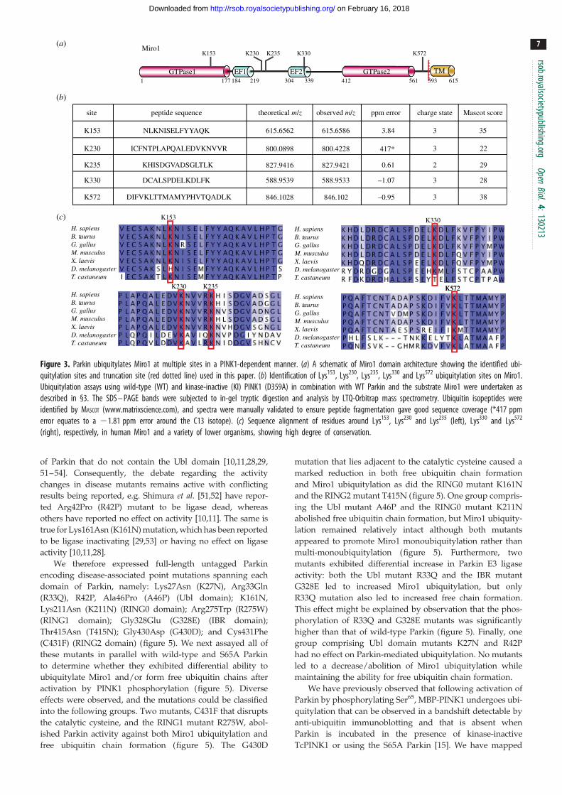
Lys153 was found in a tryptic peptide, located within the first
GTPase domain; Lys230 and Lys235 were found carrying di-Gly
remnants in two independent peptides found within the central
linker region; and Lys330 was identified in the fourth peptide
located within the second EF-hand domain of Miro1 (figure
3a–c). A fifth peptide containing a di-Gly remnant at Lys572
was located within the C-terminal non-catalytic region of
Miro1 between the second GTPase domain and the transmem-
brane domain (figure 3a–c). Parallel in-solution tryptic
digestion and analysis also identified a miscleaved peptide,
MPPPQAFTCNTADAPSKDIFVK(GG)LTTMAMYPHVTQAD
LK, spanning Lys572 and another highly conserved lysine resi-
due, Lys567 (data not shown). Peptide fragmentation pattern
analysis supported the modification occurring at Lys572. To con-
firm this we undertook mutagenesis analysis, which revealed
that a single Lys572Arg point mutant of Miro1(1–592) signifi-
cantly reduced the major band of monoubiquitylation, and
reduced the minor bands of multi-monoubiquitylation by
Parkin phosphorylated by PINK1 (the electronic supplementary
material, figure S3). In contrast, mutation of the Lys567 residue
had no effect on ubiquitylation of Miro1 (the electronic sup-
plementary material, figure S3). All sites identified are highly
conserved (figure 3c), and these analyses indicate that Miro1
undergoes multi-monoubiquitylation and that Lys153, Lys230,
Lys235, Lys330 and Lys572 are the major sites of Miro1 ubiquityla-
tion targeted by activated Parkin. Several of these residues in
Miro1 (Lys153, Lys235 and Lys572) were also recently reported
to be ubiquitylated in vivo in cells overexpressing tagged
Parkin [32].
4.4. E2s exhibit differential effects on Parkin-mediatedubiquitylation
The identity of the physiological E2 that interacts with Parkin
remains unknown. Previous studies have suggested that E2s
play a critical role in controlling activity and specificity of
RING E3 ligases, whereas the substrate specificity of HECT
E3 ligases is conferred mainly via the E3–substrate interaction
[46,47]. Parkin has previously been reported to partner with
several ubiquitin-conjugating E2 enzymes, including UbcH7
(UBE2L3) [5,12], UbcH8 (UBE2L6) [48], UBC6 (UBE2J1) [49],
UBC7 (UBE2G1, UBE2G2) [49] and Ubc13/Uev1a heterodimer
(UBE2N/UBE2V1) [50]. Given that Parkin possesses both
RING and HECT-like properties, it is not obvious how the
nature of the ubiquitin conjugates would be influenced by
the E2. We therefore decided to investigate how a panel of 25
E2 ligases impacted on the ability of Parkin to ubiquitylate
Miro1 and induce formation of free polyubiquitin chains.
This revealed that 23 of the 25 enzymes tested catalysed
Miro1 ubiquitylation in a Parkin Ser65 phosphorylation-depen-
dent manner (figure 4). Interestingly, these could be divided
into two differential groups: one group of E2s catalysed
robust free ubiquitin chain formation in addition to Miro1 ubi-
quitylation (UBE2D1, UBE2D2, UBE2D3, UBE2D4, UBE2E1,
UBE2E3, UBE2J2, UBE2L3, UBE2N1 (weakly)), whereas the
other group of E2s preferentially catalysed Miro1 ubiquityla-
tion but no significant free ubiquitin chain formation
(UBE2A, UBE2B, UBE2C, UBE2E2, UBE2G2, UBE2H, UBE2J1,
UBE2K, UBE2O (weakly), UBE2R1, UBE2R2, UBE2S, UBE2T,
UBE2Z (weakly)). In addition, two E2s catalysed Miro1 ubiqui-
tylation in a Parkin-independent manner (UBE2Q and UBE2W).
4.5. Parkin disease-associated mutants exhibitdifferential effects on Parkin-mediatedubiquitylation
We next investigated the effect of Parkin disease-associated
point mutations in the Miro1 substrate-based assay of E3
ligase activity. While the impact of mutations has been repor-
ted in previous studies, the majority of these have measured
Parkin auto-ubiquitylation activity using either N-terminal-
tagged versions of Parkin or N-terminally truncated forms
H. sapiensB. taurusG. gallusM. musculusX. laevisD. melanogasterT. castaneum
K572
K153
H. sapiensB. taurusG. gallusM. musculusX. laevisD. melanogasterT. castaneum
H. sapiensB. taurusG. gallusM. musculusX. laevisD. melanogasterT. castaneum
K230 K235 K572
H. sapiensB. taurusG. gallusM. musculusX. laevisD. melanogasterT. castaneum
K330
site peptide sequence
K153
K230
K235
K330
K572
NLKNISELFYYAQK
ICFNTPLAPQALEDVKNVVR
KHISDGVADSGLTLK
DCALSPDELKDLFK
DIFVKLTTMAMYPHVTQADLK
theoretical m/z observed m/z ppm error charge state Mascot score
827.9416 827.9421 0.61 2 29
615.6562 615.6586 3.84 3 35
588.9539 588.9533 –1.07 3 28
800.0898 800.4228 417* 223
846.1028 846.102 –0.95 3 38
412 561304 339219177 1841
Miro1
593 615
K153 K572K230 K235 K330
GTPase1 GTPase2EF1 EF2 TM
(b)
(a)
(c)
Figure 3. Parkin ubiquitylates Miro1 at multiple sites in a PINK1-dependent manner. (a) A schematic of Miro1 domain architecture showing the identified ubi-quitylation sites and truncation site (red dotted line) used in this paper. (b) Identification of Lys153, Lys230, Lys235, Lys330 and Lys572 ubiquitylation sites on Miro1.Ubiquitylation assays using wild-type (WT) and kinase-inactive (KI) PINK1 (D359A) in combination with WT Parkin and the substrate Miro1 were undertaken asdescribed in §3. The SDS – PAGE bands were subjected to in-gel tryptic digestion and analysis by LTQ-Orbitrap mass spectrometry. Ubiquitin isopeptides wereidentified by MASCOT (www.matrixscience.com), and spectra were manually validated to ensure peptide fragmentation gave good sequence coverage (*417 ppmerror equates to a 21.81 ppm error around the C13 isotope). (c) Sequence alignment of residues around Lys153, Lys230 and Lys235 (left), Lys330 and Lys572
(right), respectively, in human Miro1 and a variety of lower organisms, showing high degree of conservation.
rsob.royalsocietypublishing.orgOpen
Biol.4:130213
7
on February 16, 2018http://rsob.royalsocietypublishing.org/Downloaded from
of Parkin that do not contain the Ubl domain [10,11,28,29,
51–54]. Consequently, the debate regarding the activity
changes in disease mutants remains active with conflicting
results being reported, e.g. Shimura et al. [51,52] have repor-
ted Arg42Pro (R42P) mutant to be ligase dead, whereas
others have reported no effect on activity [10,11]. The same is
true for Lys161Asn (K161N) mutation, which has been reported
to be ligase inactivating [29,53] or having no effect on ligase
activity [10,11,28].
We therefore expressed full-length untagged Parkin
encoding disease-associated point mutations spanning each
domain of Parkin, namely: Lys27Asn (K27N), Arg33Gln
(R33Q), R42P, Ala46Pro (A46P) (Ubl domain); K161N,
Lys211Asn (K211N) (RING0 domain); Arg275Trp (R275W)
(RING1 domain); Gly328Glu (G328E) (IBR domain);
Thr415Asn (T415N); Gly430Asp (G430D); and Cys431Phe
(C431F) (RING2 domain) (figure 5). We next assayed all of
these mutants in parallel with wild-type and S65A Parkin
to determine whether they exhibited differential ability to
ubiquitylate Miro1 and/or form free ubiquitin chains after
activation by PINK1 phosphorylation (figure 5). Diverse
effects were observed, and the mutations could be classified
into the following groups. Two mutants, C431F that disrupts
the catalytic cysteine, and the RING1 mutant R275W, abol-
ished Parkin activity against both Miro1 ubiquitylation and
free ubiquitin chain formation (figure 5). The G430D
mutation that lies adjacent to the catalytic cysteine caused a
marked reduction in both free ubiquitin chain formation
and Miro1 ubiquitylation as did the RING0 mutant K161N
and the RING2 mutant T415N (figure 5). One group compris-
ing the Ubl mutant A46P and the RING0 mutant K211N
abolished free ubiquitin chain formation, but Miro1 ubiquity-
lation remained relatively intact although both mutants
appeared to promote Miro1 monoubiquitylation rather than
multi-monoubiquitylation (figure 5). Furthermore, two
mutants exhibited differential increase in Parkin E3 ligase
activity: both the Ubl mutant R33Q and the IBR mutant
G328E led to increased Miro1 ubiquitylation, but only
R33Q mutation also led to increased free chain formation.
This effect might be explained by observation that the phos-
phorylation of R33Q and G328E mutants was significantly
higher than that of wild-type Parkin (figure 5). Finally, one
group comprising Ubl domain mutants K27N and R42P
had no effect on Parkin-mediated ubiquitylation. No mutants
led to a decrease/abolition of Miro1 ubiquitylation while
maintaining the ability for free ubiquitin chain formation.
We have previously observed that following activation of
Parkin by phosphorylating Ser65, MBP-PINK1 undergoes ubi-
quitylation that can be observed in a bandshift detectable by
anti-ubiquitin immunoblotting and that is absent when
Parkin is incubated in the presence of kinase-inactive
TcPINK1 or using the S65A Parkin [15]. We have mapped

UBE2C UBE2D1UBE2A UBE2B UBE2HUBE2G2UBE2E3UBE2D3UBE2D2 UBE2E1 UBE2E2UBE2D4
UBE2L3 UBE2N1 UBE2R1 UBE2R2UBE2J1 UBE2J2 UBE2Q UBE2W UBE2ZUBE2SUBE2O UBE2T
PINK1:Parkin (WT):
UBE2K
ubiquitin:
Miro1:
PINK1:
––
WT+
KI+
––
WT+
KI+
––
WT+
KI+
––
WT+
KI+
––
WT+
KI+
––
WT+
KI+
––
WT+
KI+
––
WT+
KI+
––
WT+
KI+
––
WT+
KI+
––
WT+
KI+
––
WT+
KI+
––
WT+
KI+
––
WT+
KI+
––
WT+
KI+
––
WT+
KI+
––
WT+
KI+
––
WT+
KI+
––
WT+
KI+
––
WT+
KI+
––
WT+
KI+
––
WT+
KI+
––
WT+
KI+
––
WT+
KI+
––
WT+
KI+
PINK1:Parkin (WT):
ubiquitin:
Miro1:
PINK1:
+ +
Figure 4. Parkin can interact with multiple different E2 conjugating enzymes to catalyse Miro1 ubiquitylation with or without free ubiquitin chain formation. An E2scan of 25 different E2 conjugating enzymes was undertaken. Two micrograms of wild-type Parkin was incubated with 1 mg of wild-type (WT) or kinase-inactive(KI) (D359A) MBP-TcPINK in a kinase reaction for 60 min as described in §3. Activated Parkin was then added into pre-assembled ubiquitylation reactions containing1 mg of the E2 conjugating enzyme as indicated. Reactions were terminated after 60 min by addition of SDS – PAGE loading buffer and resolved by SDS – PAGE.Miro1, ubiquitin and PINK1 were detected by immunoblotting using anti-SUMO, anti-FLAG and anti-MBP antibodies, respectively.
rsob.royalsocietypublishing.orgOpen
Biol.4:130213
8
on February 16, 2018http://rsob.royalsocietypublishing.org/Downloaded from
the site of ubiquitylation to a lysine residue on MBP (Lys306
lying in the SYEEELVKDPR sequence motif; data not shown).
We observed that MBP-PINK1 ubiquitylation was lost in
mutants that led to decrease in Miro1 ubiquitylation or free
chain formation (A46P, K211N) or both (S65A, R275W,
C431F, G430D, K161N, T415N; figure 5) and was unaltered
in mutants that had no effect or increased Parkin activity
(WT, K27N, R33Q, R42P, G328E; figure 5).
4.6. Phosphorylation of Ser65 promotes discharge ofubiquitin from UbcH7-loaded E2 ligase: impact ofdisease-associated point mutations
We next investigated the mechanism of Parkin activation
upon PINK1-dependent phosphorylation of Parkin at Ser65.
Given the direct interaction of the Ubl domain with the
RING1 domain [29], we hypothesized that Ser65 phosphoryl-
ation might influence binding of ubiquitin-loaded E2 to the
RING1 and/or E2-mediated ubiquitin transfer. We therefore
investigated whether phosphorylation of Parkin Ser65 influ-
ences its ability to induce discharge of ubiquitin from a
ubiquitin-loaded E2, UbcH7 (E2-Ub). To load UbcH7 with
ubiquitin, we incubated E1 (UBE1), UbcH7, and ubiquitin
in the presence of Mg-ATP for 60 min at 308C. Non-
phosphorylated or TcPINK1-phosphorylated Parkin was then
added to the reaction mixture for 15 min. Reactions were termi-
nated using LDS loading dye and immediately analysed by
electrophoresis on a polyacrylamide gel that was subsequently
stained with Coomassie. This analysis enabled the facile dis-
crimination of ubiquitin-conjugated UbcH7 from non-
conjugated UbcH7. Wild-type non-phosphorylated Parkin (in
the absence of PINK1 and in the presence of kinase-inactive
TcPINK1) failed to mediate significant discharge of ubiquitin
from UbcH7; however, Parkin that was phosphorylated by
wild-type TcPINK1 induced a robust ubiquitin discharge illus-
trated by reduction of UbcH7–Ub thioester band (figure 6a).
We did not observe any Parkin–ubiquitin thioester, which
is consistent with previous analysis of full-length Parkin
[4,5,32]. A time-course analysis revealed that under the
conditions used maximal ubiquitin discharge induced by
PINK1-phosphorylated Parkin occurred within 4–5 min
(figure 6b). Consistent with the requirement of phosphoryl-
ation by PINK1, the Parkin Ser65Ala mutation prevented
ubiquitin discharge from UbcH7 (figure 6c).
We next investigated the effects of disease-associated
mutations on the ubiquitin discharge from UbcH7 after acti-
vation by TcPINK1. Parkin mutants that exhibited normal
or increased ubiquitylation of Miro1, namely K27N, R33Q,
R42P and G328E, showed no significant changes in the ubi-
quitin discharge ability (figure 6d ). Strikingly, we observed
a Parkin–ubiquitin thioester for the R33Q mutant, suggesting
that this mutation may lead to conformational changes that
render the complex more stable when compared with
wild-type Parkin (figure 6d ).
Parkin mutants A46P, R275W and T415N were similar to
the S65A mutant and the catalytic active site disease mutant
C431F and showed significantly reduced E2-ubiquitin dis-
charge ability, suggesting that these residues are required
for efficient ubiquitin discharge upon Parkin Ser65 phos-
phorylation and E2 binding to Parkin. The remaining
mutants comprising RING0 mutants K161N, K211N and
RING2 mutant G430D exhibited intact or modestly reduced
(K211N) Parkin phosphorylation-dependent E2 discharge.

WT K27N R33Q R42P A46P S65A
PINK1 WTPINK1 KDNo PINK1
++
++
+
++
+
++
+
++
+
++
+
+
Parkin: K161N K211N R275W G328E T415N G430D
++
++
+
++
+
++
+
++
+
++
+
+
C431F
++
+
ubiquitin:
Miro1:
PINK1:
autorad
PINK1:
Parkin:
K27
N
R33
QR
42P
T41
5N
C43
1FG
430D
G32
8E
R27
5W
K21
1N
K16
1N
A46
P
S65A
Ubl RING0 RING1 RING2IBR
1 76 145 215 237 292 327 378 415 465
Figure 5. Heterogeneity of the effects displayed by Parkinson’s disease-associated point mutations. (upper panel) A schematic of Parkin domain architecture show-ing the location of disease-associated Parkin mutants. (lower panel) Parkin mutants exhibit diverse effects on E3 ligase activity. Assays using wild-type (WT) andkinase-inactive (KI) PINK1 (D359A) in combination with WT and indicated mutants of Parkin and the substrate Miro1 were undertaken as described in §3. A kinasereaction including 0.1 mM [g-32P] ATP (approx. 500 cpm pmol21) was carried out in parallel for 60 min to confirm the phosphorylation as described in methods.Reactions were terminated after 60 min by addition of SDS loading buffer and resolved by SDS – PAGE. Miro1, Ubiquitin, Parkin and PINK1 were detected usinganti-SUMO, anti-FLAG, anti-Parkin and anti-MBP antibodies, respectively. Representative of three independent experiments.
rsob.royalsocietypublishing.orgOpen
Biol.4:130213
9
on February 16, 2018http://rsob.royalsocietypublishing.org/Downloaded from
5. DiscussionThis study provides fundamental evidence that PINK1
phosphorylation at Ser65 activates Parkin E3 ligase. Most impor-
tantly, by elaborating novel in vitro assays to assess Parkin
activity, we demonstrate that phosphorylation of Ser65 by
PINK1 is critical to enable Parkin to ubiquitylate its sub-
strate Miro1 and induce formation of free ubiquitin chains
(figure 1). Importantly, this is dependent on full-length Parkin,
because DUbl-Parkin failed to ubiquitylate Miro1 (figure 2).
This suggests that phosphorylation at Ser65 may not act exclu-
sively in relieving autoinhibition but may also have an
additional role in Parkin activation. The importance of the Ubl
domain in Parkin activation is also underscored by a recent
study in which it was observed that DUbl-Parkin prevented for-
mation of a Parkin C431S oxyester in cells in response to
mitochondrial depolarization [55]. Using an E2-ubiquitin dis-
charge assay, we demonstrated that Ser65 phosphorylation of
Parkin is critical for efficient discharge of ubiquitin from the
UbcH7 E2 ligase (figure 6a–d). Furthermore, we provide new
mechanistic insights into the pathogenicity of human disease-
associated mutations of Parkin (summarized in the electronic
supplementary material, table S1).
5.1. Miro1 is a direct Parkin substrateOur study is the first to show that Miro1 is a direct substrate
of Parkin in vitro. Two previous studies suggested that the
levels of Miro1 may be regulated by PINK1 and Parkin, but
results were conflicting. Wang et al. [44] reported that overex-
pression of PINK1 and/or Parkin led to decreased Miro1
levels in HEK 293T cells, and the authors reported that this
was mediated by PINK1-dependent phosphorylation of
Miro1 at Ser156. On the other hand, Liu et al. [43] found no
evidence for phosphorylation at Miro1 Ser156 and found
Miro1 levels were lower in PINK1 siRNA-targeted HeLa
cells as well as in PINK1 KO MEF cells compared with
wild-type MEF cells. We have not been able to phosphorylate
the Miro1 (1–592) fragment that lacks the transmembrane
domain with TcPINK1 (data not shown).
A recent global ubiquitylation analysis of Parkin-regu-
lated proteins reported Miro1 Lys572, Lys153, Lys194 and
Lys235 ubiquitylation in cells overexpressing tagged Parkin
stimulated with CCCP; however, it did not address whether
Parkin catalysed the ubiquitylation of these sites directly [32].
In our assay, the Lys572Arg mutant drastically reduced ubi-
quitylation as judged by Coomassie staining analysis,
suggesting that Lys572 is a major site targeted by Parkin
(the electronic supplementary material, figure S3). While we
also identified Lys153, Lys230, Lys235 and Lys330 as direct
sites of Parkin ubiquitylation, we cannot rule out additional
sites such as Lys194, which may be of lower stoichiometry
(figure 3). Miro1 plays a crucial role in mitochondrial traffick-
ing by tethering mitochondria to KIF5 motor proteins,
enabling mitochondria to be transported along microtubules
[56]. Several of the sites we have identified lie within or near

250150100
75
50
37
252015
Parkin
E2-Ub
E2
PINK1E1
*
0
5
1012
E2/
E2-
Ub
PINK1:Parkin (WT):
Ube-1PINK1
Parkin
UbcH7-Ub
UbcH7
25015010075
50
37
252015
PINK1 (WT):Parkin:
Ube-1PINK1
Parkin
UbcH7-Ub
UbcH7
25015010075
50
37
252015
Ube-1PINK1
Parkin
UbcH7-Ub
UbcH7
time (min): 1 2 3 4 5 7.5 10 20 30
25015010075
50
37
252015
(a) (b) (c)––
+WT
+S65A
PINK1 WT + Parkin WT–+
WT+
KI+
––
(d)
WT K27N R33Q R42P A46P S65A
PINK1 WTPINK1 KDNo PINK1
++
++
+
++
+
++
+
++
+
++
+
+
Parkin: K161N K211N R275W G328E T415N G430D
++
++
+
++
+
++
+
++
+
++
+
+
C431F– –
++
+
Figure 6. PINK1-dependent phosphorylation of Parkin Ser65 is required for discharge of ubiquitin from E2. Parkin was phosphorylated using wild-type (WT) orkinase-inactive (KI) MBP-TcPINK1. An E2 discharge assay was established by incubation of this mixture with 2 mg of UbcH7 that had been pre-incubated with0.5 mg of E1 and FLAG-ubiquitin in the presence of ATP for 60 min. Reactions were allowed to continue for 15 min (a,c,d) or as indicated (b) and stoppedusing SDS – PAGE loading buffer in absence of reducing agent. Samples were resolved by SDS – PAGE and proteins detected by Colloidal Coomassie staining.(a) Ubiquitin-loaded UbcH7 (UbcH7-Ub) was observed in the absence of Parkin (lanes 1,2). WT Parkin only in the presence of WT MBP-TcPINK1 was able toefficiently discharge UbcH7-Ub (lanes 5,6). No discharge was observed with WT Parkin alone (lanes 3,4) or WT Parkin in the presence of KI MBP-TcPINK1(lanes 7,8). (b) Time course of E2 discharge after addition of activated WT Parkin in the presence of WT MBP-TcPINK1 demonstrated rapid and maximal dischargeof UbcH7-Ub at 4 min. (c) Abrogation of UbcH7-Ub discharge by Parkin Ser65Ala (S65A; lanes 5,6) in contrast to WT Parkin in the presence of WT PINK1 (lanes 3,4).(d ) Comparison of the effects Parkin disease mutations on ubiquitin discharge from UbcH7. Red dotted line indicates the WT activity. K27N, R33Q, R42P, K161N,G430D and G328E mutants showed no significant changes in activity. A46P, S65A, K211N, R275W, T415N and C431F displayed markedly decreased E2-ubiquitindischarge ability. Asterisk indicates the R33Q Parkin – ubiquitin thioester. Representative of three independent experiments.
rsob.royalsocietypublishing.orgOpen
Biol.4:130213
10
on February 16, 2018http://rsob.royalsocietypublishing.org/Downloaded from
functional domains of Miro1, including Lys153 that is located
within the N-terminal GTPase domain of Miro1, Lys330 that
lies within the second EF hand domain and Lys572 that lies in
a C-terminal linker region near its transmembrane domain
that localizes Miro1 to the outer mitochondrial membrane. It
would be exciting to test whether Parkin-mediated ubiquityla-
tion of Miro1 leads to alteration of its GTPase activity,
localization, calcium binding or role in mitochondrial transport.
The finding that PINK1-activated Parkin induces multi-
monoubiquitylation of Miro1 rather than attachment of a
polyubiquitin chain highlights the potential diversity of
Parkin’s catalytic activity. Previous studies that have used
Parkin with activating N-terminal tags have also observed
monoubiquitylation activity. For example, Tanaka’s laboratory
first reported that Parkin could catalyse monoubiquitylation
in vitro using a pseudo-substrate assay in which MBP-fused
Parkin targeted residues within MBP in cis [11]. Multi-
monoubiquitylation activity has also been reported in an
auto-ubiquitylation assay using GST-Parkin [10]. Future
work expanding our initial analysis to test a variety of reported
substrates of Parkin, including mitochondrial proteins such as
VDAC1 and Tom70 as well as non-mitochondrial proteins,
e.g. CDCrel1, Pael-R and PARIS [33], will be crucial in deter-
mining the mechanistic qualities and functional effects of
ubiquitylation by Parkin. Monoubiquitylation of substrates
has previously been shown to be important for histone
regulation, DNA repair and viral budding, whereas multi-
monoubiquitylation has been implicated in endocytosis
[57,58]. The consequence of mono/multi-monoubiquitylation
of outer mitochondrial membrane proteins such as Miro1 is
unknown. It could be critical for intermolecular signalling at
the mitochondria, because, in more well-studied systems
such as endocytosis, many proteins, e.g. Eps15, contain ubiqui-
tin interaction motifs that bind monoubiquitin [59].
Alternatively, it is possible that monoubiquitylation of Miro1
and other Parkin substrates targets these for polyubuitylation
chain extension by other E3 ligases. This has been demon-
strated for the ubiquitylation of proliferating cell nuclear
antigen which binds DNA during DNA replication [60]. How-
ever, the possibility that Parkin itself can catalyse the formation
of polyubiquitylated Miro1 under specific cellular conditions
or in the presence of a regulatory protein missing from our
assay cannot be excluded, e.g. the E4 CHIP has previously
been shown to enhance Parkin-mediated polyubiquitylation
rsob.royalsocietypublishing.orgOpen
Biol.4:130213
11
on February 16, 2018http://rsob.royalsocietypublishing.org/Downloaded from
of the substrate Pael-R [61]. In future, it would be critical to
establish whether Miro1 is multi-monoubiquitylated or polyu-
biquitylated in vivo and in the latter case to investigate whether
other E3 ligases are required to achieve this.
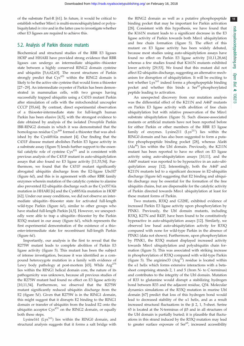
5.2. Analysis of Parkin disease mutantsBiochemical and structural studies of the RBR E3 ligases
HOIP and HHARI have provided strong evidence that RBR
ligases can undergo an intermediate ubiquitin–thioester
state between a highly conserved RING2 domain cysteine
and ubiquitin [5,6,62,63]. The recent structures of Parkin
strongly predict that Cys431 within the RING2 domain is
likely to be the active site cysteine that would form a thioester
[27–29]. An intermediate oxyester of Parkin has been demon-
strated in mammalian cells, with two groups having
successfully trapped ubiquitin using a C431S mutant Parkin
after stimulation of cells with the mitochondrial uncoupler
CCCP [55,64]. By contrast, direct experimental observation
of a thioester-intermediate state for full-length human
Parkin has been elusive [4,5], with the strongest evidence to
date obtained by analysis of the isolated Drosophila Parkin
IBR-RING2 domain in which it was demonstrated that the
homologous residue Cys449 formed a thioester that was abol-
ished by the Cys449Ala mutant [4]. Our finding that the
C431F disease mutant abolishes Parkin E3 ligase activity in
a substrate assay (figure 5) lends further support to the essen-
tial catalytic role of cysteine Cys431 and is consistent with
previous analysis of the C431F mutant in auto-ubiquitylation
assays that also found no E3 ligase activity [11,53,54]. Fur-
thermore, we found that the C431F mutant significantly
abrogated ubiquitin discharge from the E2-ligase UbcH7
(figure 6d), and this is in agreement with other RBR family
enzymes wherein mutation of the catalytic cysteine to alanine
also prevented E2-ubiquitin discharge such as the Cys357Ala
mutation in HHARI [6] and the Cys885Ala mutation in HOIP
[62]. Under our assay conditions, we did not observe an inter-
mediate ubiquitin–thioester state for activated full-length
wild-type Parkin (figure 6d ), similar to other groups who
have studied full-length Parkin [4,5]. However, we unexpect-
edly were able to trap a ubiquitin–thioester by the Parkin
R33Q mutant in our assay (figure 6d ), which represents the
first experimental demonstration of the existence of a thio-
ester-intermediate state for recombinant full-length Parkin
(figure 6d ).
Importantly, our analysis is the first to reveal that the
R275W mutant leads to complete abolition of Parkin E3
ligase activity (figure 5). This mutant has been the subject
of intense investigation, because it was identified as a com-
pound heterozygote mutation in a family with evidence of
Lewy body pathology at post-mortem [65]. While Arg275
lies within the RING1 helical domain core, the nature of its
pathogenicity was unknown, because all previous studies of
the R275W mutant had found no effect on E3 ligase activity
[10,11,54]. Furthermore, we observed that the R275W
mutant significantly reduced ubiquitin discharge from the
E2 (figure 5d ). Given that R275W is in the RING1 domain,
this might suggest that it disrupts E2 binding to the RING1
domain or transfer of ubiquitin from the loaded E2 onto the
ubiquitin acceptor Cys431 on the RING2 domain, or equally
both these steps.
Lysine161 (Lys161) lies within the RING0 domain, and
structural analysis suggests that it forms a salt bridge with
the RING2 domain as well as a putative phosphopeptide
binding pocket that may be important for Parkin activation
[28]. Consistent with this hypothesis, we have found that
the K161N mutant leads to a significant decrease in the E3
ligase activity of Parkin towards both Miro1 ubiquitylation
and free chain formation (figure 5). The effect of this
mutant on E3 ligase activity has been widely debated,
because most studies using auto-ubiquitylation assays have
found no effect on Parkin E3 ligase activity [10,11,28,66]
whereas a few studies found that K161N mutants exhibited
lower activity [29,53]. We found that this mutant did not
affect E2-ubiquitin discharge, suggesting an alternative mech-
anism for disruption of ubiquitylation. It will be exciting to
test whether Lys161 indeed forms a phosphopeptide binding
pocket and whether this binds a Ser65-phosphorylated
peptide leading to activation.
An unexpected discovery from our mutation analysis
was the differential effect of the K211N and A46P mutants
on Parkin E3 ligase activity with abolition of free chain
ubiquitylation but with relative preservation of the Miro1
substrate ubiquitylation (figure 5). Such disease-associated
mutants or artificial mutants have not been reported before
for either Parkin or other members of the RBR E3 ligase
family of enzymes. Lysine211 (Lys211) lies within the
RING0 domain and has also been suggested to form a puta-
tive phosphopeptide binding pocket [28], whereas Ala46
(Ala46) lies within the Ubl domain. Previously, the K211N
mutant has been reported to have no effect on E3 ligase
activity using auto-ubiquitylation assays [10,11], and the
A46P mutant was reported to be hyperactive in an auto-ubi-
quitylation assay [12]. Interestingly, both the A46P and
K211N mutants led to a significant decrease in E2-ubiquitin
discharge (figure 6d) suggesting that E2 binding and ubiqui-
tin discharge may be essential for the formation of the free
ubiquitin chains, but are dispensable for the catalytic activity
of Parkin directed towards Miro1 ubiquitylation at least for
these mutant forms of Parkin.
Two mutants, R33Q and G328E, exhibited evidence of
increased Parkin E3 ligase activity upon phosphorylation by
PINK1. Previously, the Ubl domain mutants, including
R33Q, K27N and R42P, have been found to be constitutively
hyperactive in auto-ubiquitylation assays [12]. Similarly, we
observed low basal auto-ubiquitylation activity for R33Q
compared with none for wild-type Parkin in the absence of
PINK1 (data not shown). Furthermore, upon phosphorylation
by PINK1, the R33Q mutant displayed increased activity
towards Miro1 ubiquitylation and polyubiquitin chain for-
mation (figure 5). This was associated with striking increase
in phosphorylation of R33Q compared with wild-type Parkin
(figure 5). The arginine33 (Arg33) residue is located within
the a1 helix which forms extensive interactions with a beta-
sheet comprising strands 2, 1 and 5 (from N- to C-terminus)
and contributes to the integrity of the Ubl domain. Mutation
of R33 to glutamine would disrupt a stabilizing hydrogen
bond between R33 and the adjacent residue, Q34. Molecular
dynamics simulations of the R33Q mutation in murine Ubl
domain [67] predict that loss of this hydrogen bond would
lead to decreased stability of the a1 helix, and as a result
increased structural fluctuations in the b 2, 1, 5-sheet. Serine
65 is located at the N-terminus of b5 and in all structures of
the Ubl domain is partially buried; it is plausible that fluctu-
ations in this strand induced by the R33Q mutation may lead
to greater surface exposure of Ser65, increased accessibility

rsob.royalsocietypublishing.orgOpen
Biol.4:130213
12
on February 16, 2018http://rsob.royalsocietypublishing.org/Downloaded from
and phosphorylation by PINK1 and a subsequent increase in E3
ligase activity (the electronic supplementary material, figure
S4). However, enhanced phosphorylation of the R33Q mutant
was not associated with an increase in UbcH7 discharge of ubi-
quitin (figure 6d) suggesting an alternative mechanism for
increased activity.
Previous reports on the effect of the G328E mutant on E3
ligase activity have been more controversial, with several
groups reporting no change in E3 ligase activity [10,11,54]
whereas one report suggested a decreased activity in auto-ubi-
quitylation assays [29]. The G328E mutant stimulated Miro1
ubiquitylation without any effect on the formation of free ubi-
quitin chains. Similar to R33Q, this was not associated with any
significant change in the E2 discharge of ubiquitin (figure 6d).
One explanation for the lack of effect of G328E on free chain for-
mation is that the Gly328 residue that is located in the RING1 :
IBR interface may directly interact with the substrate Miro1 and
the G328E mutant may stabilize this interaction leading to
enhanced Miro1 ubiquitylation. In future studies, it would be
important to determine whether the G328E mutant enzyme
has a higher affinity for its substrate.
5.3. Analysis of Parkin – E2 interactionsAn important question remaining in the field is establishing
the identity of the physiological E2(s) that Parkin interacts
with to catalyse ubiquitylation of its substrates. Extensive ana-
lyses of RING E3 ligases indicate that the diversity and
specificity of ubiquitin conjugates is significantly influenced
by partnering E2s. In contrast, for HECT E3 ligases the pattern
of ubiquitin conjugates appears to be largely independent of
the identity of the E2 and conferred principally by the E3–sub-
strate interaction. Given that Parkin possesses both RING- and
HECT-like properties, it is not obvious how the nature of the
ubiquitin conjugates would be influenced by the E2 [46,68].
We investigated how a panel of E2 ligases impacted the ability
of Parkin to monoubiquitylate Miro1. Previous studies ident-
ified UbcH7 (UBE2L3), UbcH8 (UBE2L6), UBC6 (UBE2J1),
UBC7 (UBE2G1, UBE2G2) and Ubc13/Uev1a heterodimer
(UBE2N/UBE2V1) [33,64,69,70] as partnering E2s for tagged
Parkin. Our assay enabled us to determine the specificity of
E2 conjugating enzymes that control ubiquitylation of Miro1
by Parkin in the presence or absence of Ser65 phosphorylation.
It also permitted us to investigate whether E2s played any role
in determining the pattern of ubiquitin conjugates that are
formed by Parkin in our assay. We tested 25 different E2s
and strikingly observed that the ubiquitylation of the substrate
Miro1, by Parkin, was not influenced by the vast majority of
the E2s tested. This observation suggests that Parkin exhibits
mainly HECT-like properties in which the interaction between
Parkin and Miro1 is the critical feature governing the ubiquity-
lation of Miro1. We nevertheless did observe a differential
effect of E2s in their ability to enable Parkin to catalyse the for-
mation of free ubiquitin chains, e.g. UBE2L3 enabled both
Miro1 ubiquitylation and free chain formation, whereas
UBE2H only enabled Parkin to mediate Miro1 ubiquitylation.
This does suggest that the formation of free chains may be cri-
tically dependent on the E2–E3 interaction. The molecular
mechanisms underpinning E2-mediated ubiquitin chain for-
mation remain poorly understood except for a few examples.
In yeast, the anaphase promoting complex/cyclosome (APC)
has been shown in vitro to promote multi-monoubiquitylation
of cyclin B in the presence of Ubc4, whereas Ubc1 enables it to
catalyse polyubiquitin chain formation [71]. Moreover, APC
exploits the differential selectivity of both E2s to target sub-
strates for polyubiquitylation in vivo [71]. It would be
interesting to study a RING2 HECT-defective mutant and
repeat the E2 screen to identify any E2s in which the ability
to generate ubiquitin free chains was dependent on the E2–
RING1 interaction alone. It will also be important to determine
the universality of our findings and test the E2 specificity for
other Parkin substrates we identify as being regulated by
Ser65 phosphorylation. Our data suggest that in vitro analysis
is unlikely to be helpful in pinpointing physiological E2 ligases
that act with Parkin. To address this question, in future, in vivoapproaches such as genetic screens could be used.
6. SummaryOverall, we have elaborated for the first time in the field a
substrate-based assay of untagged full-length Parkin. This
has revealed the critical importance for Ser65 phosphorylation
by PINK1 for Parkin activation and provided further evi-
dence that Miro1 is a bona fide substrate of Parkin. Our
studies reveal a critical requirement for the Ubl domain in
substrate ubiquitylation and also demonstrate that Ser65
phosphorylation by PINK1 stimulates ubiquitin discharge
from the E2 ligase UbcH7 that provides one explanation of
how phosphorylation might activate Parkin E3 ligase activity.
The assay and reagents we have developed will be extre-
mely valuable in addressing critical questions of Parkin
biology. As proof of concept, we have deployed these to
understand how Parkin disease mutations impact on catalytic
activity, and this has revealed new fundamental knowledge
on the mechanism of pathogenicity of these mutations.
Recently, novel regulatory modifications have been reported
for Parkin E3 ligase activity, including c-Abl-induced tyrosine
phosphorylation [72] and cysteine sulfhydration [73]; it will
be interesting to test the influence of these modifications on
Parkin activity in our assay. Lastly, there is increasing interest
to explore the therapeutic potential of developing small mol-
ecule activators of Parkin E3 ligase that could have the
potential to treat PD. The assay we have developed to
measure ubiquitylation of Miro1 or discharge of ubiquitin-
loaded UbcH7 could serve as a basis for setting up a screen
to identify compounds that activate Parkin.
Acknowledgements. We thank Ron Hay for the gift of anti-SUMO1 anti-body. We are grateful to the sequencing service (College of LifeSciences, University of Dundee); Axel Knebel and his protein pro-duction team; Hilary McLauchlan and the antibody purificationand protein production teams (Division of Signal TransductionTherapy (DSTT), University of Dundee) for excellent technical sup-port. We thank Ubiquigent for provision of ubiquitin reagentsincluding E2 and E1 enzymes.
Funding statement. A.K. is supported by a J. Macdonald MenziesCharitable Trust Prize Studentship. M.M.K.M. is supported by aWellcome Trust Senior Research Fellowship in Clinical Science(101022/Z/13/Z). This work was supported by the Medical ResearchCouncil; the Wellcome Trust; Parkinson’s UK; the Michael J. FoxFoundation for Parkinson’s disease research; and a Wellcome/MRCPD consortium grant to UCL Institute of Neurology, University ofSheffield and MRC-PPU of University of Dundee. We also thankthe pharmaceutical companies supporting the Division of SignalTransduction Therapy Unit (AstraZeneca, Boehringer-Ingelheim,GlaxoSmithKline, Merck KGaA, Janssen Pharmaceutica and Pfizer)for financial support.

13
on February 16, 2018http://rsob.royalsocietypublishing.org/Downloaded from
References
rsob.royalsocietypublishing.orgOpen
Biol.4:130213
1. Lees AJ, Hardy J, Revesz T. 2009 Parkinson’s disease.Lancet 373, 2055 – 2066. (doi:10.1016/S0140-6736(09)60492-X)
2. Puschmann A. 2013 Monogenic Parkinson’s diseaseand parkinsonism: clinical phenotypes andfrequencies of known mutations. ParkinsonismRelat. Dis. 19, 407 – 415. (doi:10.1016/j.parkreldis.2013.01.020)
3. Kitada T, Asakawa S, Hattori N, Matsumine H,Yamamura Y, Minoshima S, Yokochi M, Mizuno Y,Shimizu N. 1998 Mutations in the parkin genecause autosomal recessive juvenile parkinsonism.Nature 392, 605 – 608. (doi:10.1038/33416)
4. Spratt DE et al. 2013 A molecular explanation forthe recessive nature of parkin-linked Parkinson’sdisease. Nat. Commun. 4, 1983. (doi:10.1038/ncomms2983)
5. Wenzel DM, Lissounov A, Brzovic PS, Klevit RE. 2011UBCH7 reactivity profile reveals parkin and HHARI tobe RING/HECT hybrids. Nature 474, 105 – 108.(doi:10.1038/nature09966)
6. Duda DM, Olszewski JL, Schuermann JP, Kurinov I,Miller DJ, Nourse A, Alpi AF, Schulman BA. 2013Structure of HHARI, a RING-IBR-RING ubiquitinligase: autoinhibition of an ariadne-family E3 andinsights into ligation mechanism. Structure 21,1030 – 1041. (doi:10.1016/j.str.2013.04.019)
7. Iguchi M et al. 2013 Parkin catalyzed ubiquitin-estertransfer is triggered by PINK1-dependentphosphorylation. J. Biol. Chem. 288, 22 019 –22 032. (doi:10.1074/jbc.M113.467530)
8. Joch M, Ase AR, Chen CX, MacDonald PA,Kontogiannea M, Corera AT, Brice A, Seguela P, FonEA. 2007 Parkin-mediated monoubiquitination ofthe PDZ protein PICK1 regulates the activity ofacid-sensing ion channels. Mol. Biol. Cell 18,3105 – 3118. (doi:10.1091/mbc.E05-11-1027)
9. Chen D, Gao F, Li B, Wang H, Xu Y, Zhu C, Wang G.2010 Parkin mono-ubiquitinates Bcl-2 and regulatesautophagy. J. Biol. Chem. 285, 38 214 – 38 223.(doi:10.1074/jbc.M110.101469)
10. Hampe C, Ardila-Osorio H, Fournier M, Brice A, CortiO. 2006 Biochemical analysis of Parkinson’sdisease-causing variants of Parkin, an E3 ubiquitin-protein ligase with monoubiquitylation capacity.Hum. Mol. Genet. 15, 2059 – 2075. (doi:10.1093/hmg/ddl131)
11. Matsuda N, Kitami T, Suzuki T, Mizuno Y, Hattori N,Tanaka K. 2006 Diverse effects of pathogenicmutations of Parkin that catalyze multiplemonoubiquitylation in vitro. J. Biol. Chem. 281,3204 – 3209. (doi:10.1074/jbc.M510393200)
12. Chaugule VK, Burchell L, Barber KR, Sidhu A, LeslieSJ, Shaw GS, Walden H. 2011 Autoregulation ofParkin activity through its ubiquitin-like domain.EMBO J. 30, 2853 – 2867. (doi:10.1038/emboj.2011.204)
13. Burchell L, Chaugule VK, Walden H. 2012 Small,N-terminal tags activate Parkin e3 ubiquitin ligaseactivity by disrupting its autoinhibited
conformation. PLoS ONE 7, e34748. (doi:10.1371/journal.pone.0034748)
14. Valente EM et al. 2004 Hereditary early-onsetParkinson’s disease caused by mutations in PINK1.Science 304, 1158 – 1160. (doi:10.1126/science.1096284)
15. Kondapalli C et al. 2012 PINK1 is activated bymitochondrial membrane potential depolarizationand stimulates Parkin E3 ligase activity byphosphorylating serine 65. Open Biol. 2, 120080.(doi:10.1098/rsob.120080)
16. Shiba-Fukushima K, Imai Y, Yoshida S, Ishihama Y,Kanao T, Sato S, Hattori N. 2012 PINK1-mediatedphosphorylation of the Parkin ubiquitin-like domainprimes mitochondrial translocation of Parkin andregulates mitophagy. Sci. Rep. 2, 1002. (doi:10.1038/srep01002)
17. Koyano F, Okatsu K, Ishigaki S, Fujioka Y, Kimura M,Sobue G, Tanaka K, Matsuda N. 2013 The principalPINK1 and Parkin cellular events triggered inresponse to dissipation of mitochondrial membranepotential occur in primary neurons. Genes Cells 18,672 – 681. (doi:10.1111/gtc.12066)
18. Lucking CB et al. 2000 Association between early-onset Parkinson’s disease and mutations in theparkin gene. N. Engl. J. Med. 342, 1560 – 1567.(doi:10.1056/NEJM200005253422103)
19. Khan NL, Graham E, Critchley P, Schrag AE, WoodNW, Lees AJ, Bhatia KP, Quinn N. 2003 Parkindisease: a phenotypic study of a large case series.Brain J. Neurol. 126, 1279 – 1292. (doi:10.1093/brain/awg142)
20. Clark IE, Dodson MW, Jiang C, Cao JH, Huh JR, SeolJH, Yoo SJ, Hay BA, Guo M. 2006 Drosophila pink1 isrequired for mitochondrial function and interactsgenetically with parkin. Nature 441, 1162 – 1166.(doi:10.1038/nature04779)
21. Park J et al. 2006 Mitochondrial dysfunction inDrosophila PINK1 mutants is complemented byparkin. Nature 441, 1157 – 1161. (doi:10.1038/nature04788)
22. Yang Y et al. 2006 Mitochondrial pathology andmuscle and dopaminergic neuron degenerationcaused by inactivation of Drosophila Pink1 isrescued by Parkin. Proc. Natl Acad. Sci. USA 103,10 793 – 10 798. (doi:10.1073/pnas.0602493103)
23. Matsuda N et al. 2010 PINK1 stabilized bymitochondrial depolarization recruits Parkin todamaged mitochondria and activates latent Parkinfor mitophagy. J. Cell Biol. 189, 211 – 221. (doi:10.1083/jcb.200910140)
24. Narendra DP, Jin SM, Tanaka A, Suen DF, Gautier CA,Shen J, Cookson MR, Youle RJ. 2010 PINK1 isselectively stabilized on impaired mitochondria toactivate Parkin. PLoS Biol. 8, e1000298. (doi:10.1371/journal.pbio.1000298)
25. Vives-Bauza C et al. 2010 PINK1-dependentrecruitment of Parkin to mitochondria inmitophagy. Proc. Natl Acad. Sci. USA 107,378 – 383. (doi:10.1073/pnas.0911187107)
26. Geisler S, Holmstrom KM, Skujat D, Fiesel FC,Rothfuss OC, Kahle PJ, Springer W. 2010 PINK1/Parkin-mediated mitophagy is dependent on VDAC1and p62/SQSTM1. Nat. Cell Biol. 12, 119 – 131.(doi:10.1038/ncb2012)
27. Riley BE et al. 2013 Structure and function of ParkinE3 ubiquitin ligase reveals aspects of RING and HECTligases. Nat. Commun. 4, 1982. (doi:10.1038/ncomms2982)
28. Wauer T, Komander D. 2013 Structure of the humanParkin ligase domain in an autoinhibited state.EMBO J. 32, 2099 – 2112. (doi:10.1038/emboj.2013.125)
29. Trempe JF et al. 2013 Structure of Parkin revealsmechanisms for ubiquitin ligase activation. Science340, 1451 – 1455. (doi:10.1126/science.1237908)
30. Ko HS, Kim SW, Sriram SR, Dawson VL, Dawson TM.2006 Identification of far upstream element-bindingprotein-1 as an authentic Parkin substrate. J. Biol.Chem. 281, 16 193 – 16 196. (doi:10.1074/jbc.C600041200)
31. Ko HS et al. 2005 Accumulation of the authenticParkin substrate aminoacyl-tRNA synthetasecofactor, p38/JTV-1, leads to catecholaminergic celldeath. J. Neurosci. 25, 7968 – 7978. (doi:10.1523/JNEUROSCI.2172-05.2005)
32. Sarraf SA, Raman M, Guarani-Pereira V, Sowa ME,Huttlin EL, Gygi SP, Harper JW. 2013 Landscape ofthe PARKIN-dependent ubiquitylome in responseto mitochondrial depolarization. Nature 496,372 – 376. (doi:10.1038/nature12043)
33. Walden H, Martinez-Torres RJ. 2012 Regulation ofParkin E3 ubiquitin ligase activity. Cell. Mol. Life Sci.69, 3053 – 3067. (doi:10.1007/s00018-012-0978-5)
34. Moore DJ, West AB, Dawson VL, Dawson TM. 2005Molecular pathophysiology of Parkinson’s disease.Annu. Rev. Neurosci. 28, 57 – 87. (doi:10.1146/annurev.neuro.28.061604.135718)
35. Imai Y, Takahashi R. 2004 How do Parkin mutationsresult in neurodegeneration? Curr. Opin. Neurobiol.14, 384 – 389. (doi:10.1016/j.conb.2004.04.002)
36. Zhu J, Wang KZ, Chu CT. 2013 After the banquet:mitochondrial biogenesis, mitophagy and cellsurvival. Autophagy 9, 1663 – 1676. (doi:10.4161/auto.24135).
37. McInnes J. 2013 Insights on altered mitochondrialfunction and dynamics in the pathogenesis ofneurodegeneration. Transl. Neurodegenerat. 2, 12.(doi:10.1186/2047-9158-2-12)
38. Trempe JF, Fon EA. 2013 Structure and function ofParkin, PINK1, DJ-1, the three musketeers ofneuroprotection. Front. Neurol. 4, 38. (doi:10.3389/fneur.2013.00038)
39. Palacino JJ, Sagi D, Goldberg MS, Krauss S, Motz C,Wacker M, Klose J, Shen J. 2004 Mitochondrialdysfunction and oxidative damage in parkin-deficient mice. J. Biol. Chem. 279, 18 614 – 18 622.(doi:10.1074/jbc.M401135200)
40. Periquet M, Corti O, Jacquier S, Brice A. 2005Proteomic analysis of parkin knockout mice:

rsob.royalsocietypublishing.orgOpen
Biol.4:130213
14
on February 16, 2018http://rsob.royalsocietypublishing.org/Downloaded from
alterations in energy metabolism, proteinhandling and synaptic function. J. Neurochem.95, 1259 – 1276. (doi:10.1111/j.1471-4159.2005.03442.x)
41. Chan NC, Salazar AM, Pham AH, Sweredoski MJ,Kolawa NJ, Graham RL, Hess S, Chan DC. 2011Broad activation of the ubiquitin-proteasome systemby Parkin is critical for mitophagy. Hum. Mol. Genet.20, 1726 – 1737. (doi:10.1093/hmg/ddr048)
42. Jin SM, Youle RJ. 2012 PINK1- and Parkin-mediatedmitophagy at a glance. J. Cell Sci. 125, 795 – 799.(doi:10.1242/jcs.093849)
43. Liu S et al. 2012 Parkinson’s disease-associatedkinase PINK1 regulates Miro protein level andaxonal transport of mitochondria. PLoS Genet. 8,e1002537. (doi:10.1371/journal.pgen.1002537)
44. Wang X et al. 2011 PINK1 and Parkin target Mirofor phosphorylation and degradation to arrestmitochondrial motility. Cell 147, 893 – 906. (doi:10.1016/j.cell.2011.10.018)
45. MacLean B et al. 2010 Skyline: an open sourcedocument editor for creating and analyzingtargeted proteomics experiments. Bioinformatics 26,966 – 968. (doi:10.1093/bioinformatics/btq054)
46. Deshaies RJ, Joazeiro CA. 2009 RING domain E3ubiquitin ligases. Annu. Rev. Biochem. 78,399 – 434. (doi:10.1146/annurev.biochem.78.101807.093809)
47. Ye Y, Rape M. 2009 Building ubiquitin chains: E2enzymes at work. Nat. Rev. Mol. Cell Biol. 10,755 – 764. (doi:10.1038/nrm2780)
48. Zhang Y, Gao J, Chung KK, Huang H, Dawson VL,Dawson TM. 2000 Parkin functions as anE2-dependent ubiquitin-protein ligase andpromotes the degradation of the synapticvesicle-associated protein, CDCrel-1. Proc. NatlAcad. Sci. USA 97, 13 354 – 13 359. (doi:10.1073/pnas.240347797)
49. Imai Y, Soda M, Inoue H, Hattori N, Mizuno Y,Takahashi R. 2001 An unfolded putativetransmembrane polypeptide, which can lead toendoplasmic reticulum stress, is a substrate ofParkin. Cell 105, 891 – 902. (doi:10.1016/S0092-8674(01)00407-X)
50. Olzmann JA, Li L, Chudaev MV, Chen J, Perez FA,Palmiter RD, Chin LS. 2007 Parkin-mediated K63-linked polyubiquitination targets misfolded DJ-1 toaggresomes via binding to HDAC6. J. Cell Biol. 178,1025 – 1038. (doi:10.1083/jcb.200611128)
51. Shimura H et al. 2000 Familial Parkinson diseasegene product, Parkin, is a ubiquitin-protein ligase.Nat. Genet. 25, 302 – 305. (doi:10.1038/77060)
52. Shimura H et al. 2001 Ubiquitination of a new formof alpha-synuclein by Parkin from human brain:implications for Parkinson’s disease. Science 293,263 – 269. (doi:10.1126/science.1060627)
53. Ren Y, Zhao J, Feng J. 2003 Parkin binds to alpha/beta tubulin and increases their ubiquitination anddegradation. J. Neurosci. 23, 3316 – 3324.
54. Sriram SR, Li X, Ko HS, Chung KK, Wong E, Lim KL,Dawson VL, Dawson TM. 2005 Familial-associatedmutations differentially disrupt the solubility,localization, binding and ubiquitination propertiesof parkin. Hum. Mol. Genet. 14, 2571 – 2586.(doi:10.1093/hmg/ddi292)
55. Zheng X, Hunter T. 2013 Parkin mitochondrialtranslocation is achieved through a novel catalyticactivity coupled mechanism. Cell Res. 23, 886 – 897.(doi:10.1038/cr.2013.66)
56. Macaskill AF, Rinholm JE, Twelvetrees AE, Arancibia-Carcamo IL, Muir J, Fransson A, Aspenstrom P,Attwell D, Kittler JT. 2009 Miro1 is a calcium sensorfor glutamate receptor-dependent localization ofmitochondria at synapses. Neuron 61, 541 – 555.(doi:10.1016/j.neuron.2009.01.030)
57. Hicke L. 2001 Protein regulation by monoubiquitin.Nat. Rev. Mol. Cell Biol. 2, 195 – 201. (doi:10.1038/35056583)
58. Sadowski M, Suryadinata R, Tan AR, Roesley SN,Sarcevic B. 2012 Protein monoubiquitination andpolyubiquitination generate structural diversity tocontrol distinct biological processes. IUBMB Life 64,136 – 142. (doi:10.1002/iub.589)
59. Husnjak K, Dikic I. 2012 Ubiquitin-binding proteins:decoders of ubiquitin-mediated cellular functions.Annu. Rev. Biochem. 81, 291 – 322. (doi:10.1146/annurev-biochem-051810-094654)
60. Bergink S, Jentsch S. 2009 Principles of ubiquitinand SUMO modifications in DNA repair. Nature 458,461 – 467. (doi:10.1038/nature07963)
61. Imai Y, Soda M, Hatakeyama S, Akagi T, HashikawaT, Nakayama KI, Takahashi R. 2002 CHIP isassociated with Parkin, a gene responsible forfamilial Parkinson’s disease, and enhances itsubiquitin ligase activity. Mol. Cell 10, 55 – 67.(doi:10.1016/S1097-2765(02)00583-X)
62. Stieglitz B, Morris-Davies AC, Koliopoulos MG,Christodoulou E, Rittinger K. 2012 LUBAC
synthesizes linear ubiquitin chains via a thioesterintermediate. EMBO Rep. 13, 840 – 846. (doi:10.1038/embor.2012.105)
63. Stieglitz B et al. 2013 Structural basis for ligase-specific conjugation of linear ubiquitin chains byHOIP. Nature 503, 422 – 426. (doi:10.1038/nature12638)
64. Lazarou M, Narendra DP, Jin SM, Tekle E, BanerjeeS, Youle RJ. 2013 PINK1 drives Parkin self-association and HECT-like E3 activity upstream ofmitochondrial binding. J. Cell Biol. 200, 163 – 172.(doi:10.1083/jcb.201210111)
65. Farrer M et al. 2001 Lewy bodies and parkinsonismin families with parkin mutations. Ann. Neurol. 50,293 – 300. (doi:10.1002/ana.1132)
66. Gu WJ et al. 2003 The C289G and C418R missensemutations cause rapid sequestration of humanParkin into insoluble aggregates. Neurobiol. Dis. 14,357 – 364. (doi:10.1016/j.nbd.2003.08.011)
67. Tomoo K, Mukai Y, In Y, Miyagawa H, Kitamura K,Yamano A, Shindo H, Ishida T. 2008 Crystal structureand molecular dynamics simulation of ubiquitin-likedomain of murine parkin. Biochim. Biophys. Acta1784, 1059 – 1067. (doi:10.1016/j.bbapap.2008.04.009)
68. Metzger MB, Pruneda JN, Klevit RE, Weissman AM.2013 RING-type E3 ligases: Master manipulators ofE2 ubiquitin-conjugating enzymes andubiquitination. Biochim. Biophys. Acta 1843,47 – 60. (doi:10.1016/j.bbamcr.2013.05.026)
69. Dawson TM, Dawson VL. 2010 The role of Parkin infamilial and sporadic Parkinson’s disease. Mov.Disord. Soc. 25(Suppl. 1), S32 – S39. (doi:10.1002/mds.22798)
70. Wenzel DM, Klevit RE. 2012 Following Ariadne’sthread: a new perspective on RBR ubiquitin ligases.BMC Biol. 10, 24. (doi:10.1186/1741-7007-10-24)
71. Rodrigo-Brenni MC, Morgan DO. 2007 SequentialE2s drive polyubiquitin chain assembly on APCtargets. Cell 130, 127 – 139. (doi:10.1016/j.cell.2007.05.027)
72. Ko HS et al. 2010 Phosphorylation by the c-Ablprotein tyrosine kinase inhibits parkin’subiquitination and protective function. Proc. NatlAcad. Sci. USA 107, 16 691 – 16 696. (doi:10.1073/pnas.1006083107)
73. Vandiver MS et al. 2013 Sulfhydration mediatesneuroprotective actions of parkin. Nat. Commun. 4,1626. (doi:10.1038/ncomms2623)


![Multitasking guardian of mitochondrial quality: Parkin …...the autoubiquitination activity of Parkin [41]. Involvement of Parkin in mitochondrial processes As an E3 ligase, Parkin](https://static.fdocuments.in/doc/165x107/60ff3ba3c386cc67f77a5535/multitasking-guardian-of-mitochondrial-quality-parkin-the-autoubiquitination.jpg)